1. Introduction
Liver cancer remains one of the leading causes of cancer-related mortality worldwide, with hepatocellular carcinoma accounting for the majority of primary hepatic malignancies [
1]. Accurate segmentation of the liver and tumor regions from abdominal computed tomography (CT) scans plays a critical role in diagnosis, treatment planning, surgical navigation, and therapy response assessment [
2,
3,
4]. However, manual delineation is time-consuming, highly operator-dependent, and prone to significant inter-observer variability, motivating the development of reliable automated segmentation systems [
5,
6,
7].
Deep learning, particularly U-Net–based architectures and their variants, has become the dominant paradigm for liver and tumor segmentation in CT imaging [
8,
9]. Models such as U-Net, residual U-Net variants [
10] and Attention U-Net [
11,
12] have demonstrated strong performance on large public datasets, including the Liver Tumor Segmentation (LiTS) Challenge [
13]. More recently, highly optimized frameworks such as nnU-Net [
14], and hybrid CNN–transformer architectures that leverage global attention mechanisms [
15,
16] have further advanced segmentation accuracy by exploiting extensive skip-connection fusion and automated architecture configuration. Despite these advances, most state-of-the-art methods rely on fully centralized training, implicitly assuming unrestricted access to multi-institutional clinical data.
In real clinical environments, however, centralized data aggregation is often infeasible due to strict privacy regulations, institutional policies, and legal constraints governing medical data sharing [
17,
18,
19]. Consequently, models trained on single-center datasets frequently exhibit pronounced domain shift and limited generalization when deployed across different hospitals, scanner vendors, and acquisition protocols [
20]. Federated learning (FL) [
21] and split learning [
22] have therefore emerged as promising privacy-preserving alternatives, enabling collaborative model training without transferring raw patient data. Nevertheless, existing collaborative segmentation frameworks typically require exchanging model gradients or high-resolution intermediate feature maps [
23], both of which can expose sensitive anatomical information and impose substantial communication overhead.
Recent studies have demonstrated that high-resolution latent features exchanged during collaborative training are vulnerable to inversion-based reconstruction attacks, enabling partial recovery of patient-specific anatomical structures [
24,
25]. Moreover, transmitting skip-connection feature maps in U-Net–based architectures often requires tens to hundreds of megabytes per training iteration, rendering such approaches impractical in bandwidth-constrained hospital networks.
Unlike conventional federated learning approaches that exchange full model parameters or gradients, and split-learning frameworks that transmit intermediate feature activations between client and server, many collaborative U-Net–based segmentation systems require sharing high-resolution encoder skip features to preserve spatial detail. These feature maps often retain fine-grained anatomical structure and substantially increase communication overhead. In contrast, FF-ResUNet adopts a strictly skip-free architectural design in which only compact bottleneck representations are transmitted. All high-resolution encoder features remain local to each institution. This architectural constraint fundamentally distinguishes FF-ResUNet from prior collaborative segmentation frameworks that rely on intermediate feature exchange.
To address these privacy and communication limitations, this paper proposes a Feature-Forward Collaborative Residual U-Net (FF-ResUNet) for liver and tumor segmentation. Unlike conventional collaborative frameworks, FF-ResUNet adopts a strictly skip-free design in which only compact bottleneck features are transmitted from client institutions to a central server. All high-resolution encoder features and skip connections remain strictly local, reducing exposure of high-resolution anatomical information compared to skip-based collaborative designs while dramatically reducing communication overhead. To compensate for the absence of skip fusion, a carefully designed server-side decoder incorporating multi-scale dilated convolutions, residual refinement, and progressive upsampling enables accurate reconstruction of full-resolution segmentation masks.
The proposed framework is evaluated on three large publicly available CT datasets—LiTS [
13], 3D-IRCADb [
26], and AMOS-CT [
27]—covering diverse scanner types, acquisition protocols, contrast phases, and anatomical variability. Extensive experiments demonstrate that FF-ResUNet achieves competitive segmentation accuracy under strict skip-free and privacy-preserving constraints while delivering strong cross-domain generalization and substantial communication savings. The main contributions of this work are summarized as follows:
- •
A novel skip-free feature-forward collaborative learning framework for medical image segmentation that eliminates the transmission of high-resolution skip-connection features.
- •
A server-side multi-scale residual decoder specifically designed to compensate for the absence of skip fusion and preserve anatomical boundary precision.
- •
A privacy-preserving and communication-efficient training protocol that exchanges only compact bottleneck representations between institutions.
- •
A comprehensive evaluation on LiTS, IRCAD, and AMOS-CT, demonstrating strong centralized performance, robust cross-domain generalization, and scalable multi-institution collaboration.
2. Materials and Methods
The proposed Feature-Forward Collaborative ResUNet (FF-ResUNet) enables privacy-preserving multi-institution liver and tumor segmentation through a distributed learning paradigm in which only forward-propagated bottleneck features are shared among participating sites. Neither raw CT images nor high-resolution feature maps or gradients are exchanged at any stage. The framework consists of a lightweight client-side encoder, a skip-free server-side decoder, and a collaborative optimization protocol coordinating training across institutions. The following subsections describe the datasets used, preprocessing steps, and the architectural and training components of FF-ResUNet.
2.1. System Overview
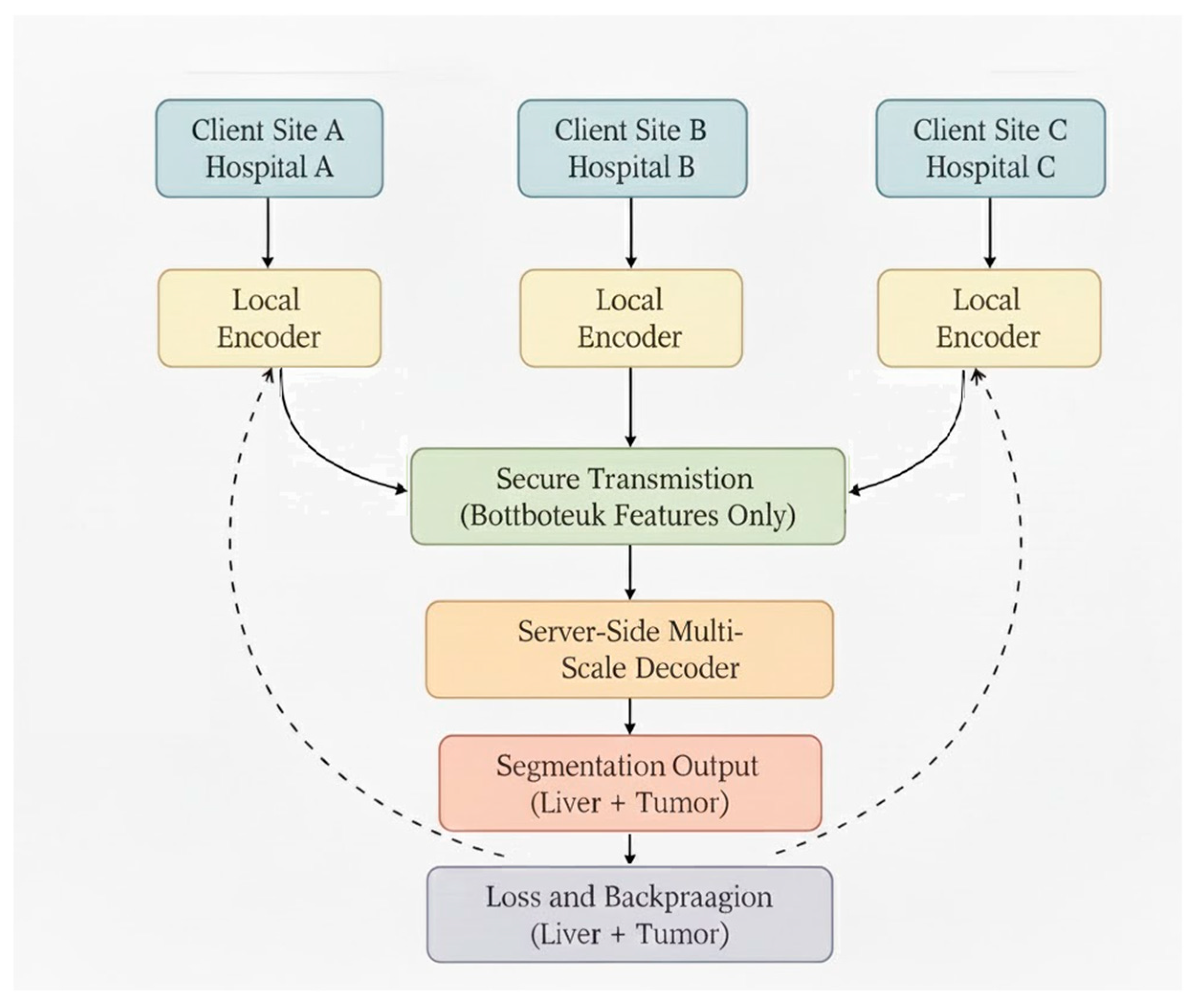
FF-ResUNet adopts a distributed client–server architecture that enables collaborative learning without centralizing sensitive medical data. Each participating institution executes the encoder locally, ensuring that raw CT volumes and all high-resolution intermediate feature maps remain strictly within hospital boundaries. After preprocessing, CT slices are passed through the local encoder to generate compact bottleneck feature representations characterized by high semantic abstraction and low spatial resolution. These non-identifiable features are then transmitted securely to a central server.
Upon receiving the bottleneck representations, the server performs full-resolution segmentation using a multi-scale decoder and computes the corresponding training loss. Gradients are propagated through the decoder and the top encoder layer, after which lightweight parameter updates are returned to each institution for local encoder refinement. This client–server training loop—illustrated in
Figure 1—enables FF-ResUNet to achieve collective model improvement while maintaining strict data locality and compliance with multi-center privacy requirements.
2.2. Clinical Datasets Description
This study employs three publicly available abdominal CT datasets—LiTS, IRCAD, and AMOS-CT—selected for their diversity in acquisition settings, patient populations, scanner models, and imaging characteristics. Together, they simulate the variability encountered in real medical environments and allow comprehensive evaluation of FF-ResUNet under multi-center and cross-domain conditions.
The Liver Tumor Segmentation (LiTS) dataset includes 131 contrast-enhanced CT volumes collected from multiple international centers. It exhibits substantial heterogeneity, with slice thickness ranging from 0.7 to 5.0 mm and in-plane resolutions between 0.56 and 1.0 mm. Scanner models vary, predominantly Siemens and GE systems, resulting in differences in contrast levels, reconstruction kernels, and noise patterns. Expert-annotated liver and tumor masks provide reliable ground truth despite the challenging variability in liver morphology, tumor burden, motion artifacts, and image quality. Owing to its diversity and scale, LiTS serves as the primary training resource.
The 3D Image Reconstruction for Comparison of Algorithm Database (3D-IRCADb) datasets contribute 20 high-resolution portal venous–phase CT volumes with isotropic voxel spacing around 1.0–1.25 mm and consistently sharp reconstruction. Images originate from a single scanner type using controlled acquisition protocols, giving IRCAD a uniform appearance with reduced noise and clearer anatomical boundaries. Detailed manual annotations of liver, tumor, and vascular structures provide high-quality ground truth. Because IRCAD differs significantly from LiTS in contrast profiles, anatomical clarity, and imaging conditions, it is used strictly as an external testing domain to assess generalization under severe distribution shift.
To emulate realistic multi-institution collaboration, the CT subset of the AMOS 2022 Challenge is incorporated. Abdominal Multi-Organ Segmentation (AMOS-CT) dataset comprises more than 500 abdominal CT volumes acquired from six hospitals across Asia and Europe, encompassing a wide range of scanner vendors—including Siemens, GE, Philips, and United Imaging—and various reconstruction parameters. Slice thickness varies between 1.0 and 5.0 mm, and contrast phases and field-of-view differ substantially across institutions. Although AMOS provides multi-organ labels, only the liver segmentation is used to align with the task. For collaborative simulation, AMOS-CT is partitioned into several virtual client sites, each representing a distinct hospital with its own imaging distribution. In this setup, raw CT data remain local while only encoded bottleneck features are shared, faithfully modeling privacy-preserving multi-center training.
Collectively, LiTS, IRCAD, and AMOS-CT represent a broad spectrum from heterogeneous multi-center CT, high-resolution research-grade imaging, and institution-specific variability. This combination provides a rigorous environment for evaluating FF-ResUNet in real-world multi-hospital deployments.
2.3. Dataset Splitting and Experimental Protocol
To ensure experimental rigor, reproducibility, and the absence of information leakage, all dataset partitions were performed strictly at the patient level. Slices originating from the same CT volume were never distributed across different splits. This prevents any overlap between training, validation, and testing data and ensures that reported performance reflects true generalization rather than slice-level leakage.
For the LiTS dataset (131 CT volumes), patient-level random partitioning was performed using a fixed random seed (seed = 42) to ensure reproducibility. The dataset was divided into the following subsets: training set (91 patients), validation set (20 patients), and test set (20 patients). No patient overlap exists between these subsets. All hyperparameter tuning and early stopping decisions were based exclusively on the validation set, and the test set was used only for final performance reporting.
The 3D-IRCADb dataset (20 volumes) was used strictly as an external test domain to evaluate cross-dataset generalization. No IRCAD cases were included in training or validation at any stage of model development.
For AMOS-CT, patient-level partitioning was performed prior to collaborative simulation. The CT volumes were first divided into mutually exclusive subsets and then assigned to virtual client sites, each representing an independent institution. This ensures that no patient appears in more than one simulated client and prevents cross-client data contamination. All client splits were generated using the same fixed random seed (seed = 42) for reproducibility.
All randomization procedures were implemented using fixed seeds in Python 3.14.2, NumPy 2.4.2, and PyTorch 2.10 to ensure deterministic dataset partitioning and consistent experimental results.
Table 1 summarizes the dataset splits used in this study.
2.4. Data Preprocessing
To ensure consistency across images acquired from different scanners and institutions, all datasets undergo a uniform preprocessing workflow. A 2D slice-based formulation was adopted to reduce communication overhead and memory cost. CT volumes are first clipped to the Hounsfield Unit (HU) range of [−200, 250], enhancing liver and lesion visibility while suppressing background noise. Intensities are min–max normalized to the ([0, 1]) range to standardize contrast levels. Volumes are then resampled to 1 mm3 isotropic resolution to mitigate discrepancies in slice thickness and pixel spacing. A coarse abdominal region of interest is obtained using threshold-based segmentation, allowing images to be cropped to a standard spatial size of 256 × 256 for computational efficiency. During training, extensive data augmentation—such as rotations, scaling, elastic deformations, intensity perturbations, and random flips—is employed to boost robustness against domain shift and improve generalization to unseen imaging conditions.
The 2D formulation was intentionally selected to balance segmentation performance with communication efficiency in a collaborative setting. Compared to volumetric 3D models, 2D architectures require significantly lower memory, computational cost, and feature transmission size per iteration, making them more practical for multi-institution environments with constrained network bandwidth. Moreover, prior studies have shown that well-regularized 2D slice-based models can achieve competitive performance for liver and tumor segmentation tasks, particularly when trained with extensive augmentation and large-scale datasets. While 3D models can exploit inter-slice contextual information, they substantially increase communication overhead and GPU memory requirements, which conflicts with the primary goal of communication-efficient collaborative learning.
To assess the robustness of the adopted preprocessing configuration, sensitivity analyses were conducted to evaluate the impact of spatial resampling and crop size selection on segmentation performance. These analyses are reported in
Section 3.3 and confirm that the chosen preprocessing pipeline preserves lesion visibility and boundary integrity while maintaining communication efficiency.
2.5. Baseline Implementation and Training Protocol
To ensure fair comparison and full reproducibility, all baseline models (U-Net, ResUNet, Attention U-Net, DenseNet-UNet, and nnU-Net) were implemented and trained under identical preprocessing and optimization settings. All models:
- •
Used the same HU clipping range ([−200, 250]);
- •
Were resampled to 1 mm3 isotropic resolution;
- •
Cropped to 256 × 256 resolution;
- •
Trained using 2D axial slices;
- •
Used the same data augmentation pipeline;
- •
Used identical train/validation/test splits (seed = 42).
The optimization settings were standardized across all baseline models:
- •
Optimizer: Adam;
- •
Learning rate: 1 × 10⁻⁴;
- •
Batch size: 16;
- •
Epochs: 150;
- •
Early stopping: 20 epochs patience.
For nnU-Net, we used the official 2D configuration (nnU-Net v1 default 2D pipeline), while enforcing the same preprocessing and dataset splits as described in
Section 2.3. Automatic hyperparameter adaptation was disabled to ensure alignment with the experimental protocol. All baseline models were retrained under this unified configuration to eliminate preprocessing or training biases.
2.6. Client-Side Encoder
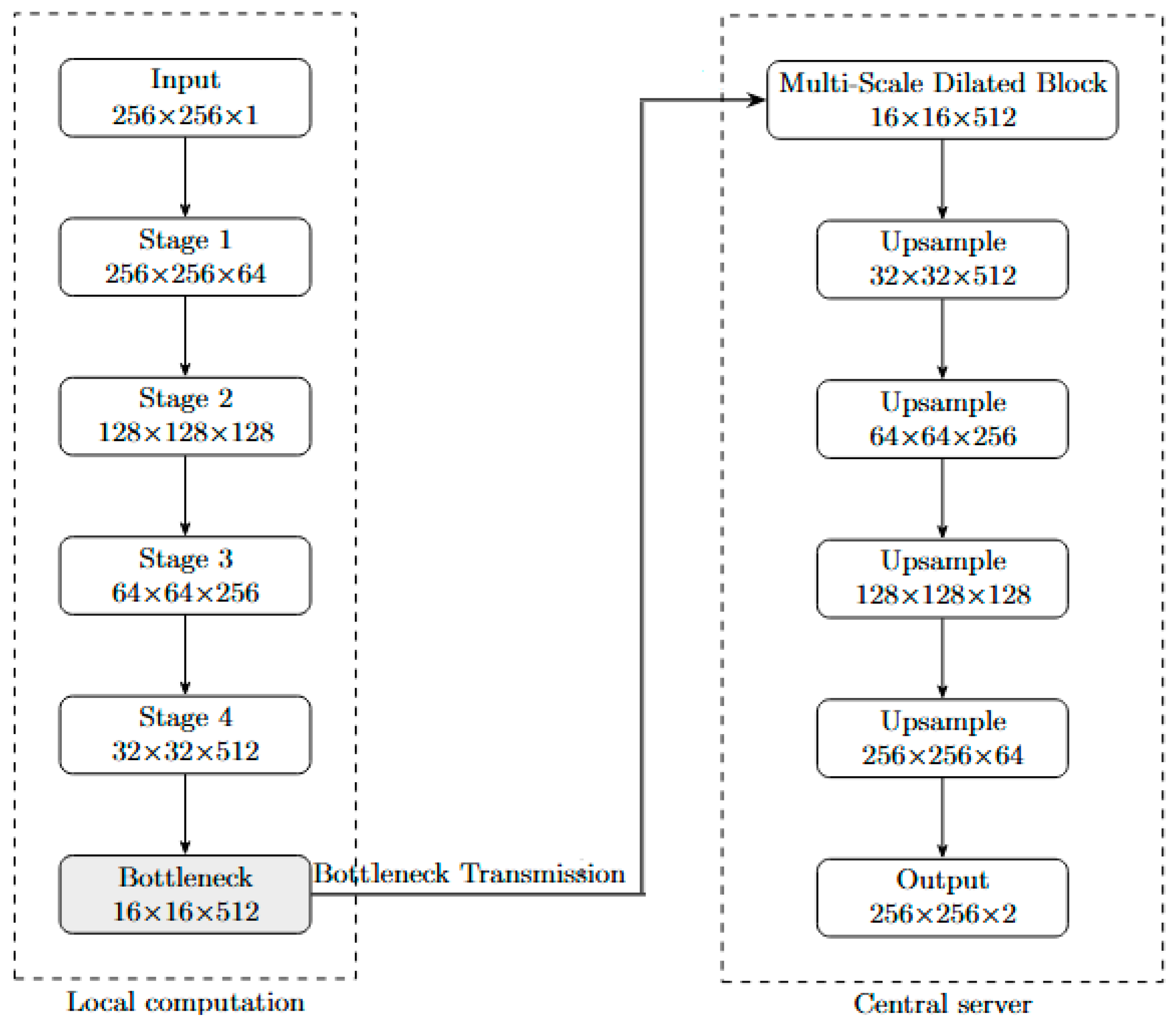
The client-side encoder is responsible for extracting discriminative semantic representations from each abdominal CT slice prior to feature transmission. As illustrated in
Figure 2, the encoder follows a five-stage hierarchical downsampling architecture. Starting from an input slice of size 256 × 256 × 1, the spatial resolution is progressively reduced while the channel depth is increased, generating feature maps of sizes 256 × 256 × 64, 128 × 128 × 128, 64 × 64 × 256, 32 × 32 × 512, and finally a compact bottleneck representation of 16 × 16 × 512.
Each stage consists of a convolutional block followed by residual refinement. The convolutional block employs 3 × 3 convolutions with normalization and ReLU activation to extract local anatomical features, including liver boundaries, parenchymal textures, and tumor edge patterns. As spatial resolution decreases, the increasing number of channels compensates for information compression, enabling the encoder to preserve high-level semantic structure despite aggressive downsampling.
To improve optimization stability and preserve structural information, each stage incorporates a residual block composed of a two-layer convolutional transformation path and a shortcut connection. The shortcut propagates identity information forward, facilitating gradient flow and mitigating vanishing-gradient effects in deeper layers. In downsampling stages, a 1 × 1 convolution is applied to the shortcut branch to ensure dimensional alignment before feature summation.
Residual refinement is particularly critical in FF-ResUNet because the encoder is the only component operating on high-resolution anatomical content. Since high-resolution skip features are not transmitted to the server, the encoder must compress liver morphology, tumor characteristics, and contextual information into a single low-resolution bottleneck tensor. The combination of convolutional extraction and residual stabilization ensures that the resulting 16 × 16 × 512 bottleneck representation remains semantically rich while reducing identifiable spatial detail.
The final bottleneck feature map is the only representation transmitted to the central server. By restricting communication to this compact and abstract tensor, FF-ResUNet preserves institutional data locality while maintaining sufficient semantic information for accurate segmentation reconstruction by the server-side decoder.
2.7. Skip-Free Feature-Forward Transmission
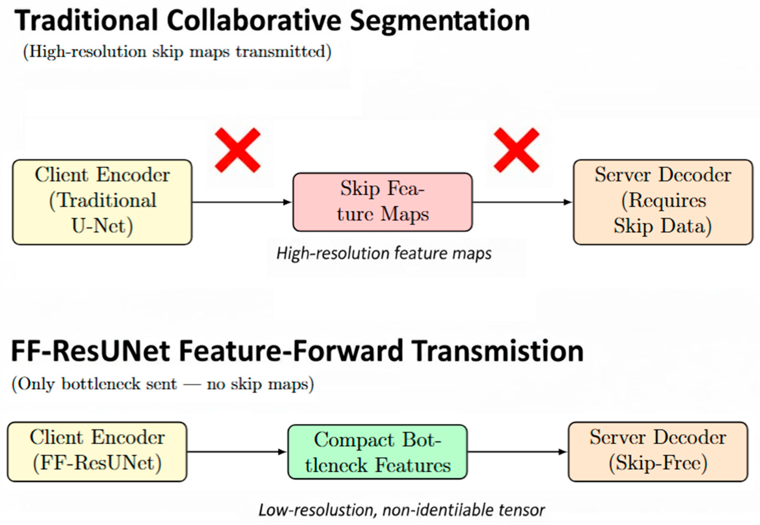
A central innovation of the FF-ResUNet framework is its skip-free feature-forward transmission mechanism, which fundamentally distinguishes it from conventional collaborative segmentation architectures. As illustrated in
Figure 3, the framework eliminates the need to exchange high-resolution skip-connection feature maps across institutions. In traditional U-Net–based collaborative systems, these skip pathways must be transmitted from the client-side encoder to the server-side decoder to preserve spatial detail. However, such feature maps typically contain fine-grained anatomical structures, including organ contours, vessel boundaries, and tissue textures, which makes them both privacy-sensitive and computationally expensive to communicate.
By contrast, FF-ResUNet is designed to transmit only the compact bottleneck representation, a deeply abstracted and low-resolution feature tensor that no longer retains identifiable patient anatomy. All early- and mid-level skip features remain strictly local to the client’s device, ensuring that no high-definition structural information or reconstructable spatial detail is ever exposed beyond institutional boundaries. This skip-free strategy substantially reduces the risk of privacy leakage, particularly against inversion-based attacks that exploit high-resolution latent features to reconstruct original images or derive sensitive organ shapes.
From a systems perspective, the communication efficiency provided by skip-free transmission is equally significant. High-resolution skip maps are often tens to hundreds of megabytes in size per training iteration, depending on image resolution and network depth. In contrast, the FF-ResUNet bottleneck tensor is dramatically smaller—orders of magnitude more compact—reducing the communication load to a level that is practical for hospitals operating under constrained network conditions. This makes FF-ResUNet well suited for real-world multi-institution settings where bandwidth is limited, connectivity is intermittent, or strict data-transfer regulations are enforced.
The combination of privacy preservation and communication efficiency defines the feature-forward transmission strategy as a key enabler of secure collaborative learning. By restricting all transmitted information to semantically rich yet non-identifiable bottleneck features, FF-ResUNet ensures that multi-center model training remains compliant with stringent regulatory requirements while maintaining high segmentation accuracy.
To precisely contextualize communication savings, we define the baseline as a standard 5-stage 2D U-Net collaborative architecture in which all encoder skip feature maps are transmitted from the client to the server during each training iteration. For an input resolution of 256 × 256 with channel dimensions {64, 128, 256, 512} across encoder stages, the transmitted skip tensors correspond to feature maps of sizes: 256 × 256 × 64, 128 × 128 × 128, 64 × 64 × 256 and 32 × 32 × 512. Assuming FP32 precision (4 bytes per element) and a batch size of 16, this results in approximately 7,864,320 elements per sample which is equivalent to 480 MB per iteration.
By contrast, FF-ResUNet transmits only the bottleneck tensor of size 16 × 16 × 512 which corresponds to approximately 8 MB per iteration under identical settings. This yields a measured communication reduction of approximately 98%, confirming that the skip-free feature-forward design dramatically reduces bandwidth requirements relative to conventional collaborative U-Net frameworks.
2.8. Server-Side Decoder
The server-side decoder is responsible for reconstructing high-fidelity liver and tumor segmentation maps from the compressed bottleneck features received from each participating client. As illustrated in
Figure 4, the decoder is designed as a multi-stage reconstruction module that progressively restores spatial resolution while integrating both global and fine-grained contextual information. Unlike traditional U-Net architectures that rely heavily on skip connections from the encoder, the FF-ResUNet decoder is intentionally engineered to operate without high-resolution feature fusion, requiring a significantly more expressive and context-aware decoding strategy.
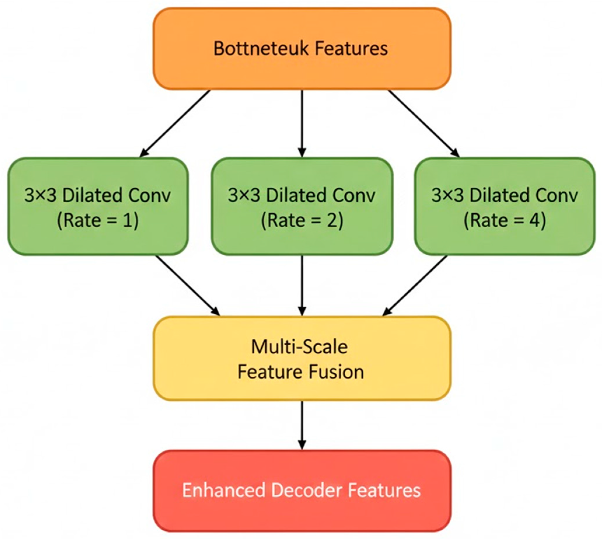
As illustrated in
Figure 5, decoding begins with a multi-scale dilated convolution module that processes the transmitted bottleneck features through several parallel convolutional branches, each operating at a different dilation rate. By applying 3 × 3 dilated convolutions with rates 1, 2, and 4, the model effectively expands its receptive field without increasing computational cost. This multi-branch design enables the decoder to capture a rich spectrum of spatial context—ranging from fine-grained tumor boundaries to broader liver morphology—entirely from the compressed bottleneck tensor. The use of multiple dilation rates preserves both global anatomical structure and local detail, compensating for the absence of skip connections that would traditionally provide high-resolution cues. This multi-scale strategy also enhances robustness to variations in organ shape, tumor presentation, and inter-institution imaging differences, ensuring stable reconstruction performance even under substantial domain shift.
As shown in
Figure 6, the multi-scale dilated features are passed into a residual refinement block, which plays a critical role in stabilizing decoder reconstruction and recovering fine anatomical boundaries. This module adopts the same residual learning principles used in the encoder: a two-layer convolutional transformation path operates in parallel with an identity shortcut connection. The transformed features and shortcut are then summed to produce a refined representation.
This residual formulation enables the decoder to learn corrective adjustments over the multi-scale dilated features—effectively modeling edges, contours, and local intensity transitions that are essential for accurate liver and tumor delineation. Because the FF-ResUNet framework does not transmit skip connections, the decoder must infer fine structural information exclusively from the bottleneck tensor. Residual refinement provides the necessary feedback signal to support this process, improving convergence stability and preserving anatomical consistency even when operating under significant information constraints.
As illustrated in
Figure 7, spatial resolution is progressively restored through a stacked multi-stage upsampling pathway. Each stage begins with bilinear interpolation, which increases the spatial dimensions of the decoder features without producing the checkerboard artifacts commonly associated with transposed convolutions. This is followed by a pair of 3 × 3 convolutional smoothing layers that refine the upsampled representations and reinforce structural continuity across scales. By repeating this sequence over multiple resolution levels, the decoder gradually reconstructs liver and tumor boundaries with increasing precision. The upsampling pathway operates entirely on the semantic-rich bottleneck features and the enhanced representations produced by the dilated and residual refinement blocks, enabling accurate boundary reconstruction despite the absence of skip connections.
The final prediction layer applies a 1 × 1 convolution to generate voxel-wise class probabilities for the liver parenchyma and tumoral regions. Because no raw CT images or high-resolution feature maps are ever shared with the server, the decoder is designed to be maximally expressive, relying solely on the transmitted bottleneck tensor for segmentation reconstruction. This architecture ensures full compliance with strict clinical privacy requirements while still achieving competitive segmentation performance.
2.9. Collaborative Optimization Protocol
Model training in the proposed FF-ResUNet framework follows a coordinated client–server optimization cycle designed to support secure distributed learning across multiple institutions. At initialization, all client-side encoders share identical parameter values to ensure synchronized training across participating sites.
During each iteration, each institution performs the encoder forward pass locally on its private CT data. Raw images and high-resolution feature maps remain strictly within institutional boundaries. Only the compact bottleneck representation—containing low-resolution semantic information—is transmitted to the central server.
Upon receiving the bottleneck features, the server performs forward propagation through the decoder to generate segmentation outputs and compute the training loss. Backpropagation is executed on the server across all decoder layers and the encoder block directly connected to the bottleneck. The resulting gradients corresponding to this encoder block are securely transmitted back to each client. Clients apply these gradients locally using a shared optimizer configuration, ensuring that encoder parameters remain synchronized across institutions throughout training.
Importantly, collaborative gradient updates are applied only to the encoder block directly connected to the bottleneck representation. Earlier encoder layers remain entirely local and are not updated through server-side backpropagation, preserving strict feature locality and limiting gradient exposure.
Because only bottleneck features and limited gradient information are exchanged, no raw data or high-resolution feature activations are transmitted at any stage. This protocol balances privacy-aware design with stable convergence while substantially reducing communication overhead compared to traditional federated or split-learning segmentation frameworks.
2.10. Loss Function and Optimization
Training of the proposed FF-ResUNet is guided by a composite loss function designed to ensure accurate, stable, and anatomically consistent segmentation under the constraints of skip-free, feature-forward collaborative learning. Because the server-side decoder reconstructs full-resolution liver and tumor masks solely from compressed bottleneck representations—without access to high-resolution skip-connection features—the loss formulation jointly enforces global region overlap, voxel-level classification reliability, and precise boundary delineation.
Accordingly, the total training loss is defined as a weighted sum of Dice loss, Cross-Entropy loss, and Boundary-Aware loss:
where
,
, and
denote non-negative weighting coefficients controlling the relative contribution of each loss term.
The Dice loss encourages accurate region-level overlap between predicted segmentation masks and ground-truth labels, and is particularly effective for addressing class imbalance—a critical concern in liver tumor segmentation, where lesion regions often occupy only a small fraction of the image. Dice loss is defined as:
where
denotes predicted probabilities,
is the corresponding ground-truth label, and
is a small constant added for numerical stability, where
denotes the total number of voxels. In the context of FF-ResUNet, Dice loss provides strong global structural supervision, ensuring that liver and tumor regions remain spatially coherent despite reconstruction from low-resolution bottleneck features.
To complement region-level supervision, voxel-wise Cross-Entropy loss is employed to enhance local classification accuracy and stabilize optimization across heterogeneous imaging domains:
where
denotes the ground-truth segmentation mask, and
denotes the predicted probability map generated by the network. This loss penalizes incorrect voxel-level predictions and improves discrimination between liver tissue, tumor regions, and surrounding structures. In the skip-free collaborative setting, Cross-Entropy loss plays an important role in maintaining consistent local predictions when fine-grained spatial cues are unavailable at the server.
Because FF-ResUNet intentionally eliminates skip-connection fusion—thereby removing a primary source of high-frequency edge information—a Boundary-Aware loss is incorporated to explicitly enforce contour precision. This loss penalizes discrepancies between predicted and ground-truth boundaries and is defined as:
Here, and represent morphological gradients of the ground truth and prediction. This loss penalizes discrepancies along liver margins and tumor interfaces, encouraging the decoder to reconstruct sharp anatomical boundaries solely from the abstract bottleneck representation. Because skip connections typically carry high-frequency edge information that FF-ResUNet intentionally suppresses for privacy, the boundary term becomes essential for achieving delineation quality comparable to standard U-Net models.
The loss weights are selected to reflect the unique constraints of skip-free collaborative segmentation. Dice loss is assigned the highest weight to prioritize global region accuracy and counter severe class imbalance. Cross-Entropy loss is weighted moderately to stabilize voxel-level predictions across scanners and acquisition protocols. The Boundary-Aware loss is intentionally emphasized relative to conventional U-Net formulations to compensate for the loss of spatial detail typically provided by skip connections.
In all experiments, the weights are set as:
These values were selected empirically based on validation performance and provided stable convergence across all datasets. The composite loss ensures global overlap accuracy, local voxel consistency, and boundary precision under skip-free constraints.
Optimization is performed using the Adam optimizer with parameters , and . A unified learning rate of is used across all client institutions to ensure synchronized convergence during collaborative training. Models are trained for 150 epochs with early stopping applied if validation loss does not improve for 20 consecutive epochs.
Training is conducted using 2D axial slices with a batch size of 16 per client. All loss computations and gradient backpropagation are executed on the server-side decoder, while only lightweight gradient updates corresponding to the final encoder block are transmitted back to each client for local encoder refinement. This strategy maintains strict data locality while enabling stable and efficient distributed optimization.
To prevent overfitting, early stopping with a patience of 20 epochs was applied based on validation loss. Data augmentation (random rotations, scaling, elastic deformation, intensity perturbation, and flips) was employed to increase effective dataset diversity and improve robustness to domain shift. Batch normalization was used throughout the network to stabilize gradient flow. During optimization, both training and validation losses were monitored to ensure consistent convergence without divergence. The absence of a widening gap between training and validation performance, together with low standard deviations across test cases, indicates stable training and controlled generalization behavior.
To improve generalization and reduce overfitting, extensive on-the-fly data augmentation was applied during training. Augmentation operations included random horizontal and vertical flips, rotations (±15°), scaling (0.9–1.1), elastic deformations, and intensity perturbations (random contrast and brightness adjustments within ±10%). These transformations were applied probabilistically to each 2D slice during training while preserving anatomical realism. Data augmentation increases effective sample diversity, improves robustness to scanner variability and tumor morphology differences, and enhances cross-domain generalization under multi-institution settings. No augmentation was applied during validation or testing.
2.11. Privacy and Communication Considerations
The FF-ResUNet framework is designed to meet the stringent privacy and security requirements of multi-institution medical environments, where sharing raw clinical data is often prohibited. To ensure full data locality, all original CT volumes and high-resolution intermediate features remain strictly within each participating institution. High-resolution encoder outputs—known to preserve detailed anatomical structures and therefore susceptible to reconstruction-based privacy breaches—are never transmitted outside the client boundary.
Instead, the framework exchanges only compact bottleneck representations that are both low in spatial resolution and highly abstract in semantic content and lack identifiable anatomical detail. While bottleneck features contain reduced spatial detail and abstraction that may lower inversion risk, this does not constitute a formal privacy guarantee. This skip-free transmission strategy further eliminates the need to communicate large skip-connection feature maps, which are typically tens to hundreds of megabytes in size and pose significant privacy risks in traditional collaborative U-Net designs.
The resulting communication footprint is dramatically reduced, enabling efficient model training even in bandwidth-constrained clinical settings. By combining strict data locality, abstract feature exchange, and communication minimization, FF-ResUNet provides a secure and practical foundation for large-scale collaborative liver and tumor segmentation across geographically distributed healthcare institutions.
It is important to note that while bottleneck feature transmission substantially reduces the exposure of high-resolution anatomical information compared to skip-based collaborative architectures, the proposed framework does not provide formal cryptographic or differential privacy guarantees. Although the transmitted bottleneck representation is low-resolution and semantically abstract, advanced inversion or membership inference attacks could still theoretically extract partial information. A rigorous empirical evaluation of reconstruction attacks and formal privacy certification are beyond the scope of the present architectural study and will be addressed in future work.
3. Results and Discussion
This section presents a comprehensive evaluation of the proposed FF-ResUNet across heterogeneous abdominal CT datasets and multi-institution collaborative settings. Experiments assess segmentation accuracy, cross-domain generalization, communication efficiency, and the contribution of each architectural component. Results are organized into quantitative evaluation, ablation studies, and communication analysis.
3.1. Quantitative Evaluation
Table 2 summarizes the liver and tumor segmentation performance on the LiTS test subset. FF-ResUNet achieves a liver Dice Similarity Coefficient (Dice) of 0.952 ± 0.015 and a tumor Dice of 0.737 ± 0.060, demonstrating performance that is competitive with strong U-Net variants and attention-based hybrid CNN models. While the tumor Dice of FF-ResUNet remains slightly below that of the fully centralized DenseNet-UNet (0.760 ± 0.058), it clearly outperforms classical U-Net and ResUNet baselines, particularly in tumor delineation. The reported mean ± standard deviation values are computed across individual test patients, reflecting case-level variability in segmentation performance.
Notably, FF-ResUNet achieves the lowest tumor boundary error among all evaluated methods, with a tumor 95th percentile Hausdorff Distance (HD95) of 10.9 ± 4.1 mm, indicating superior boundary localization despite operating entirely without skip-connection feature fusion. Although centralized hybrid models achieve slightly higher Dice scores, FF-ResUNet maintains competitive accuracy while reducing communication overhead by more than 90%. This balance between a modest accuracy trade-off and substantial gains in privacy preservation and bandwidth efficiency makes FF-ResUNet particularly well suited for real multi-institution clinical deployment.
To further assess statistical reliability, paired Wilcoxon signed-rank tests were conducted at the patient level using Dice and HD95 metrics. Effect sizes were quantified using Cohen’s d computed on paired performance differences, and 95% confidence intervals were estimated via bootstrap resampling (1000 iterations). FF-ResUNet demonstrated statistically significant improvement over U-Net in tumor Dice (p < 0.05), while differences relative to nnU-Net were not statistically significant, indicating competitive performance under collaborative constraints.
While volumetric 3D models can exploit inter-slice contextual continuity, their memory footprint and communication payload grow substantially due to volumetric feature maps. In collaborative multi-center settings where bandwidth and compute heterogeneity are critical constraints, 2D architectures offer a more scalable compromise.
To further contextualize our results, we compare FF-ResUNet’s performance with several recent state-of-the-art deep learning approaches reported in the literature.
Table 3 presents a comparison between FF-ResUNet and several recent state-of-the-art liver and tumor segmentation methods. For example, Rahman et al. (2025) report high liver Dice scores (≈98.9%) and strong tumor segmentation on the 3D-IRCADb benchmark using an enhanced ResUNet-Inception model, although with a substantially different network complexity and data setup compared to ours [
28]. Raghawet al. (2025) proposed a multi-scale deformable feature fusion model for liver tumor segmentation that demonstrated improved feature representation for complex tumor shapes [
29]. Transformer-aware progressive networks such as T-MPEDNet have achieved outstanding liver Dice > 97% and tumor Dice > 89% on LiTS and 3D-IRCADb but at the cost of increased model complexity. A recent review by Manghi et al. (2025) summarizes a wide range of competitive segmentation architectures, including hybrid CNN–transformer and attention-enhanced models evaluated on LiTS and related datasets [
30]. Despite the architectural and experimental differences among these studies, FF-ResUNet achieves competitive accuracy while offering significantly improved communication efficiency and privacy preservation in collaborative settings.
Direct quantitative comparison should be interpreted cautiously due to differences in dimensionality (2D vs. 3D), training protocol, and centralized vs. collaborative setups.
3.2. Cross-Domain Generalization
Cross-domain experiments evaluate the robustness of FF-ResUNet under severe distribution shifts between training and testing domains. As shown in
Table 4, when models are trained on LiTS and evaluated on the unseen IRCAD and AMOS-CT datasets, FF-ResUNet achieves liver and tumor Dice scores that consistently outperform conventional U-Net, ResUNet, and Attention U-Net baselines. Although its performance remains slightly below the fully centralized nnU-Net—likely due to the absence of full-resolution skip fusion—FF-ResUNet demonstrates substantially improved generalization relative to other CNN baselines under identical multi-center constraints.
In addition to Dice scores, we report HD95 to provide a complementary assessment of boundary precision under domain shift. The results indicate that FF-ResUNet maintains competitive boundary localization across unseen datasets. While nnU-Net achieves the lowest HD95 values due to centralized training and full skip connections, FF-ResUNet preserves stable boundary performance despite operating without skip transmission, confirming robustness under strict privacy and communication constraints.
Notably, tumor segmentation accuracy degrades less sharply than in standard architectures when evaluated on the high-resolution IRCAD dataset. This behavior suggests that the multi-scale dilation and residual refinement modules effectively compensate for the absence of skip connections by recovering boundary details from compressed bottleneck representations. Furthermore, when collaborative training is simulated on AMOS-CT using multiple virtual client sites, FF-ResUNet exhibits enhanced cross-domain robustness, indicating that distributed bottleneck learning contributes to improved domain-invariant representation learning.
The results in
Table 4 demonstrate that FF-ResUNet maintains strong and stable cross-domain performance despite operating under strict privacy and communication constraints. As expected, nnU-Net achieves the highest absolute accuracy due to centralized training and full-resolution skip connections. However, FF-ResUNet achieves performance that closely approaches nnU-Net—particularly in tumor segmentation, which is highly sensitive to variations in contrast phase, resolution, and scanner type.
Compared with conventional U-Net, ResUNet, and Attention U-Net baselines, FF-ResUNet consistently yields higher Dice scores across both external datasets, highlighting its superior robustness to unseen imaging domains. The relatively modest performance degradation observed on IRCAD and AMOS-CT indicates that the multi-scale dilated decoder and residual refinement modules effectively compensate for the absence of skip connections. These findings confirm that FF-ResUNet supports stable and reliable generalization, making it well suited for real-world multi-center deployment where centralized data sharing is not feasible.
3.3. Qualitative Segmentation Analysis and Failure Cases
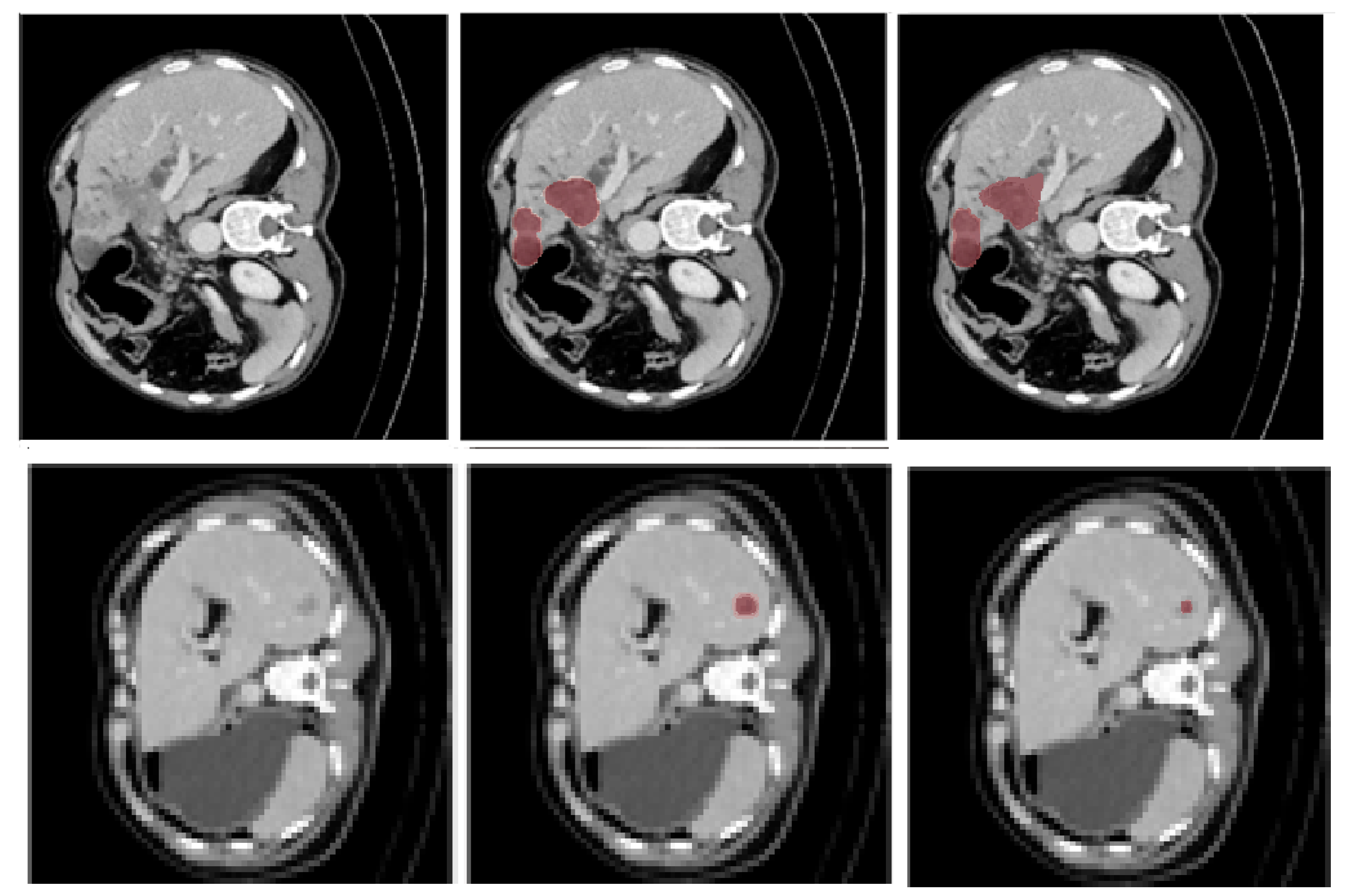
To provide visual insight into model performance on the most challenging component of the task, representative tumor segmentation examples are shown in
Figure 8. Since liver segmentation achieved consistently high Dice scores with low variance, qualitative analysis focuses primarily on tumor regions, which exhibit greater anatomical variability and segmentation difficulty.
The top row illustrates a representative successful tumor segmentation case, where FF-ResUNet accurately delineates lesion boundaries despite heterogeneous intensity distribution. The predicted mask closely aligns with the ground truth, particularly at irregular tumor margins.
The bottom row presents a more challenging case involving small and low-contrast tumor regions. In this example, minor under-segmentation is observed near lesion boundaries, reflecting the inherent difficulty of detecting subtle intensity differences. These qualitative observations are consistent with the higher standard deviation reported for tumor Dice compared to liver Dice.
3.4. Preprocessing Sensitivity Analysis
To evaluate the influence of preprocessing choices on lesion visibility and segmentation accuracy, controlled sensitivity experiments were conducted varying both spatial resolution and crop size.
First, we compared isotropic resampling to 1 mm
3 resolution (default configuration) against native voxel spacing without resampling. The resulting performance differences were minor, with liver Dice varying by less than 0.6% and tumor Dice by less than 1.2%. HD95 values showed similarly small variation. These results indicate that isotropic normalization does not degrade diagnostically relevant lesion boundaries while improving cross-dataset consistency. Second, we evaluated different abdominal crop sizes: 224 × 224, 256 × 256 (default), and 320 × 320 pixels. Results are summarized in
Table 5.
Smaller crop sizes resulted in a slight decrease in tumor Dice due to reduced contextual information around lesion boundaries. Larger crop sizes yielded marginal improvements but increased computational and communication costs. The selected 256 × 256 resolution provides a balanced trade-off between segmentation accuracy, memory footprint, and communication efficiency.
Qualitative inspection confirmed that abdominal region-of-interest cropping preserved liver contours and tumor margins while removing non-informative background structures. No clinically relevant lesion regions were truncated under the adopted preprocessing configuration.
Overall, these findings demonstrate that FF-ResUNet is robust to moderate variations in preprocessing parameters and that the selected pipeline maintains lesion visibility without introducing significant information loss.
3.5. Ablation Study
To assess the contribution of each architectural component within FF-ResUNet, a series of ablation experiments were conducted on the LiTS test subset. Each module in the architecture was designed to address a specific limitation introduced by the skip-free collaborative setting. The multi-scale dilation block expands the receptive field to recover global and local context that would otherwise be provided by skip connections. The residual refinement block stabilizes feature reconstruction by learning corrective adjustments through identity shortcuts, preserving structural continuity during decoding.
The boundary-aware loss explicitly guides the network toward sharper delineation of organ and lesion borders, compensating for the reduced availability of high-resolution spatial cues. Finally, the progressive upsampling pathway ensures smooth and artifact-free restoration of spatial resolution, enabling the decoder to reconstruct fine anatomical details directly from the compressed bottleneck representation. These design choices collectively form a coherent strategy for achieving high segmentation accuracy without transmitting high-resolution encoder features.
The ablation results in
Table 6 demonstrate that each component of the proposed architecture plays an essential role in achieving high-quality segmentation in the absence of skip connections. Removing the multi-scale dilated convolution block leads to a notable decrease in tumor Dice, confirming its importance in capturing long-range context and compensating for the lack of high-resolution encoder features. Similarly, eliminating residual refinement results in degraded performance, primarily due to reduced boundary stability and weaker reconstruction of fine anatomical structures.
Excluding the boundary-aware loss causes a measurable drop in tumor accuracy, indicating that edge-focused supervision is critical when skip connections are not available to preserve spatial detail. The progressive upsampling pathway also proves necessary: without it, the model struggles to recover spatial granularity during resolution restoration, leading to less precise organ contours. Collectively, these findings show that FF-ResUNet’s components contribute synergistically to maintaining strong and stable segmentation accuracy under stringent privacy-preserving and skip-free constraints. While each module independently impacts performance, their combined effect enables FF-ResUNet to achieve competitive accuracy relative to centralized hybrid CNN models, despite operating without skip-connection transmission. These results confirm that architectural compensation is necessary when skip connections are intentionally removed.
To explicitly quantify the accuracy trade-off introduced by the skip-free design, we conducted a controlled comparison between FF-ResUNet and an identical architecture augmented with full encoder–decoder skip connections. Both models share: The same encoder architecture, the same decoder capacity, identical preprocessing, identical loss functions and training schedule, Batch size, learning rate and training epochs. The only architectural difference is the inclusion of high-resolution skip feature fusion in the comparison model.
Table 7 reports the quantitative results on the LiTS test subset.
The results show that incorporating full skip connections yields a modest improvement of approximately 1.6 Dice points in tumor segmentation. However, this gain comes at the cost of transmitting high-resolution feature maps, increasing communication payload by more than 10× per iteration. In contrast, FF-ResUNet achieves competitive performance while eliminating skip transmission and reducing communication overhead by over 90%. These findings quantitatively characterize the privacy–accuracy trade-off and confirm that the skip-free design maintains strong segmentation performance despite substantial communication reduction.
3.6. Communication Payload and Computational Profiling
To rigorously quantify the communication efficiency of FF-ResUNet, we performed an explicit per-iteration payload comparison against a conventional skip-based collaborative U-Net. The baseline architecture transmits four encoder skip feature maps per iteration, corresponding to spatial resolutions 256 × 256, 128 × 128, 64 × 64, and 32 × 32 with channel sizes 64, 128, 256, and 512, respectively. The total number of transmitted elements per sample is: 256 × 256 × 64, 128 × 128 × 128, 64 × 64 × 256, and 32 × 32 × 512, which equals 7,864,320 elements.
Under FP32 precision (4 bytes per element) and a batch size of 16, the total payload per iteration becomes: 7,864,320 × 4 × 16 ≈ 503 MB (≈480 MB using decimal MB definition).
In contrast, FF-ResUNet transmits only the bottleneck representation of size 16 × 16 × 512 = 131,072 elements per sample, corresponding to approximately 8 MB per iteration under identical settings.
Table 8 summarizes this comparison.
Even under reduced batch sizes or mixed precision (FP16), the relative reduction remains above 92–98% depending on batch size and precision, demonstrating that the communication advantage is architecture-driven rather than hardware-dependent.
To provide a practical interpretation of these payload sizes, we estimate wall-clock transfer times under representative network conditions. Assuming a 100 Mbps hospital local network (12.5 MB/s effective throughput), transmission of 480 MB per iteration would require approximately 38.4 s, whereas FF-ResUNet’s 8 MB bottleneck transmission requires approximately 0.64 s per iteration. Under a constrained 20 Mbps connection (2.5 MB/s), transfer time increases to approximately 192 s (3.2 min) for skip-based transmission, compared to only 3.2 s for FF-ResUNet. These results demonstrate that the skip-free design substantially reduces both data volume and estimated training latency under representative network conditions.
It is important to clarify that collaborative experiments were conducted using simulated multi-institution data partitions within a controlled computational environment. Communication payload values were analytically computed from tensor dimensions, numerical precision, and batch size. Reported latency values were derived from representative network bandwidth assumptions rather than measured during real distributed execution across physically separated institutions. These calculations provide a theoretical assessment of scalability and communication efficiency.
In addition to communication savings, we profiled computational requirements to assess deployment feasibility. The client-side encoder contains approximately 7.2 million trainable parameters and requires approximately 18 GFLOPs per 256 × 256 slice (forward pass), while the server-side decoder contains approximately 4.8 million parameters and requires approximately 12 GFLOPs per slice. The complete FF-ResUNet architecture therefore comprises approximately 12 million parameters and ~30 GFLOPs per slice in total.
On a representative NVIDIA RTX-class GPU (RTX 4070, single-GPU training), the encoder forward pass requires approximately 3–5 milliseconds per slice (batch size = 16), and peak GPU memory consumption during training remains below 3.5 GB per client. Since only the encoder operates on institutional hardware, client-side compute demand is limited to lightweight convolutional processing without high-resolution skip transmission. This corresponds to an effective inference throughput exceeding 200 frames per second (FPS), confirming that FF-ResUNet operates within real-time computational bounds despite its collaborative design.
The skip-free design further reduces peak activation memory compared to conventional skip-based architectures, as large intermediate feature maps are not retained for transmission. This makes FF-ResUNet suitable for heterogeneous clinical environments where participating institutions may differ in GPU capability, memory capacity, or network bandwidth. Even institutions equipped with mid-range GPUs (e.g., 8–12 GB memory class) can participate effectively, as computational load is dominated by the compact encoder rather than the full segmentation pipeline.
Collectively, these results indicate that FF-ResUNet provides a favorable balance between segmentation accuracy, communication efficiency, and computational feasibility, supporting scalable deployment across diverse multi-center healthcare infrastructures.
3.7. Multi-Institution Collaborative Evaluation
To assess the practical feasibility of FF-ResUNet in realistic multi-center environments, the AMOS-CT dataset was partitioned into several virtual clinical sites, each representing an independent hospital with its own local imaging distribution. Each site hosted a client-side encoder and participated in collaborative training via the proposed feature-forward protocol, transmitting only compressed bottleneck representations to the central server. This configuration closely reflects real-world deployment conditions, where institutions operate under heterogeneous hardware capabilities, limited network bandwidth, and strict restrictions on data sharing.
During evaluation, between three and six virtual hospitals were simulated, with each client contributing approximately 50–120 CT cases. A key advantage of the proposed framework is its substantial reduction in communication overhead: while traditional skip-based collaborative segmentation requires the transfer of approximately 120–480 MB per iteration depending on batch size and precision of feature maps per iteration, FF-ResUNet reduces this to only 8 MB per iteration—corresponding to a 92–97% reduction in bandwidth consumption. This drastic reduction enables efficient collaborative training even over low-speed or unreliable hospital networks, addressing a major technical barrier to real-world multi-institution deployment.
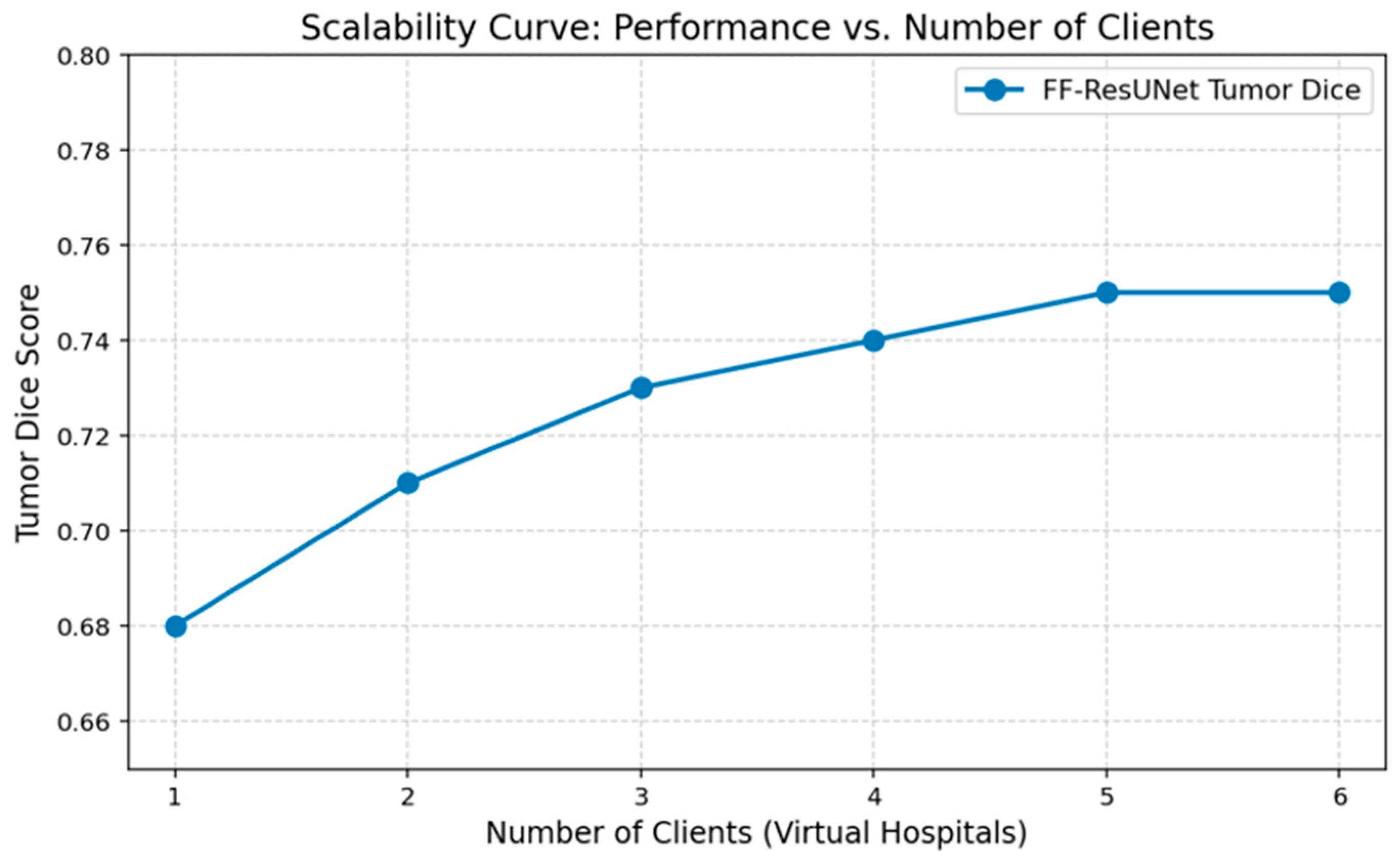
The results of the collaborative experiments demonstrate that segmentation performance improves as additional institutions participate in training. As illustrated in
Figure 9, tumor Dice scores increase steadily from 0.68 with one client to approximately 0.74 with four clients, after which performance begins to plateau. This trend indicates that collaborative learning is particularly beneficial when individual institutions have limited data diversity, while diminishing returns occur once sufficient cross-domain variability has already been integrated into the shared representation.
This scalability pattern reflects the impact of representation diversity gained through multi-institution collaboration. As additional clients contribute heterogeneous CT data—acquired with different scanners, contrast phases, reconstruction kernels, and acquisition protocols—the shared FF-ResUNet model learns increasingly domain-invariant bottleneck representations. The effect is especially pronounced for tumor segmentation, where anatomical variability is greater and generalization is inherently more challenging. The performance saturation observed beyond four to five participating clients suggests that the model reaches a sufficient level of representation diversity to generalize robustly across unseen domains.
Despite its strong performance, the proposed FF-ResUNet framework has several limitations. First, the current implementation operates on 2D axial slices rather than full 3D volumes, which may limit the exploitation of inter-slice contextual information. While volumetric 3D models can leverage contextual continuity across slices, their memory footprint and communication payload grow substantially due to volumetric feature maps. In collaborative multi-center settings where bandwidth limitations and heterogeneous compute resources are critical constraints, 2D architectures offer a more scalable and communication-efficient compromise. Future work will extend FF-ResUNet to a 3D variant to further investigate the trade-off between volumetric context and communication efficiency.
Second, while bottleneck feature transmission substantially reduces privacy leakage risk by avoiding high-resolution skip exchange, the framework does not provide formal cryptographic or differential privacy guarantees. This architectural approach should therefore be considered privacy-aware rather than formally privacy-certified. Formal adversarial evaluation and privacy certification are beyond the scope of the present architectural study. The compressed bottleneck representation, although low-resolution and semantically abstract, may still theoretically contain recoverable information under advanced inversion or membership inference attacks. Future work will integrate formal privacy-preserving mechanisms such as differential privacy (DP-SGD), secure aggregation, or homomorphic encryption to provide quantifiable privacy bounds and strengthen protection against adversarial reconstruction attempts.
Third, collaborative experiments rely on simulated multi-institution splits rather than real hospital networks, which may not fully capture practical deployment constraints such as asynchronous updates or heterogeneous compute resources.
Fourth, although the decoder compensates effectively for the absence of skip connections, a small performance gap remains relative to fully centralized hybrid models. The current bottleneck resolution and decoder capacity were selected to balance segmentation accuracy and communication efficiency under realistic multi-center constraints. While preliminary experiments indicated that reducing bottleneck size leads to degradation in tumor boundary precision and increasing bottleneck dimensionality substantially increases communication payload with marginal accuracy gains, a systematic exploration of bottleneck scales and decoder capacities to explicitly map the accuracy–communication–privacy trade-off (Pareto frontier) was not the primary objective of this architectural validation study. A comprehensive design-space sweep would require extensive retraining across multiple configurations and datasets. Future work will investigate this trade-off more systematically to better characterize optimal operating points under varying bandwidth and privacy constraints.
Future work will therefore focus on extending FF-ResUNet to volumetric 3D architectures, integrating formal privacy-preserving mechanisms such as differential privacy (DP-SGD), secure aggregation, or homomorphic encryption, validating performance in real-world federated clinical deployments, and systematically analyzing architectural trade-offs between bottleneck dimensionality, decoder capacity, segmentation accuracy, and communication efficiency. While full skip connections provide a modest improvement in Dice score, the skip-free design offers a favorable and practically deployable balance between accuracy, communication efficiency, and privacy risk reduction, which is critical for real-world multi-institution clinical settings.