Next Issue
Volume 5, June
Previous Issue
Volume 4, December


Osteology, Volume 5, Issue 1 (March 2025) – 10 articles

Cover Story (view full-size image): 
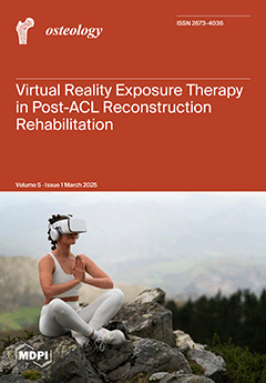
Anterior cruciate ligament reconstruction is a common surgical procedure among active individuals, yet postoperative rehabilitation remains challenging. Virtual Reality Exposure Therapy has emerged as a promising tool to enhance rehabilitation outcomes. This meta-analysis evaluates the efficacy of VRET in post-ACL reconstruction rehabilitation by analyzing studies comparing VRET with conventional therapies. The findings suggest that VRET significantly improves functional recovery and pain reduction, underscoring its potential as an innovative adjunct in postoperative rehabilitation programs. View this paper
  • Issues are regarded as officially published after their release is announced to the table of contents alert mailing list.
  • You may sign up for e-mail alerts to receive table of contents of newly released issues.
  • PDF is the official format for papers published in both, html and pdf forms. To view the papers in pdf format, click on the "PDF Full-text" link, and use the free Adobe Reader to open them.
Order results
Result details
Select all
Export citation of selected articles as:
9 pages, 591 KiB  
Article
Physiotherapy Intervention on Functional Health in Aging on Functional Capacity, Risk of Falls, Cognitive Function, and Back Pain
by Gustavo Desouzart
Osteology 2025, 5(1), 10; https://doi.org/10.3390/osteology5010010 - 10 Mar 2025
Viewed by 648
Abstract
Background/Objectives: Aging is associated with a progressive decline in biological function due to a complex interplay of physical, psychological, and social factors. This randomized controlled trial aimed to evaluate the effects of a functional health education program on functional capacity in older [...] Read more.
Background/Objectives: Aging is associated with a progressive decline in biological function due to a complex interplay of physical, psychological, and social factors. This randomized controlled trial aimed to evaluate the effects of a functional health education program on functional capacity in older adults. Methods: Twenty participants (mean age: 80.70 ± 5.992 years) were randomized to either an experimental group that received a 12-week exercise program or a control group. The exercise program included aerobic, flexibility, strength, and cognitive components. Outcomes were assessed using the Timed Up and Go (TUG) test, Falls Efficacy Scale (FES), and Visual Analog Scale (VAS) for pain. Results: In the population studied, 80% of the older adults indicated some type of back pain. The experimental group results showed a significant reduction in back pain (p = 0.032) and risk of falling (p = 0.013). Additionally, the experimental group demonstrated significant improvements in functional capacity (p = 0.016) and cognitive capacity (p = 0.023). Conclusions: This study demonstrated that a specific activity significantly improved participants’ perceived risk of falls, functionality, cognitive function, and reduced back pain complaints in the experimental group. Full article
Show Figures

Figure 1

11 pages, 1621 KiB  
Article
Association Between Bone Mineral Density Around the Stem, Morphology of the Proximal Femur, and Effects of Osteoporosis Treatment in Patients with Femoral Neck Fracture
by Keisuke Oe, Shinya Hayashi, Tomoaki Fukui, Yoshitada Sakai, Shunsuke Takahara, Takashi Iwakura, Atsushi Sakurai, Etsuo Shoda, Ryosuke Kuroda and Takahiro Niikura
Osteology 2025, 5(1), 9; https://doi.org/10.3390/osteology5010009 - 4 Mar 2025
Viewed by 430
Abstract
Background/Objectives: The aim of this study was to evaluate changes in bone mineral density (BMD) around the stem in elderly patients with femoral neck fractures who underwent hemiarthroplasty using a collared and full hydroxyapatite coated cementless stem, as assessed using the Dorr classification [...] Read more.
Background/Objectives: The aim of this study was to evaluate changes in bone mineral density (BMD) around the stem in elderly patients with femoral neck fractures who underwent hemiarthroplasty using a collared and full hydroxyapatite coated cementless stem, as assessed using the Dorr classification and with anti-osteoporosis drug intervention. Methods: This study followed 85 older patients with femoral neck fractures classified by Dorr’s classification. We measured their BMD around the stem using dual-energy X-ray absorptiometry according to Gruen 7 zones classification and clinical scores. We compared the rate of BMD change based on Dorr’s classification and clinical scores. We also investigated the effect of osteoporosis treatment interventions on the rate of BMD change. The study followed up with the patients for one year after surgery. Results: After excluding patients with missing data, 40 patients were included in the analysis. The rate of change in BMD in zone 2 was significantly reduced in Dorr type C compared to Dorr type B. Clinical scores did not significantly differ between the three groups. Regarding the association between osteoporosis treatment and the rate of BMD change, the pre-injury intervention group had a significantly suppressed decline in the rate of BMD change in zones 1 and 7 compared to the post-injury intervention and no-intervention groups. Conclusions: Careful follow-up examination is crucial when performing hemiarthroplasty in patients with Dorr type C femoral neck fractures because the rate of BMD change may decline postoperatively. Pre-injury osteoporosis intervention therapy may suppress BMD loss around the stem. Full article
Show Figures

Figure 1

19 pages, 3660 KiB  
Systematic Review
Effectiveness of Virtual Reality Exposure Therapy for Postoperative Rehabilitation Following Cruciate Ligament Reconstruction: A Systematic Review and Meta-Analysis
by Sebastián Eustaquio Martín Pérez, Carmen Pérez Canosa, Iván Pérez Aguiar, Alexandra Marina Medina Rodríguez and Isidro Miguel Martín Pérez
Osteology 2025, 5(1), 8; https://doi.org/10.3390/osteology5010008 - 17 Feb 2025
Viewed by 1029
Abstract
Background/Objectives: Cruciate ligament injuries, particularly those involving the anterior cruciate ligament and posterior cruciate ligament, are common among active individuals and often require surgical reconstruction followed by intensive rehabilitation to restore knee stability, movement, and strength. Virtual reality exposure therapy has emerged [...] Read more.
Background/Objectives: Cruciate ligament injuries, particularly those involving the anterior cruciate ligament and posterior cruciate ligament, are common among active individuals and often require surgical reconstruction followed by intensive rehabilitation to restore knee stability, movement, and strength. Virtual reality exposure therapy has emerged as a potentially beneficial adjunct to traditional rehabilitation, offering immersive, interactive environments that may aid in pain relief, balance, proprioception, and functional recovery. This meta-analysis aimed to evaluate the efficacy of VRET compared to conventional rehabilitation for postoperative cruciate ligament reconstruction, focusing on outcomes in pain, balance, proprioception, and the knee flexion range of motion. Methods: A systematic review and meta-analysis were conducted following the PRISMA guidelines and registered in PROSPERO (CRD42024604706). A comprehensive search across databases including MEDLINE (PubMed), SPORTDiscus, ScienceDirect, Web of Science (WOS), Cochrane Library, Scopus, and EBSCOhost included studies from inception until the date of search, using terms such as “cruciate ligament”, “virtual reality”, “rehabilitation”, “pain”, and “balance”, combined with Booleans “AND” and “OR”. Methodological quality, risk of bias, and recommendation strength were assessed using PEDro Scale, Cochrane Risk of Bias Tool (RoB 2.0), and GRADE, respectively. Results: Eleven studies (n = 387) met the inclusion criteria, involving patients who had undergone ACL or PCL reconstruction. Virtual reality exposure therapy showed significant benefits in reducing pain intensity [SMD = −2.33, 95% CI: −4.24 to −0.42, Z = 2.40, p = 0.02], improving proprioception, and enhancing the knee flexion range of motion. However, the results for static balance [SMD = −0.37, 95% CI: −1.62 to 0.88, Z = 0.58, p = 0.56] and dynamic balance [SMD = −0.37, 95% CI: −1.83 to 1.09, Z = 0.50, p = 0.62] were mixed and not statistically significant. Conclusions: Virtual reality exposure therapy is an effective adjunct therapy to postoperative rehabilitation for cruciate ligament reconstruction, particularly in reducing pain and enhancing proprioception. However, the small sample sizes and variability across studies underscore the need for further research with larger cohorts to validate these benefits in diverse patient populations. Full article
Show Figures

Figure 1

11 pages, 701 KiB  
Review
Investigation and Management of Painful Os Peroneum Syndrome: A Narrative Review of the Current Literature
by Marwan Tahoun, Abdul-Hadi Kafagi, Rana Tahoun, Omar Tariq Al Zareeni, Anand Pillai, Hashim Abdelrazik and Khaled El Aloul
Osteology 2025, 5(1), 7; https://doi.org/10.3390/osteology5010007 - 13 Feb 2025
Viewed by 956
Abstract
Painful Os Peroneum Syndrome (POPS) is a rare cause of lateral midfoot pain due to pathologies involving the os peroneum, an accessory ossicle within the peroneus longus tendon. POPS may result from fractures, tendon tears, tenosynovitis, or degenerative changes. This review evaluates outcomes [...] Read more.
Painful Os Peroneum Syndrome (POPS) is a rare cause of lateral midfoot pain due to pathologies involving the os peroneum, an accessory ossicle within the peroneus longus tendon. POPS may result from fractures, tendon tears, tenosynovitis, or degenerative changes. This review evaluates outcomes of conservative and surgical management of POPS based on the current literature. A comprehensive review of published case reports, series, and relevant studies on POPS management was conducted. Clinical outcomes, radiographic findings, and complications following conservative and surgical interventions were analysed. Conservative management, including immobilisation, physiotherapy, cryotherapy, and corticosteroid injections, showed variable success, particularly in nondisplaced fractures or inflammatory causes. Surgical management demonstrated better outcomes in severe cases, including displaced fractures or tendon ruptures. Techniques such as os peroneum excision, tendon repair, tenodesis to the peroneus brevis, and tendon grafting yielded high patient satisfaction, reduced pain scores, and improved functional outcomes. Reported complications included delayed wound healing, sural neuritis, and chronic pain. Despite promising surgical results, a lack of standardised treatment protocols remains a challenge. Both conservative and surgical management of POPS can be effective, depending on injury severity and pathology. Surgical intervention appears superior for advanced cases, especially with tendon rupture or os peroneum fracture. Further research with standardised scoring systems and larger cohorts is needed to establish optimal treatment algorithms and improve clinical outcomes. Full article
Show Figures

Figure 1

17 pages, 1118 KiB  
Article
The Validation of the Tanner–Whitehouse 3 Method for Radiological Bone Assessments in a Pediatric Population from the Canary Islands
by Sebastián Eustaquio Martín Pérez, Isidro Miguel Martín Pérez, Ruth Molina Suárez, Jesús María Vega González and Alfonso Miguel García Hernández
Osteology 2025, 5(1), 6; https://doi.org/10.3390/osteology5010006 - 6 Feb 2025
Viewed by 820
Abstract
Background/Objectives: Bone age assessments are essential for evaluating the growth and skeletal development of children and adolescents, influenced by factors such as genetics, ethnicity, culture, and nutrition. Clinical standards for these assessments must be adapted to the specific populations under study. This [...] Read more.
Background/Objectives: Bone age assessments are essential for evaluating the growth and skeletal development of children and adolescents, influenced by factors such as genetics, ethnicity, culture, and nutrition. Clinical standards for these assessments must be adapted to the specific populations under study. This study validates the use of the Tanner–Whitehouse 3 method for determining bone age in pediatric and adolescent populations in the Canary Islands. Methods: This cross-sectional study analyzed 214 posteroanterior radiographs of the left hand and wrist from 80 females and 134 males, aged between 5 and 18 years. The radiographs were independently evaluated by three raters: a Radiologist Specialist (Rater 1), a General Practitioner (Rater 2), and a Medical Student (Rater 3). Intra- and inter-rater reliability were assessed using intraclass correlation coefficients (ICCs). Accuracy was evaluated by comparing estimated bone age with chronological age, stratified by sex and developmental stage. Results: Strong intra-rater reliability was observed across all raters. Raters 1 and 2 demonstrated excellent consistency (ICCs: 0.990–0.996), while Rater 3 exhibited slightly lower yet robust reliability (ICCs: 0.921–0.976). Inter-rater agreement was high between Raters 1 and 2 but decreased with Rater 3, reflecting the influence of experience (ICCs: 0.812–0.912). Bone age was underestimated in preschool children (mean difference: 3.712 mos.; 95% CI: 1.290–6.130; p = 0.199) and school-age males (mean difference: 3.978 mos.; 95% CI: −12.550 to 4.590; p = 0.926), with minimal discrepancies in females. In teenagers, the Tanner–Whitehouse 3 method slightly overestimated bone age (mean difference: −0.360 mos.; 95% CI: −0.770 to −0.954; p = 0.299). Conclusions: In conclusion, the Tanner–Whitehouse 3 method demonstrates overall precision and reliability but requires caution, as it underestimates bone age in preschool children and school-age males, while slightly overestimating it in adolescents. Full article
Show Figures

Figure 1

11 pages, 854 KiB  
Article
Risk-Weight Calculation of Candidate Risk Factors for Incidental Osteoporotic Fracture in Patients with Rheumatic Diseases: A Potentially Accurate Approach
by Ichiro Yoshii, Naoya Sawada and Tatsumi Chijiwa
Osteology 2025, 5(1), 5; https://doi.org/10.3390/osteology5010005 - 27 Jan 2025
Viewed by 573
Abstract
Background/Objectives: To assess the risk of osteoporotic fractures in patients with rheumatic diseases (RDs), we introduced a new approach for predicting incident osteoporotic fractures (OF), employing a risk-weight calculation for each candidate risk factor. Methods: RD outpatients were picked up, and [...] Read more.
Background/Objectives: To assess the risk of osteoporotic fractures in patients with rheumatic diseases (RDs), we introduced a new approach for predicting incident osteoporotic fractures (OF), employing a risk-weight calculation for each candidate risk factor. Methods: RD outpatients were picked up, and their histories, including OFs, were studied. A Cox regression analysis that evaluated candidate risk factors was conducted with a multivariate model. The variants were selected as candidate risk factors that showed statistical significance using a univariate model. Using the risk ratio or the β-value and p-value, different approaches to acquire a total risk weight (TRW) for each patient were determined to compare the sensitivity and specificity among the approach methods. The cut-off index (COI) was determined using receiver operating characteristic analysis. Sensitivity and specificity for incident OFs were determined using the Kaplan–Meier survival analysis. Results: In a total of 1228 patients, incidental OF occurred in 179 (14.58%) who were included. Factors with significantly higher risk ratios were a history of vertebral and non-vertebral fractures (p < 0.001), cognitive impairment (p < 0.001), anti-osteoporosis drug intervention (p < 0.001), and rehabilitation (p < 0.001). The excellent approach to acquire the best sensitivity and specificity was to calculate the β-value multiplied by the logarithm of the p-value based on 0.05, including non-significant factors (sensitivity: 31.2%, specificity: 94.9%, and area under the curve (AUC): 0.774) compared to 29.4%, 91.6%, and 0.723, respectively, with a counted significant risk factors approach. Conclusions: This novel approach, which includes non-significant factors, can achieve a more accurate sensitivity and specificity to accidental OF in patients with RDs. Full article
Show Figures

Figure 1

8 pages, 1311 KiB  
Article
Subpectoral Biceps Tenodesis with a Bicortical Button and Proximity to the Axillary Nerve: An Anatomical Study
by John L. Eakin, Ryan J. Whelan, Jason T. Goodrum and Kyle E. Swanson
Osteology 2025, 5(1), 4; https://doi.org/10.3390/osteology5010004 - 16 Jan 2025
Viewed by 606
Abstract
Background/Objectives: Subpectoral biceps tenodesis using a bicortical button has been associated with the risk of damage to the axillary nerve. The purpose of this study is to describe the anatomical relationships between the long head of the biceps tendon tenodesis location, the [...] Read more.
Background/Objectives: Subpectoral biceps tenodesis using a bicortical button has been associated with the risk of damage to the axillary nerve. The purpose of this study is to describe the anatomical relationships between the long head of the biceps tendon tenodesis location, the bicortical tenodesis button, the pectoralis major tendon, and the axillary nerve to help clarify the safety of using bicortical button fixation. Methods: Subpectoral biceps tenodesis with a bicortical button was performed on ten cadaveric shoulders. The specimens were dissected to identify the tenodesis button, the pectoralis major tendon, and the axillary nerve. Distances were measured between the anterior cortical drill hole and the proximal border of the pectoralis major tendon and between the tenodesis button and the axillary nerve. Results: The distance between the tenodesis button and the axillary nerve demonstrated a very high positive and statistically significant correlation with the distance between the anterior cortical drill hole and the proximal border of the pectoralis major tendon (R = 0.9586, p = 1.22 × 10 −5). Conclusions: The proximal border of the pectoralis major tendon correlates with the level of the anterior branch of the axillary nerve. Subpectoral biceps tenodesis using a bicortical button is a safe procedure with regard to the axillary nerve given it is performed well below the proximal border of the pectoralis major tendon. The inferior border of the pectoralis major tendon is a useful intraoperative landmark. Full article
(This article belongs to the Special Issue Current Trends in Sports Medicine Based on Orthopedics and Osteology)
Show Figures

Figure 1

19 pages, 5232 KiB  
Systematic Review
Comparative Outcomes of Minimally Invasive Versus Open Hallux Valgus Surgery: A Systematic Review and Meta-Analysis
by Abdul-Hadi Kafagi, Abdul-Rhaman Kafagi, Marwan Tahoun, Omar Tariq Al Zareeni, Khaled El Aloul, Mohammad Usman Ahmad and Anand Pillai
Osteology 2025, 5(1), 3; https://doi.org/10.3390/osteology5010003 - 9 Jan 2025
Viewed by 1645
Abstract
Objectives: To compare the safety and efficacy of open surgery (OS) and minimally invasive surgery (MIS) techniques in the correction of symptomatic hallux valgus (HV). Methods: A systematic review of studies up to January 2024 was conducted, identifying all the relevant literature comparing [...] Read more.
Objectives: To compare the safety and efficacy of open surgery (OS) and minimally invasive surgery (MIS) techniques in the correction of symptomatic hallux valgus (HV). Methods: A systematic review of studies up to January 2024 was conducted, identifying all the relevant literature comparing OS and MIS for symptomatic HV. Searches were performed across major databases including MEDLINE, Cochrane and EMBASE. A total of 32 studies were included, comprising randomised control trials, prospective and retrospective cohort studies as well as grey literature. Key outcomes assessed included radiographic measures such as the hallux valgus angle (HVA), intermetatarsal angle (IMA), and distal metatarsal articular angle (DMAA), with preoperative and postoperative angles analysed to calculate the power of correction. Secondary outcomes included American Orthopaedic Foot and Ankle Society (AOFAS) scores, operative time, hospital stay duration, radiation exposure and complication rates. Both fixed-effect and random-effects models were applied based on the observed heterogeneity in the data. Results: Thirty-two studies with 2423 patients contributed to the summative outcome. Postoperative HVA and IMA were comparable between OS and MIS groups. However, MIS showed a significantly lower DMAA angle (MD = −0.90, CI: −1.55 to −0.25, p = 0.01). In radiographic correction analysis, MIS demonstrated significantly greater DMAA correction (MD = 1.09, CI: 0.43 to 1.75, p = 0.001). The odds of hardware removal were significantly higher with MIS (OR = 2.37, CI: 1.41 to 4.00, p = 0.001). Functional analysis showed that MIS achieved significantly higher postoperative AOFAS scores (MD = 2.52, CI: 0.92 to 4.13, p = 0.002). MIS was associated with a shorter operative (MD = −12.07 min, CI: −17.02 to −7.11, p < 0.00001) and a significantly shorter hospital stay (MD = −0.76, CI: −1.30 to −0.21, p = 0.007). MIS was linked to higher radiation exposure (MD = 51.18, CI: 28.71 to 73.65, p < 0.00001). Conclusions: There is no definitive superiority between MIS and OS for hallux valgus correction. While MIS offers benefits such as improved DMAA correction, higher functional AOFAS scores, shorter operative times and reduced hospital stays, it also carries risks like increased radiation exposure and a higher rate of hardware removal. The decision between MIS and OS should be personalised, taking into account the specific needs and circumstances of each patient. Larger studies are warranted to validate these findings as newer MIS techniques continue to emerge and evolve. Full article
Show Figures

Figure 1

7 pages, 1341 KiB  
Article
The Diagnostic Accuracy of the Hyperextension–Internal Rotation (HERI) Test in Assessing Anterior Glenohumeral Instability
by Adel Alahaidib, Mohammad Almashouq, Laila Alsabbagh and Abdulaziz Alahaideb
Osteology 2025, 5(1), 2; https://doi.org/10.3390/osteology5010002 - 30 Dec 2024
Viewed by 782
Abstract
Background: To assess the diagnostic accuracy of the hyperextension–internal rotation (HERI) test to assess anterior glenohumeral instability. Methods: This study evaluated a total of 100 patients. The HERI test was performed on both the stable and unstable shoulder of each patient. The range [...] Read more.
Background: To assess the diagnostic accuracy of the hyperextension–internal rotation (HERI) test to assess anterior glenohumeral instability. Methods: This study evaluated a total of 100 patients. The HERI test was performed on both the stable and unstable shoulder of each patient. The range of extension (°) was measured by a goniometer on both shoulders. Statistical analysis was performed to assess the diagnostic accuracy of the HERI test. Results: A total of 89% of patients showed evidence of an increased extension angle on the abnormal side by a mean of 84.19° compared with the normal contralateral side with a mean of 71.23°. The mean difference in extension angles between the normal and abnormal sides was 12.96°. In diagnosing anterior glenohumeral instability, the HERI test had a sensitivity of 88.8% (95% CI, 79.6–98%), a specificity of 87.3% (95% CI, 80.7–93.9%), and an accuracy of 88%. The positive predictive value was 87% (95% CI, 83.4–92.5%); the negative predictive value was 89% (95% CI, 84.6–93.3%); the positive likelihood ratio was 6.97 (95% CI, 3.4–10.4); the negative likelihood ratio was 0.13 (95% CI, 0.08–0.17); and the diagnostic odds ratio was 54.15 (95% CI, 47.2–61.0). Conclusions: The results of this diagnostic study indicate that the HERI test is a highly sensitive and specific test in assessing anterior glenohumeral instability without causing pain or producing a sense of instability. Full article
Show Figures

Figure 1

10 pages, 252 KiB  
Article
Racial Disparities in Total Ankle Arthroplasty Utilization: A National Database Analysis
by Jason Long, Isabel Shaffrey, Richard Danilkowicz, Jaewhan Kim, Nathan Grimm, Albert Anastasio and Samuel Adams
Osteology 2025, 5(1), 1; https://doi.org/10.3390/osteology5010001 - 27 Dec 2024
Viewed by 817
Abstract
Background: Total ankle arthroplasty (TAA) is successful at reducing pain and improving patient satisfaction. A paucity of literature exists regarding racial disparities in TAA. The aim of this study was to update the literature, analyze utilization rates, and detect differences in postoperative outcomes [...] Read more.
Background: Total ankle arthroplasty (TAA) is successful at reducing pain and improving patient satisfaction. A paucity of literature exists regarding racial disparities in TAA. The aim of this study was to update the literature, analyze utilization rates, and detect differences in postoperative outcomes between the racial/ethnic groups. Methods: A retrospective study was performed utilizing the National Surgical Quality Improvement Program (NSQIP) database between the years 2012 and 2018. The postoperative complications were identified as outcomes. Patients were categorized based on race/ethnicity for comparison. A p-value less than 0.05 was considered significant. Results: 1164 patients met criteria, 1051 (90.3%) were White, 113 (9.7%) were Non-White, a nearly 10-fold difference in utilization rate. The mean age of White patients undergoing TAA was older than Non-White patients, 63.7 and 55.3 years respectively (p-value < 0.01). Incidence of postoperative complications showed no significant differences based on racial group. Conclusions: Our results found a nearly 10-fold difference in White patients undergoing TAA compared to other racial/ethnic groups. This highlights the continued racial disparities present in TAA. Further efforts are needed to improve the proportion of minority populations who undergo TAA to bridge the current racial disparities present in the field of TAA. Full article
Previous Issue
Next Issue
Back to TopTop