Article

Association Between Bone Mineral Density Around the Stem, Morphology of the Proximal Femur, and Effects of Osteoporosis Treatment in Patients with Femoral Neck Fracture

1
Department of Orthopaedic Surgery, Kobe University Graduate School of Medicine, 7-5-1 Kusunoki-cho, Chuo-ku, Kobe 650-0017, Japan
2
Division of Rehabilitation Medicine, Kobe University Graduate School of Medicine, 7-5-1 Kusunoki-cho, Chuo-ku, Kobe 650-0017, Japan
3
Department of Orthopaedic Surgery, Hyogo Prefectural Kakogawa Medical Center, 203 Kanno-cho, Kanno, Kakogawa 675-8555, Japan
4
Department of Orthopaedic Surgery, Sanda City Hospital, 3-1-1 Keyakidai, Sanda 669-1321, Japan
5
Department of Orthopaedic Surgery, Hyogo Prefectural Awaji Medical Center, 1-1-137 Shioya, Sumoto 656-0021, Japan
6
Department of Orthopaedic Surgery, Nishinomiya Watanabe Hospital, 10-22 Murokawa-cho, Nishinomiya 662-0863, Japan
7
Department of Orthopaedic Surgery, Hyogo Prefectural Nishinomiya Hospital, 13-9 Rokutanji-cho, Nishinomiya 662-0918, Japan
*
Author to whom correspondence should be addressed.
Osteology 2025, 5(1), 9; https://doi.org/10.3390/osteology5010009
Submission received: 23 December 2024 / Revised: 17 January 2025 / Accepted: 3 March 2025 / Published: 4 March 2025

Abstract

:
Background/Objectives: The aim of this study was to evaluate changes in bone mineral density (BMD) around the stem in elderly patients with femoral neck fractures who underwent hemiarthroplasty using a collared and full hydroxyapatite coated cementless stem, as assessed using the Dorr classification and with anti-osteoporosis drug intervention. Methods: This study followed 85 older patients with femoral neck fractures classified by Dorr’s classification. We measured their BMD around the stem using dual-energy X-ray absorptiometry according to Gruen 7 zones classification and clinical scores. We compared the rate of BMD change based on Dorr’s classification and clinical scores. We also investigated the effect of osteoporosis treatment interventions on the rate of BMD change. The study followed up with the patients for one year after surgery. Results: After excluding patients with missing data, 40 patients were included in the analysis. The rate of change in BMD in zone 2 was significantly reduced in Dorr type C compared to Dorr type B. Clinical scores did not significantly differ between the three groups. Regarding the association between osteoporosis treatment and the rate of BMD change, the pre-injury intervention group had a significantly suppressed decline in the rate of BMD change in zones 1 and 7 compared to the post-injury intervention and no-intervention groups. Conclusions: Careful follow-up examination is crucial when performing hemiarthroplasty in patients with Dorr type C femoral neck fractures because the rate of BMD change may decline postoperatively. Pre-injury osteoporosis intervention therapy may suppress BMD loss around the stem.

1. Introduction

Hip replacement surgery is the most common treatment for displaced femoral neck fractures in older patients due to the poor results of osteosynthesis. Because of low activity and surgery invasiveness, hemiarthroplasty (bipolar hip arthroplasty: BHA) is frequently performed instead of total hip arthroplasty (THA) [1,2,3]. Poor initial fixation of the stem in surgical treatment affects postoperative pain, loosening and subsidence, and fractures around the stem. Initial fixation is also affected by the stem design and periprosthetic bone mineral density (BMD), particularly for cementless stems [4,5,6]. Therefore, stems are divided into cemented and cementless stems. Cementless stems account for approximately 70% of THA cases [1,7,8,9,10]. The ACTIS stem (Depuy Synthes, Warsaw, IN, USA) is a triple-tapered short stem with a medial collar that provides added stability and has a porous sintered titanium bead coating proximally, a grit-blasted surface distally, and an all-over HA coating to prevent subsidence. This prosthesis has been available since 2016 (Figure 1). Additionally, the implant is inserted using a hybrid compression and cutting broach, which allows the cancellous bone to be compressed towards the cortical edge until axial and rotational stability is achieved. Cancellous bone is maintained without the implant contacting the femoral cortex, and the HA-coated femoral stem is inserted into a sleeve of compressed cancellous bone. Although there are some treatment reports on THA using the ACTIS stem, there are few reports on BHA for femoral neck fractures [11,12,13]. Past reports have shown that the full HA-coated compaction stem has a good reputation as a stem with fewer complications in THA. On the other hand, there are reports that administering osteoporosis medications such as alendronate can prevent the decline in BMD around the stem after THA. However, most of these reports involved administering drugs after surgery [14,15,16,17]. We hypothesized that using the ACTIS stem in BHA for femoral neck fractures will result in reduced BMD loss, regardless of the medullary canal shape, and that administration of anti-osteoporosis drugs can mitigate BMD loss around the stem. As there have been no reports to investigate the BMD around the ACTIS stem for femoral neck fractures in older patients, we investigated changes in BMD around the stem over time, from the medullary canal shape to the osteoporosis treatment intervention. We also examined differences depending on the timing of the intervention with anti-osteoporosis treatment.

2. Materials and Methods

2.1. Study Design

This multicenter clinical trial began on 28 November 2018 (when the first patient provided informed consent). This is a prospective longitudinal study without a control group. It ended on 28 February 2021 (the day when the 1-year follow-up period of the last patient was completed). Eighty-five patients from 12 institutions were enrolled in this study. Of these, two patients died, and 22 withdrew due to various factors (10, 2, 2, and 8 patients were COVID-19 related events, withdrew their consent, were affected by new diseases, and withdrew their participation for unknown reasons, respectively). Data were missing for 21 out of the follow-up patients; therefore, data from 40 patients were included in the final analysis (Figure 2). Patients aged >65 years with displaced femoral neck fractures (Garden stages 3 and 4) and mobility before fracture were included. Those with an inability to walk, previous hip fractures, rheumatism, or dementia were excluded because these may affect BMD changes and postoperative movement recovery. Additionally, demographic factors (i.e., age, body mass index [BMI], and sex) that may have influenced the BMD and clinical outcomes were investigated.

2.2. Sample Size Estimation

A power analysis was performed to plan the study, aiming to collect 100 samples in total, with at least 10 samples in each group. However, this did not work out as expected for the reasons shown in Figure 2.

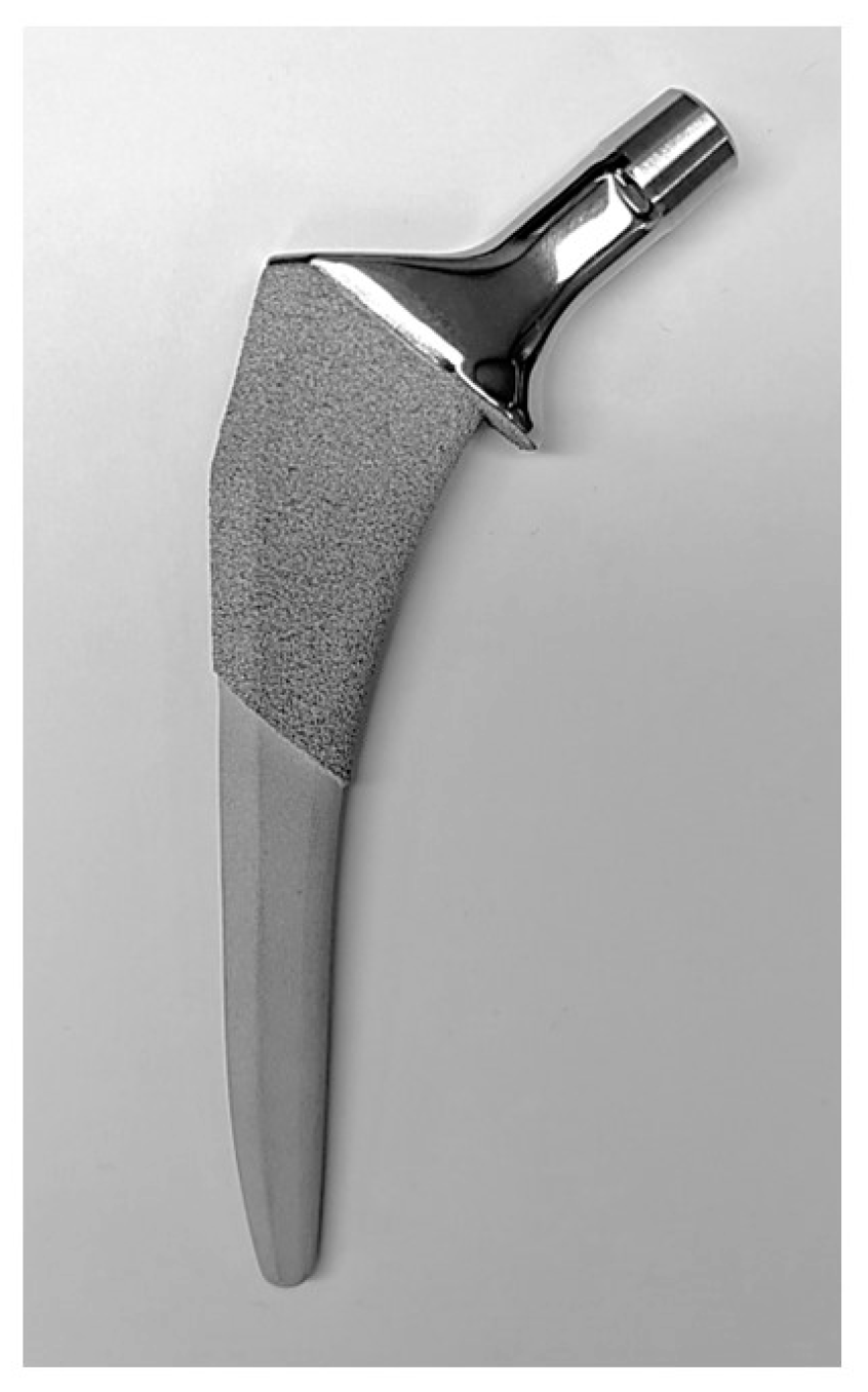
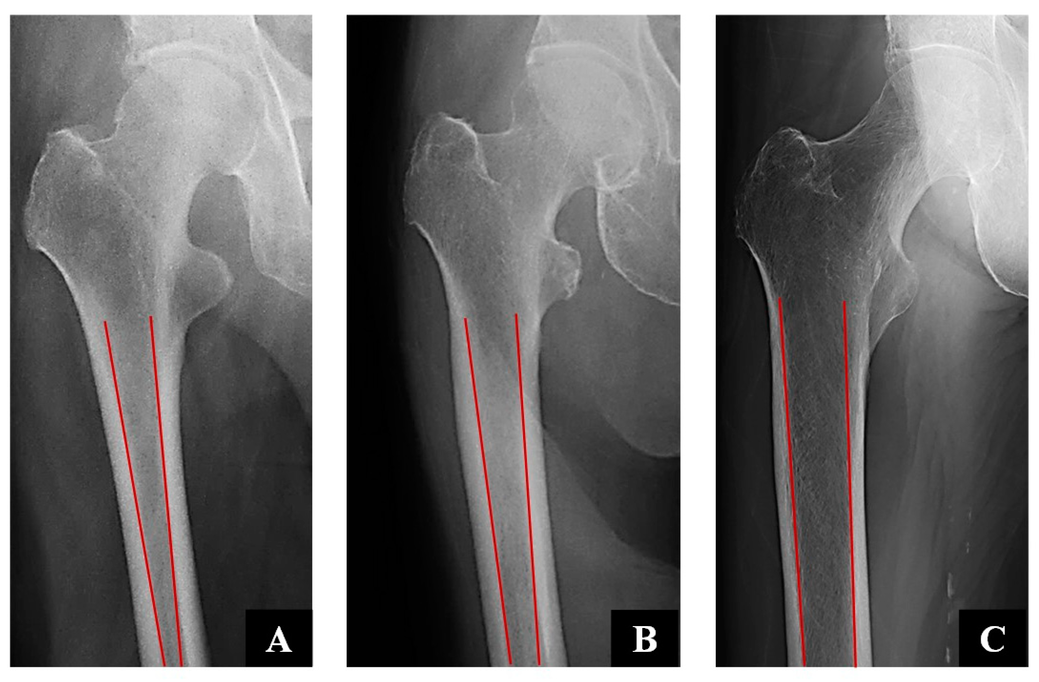
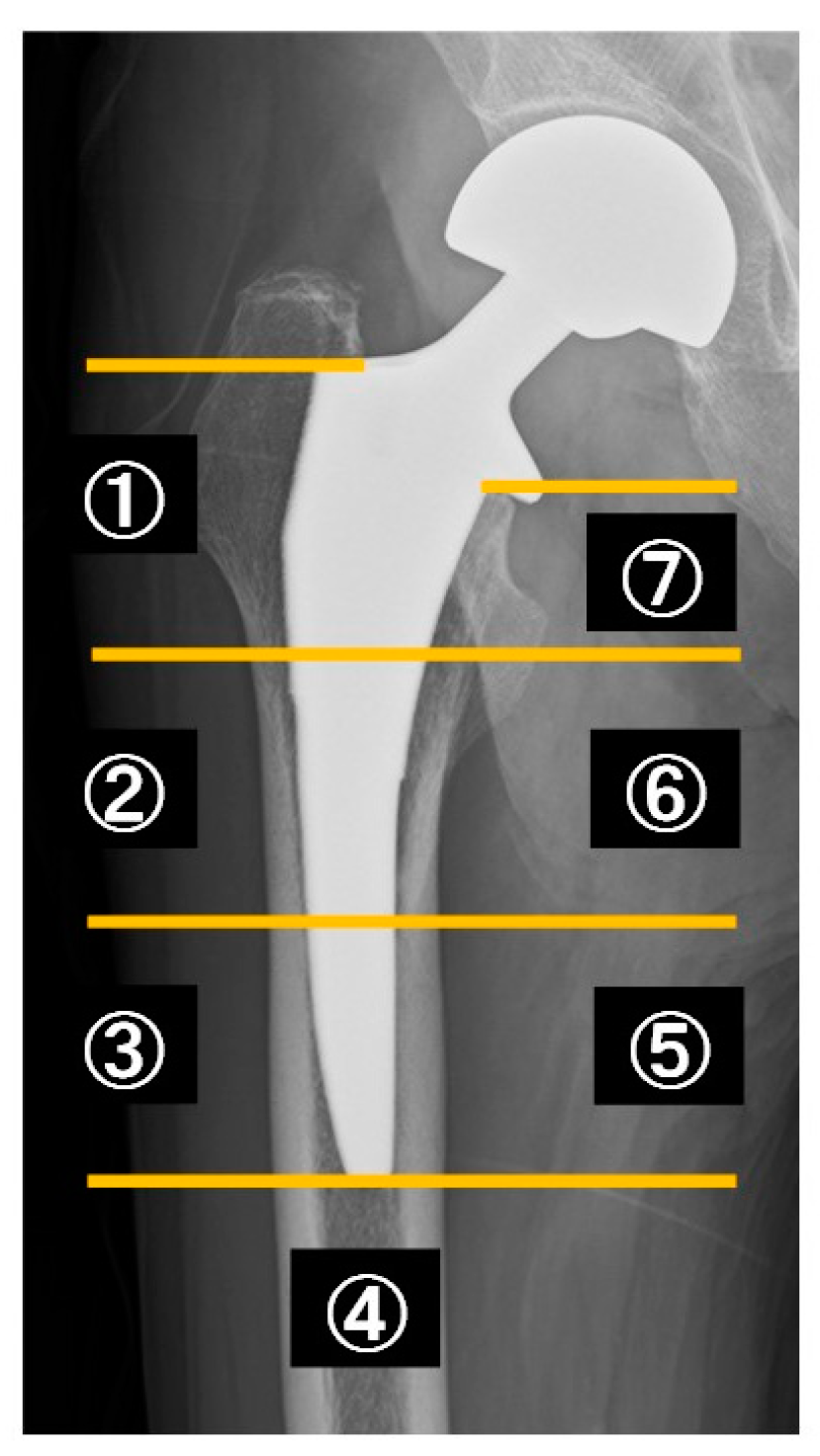
2.3. Clinical and Radiographical Assessment

Based on radiographic findings, patients were divided into types A, B, and C according to the Dorr classification. Those with a narrow medullary canal size were classified as Type A, those with a normal size as Type B, and those with a wide medullary canal as Type C [18] (Figure 3). The surgical approach was posterior in all cases. The same stem model was used, but the size was selected by the surgeon for each case depending on the shape of the medullary canal (Table 1). Stem sizes from 3 to 6 were commonly used. There was no set protocol for rehabilitation. Evaluations were conducted using radiographics (evaluation of implant loosening, subsidence, and fracture), dual-energy X-ray absorptiometry (DEXA), and clinical scores (Harris Hip Score [HHS] and EuroQoL 5-Dimension 5-Level [EQ-5D-5L]) [19,20,21]. Follow-up was at three points: at the time of discharge (within 1 month after surgery), 6 months, and 12 months.

2.4. DEXA Measurements

Seven Gruen zones were used to determine the BMD around each patient’s stem [22] using DEXA (Figure 4). Measurements were taken within 1, 6, and 12 months postoperatively. DEXA is a well-documented method to monitor bone quality after THA and BHA and detects even small changes in BMD around the stem. We referred to past reports for determining the timing of the DEXA exam [23,24].
The DEXA models used were GE (Lunar and Prodigy, Madison, WI, USA) and Hologic (Horizon, Explorer, and Discovery, Marlborough, MA, USA), each within six hospitals. Patients were positioned supine with their leg in a neutral position, with knee and foot support to allow scanning of the anterior–posterior projection of the proximal femur, including the area distal to the prosthesis, using an edge-detection technique.
Postoperative peri-prosthetic BMD was determined in the Gruen zone in seven regions of interest (ROIs) [22]. The seven Gruen zones were positioned on the distal tip and shoulder of the prosthesis. The two most proximal Gruen zones (1 and 7) were also combined to form a proximal femur ROI, representing the region where the porous coating of the uncemented stems is typically located. The BMD area was expressed as grams per square centimeter (g/cm2). A technician from the radiology department assessed the BMD around the stem at 1 month postoperatively (baseline BMD) and 6 and 12 months later. The BMD ratio was calculated by dividing the BMD values at 6 and 12 months by the BMD values obtained within 1 month after surgery. If the BMD change is more than 1, it indicates BMD increased, if less than 1, it indicates BMD decreased.

2.5. Osteoporosis Treatment

Based on previous reports that administration of anti-osteoporotic drugs after THA surgery suppresses BMD loss around the stem, we investigated the number of cases where osteoporosis treatment intervention was performed after femoral neck fracture (post-injury intervention group), where treatment intervention was performed before femoral neck fracture (pre-injury intervention group), and where no osteoporosis treatment was performed (no-intervention group) [14,15,16,17]. At the time of the final survey, we also investigated osteoporosis medications.

2.6. Statistical Analysis

The Kruskal–Wallis test was used to compare the change rate in the BMD around the stem over time and clinical scores in patients according to the Dorr classification and osteoporosis therapy intervention (Table 1 and Table 2). The chi-square test was used to examine the Dorr classification, sex, and osteoporosis therapy intervention (Table 1 and Table 2). All statistical analyses were performed using IBM SPSS (IBM, Armonk, NY, USA) version 26 for Windows. A p-value < 0.05 was considered statistically significant.

2.7. Ethical Statement

This study was conducted in accordance with the principles embodied in the Declaration of Helsinki and ICMJE. It was approved by the institutional review boards of the 12 participating hospitals on 23 October 2018 (approval no. 180206). Written informed consent was obtained from all participants.

3. Results

The mean patient age at the time of operation was 80.3 ± 6.3 years. The mean BMI was 20.8 ± 2.5 kg/m2. The HHS and EQ-5D-5L were 82.3 ± 15.8 and 0.78 ± 0.19 points, respectively. There were 6, 24, and 10 patients in the Dorr type A, B, and C groups. Table 1 shows patient backgrounds as assessed using the Dorr classification.
At 12 months, there was a significant difference in the BMD change rate at zone 2 between the Dorr B and C groups. The BMD change rate was significantly lower in Dorr C than in the Dorr B group (p = 0.036). There were no significant differences in any zone between the Dorr A and B groups and between the Dorr A and C groups. There was no significant difference between the three groups (Dorr A, B, and C) regarding HHS and EQ-5D-5L (Table 2).
Regarding osteoporosis treatment, there were 6, 19, and 15 patients in the pre-injury, post-injury, and no-intervention groups, respectively. There was no significant difference between the three groups in age, BMI, HHS, and EQ-5D-5L. Regarding the association between the osteoporosis treatment intervention and BMD, there was a significant difference between the pre-injury and no-intervention groups at 12 months in zones 1, 6, and 7. The decline in the BMD change rate was significantly suppressed in the pre-injury intervention group compared to the no-intervention group (between-group comparison at 12 months in zone 1: p = 0.039, at 6 months in zone 7: p = 0.005, and 12 months in zone 7: p = 0.029). Moreover, at 6 and 12 months in zone 7, there was a significant difference between the pre-injury and post-injury intervention groups. The decline in the BMD change rate was significantly suppressed in the pre-injury intervention group than in the post-injury intervention group (between-group comparison at 6 months in zone 7: p = 0.003 and 12 months in zone 7: p = 0.004). However, there was no significant difference in the BMD change rate between the post-injury and no-intervention groups (Table 3).
Table 4 shows the type of anti-osteoporotic drugs administered. Vitamin D and bisphosphonate preparations were frequently used, accounting for over 80% of the total.
Regarding radiographic evaluation, there were no peri-implant fractures, evident stem subsidence, or major complications during the follow-up period in the 40 patients analyzed.
  • Case presentation
A 79-year-old woman fell and sustained a displaced left femoral neck fracture. The medullary canal shape was Dorr C. BHA was performed the day after the injury. The radiograph immediately after surgery showed that a slightly smaller stem was selected. There was no significant subsidence, but the radiograph taken 12 months later showed stress shielding around the stem. Stress shielding was observed especially in Gruen zone 2 and 7 (Figure 5). Compared to immediately after surgery, 12 months later, there was a decrease in BMD of 18.6% in zone 2 and 32.7% in zone 7.

4. Discussion

In this study, we evaluated older patients with femoral neck fractures who underwent BHA using a full hydroxyapatite-collared compaction short stem according to Dorr classification to assess the change in BMD around the stem. The present results differ from our hypothesis that BMD around the stem can be maintained without being affected by the shape of the medullary canal, even in femoral neck fractures in older adults. BMD around the stem was preserved regardless of the medullary canal shape in a case series of THA with ACTIS stems, which included patients with hip osteoarthritis undergoing THA who were relatively young (mean age, 65 years) [11]. Our cohort of patients with femoral neck fractures was older (mean age, 80 years); thus, advanced osteoporosis was predicted, with a more significant decrease in BMD over time, especially in Dorr type C cases and cases with a wider medullary canal.
In this study, the significant BMD loss in Dorr C zone 2 can be attributed to a mismatch between the stem size and the medullary canal in patients with a wide medullary canal, such as Dorr type C cases. We believe surgeons may opt for a smaller stem size due to concerns about intraoperative femoral fractures (case presentation). According to the stem concept, this may have prevented load transfer, resulting in proximal BMD loss. In this study, fortunately, there were no cases of peri-stem fracture or implant subsidence in patients with Dorr type C; nonetheless, careful follow-up is necessary. We believe that BMD loss is less of a problem in Dorr A and B, but for medullary canals of Dorr type C, a cemented stem may be considered because BMD loss may progress.
Regarding the second hypothesis that anti-osteoporotic drug interventions can suppress BMD loss around the stem, there was a significant difference between the pre-injury and no-intervention groups and the pre-injury and post-injury intervention groups. The use of anti-osteoporotic drugs and changes in BMD around the stem were critical. Patients who took medication before the femoral neck fracture had suppressed BMD loss around the stem. When older people are diagnosed with osteoporosis based on DEXA scan results and previous fragility fractures, proactively intervening with anti-osteoporotic drugs may prevent secondary fractures and limit BMD reduction around the stem. It is possible to suppress the decrease in BMD around the stem, implying that postoperative periprosthetic fractures and stem subsidence and loosening can be prevented. Some studies have revealed that using anti-osteoporotic drugs after THA suppressed the decreased BMD around the stem [14,15,16,17]. However, no reports on drug intervention for osteoporosis before the injury and BMD around the stem have been published. We believe that proactively diagnosing osteoporosis and providing treatment will not only prevent femoral neck fractures but will also lead to reduced bone density around the stem after BHA.
Regarding clinical scores, Hayashi et al. found a correlation between daily activity and periprosthetic BMD in THAs [25]. In contrast, Murat et al. found no correlation between the clinical scores and peri-stem BMD in femoral neck fractures [26]. Regarding our clinical scores studied in femoral neck fractures, no significant differences were found among the three medullary canal shapes, suggesting that the shape of the medullary canal did not affect the clinical scores during our follow-up period. This may be due to the older age of the neck fracture patients compared to the age of the THA patients. In addition, the short follow-up period of this study (12 months) may have also influenced the results.
This study had some limitations. First, the sample size was small. Before this study, we performed a power analysis; at least 10 cases in each group were needed. However, the statistical power was insufficient because of dropout cases and missing data. Second, the postoperative follow-up period was only one year. However, this study was conducted in an older population, so long-term follow-up was difficult for various reasons. Third, patient data could not be collected for one-quarter of the study participants. Fourth, only one type of stem was investigated; thus, cemented or other cementless stems could have affected our study results, as it is known that stem design affects bone density around the stem [27]. Fifth, we could not investigate the rate of change in BMD by type of osteoporosis medication. Finally, the duration of osteoporosis treatment prior to injury was not investigated. To confirm the changes in BMD around the stem and the outcomes of anti-osteoporotic drugs in BHA patients, a comparative study with a longer follow-up period with a larger number of participants and a different stem design is desirable.

5. Conclusions

The BMD change rate in zone 2 was significantly reduced in Dorr type C compared to Dorr type B. Regarding the association between osteoporosis treatment and the BMD change rate, the pre-injury intervention group had significantly suppressed BMD change rate decline relative to the post-injury intervention and no-intervention groups in zones 1 and 7. A careful follow-up examination is essential when performing BHA in older patients with femoral neck fractures of Dorr type C because BMD is likely to decline postoperatively in the proximal area. It has been proposed that administering anti-osteoporotic drugs before femoral neck fracture can suppress the loss of BMD around the stem.

Author Contributions

Conceptualization, K.O., S.H., R.K. and T.N.; methodology, K.O. and S.H.; software, K.O. and Y.S.; validation, K.O., S.H., T.F. and T.N.; formal analysis, K.O. and Y.S.; investigation, K.O., S.T., T.I., A.S., E.S. and T.N.; resources, K.O., S.H. and T.F.; data curation, K.O. and Y.S.; writing—original draft preparation, K.O. and S.H.; writing—review and editing, K.O., S.H. and T.N.; visualization, K.O.; supervision, R.K. and T.N.; project administration, K.O. and T.N.; funding acquisition, none. All authors have read and agreed to the published version of the manuscript.

Funding

This research received no external funding.

Institutional Review Board Statement

This study was conducted in accordance with the principles embodied in the Declaration of Helsinki and ICMJE. It was approved by the institutional review boards of the 12 participating hospitals on 23 October 2018 (approval no. 180206).

Informed Consent Statement

Informed consent was obtained from all subjects involved in the study.

Data Availability Statement

The datasets used and/or analyzed during the current study are available from the corresponding author on reasonable request.

Acknowledgments

We thank Masaya Tsunoda, Katsunori Shibahara, Shinpei Kitada, Kenjiro Ito, Yasuhiro Ueda, Kohei Kawakita, Takuma Ozaki, Takahiro Fukazawa, Yohei Kumabe, and Yu Sasaki for patient follow-up and data collection. We would like to express our sincere gratitude to the doctors who cooperated with us during the study.

Conflicts of Interest

The authors declare no conflicts of interest.

Abbreviations

The following abbreviations are used in this manuscript:
BHABipolar hip arthroplasty
BMDBone mineral density
BMIBody mass index
DEXADual-energy X-ray absorptiometry
EQ5D-5LEuroQoL 5-Dimension 5-Level
HHSHarris Hip Score
ROIRegion of interest
THATotal hip arthroplasty
SERMSelective estrogen receptor modulator

References

  1. Vidovic, D.; Matejcic, A.; Punda, M.; Ivica, M.; Tomljenovic, M.; Bekavac-Beslin, M.; Mijic, A.; Bakota, B. Periprosthetic bone loss following hemiarthroplasty: A comparison between cemented and cementless hip prosthesis. Injury 2013, 44, S62–S66. [Google Scholar] [CrossRef]
  2. Burgers, P.T.P.W.; Van Geene, A.R.; Van Den Bekerom, M.P.J.; Van Lieshout, E.M.M.; Blom, B.; Aleem, I.S.; Bhandari, M.; Poolman, R.W. Total hip arthroplasty versus hemiarthroplasty for displaced femoral neck fractures in the healthy elderly: A meta-analysis and systematic review of randomized trials. Int. Orthop. 2012, 36, 1549–1560. [Google Scholar] [CrossRef] [PubMed]
  3. Bhandari, M.; Devereaux, P.; TornettaIII, P.; Swiontkowski, M.F.; Berry, D.J.; Haidukewych, G.; Schemitsch, E.H.; Hanson, B.P.; Koval, K.; Dirschl, D.; et al. Operative management of displaced femoral neck fractures in elderly patients. J. Bone Jt. Surg. 2005, 87, 2122–2130. [Google Scholar] [CrossRef] [PubMed]
  4. Iwamoto, Y.; Kijima, H.; Tazawa, H.; Konishi, N.; Kubota, H.; Yamada, S.; Tani, T.; Kamo, K.; Suzuki, N.; Okudera, Y.; et al. Initial rotational instability of the tapered wedge-shaped type cementless stem. Adv. Orthop. 2020, 2020, 2180260. [Google Scholar] [CrossRef]
  5. Fujii, M.; Kitamura, K.; Ikemura, S.; Hamai, S.; Motomura, G.; Nakashima, Y. Pneumatic femoral broaching decreases post-operative subsidence of a cementless taper-wedge stem. Int. Orthop. 2022, 46, 233–240. [Google Scholar] [CrossRef] [PubMed]
  6. Leiss, F.; Goetz, J.S.; Schindler, M.; Reinhard, J.; Müller, K.; Grifka, J.; Greimel, F.; Meyer, M. Influence of bone mineral density on femoral stem subsidence after cementless THA. Arch. Orthop. Trauma Surg. 2024, 144, 451–458. [Google Scholar] [CrossRef]
  7. Bunyoz, K.I.; Malchau, E.; Malchau, H.; Troelsen, A. Has the use of fixation techniques in THA changed in this decade? The uncemented paradox revisited. Clin. Orthop. Relat. Res. 2020, 478, 697–704. [Google Scholar] [CrossRef]
  8. Springer, B.D.; Etkin, C.D.; Shores, P.B.; Gioe, T.J.; Lewallen, D.G.; Bozic, K.J. Perioperative periprosthetic femur fractures are strongly correlated with fixation method: An analysis from the american joint replacement registry. J. Arthroplast. 2019, 34, S352–S354. [Google Scholar] [CrossRef]
  9. Veldman, H.D.; Heyligers, I.C.; Grimm, B.; Boymans, T.A.E.J. Cemented versus cementless hemiarthroplasty for a displaced fracture of the femoral neck: A systematic review and meta-analysis of current generation hip stems. Bone Jt. J. 2017, 99-B, 421–431. [Google Scholar] [CrossRef]
  10. Ekman, E.; Laaksonen, I.; Isotalo, K.; Liukas, A.; Vahlberg, T.; Mäkelä, K. Cementing does not increase the immediate postoperative risk of death after total hip arthroplasty or hemiarthroplasty: A hospital-based study of 10,677 patients. Acta Orthop. 2019, 90, 270–274. [Google Scholar] [CrossRef]
  11. Hayashi, S.; Hashimoto, S.; Kuroda, Y.; Nakano, N.; Matsumoto, T.; Kamenaga, T.; Niikura, T.; Kuroda, R. Hydroxyapatite-coated compaction short stem represents a characteristic pattern of peri-prosthetic bone remodelling after total hip arthroplasty. Arch. Orthop. Trauma Surg. 2022, 142, 2903–2910. [Google Scholar] [CrossRef] [PubMed]
  12. Kaszuba, S.V.; Cipparrone, N.; Gordon, A.C. The Actis and Corail femoral stems provide for similar clinical and radiographic outcomes in total hip arthroplasty. HSS J. 2020, 16, 412–419. [Google Scholar] [CrossRef] [PubMed]
  13. Chitnis, A.S.; Mantel, J.; Ruppenkamp, J.; Bourcet, A.; Holy, C.E. Survival analysis for all-cause revision following primary total hip arthroplasty with a medial collared, triple-tapered primary hip stem versus other implants in real-world settings. Curr. Med. Res. Opin. 2020, 36, 1839–1845. [Google Scholar] [CrossRef] [PubMed]
  14. Hatano, M.; Koizumi, Y.; Yamamoto, N.; Miyoshi, K.; Kawabata, K.; Tanaka, T.; Tanaka, S.; Shiroshita, A.; Kataoka, Y. Anti-osteoporotic drug efficacy for periprosthetic bone loss after total hip arthroplasty: A systematic review and network meta-analysis. J. Orthop. Sci. 2025, 30, 126–135. [Google Scholar] [CrossRef]
  15. Chen, X.; Shen, Y.; Ye, C.; Mumingjiang, Y.; Lu, J.; Yu, Y. Prophylactic efficacy on periprosthetic bone loss in calcar region after total hip arthroplasty of antiosteoporotic drugs: A network meta-analysis of randomised controlled studies. Postgrad. Med. J. 2021, 97, 150–155. [Google Scholar] [CrossRef]
  16. Arabmotlagh, M.; Rittmeister, M.; Hennigs, T. Alendronate prevents femoral periprosthetic bone loss following total hip arthroplasty: Prospective randomized double-blind study. J. Orthop. Res. 2006, 24, 1336–1341. [Google Scholar] [CrossRef]
  17. Zeng, Y.; Lai, O.; Shen, B.; Yang, J.; Zhou, Z.; Kang, P.; Pei, F. A systematic review assessing the effectiveness of alendronate in reducing periprosthetic bone loss after cementless primary THA. Orthopedics 2011, 34. [Google Scholar] [CrossRef]
  18. Dorr, L.D.; Faugere, M.-C.; Mackel, A.M.; Gruen, T.A.; Bognar, B.; Malluche, H.H. Structural and cellular assessment of bone quality of proximal femur. Bone 1993, 14, 231–242. [Google Scholar] [CrossRef]
  19. Söderman, P.; Malchau, H. Is the Harris hip score system useful to study the outcome of total hip replacement? Clin. Orthop. Relat. Res. 2001, 384, 189–197. [Google Scholar] [CrossRef]
  20. Shiroiwa, T.; Ikeda, S.; Noto, S.; Igarashi, A.; Fukuda, T.; Saito, S.; Shimozuma, K. Comparison of value set based on DCE and/or TTO data: Scoring for EQ-5D-5L health states in Japan. Value Health 2016, 19, 648–654. [Google Scholar] [CrossRef]
  21. Greene, M.E.; Rader, K.A.; Garellick, G.; Malchau, H.; Freiberg, A.A.; Rolfson, O. The EQ-5D-5L improves on the EQ-5D-3L for health-related quality-of-life assessment in patients undergoing total hip arthroplasty. Clin. Orthop. Relat. Res. 2015, 473, 3383–3390. [Google Scholar] [CrossRef]
  22. Gruen, T.A.; McNeice, G.M.; Amstutz, H.C. “Modes of failure” of cemented stem-type femoral components: A radiographic analysis of loosening. Clin. Orthop. Relat. Res. 1979, 141, 17–27. [Google Scholar] [CrossRef]
  23. Kilgus, D.; Shimaoka, E.; Tipton, J.; Eberle, R. Dual-energy X-Ray absorptiometry measurement of bone mineral density around porous-coated cementless femoral implants. Methods and preliminary results. J. Bone Jt. Surgery. Br. Vol. 1993, 75-B, 279–287. [Google Scholar] [CrossRef] [PubMed]
  24. Kröger, H.; Miettinen, H.; Arnala, I.; Koski, E.; Rushton, N.; Suomalainen, O. Evaluation of periprosthetic bone using dual-energy X-ray absorptiometry: Precision of the method and effect of operation on bone mineral density. J. Bone Miner. Res. 1996, 11, 1526–1530. [Google Scholar] [CrossRef] [PubMed]
  25. Hayashi, S.; Nishiyama, T.; Fujishiro, T.; Kanzaki, N.; Hashimoto, S.; Kurosaka, M. Periprosthetic bone mineral density with a cementless triple tapered stem is dependent on daily activity. Int. Orthop. 2012, 36, 1137–1142. [Google Scholar] [CrossRef]
  26. Murat, M.; Ermutlu, C.; Unkar, E.A.; Topalhafızoglu, S.; Şenel, A.; Öztürkmen, Y. Bone loss following cementless hemiarthroplasty for the treatment of femoral neck fracture. Indian J. Orthop. 2020, 54, 454–462. [Google Scholar] [CrossRef]
  27. Damborg, F.; Nissen, N.; I Jørgensen, H.R.; Abrahamsen, B.; Brixen, K. Changes in bone mineral density (BMD) around the cemented Exeter stem: A prospective study in 18 women with 5 years follow-up. Acta Orthop. 2008, 79, 494–498. [Google Scholar] [CrossRef]
Figure 1. Photograph of the full HA compaction short stem (ACTIS).
Osteology 05 00009 g001
Figure 2. Patient flow diagram. The study enrolled 85 patients; of these, 2 died, 10 had coronavirus disease, 2 withdrew their consent, 2 were affected by new diseases, and 8 withdrew for unknown reasons. Out of 61 follow-up patients, 21 had incomplete data sets of bone mineral density; therefore, data from 40 patients were included in the final analysis.
Osteology 05 00009 g002
Figure 3. This is the radiograph showing the medullary canal shapes of Types (AC) according to the Dorr classification. Type (A) has a narrow medullary cavity, type (B) is normal, and type (C) is wide.
Osteology 05 00009 g003
Figure 4. This is a diagram of the Gruen zone. It is divided into seven zones.
Osteology 05 00009 g004
Figure 5. (a) At the time of injury. (b) Immediately after surgery. (c) Six months later. (d) Twelve months later.
Osteology 05 00009 g005
Table 1. This is a list of selected stem sizes.
Stem SizeNumber
11
23
39
48
58
67
73
81
Table 2. Patient demographics were assessed using the Dorr classification and the association between the Dorr classification and BMD change rate.
Dorr ADorr BDorr Cp ValueX2 Value
Number of Patients62410--
Female, n (%)3 (50%)16 (66.7%)9 (90%)0.2043.175
Age (years)81.8 ± 4.580.0 ± 6.680.1 ± 7.30.173-
BMI (kg/m2)21.9 ± 3.120.8 ± 2.719.9 ± 1.80.338-
HHS (points)93.5 ± 6.081.9 ± 15.675.9 ± 19.00.087-
EQ-5D-5L (score)0.88 ± 0.110.76 ± 0.210.75 ± 0.180.287-
Zone areaTimeBMD change rate
Zone 16 m0.90 ± 0.090.89 ± 0.110.88 ± 0.120.218-
12 m0.86 ± 0.120.86 ± 0.150.85 ± 0.130.943-
Zone 26 m0.91 ± 0.050.90 ± 0.120.83 ± 0.150.200-
12 m0.88 ± 0.070.88 ± 0.16 *0.77 ± 0.09 *0.036 *-
Zone 36 m0.98 ± 0.030.96 ± 0.060.96 ± 0.090.673-
12 m0.96 ± 0.020.95 ± 0.080.97 ± 0.070.771-
Zone 46 m0.99 ± 0.020.98 ± 0.050.97 ± 0.050.769-
12 m0.98 ± 0.020.97 ± 0.050.97 ± 0.060.985-
Zone 56 m0.98 ± 0.030.97 ± 0.080.97 ± 0.080.948-
12 m0.96 ± 0.030.92 ± 0.210.98 ± 0.090.429-
Zone 66 m0.91 ± 0.080.91 ± 0.120.88 ± 0.150.911-
12 m0.90 ± 0.130.94 ± 0.100.82 ± 0.150.169-
Zone 76 m0.86 ± 0.150.78 ± 0.130.71 ± 0.160.298-
12 m0.86 ± 0.210.80 ± 0.160.67 ± 0.100.113-
* p < 0.05. Dorr A, B, and C were compared and the groups with significant differences were marked with *.
Table 3. The patient background is classified based on osteoporosis therapy intervention and the association between the osteoporosis treatment intervention and BMD change rate.
Pre-Injury InterventionPost-Injury InterventionNo Interventionp ValueX2 Value
Number of Patients61915--
Dorr typeA, 2; B, 3; C, 1A, 1; B, 13; C, 5A, 3; B, 8; C, 40.523-
Female, n (%)5 (83.3%)13 (68.4%)10 (66.7%)0.7370.610
Age (years)84.2 ± 3.379.9 ± 6.879.2 ± 6.60.129-
BMI (kg/m2)21.1 ± 2.121.5 ± 2.719.7 ± 2.30.902-
HHS (points)83.7 ± 16.379.6 ± 18.385.2 ± 12.90.832-
EQ-5D-5L (score)0.82 ± 0.180.77 ± 0.220.77 ± 0.160.743-
Zone areaTimeBMD change rate
Zone 16 m0.94 ± 0.100.86 ± 0.110.87 ± 0.110.351-
12 m0.95 ± 0.11 +0.88 ± 0.130.80 ± 0.14 +0.039 +-
Zone 26 m0.93 ± 0.080.86 ± 0.160.89 ± 0.070.699-
12 m0.92 ± 0.090.86 ± 0.180.83 ± 0.100.357-
Zone 36 m1.01 ± 0.040.95 ± 0.060.97 ± 0.060.920-
12 m0.98 ± 0.030.96 ± 0.090.94 ± 0.050.336-
Zone 46 m1.00 ± 0.040.97 ± 0.040.98 ± 0.050.446-
12 m0.99 ± 0.020.97 ± 0.050.97 ± 0.040.552-
Zone 56 m0.98 ± 0.080.97 ± 0.070.96 ± 0.080.974-
12 m0.97 ± 0.040.96 ± 0.080.89 ± 0.260.700-
Zone 66 m0.98 ± 0.100.90 ± 0.130.87 ± 0.100.076-
12 m0.94 ± 0.100.92 ± 0.150.89 ± 0.090.387-
Zone 76 m0.94 ± 0.09 #+0.73 ± 0.14 #0.76 ± 0.12 +0.003 #, 0.005 +-
12 m0.93 ± 0.11 #+0.74 ± 0.19 #0.78 ± 0.14 +0.004 #, 0.029 +-
+p < 0.05, # p < 0.05. Pre-injury intervention of anti-osteoporotic drugs, post-injury intervention, and no intervention were compared and the groups with significant differences were marked with + and #.
Table 4. Type and number of anti-osteoporotic drug.
Type of DrugNumber of Patients (n = 39)
Vitamin D17 (44%)
Bisphosphonate16 (41%)
Denosumab3 (8%)
Teriparatide2 (5%)
SERM1 (2%)
Disclaimer/Publisher’s Note: The statements, opinions and data contained in all publications are solely those of the individual author(s) and contributor(s) and not of MDPI and/or the editor(s). MDPI and/or the editor(s) disclaim responsibility for any injury to people or property resulting from any ideas, methods, instructions or products referred to in the content.

