Abstract
The study seeks to unravel the intricate operational sequence involved in preparing prehistoric pigments for rock art within the Puerto Roque rock art shelter. Sixteen pigment samples were meticulously collected from specific figurative representations. Additionally, three ochre samples were sourced from the shelter’s soil. Employing a comprehensive multi-analytical approach, including Raman microspectroscopy, X-ray microfluorescence (EDxrf), Fourier-transform infrared spectroscopy (ATR-FTIR), and scanning electron microscopy (SEM-EDS), all nineteen samples underwent thorough analysis. Notably, darker pigments revealed a composition of hematite with the added presence of carbon. Meanwhile, one sample exhibited an orange hue primarily composed of goethite and bright red pigments predominantly characterised by hematite, as confirmed by Raman analysis. EDxrf analysis demonstrated an elemental composition akin to the three ochre pieces examined. Furthermore, ATR-FTIR spectroscopy suggested the potential influence of a heating process in enhancing red coloration, corroborated by spectral results from specific samples. This finding aligns with prior research, underscoring the technological sophistication embedded in early artistic endeavours.
1. Introduction
The “Puerto Roque” open-air rock art shelter is a prehistoric painted natural shelter situated on the southern façade of the “Peñas de Puerto Roque” mountain range in Valencia de Alcántara, Extremadura, Spain (see Figure 1). This extensive shelter spans 7.5 m in length, 5.4 m in height, and reaches a depth of 4.2 m. It occupies an elevated position on the rocky cliff at an altitude of 725 m (See Figure 1C). The shelter’s quartzite walls are adorned with reddish paintings portraying both human figures and abstract forms, all of which are situated within the framework of the “Iberian Peninsula Schematic Rock Art style”. The schematic style characterises figures that morphologically simplify a human figure, an object, an animal, or other natural or artificial elements, or that even go as far as total abstraction with the representation of geometric symbols. These representations respond to certain formal, technical, and thematic rules [1].
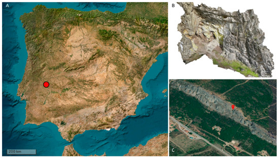
Figure 1.
(A) Localization of Abrigo Puerto Roque (Valencia de Alcántara, Cáceres, Spain); (B) 3D representation of the shelter; (C) elevated position on the rocky cliff.
It surfaces in conjunction with animal husbandry and agriculture, transitioning from the initial egalitarian and tribal structures of the Neolithic era to the hierarchical societies of the Bronze Age, marking a pivotal moment culminating in its eventual disappearance. The entire course can be meticulously traced by examining the distribution and internal organisation of its symbolic system, enabled by the widespread recording of its expressions across the geography of the Iberian Peninsula [2,3,4].
Our overarching objective centred on discerning and comprehending the presence of organic elements embedded within the portrayed imagery. Furthermore, the abundance of panels adorned with various hues of pigment on distinct figures within the shelter presented a significant opportunity for a detailed diagnosis of pigment compositions [5,6]. This unique assemblage of pigmented panels not only fuelled our curiosity but also served as a compelling prospect to delve into the nuanced intricacies of pigment composition within this archaeological context [7,8,9,10,11,12]. Considering the intricate nature of this archaeological site, a pivotal question emerged: Did the artists utilise specific pigment recipes for the creations within this shelter? In pursuit of answers, our research aimed to unravel the operational sequence and intricate processes involved in the application of pigments.
Sixteen rock panels adorned with painted figures have been identified and thoroughly documented using a collection of schematic representations. A noteworthy aspect is that many of these panels are oriented towards the viewer, suggesting a public-facing purpose, meaning the paintings are facing outward, and they are distributed across rock surfaces of varying dimensions. The application of pigments to create these rock paintings was most likely accomplished using fingers as the primary tool (see Figure 2).
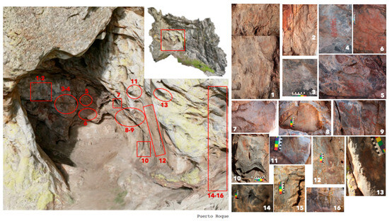
Figure 2.
Distribution of the rock art panels in the Puerto Roque shelter.
Regrettably, the high density of these figures does not provide a discernible process for determining the diachronic evolution of the depicted images at this site. Many of the figures are rendered in various shades of red, spanning from orange hues to deeper and darker tones. These disparities in coloration could be attributed to a multitude of factors, such as the state of conservation, environmental and biological degradation, interactions with the rock surface, variable adherence to the rock substrate, or the utilisation of different pigment types or even different chronologies. Notably, the darker shades of red, at times resembling garnet minerals, hold greater prominence than the orange variants.
Within the shelter, a rich assemblage of schematic red paintings predominates, encompassing a diverse range of imagery. This array includes several typologies of figures, notably “ramiforms” (a preferred Iberian term for what is also described as a tree-like linear motif), circular-shaped figures, lines, and anthropomorphic representations (see Figure 3).
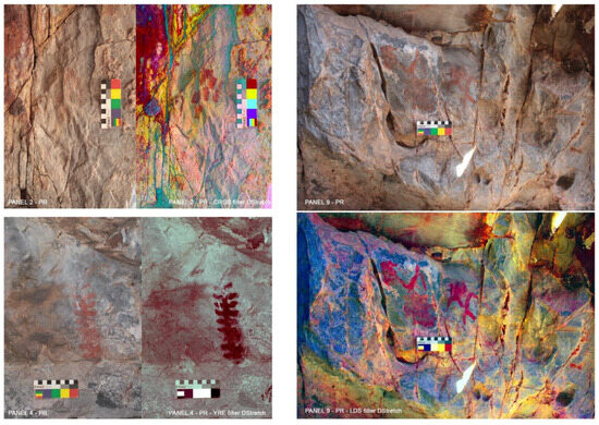
Figure 3.
Examples of figures from Puerto Roque shelter within the so-called Iberian Peninsula Schematic Rock Art Style. Top Left: Panel 2 from Puerto Roque original picture and with CRGB filter of DStretch; Right: Panel 9 of Puerto Roque original picture and LDS filter DStretch; Bottom Left: Panel 4 from Puerto Roque original picture and YRE filter from DStretch.
While numerous studies aimed at comprehending this distinctive style of rock art studies have been conducted since the early 20th century, commencing with the work of Henri Breuil, it is noteworthy that only in recent times have archaeometric investigations been applied to rock art sites in this region [13,14,15,16,17].
In other regions of Spain, extensive research endeavours have sought to establish a comprehensive understanding of prehistoric pigment compositions, encompassing various chronologies and artistic styles [18,19,20,21,22,23,24].
At the Puerto Roque shelter, we implemented a multi-instrumental protocol to methodically analyse the chemical and mineral composition of the prehistoric paintings. The survey area was focused on the north wall of the shelter, measuring approximately 1.30 m in width along the east–west axis and 1.05 m in length along the north–south axis [12].
2. Materials and Methods
Sixteen pigment samples (Lab Code: Puerto_1 to Puerto_16) were collected from specific figurative representations within the Puerto Roque rock art shelter, along with three samples of ochre sourced from the shelter’s soil (samples Puerto_17, Puerto_18 and Puerto_19).
The sampling strategy for the 16 panels in the Puerto region was meticulously designed to capture the diverse array of rock art manifestations, each presenting unique characteristics. The selection of these panels was guided by the recognition of various artistic techniques employed in the creation of the paintings, the nuanced hues exhibited, prevalent erosion issues, potential effects of sunlight exposure, and the intriguing hypothesis of distinct chronological periods due to several superimpositions detected. The ochre samples (Puerto_17, Puerto_18, Puerto_19) were considered as a possible raw material used to prepare the pigment [25,26,27,28,29,30].
The consideration of various factors such as artistic techniques, hues, erosion patterns, sunlight impact, and chronological hypotheses ensures a holistic scientific approach to the study, fostering a deeper understanding of the cultural and environmental dynamics that shaped these remains of prehistoric art.
Sample 1 (Puerto_01) comes from dark red digit paintings made directly with the fingers, in what is known in Spanish as “Flat Paint”. This sample provides insights into the artistic process involved in creating dark red digit paintings, with a focus on the specific technique employed which, given the different number of techniques used in Puerto Roque, was considered technique 1.
The sample labelled Puerto_02 consists of light red digit paintings that are less visible. An inquiry arises regarding whether the reduced visibility is attributed to a different chronology or erosion. This prompts further investigation to discern the factors influencing the visibility and potentially understand the temporal context of these paintings. Sample Puerto_03 is characterised by a dark red pigment forming linear figures, which raises questions about the technique involved, specifically, whether a brush was used (Technique 2). Understanding the artistic methods employed in creating linear figures contributes to the broader interpretation.
A distinctive panel, in which sample Puerto_04 was taken, showcases a very visible plant motif made with fingers. This non-geometric figure prompts speculation about its purpose, considering it is regarded as one of the oldest figures of Schematic Rock Art. Overall, the analysis of the figurative typologies that appear to decorate some mobile objects in post-paleolithic Iberian territory shows that, individually, soliforms, ramiforms, series of angled motifs, zig-zags, and some anthropomorphic and zoomorphic motifs are already represented on ceramics with printed, cardial and non-cardial decorations from the Early and Middle Neolithic [31,32]. This makes the ramiform figure one of the first figures to be represented in peninsular schematic art.
An intriguing sample, Puerto_05, features an accumulation of pigment next to very visible human figures. The purpose of this pigment accumulation becomes a focal point for investigation.
Characterised by superimpositions, the figure from which sample Puerto_06 comes raises questions about the mixture of pigments used in the artwork. Sample Puerto_07 comes from a figure with a high level of erosion and incrustations on top, resulting in a different hue. Investigating the impact of erosion on artistic elements contributes to understanding preservation challenges. Sample Puerto_08 stands out as a figure with distinct characteristics with a pastier texture and a different hue. Superimpositions and good quality pigments characterise Puerto_09, highlighting figures with exceptional aesthetic qualities.
Sample Puerto_10 features orange figures, potentially impacted by the sun. Exploring the impact of sunlight on the pigments provides insights into environmental influences on the paintings.
Sample Puerto_11 is described as difficult to visualise and raises questions about whether this difficulty is due to erosion or a different technique. Darkly coloured figures above dark fungi prompted further investigation. Sample Puerto_12 involves using fingers to paint and splashing pigment (Technique 3), representing a mixture of techniques. Exploring the artistic approach involving finger painting and pigment splashing adds depth to the analysis, especially the use of different techniques on the same panel.
A faded figure that is difficult to see characterises sample Puerto_13. Understanding the challenges in visibility and potential causes contributes to interpreting the significance of the figure.
Linear figures made from small dots, potentially created with a brush tool characterise Puerto_14 and samples Puerto_15 features extremely blurred orange figures, almost imperceptible to the naked eye. Questions arise about whether the blurriness is due to antiquity or erosion. Small, hard-to-see red dots characterise sample Puerto_16. DStretch filters helped to see how small and different these figures are if one compared them with all other figures. One questions if this means that the artists employed a different technique (technique 3).
Ethical removal techniques, adhering to the code of ethics and guidelines established by the American Institute for Conservation [33], were employed to ensure the preservation of the artwork [34,35]. Each sample, weighing between 10 and 100 mg, was extracted from areas where pigment was readily observable using a sterilised tungsten scalpel and placed in 0.5 mL microcentrifuge tubes [36].
2.1. Micro-Raman Spectroscopy
This technique played a crucial role in identifying the mineralogical composition of the pigment samples. It provides valuable insights into the composition of materials [37,38,39,40,41,42]. It allows for differentiation between different materials, facilitating provenance studies and archaeological interpretations. This technique is particularly useful for distinguishing between different types of pigments and their mineral components [43].
The measurements were conducted with a LabRam HR800 spectrometer (Horiba Jobin Yvon, Palaiseau, France), coupled with an Olympus BXFM optical microscope (Olympus, Tokyo, Japan). The spectrometer was equipped with an air-cooled CCD detector (1024 × 256 pixels) maintained at −70 °C and utilised gratings with 600 and 1800 grooves/mm. A laser beam with a diameter of 1 mm was focused on the samples, providing a spectral resolution of approximately 4 cm−1. The He Ne laser line at 632.82 nm served as the excitation source, filtered to maintain laser power between 0.2 and 10 mW. Exposure time, beam power, and accumulations were adjusted individually for each sample to acquire informative spectra while safeguarding against sample alteration. Raman spectra were recorded in the range of 200–2000 cm−1, with exposure times of 5–16 s and 5–11 accumulations. The laser beam was focused onto the samples using 10× and 50× microscope objectives, with a spot size diameter of about 2–3 μm. The wavelength scale was calibrated using a Silicon standard (520.5 cm−1), and acquired spectra were compared with published scientific data and reference databases, such as Horiba Lab-Spec 5 (Horiba) and RRUFF (RRUFF, University of Arizona, Tucson, AZ, USA).
2.2. Fourier-Transform Infrared Spectroscopy (ATR-FTIR)
ATR-FTIR is employed to differentiate between various types of clay minerals and provides insights into their structure, composition, and structural changes resulting from chemical modifications [44]. However, accurately identifying clay minerals with this method can be challenging due to their variable chemical composition and structural disorders. Additionally, clay minerals often occur as mixtures with varying ratios of different clay minerals, further complicating their identification [45]. ATR-FTIR spectra were acquired using a Bruker Alpha FT-IR spectrometer with Opus 7.5 software and an Attenuated Total Reflection (ATR) sampling device. The ATR-FTIR spectrometer featured a global source, a KBr beam splitter, and a Deuterated Lanthanum α Alanine-doped TriGlycine Sulphate detector operating at room temperature. The ATR sampling device employed a diamond internal reflection element (IRE) in a single-reflection configuration. Spectra were recorded over the spectral range of 400–4000 cm−1 at a resolution of 4cm−1, with 24 scans.
2.3. Energy-Dispersive X-ray Micro-Fluorescence (EDxrf)
EDxrf spectra of red ochre collected at the Puerto Roque rock art shelter provide valuable information about the elemental composition of the samples. This technique is instrumental in identifying the elemental constituents of the pigments [41,46,47,48,49,50].
EDxrf analysis was performed using a portable Bruker ARTAX 200 μEDxrf spectrometer equipped (Billerica, MA, USA) with a Mo X-ray tube and a collimator with a diameter of 200 μm. The X-fluorescence phenomenon occurs because of the atoms returning to their stable configuration and emitting the characteristic X photons. The analysis was conducted with voltages ranging from 15 to 50 kV and current levels between 1500 μA and 700 μA. The acquisition time for each sample was 60 s, and helium flow was employed to enhance the identification of light elements (e.g., Na, etc.). XRF spectra were acquired and analysed using ARTAXControl 7.2 software.
2.4. Scanning Electron Microscopy (SEM)
This method is useful for acquiring micrographs, elemental maps, and EDS spectra of the analysed samples. For microstructural characterization and determination of the chemical composition of the samples, a ZEISS EVO MA 15 scanning electron microscope (SEM) coupled (Jena, Germany) with an energy-dispersive X-ray spectroscopy (EDS) system (Aztec, Oxford, UK) was used. The SEM is equipped with a silicon drift detector (SDD), a “LaB6” filament as an electron source, and cobalt as a calibration standard. With this instrument, even small rock and pigment fragments can be studied at 20 kV and a working distance of 8.5 mm under high vacuum using backscattered electrons (BSE). Samples analysed with the scanning electron microscope can be analysed as they are or with surface treatment. If the sample is treated for analysis, it can affect the use of other analytical techniques on the same sample. To avoid this issue, samples can be analysed without prior surface treatment and under variable pressure conditions. In order not to create an irreversible situation of the samples, we chose not to work in high vacuum by coating the samples with gold or graphite. The only sample that was analysed with SEM-EDS (Puerto_12) was not pre-treated and was analysed under variable pressure, in low vacuum.
With the scanning electron microscope, there is also the possibility of performing the SEM-EDS chemical mapping technique [51]. This technique is used to investigate the distribution of elements within the samples and is widely used to differentiate between rock, pigments, and patinas.
3. Results and Discussion
Some of the samples show a lack of results in some of the techniques applied due to the small size of the samples taken. The summarised results from all different techniques applied to the 19 samples from Puerto Roque are in Table 1. To check the location of the samples and the respective Micro-Raman, EDxrf ATR-FTIR and SEM results, please see Supplementary Materials. The ATR-FTIR analysis of samples Puerto_4, Puerto_6, Puerto_12, and Puerto_15 did not return any readable results. In the case of sample Puerto_12, because it was larger in quantity than the other samples, an SEM-EDS analysis was also carried out.

Table 1.
Archaeometric results of multi-instrumental application (EDxrf, Raman, SEM-EDS, ATR-FTIR) to rock art paintings of Abrigo Puerto Rock.
3.1. Raman Spectra Results
The Raman spectroscopy results confirm that the red figures at the Abrigo Puerto Roque rock art shelter primarily consist of hematite and, in a few cases, of goethite or goethite and hematite. The characteristic peaks of hematite, observed at 222, 289, 406, 607, 658, and 1313 cm−1, are in line with findings from previous studies on prehistoric rock art pigments [20,22,36,52]. Hematite is a common mineral and a key component of natural earth ochres, which have been used in painting since prehistoric times [53]. However, Raman bands associated with clay minerals were not detected in the spectra due to the strong fluorescence background from aluminosilicates [19].
An intriguing aspect of the study is the presence of a double peak at 605 and 656 cm−1. These peaks can be attributed to a disordered phase of hematite, similarly to what is found in natural earth ochres [52,54]. However, it is important to note that several factors, both anthropogenic and natural, such as heating, grinding, biodegradation, and weathering, can also lead to the appearance of these peaks [55,56,57]. Raman spectroscopy alone may not distinguish between these factors [54,58].
3.2. Fourier-Transform Infrared Spectroscopy (ATR-FTIR) Results
The ATR-FTIR results corroborate the findings obtained from other methods. The databases already used in other studies [15,17,59,60] were used to assign and interpret the spectra obtained through the ATR-FTIR analysis of the samples analysed. ATR-FTIR spectra are composed of peaks that, based on the detectability of the sample, indicate clay minerals and their structure, composition and structural changes resulting from chemical modifications. In these spectra, peaks with different intensities are identified such as very strong (vs), strong (s), medium (m), weak (w), very weak (vw), and shoulder (sh).
Specifically (Table 2), the Puerto_01 sample presents peaks at 3361 w, 3299 w, 1670 m, 1628 m, 1457 m, 1423 m, 1013 vvs, 776 s, 689 m, 526 m, and 450 vs cm−1. The particular slope of the spectrum in the interval 3500–3100 cm−1 is reflected in the slope observed in [59] in clay heated to 60 °C. This finding suggests that water absorption in this sample is partially obscured by the OH stretching structural band.
Table 2.
FTIR results interpretation.
The Puerto_03 sample has peaks at 3627, 1429, 1010 vvs, 917, 795, 775, 692, 531 and 461 vs cm−1. The peaks around the value of 3620 cm−1 are generally attributed to the internal hydroxyl groups of the clay located between the tetrahedral and octahedral sheets of its structure [59] indicating the clay heating process. The presence of hematite is identified thanks to the absorption at 531 cm−1.
Puerto_05 sample reveals peaks at 3490, 1430, 1079 vvs, 794, 777, 692, 520, and 462 vs cm−1, with a broad band near 3430 cm−1; this band is indicative of H-O-H vibrations of water adsorbed [59] related also to heated clay.
Sample Puerto_07 shows peaks at 3399, 1722, 1700, 1614, 1575, 1466, 1414, 1322, 1160, 1080 vvs, 796, 779, 520, and 459 s cm−1, suggesting the presence of quartz, hematite, and incrustations due to natural action. The encrustation appears to be correlated with the presence of C=O stretching and thus reportable as whewellite or weddellite.
The Puerto_8 sample peaks at 1624 (-OH), 1431 (-CO), 1060 vvs, 953, 791, 543 and 455 vs cm−1 are associated with quartz substrate of the rock and its impurity.
Sample Puerto_09 shows peaks at 1431, 1074 vvs, 780, 678, 614, 517, and 466 s cm−1, indicating a quartz and hematite composition.
The Puerto_11 sample peaks at 3220, 1668, 1633, 1457, 1429, 1016vvs, 776, 689, 605, 522 and 458 vs cm−1 reveal a clay-based sample composition.
The collective findings present a thorough comprehension of the mineralogical composition inherent in the pigments employed in the rock art adorning Abrigo de Puerto Roque. These results not only shed light on the intricate makeup of the pigments but also indicate the alteration processes they may have undergone over time. This exploration into the mineralogical aspects enriches our understanding of the geological origins and transformations that contribute to the vibrant palette seen in rock art. It is essential to acknowledge, however, that some samples exhibit a paucity of results in specific techniques. This limitation stems from the inherently diminutive size of the samples procured during the sampling process. The petite nature of these samples poses a challenge in executing certain analytical techniques, thereby restricting the depth of insights that can be gleaned. Despite this constraint, the results still furnish valuable data, underscoring the importance of judiciously considering the sample size in future analyses. In moving forward, addressing the sample size limitations will be pivotal in unlocking a more comprehensive understanding of the rock art pigments. Adopting methodologies that accommodate the nuances of smaller samples and exploring complementary techniques could prove instrumental in overcoming these challenges. This iterative refinement in sampling strategies and analytical approaches will undoubtedly contribute to a more nuanced and detailed comprehension of the mineralogical intricacies inherent in the captivating rock art of Abrigo Puerto Roque.
ATR-FTIR results for the remaining samples are as follows:
Sample Puerto_14: Presents peaks at 1080 vs, 794, 778, 693, 641, 518, and 457 vvs cm−1, indicating the presence of both quartz and hematite. Sample Puerto_16 displays peaks at 1161, 1081 vs, 795, 778, 693, 641, 563, 517, and 459 vvs cm−1, also suggesting the presence of quartz and hematite. Sample Puerto 17 shows peaks at 3149, 1641, 1009 vvs, 900 vs, 790 vs, 617, 533, and 467 cm−1, which correspond to a composition consistent with natural ochre composed of goethite. Sample Puerto 18 presents peaks at 3331, 1624, 1426, 985 vvs, 625, 524, and 436 cm−1, identifying it as natural brown ochre. Example Puerto 19 presents peaks at 3081, 1563, 1034 vs, 891 vs, 794 vs, 523 s, and 436 vvs cm−1, also indicating a characteristic composition of natural ochre composed of goethite. Specifically, the peak at 3081 cm−1 turns out to be CH3/NH3 of organic material (probably fungi).
Significantly, it is imperative to underscore that Samples Puerto_4, Puerto_6, Puerto_10, Puerto_12, and Puerto_15, when subjected to analysis with the ATR-FTIR equipment, did not produce spectra deemed reliable for interpretation. This noteworthy observation adds a layer of complexity to our exploration of pigment composition, shedding light on both the capabilities and limitations of the analytical methods employed. While the absence of reliable spectra in these particular samples presents a challenge in drawing direct conclusions about their molecular composition, it intriguingly aligns with and fortifies the earlier findings regarding the prevalent use of hematite and natural earth ochre in the rock art at Abrigo Puerto Roque. This concordance with previous results serves to reinforce the robustness and reliability of the data obtained from other samples, underlining the consistency in the pigments employed across the artistic depictions. In conclusion, these results not only deepen our understanding of pigment composition but also underscore the significance of methodological considerations in the scientific study of rock art. The nuances revealed by the limitations in spectral data contribute to a more comprehensive appreciation of the analytical process and, in tandem with previous findings, strengthen our comprehension of the rich and diverse palette employed by the ancient artists at Abrigo Puerto Roque.
3.3. X-ray Fluorescence (EDxrf) Results
The results obtained from EDxrf analysis of samples collected from the red figures at the Abrigo Puerto Roque rock art shelter reveal that these red pigments predominantly consist of iron (Fe) and, in some instances, calcium (Ca). Additionally, there is a presence of silicon (Si), likely associated with the underlying quartzite substrate of the rock or accessory minerals of haematite in natural earth ochres.
Remarkably, the samples numbered 01, 03, 05, 07, 09, and 11, distinguished by their vibrant red hues, display a striking resemblance in the composition to the ochre pieces meticulously examined—specifically, samples Puerto_17, Puerto_18, and Puerto_19. This compelling similarity strongly implies a significant correlation between the red pigments employed in these figurative representations and the ochre derived from the shelter’s environs, as has been identified at other sites in the Arronches Region [61]. The convergence of elemental composition between the vividly red samples and the ochre specimens extracted from the shelter establishes a tangible link, accentuating the coherence in the pigments’ origins. This finding not only underscores the consistency in the colour palette chosen by the artisans but also provides crucial insights into the sourcing and utilisation of ochre in the creation of the rock art at Abrigo Puerto Roque.
3.4. SEM-EDS Results
SEM-EDS analysis, which was made in only one sample (Puerto_12) has confirmed the presence of Fe in high proportions, and, on the other hand, it has highlighted the presence of other elements such as Si probably referable to substrate rock. In addition to a high concentration of Fe, SEM-EDS analyses on red samples also revealed the presence of Ca and some K. Thanks to the thickness of the pictorial layer, it is possible to attribute these elements to the concretion matrix and not to pigment or the rock substratum, reinforcing the hypothesis that red pigment has been obtained from natural red ochre [62] whose ochres are very common in the study area. Moreover, the red and the dark red pigments show a different chemical composition, suggesting that they could come from different ochres. In any case, further trace elements analyses of the red pigments should be performed in order to confirm this hypothesis. Ochre does not seem to be the only raw material used to make red pigment. The analysis of the Sample 12 taken from Abrigo Puerto Roque suggests the use of a clay component in association with hematite, the red ochre sample presented Fe, and also Al and K, as well as P, from alterations of the rock surface (patina).
This collective set of results furnishes a thorough and comprehensive insight into the intricate mineralogical composition characterising the pigments employed in the rock art adorning Abrigo Puerto Roque. Not only do these findings illuminate the specific minerals constituting the pigments, but they also contribute valuable perspectives on the potential origins of these minerals and the alteration processes they may have undergone.
In the meticulous analysis of samples extracted from the Puerto Roque rock art shelter, a compelling revelation emerges. The presence of calcium peaks in Samples Puerto_01 and Puerto_11 aligns with the results obtained from EDxrf. Sample Puerto_04 contains charcoal in addition to red ochre, indicating that charcoal was likely added during the pigment preparation process. Sample Puerto_05 contains silicates along with hematite. Samples Puerto_06 and Puerto_07 contain hematite, quartz, and incrustations due to natural actions. Samples Puerto_09, Puerto_10, and Puerto_15 primarily consist of hematite. Sample Puerto_08 contains goethite and hematite, resembling natural earth ochre (Sample Puerto_18). Sample Puerto_12 includes Fe, Al, K, and P, likely from alteration of the rock surface (patina). Samples Puerto_13 to Puerto_16 are primarily composed of Fe and Ca, with both elements identified in the EDxrf spectra.
Although in the area of Spanish Extremadura, organic compounds in schematic art paintings have already been identified in other shelters previously [15,16], it is noteworthy that within the realm of studies focusing on rock art pigments, the identification of organic binders remains a rare occurrence when employing the aforementioned techniques. This observation accentuates the prevailing dominance of inorganic components and the products of deterioration in the composition of these artistic materials. The scarcity of detected organic binders underscores the challenges and complexities associated with their preservation over time, shedding light on the resilient and enduring nature of the inorganic elements present in rock art pigments [63]. This nuanced understanding invites further exploration into the dynamic interplay between organic and inorganic components, offering valuable insights into the long-term preservation and alterations that shape the captivating world of rock art [17,64]. In any case, our inability to find organic compounds could be due to the degradation of binders, which could result in oxalates being detected in some samples [65].
4. Conclusions
Archaeometric studies were meticulously carried out on samples extracted from the prehistoric paintings within the Puerto Roque open-air shelter in the Spanish Extremadura. The findings point towards the plausible use of red hematite pigments sourced from diverse regional ochres, with the additional detection of clay minerals, as indicated in the works of [15,66,67]. The revelation of hematite, clay minerals, and quartz through Raman spectroscopy, coupled with the significant iron oxides and hydroxides abundance identified by ATR-FTIR, strongly indicates the utilisation of ochre pigments to achieve the striking red hues in the rock art [68,69]. Furthermore, the likelihood of a heating process enhancing the red coloration is supported by the spectra obtained from samples Puerto_1, Puerto_3, and Puerto_5, aligning with the findings of [70,71]. The SEM analysis of sample Puerto_12 returned a response consistent with that obtained from the XRF analysis performed on the same sample.
These advanced analytical techniques not only contribute to the elucidation of pigment composition and preparation methods but also play a pivotal role in the ongoing monitoring and conservation of rock art sites. Recognizing that each rock art site has a unique microclimate that affects the preservation of pigments and substrates, the application of archaeometric studies at a local level becomes paramount to ensure good preservation [72]. This localised approach is crucial for making informed decisions that contribute to the preservation and safeguarding of these invaluable cultural resources. It underscores the interdisciplinary synergy between archaeology and material science in unravelling the mysteries of the past while ensuring the sustainable conservation of our cultural heritage [73].
Understanding the intricate processes involved in the preparation of prehistoric pigments for rock art holds paramount significance in unravelling the artistic techniques employed by prehistoric artists. The strong indication of ochre pigment utilisation to achieve the vibrant red hues in rock art not only adds depth to our comprehension of artistic choices but also unveils the resourcefulness of those peoples in utilising locally available materials. This insight contributes to a broader understanding of cultural practices, providing a lens through which we can explore the artistic preferences and material culture of prehistoric communities [74,75]. Furthermore, the discerned likelihood of a heating process contributing to the enhancement of red coloration, as supported by the spectra from specific samples and aligned with previous research findings, underscores the technological sophistication of these early artistic endeavours. Delving into the preparation techniques unveils a deliberate and intentional approach to colour manipulation, shedding light on the nuanced understanding ancient communities possessed about the transformative potential of their materials. This knowledge is crucial not only for unravelling the aesthetics of rock art but also for appreciating the technological ingenuity that underpinned these artistic expressions. In essence, the study of preparation processes for prehistoric pigments becomes a window into the cultural and technological tapestry of ancient societies, enriching our narrative of human creativity and innovation.
In summary, the amalgamation of diverse analytical techniques has demonstrated its efficacy in unveiling the intricate mineralogical components embedded within the pigments, thereby illuminating the nuanced world of their preparation techniques. The identification of multiple processes employed in transforming and preparing these pigments strongly implies purposeful actions within a specific cultural and artistic context. This insight suggests a meticulous and intentional selection of raw materials and techniques, with colour emerging as a significant criterion guiding these choices. The varied processes involved in pigment transformation and preparation unveil a sophisticated activity that demands not only specific knowledge but also dedicated time within the community. This intricate practice implies an initial conceptualization, wherein the final objective was likely predetermined. The deliberate nature of these choices emphasises the cultural significance of the pigments and the meticulous approach taken by the creators in transforming natural materials into vibrant expressions of art. This intricate dance of preparation techniques, driven by intentional decisions, underscores the rich tapestry of knowledge and craftsmanship embedded within the community, revealing rock art as a dynamic and purposeful cultural practice.
Supplementary Materials
The following supporting information can be downloaded at: https://www.mdpi.com/article/10.3390/heritage7030053/s1.
Author Contributions
Conceptualization, H.G., H.C., S.G., V.L., M.N., N.E., E.M. and P.R.; methodology, H.G., H.C., S.G., V.L., M.N., N.E., E.M. and P.R.; software, H.G., H.C., S.G., V.L., M.N., N.E., E.M. and P.R.; validation, H.G., H.C., S.G., V.L., M.N., N.E., E.M. and P.R.; formal analysis, H.G., H.C., S.G., V.L., M.N., N.E., E.M. and P.R.; investigation, H.G., H.C., S.G., V.L., M.N., N.E., E.M. and P.R.; resources, H.G., H.C., S.G., V.L., M.N., N.E., E.M. and P.R.; data curation, H.G., H.C., S.G., V.L., M.N., N.E., E.M. and P.R.; writing—original draft preparation, H.G., H.C., S.G., V.L., M.N., N.E., E.M. and P.R.; writing—review and editing, H.G., H.C., S.G., V.L., M.N., N.E., E.M. and P.R.; visualization, H.G., H.C., S.G., V.L., M.N., N.E., E.M. and P.R.; supervision, H.G., H.C., S.G., V.L., M.N., N.E., E.M. and P.R.; project administration, H.G., H.C., S.G., V.L., M.N., N.E., E.M. and P.R.; funding acquisition, H.G., H.C., S.G., V.L., M.N., N.E., E.M. and P.R. All authors have read and agreed to the published version of the manuscript.
Funding
This research received no external funding.
Data Availability Statement
The data presented in this study are available on request from the corresponding author.
Conflicts of Interest
The authors declare no conflict of interest.
References
- Oliveira, J.; Torres, M.F. O Abrigo do Ninho do Bufo-o painel da parturiente e o seu contexto (Marvão–Portugal). Sci. Antiq. 2021, 24–51. Available online: https://www.scientiaantiquitatis.uevora.pt/index.php/SA/article/view/313 (accessed on 15 February 2024).
- González Cordero, A. Datos para Ia conrextualizaci6n del arte rupestre en Ia Alta Extremadura. Zephyrus 1999, 52, 191–220. [Google Scholar]
- Rogerio-Candelera, M.A.; Vanhaecke, F.; Resano, M.; Marzo, P.; Porca, E.; Alloza, R.; Saiz-Jimenez, C. Combinação de análise de imagens e técnicas analíticas para a distinção de diferentes fases num painel rochoso (La Coquinera II, Obón, Teruel). In Proceedings of the Actas IV Congreso El Arte Rupestre del Arco Mediterráneo de la Península Ibérica, Valência, Spain, 4–5 December 2008; pp. 327–334. [Google Scholar]
- Collado Giraldo, H.; García Arranz, J.J. Reflexiones sobre la fase inicial del arte rupestre esquemático en Extremadura a raíz de las recientes investigaciones. In Actas del II Congreso de Arte Rupestre Esquemático en la Península Ibérica: Comarca de Los Vélez, 5–8 de mayo 2010; Ayuntamiento de Vélez Blanco: Almería, Spain, 2013; pp. 287–299. [Google Scholar]
- Edwards, H.G.M.; Newton, E.M.; Russ, J. Raman spectroscopic analysis of pigments and substrata in prehistoric rock art. J. Mol. Struct. 2000, 550, 245–256. [Google Scholar] [CrossRef]
- Hoerlé, S.; Bertrand, L.; Mguni, S.; Jacobson, L. Microanalysis and Dating for Rock Art Studies: Towards a Common Analytical Strategy. S. Afr. Archaeol. Bull. 2010, 65, 221–228. [Google Scholar]
- Fleming, M.I.A. Aplicação da arqueometria no estudo de coleções arqueológicas. Revista CPC 2007, 6, 219–230. [Google Scholar] [CrossRef]
- Resano, M.; García-Ruiz, E.; Alloza, R.; Marzo, M.P.; Vandenabeele, P.; Vanhaecke, F. Espectrometria de massa por ablação a laser com plasma indutivamente acoplado para acaracterização de pigmentos em arte rupestre pré-histórica. Anal. Chem. 2007, 79, 8947–8955. [Google Scholar] [CrossRef]
- Lavé, J. Painted rock art and the archaeology of performance: The Magura Cave, Bulgaria. J. Archaeol. Method Theory 2013, 20, 446–476. [Google Scholar]
- Müller, C.M.; Pejcic, B.; Esteban, L.; Piane, C.; Raven, M.; Mizaikoff, B. Infrared Attenuated Total Reflectance Spectroscopy: An Innovative Strategy for Analyzing Mineral Components in Energy Relevant Systems. Sci. Rep. 2014, 4, 6764. [Google Scholar] [CrossRef]
- Franquelo, M.L.; Perez-Rodriguez, J.L. A new approach to the determination of the synthetic or natural origin of red pigments through spectroscopic analysis. Spectrochim. Acta Part A Mol. Biomol. Spectrosc. 2016, 166, 103–111. [Google Scholar] [CrossRef]
- Huntley, J.; Wallis, L.A.; Stephenson, B.; Davis, A. A multi-technique approach to contextualizing painted rock art in the Central Pilbara of Western Australia: Integrating in-field and laboratory methods. Quat. Int. 2021, 572, 52–73. [Google Scholar] [CrossRef]
- Gomes, H.; Collado, H.; Martins, A.; Nash, G.H.; Rosina, P.; Vaccaro, C.; Volpe, L. Pigment in western Iberian schematic rock art: An analytical approach. Mediterr. Archaeol. Archaeom. 2015, 15, 163–175. [Google Scholar] [CrossRef]
- Rosina, P.; Gomes, H.; Collado, H.; Nicoli, M.; Volpe, L.; Vaccaro, C. Μicro-Raman spectroscopy for the characterization of rock-art pigments from Abrigo del Águila (Badajoz–Spain). Opt. Laser Technol. 2018, 102, 274–281. [Google Scholar] [CrossRef]
- Rosina, P.; Collado, H.; Garcês, S.; Gomes, H.; Eftekhari, N.; Nicoli, M.; Vaccaro, C. Benquerencia (La Serena–Spain) rock art: An integrated spectroscopy analysis with FTIR and Raman. Heliyon 2019, 5, e02561. [Google Scholar] [CrossRef]
- Garcês, S.; Collado, H.; Rosina, P.; Gomes, H.; Nash, G.; Nicoli, M.; Vaccaro, C. Identification of organic material in Los Buitres 1 rock art shelter, Badajoz, Spain. Complutum 2022, 33, 347–361. [Google Scholar] [CrossRef]
- Nicoli, M.; Eftekhari, N.; Vaccaro, C.; Collado Giraldo, H.; Garcês, S.; Gomes, H.; Lattao, V.; Rosina, P. A multi-analytical evaluation of the depositional pattern on open-air rock art panels at “Abrigo del Lince” (Badajoz, Spain). Environ. Sci. Pollut. Res. 2023, 30, 24344–24360. [Google Scholar] [CrossRef]
- Hernanz, A.; Mas, M.; Gavilán, B.; Hernández, B. Raman microscopy and IR spectroscopy of prehistoric paintings from Los Murciélagos cave (Zuheros, Córdoba, Spain). J. Raman Spectrosc. 2006, 37, 492–497. [Google Scholar] [CrossRef]
- Hernanz, A.; Ruiz-López, J.; Gavira Vallejo, J.M. Pigmentos, aglutinantes y pátinas: Caracterización fisicoquímica de la tecnología de las pinturas rupestres levantinas. In The Levantine Question. Post-Paleolithic Rock Art in the Iberian Peninsula; García Arranz, J.J., Collado Giraldo, H., Nash, G., Eds.; Universidad de Extremadura: Badajoz, Spain, 2010; pp. 345–365. [Google Scholar] [CrossRef]
- Hernanz, A.; Gavira-Vallejo, J.M.; Ruiz-López, J.F.; Martin, S.; Maroto-Valiente, Á.; Balbín-Behrmann, R.; Menédez, M.; Alcolea-González, J.J. Spectroscopy of Palaeolithic rock paintings from the Tito Bustillo and El Buxu Caves, Asturias, Spain. J. Raman Spectrosc. 2012, 43, 1644–1650. [Google Scholar] [CrossRef]
- Baldellou, V.; Alloza, R. El análisis de pigmentos en Aragón: Otra forma de documentar el arte rupestre. In Proceedings of the Jornadas Técnicas para la gestión del arte rupestre, Património Mundial. Parque Cultural del Río Vero, Alquézar-Huesca, Comarca de Somontano de Barbaste, Spain, 28–31 May 2012; pp. 73–83. [Google Scholar]
- Iriarte, M.; Hernan, A.; Ruiz-López, J.; Martin, S. Raman Spectroscopy of Prehistoric Paintings from the Abrigo Remacha Rock Shelter (Villaseca, Segovia, Spain). J. Raman Spectrosc. 2013, 44, 1557–1562. [Google Scholar] [CrossRef]
- Mas, M.; Jorge, A.; Gavilán, B.; Solís, M.; Parra, E.; Pérez, P.P. Minateda Rock Shelters (Albacete) And Post-Palaeolithic Art of the Mediterranean Basin in Spain: Pigments, surfaces and patinas. J. Archaeol. Sci. 2013, 40, 4635–4647. [Google Scholar] [CrossRef]
- López-Montalvo, E.; Roldán, C.; Badal, E.; Murcia-Mascarós, S.; Villaverde, V. Identification of plant cells in black pigments of prehistoric Spanish Levantine rock art by means of a multi-analytical approach. A new method for social identity materialisation using chaîne opératoire. PLoS ONE 2017, 12, e0172225. [Google Scholar] [CrossRef]
- Thomas, A. Colours from the Earth; Van Nostrand Reinhold: New York, NY, USA, 1980. [Google Scholar]
- Fuller, C. Natural Colored Iron Oxide Pigments. In Pigment Handbook, 2nd ed.; Lewis, P., Ed.; John Wiley Sons: New York, NY, USA, 1988; pp. 281–286. [Google Scholar]
- Menu, M.; Walter, P. Matières picturales et techniques de peinture. In La Conservation des Grottes Ornées; Brunet, J., Vouvé, J., Eds.; CNRS: París, France, 1996; pp. 31–41. [Google Scholar]
- Elias, M.; Chartier, C.; Prévot, G.; Garay, H.; Vignaud, C. The colour of ochres explained by their composition. Mater. Sci. Eng. B 2006, 127, 70–80. [Google Scholar] [CrossRef]
- Hodgskiss, T. Identifying grinding, scoring, and rubbing use-wear on experimental ochre pieces. J. Archaeol. Sci. 2010, 37, 3344–3358. [Google Scholar] [CrossRef]
- Gialanella, S.; Belli, R.; Dalmeri, G.; Lonardelli, I.; Mattarelli, M.; Montagna, M.; Toniutti, L. Artificial or natural origin of hematite-based red pigments. Archaeometry 2011, 53, 950–962. [Google Scholar] [CrossRef]
- Martí Oliver, B.; Hernandez Perez, M.S. El Neolític Valencià. Arte Rupestre i cultura material. In Serie d’Investigació Prehistórica de la Diputación de Valencià; Servei d’Investigacio Prehistorica: Valencia, Spain, 1988; 116p. [Google Scholar]
- Carrasco Rus, J.; Navarrete Enciso, M.S.; Pachón Romero, J.A. Nuevos datos para el estudio de representaciones zoomorfas en el arte esquemático de Andalucía. Rev. Tabona 2005, 13, 41–54. [Google Scholar]
- A.I.C. American Institute for Conservation. Code of Ethics and Guidelines for Practice. AIC. 2023. Available online: https://www.culturalheritage.org/ (accessed on 15 February 2024).
- Huntley, J.; Westaway, K.E.; Gore, D.B.; Aubert, M.; Ross, J.; Morwood, M.J. Non-Destructive or Noninvasive? The Potential Effect of X-ray Fluorescence Spectrometers on Luminescence Age Estimates of Archaeological Samples. Geoarchaeology 2016, 31, 592–602. [Google Scholar] [CrossRef]
- Horn, K.R.; Walker, G.; Winton, V.; Ramanaidou, E.; Hamlett, C.; Hamlett, B. Field characterization of rock art paintings using non-invasive reflectance spectroscopy in the search for organic paint binders at Genealogy and Stickman Rock-shelters in the Weld Range (Western Australia). J. Archaeol. Sci. Rep. 2020, 34, 102617. [Google Scholar]
- Wainright, I.N.M.; Helwig, K.; Rolandi, D.S.; Gradin, C.; Podestá, M.M.; Onetto, M.; Achero, C.A. Rock Paintings Conservation and Pigment Analysis at Cueva de las Manos and Cerro de los Indios, Santa Cruz (Patagonia). In ICOM Committee for Conservation, 13th Triennial Meeting, Rio de Janeiro, 22–27 September 2002; Vontobel, R., Ed.; James and James; Science Publishers: London, UK, 2002; pp. 582–589. [Google Scholar]
- Castellucci, E.M.; Perardi, A.; Zoppi, A. La Spettroscopia Micro-Raman E Le Sue Applicazioni/Micro-Raman Spectroscopy and Its Applications. OPD Restauro 1999, 11, 16–29. [Google Scholar]
- Burgio, L.; Clark, R.J.H. Library of FT-Raman spectra of pigments, minerals, pigment media and varnishes, and supplement to existing library of Raman spectra of pigments with visible excitation. Acta Spectrochim. Part A 2001, 57, 1491–1521. [Google Scholar] [CrossRef]
- Vandenabeele, P.; Edwards, H.G.M.; Moens, L. A Decade of Raman Spectroscopy in Art andArchaeology. Chem. Rev. 2007, 107, 675–686. [Google Scholar] [CrossRef] [PubMed]
- Buzgar, N.; Apopei, A.I. The Raman study on certain carbonates. Analele Scintifice Ale Univ. Al. I. Cuza Iasi 2009, 55, 97–112. [Google Scholar]
- Lebon, M.; Beck, L.; Lahli, S.; Rousselière, H.; Castaing, J.; Durán, A. Étude de parois ornées par Analyses In Situ. Apports, Limites et Potentiel des Techniques de DRX-XRF, m-Raman Portables: L’exemple de Rouffignac”, MADAPCA-2011.Lucile Beck, Hélène Rousselière, Jacques Castaing, Adrian Duran, Matthieu Lebon, Sophia Lahlil et Frédéric Plassard, « Analyse In Situ des Dessins Préhistoriques de la Grotte de Rouffignac par Fluorescence X et Diffraction X portable », ArcheoSciences [En ligne], 36 | 2012, mis en Ligne le 31 Décembre 2014, Consulté le 20 Février 2024. Available online: http://journals.openedition.org/archeosciences/3874 (accessed on 15 February 2024).
- Caggiani, M.C.; Cosentino, A.; Mangone, A. Pigments Checker version 3.0, a handy set for conservation scientists: A free online Raman spectra database. Microchem. J. 2016, 129, 123–132. [Google Scholar] [CrossRef]
- Prinsloo, L.C.; Tournié, A.; Colomban, P.; Paris, C.; Bassett, S.T. In search of the optimum Raman/IR signatures of potential ingredients used in San/Bushman rock art paint. J. Archaeol. Sci. 2013, 40, 2981–2990. [Google Scholar] [CrossRef]
- Bhargava, R.; Wang, S.Q.; Koenig, J.L. FTIR microspectroscopy of polymeric systems. Adv. Polym. Sci. 2003, 163, 137–191. [Google Scholar]
- Prinsloo, L.C.; Wadley, L.; Lombard, M. Infrared reflectance spectroscopy as an analytical technique for the study of residues on stone tools: Potential and challenges. J. Archaeol. Sci. 2014, 41, 732–739. [Google Scholar] [CrossRef]
- Mauser, K.E.; Mueller, L. Detection limits in X-ray fluorescence analysis. In Proceedings of the Second International Symposium on Analytical Chemistry in the Exploration, Mining and Processing of Materials, Pretoria, South Africa, 15–19 April 1985; p. 348. [Google Scholar]
- Clark, R.J.H.; Curri, M.L. The identification by Raman Microscopy and X-ray diffraction of iron oxide pigments and of the red pigments found on Italian pottery fragments. J. Mol. Struct. 1998, 440, 105–111. [Google Scholar] [CrossRef]
- Börjesson, J.; Mattsson, S.; Mori, Y.; Zucchiatti, A.; Ninomiya, T.; Szalóki, I.; Osán, J.; de Hoog, J.; Van Grieken, R. New Applications. In X-ray Spectrometry: Recent Technological Advances; John Wiley & Sons: Hoboken, NJ, USA, 2004; pp. 487–592. [Google Scholar]
- Verma, H.R. X-ray fluorescence (XRF) and particle-induced X-ray emission (PIXE). In Atomic and Nuclear Analytical Methods: XRF, Mössbauer, XPS, NAA and B63Ion-Beam Spectroscopic Techniques; Springer: Berlin/Heidelberg, Germany, 2007; pp. 1–90. [Google Scholar]
- Roldán, C.; Murcia-Mascarós, S.; Ferrero, J.; Villaverdem, V.; Martínez, R.; Guillem, P. Application of field portable EDXRF spectrometry to analysis of pigments of Levantine rock art. X-ray Spectrom. 2010, 39, 243–250. [Google Scholar] [CrossRef]
- Josa, V.G.; Bertolino, S.R.; Laguens, A.; Riveros, J.A.; Castellano, G. X-ray and scanning electron microscopy archaeometric studies of pigments from the Aguada culture, Argentina. Microchem. J. 2010, 96, 259–268. [Google Scholar] [CrossRef]
- Eastaugh, N.; Walsh, V.; Chaplin, T.; Siddall, R. Pigment compendium. In A dictionary and Optical Microscopy of Historical Pigments; Elsevier Butterworth-Heinemann: Oxford, UK, 2008. [Google Scholar]
- Lofrumento, C.; Ricci, M.; Bachechi, L.; De Feo, D.; Castellucci, E.M. The first spectroscopic analysis of Ethiopian prehistoric rock painting. J. Raman Spectrosc. 2012, 43, 809–816. [Google Scholar] [CrossRef]
- Faria, D.L.A.; Lopes, F.N. Heated goethite and natural Haematite: Can Raman spectroscopy be used to differentiate them? Vib. Spectrosc. 2007, 45, 117–121. [Google Scholar] [CrossRef]
- Frost, R.L. Raman spectroscopy of natural oxalates. Anal. Chim. Acta 2004, 517, 207–214. [Google Scholar] [CrossRef]
- Pavia, D.L.; Lampman, G.M.; Kritz, G.; Engel, R.G. Introduction to Organic Laboratory Techniques, 4th ed.; Thomson Brooks/Cole: Pacific Grove, CA, USA, 2006; pp. 797–817. [Google Scholar]
- Carvalho, E. Weathering processes and rock art preservation: The case of an open-air site in NE Iberia. J. Cult. Herit. 2020, 45, 128–137. [Google Scholar]
- Frost, R.L.; Ding, Z.; Ruan, H.D. Thermal analysis of goethite. Relevance to Australian indigenous art. J. Therm. Anal Calorim. 2003, 71, 783–797. [Google Scholar] [CrossRef]
- Madejova, J. FTIR Techniques in Clay Mineral Studies. Vib. Spectrosc. 2003, 31, 1–10. [Google Scholar] [CrossRef]
- Garcês, S.; Gomes, H.; Haddad, L.; Cura, P.; Rosina, P. In search of the ATR-FTIR signatures of experimentally mixed ingredients presumably used in prehistoric rock art. Rock Art Res. 2019, 36, 182–188. [Google Scholar]
- Nuevo, M.J.; Martín Sánchez, A.; Oliveira, C.; De Oliveira, J. In situ energy dispersive X-ray fluorescence analysis of rock art pigments from the ‘Abrigo dos Gaivões’ and ‘Igreja dos Mouros’ caves (Portugal). X-ray Spectrom. 2012, 41, 1–5. [Google Scholar] [CrossRef]
- Bikiaris, D.; Daniilia, S.; Sotiropoulou, S.; Katsimbiri, O.; Pavlidou, E.; Moutsatsou, A.P.; Chryssoulakis, Y. Ochre-differentiation through micro-Raman and micro-FTIR spectroscopies: Application on wall paintings at Meteora and Mount Athos, Greece. Spectrochim Acta A 2000, 56, 3–18. [Google Scholar] [CrossRef] [PubMed]
- Livingston, A.; Robinson, E.; Armitage, R.A. Characterizing the binders in rock paintings by THM-GC–MS: La Casa de Las Golondrinas, Guatemala, a cautionary tale for radiocarbon dating. Int. J. Mass Spectrom. 2009, 284, 142–151. [Google Scholar] [CrossRef]
- Pozo-Antonio, J.S.; Rivas, T.; Carrera, F.; García, L. Deterioration processes affecting prehistoric rock art engravings in granite in NW Spain. Earth Surf. Process. Landf. 2018, 43, 2435–2448. [Google Scholar] [CrossRef]
- Sotiropoulou, S.; Papliaka, Z.E.; Vaccari, L. Micro FTIR imaging for the investigation of deteriorated organic binders in wall painting stratigraphies of different techniques and periods. Microchem. J. 2016, 124, 559–567. [Google Scholar] [CrossRef]
- Hradil, D.; Grygar, T.; Hradilova, J.; Bezdic, P. Clay and iron oxide pigments in the history of painting. Clay Sci. 2003, 22, 223–236. [Google Scholar] [CrossRef]
- Dayet, L. Invasive and non-invasive analyses of ochre and iron-based pigment raw materials: A methodological perspective. Minerals 2021, 11, 210. [Google Scholar] [CrossRef]
- Oliveira, C.; Bettencourt, A.M.S.; Araújo, A.; Gonçalves, L.; Kuźniarska-Biernacka, I.; Costa, A.L. Integrated analytical techniques for the study of colouring materials from two megalithic barrows. Archaeometry 2017, 59, 1065–1081. [Google Scholar] [CrossRef]
- Oliveira, C.; Bettencourt, A.M.; Goncalves, L.; Alves, M.C.; Ribeiro, A.T.; Barbosa, A.; Martín-Seijo, M.; Ribeiro, J.; Guedes, J.; Delerue-Matos, C. A multi-analytical study of rock paintings from Leandro 5 Megalithic barrow, North-Western Portugal. Rock Art Res. 2019, 36, 164–172. [Google Scholar]
- Pomiès, M.P.; Menu, M. Red palaeolithic pigments: Natural hematite or heated goethite? Archaeometry 1999, 41, 275–285. [Google Scholar] [CrossRef]
- Gomes, H.; Rosina, P.; Collado Giraldo, H.; García Arranz, J.J.; Da Silva Nobre, L.F.; Domínguez García, I.M.; Rivera Rubio, E.; Rodríguez Dorado, L.; Torrado Cárdeno, J.M.; Villalba de Alvarado, M.; et al. Archaeometric characterization analyses on rock art pigments and natural concretions at Friso del Terror-Monfragüe National Park, Cáceres, Spain. Sobre Rocas Huesos Las Soc. Prehistóricas Sus. Manifestaciones Plásticas 2015, 411–423. [Google Scholar] [CrossRef]
- Milazzo, M.; Ludwig, N. Misurare l’arte: Analisi Scientifiche per lo Studio dei Beni Culturali; The Thief Bruno: Milan, Italy, 2010. [Google Scholar]
- Ospitali, F.; Smith, D.C.; Lorblanchet, M. Preliminary investigations by Raman microscopy of prehistoric pigments in the wall-painted cave at Roucadour, Quercy, France. J. Raman Spectrosc. 2006, 37, 1063–1071. [Google Scholar] [CrossRef]
- Wreschner, E.E. Red Ochre and Human Evolution: A Case for Discussion. Curr. Anthropol. 1980, 21, 631–644. [Google Scholar] [CrossRef]
- Pomiès, M.P.; Barbaza, M.; Menu, M.; Vignaud, C. Préparation des pigments rouges préhistoriques par chauffage. L’Anthropologie 1999, 103, 503–518. [Google Scholar]


Disclaimer/Publisher’s Note: The statements, opinions and data contained in all publications are solely those of the individual author(s) and contributor(s) and not of MDPI and/or the editor(s). MDPI and/or the editor(s) disclaim responsibility for any injury to people or property resulting from any ideas, methods, instructions or products referred to in the content. |
© 2024 by the authors. Licensee MDPI, Basel, Switzerland. This article is an open access article distributed under the terms and conditions of the Creative Commons Attribution (CC BY) license (https://creativecommons.org/licenses/by/4.0/).