



Next Issue
Volume 9, September
Previous Issue
Volume 9, March


Reports, Volume 9, Issue 2 (June 2026) – 101 articles
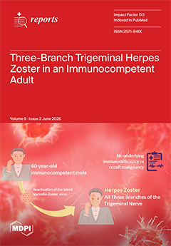
Cover Story (view full-size image): Herpes zoster typically restricts itself to a single unilateral dermatome. Simultaneous reactivation across all three branches of the trigeminal nerve—ophthalmic (V1), maxillary (V2), and mandibular (V3)—is an exceptionally rare clinical entity in the absence of underlying immunosuppression. We present a unique case of a 60-year-old immunocompetent male who presented with a rapid, painful, and extensive unilateral vesiculobullous eruption spanning the entire left side of his face and intraoral mucosa. A comprehensive diagnostic workup definitively ruled out occult malignancies and systemic immunodeficiencies, confirming the diagnosis via PCR and serology testing. Immediate administration of oral valacyclovir led to rapid crusting and full resolution within a week, completely preventing permanent ocular or neurological sequelae. View this paper
  • Issues are regarded as officially published after their release is announced to the table of contents alert mailing list.
  • You may sign up for e-mail alerts to receive table of contents of newly released issues.
  • PDF is the official format for papers published in both, html and pdf forms. To view the papers in pdf format, click on the "PDF Full-text" link, and use the free Adobe Reader to open them.
Order results
Result details
Section
Select all
Export citation of selected articles as:
12 pages, 8712 KB  
Article
Clinical Outcomes of the Canine Bypass Anchorage Technique for Severe Maxillary Bone Deficiency: A Case Report Series
by Calin Romulus Fodor, Marta Bieńkowska, Bartosz Dalewski and Łukasz Pałka
Reports 2026, 9(2), 195; https://doi.org/10.3390/reports9020195 - 22 Jun 2026
Viewed by 988
Abstract
Background/Objectives: Advanced implant anchorage techniques are increasingly used to manage severe maxillary bone deficiency and to avoid extensive bone augmentation procedures. This case series report aimed to describe the canine bypass anchorage technique and to evaluate the short- to medium-term clinical outcomes and [...] Read more.
Background/Objectives: Advanced implant anchorage techniques are increasingly used to manage severe maxillary bone deficiency and to avoid extensive bone augmentation procedures. This case series report aimed to describe the canine bypass anchorage technique and to evaluate the short- to medium-term clinical outcomes and survival of implants placed using this approach. Materials and Methods: Thirteen patients presenting with missing maxillary premolars or posterior segments and insufficient alveolar bone height for conventional axial implant placement were treated using the canine bypass technique. A total of 19 long one-piece implants were inserted palatally to the canine root, engaging distant cortical bone of the nasal cavity and/or palatal alveolar process. Pre- and postoperative cone-beam computed tomography (CBCT) examinations were performed to assess implant positioning and anchorage. Patients were followed up to 3.5 years. Results: The mean follow-up period was 26.1 ± 10.8 months. Nasal cortical anchorage was achieved in 84.2% of implants, and palatal cortical anchorage in 73.7%; both anchorage types were obtained simultaneously in 57.9% of cases. The mean distance between the implant and canine root was 1.27 ± 1.4 mm (range: −1.0 to 4.5 mm), including cases of direct implant–tooth contact and periodontal ligament space transgression. All implants remained functional throughout the observation period, yielding a cumulative survival rate of 100%. Canine pulp vitality was preserved in all non-endodontically treated teeth. Conclusions: Within the limitations of this case series report, the canine bypass anchorage technique appears to be a feasible and minimally invasive treatment option for maxillary rehabilitation with implant-supported restoration in selected patients with severe bone deficiency, potentially allowing avoidance of sinus augmentation procedures. Further prospective studies with larger patient cohorts and longer follow-up periods are required to confirm the long-term safety, predictability, and clinical applicability of this approach. Full article
(This article belongs to the Section Dentistry/Oral Medicine)
Show Figures

Figure 1

13 pages, 1550 KB  
Case Report
Clinical Decision-Making and Multidisciplinary Management of Peristomal Pyoderma Gangrenosum in Stage IVB Rectal Cancer: A Case Report—Corticosteroid Response but Fatal Cancer Progression
by Hiroshi Tanabe, Mari Ogawa, Mari Kita and Takeshi Kotake
Reports 2026, 9(2), 194; https://doi.org/10.3390/reports9020194 - 22 Jun 2026
Viewed by 569
Abstract
Background and Clinical Significance: Peristomal pyoderma gangrenosum (PPG) is a rare subtype of pyoderma gangrenosum, most commonly associated with inflammatory bowel disease or haematologic disorders. Its occurrence in patients with solid malignancies is uncommon. PPG in an oncologic setting poses diagnostic and therapeutic [...] Read more.
Background and Clinical Significance: Peristomal pyoderma gangrenosum (PPG) is a rare subtype of pyoderma gangrenosum, most commonly associated with inflammatory bowel disease or haematologic disorders. Its occurrence in patients with solid malignancies is uncommon. PPG in an oncologic setting poses diagnostic and therapeutic challenges because systemic immunosuppressive therapy, wound care, and ongoing chemotherapy must be carefully balanced; Case Presentation: We report the case of a Japanese man in his 50s with stage IVB rectal adenocarcinoma who developed rapidly progressive peristomal ulceration clinically consistent with PPG around a colostomy 12 weeks after initiation of panitumumab-containing systemic chemotherapy. The diagnosis was made on clinical grounds and was strongly supported by the clinical morphology, exclusion of major mimickers, and response to systemic corticosteroid therapy, although histopathological confirmation was not obtained. Because existing diagnostic criteria for pyoderma gangrenosum are not specifically designed for peristomal disease, they were used as supportive rather than definitive diagnostic tools. Skin biopsy was avoided due to the risk of pathergy at the peristomal site. Superficial cultures were not obtained because frequent cleansing and faecal contamination were likely to compromise diagnostic accuracy. To minimise mechanical pathergy, the stoma appliance was changed from a one-piece soft convex system to a two-piece flat system. Multidisciplinary management, including systemic corticosteroids, meticulous stoma care, and selective ultrasonic debridement, resulted in complete epithelialisation by Week 26. Chemotherapy was temporarily withheld during the active inflammatory phase and later resumed. Despite successful control of the peristomal ulceration, the patient died from progressive malignancy at Week 34; Conclusions: This case highlights the clinical challenge of balancing immunosuppressive therapy for clinically suspected PPG with ongoing oncologic treatment. Mechanical pathergy related to stoma appliance use was considered a more likely precipitating factor than chemotherapy alone, although panitumumab may have contributed to impaired cutaneous repair. Close collaboration among dermatologists, oncologists, surgeons, WOC nurses, and family caregivers is essential for multidisciplinary decision-making in complex oncologic settings. Full article
Show Figures

Graphical abstract

6 pages, 3712 KB  
Case Report
Inguinal Hernia Containing the Bladder and Postoperative Appearance: A Multimodality Case Report
by Hala Jasim, Orhan K. Öz and Joseph Frankl
Reports 2026, 9(2), 193; https://doi.org/10.3390/reports9020193 - 20 Jun 2026
Viewed by 404
Abstract
Background and Clinical Significance: Many diagnostic radiopharmaceuticals are excreted in the urine. This can pose a diagnostic challenge when urine-containing structures are in atypical locations, particularly in review of planar imaging without anatomical details from cross-sectional imaging. This case highlights a challenging 99m [...] Read more.
Background and Clinical Significance: Many diagnostic radiopharmaceuticals are excreted in the urine. This can pose a diagnostic challenge when urine-containing structures are in atypical locations, particularly in review of planar imaging without anatomical details from cross-sectional imaging. This case highlights a challenging 99mTc-methylene diphosphonate (99mTc-MDP) bone scan in a patient with an inguinal hernia containing a portion of the urinary bladder. Subsequently, we review diagnostic challenges on conventional and molecular imaging following surgical repair of the inguinal hernia. Case Presentation: A 79-year-old man with prostate cancer underwent initial staging prior to prostatectomy with 99mTc-MDP bone scintigraphy. Anterior and posterior images showed focal uptake overlying the pubic symphysis. Lateral views showed that the activity was extraosseous. Follow-up CT urography showed a bladder hernia as the cause of the abnormality on bone scan. Prostatectomy and inguinal hernia repair were performed as a combination case. Four years postoperatively, follow-up 68Ga-PSMA-11 positron emission tomography/computed tomography (PET/CT) showed no recurrence. The CT component of the exam showed an intermediate-density focus at the right inguinal hernia repair site, corresponding to a plugoma related to a polypropylene mesh plug, and a hyperattenuating Gore-Tex mesh repair of the left inguinal hernia. Conclusions: This case highlights the importance of lateral projections in resolving scintigraphic pitfalls and recognizing mesh-related imaging appearances to prevent misinterpretation. Full article
Show Figures

Figure 1

7 pages, 4492 KB  
Case Report
Myopericarditis Secondary to Toxoplasma Gondii Infection in an Immunocompetent Young Male—A Case Report
by Niall Leahy, Sandra Quinn and Derek Crinion
Reports 2026, 9(2), 192; https://doi.org/10.3390/reports9020192 - 20 Jun 2026
Viewed by 444
Abstract
Background and Clinical Significance: Inflammatory myopericardial syndrome is an umbrella term recently introduced by the European Society of Cardiology, which encapsulates the overlap that exists in clinical practice between myocardial and pericardial disease. It has a heterogeneous aetiology and a broad spectrum [...] Read more.
Background and Clinical Significance: Inflammatory myopericardial syndrome is an umbrella term recently introduced by the European Society of Cardiology, which encapsulates the overlap that exists in clinical practice between myocardial and pericardial disease. It has a heterogeneous aetiology and a broad spectrum of severity in terms of its clinical features. Toxoplasma gondii is a rare but recognised infectious cause of myopericarditis and is typically seen in immunocompromised individuals. Case Presentation: We present the case of a young, immunocompetent male, presenting with pleuritic chest pain following a recent flu-like illness. Investigations revealed an acute myocardial injury based on elevated troponin T levels, in the absence of ventricular dysfunction. Toxoplasma immunoserology was consistent with primary toxoplasma infection. The remainder of his viral panel was negative. There was prompt symptom improvement following commencement of treatment with colchicine and a non-steroidal anti-inflammatory agent. Cardiac magnetic resonance imaging post-discharge revealed findings consistent with prior myocarditis. Conclusions: This case is an example of the rare occurrence of toxoplasma myopericarditis in an immunocompetent individual. Cardiac MRI is an invaluable imaging modality used to evaluate myocardial function and tissue characteristics in patients presenting with inflammatory myopericardial syndrome. Full article
(This article belongs to the Section Cardiology/Cardiovascular Medicine)
Show Figures

Figure 1

7 pages, 336 KB  
Case Report
Cerebral Amyloid Angiopathy Presenting as Lobar Intracerebral Hemorrhage with Cognitive Decline in an 80-Year-Old Patient: A Clinicoradiologic Case Report
by Riana Tarabocchia, Kiran Javaid, Rahul Mittal, Maria Balabanian and Rory Ulloque
Reports 2026, 9(2), 191; https://doi.org/10.3390/reports9020191 - 18 Jun 2026
Viewed by 702
Abstract
Background and Clinical Significance: Cerebral amyloid angiopathy (CAA) is a neurovascular disorder characterized by the deposition of amyloid beta (Aβ) peptides within the walls of small-to-medium-sized cerebral vessels, leading to vascular fragility and an increased risk of lobar intracerebral hemorrhage [...] Read more.
Background and Clinical Significance: Cerebral amyloid angiopathy (CAA) is a neurovascular disorder characterized by the deposition of amyloid beta (Aβ) peptides within the walls of small-to-medium-sized cerebral vessels, leading to vascular fragility and an increased risk of lobar intracerebral hemorrhage (ICH), cognitive decline, and recurrent stroke. CAA is an important cause of spontaneous ICH in elderly patients and may be underrecognized, particularly when presenting with acute neurologic symptoms that mimic ischemic stroke. Early identification has significant implications for management, prognosis, and secondary prevention. Case Presentation: An 80-year-old male presented to the emergency department with incoherent speech, rambling, and severe headache concerning for acute stroke. His medical history was notable for a prior cerebrovascular accident, hypertension, diabetes mellitus, benign prostatic hyperplasia, and recent evaluation for dementia-like symptoms. Initial neuroimaging revealed a 3.2 cm intraparenchymal hemorrhage in the left occipital lobe with surrounding edema. Subsequent MRI demonstrated a lobar hemorrhage pattern suggestive of CAA based on imaging findings and clinical context. The patient was admitted to the intensive care unit (ICU) for close neurologic monitoring. He remained hemodynamically stable with no new motor or sensory deficits. Over a three-day hospital course, his speech and visual deficits improved. Blood pressure was carefully controlled, and repeat imaging demonstrated stable hemorrhage without progression. He was diagnosed with probable CAA and discharged home with supportive services. Conclusions: This case highlights the importance of considering cerebral amyloid angiopathy in elderly patients presenting with spontaneous lobar intracerebral hemorrhage and cognitive symptoms. Prompt recognition and appropriate neuroimaging are critical for diagnosis, risk stratification, and guiding management. Full article
Show Figures

Figure 1

10 pages, 5809 KB  
Case Report
Pre-Eruptive Intracoronal Resorption: Report of Two Cases
by Līna Petrova, Jūlija Ustiča, Elīna Rasčevska and Shaju Jacob Pulikkotil
Reports 2026, 9(2), 190; https://doi.org/10.3390/reports9020190 - 17 Jun 2026
Viewed by 426
Abstract
Background and Clinical Significance: Pre-eruptive intracoronal resorption is a rare developmental anomaly resembling occlusal caries despite the absence of an external breach. Case Presentation: We report of two cases. The first case involves a 9-year-old girl with PEIR of tooth 24 that was [...] Read more.
Background and Clinical Significance: Pre-eruptive intracoronal resorption is a rare developmental anomaly resembling occlusal caries despite the absence of an external breach. Case Presentation: We report of two cases. The first case involves a 9-year-old girl with PEIR of tooth 24 that was not identified on a panoramic radiograph taken one year earlier. The lesion later became clinically evident, presenting with symptoms and discoloration, and progressed to irreversible pulpitis requiring pulpotomy. This case highlights the importance of careful interpretation of paediatric panoramic radiographs and timely intervention to preserve pulp vitality in developing permanent teeth. The second case concerns a 16-year-old girl in whom PEIR was incidentally detected on cone-beam computed tomography (CBCT) in tooth 38. As the tooth has not yet erupted, its future clinical presentation and progression remain uncertain. To the best of the authors’ knowledge, there are no published reports specifically describing PEIR in patients from the Baltic region. Conclusions: Early radiographic detection of pre-eruptive intracoronal resorption is essential to prevent pulpal involvement and improve treatment outcomes, particularly when combined with careful interpretation of routine paediatric radiographs to minimize the risk of delayed diagnosis. This case emphasizes the importance of recognizing asymptomatic disease progression and integrating multidisciplinary approach to provide individualized treatment planning. Full article
(This article belongs to the Special Issue Case Reports in Oral Diseases)
Show Figures

Figure 1

12 pages, 431 KB  
Article
Challenges of the Oxycodone Hydrochloride Shortage
by Gursan Gunes Yenidogan, Nagihan Duran Yakar, Enise Alioglu, Salim Taner Gözükızıl and Aysegul Bilen
Reports 2026, 9(2), 189; https://doi.org/10.3390/reports9020189 - 17 Jun 2026
Viewed by 798
Abstract
Objectives: To evaluate the clinical impact and treatment adaptations during the immediate-release oxycodone hydrochloride shortage. Methods: This retrospective, observational study was conducted during the oxycodone shortage period (May 2024–March 2025) in patients with cancer pain. Pain intensity was assessed using the Numerical Rating [...] Read more.
Objectives: To evaluate the clinical impact and treatment adaptations during the immediate-release oxycodone hydrochloride shortage. Methods: This retrospective, observational study was conducted during the oxycodone shortage period (May 2024–March 2025) in patients with cancer pain. Pain intensity was assessed using the Numerical Rating Scale (NRS) at baseline (prior to switching, while receiving oxycodone) and at follow-up (after switching to alternative analgesics). Changes in pain intensity were evaluated using within-patient differences (ΔNRS), with clinically meaningful worsening defined as an increase of ≥2 points. Descriptive and inferential statistics were used to summarize patient characteristics and outcomes. Results: Of 300 patients screened, 55 met inclusion criteria (mean age 65.2 ± 11.0 years; 63.6% male). Pain intensity increased significantly following treatment modification during the period of oxycodone unavailability, with mean NRS scores rising from 4.3 ± 1.7 to 5.9 ± 2.5 (p < 0.001). The mean ΔNRS was +1.56 (95% CI 0.79–2.34), with clinically meaningful worsening observed in 36 patients (65.5%). No statistically significant association was observed between substitute analgesic type and clinically meaningful worsening (p = 0.11). Conclusions: The oxycodone shortage was associated with worsened pain control and increased need for treatment modifications in cancer patients, highlighting the importance of uninterrupted access to essential opioids. Full article
(This article belongs to the Section Epidemiology/Public Health)
Show Figures

Figure 1

22 pages, 547 KB  
Case Report
Tumefactive Multiple Sclerosis Mimicking a High-Grade Glioma: A Case Report and Literature Review
by Maria P. Fernandez-Gomez, Luis Rafael Moscote-Salazar, Jesus Francisco Saltaren Fonseca, Guillermo de Jesus Aguirre Vera, Willem Calderon Miranda and Jose Valerio
Reports 2026, 9(2), 188; https://doi.org/10.3390/reports9020188 - 16 Jun 2026
Viewed by 617
Abstract
Background and Clinical Significance: Tumefactive Multiple Sclerosis (TMS) represents a rare and diagnostically challenging form of demyelinating disease characterized by large space-occupying lesions that can closely mimic intracranial neoplasms, abscesses, and other inflammatory or vascular conditions. Case Presentation: The case highlights the overlapping [...] Read more.
Background and Clinical Significance: Tumefactive Multiple Sclerosis (TMS) represents a rare and diagnostically challenging form of demyelinating disease characterized by large space-occupying lesions that can closely mimic intracranial neoplasms, abscesses, and other inflammatory or vascular conditions. Case Presentation: The case highlights the overlapping radiologic features that frequently lead to diagnostic uncertainty and underscores the importance of careful interpretation of multimodal imaging and ancillary studies. Overall a comprehensive multidisciplinary evaluation is essential to reduce the risk of misdiagnosis and avoid unnecessary invasive interventions. Conclusions: This review summarizes current evidence regarding the diagnostic approach, imaging characteristics, and therapeutic strategies for tumefactive demyelinating lesions. Additionally, we present a clinical case that illustrates the diagnostic complexity of this entity, in which neuroimaging findings and cerebrospinal fluid analysis supported a demyelinating rather than neoplastic process. Full article
Show Figures

Figure 1

9 pages, 4008 KB  
Case Report
A Rare Case Reveals Important Consideration of the Diagnosis of Giant Cell Arteritis in Patients with Bilateral Painful Optic Perineuritis
by Jordan Santos, Faraz Behzadi, S. Mozammil Alam, Thomas C. Varkey, David C. Maeng, Ghassan J. Ibrahim, Trent H. Smith and Alan Wang
Reports 2026, 9(2), 187; https://doi.org/10.3390/reports9020187 - 15 Jun 2026
Viewed by 574
Abstract
Background and Clinical Significance: Giant cell arteritis (GCA) is an autoimmune vasculitis of both medium and large-sized vessels typically affecting females 50 years of age or older. Severe complications can include permanent visual loss, acute coronary syndrome, or stroke. This case will present [...] Read more.
Background and Clinical Significance: Giant cell arteritis (GCA) is an autoimmune vasculitis of both medium and large-sized vessels typically affecting females 50 years of age or older. Severe complications can include permanent visual loss, acute coronary syndrome, or stroke. This case will present an atypical presentation of bilateral OPN which can be a rare manifestation of GCA; Case Presentation: Our patient developed acute, painful worsening central vision loss progressing from right eye to left with bilateral extraocular motility restriction and magnetic resonance image (MRI) revealed bilateral, circumferential optic nerve sheath enhancement suggesting optic perineuritis (OPN). Temporal artery biopsy confirmed GCA with bilateral temporal arteritis. The patient was treated with a high dose course of corticosteroids followed by a taper and was started on upadacitinib with symptomatic improvement; Conclusions: This case underscores OPN as a rarer manifestation of giant cell arteritis that can present with bilateral painful eye movements and vision loss. Early recognition and prompt corticosteroid therapy are essential to prevent irreversible visual impairment. Full article
Show Figures

Figure 1

11 pages, 10574 KB  
Case Report
Non-Surgical Periodontal Defect Fill and Spontaneous Tooth Repositioning: A Case Report
by Hani T. Fadel
Reports 2026, 9(2), 186; https://doi.org/10.3390/reports9020186 - 15 Jun 2026
Viewed by 467
Abstract
Background and Clinical Significance: Advanced periodontitis destroys tooth-supporting structures and causes pathologic tooth migration (PTM) and functional and esthetic repercussions. This report illustrates how non-surgical periodontal therapy (NSPT) addresses such concerns. Case Presentation: A 34-year-old woman (healthy, who quit smoking) complained [...] Read more.
Background and Clinical Significance: Advanced periodontitis destroys tooth-supporting structures and causes pathologic tooth migration (PTM) and functional and esthetic repercussions. This report illustrates how non-surgical periodontal therapy (NSPT) addresses such concerns. Case Presentation: A 34-year-old woman (healthy, who quit smoking) complained of a space between her upper front teeth. Based on clinical and radiographic data, she had localized periodontitis stage III grade C with intrabony defects, deep pockets, and a diastema between teeth # 12 and 11. The treatment plan involved improvement of self-performed oral hygiene measures and NSPT. Initially, improvements in plaque, bleeding, and pocket depth scores were observed. Non-surgical re-treatment of residual sites was associated with further improvements and complete closure of the diastema, along with patient satisfaction with the outcome. Conclusions: The potential of NSPT in improving periodontal clinical parameters and the association with spontaneous tooth repositioning and apparent filling of intrabony defects radiographically is demonstrated, ultimately addressing the patient’s functional and esthetic concerns. Full article
(This article belongs to the Special Issue Case Reports in Oral Diseases)
Show Figures

Figure 1

13 pages, 63394 KB  
Case Report
Metastatic Anaplastic Thyroid Carcinoma Presenting with Gastrointestinal Bleeding: A Case Report and Literature Review
by Hassan Al-Thani, Husham Abdelrahman, Maryam Al-Sulaiti, Abdelhakem Tabeb, Mahir Petkar, Noora Al-Thani and Ayman El-Menyar
Reports 2026, 9(2), 185; https://doi.org/10.3390/reports9020185 - 14 Jun 2026
Viewed by 474
Abstract
Background and Clinical Significance: Thyroid cancer is increasing, particularly the differentiated type, with decreasing incidence of the anaplastic type. Anaplastic thyroid carcinoma (ATC) is a rare, aggressive, and often lethal form. It frequently presents with metastatic disease, regional and systemic, with common [...] Read more.
Background and Clinical Significance: Thyroid cancer is increasing, particularly the differentiated type, with decreasing incidence of the anaplastic type. Anaplastic thyroid carcinoma (ATC) is a rare, aggressive, and often lethal form. It frequently presents with metastatic disease, regional and systemic, with common distant metastasis to the lung, bone, brain, and adrenal, and rarely to other places; Case presentation: A 74-year-old Arab male presented with symptomatic anemia and melena and was admitted for investigation of the cause. The patient was found to have a large retrosternal goiter and gastric tumor. CT scan showed a pedunculated, nonobstructive mass, suggestive of a GIST or leiomyoma. The neck mass presented with compressive symptoms. He underwent a combined neck and abdominal surgical resection based on a multidisciplinary team decision, as prior biopsies were not conclusive. The final pathology report identified similar tumors in the two specimens and suggested an anaplastic thyroid carcinoma as the primary tumor with metastasis to the stomach. Furthermore, the workup, including a PET scan 2 weeks post-surgery, revealed widespread metastases in the bone, lung, and liver, and the treatment was palliative. He was followed up in the outpatient clinic for 4 and a half months post-operatively. The patient developed sepsis and cardiopulmonary arrest and died; Conclusions: ATC can metastasize to many places in the body, including the stomach (as shown in our case), which can cause significant upper gastrointestinal bleeding and anemia. Metastatic ATC carries a poor prognosis; thus, physicians need to keep a high index of suspicion in approaching similar cases. A multidisciplinary approach for the management is of utmost importance for appropriate treatment. This disease’s pathology, behavior, and targeted new treatment modalities must be explored further. Full article
(This article belongs to the Collection Clinical Research in Oncology)
Show Figures

Figure 1

14 pages, 11573 KB  
Case Report
Spontaneous Bilateral Renal Forniceal Rupture Secondary to Acute Urinary Retention in a Patient with Prior Prostate Radiotherapy: A Case Report
by Timoleon Giannakas, Dimitrios Deligiannis, Panagiotis Mitsos, Anna Papakonstantinou, Marios Stavropoulos and Aris Kaltsas
Reports 2026, 9(2), 184; https://doi.org/10.3390/reports9020184 - 12 Jun 2026
Viewed by 740
Abstract
Background and Clinical Significance: Spontaneous renal forniceal rupture is an uncommon complication of obstructive uropathy and is classically associated with ureteric calculi rather than distal urinary retention. Bilateral retention-related rupture appears to be exceptionally rare and may be diagnostically challenging when renal function [...] Read more.
Background and Clinical Significance: Spontaneous renal forniceal rupture is an uncommon complication of obstructive uropathy and is classically associated with ureteric calculi rather than distal urinary retention. Bilateral retention-related rupture appears to be exceptionally rare and may be diagnostically challenging when renal function begins to improve after bladder decompression; Case Presentation: An 82-year-old man with a history of prostate cancer treated five years earlier with external beam radiotherapy and androgen deprivation therapy presented with acute abdominal pain radiating to both flanks and inability to void. Bedside ultrasonography showed urinary retention and bilateral hydronephrosis, and a 16-Fr Foley catheter drained 900 mL of urine. Admission evaluation showed severe acute kidney injury, microscopic hematuria, minimal leukocyturia, and elevated inflammatory markers. Post-obstructive diuresis developed after bladder decompression. CT urography with excretory-phase imaging on hospital day 3 demonstrated severe bilateral hydroureteronephrosis with bilateral renal forniceal rupture and associated urinomas, including a larger left-sided collection extending toward the psoas compartment. Bilateral percutaneous nephrostomies were placed on hospital day 4 for upper-tract diversion. Immediate nephrostography showed no active contrast extravasation. At one-month follow-up, combined CT and nephrostographic assessment confirmed complete resolution of the bilateral urinomas without persistent leak, and the nephrostomy tubes were removed; Conclusions: This case suggests that urinary retention in an older man with prior prostate radiotherapy may reflect radiation-associated outlet pathology and/or impaired detrusor function rather than simple prostate enlargement. Delayed-phase CT urography was essential for diagnosis, and active bilateral diversion was justified by bilateral rupture, acute kidney injury, and the extent of urinary extravasation. The report expands the limited PubMed-indexed literature on retention-related upper urinary tract rupture and supports cautious follow-up aimed at defining the underlying mechanism of retention. Full article
(This article belongs to the Special Issue When Urology Surprises: Educational and Rare Clinical Cases)
Show Figures

Figure 1

15 pages, 359 KB  
Case Report
Morita Therapy-Based Nursing Support for Socially Withdrawn Japanese Youth (Hikikomori) with Gaze Phobia: A Case Report
by Mikie Ebihara, Miwa Yoshida, Kohei Handa, Katsuharu Yano, Tomoko Omiya and Kei Nakamura
Reports 2026, 9(2), 183; https://doi.org/10.3390/reports9020183 - 11 Jun 2026
Viewed by 626
Abstract
Background and Clinical Significance: “Hikikomori”—a state of prolonged social withdrawal affecting an estimated 2% of Japan’s working-age population—is frequently associated with underlying anxiety disorders, such as gaze phobia, and contributes to the socio-economic burden known as the “8050 problem,” in which aging [...] Read more.
Background and Clinical Significance: “Hikikomori”—a state of prolonged social withdrawal affecting an estimated 2% of Japan’s working-age population—is frequently associated with underlying anxiety disorders, such as gaze phobia, and contributes to the socio-economic burden known as the “8050 problem,” in which aging parents support their socially isolated adult children. While Morita therapy is effective for such conditions, nursing support has historically lacked a systematic theoretical framework. This case report presents a novel nursing model analyzing the transformation process from toraware (mental preoccupation) toward mokuteki-hon-i (purpose-driven action). It proposes the ‘side-by-side’ nursing approach as a potentially important element in supporting patient autonomy in similar clinical settings. Case Presentation: A man in his 20s, diagnosed with gaze phobia and experiencing long-term withdrawal following traumatic bullying, was referred to our specialized short-care program. After initial preparation through structured psychoeducation regarding Morita therapy principles (toraware, sei-no-yokubo, mokuteki-hon-i), he participated in a 14-month Morita therapy-based short-care program combining individual and group interventions. Initially, the patient exhibited severe social avoidance and was trapped in a cycle of seishin-kogo-sayo (psychic interaction). Nurses applied ‘Strategic Inattention to Symptoms’ (shojo-fumon) and provided specific role suggestions, such as serving as a secretary in group discussions, to elicit his sei-no-yokubo (desire for life). Through the reframing of his anxiety as a constructive drive, the patient shifted to a purpose-driven stance. Outcomes showed improved self-adjustment skills in public spaces and successful social reintegration through sustained part-time employment. Conclusions: Nursing care characterized by ‘intentional non-intervention’—which involves waiting in a ‘side-by-side’ manner within a minimally structured environment—may contribute to fostering patient autonomy in similar clinical contexts. This ‘experience-oriented’ approach appeared to elicit inner strengths and support self-regulation in this case, warranting further investigation in multi-case designs. The relative contributions of individual nursing support and group therapeutic milieu cannot be disentangled in a single-case design. Full article
Show Figures

Figure 1

12 pages, 15695 KB  
Case Report
Torsion of the Vermiform Appendix in an 18-Day-Old Neonate: A Case Report from Romania and Review of the Literature
by Paul Tchouala Tchakoute, Alin Iuhas, Vlad-Ionuț Nechita, Andrei Vasile Pașcalău and Ion Cosmin Puia
Reports 2026, 9(2), 182; https://doi.org/10.3390/reports9020182 - 10 Jun 2026
Viewed by 615
Abstract
Background and Clinical Significance: Torsion of the vermiform appendix is a rare condition with a clinical presentation closely resembling acute appendicitis, while preoperative investigations are of limited value in distinguishing between the two entities. In most cases, the definitive diagnosis is made incidentally [...] Read more.
Background and Clinical Significance: Torsion of the vermiform appendix is a rare condition with a clinical presentation closely resembling acute appendicitis, while preoperative investigations are of limited value in distinguishing between the two entities. In most cases, the definitive diagnosis is made incidentally during surgery. Case Presentation: The authors present the case of an 18-day-old female neonate who presented with marked abdominal distension, diffuse spontaneous and palpation-induced abdominal pain, guarding, and signs of peritoneal irritation. The clinical manifestations and paraclinical findings mimicked a neonatal intestinal obstruction; however, intraoperative exploration revealed a gangrenous vermiform appendix twisted 240° anticlockwise, associated with a fibrinous pseudomembrane and multiple enlarged mesenteric lymph nodes. Although the initial therapeutic strategy was to perform a laparoscopy, severe abdominal distension caused by marked aerocolia necessitated conversion to a supra- and infraumbilical midline laparotomy. We thus describe, to the best of our knowledge, one of the youngest neonatal cases of appendiceal torsion reported in the literature. Conclusions: Although rare, appendiceal torsion should be considered in the differential diagnosis of neonatal acute abdomen, and timely surgical exploration is key to achieving a favorable outcome. Full article
(This article belongs to the Section Surgery)
Show Figures

Figure 1

11 pages, 843 KB  
Case Report
Mikulicz Disease Revealing IgG4-Related Tubulointerstitial Nephritis: A Case Report and Literature Review
by Lissethkaren Alvarez Vargas, Celia Rodríguez Tudero, Elena Jiménez Mayor, Avinash Chandu Nanwani, Esperanza Moral Berrio, Juan Daniel Díaz García, Arturo Villalobos Navarro, Emily Rosario Chamorro Asto, Michael Cieza Terrones and José C. De La Flor
Reports 2026, 9(2), 181; https://doi.org/10.3390/reports9020181 - 10 Jun 2026
Viewed by 796
Abstract
Background and Clinical Significance: IgG4-related disease (IgG4-RD) is a chronic fibroinflammatory, immune-mediated multisystem disorder that can mimic neoplastic, infectious, or autoimmune conditions. Among its head-and-neck manifestations, IgG4-related dacryoadenitis and sialadenitis, historically referred to as Mikulicz disease, should be distinguished from the classical Mikulicz [...] Read more.
Background and Clinical Significance: IgG4-related disease (IgG4-RD) is a chronic fibroinflammatory, immune-mediated multisystem disorder that can mimic neoplastic, infectious, or autoimmune conditions. Among its head-and-neck manifestations, IgG4-related dacryoadenitis and sialadenitis, historically referred to as Mikulicz disease, should be distinguished from the classical Mikulicz syndrome, which describes secondary lacrimal and salivary gland enlargement due to other systemic disorders. Renal involvement, most commonly in the form of IgG4-related tubulointerstitial nephritis (IgG4-TIN), is less frequent but carries major prognostic implications because delayed diagnosis may lead to irreversible kidney damage. Case Presentation: A 49-year-old man with no relevant past medical history presented with a 2-year history of intermittent polyuria and foamy urine. Laboratory testing revealed advanced kidney dysfunction, with serum creatinine of 4.2 mg/dL, estimated glomerular filtration rate of 16 mL/min/1.73 m2, and proteinuria of 2874 mg/day. Physical examination showed bilateral parotid enlargement, upper eyelid edema, lacrimal gland enlargement, and sicca symptoms, raising suspicion for IgG4-related dacryoadenitis and sialadenitis (Mikulicz disease). Further work-up demonstrated marked eosinophilia, polyclonal hypergammaglobulinemia, and significantly elevated serum IgG4 levels (3180 mg/dL), while infectious serologies and autoimmune studies were negative. Kidney biopsy revealed plasma cell-rich tubulointerstitial nephritis with lymphoplasmacytic and eosinophilic infiltrates, interstitial fibrosis, tubular atrophy, and more than 40 IgG4-positive plasma cells per high-power field, supporting the diagnosis of IgG4-related tubulointerstitial nephritis in the setting of systemic IgG4-RD. Treatment with prednisone followed by mycophenolate mofetil led to improvement in glandular manifestations and a partial reduction in proteinuria, but renal recovery remained incomplete. The patient subsequently developed a severe pulmonary infection complicated by sepsis and oligoanuric acute kidney injury superimposed on chronic kidney disease, and ultimately progressed to end-stage kidney disease requiring chronic maintenance hemodialysis. Conclusions: This case highlights that a Mikulicz disease phenotype may represent the initial manifestation of systemic IgG4-RD and should prompt evaluation for extraglandular involvement, particularly renal disease. In patients with glandular enlargement, eosinophilia, hypergammaglobulinemia, and unexplained renal dysfunction, IgG4-RD should be actively considered. Kidney biopsy remains essential for diagnostic confirmation and prognostic assessment, as delayed recognition may result in irreversible renal damage and progression to end-stage kidney disease. Full article
(This article belongs to the Section Nephrology/Urology)
Show Figures

Figure 1

9 pages, 1524 KB  
Case Report
Acute Forearm and Hand Compartment Syndrome in a Child Following Delayed Presentation of Forearm Trauma: A Case Report and Literature Review
by Abdulmonem Alsiddiky, Mishari Alanezi, Nouf Alabdulkarim, Bandar Aljammaz, Othman Alabdullah, Saad Alkahtani, Razan Alshatwi and Abdulrahman Alrajhi
Reports 2026, 9(2), 180; https://doi.org/10.3390/reports9020180 - 10 Jun 2026
Viewed by 545
Abstract
Background and Clinical Significance: Acute compartment syndrome is a rare but limb-threatening emergency in pediatric patients. While most cases follow high-energy trauma or displaced fractures, acute compartment syndrome precipitated by initially underestimated forearm injuries is uncommon and may create a significant diagnostic challenge, [...] Read more.
Background and Clinical Significance: Acute compartment syndrome is a rare but limb-threatening emergency in pediatric patients. While most cases follow high-energy trauma or displaced fractures, acute compartment syndrome precipitated by initially underestimated forearm injuries is uncommon and may create a significant diagnostic challenge, particularly in young children who exhibit atypical clinical presentations, such as escalating anxiety and analgesic requirements, rather than classic ischemic signs. Case Presentation: We report the case of a 4-year-old girl who developed severe forearm and hand compartment syndrome following a delayed presentation after a fall from a height of 2–2.5 m onto the left upper extremity. Initial evaluation revealed progressive tense swelling, severe pain with passive stretch, diminished distal perfusion, and radiographic evidence of distal radius-ulna buckle fractures associated with a proximal ulna fracture. Emergent surgical decompression via extensive volar and dorsal fasciotomies revealed markedly elevated compartment pressures. Intraoperatively, deep volar muscle ischemia and necrosis were identified, requiring carpal tunnel release, serial debridements, and complex staged wound management. Multidisciplinary care and ongoing rehabilitation were essential for limb salvage and functional recovery. Conclusions: This case underscores the profound unpredictability of pediatric compartment syndrome and demonstrates that even classically stable, benign fractures can initiate a devastating ischemic cascade. A high index of suspicion, regardless of the injury mechanism, along with early recognition and prompt surgical intervention, is absolutely critical for preventing irreversible myoneural damage and optimizing management outcomes in pediatric patients. Full article
(This article belongs to the Section Orthopaedics/Rehabilitation/Physical Therapy)
Show Figures

Figure 1

9 pages, 393 KB  
Brief Report
Outcomes of Lurbinectedin in Central Nervous System Metastases of Small Cell Lung Cancer: A Single-Institution Retrospective Case Series
by Navanita Biswas, Carolin Schmidt and Shoja Rahimian
Reports 2026, 9(2), 179; https://doi.org/10.3390/reports9020179 - 7 Jun 2026
Viewed by 633
Abstract
Background/Objectives: Central nervous system (CNS) metastases are a frequent and morbid complication of small cell lung cancer (SCLC), with limited effective systemic treatment options. Lurbinectedin has demonstrated systemic activity in relapsed SCLC; however, its intracranial efficacy remains unclear because patients with active [...] Read more.
Background/Objectives: Central nervous system (CNS) metastases are a frequent and morbid complication of small cell lung cancer (SCLC), with limited effective systemic treatment options. Lurbinectedin has demonstrated systemic activity in relapsed SCLC; however, its intracranial efficacy remains unclear because patients with active CNS disease were underrepresented in pivotal trials. We evaluated real-world intracranial outcomes of lurbinectedin in patients with SCLC and CNS metastases. Methods: A single-institution retrospective case series was conducted among adult patients with histologically confirmed SCLC and radiologic CNS metastases treated with lurbinectedin between July 2020 and April 2025. Primary endpoints were CNS disease control rate (CNS-DCR), defined as radiographic stability or improvement lasting ≥8 weeks, and intracranial progression-free survival (iPFS), defined as time from lurbinectedin initiation to clinical or radiographic CNS progression or death. Results: Thirty patients received lurbinectedin; 14 (46.7%) had CNS metastases at any time. Five patients (16.7%) had baseline CNS metastases prior to lurbinectedin initiation, while nine (30.0%) developed CNS metastases during treatment. Among patients with baseline CNS disease, one patient demonstrated radiographic intracranial improvement at approximately 4 months; however, systemic progression at 5 months limited further assessment of response duration. The remaining four patients experienced intracranial progression within 2–4 months. One of five patients with baseline CNS metastases met the predefined CNS disease control endpoint; this descriptive proportion corresponds to 20% within our small sample. Median iPFS was approximately 2.5 months. No CNS-specific adverse events attributable to lurbinectedin were observed. Conclusions: In this single-institution retrospective case series, limited intracranial disease control was observed among SCLC patients with baseline CNS metastases treated with lurbinectedin. Given the small number of evaluable patients, these findings should be interpreted as descriptive and hypothesis-generating rather than a conclusive efficacy analysis. Prospective studies incorporating CNS-specific endpoints are needed to better define the role of lurbinectedin and other systemic therapies in intracranial disease management. Full article
(This article belongs to the Section Oncology)
Show Figures

Figure 1

8 pages, 1566 KB  
Case Report
Structural Mimics of Recurrent Bell’s Palsy: A Case Report Highlighting Diagnostic Red Flags in Facial Nerve Schwannoma and Metastasis
by Sohyeon Kim, Hyesoo Kwon and Hung Youl Seok
Reports 2026, 9(2), 178; https://doi.org/10.3390/reports9020178 - 7 Jun 2026
Viewed by 451
Abstract
Background and Clinical Significance: Recurrent peripheral facial palsy is most often attributed to idiopathic Bell’s palsy (BP), yet a minority of patients harbor underlying structural lesions. Early recognition of such lesions is essential for timely and appropriate management. Case Presentation: We [...] Read more.
Background and Clinical Significance: Recurrent peripheral facial palsy is most often attributed to idiopathic Bell’s palsy (BP), yet a minority of patients harbor underlying structural lesions. Early recognition of such lesions is essential for timely and appropriate management. Case Presentation: We retrospectively reviewed recurrent unilateral facial palsy at a single tertiary center and selected two illustrative patients in whom serial clinical, electrodiagnostic, and magnetic resonance imaging (MRI) findings ultimately indicated structural pathology. The first patient experienced three strictly right-sided episodes over 3.5 years, with worsening House–Brackmann grades (II → III → V) and progressive axonal loss on serial facial nerve conduction studies, culminating in a mass-like enhancing lesion at the geniculate ganglion consistent with schwannoma. The second patient had two left-sided recurrences within seven months. MRI evolved from a subtle intrameatal nodular suspicion without enhancement to a clearly enhancing intrameatal nodule with dural thickening. Additional intracranial nodular enhancements were discovered later and the patient was diagnosed with leptomeningeal metastasis. Conclusions: These cases illustrate that strictly ipsilateral recurrences, progressive electrophysiologic deterioration, and atypical or evolving MRI enhancement patterns should prompt thorough re-evaluation for structural causes rather than repeated labeling as idiopathic BP. Full article
Show Figures

Figure 1

10 pages, 28982 KB  
Case Report
Surgical Management of Inframammary Hidradenitis Suppurativa with Reduction Mammaplasty Technique: A Report of Two Cases
by Enrico Caporali, Paolo Toninello, Monia Maritan, Alessandro Gatti and Giorgio Berna
Reports 2026, 9(2), 177; https://doi.org/10.3390/reports9020177 - 6 Jun 2026
Viewed by 570
Abstract
Background and Clinical Significance: Hidradenitis suppurativa (HS) is a chronic, debilitating skin disorder that often affects the inframammary fold (IMF). While surgical management, particularly wide local excision (WLE), is the gold standard for severe cases, less is known about the role of [...] Read more.
Background and Clinical Significance: Hidradenitis suppurativa (HS) is a chronic, debilitating skin disorder that often affects the inframammary fold (IMF). While surgical management, particularly wide local excision (WLE), is the gold standard for severe cases, less is known about the role of breast surgery techniques in treating HS in this area; Case Presentation: This report presents two cases of female patients with bilateral inframammary HS and mammary hypertrophy, both treated with reduction mammaplasty to excise diseased tissue while addressing breast volume and contour. Both patients had experienced inadequate response to medical therapies, including biologic treatments, and presented with distinct clinical features—one with significant asymmetry and active disease, and the other with more scarring and hypertrophic lesions. During follow-up, no recurrence of disease was observed and both patients reported improved breast appearance and satisfaction; Conclusions: These cases underscore the importance of a multidisciplinary approach, with dermatologists and surgeons both playing a key role in the management of this complex condition. Full article
(This article belongs to the Section Surgery)
Show Figures

Figure 1

13 pages, 358 KB  
Article
Association of Early Albumin Administration with Clinical Outcomes in Patients Hospitalized with Spontaneous Bacterial Peritonitis: A Propensity-Matched Cohort Study
by Noor Albusta, Mohamed Abdulla, Sara Isa and Rehab Almarzooq
Reports 2026, 9(2), 176; https://doi.org/10.3390/reports9020176 - 6 Jun 2026
Viewed by 507
Abstract
Background/Objectives: Spontaneous bacterial peritonitis (SBP) is a serious complication of decompensated cirrhosis and is associated with acute kidney injury (AKI), organ failure, and death. Intravenous albumin is recommended in SBP because it reduces renal impairment and mortality, particularly in patients at higher risk [...] Read more.
Background/Objectives: Spontaneous bacterial peritonitis (SBP) is a serious complication of decompensated cirrhosis and is associated with acute kidney injury (AKI), organ failure, and death. Intravenous albumin is recommended in SBP because it reduces renal impairment and mortality, particularly in patients at higher risk of circulatory dysfunction and hepatorenal complications. However, the prognostic impact of early albumin administration on clinical outcomes in hospitalized SBP patients remains incompletely characterized in real-world practice. This study aimed to assess the association between early albumin administration and clinical outcomes in patients hospitalized with SBP compared to those without early albumin. Methods: A retrospective cohort study was conducted using the TriNetX US Collaborative Research Network, including adults hospitalized with SBP through February 2026. Patients were divided into those receiving early albumin administration (n = 1248) and those without early albumin (n = 4932) within 24 h of index SBP diagnosis. Propensity score matching (1:1) balanced cohorts (n = 1230 each) for demographics, comorbidities, liver disease severity surrogates, medications, and laboratory values. Relative risks (RR), risk differences (RD), and hazard ratios (HR) were calculated using propensity-matched and Cox proportional hazard models. Results: Early albumin administration was associated with significantly lower all-cause mortality (RR 0.620; 95% CI: 0.441–0.871; p = 0.005 at 5 days; RR 0.770; 95% CI: 0.651–0.910; p = 0.002 at 90 days). Secondary outcomes showed reduced risks for acute kidney injury (RR 0.654; 95% CI: 0.553–0.774; p < 0.001 at 5 days; RR 0.798; 95% CI: 0.706–0.903; p < 0.001 at 90 days), hepatorenal syndrome–AKI (RR 0.598; 95% CI: 0.445–0.804; p < 0.001 at 5 days; RR 0.756; 95% CI: 0.613–0.932; p = 0.009 at 90 days), vasopressor requirement (RR 0.633; 95% CI: 0.489–0.820; p < 0.001 at 5 days; RR 0.712; 95% CI: 0.572–0.887; p = 0.002 at 30 days), and renal replacement therapy (RR 0.533; 95% CI: 0.324–0.878; p = 0.011 at 5 days; RR 0.642; 95% CI: 0.442–0.932; p = 0.019 at 30 days). Cox models confirmed statistically significant risk reductions for all primary and secondary outcomes, including ICU admission (HR 0.82; 95% CI: 0.73–0.92; p = 0.001) and 30-day readmission (HR 0.84; 95% CI: 0.73–0.97; p = 0.015). Associations were strongest in the early period and attenuated over time. Conclusions: Early albumin administration was associated with reduced risks of mortality, AKI, HRS-AKI, and hemodynamic instability in patients hospitalized with SBP, with attenuation over time. These findings support timely implementation of guideline-concordant albumin therapy, although residual confounding cannot be excluded. Full article
Show Figures

Figure 1

21 pages, 40077 KB  
Case Report
Interdisciplinary Management of Severe Skeletal Class II Malocclusion with Three-Piece Le Fort I and Bilateral Sagittal Split Osteotomy: A Case Report
by Tatiana-Maria Coman, Simion Bran, Andrei-Mario Bădărău-Șuster, Mariana Păcurar and Sorin-Claudiu Popșor
Reports 2026, 9(2), 175; https://doi.org/10.3390/reports9020175 - 5 Jun 2026
Viewed by 768
Abstract
Background and Clinical Significance: Severe skeletal Class II malocclusion associated with periodontal compromise and posterior edentulism requires a carefully sequenced interdisciplinary treatment approach integrating orthodontic, surgical, periodontal, and prosthetic rehabilitation. Case Presentation: This case report describes the comprehensive interdisciplinary management of a 21-year-old [...] Read more.
Background and Clinical Significance: Severe skeletal Class II malocclusion associated with periodontal compromise and posterior edentulism requires a carefully sequenced interdisciplinary treatment approach integrating orthodontic, surgical, periodontal, and prosthetic rehabilitation. Case Presentation: This case report describes the comprehensive interdisciplinary management of a 21-year-old female patient presenting with skeletal Class II malocclusion, severe mandibular retrognathia, vertical maxillary excess, labial incompetence, temporomandibular joint (TMJ) dysfunction and periodontal deficiencies. The treatment sequence involved occlusal splint therapy, pre-surgical orthodontic decompensation, bimaxillary orthognathic surgery using a segmental Le Fort I osteotomy and bilateral sagittal split osteotomy (BSSO), postoperative orthodontic refinement with aligners, periodontal plastic surgery using the Zucchelli technique and guided bone regeneration (GBR) with implant placement in the posterior mandible. At the four-year follow-up, the patient demonstrated complete root coverage, stable skeletal correction, and satisfactory implant integration, with maintenance of functional and aesthetic outcomes over time. Conclusions: This report highlights the importance of precise preoperative planning and the synergy between orthodontics, orthognathic surgery and periodontics in achieving optimal functional and aesthetic results. Full article
Show Figures

Figure 1

7 pages, 3490 KB  
Case Report
Diagnostic and Therapeutic Pitfalls Encountered in a Young Adult Patient with a Symptomatic Chronic Subdural Hematoma Mimicking a Subacute Epidural Hematoma in the Presence of a Galassi Grade III Arachnoid Cyst: Case Report
by Marios Theologou, Nikolaos Syrmos and Vaitsa Giannouli
Reports 2026, 9(2), 174; https://doi.org/10.3390/reports9020174 - 4 Jun 2026
Viewed by 749
Abstract
Background and Clinical Significance: Chronic subdural hematomas (cSDHs) present characteristic imaging findings, making the diagnosis straightforward. In rare cases, arachnoid cysts (ACs) may be associated with their formation. There is still no consensus regarding their treatment; Case Presentation: A young adult male [...] Read more.
Background and Clinical Significance: Chronic subdural hematomas (cSDHs) present characteristic imaging findings, making the diagnosis straightforward. In rare cases, arachnoid cysts (ACs) may be associated with their formation. There is still no consensus regarding their treatment; Case Presentation: A young adult male presented with occipital headache. Neurological examination was normal. Laboratory investigations were within physiological limits. A CT scan revealed the presence of a Galassi Grade III temporo-parietal AC accompanied by a parietal epidural hematoma (EDH) on the right side. His medical history was significant for treated hypertension. There was no use of anticoagulants, antiplatelets, or history of trauma. Vascular pathology was excluded by MRA/MRV. He was discharged for home care and was readmitted 10 days later after a repeat CT scan. A brief cognitive assessment with the Mini-Mental State Examination (MMSE) revealed mild cognitive impairment. A burr-hole evacuation was performed, and a drainage catheter was left in place for 24 h. Intraoperative findings were consistent with a chronic subdural hematoma. The patient was discharged with complete resolution of symptoms. A follow-up CT scan performed one month postoperatively confirmed the favorable result. Cognitive functions were normal on follow-up; Conclusions: A SDH may mimic the characteristics of an EDH in the presence of an AC. The most common symptom is cephalalgia. Neurocognitive impairment may occur secondary to elevated intracranial pressure. A burr-hole hematoma evacuation may be sufficient. Further treatment should be considered only in the case of complications associated with ACs. Full article
Show Figures

Figure 1

8 pages, 642 KB  
Case Report
Giardiasis and Bupropion-Associated Gastrointestinal Symptoms in a Returning Traveler: A Diagnostic Challenge Case Report
by Lucy T. Liu, Xiaoyi Zhang, Samira Hayee and Muhammad Fahimuddin
Reports 2026, 9(2), 173; https://doi.org/10.3390/reports9020173 - 4 Jun 2026
Viewed by 605
Abstract
Background and Clinical Significance: Giardia lamblia is a protozoan parasite that causes giardiasis, a gastrointestinal disease. The disease commonly manifests with abdominal pain, diarrhea, and emesis. Many giardia infections, however, are asymptomatic and can be easily spread to others. Bupropion, a norepinephrine [...] Read more.
Background and Clinical Significance: Giardia lamblia is a protozoan parasite that causes giardiasis, a gastrointestinal disease. The disease commonly manifests with abdominal pain, diarrhea, and emesis. Many giardia infections, however, are asymptomatic and can be easily spread to others. Bupropion, a norepinephrine and dopamine reuptake inhibitor, can have side effects of nausea and abdominal pain, which can mimic or overlap with symptoms of a Giardia lamblia infection. Case Presentation: We present a 39-year-old man who has sex with men (MSM) who presented with abdominal pain with nausea and vomiting. His symptoms began 3 months after returning from a trip to Peru and 1 month after starting bupropion. Bupropion was discontinued upon admission due to suspicion of medication-induced adverse effects. CT imaging revealed thickening of the transverse colon, suggestive of an infectious or inflammatory etiology. The patient’s GI-PCR returned positive for Giardia lamblia, and the patient was treated with metronidazole. While the temporal relationship suggests an association, it remains unclear whether the clinical presentation was primarily driven by bupropion, the Giardia infection, or a synergistic effect of both. A norovirus result was indeterminate, and the patient improved after bupropion discontinuation but before metronidazole initiation, leaving open the possibility of a self-limited viral gastroenteritis. Conclusions: This case highlights a possible association between bupropion initiation and the temporal association between bupropion initiation and the detection of symptomatic giardiasis in an MSM patient. Clinicians may consider stool testing for Giardia before attributing gastrointestinal symptoms solely to medication effects. Giardia lamblia should be considered in the differential diagnosis of gastrointestinal symptoms for MSM patients, particularly when compatible exposure history, travel history, or enteric infection risk factors are present. Full article
Show Figures

Figure 1

10 pages, 4813 KB  
Case Report
Locoregional Treatment of Bone Metastases in a Lung Cancer Patient: A Case Report Using Multiple Techniques: Electrochemotherapy, Cryoablation, and Cementoplasty
by Francesco Fiore, Salvatore Stilo, Luca Tarotto, Emanuela Federico, Noemi Brignola, Gaetano Sicuranza and Roberto D’Angelo
Reports 2026, 9(2), 172; https://doi.org/10.3390/reports9020172 - 1 Jun 2026
Viewed by 641
Abstract
Background and Clinical Significance: Bone involvement is a common and debilitating manifestation of advanced malignancies, with a substantial negative impact on patients’ functional status, quality of life, and overall prognosis. Management is primarily palliative and may include several locoregional approaches such as [...] Read more.
Background and Clinical Significance: Bone involvement is a common and debilitating manifestation of advanced malignancies, with a substantial negative impact on patients’ functional status, quality of life, and overall prognosis. Management is primarily palliative and may include several locoregional approaches such as radiotherapy, surgical stabilization, cementoplasty, thermal or cryoablation, and high-intensity focused ultrasound. Electrochemotherapy (ECT) is an emerging non-thermal ablative technique that combines limited invasiveness with short procedural times and a favorable safety profile. Case Presentation: We report the case of a patient with oligometastatic lung cancer presenting with a painful rib metastasis refractory to radiotherapy. The patient had previously undergone radiotherapy to the right femoral head and the eighth rib, followed by cryoablation combined with cementoplasty for the femoral lesion and cryoablation of the rib. At one-year follow-up after cryoablation combined with bone cementoplasty, computed tomography demonstrated progression with the appearance of a new symptomatic rib lesion unresponsive to further radiotherapy. Percutaneous ECT was therefore performed under general anesthesia, supplemented with an erector spinae plane block. A total of twelve 18-gauge needle electrodes were accurately positioned under fluoroscopic guidance. Follow-up imaging at three months showed complete local tumor resolution, accompanied by marked and sustained pain relief. Conclusions: This experience supports the role of ECT as an effective salvage locoregional treatment option in selected patients with bone metastases resistant to conventional therapies. Full article
Show Figures

Figure 1

9 pages, 5382 KB  
Case Report
Multidermatomal Herpes Zoster Involving All Three Branches of the Trigeminal Nerve in an Immunocompetent Adult: A Case Report
by Vasileios Petrakis, Periklis Panagopoulos, Maria Panopoulou, Dimitrios Papazoglou and Antonios Karpouzis
Reports 2026, 9(2), 171; https://doi.org/10.3390/reports9020171 - 29 May 2026
Viewed by 2113
Abstract
Background and Clinical Significance: Herpes zoster (HZ), caused by the reactivation of the latent Varicella-Zoster virus (VZV), typically is confined to a single dermatome in immunocompetent individuals. Thus, multidermatomal involvement, particularly simultaneous reactivation across all three branches of the trigeminal nerve, is [...] Read more.
Background and Clinical Significance: Herpes zoster (HZ), caused by the reactivation of the latent Varicella-Zoster virus (VZV), typically is confined to a single dermatome in immunocompetent individuals. Thus, multidermatomal involvement, particularly simultaneous reactivation across all three branches of the trigeminal nerve, is exceedingly rare without history of immunosuppression. Case Presentation: We present the case of a 60-year-old immunocompetent male who presented to the Emergency Department with a two-day history of a rapidly progressive, painful vesicular eruption over the entire left side of his face, including the intraoral mucosa. Clinical evaluation, polymerase chain reaction (PCR) and serology testing confirmed VZV reactivation across the V1, V2, and V3 dermatomes. Extensive diagnostic workup, including HIV serology and whole-body computed tomography, revealed no underlying immunodeficiency or occult malignancy. The patient was treated promptly with oral valacyclovir and topical ointments, resulting in rapid crusting and healing within one week without severe complications. Conclusions: This case highlights that multidermatomal trigeminal HZ can occur in healthy individuals and emphasizes the importance of prompt diagnostic workup and antiviral therapy to prevent devastating ocular and neurological sequelae. Full article
Show Figures

Figure 1

5 pages, 4002 KB  
Interesting Images
Progressive Multifocal Leukoencephalopathy or Lymphoma? A Massive Unilateral Hemispheric Mimicker in a Patient Undergoing Lymphoma Treatment
by Koji Hayashi, Mamiko Sato, Hiroki Tsukamoto, Eiju Negoro and Takahiro Yamauchi
Reports 2026, 9(2), 170; https://doi.org/10.3390/reports9020170 - 29 May 2026
Viewed by 812
Abstract
An 81-year-old woman with follicular lymphoma treated with obinutuzumab and bendamustine developed cognitive impairment and dysarthria. Three months before death, neurological exams showed dysarthria, right hemiparesis, and gait disturbance. Blood tests showed lymphocytopenia (lymphocyte 10.4%). Cerebrospinal fluid (CSF) findings were unremarkable, including with [...] Read more.
An 81-year-old woman with follicular lymphoma treated with obinutuzumab and bendamustine developed cognitive impairment and dysarthria. Three months before death, neurological exams showed dysarthria, right hemiparesis, and gait disturbance. Blood tests showed lymphocytopenia (lymphocyte 10.4%). Cerebrospinal fluid (CSF) findings were unremarkable, including with respect to cytology. Brain MRI demonstrated a mass-like hyperintense lesion in the left parietal lobe and band-like abnormalities in the left fronto-temporal white matter that lacked contrast enhancement. Symptoms progressed to hemiplegia and mutism; severe dysphagia eventually necessitated intravenous fluid management. Follow-up MRI one month before death revealed a lesion encompassing nearly the entire left hemisphere, with hyperperfusion observed during arterial spin labeling (ASL). JC virus was detected in CSF (221 copy/mL), confirming that the patient had progressive multifocal leukoencephalopathy (PML). Subsequently, she exhibited poor arousal, followed by death. Here, lymphoma recurrence or PML was suspected due to a post-chemotherapy unilateral expanding brain lesion. These conditions are usually differentiated by contrast-enhancement patterns, but PML can also enhance during immune reconstitution. Moreover, lesions rarely cause a mass effect and more often exhibit hyperperfusion, which may aid in diagnosis. While unilateral PML has been reported, especially in the early stage, such an extensive lesion involving nearly an entire single hemisphere, as seen in our case, is rare. Full article
(This article belongs to the Section Neurology)
Show Figures

Figure 1

14 pages, 2711 KB  
Article
Predictive Analysis of Extubation Failure in the Paediatric Intensive Care Unit in Bloemfontein, South Africa
by Mbaya Buankuna, Joseph B. Sempa, Olive P. Khaliq and Michael A. Pienaar
Reports 2026, 9(2), 169; https://doi.org/10.3390/reports9020169 - 28 May 2026
Viewed by 622
Abstract
Background: Extubation failure (EF) is a significant complication, and it is associated with increased mortality, prolonged hospital stays and extended mechanical ventilation (MV). Determining reliable predictors of EF could improve the clinical decision-making and outcomes. Objectives: Determine the outcomes and predictors of EF [...] Read more.
Background: Extubation failure (EF) is a significant complication, and it is associated with increased mortality, prolonged hospital stays and extended mechanical ventilation (MV). Determining reliable predictors of EF could improve the clinical decision-making and outcomes. Objectives: Determine the outcomes and predictors of EF in a paediatric intensive care unit (PICU) and develop predictive models using machine learning algorithms. Methods: A retrospective cohort study (n = 824) was conducted in two PICUs in participants who underwent planned extubation (January 2018–December 2022). Demographic characteristics, clinical parameters, ventilator setting, laboratory findings and extubation outcomes were collected. Univariate and multivariate analysis were performed to identify significant predictors of EF. Six machine learning algorithms—Logistic Regression (LR), Artificial Neural Network (ANN), Extreme Gradient Boosting (XGBoost), Random Forest (RF), Support Vector Machine (SVM) and Decision Tree (DT)—were developed and validated for prediction of EF. Results: The overall EF rate was 231 (28%). Multivariate analysis identified a mechanical ventilation for a duration of 3 days or more (aOR 4.49, 95% CI 3.24–6.57, p < 0.001), use of neuromuscular blockade (aOR 1.32, 95% CI 1.07–1.63, p = 0.009), and administration of vasopressors (aOR 1.57, 95% CI 1.24–2.01, p < 0.001) as significant independent predictors of EF. The ANN and LR models demonstrated the highest performance with AUCROC of 0.87 ± 0.04 and 0.86 ± 0.02, respectively. Conclusions: Extubation failure was common in our setting (28%) compared to other studies. Days of ventilation, undernutrition, use of neuromuscular blockade, use of vasopressors or inotropes and CNS comorbidity were associated with EF. The main cause of EF was upper airway obstruction. Full article
Show Figures

Figure 1

12 pages, 3047 KB  
Case Report
The Complementary Role of Optical Coherence Tomography and Fluorescein Angiography in Diagnosing and Monitoring Retinal Vascular Status in Susac Syndrome: Two Case Reports
by Zuzanna Wilk, Olga Kaczmarek, Sławomir Liberski, Danuta Nikratowicz, Szczepan Cofta, Goran Petrovski and Jarosław Kocięcki
Reports 2026, 9(2), 168; https://doi.org/10.3390/reports9020168 - 27 May 2026
Viewed by 600
Abstract
Background and Clinical Significance: Susac syndrome is a rare autoimmune-mediated microangiopathy characterized by the triad of encephalopathy, branch retinal artery occlusion (BRAO), and sensorineural hearing loss. Due to its variable onset and protean manifestations, the syndrome is frequently misdiagnosed, potentially leading to [...] Read more.
Background and Clinical Significance: Susac syndrome is a rare autoimmune-mediated microangiopathy characterized by the triad of encephalopathy, branch retinal artery occlusion (BRAO), and sensorineural hearing loss. Due to its variable onset and protean manifestations, the syndrome is frequently misdiagnosed, potentially leading to delayed treatment and irreversible organ damage. Ocular involvement is common and often provides the first diagnostic clue. Multimodal imaging, particularly fluorescein angiography (FA) and optical coherence tomography (OCT) as well as optical coherence tomography angiography (OCT-A), enables the detection of both acute and chronic ischemic retinal changes. Their complementary application yields critical insights into disease activity, supports monitoring of relapses, and guides therapeutic strategies. Case Presentation: We describe two patients with Susac syndrome presenting with distinct ocular and neurological features. A 43-year-old male developed recurrent BRAOs in both eyes, documented by FA, OCT, and OCT-A, with preserved best-corrected visual acuity (BCVA) of 0.00 logMAR in both eyes (OU). OCT demonstrated progressive thinning of the retinal nerve fiber layer (RNFL) and inner retinal layers, consistent with sequelae of microinfarctions, while FA revealed focal arteriolar wall hyperfluorescence. Immunosuppressive therapy with corticosteroids and mycophenolate mofetil stabilized his condition. A 31-year-old female with a history of migraine and encephalopathy showed thinning of the RNFL and ganglion cell layer (GCL) with macular atrophy on OCT. FA demonstrated peripheral arteriolar wall hyperfluorescence and microaneurysms. Despite these structural alterations, visual acuity remained unaffected. Serial imaging initially demonstrated mild progression on OCT and OCT-A, followed by disease stabilization under systemic immunosuppressive therapy. Conclusions: These cases highlight the pivotal role of multimodal imaging in the early recognition and long-term monitoring of Susac syndrome. OCT provides a detailed assessment of retinal microinfarctions and chronic atrophy, while FA remains indispensable for detecting vascular leakage and disease activity. The complementary use of OCT, OCT-A, and FA enhances diagnostic accuracy, facilitates timely therapeutic interventions, and supports individualized management. Regular ophthalmological monitoring, including advanced imaging modalities, should be considered an essential component of care in Susac syndrome. Full article
(This article belongs to the Section Ophthalmology)
Show Figures

Figure 1

10 pages, 2655 KB  
Case Report
Case Report—Uterine Necrosis: A Rare Complication of Uterine Artery Embolization in Postpartum Hemorrhage
by Soobin Lee, Nari Kim, Myung Shin Shin, Haeyoun Kang and Sang Hee Jung
Reports 2026, 9(2), 167; https://doi.org/10.3390/reports9020167 - 24 May 2026
Viewed by 908
Abstract
Background and Clinical Significance: Postpartum hemorrhage (PPH) is a leading cause of maternal mortality worldwide. Among its various etiologies, uterine atony accounts for approximately 70% of cases, while other causes include genital tract trauma, pathologic placentation, and intrapelvic arterial injury. Uterine artery embolization [...] Read more.
Background and Clinical Significance: Postpartum hemorrhage (PPH) is a leading cause of maternal mortality worldwide. Among its various etiologies, uterine atony accounts for approximately 70% of cases, while other causes include genital tract trauma, pathologic placentation, and intrapelvic arterial injury. Uterine artery embolization (UAE) has emerged as a preferred management option for severe PPH due to its high success rates of 89–98% and fertility preservation benefit. Despite its efficacy, UAE can lead to complications, such as pain, re-bleeding, infection, persistent vaginal discharge, ovarian insufficiency, and uterine necrosis—a rare but serious complication occurring in 1.4–2.7% of cases. Case Presentation: We present three cases of uterine necrosis following UAE from a single center (CHA Bundang Medical Center) between 2003 and 2024. All patients developed persistent high-grade fever approximately two weeks after the procedure, despite an initial response to antibiotic therapy. Imaging studies, including contrast-enhanced CT and MRI, revealed uterine ischemia and necrosis, and all patients ultimately required total hysterectomy. Conclusions: Uterine necrosis is a rare but potentially life-threatening complication of UAE that should be suspected in patients with persistent high-grade fever beyond the typical post-procedural course. Early imaging evaluation, particularly with contrast-enhanced modalities, is essential for prompt diagnosis. Timely surgical intervention, including hysterectomy, may be required to prevent severe morbidity. Full article
(This article belongs to the Section Obstetrics/Gynaecology)
Show Figures

Figure 1

15 pages, 1094 KB  
Case Report
Synchronous Prostate Adenocarcinoma and Bladder Carcinoma In Situ Detected During Evaluation of Incidental PSA Elevation: A Case Report Illustrating Multimodal Diagnostic Correlation and Long-Term Follow-Up
by Simona Maria Borta, Adrian Silviu Crișan, Imola Miklos, Dana Zdremtan and Roxana Andra Coman
Reports 2026, 9(2), 166; https://doi.org/10.3390/reports9020166 - 22 May 2026
Viewed by 1575
Abstract
Background and Clinical Significance: The coexistence of synchronous urologic malignancies may present diagnostic and therapeutic challenges, particularly when symptoms are minimal or nonspecific. This case illustrates the role of multimodal diagnostic correlation in identifying a second primary urologic malignancy during the evaluation [...] Read more.
Background and Clinical Significance: The coexistence of synchronous urologic malignancies may present diagnostic and therapeutic challenges, particularly when symptoms are minimal or nonspecific. This case illustrates the role of multimodal diagnostic correlation in identifying a second primary urologic malignancy during the evaluation of incidental PSA elevation. Case presentation: Case Presentation: We report the case of a 56-year-old male presenting with minimal lower urinary tract symptoms who underwent stepwise diagnostic evaluation including PSA (prostate specific antigen), free PSA, urinary SelectMDx RT-PCR testing (reverse transcription polymerase chain reaction), multiparametric MRI (magnetic resonance imaging), transrectal biopsy and inflammatory biomarker assessment. PSA was 17.69 ng/mL with a free PSA ratio of 6.56%. SelectMDx indicated a 90% probability of prostate cancer and a 65% risk of Gleason ≥ 7 disease. mpMRI demonstrated two suspicious lesions without extracapsular extension. Biopsy confirmed acinar adenocarcinoma Gleason 7 (3 + 4), Grade Group 2. Persistent post-biopsy hematuria led to additional imaging that revealed bladder wall thickening, and cystoscopy confirmed multifocal carcinoma in situ. Radical cystoprostatectomy with orthotopic ileal neobladder reconstruction was performed. Conclusions: This case illustrates the importance of diagnostic vigilance and multimodal correlation in a minimally symptomatic patient, particularly when persistent clinical findings are not fully explained by the initial diagnosis. The findings should be interpreted as illustrative and cannot be generalized beyond the single-case context. Full article
(This article belongs to the Special Issue When Urology Surprises: Educational and Rare Clinical Cases)
Show Figures

Graphical abstract

Previous Issue
Next Issue
Back to TopTop
