Article

Encapsulation of Lactobacillus casei (ATCC 393) by Pickering-Stabilized Antibubbles as a New Method to Protect Bacteria against Low pH

by
Vida Mardani Ghahfarokhi
1,2,
Paolo P. Pescarmona
1,
Gert-Jan W. Euverink
3,* and
Albert T. Poortinga
4,*
1
Chemical Engineering Group, Engineering and Technology Institute Groningen, University of Groningen, Nijenborgh 4, 9747 AG Groningen, The Netherlands
2
Food Science and Technology Group, Urmia University, Urmia 5756151818, Iran
3
Products and Processes for Biotechnology, Engineering and Technology Institute Groningen, University of Groningen, Nijenborgh 4, 9747 AG Groningen, The Netherlands
4
Polymer Technology, Eindhoven University of Technology, 5612 AP Eindhoven, The Netherlands
*
Authors to whom correspondence should be addressed.
Colloids Interfaces 2020, 4(3), 40; https://doi.org/10.3390/colloids4030040
Submission received: 7 June 2020 / Revised: 1 September 2020 / Accepted: 3 September 2020 / Published: 10 September 2020
(This article belongs to the Special Issue Food Colloids)

Abstract

:
Pickering-stabilized antibubbles were used as a new method to encapsulate Lactobacillus casei. Antibubbles consist of one or more liquid droplets within a shell of gas. The antibubbles were prepared from a water-in-oil-in-water (W/O/W) emulsion stabilized by silica particles, which was then freeze-dried to remove the water and oil phases, before being subsequently reconstituted in water. Different oil phases and aqueous phase compositions were tested for their effect on the survival of the bacteria. The survival of L. casei after encapsulation using decane was 29.8 ± 2.1% in antibubbles containing 10% (w/v) maltodextrin plus 8% (w/v) sucrose, which is comparable to the survival when bacteria were freeze-dried without being encapsulated. Encapsulation within antibubbles led to a 10 to 30 times higher survival of L. casei at pH 2 in comparison with unencapsulated bacteria. This study shows that probiotics can be encapsulated within a shell of gas through the use of antibubbles and that this protects probiotics against a low pH.

1. Introduction

Probiotics are live microorganisms, which, when administered in adequate amounts, confer a health benefit on the host. They can provide health benefits such as an enhancement of the immune system, prevention of allergy and intestinal infection, and can display an anticholesterolemic and anticarcinogenic activity [1]. Though different kinds of genera and species of microorganisms are considered to be probiotics, several strains from the genera Lactobacillus and Bifidobacterium constitute the main probiotic bacteria in use nowadays [2]. To exert therapeutic effects on the host, it is necessary that biologically active probiotic bacteria reach the intestine in a viable way and in sufficient numbers. However, most probiotics are highly sensitive to the harsh conditions in some foods and in the acidic-bile conditions of the gastrointestinal tract [3]. Therefore, the effective application of probiotics is a crucial challenge in the food and pharmaceutical industry. This has encouraged researchers to look for effective strategies, such as microencapsulation, to preserve cell viability and probiotic functions [4].
Microencapsulation is a technology in which bacteria are entrapped in the core of materials within a shell designated as encapsulating or wall material. Microencapsulation provides a physical barrier against environmental stress [5] and therefore minimizes the loss of viability of probiotics during processing, storage and digestion, ensuring a targeted delivery in the gut [6,7]. Microparticles’ sizes vary between a few microns to 1 mm. Various methods of encapsulation, such as emulsification, extrusion, spray drying, freeze-drying and coacervation, have been developed [8]. In spite of a large number of studies on encapsulation, this technology is still under active investigation and has attracted broad interest from the pharmaceutical and food industry for different applications.
Out of the many materials that have already been used to encapsulate actives, there is one shell component that has hitherto been overlooked: gas. Antibubbles (also referred to as inverse bubbles) consist of one or more liquid droplets within a gas shell. The antibubbles can be used to encapsulate substances by loading these droplets with the substance to be encapsulated. The shell of gas that surrounds the droplets provides an excellent barrier to nonvolatile substances. This barrier disappears almost instantaneously when the gas shell ruptures, e.g., in response to an external trigger [9]. Antibubbles thus have the potential to be used as encapsulates with good barrier properties and the possibility of a fast and complete triggered release. This would make antibubbles an ideal type of encapsulation, and hence antibubbles are attracting interest [10]. However, due to their short lifetime, antibubbles have not yet been applied. Recently, this limitation was solved by using a so-called Pickering stabilization to produce stable micro-antibubbles [11,12]. Pickering stabilization refers to the stabilization of interfaces by adsorbed solid particles instead of surfactants [13,14]. Pickering-stabilized micro-antibubbles can be produced by first making a particle-stabilized water-in-oil-in-water (W/O/W) emulsion, in which the two water phases contain a solute that becomes glassy upon drying, and in which the oil phase is volatile. The next step is to remove the water and oil by freeze-drying the W/O/W emulsion. Antibubbles are obtained when this freeze-dried material is reconstituted in water [11]. Hydrophobized silica particles are one of the most commonly applied materials in the formulation of Pickering emulsions [13]. These hydrophobized silica particles are allowed in the use of food, feed, cosmetics and oral pharmaceuticals [15].
To date, neither the encapsulation of probiotics in antibubbles nor the use of antibubbles for controlled release purposes has been reported. This work provides an initial check on the potential of antibubbles for the encapsulation of L. casei ATCC 393 as a probiotic model organism. We studied the survival of the bacteria during encapsulation, the encapsulation efficiency and the protective effect of the encapsulation against a low pH, and we determined if bile salts can trigger the release of the encapsulated bacteria.

2. Materials and Methods

2.1. Materials

Maltodextrin (DE 13.0–17.0) was obtained from Sigma–Aldrich (Ltd, Lincoln, NE, USA). Sucrose was purchased from a local grocery store. Lactobacilli de Man Rogosa Sharpe (MRS) broth and agar were obtained from Oxoid (Ltd, Wiltshire, England) and Becton, Dickinson and Company (East Rutherford, NJ, USA), respectively. Aerosil R972-hydrophobized fumed silica particles were supplied by Evonik. Decane was purchased from TCI (Gillman, Australia). Cyclohexane was obtained from Sigma–Aldrich (Ltd, Lincoln, NE, USA). All water used in the experiments was Milli Q grade.

2.2. Bacteria, Culture Conditions and Preparation of Cell Concentrate

L. casei ATCC 393 was a kind gift from Prof. Dr. Jan Kok (Molecular Genetics, University of Groningen). Single colonies of L. casei were obtained after 48 h of incubation at 37 °C on MRS-agar. A single colony was inoculated into a 100 mL fresh sterile MRS broth and incubated overnight at 37 °C in a shaking incubator at 200 rpm. A volume of 4 mL of this liquid culture was mixed with 1 mL of sterilized 80% glycerol solution, stored at −80 °C and used as stocks. Before encapsulation, single colonies grown on MRS-agar (inoculated from the −80 °C stocks) were cultured in 200 mL MRS broth for 20 h at 37 °C in a shaking incubator at 200 rpm. Bacteria were harvested by centrifugation at 8000× g for 12 min at 10 °C. The supernatant was removed, and the pellet was weighed so that the bacterial concentration could be calculated. The pellet generally contained about 5 × 10⁹ Colony-Forming Units per mL (CFU/mL).

2.3. Preparation of Antibubbles

Figure 1 shows the process (consisting of three steps: producing a double emulsion (Figure 1A), freeze-drying the double emulsion (Figure 1B) and reconstituting the resulting dried material in an aqueous phase (Figure 1C)) used to encapsulate probiotics in antibubbles. The pellet obtained by centrifuging the bacterial culture was added to a maltodextrin solution (10% w/v) with or without sucrose (8% w/v) to prepare a suspension containing 4 × 10⁸ CFU/mL probiotic bacteria. To prepare a particle-stabilized water-in-oil (W/O) emulsion, 5 mL of the suspension was added to 15 mL of oil containing 2.5% hydrophobized fumed silica particles and emulsified using a Turrax (IKA-Werke GmbH & Co. KG, Staufen im Breisgau, Germany) at 10,000 rpm for 30 s. The aqueous phase of the W/O emulsion is referred to as the ‘inner water phase’. Four different volatile oils were tested for their ability to form antibubble-encapsulated probiotics: ethyl acetate, ethyl butyrate, cyclohexane and decane. The obtained W/O emulsion was subsequently emulsified at a concentration of 25% in a solution of maltodextrin (10% w/v) containing 0.5% hydrophobized fumed silica using a Turrax mixer at 4000 rpm for 30 s to make a particle-stabilized water-in-oil-in-water emulsion. Before use, all the media and solutions were sterilized at 121 °C for 15 min in an autoclave. Dispersing the silica particles in the oil or water phase was done using an ultrasound probe (Branson Ultrasonic Sonifier S-250, Danbury, CT, USA). Particle-stabilized W/O/W emulsions were flash-frozen in liquid nitrogen, freeze-dried for 48 h using a vacuum freeze-dryer (Christ, Alpha 2–4 LD, Hagen, Germany) at a chamber pressure of 0.1 mbar. The powder obtained after freeze-drying (which we will refer to as ‘antibubble powder’) could be reconstituted in an aqueous phase, leading to the formation of a suspension of antibubbles. It is important to note that freeze-drying is therefore part of the process to encapsulate bacteria inside antibubbles. For comparison purposes, we also produced freeze-dried formulations containing unencapsulated bacteria. In that case, maltodextrin solutions (10% w/v) with or without sucrose (8% w/v) containing 4 × 10⁸ CFU/mL probiotic bacteria were flash-frozen in liquid nitrogen and freeze-dried in the same way as the W/O/W emulsions that were used to produce encapsulated bacteria. We refer to the dried material obtained this way as ‘unencapsulated bacteria powder’.

2.4. Survival of L. casei during the Encapsulation Process

To evaluate the survival of L. casei during encapsulation, 0.1 g of the antibubble powder obtained after freeze-drying was dissolved in 20 mL of the same solution used previously as the outer water phase to produce the antibubble type under investigation. This was done to match the osmotic pressure of the inner droplets of the antibubbles, in order to prevent the swelling or shrinkage of these inner droplets. Tests were performed directly after freeze-drying. To allow the counting of the number of viable bacteria, the encapsulated bacteria were released from the antibubbles by emulsifying the antibubble suspension using a Turrax mixer at 10,000 rpm for 30 s. Using unencapsulated bacteria, we checked that this emulsification treatment did not significantly affect the viability of the bacteria. The number of viable bacteria was determined via spread-plating serial 10-fold dilutions of destroyed antibubbles on MRS agar. After incubation at 37 °C for 48 h, the number of colonies was used to determine the viability of the L. casei bacteria. For the unencapsulated bacteria, 0.01 g of unencapsulated bacteria powder was added to 20 mL of the same outer water phase. In such cases, only 0.01 g of powder was used because, for these powders, the amount of CFU added was ten times higher than for the formulations containing encapsulated bacteria. The survival of bacteria during encapsulation and freeze-drying was calculated as the ratio between the measured number of CFU and the number of CFU added to the formulation.
To evaluate the entrapment efficiency, 0.1 g of the antibubble powder was suspended in 20 mL of the same solution used previously as the outer water phase to produce this antibubble type. The suspension was left to stand for 10 min to allow the antibubbles to rise to the top of the suspension. One mL of the supernatant below this antibubble foam was taken using a sterile syringe and cultured on MRS agar at 37 °C for 48 h to count the number of bacteria that were not entrapped inside the antibubbles, i.e., that were unencapsulated.
The following equation was used to evaluate the entrapment efficiency:
Entrapment   efficiency   ( % )   =   ( X Y X )   ×   100
where X is the total number of viable bacteria and Y is the number of viable unencapsulated bacteria in the antibubble powder.

2.5. Triggered Release along the Digestive Tract

To have a first indication of whether the release of the bacteria along the digestive tract could be triggered, 10% of a 5% bile extract (Sigma B3883) solution was added to an antibubble suspension, and the condition of the antibubbles was observed microscopically.

2.6. Survival of Unencapsulated and Encapsulated L. casei at Low pH

To evaluate the protective effect of the encapsulation against a low pH, 0.1 g of the antibubble powder (directly after freeze-drying) was added to 20 mL of the same solution used previously as the outer water phase to produce this antibubble type and was adjusted to pH 2 with 6 M HCl. The suspensions were incubated at room temperature for 1 h. After this, the suspension was neutralized by adding 200 mM phosphate buffer pH 6.9. The encapsulated bacteria were released from the antibubbles by emulsifying the suspension with a Turrax mixer at 10,000 rpm for 30 s and were serially diluted and cultured on MRS agar at 37 °C for 48 h to determine the number of viable bacteria. For the unencapsulated bacteria, 0.01 g of unencapsulated bacteria powder was used.

2.7. Statistical Analysis

All experiments were carried out in triplicate, and the values were expressed as the mean value ± standard deviation. The significance of the differences was determined using a Student’s t-test (α = 0.05) in Microsoft Excel.

3. Results and Discussion

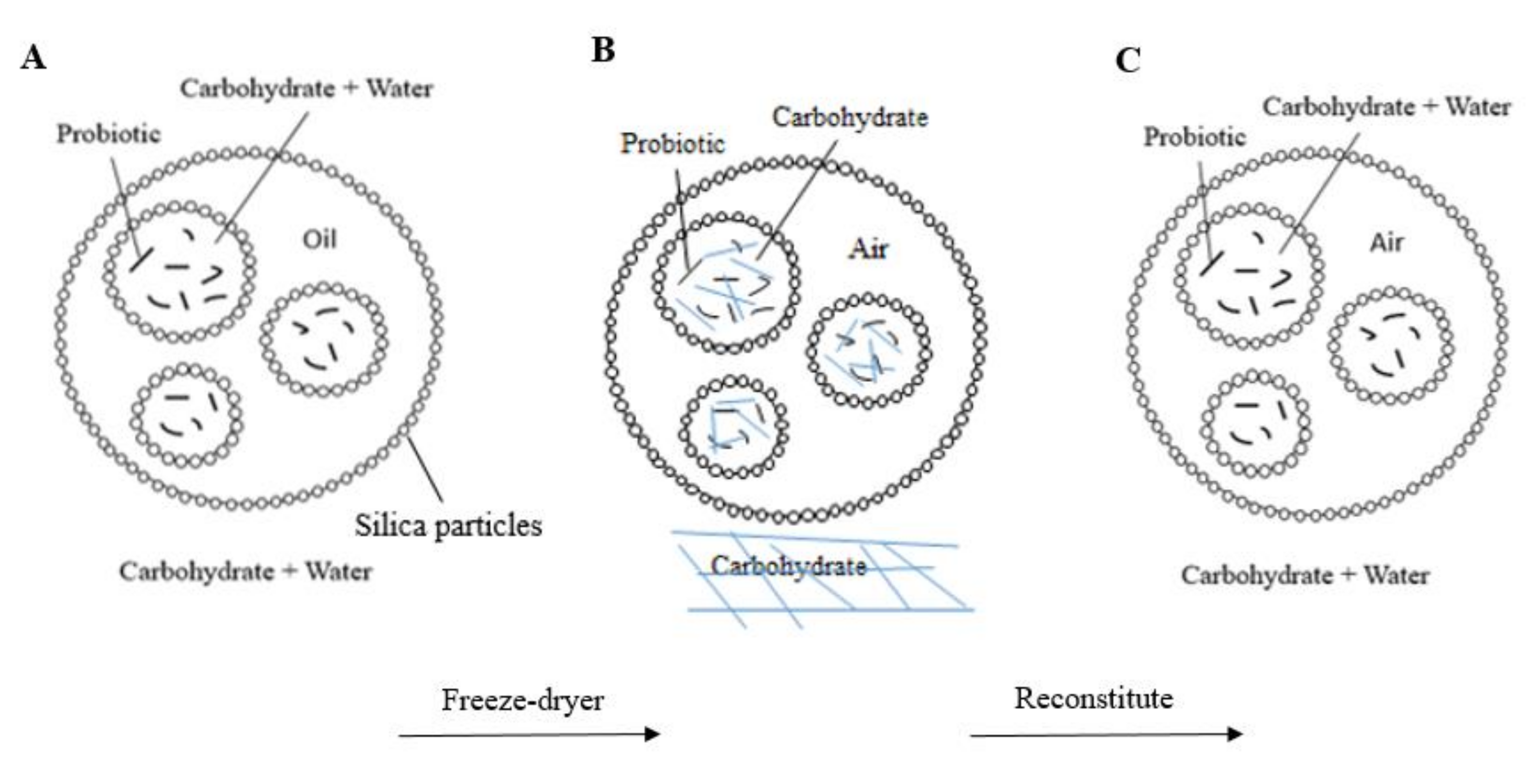
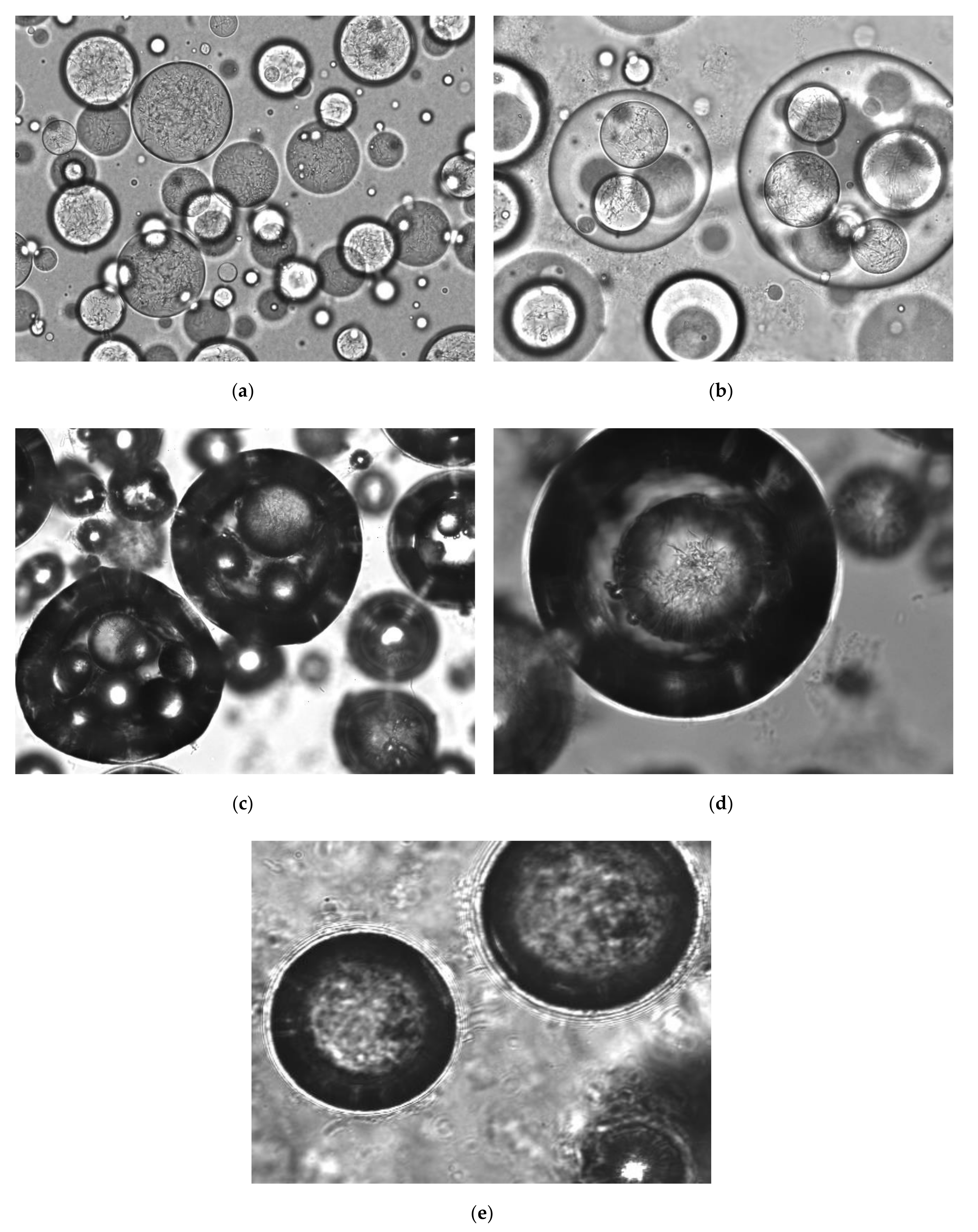
3.1. Antibubble Production

Antibubble-encapsulated bacteria were obtained by producing a W/O/W emulsion stabilized by hydrophobized silica particles and with L. casei incorporated in the inner water phase of the W/O/W emulsion. Both the water and the volatile oil were subsequently removed by freeze-drying, giving a powder that, upon reconstitution in water, formed antibubbles containing probiotic bacteria.
Preliminary experiments to determine the appropriate volatile oil, using ethyl acetate, ethyl butyrate, cyclohexane and decane, revealed that the use of ethyl acetate or ethyl butyrate did not allow the formation of Pickering-stabilized W/O/W emulsions. The relatively high polarity of these compounds probably requires particles with a different hydrophobicity to stabilize the interface between the oil and water phases [16]. On the other hand, cyclohexane and decane allowed for the preparation of Pickering-stabilized W/O/W emulsions and were thus selected to produce four types of antibubbles, which were obtained by combining cyclohexane or decane with maltodextrin, either alone or with additional sucrose, in the inner and outer water phase (Table 1).
The structure of the antibubbles in a stepwise production using decane and 10% maltodextrin + 8% sucrose, visualized by phase-contrast microscopical imaging, is shown in Figure 2. Images were also obtained for the other formulations, but these are not shown because no differences in structure were observed, probably because the presence or absence of sucrose and the use of a different oil had a small influence on the viscosity and hence on the emulsification process. In Figure 2a–d, bacteria can be observed inside the water droplets. Figure 2a shows the W/O emulsion obtained when emulsifying the bacterial suspension in the oil phase. Figure 2b shows the W/O/W emulsion obtained after emulsifying the W/O emulsion in the outer aqueous phase. The suspension of antibubbles obtained after freeze-drying the W/O/W of Figure 2b and reconstituting the subsequently obtained antibubble powder in an aqueous phase is shown in Figure 2c,d. A clear difference is observed between the droplets comprising oil (Figure 2b) and those in which the oil has been removed and replaced with gas (Figure 2c). The replacement of oil with gas increases the scattering of light, and the antibubbles are therefore darker than the double emulsion droplets. We used microscopic images to determine the size distribution of the antibubbles and found a unimodal distribution with a number-averaged size of 83 ± 33 μm. Furthermore, it can be seen that the antibubbles have the same morphology as the double emulsion droplets from which they originate, i.e., the freeze-drying process has little influence on the morphology of the structures. It is important to note that the droplets of the bacterial suspension in Figure 2c are separated from the outer aqueous phase by a gas phase. There is no aqueous connection between these inner droplets and the outer phase, as this would lead to an almost instantaneous coalescence of the droplets with the outer phase, as was shown in previous work [11]. Inside the inner droplets, a Brownian motion of the bacteria could be observed. This leads us to believe that the inner droplets are in fact droplets, i.e., they are liquid. We expect that during reconstitution of the antibubble powder the dry cores inside the antibubbles quickly rehydrate, since water vapor can pass through the gas shell, and that this makes the cores liquid again.

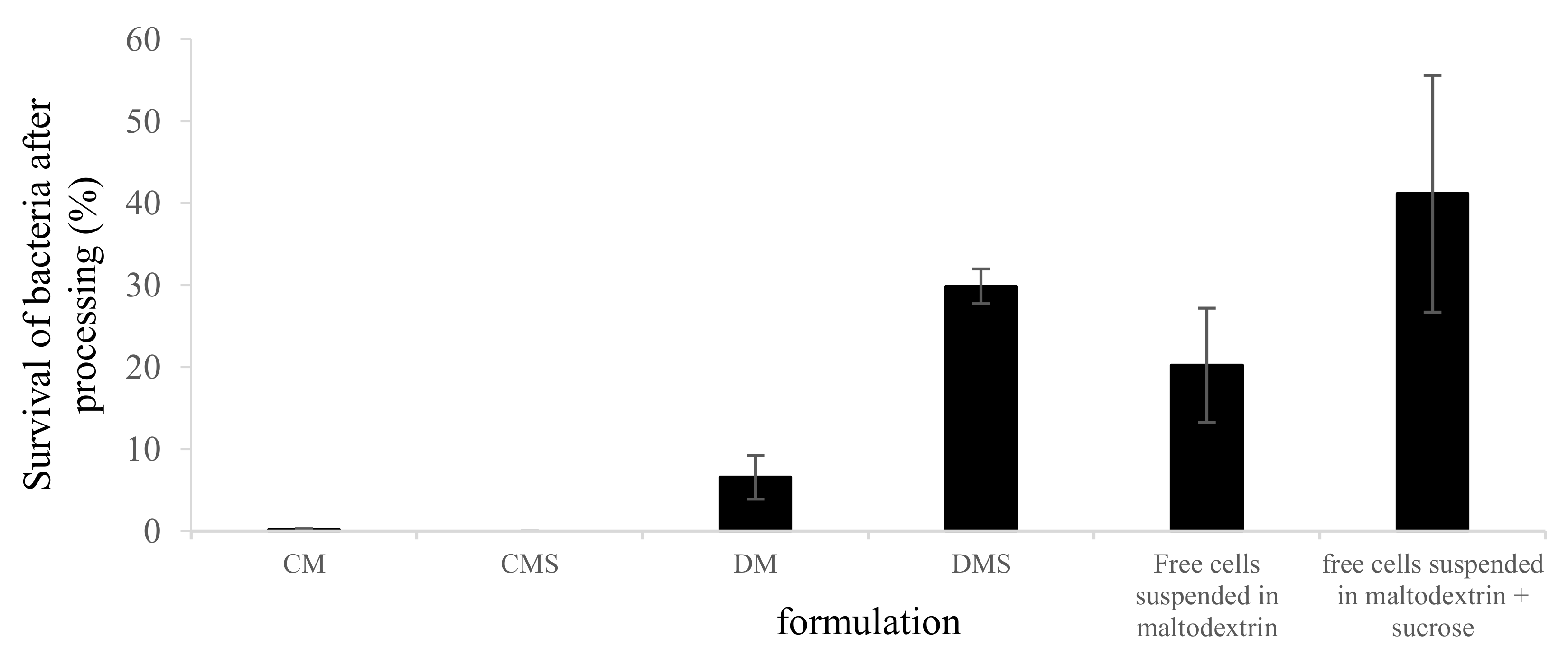
3.2. Survival of L. casei after Freeze-Drying and after Encapsulation inside Antibubbles

The survival of L. casei after encapsulation was significantly different (p-value < 0.001) for DM antibubbles (6.5 ± 2.6% survival) and DMS antibubbles (29.8 ± 2.1%), as shown in Figure 3. On the other hand, when cyclohexane was used as the oil phase, no viable bacteria were found in the resulting antibubbles. This can be explained by the fact that decane has an octanol-water partitioning coefficient log P = 5.6, whereas for cyclohexane, log P = 3.4. Solvents with a log P > 5 are considered relatively nontoxic to bacteria [17]. For comparison, we also determined the survival of unencapsulated bacteria during freeze-drying. The survival of the freeze-dried unencapsulated bacteria resuspended in 10% maltodextrin or 10% maltodextrin + 8% sucrose was 20.2 ± 6.9% and 41.2 ± 14.4% (difference not significant), respectively (Figure 3). Encapsulation thus led to a survival that was 13.7 and 11.4% lower for the formulations with maltodextrin (significant difference with p = 0.02) and maltodextrin + sucrose (difference not significant), respectively, than when the bacteria were not encapsulated but only freeze-dried. The decreased survival can be explained in view of the fact that, during the encapsulation process during the formation of the W/O emulsion, (part of) the bacteria come in contact with the oil, which might interfere with the bacterial cell membrane integrity when combined with the high shear forces in the emulsification process using a Turrax mixer [18]. Even though decane is relatively nontoxic, this may still decrease the survival somewhat. Although encapsulation lowered the survival when compared to freeze-drying alone, this decrease in survival was considerably smaller than the decrease in survival resulting from freeze-drying.
It is well-known that, in general, only part of the bacteria survives freeze-drying. The survival degree that we observed after freeze-drying was comparable to the survival found in the literature for the same strain [19,20]. The removal of both unbound and bound (hydrogen-bonded) water can occur during freeze-drying and cause damage to bacteria [19]. Applying proteins and sugars as an additive can counteract these negative effects. Previously, Miao et al. (2008) reported that disaccharides replace the hydrogen bonds through interactions with the polar sites of the cell membrane and cellular proteins, stabilizing their physical state and structures [21]. This can explain why, in our work, a mixture of maltodextrin and sucrose resulted in a higher recovery of bacteria, both after encapsulation and after freeze-drying alone, than when using only maltodextrin.
In literature reports, other encapsulation technologies also showed a reduction in the viability of the bacteria. L. plantarum showed a 37% and 16% survival during encapsulation within a skim milk-inulin-sodium alginate and an inulin-sodium alginate matrix, respectively [22]. Yao et al. (2018) showed that the microgel preparation procedure for the encapsulation of Pediococcus pentosaceus Li05 reduced the cell viability to 22% [3]. The survival of bacteria found in these studies is comparable with the survival of bacteria found for the antibubble encapsulation process described in this paper. This indicates that antibubble encapsulation is a good alternative technology in terms of the survival of bacteria during the encapsulation process.
The entrapment efficiency was measured for the antibubble variant DMS and was found to be 88.7 ± 2.8%. This means that the great majority of the bacteria in this formulation are actually entrapped within the antibubbles.

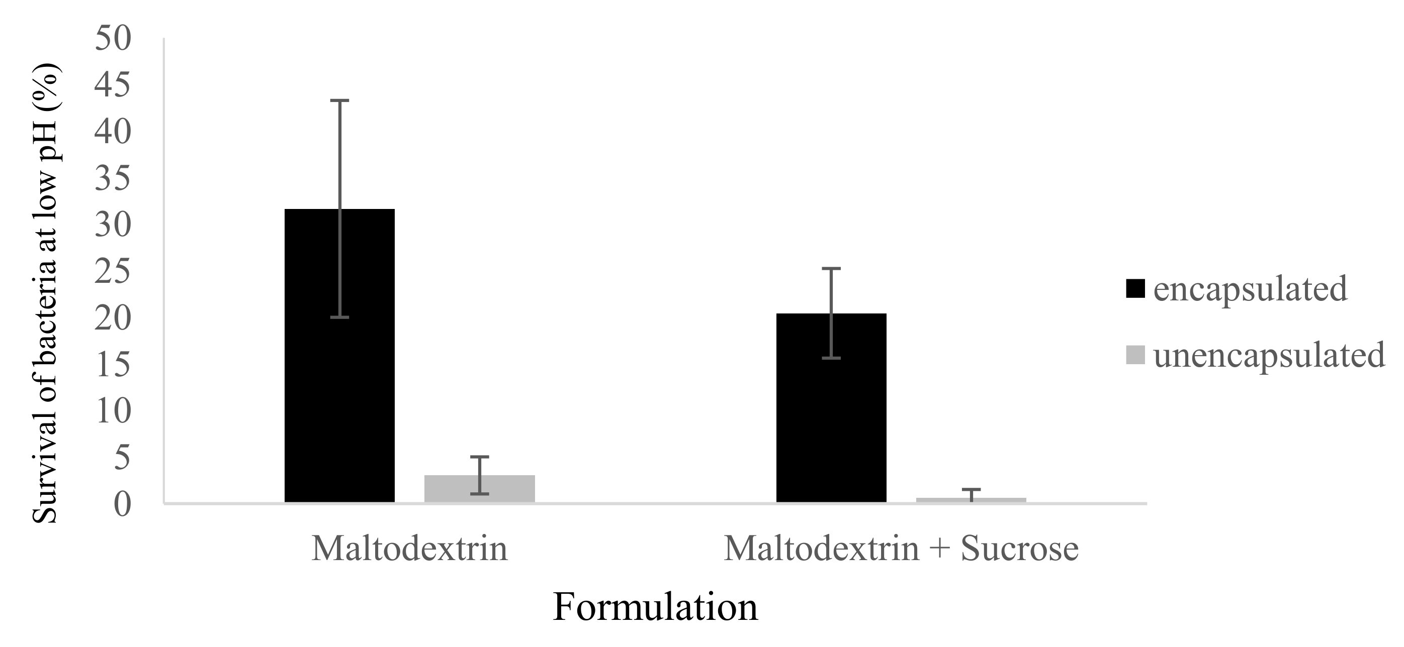
3.3. Survival of Unencapsulated Versus Encapsulated L. casei at Low pH

An important factor in the application of probiotics is their survival at a low pH. The survival of bacteria at a low pH is limited because of the energy that the bacteria need to spend to maintain a neutral intracellular pH, thus preventing vital metabolic reactions from taking place [23]. However, probiotics should resist the stressful conditions of the stomach to be able to exert beneficial effects. Therefore, one of the most important goals of encapsulation is to enhance the low pH tolerance of probiotics. The pH of gastric juices is about 1.5–3 [24]. To determine the protective effect of antibubbles for L. casei against acidic gastric conditions, the survival of unencapsulated versus encapsulated bacteria after 60 min at pH 2 was investigated. This time was taken as an indication of the in vivo residence time in the stomach. As encapsulated formulations, only the formulations DM and DMS were included, since the formulations CM and CMS were found (Section 3.2) to contain a very low number of living bacteria. An analysis by microscopy of the antibubbles did not show severe damage to the antibubbles at the end of the low pH incubation period (date not shown). Significant differences were found in the survival of unencapsulated bacteria and encapsulated bacteria. The survival of the bacteria after exposure to a low pH was 31.6 ± 11.6% (0.5 log reduction) and 20.4 ± 4.9% (0.7 log reduction) of the initial population found in the DM and DMS antibubbles (difference not significant), respectively (Figure 4). In contrast, there was a huge loss of unencapsulated bacteria at pH 2. Only 3.0 ± 2.0% (1.5 log reduction) and 0.6 ± 0.6% (2.2 log reduction) of unencapsulated bacteria suspended in 10% maltodextrin and 10% maltodextrin + 8% sucrose, respectively, survived exposure to pH 2 solutions. Sanchez et al. (2017) reported a continuous decrease in viable bacteria, reaching 0 CFU/g for unencapsulated Lactobacillus bacteria after 60 min in an acidic environment [25]. This is in agreement with our survival results for unencapsulated bacteria.
Our results show that antibubbles protect bacteria against exposure to low pH for at least one hour. The difference in survival between encapsulated and unencapsulated bacteria is significant, both for the formulations with only maltodextrin (p = 0.03) and for the formulations with maltodextrin and sucrose (p = 0.02). However, even for the encapsulated bacteria, the observed survival is lower than 100%. To better understand the protective effect of the gas shell against low pH, we performed a separate experiment in which we determined the survival of bacteria after 1 h in a 10% maltodextrin solution, i.e., the inner water phase encapsulated inside the antibubbles. This survival was found to be 43.6%. This survival is within the range of the 31.6 ± 11.6% survival found for bacteria in maltodextrin solution encapsulated within antibubbles. This suggests that the fact that the survival at a low pH is lower than 100% is not the result of the low pH of the solution in which the antibubbles are dispersed but rather an effect of an incomplete survival of the bacteria in the inner water phase. In the future, in order to further enhance survival, the composition of the inner water phase could be optimized to better sustain the viability of the encapsulated bacteria.
It is worth comparing the protective effect against a low pH that we found for antibubbles with the protective effect of other encapsulation methods. Heidebach et al. (2009) encapsulated Lactobacilli in hydrogel beads made of crosslinked caseinate with a size of around 200 micron and observed a survival of less than 1% after one hour at pH 2.5 at 37 °C [26]. Chandramouli et al. (2004) encapsulated Lactobacilli in 200-micron alginate beads and found that 1% survived after one hour at pH 2 at 37 °C [27]. Mandal et al. (2006) found that less than 5% of L. casei encapsulated in mm-sized alginate beads survived after a one-hour incubation at pH 1.5 at 37 °C [28]. Although more work needs to be done, e.g., incubation tests at 37 °C, we believe that the protective effect of antibubbles against a low pH reported here is also promising when compared with the results obtained using conventional methods such as encapsulation inside hydrogel beads.
Finally, we observed that adding a relevant concentration of bile salt to an antibubble suspension quickly led to the coalescence of the inner water droplets contained by the antibubbles with the aqueous phase that surrounded the antibubbles, i.e., the cores of the antibubbles were released: comparing Figure 2e (after adding bile) with Figure 2c showed that the droplets of the inner water phase (that contained the bacteria) present inside the bubbles in Figure 2c had disappeared. This is attributed to the fact that bile salt is a surfactant that changes the wetting conditions of the hydrophobized fumed silica, leading to the destabilization of the air shell and concomitant release of the inner droplets. Such a release, triggered by a change in wetting conditions, has previously been described by Poortinga (2013) [11] and Yohe et al. (2012) [29]. Further steps should be taken to more closely mimic the in vivo situation, e.g., by adding gastric and intestinal enzymes and performing the incubation at 37 °C. However, we feel that our finding that probiotic bacteria were released in the presence of a relevant concentration of bile salt is a clear indication that antibubble-encapsulated probiotics will be released along the intestinal tract, which is crucial for the probiotics to be functional.

4. Conclusions and Outlook

We have shown that L. casei can be encapsulated by Pickering-stabilized antibubbles. The survival of L. casei during the encapsulation process was comparable to the survival of L. casei during freeze-drying, provided that decane was used as the volatile oil during the encapsulation process. Notably, encapsulation within antibubbles improved the viability of the bacteria after a 1 h incubation at a low pH, demonstrating that this encapsulation approach may find application, e.g., to increase the survival of probiotic bacteria along the digestive tract or to maintain the viability of probiotic bacteria in acidic foods. We also observed that bile salts triggered the release of the encapsulated bacteria, i.e., it is to be expected that bacteria will be released in the intestines, and in this way they can induce their health benefits. To prove this, incubation tests need to be done under conditions that more closely resemble in vivo conditions. Since bile salt can also decrease the viability of probiotics, a further improvement would be to produce antibubble-encapsulated probiotics that are released only in the large intestines. Additionally, it would be interesting to test the protective effect against low pH for incubation times lasting weeks, which would be representative of food uses like acidic dairy products or fruit juices. The stability of the antibubble powder obtained after freeze-drying has not yet been consistently tested in our current work, but since probiotics are generally supplied in a freeze-dried form, this stability is expected to be sufficient. The antibubbles consisted of maltodextrin, hydrophobized silica and gas (air), and were produced using a volatile oil (in our case decane) as a processing aid, i.e., the oil is used during production but is then removed, so that the final product should be nearly free of the oil. Maltodextrin is a common ingredient in food and pharmaceutical products. The used hydrophobized silica is allowed for use in oral pharmaceuticals. The use of alkanes in the production of oral pharmaceutical products is allowed, provided that the residual amount of alkanes is below the limits given by the European Pharmacopeia. Therefore, we expect that the antibubbles described here may be used in the formulation of pharmaceutical probiotic products. To allow use in food, it may help to replace the hydrophobized silica with hydrophobic particles that are more commonly used in food. This should be feasible based on the work already done on food-grade Pickering stabilization [30]. Furthermore, although trace amounts of decane are naturally present in foods like oregano and licorice, further investigations should be conducted on which volatile oil can best be used as a processing aid to produce a food-grade formulation. In conclusion, we believe that, although more work is still needed before a practical implementation is possible, we have presented a potentially very versatile new method for the encapsulation of probiotics.

Author Contributions

Conceptualization, A.T.P.; methodology, V.M.G., P.P.P., G.-J.W.E. and A.T.P.; formal analysis, V.M.G., P.P.P., G.-J.W.E. and A.T.P.; investigation and writing—original draft preparation, V.M.G. and A.T.P.; writing—review and editing, V.M.G., P.P.P., G.-J.W.E. and A.T.P. All authors have read and agreed to the published version of the manuscript.

Funding

This research received no external funding.

Conflicts of Interest

A.T.P. started a company to commercialize the antibubble encapsulation method.

References

  1. Nagpal, R.; Kumar, A.; Kumar, M.; Behare, P.; Jain, S.; Yadav, H. Probiotics, their health benefits and applications for developing healthier foods: A review. FEMS Microbiol. Lett. 2012, 334, 1–15. [Google Scholar] [CrossRef] [PubMed] [Green Version]
  2. Gomes, A.M.P.; Malcata, F.X. Bifidobacterium spp. and Lactobacillus acidophilus: Biological, biochemical, technological and therapeutical properties relevant for use as probiotics. Trends Food Sci. Technol. 1991, 10, 139–157. [Google Scholar] [CrossRef]
  3. Yao, M.; Li, B.; Ye, H.; Huang, W.; Luo, Q.; Xiao, H.; Mcclements, D.J.; Li, L. Enhanced viability of probiotics (Pediococcus pentosaceus Li05) by encapsulation in microgels doped with inorganic nanoparticles. Food Hydrocoll. 2018, 83, 246–252. [Google Scholar] [CrossRef]
  4. Mortazavian, A.M.; Razavi, S.H.; Ehsani, M.R.; Sohrabvandi, S. Principles and methods of microencapsulation of probiotic microorganisms. Iran. J. Biotechnoly 2007, 5, 1–18. [Google Scholar]
  5. Burgain, J.; Gaiani, C.; Linder, M.; Scher, J. Encapsulation of probiotic living bacteria: From laboratory scale to industrial applications. J. Food Eng. 2011, 104, 467–483. [Google Scholar] [CrossRef]
  6. Heidebach, T.; Först, P.; Kulozik, U. Microencapsulation of probiotic bacteria for food applications. Crit. Rev. Food Sci. Nutr. 2012, 52, 291–311. [Google Scholar] [CrossRef]
  7. Dimitrellou, D.; Kandylis, P.; Petrovic, T.; Dimitrijevicbrankovic, S.; Levic, S.; Nedovic, V.; Kourkoutas, Y. Survival of spray dried microencapsulated Lactobacillus casei ATCC 393 in simulated gastrointestinal conditions and fermented milk. LWT Food Sci. Technol. 2016, 71, 169–174. [Google Scholar] [CrossRef]
  8. Anal, A.K.; Singh, H. Recent advances in microencapsulation of probiotics for industrial applications and targeted delivery. Trends Food Sci. Technol. 2007, 18, 240–251. [Google Scholar] [CrossRef]
  9. Silpe, J.E.; Nunes, J.K.; Poortinga, A.T.; Stone, H.A. Generation of Antibubbles from Core–Shell Double Emulsion Templates Produced by Microfluidics. Langmuir 2013, 29, 8782–8787. [Google Scholar] [CrossRef]
  10. Vitry, Y.; Dorbolo, S.; Vermant, J.; Scheid, B. Controlling the lifetime of antibubbles. Adv. Colloid Interface Sci. 2019, 270, 73–86. [Google Scholar] [CrossRef] [Green Version]
  11. Poortinga, A.T. Micron-sized antibubbles with tunable stability. Colloids Surf. A 2013, 419, 15–20. [Google Scholar] [CrossRef]
  12. Poortinga, A.T. Long-Lived Antibubbles: Stable Antibubbles through Pickering Stabilization. Langmuir 2011, 27, 2138–2141. [Google Scholar] [CrossRef] [PubMed]
  13. Chevalier, Y.; Bolzinger, M.A. Emulsions stabilized with solid nanoparticles: Pickering emulsions. Colloids Surf. A 2013, 439, 23–34. [Google Scholar] [CrossRef]
  14. Marefati, A.; Bertranda, M.; Sjööa, M.; Dejmek, P.; Rayner, M. Storage and digestion stability of encapsulated curcumin in emulsions based on starch granule Pickering stabilization. Food Hydrocoll. 2017, 63, 309–320. [Google Scholar] [CrossRef]
  15. Villota, R.; Hawkes, J.G.; Cockrane, H. Food applications and the toxicological and nutritional implications of amorphous silicon dioxide. Crit. Rev. Food Sci. 1986, 23, 289–321. [Google Scholar] [CrossRef]
  16. Binks, B.P.; Clint, J.H. Solid wettability from surface energy components: Relevance to Pickering emulsions. Langmuir 2002, 18, 1270–1273. [Google Scholar] [CrossRef]
  17. Heipieper, H.J.; Weber, F.J.; Sikkema, J.; Keweloh, H.; de Bont, J.A.M. Mechanisms behind resistance of whole bacteria to toxic organic solvents. Trends Biotechnol. 1994, 12, 409–415. [Google Scholar] [CrossRef]
  18. Capela, P.; Hay, T.K.C.; Shah, N.P. Effect of homogenization on bead size and survival of encapsulated probiotic bacteria. Food Res. Int. 2007, 40, 1261–1269. [Google Scholar] [CrossRef]
  19. Dimitrellou, D.; Kandylis, P.; Kourkoutas, Y. Effect of cooling rate, freeze-drying, and storage on survival of free and immobilized Lactobacillus casei ATCC 393. LWT Food Sci. Technol. 2016, 69, 468–473. [Google Scholar] [CrossRef]
  20. Xu, M.; Gagné-Bourque, F.; Dumont, M.J.; Jabaji, S. Encapsulation of Lactobacillus casei ATCC 393 bacteria and evaluation of their survival after freeze-drying, storage and under gastrointestinal conditions. J. Food Eng. 2016, 168, 52–59. [Google Scholar] [CrossRef]
  21. Miao, S.; Mills, S.; Stanton, C.; Fitzgerald, G.F.; Roos, Y.; Ross, R.P. Effect of disaccharides on survival during storage of freeze dried probiotics. Dairy Sci. Technol. 2008, 88, 19–30. [Google Scholar] [CrossRef] [Green Version]
  22. Wang, L.; Yu, X.; Xu, H.; Aguilar, Z.P.; Wei, H. Effect of skim milk coated inulin-alginate encapsulation beads on viability and gene expression of Lactobacillus plantarum during freeze-drying. LWT Food Sci. Technol. 2016, 68, 8–13. [Google Scholar] [CrossRef]
  23. Cotter, P.D.; Hill, C. Surviving the acid test: Responses of Gram-positive bacteria to low pH. Microbiol. Mol. Biol. Rev. 2003, 67, 429–453. [Google Scholar] [CrossRef] [PubMed] [Green Version]
  24. Kos, B.; Suskovic, J.; Goreta, J.; Matosic, S. Effect of protectors on the viability of Lactobacillus acidophilus M92 in simulated gastrointestinal conditions. Food Technol. Biotechnol. 2000, 38, 121–127. [Google Scholar]
  25. Sanchez, M.T.; Ruiz, M.A.; Lasserrot, A.; Hormigo, M.; Morales, M.E. An improved ionic gelation method to encapsulate Lactobacillus spp. bacteria: Protection, survival and stability study. Food Hydrocoll. 2017, 69, 67–75. [Google Scholar] [CrossRef]
  26. Heidebach, T.; Först, P.; Kulozik, U. Transglutaminase-induced caseinate gelation for the microencapsulation of probiotic bacteria. Int. Dairy J. 2009, 19, 77–84. [Google Scholar] [CrossRef]
  27. Chandramouli, V.; Kailasapathy, K.; Peiris, P.; Jones, M.R. An improved method of microencapsulation and its evaluation to protect Lactobacillus spp. in simulated gastric conditions. J. Microbiol. Methods 2004, 56, 27–35. [Google Scholar] [CrossRef]
  28. Mandal, S.; Puniya, A.K.; Singh, K. Effect of alginate concentrations on survival of microencapsulated Lactobacillus casei NCDC-298. Int. Dairy J. 2006, 16, 1190–1195. [Google Scholar] [CrossRef]
  29. Yohe, S.T.; Colson, Y.L.; Grinstaff, M.W. Superhydrophobic materials for tunable drug release: Using displacement of air to control delivery rates. J. Am. Chem. Soc. 2012, 134, 2016–2019. [Google Scholar] [CrossRef] [Green Version]
  30. Binks, B.P.; Muijlwijk, K.; Koman, H.; Poortinga, A.T. Food-grade Pickering stabilisation of foams by in situ hydrophobisation of calcium carbonate particles. Food Hydrocoll. 2017, 63, 585–592. [Google Scholar] [CrossRef]
Figure 1. Schematic representation of the structure and stepwise production of antibubble-encapsulated bacteria. (A): an oil droplet containing emulsified water droplets containing bacteria, as produced by two consecutive emulsification steps. This water-in-oil-in-water (W/O/W) emulsion is stabilized by silica particles adsorbed at the interfaces. (B): antibubble powder structure, obtained after freeze-drying the W/O/W emulsion. This removes the volatile oil from the W/O/W emulsion and converts the inner and outer aqueous phases of the W/O/W emulsion into a dry carbohydrate matrix. (C): antibubble structure obtained after reconstituting the antibubble powder in an aqueous phase. This leads to the rehydration of both the inner and outer carbohydrate matrices of the antibubble powder. The resulting structure consists of bubbles that contain droplets, with the droplets containing bacteria. One such bubble is shown. The blue dashes in the middle image indicate that these phases have become porous solid matrices due to freeze-drying. The arrows below the figure indicate the processing step that converts one structure into the other.
Colloids 04 00040 g001
Figure 2. Optical micrographs of the structure and stepwise production of the antibubbles using decane and 10% maltodextrin + 8% sucrose. (a) Pickering-stabilized W/O emulsion; (b) Pickering-stabilized W/O/W emulsion; (c) Antibubbles generated after freeze-drying the W/O/W emulsion and reconstituting the resulting material; (d) as (c) but with a higher magnification; (e) Antibubbles after the addition of bile salt. Scale bar represents 25 µm (panel d) or 50 µm (other panels).
Colloids 04 00040 g002
Figure 3. Survival of bacteria after processing. Processing consists of encapsulation (formulations CM, CMS, DM, DMS) or only freeze-drying (other two formulations). Values shown are means ± standards deviations.
Colloids 04 00040 g003
Figure 4. Survival of unencapsulated and encapsulated bacteria after 1 h at pH 2. Values shown are means ± standards deviations. Note that the encapsulated formulations indicated with ‘Maltodextrin’ and ‘Maltodextrin + Sucrose’ are also referred to as DM and DMS, respectively.
Colloids 04 00040 g004
Table 1. Compositions that allowed for the successful production of antibubbles. C: cyclohexane; D: decane; M: maltodextrin; S: sucrose.
AntibubbleOil PhaseInner Water Phase
CMCyclohexane10% Maltodextrin
CMS10% Maltodextrin + 8% Sucrose
DMDecane10% Maltodextrin
DMS10% Maltodextrin + 8% Sucrose

