Proceeding Paper

Modification of Glial Attachment by Surface Nanostructuring of SU-8 Thin Films †

1
MTA EK MFA MEMS Laboratory, H-1083 Budapest, Hungary
2
Faculty of Information Technology, Pázmány Péter Catholic University, H-1083 Budapest, Hungary
3
Institute of Biology, Department of Physiology and Neurobiology, Eötvös Loránd University, H-1053 Budapest, Hungary
4
Institute of Biology, Department of Anatomy, Cell and Developmental Biology, Eötvös Loránd University, H-1053 Budapest, Hungary
*
Author to whom correspondence should be addressed.
Presented at the Eurosensors 2018 Conference, Graz, Austria, 9–12 September 2018.
Proceedings 2018, 2(13), 1016; https://doi.org/10.3390/proceedings2131016
Published: 19 November 2018
(This article belongs to the Proceedings of EUROSENSORS 2018)

Abstract

:
Various methods are currently under development to enhance the biocompatibility of neural electrodes and to minimize the reactive gliosis around the implant surface. As cells in their native microenvironment interact with 3D nanoscale topographies of the extracellular matrix, physical modification of implant surfaces may provide an alternative solution to the negative tissue response by imitating the structure of the extracellular matrix, and therefore affecting the attachment and behavior of neurons and glial cells. The attachment of primary mouse astrocytes on nanostructured SU8 polymer surfaces fabricated by e-beam lithography was investigated in our study. We found that attachment of primary mouse astrocytes on silicon-SU8 surfaces is strongly influenced by the surface topography.

1. Introduction

The long-term application of implanted CNS sensors and stimulators is currently limited by the immuneresponse of the brain tissue, affecting both the performance of the device and the integrity of the neural network. Implants are required to limit the activation of astrocytes, which would ultimately lead to insulation from the surrounding tissue [1]. Topographical modification of implant surfaces may serve as a possible solution, since imitating the structure of the extracellular matrix has an influence on both attachment and behavior of neural cells [1,2].
Various groups showed the impact of surface topography on cell attachment and development: the fill factor of micropillars influences cell development [3]; cells sense the depth of micro gratings [4]; the distance between a micropattern and soma of a neuron affects the neurite outgrowth [5]. The neurites length, spreading, branching were studied to get more information about the interaction between the artificial surface and the cells [6].

2. Materials and Methods

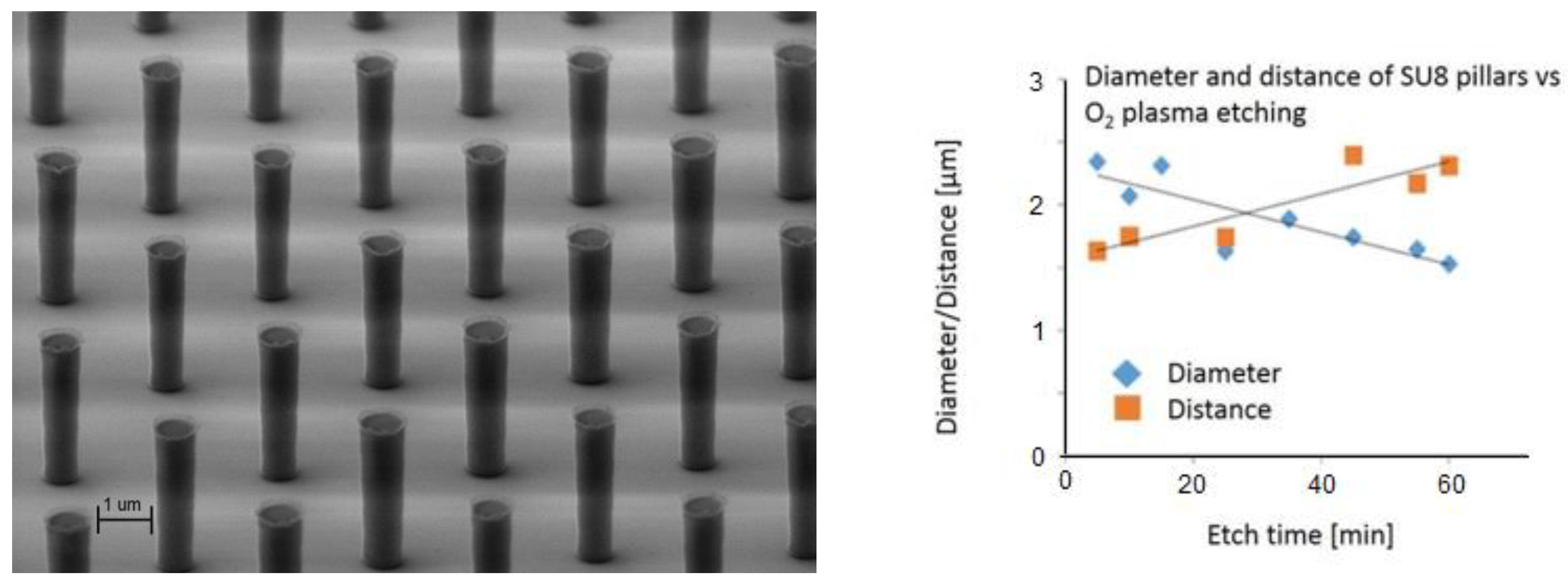
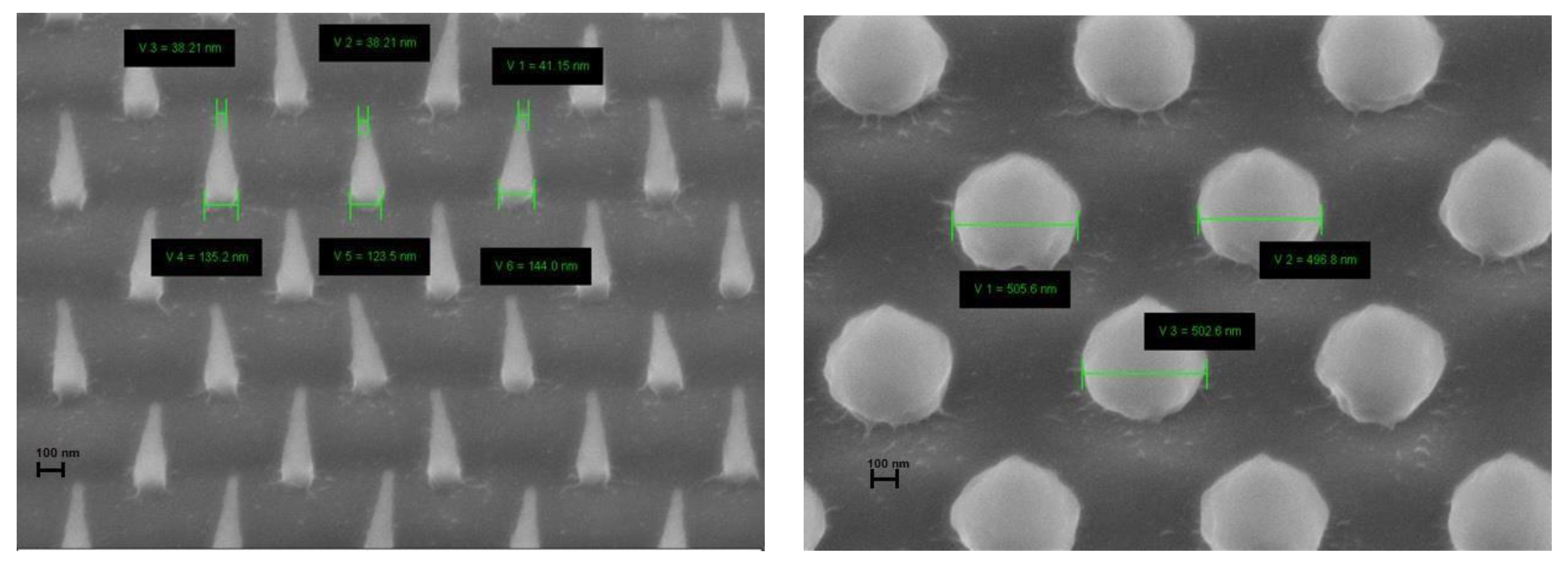
2.1. Surface Patterning

To create our surface pattern, we used the epoxy-based negative photoresist, SU-8. This material is an epoxy-based negative photoresist, which is as biocompatible as medical steel [7].
In our work, SU-8 structures were patterned by standard photolithography and electron beam lithography depending on the desired feature size. SU-8 2000.5 was used to form the 2 microns high micro- and nanostructures. The applied dose in the Raith 150 E-beam Litography system was 100 mJ/cm2. The post exposure bake was performed at 95 °C for 10 min.
The optimal irradiation depends on the sensitivity of the photoresist type, its thickness and the electron energy. These parameters were investigated preliminarily to form nanostructures with the desired geometry. To produce structures at the sub-micron scale, O2 plasma etching was used. Representative features are shown in Figure 1 and Figure 2.

2.2. Cell Culture

Primary astrocytes were prepared postnatally from 3-day-old mouse pups according to the method described in [8]. Cultures were maintained in HDMEM (Sigma) with 10% FCS (Gibco), 2 mM glutamine (Sigma, Kawasaki, Japan), 40 μg/mL gentamicin (Hungaropharma, Budapest, Hungary) and 2.5 μg/mL amphotericin B (Sigma-Aldrich Co. Ltd., Gillingham, United Kingdom). Cells were allowed to proliferate and passaged 1× with 0.05% trypsin – 0.02% EDTA (Sigma-Aldrich Co. Ltd., Gillingham, United Kingdom) before being seeded onto the test surfaces. Test surfaces were sterilized at 180 °C for 4 h then placed in 24-well culture plates. No additional surface treatment was applied. Astrocytes were seeded at starting densities of 210 cells/mm2. Cultures were kept at 37 °C in a 5% CO2 atmosphere and fixed after 24 or 48 h.

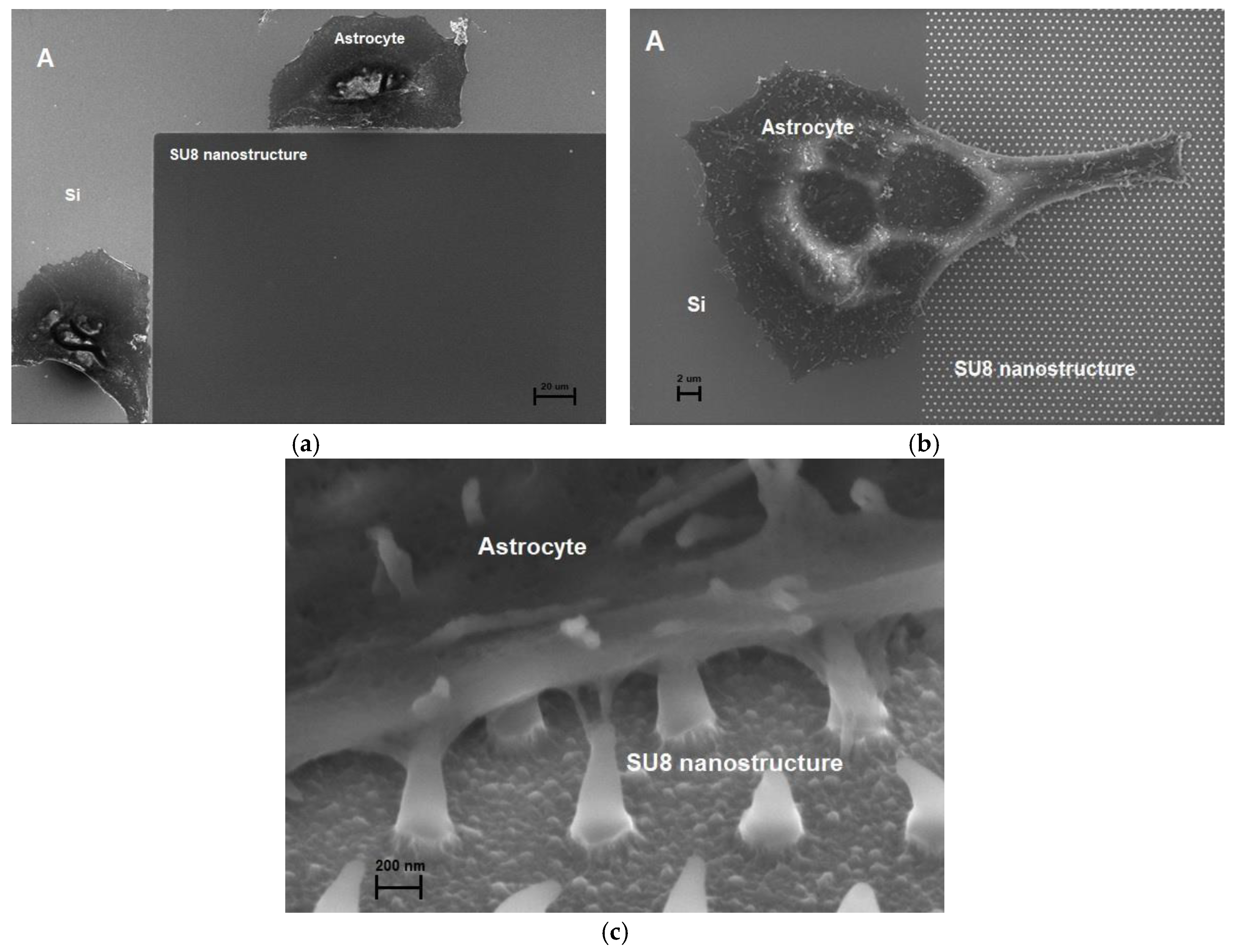
3. Results and Discussion

In vitro test chips were designed to investigate the cell-nanostructure interaction on different SU-8 surfaces using scanning electron microscopy. We found that shape and density of SU-8 nanostructures strongly affect the attachment of primary mouse astrocytes, opening up the way to control glial attachment by local nanoscale surface modification of polymer thin films. Representative results are shown in Figure 3.

Acknowledgments

The authors are grateful for the technical staff of MEMS Lab in MTA EK MFA. This work was supported by the Hungarian Scientific Research Fund (OTKA NN 116550), the Hungarian Brain Research Program (2017_1.2.1_NKP-2017-00002). A. Pongracz is thankful for the support of the Bolyai Research Fellowship. Zs. Bérces is thankful for the KAP 15-190-3.3-ITK grants. The support of the European Union through the grant EFOP-3.6.3-VEKOP-16-2017-00002 cofinanced by the European Social Fund is also acknowledged.

Conflicts of Interest

The authors declare no conflict of interest.

References

  1. Jorfi, M.; Skousen, J.L.; Weder, C.; Capadona, J.R. Progress towards biocompatible intracortical microelectrodes for neural interfacing applications. J. Neural Eng. 2014, 12, 011001. [Google Scholar] [CrossRef] [PubMed]
  2. Piret, G.; Perez, M.T.; Prinz, C.N. Support of neuronal growth over glial growth and guidance of optic nerve axons by vertical nanowire arrays. ACS Appl. Mater. Interfaces 2015, 7, 18944–18948. [Google Scholar] [CrossRef] [PubMed]
  3. Jeon, H.; Simon, C.G.; Kim, G. A mini-review: Cell response to microscale, nanoscale, and hierarchical patterning of surface structure. J. Biomed. Mater. Res. Part B Appl. Biomater. 2014, 102, 1580–1594. [Google Scholar] [CrossRef] [PubMed]
  4. Chua, J.S.; Chng, C.P.; Moe, A.A.K.; Tann, J.Y.; Goh, E.L.; Chiam, K.H.; Yim, E.K. Extending neurites sense the depth of the underlying topography during neuronal differentiation and contact guidance. Biomaterials 2014, 35, 7750–7761. [Google Scholar] [CrossRef]
  5. Mahoney, M.J.; Chen, R.R.; Tan, J.; Saltzman, W.M. The influence of microchannels on neurite growth and architecture. Biomaterials 2005, 26, 771–778. [Google Scholar] [CrossRef]
  6. Li, W.; Tang, Q.Y.; Jadhav, A.D.; Narang, A.; Qian, W.X.; Shi, P.; Pang, S.W. Largescale topographical screen for investigation of physical neural-guidance cues. Sci. Rep. 2015, 5, 8644. [Google Scholar] [CrossRef] [PubMed]
  7. Nemani, K.V.; Moodie, K.L.; Brennick, J.B.; Su, A.; Gimi, B. In vitro and in vivo evaluation of SU-8 biocompatibility. Mater. Sci. Eng. C 2013, 33, 4453–4459. [Google Scholar] [CrossRef] [PubMed]
  8. Tárnok, K.; Szilágyi, L.; Berki, T.; Németh, P.; Gráf, L.; Schlett, K. Anoxia leads to a rapid translocation of human trypsinogen 4 to the plasma membrane of cultured astrocytes. J. Neurochem. 2010, 115, 314–324. [Google Scholar] [CrossRef] [PubMed]
Figure 1. Representative microstructures (left) and relationship between the O2 plasma etching time and the feature size of the pillars (right).
Proceedings 02 01016 g001
Figure 2. Spiky (left) and rounded (right) SU-8 nanostructures fabricated using optimized e-beam lithography parameters. Scale bars represent 100 nm.
Proceedings 02 01016 g002
Figure 3. Primary mouse astrocytes avoid type A nanostructured SU-8 surfaces (a), while spread over type B nanostructured surfaces (b) and attach to spiky SU-8 nanopillars (c).
Proceedings 02 01016 g003
Publisher’s Note: MDPI stays neutral with regard to jurisdictional claims in published maps and institutional affiliations.

