Article

Combined Effects of Temperature and Salinity on Antioxidants in the Immune System of the Pearl Oyster Pinctada fucata

1
Sanya Tropical Fisheries Research Institute, Sanya 572018, China
2
Key Laboratory of Efficient Utilization and Processing of Marine Fishery Resources of Hainan Province, Sanya 572018, China
3
Tropical Aquaculture Research and Development Center, South China Sea Fisheries Research Institute, Chinese Academy of Fishery Sciences, Sanya 572018, China
4
Changdao Enhancement and Experiment Station, Chinese Academy of Fishery Sciences, Yantai 265800, China
*
Authors to whom correspondence should be addressed.
Fishes 2022, 7(5), 260; https://doi.org/10.3390/fishes7050260
Submission received: 23 August 2022 / Revised: 18 September 2022 / Accepted: 19 September 2022 / Published: 27 September 2022
(This article belongs to the Special Issue Mollusk Genetic Diversity and Breeding Technology)

Abstract

:
A water environment can impact many physiological processes of aquatic animals. The antioxidant response of immune system of the pearl oyster to temperature and salinity is of great significance to health. This study analyzed the physiological changes and immune responses under different temperature and salinity levels (temperature: 20, 25, and 30 °C; salinity: 23, 28, and 33‰) in the short term (7 d) in the hepatopancreas and other tissues of 405 pearl oysters. The combined effects of temperature and salinity on antioxidants in the immune system of the pearl oyster were evaluated via response surface methodology and Box–Benhnken design (BBD) under laboratory conditions. The secondary effects of salinity on the activities of glutathione peroxidase (GSH-PX), catalase (CAT), and superoxide dismutase (SOD) were significant, and CAT and SOD showed an inverted U-shaped trend with the increase in salinity. Temperature significantly impacted GSH-PX, CAT, glutathione (GSH), and SOD in primary and secondary effects, and the maximum values of CAT, GSH, and SOD were observed. The phenoloxidase (POX) and alkaline phosphatase (AKP) activities increased with the increasing temperatures. The interaction of temperature and salinity was significant on CAT and SOD, but was not significant on GSH-PX. The interaction between salinity and temperature on AKP was significant, which was contrary to the results of AKP in the hemolymph. The expression levels of antioxidant genes varied between tissues, and the expression levels of different genes in the same tissue were different. Appropriate immunity and antioxidant index models were established under the combined temperature and salinity conditions. The optimal combination of temperature and salinity was 24.95 °C and 28.11‰, respectively, and the desirability was 0.803. This study provides theoretical reference points for the pearl oyster to respond to temperature and salinity changes and can be used to establish an index model for shellfish aquaculture.

Graphical Abstract

1. Introduction

Antioxidants in the immune system are no doubt run through the whole process of ontogenetic organisms, in which they are exposed to changes in environmental factors. It is well known that aquatic livestock in their natural habitats undergo the simultaneous impacts of various environmental variables [1]. Among them, salinity and temperature are the most significant environmental factors affecting aquatic organisms, which can indirectly or directly regulate the rate of all bioprocesses, such as development, growth, survival, and reproduction [1,2,3]. Salinity and temperature have thus been described as a dominant “ecological principal factor” for many marine species [1]. However, most of the auto-biologic studies on bivalves have mainly concentrated on single effects rather than multiple environmental factors. For example, some studies by Japanese researchers were in the context of a single-factor influence of salinity or temperature on the pearl oyster [4,5,6,7,8]. Wang et al. [1] suggest that the interaction between single factors is critical, and single-factor experiments cannot provide information on interactive effects. Multiple factors may have posed for the mass mortalities of Japanese pearl oysters since the 1960s, and it has long been recognized that the influence of one factor can be modified by another factor [9]. Accordingly, a study of the combined effects of multiple environmental factors is necessary.
The Box–Benhnken design (BBD) was created byBox [10], and firstly applied byDavis [11], who investigated ovum development, growth, and larval survival of hard clam Mercenaria mercenaria and American oyster Crassostrea virginia. Such a technique makes multi-factor synergy possible under broader environmental conditions rather than through laboratory experiments, and its first application was under the combined effect of salinity and temperature factors on bivalve larvae [1]. Subsequently, the combined influences of the two factors have been investigated for numerous mollusks, such as European flat oyster Ostrea edulis [12], mussel Mytilus edulis [13], bivalve molluso Adula californiensis (Pelecypoda: Mytilidae) [14], brackish water clam Rangia cuneata [15], Chinese pearl oyster Pinctada fucata martensii [1], pearl oyster Pinctada imbricata Röding [9], coot clam Mulinia lateralis [16], Northern Bay scallop Argopecten irradians irradians [17], noble scallop Chlamys nobilis [18], Mediterranean mussel Mytilus galloprovincialis, and Japanese oyster Crassostrea gigas [19].
The pearl oyster is the main shellfish species for cultivating seawater pearls worldwide [1]. Since 1949, the pearl oyster has been cultivated in Guangdong, Guangxi, and Hainan Provinces of China until pearl production peaked in the 1990s. The yield of seawater pearl in China reached 6 tons in 2010, and the gross value was about USD 20 million. The pearl oyster has greatly promoted the development of the pearl industry in China and created considerable economic income for the nation [20]. However, massive mortalities of the pearl oyster are frequently reported and have caused considerable economic losses in the past 10 years. A series of studies have been conducted to understand the causes of such mortalities. Evidence has indicated that temperature, salinity, dissolved oxygen, and pH can change the physiological indexes of the pearl oyster, including cellular immune level, oxygen consumption rate, osmotic pressure (OSM), and ammonia excretion rate, as well as inhibit growth, disturb the balance of basal metabolism, and even lead to death [20,21,22,23]. All of the evidence reveals that massive mortality of the pearl oyster is closely related to these environmental changes. To date, most studies have focused on one environmental factor at a time, while the existing multi-factor studies have focused on the immunological expression [24], survival, growth [25], energy budget [26], fertilization, hatching [1], transcriptome, and biomineralization [27]. Therefore, it is necessary to explore in detail how salinity and temperature, in particular, jointly influence antioxidants in the immune system, and to determine the optimal combination of factors with practical significance. Undoubtedly, insight into such issues will be beneficial for maintaining the optimal living conditions of the pearl oyster, assuring their development, growth, and survival, thereby reducing economic losses and increasing production.
The objectives of this study were to (1) examine the synergistic effects of salinity and temperature on antioxidants in the immune system in the pearl oyster using response surface method and BBD, (2) model the relationship of antioxidants in the immune system with salinity and temperature, and (3) determine the optimal salinity–temperature combination using the statistical optimization technique. The results from the present study will provide a theoretical reference for shellfish culture with the variation of two environmental factors and the establishment of index models for P. pucata aquaculture.

2. Materials and Methods

2.1. Experimental Animals and Design

The pearl oysters with a similar size (body mass: 34.19 ± 1.39 g, shell length: 50.75 ± 1.43 mm) were collected from the South China Sea and then acclimated in the laboratory of Lingshui Station (Hainan, China) for 7 days. All of the pearl oysters were conditioned in a 5000 L cement tank at salinity of 33 ± 1‰ and 25 ± 1 °C prior to the experiment. During the acclimation, the ambient parameter remained pH 8.0 ± 0.1, ammonia nitrogen < 0.01 mg/L, DO > 6.5 mg/L, nitrite nitrogen < 0.02 mg/L, and light intensity < 500 Lx with a natural photoperiod. The oysters were fed with algae Platymonas subcordiformis (2 × 10⁵ cells/mL) once a day at regular intervals (9:00–9:30 a.m.). Half of the seawater was replaced, and feces and feed residues were siphoned off daily. Dead pearl oysters were immediately removed from the tank. The antioxidants (AKP, GSH-PX, MDA, SOD, CAT, GSH, POX, and GPX) in the immune system were assessed using a Box–Benhnken design (BBD) with either two factors or explanatory variables, salinity (S, ‰) and temperature (T, °C). Each factor contained three levels coded as −1, 0, and 1, respectively (Table 1). There were nine combined temperature–salinity treatments in the experiment, where the combination of 30 °C and 33‰ was set as the control, each treatment had three replicates, and each replicate contained 15 pearl oysters. The experimental design and results of biochemical indicators related to immunity and antioxidation and relative expression of antioxidant genes response surface are shown in Table 2 and Table 3, respectively.
Throughout the experiment, temperature ranged from 20 °C to 30 °C, and the minimum salinity was set at 23‰, with the maximum at 33‰. Preliminary trials, based upon the seasonal changes in temperature and salinity of the seawater in the South China Sea, were conducted to set the temperature and salinity ranges. A heater and ice block were used to maintain the temperature. All salinities were prepared by adding sea salts or tap water with 24 h aeration to the natural double-filtered seawater, and a salinometer (ATAGO S-10E) was used to measure salinity.

2.2. Collection and Determination of Hemolymph and Hepatopancreas

After the experiment, the biological samples were not anesthetized before collection. Three pearl oysters were randomly collected from each replicate. The hemolymph was extracted from the pericardium of each shellfish. The shell was opened, and the adductor muscle was cut off along the left shell. Then, hemolymph was extracted from the exposed heart cavity with a 2 mL syringe. Serum biochemical indexes were determined according to the kit instructions of the Nanjing Jiancheng Bioengineering Institute (Nanjing, China). The test variables included phenoloxidase content (POX, No. H247), alkaline phosphatase activity (AKP, No. A059-2-2), and the total protein content (TP, No. A045-4-4).
After the hemolymph was collected, the hepatopancreas was quickly cut off on an ice tray with scissors. After being rinsed with pre-cooling 0.9% normal saline and blotted with clean filter paper, the tissue samples were quickly placed in 2 mL centrifuge tubes and stored at −80 °C. The biochemical parameters in the hepatopancreas were determined according to the instructions of the manufacturer (Nanjing Jiancheng Institute of Biological Engineering, Nanjing, China), including AKP, glutathione peroxidase activity (GSH-PX, No. A005-1-2), malondialdehyde content (MDA, No. A003-1-2), total superoxide dismutase activity (SOD, No. A001-3-2), catalase activity (CAT, No. A007-1-1), reduced glutathione content (GSH, No. A006-2-1), and TP.

2.3. Primer Design and Gene Expression

The mantle, gonad, adductor muscle, and foot tissues of pearl oysters were collected and kept in RNAlater (minimize RNA damage) during storage to analyze the relative expression of antioxidant genes. According toWei et al. [28], Gu et al. [29], and Adzigbli et al. [30], primers and genes design were selected for qPCR analysis in this study (Table 4). RNA was extracted with Total RNA Isolation Reagent (Trizol, Biosharp BS259A) (Lianshimall.com, Beijing, China) and subsequently isolated in a chloroform layer, precipitated in isopropanol [31].
The obtained RNA pellet was washed in 1 mL 75% ethanol and air-dried before resuspension in RNase-free water. The concentration and integrity of isolated RNA were quantified and assessed using an ND 5000 spectrophotometer (BioTeke Corporation, Beijing, China) and 1% agarose gel (4S GelRed, 10,000X in water (Sangon Biotech, Order NO. A616697) was used for RNA staining on the agarose gel electrophoresis, respectively. Among them, high-quality RNA (total RNA > 10 μg OD260/230 ≥ 2.0, OD260/280 = 1.8~2.2, 28S:18S ≥ 1.0) was used for subsequent experiments; the criteria refer toFu et al. [32]. Immediately, RNA was reverse transcribed into cDNA (stored at −20 °C until further use) using cDNA Synthesis SuperMix and TransScript-Uni One-Step gDNARemoval (Beijing TransGen Biotech Co., Ltd., Beijing, China) according the manufacturer’s instructions [31].
The qPCR on the antioxidant-related genes was performed with real-time qPCR analysis (Hangzhou Longgene Scientific Instrument Co., Ltd., Hangzhou, China) using SYBR Green (Tiangen Biotech Co., Ltd., Beijing, China). Then, 20 μL of reaction including 10 μL 2 × RealUniversal PreMix, 0.6 μL of each primer (10 μM), and 2 μL of diluted cDNA was initially denatured at 95 °C for 15 min and then amplified for 40 cycles (95 °C, 10 s; 58 °C, 20 s; and 72 °C, 30 s) [31]. For each sample, the PCR reactions were performed in triplicate. At the end of each cycle, the melting curve analysis of the primers was performed to ensure only specific products were obtained with no formation of primer dimers. No template control was included with each assay to verify that PCR master mixes were free of contamination. The expression levels of antioxidant-related genes were normalized based on the level of housekeeping gene (β-actin) [31,33]. After verification of PCR efficiency of around 100%, the gene expression data were analyzed based on the 2△△CT method [34,35].

2.4. Statistical Analysis

The Box–Benhnken design (BBD) was used to accurately estimate the effects of salinity and temperature. The response surface of the fitted experimental data largely determines the relationship between the response and the quantitative correlation factor, so the three-dimensional response surface plot can reflect the relationship between salinity temperature and response [36,37]. Data analysis and image plotting were performed using DX10 (Design Expert 10 (32-bit)). Assume that the following models describe the response surface properties:
Y = β 0 + β 1 S + β 2 T + β 12 S × T + β 11 S 2 + β 22 T 2 + ε
In the formula, Y : the response of immune and antioxidant related indicators; β 0 : a constant; β 1 and β 2 : the linear effect of salinity and temperature, respectively; β 11 and β 22 : the secondary effect of salinity and temperature, respectively; β 12 : the combined effect of salinity and temperature; ε : the mean; and the random error was zero in line with the normal distribution.
The least square method was used to calculate the regression coefficients of these experimental points. The determination coefficient R2 of the above model represents the fitting situation, and the statistical significance was determined by F test. p < 0.05 of the items included in the above model was considered statistically significant or significant. The adequacy and importance of the established model can be proven by the generated variance analysis table. After constructing the model, the response was optimized by the optimization program of DX10 software, and the optimal condition set was obtained by transforming the minimum or maximum function [1]. The model equations of immune and antioxidant responses were further optimized by the method ofMontgomery [37].

3. Results

3.1. Model Significance Analysis

The regression equations of salinity and temperature on the antioxidant and physiological immune indexes of the pearl oyster (each coefficient was the actual value) established in the experiment were as follows:
AKP = −3.20 × 10⁶ + 27,693.38 × T + 2.08 × 10⁵ × S − 1284.8657 × T × S + 407.8005 × T2 − 3092.1995 × S2
GSH−PX = +199.79 − 5.07 × T − 5.18 × S + 6.82 × 10⁻³ × T × S + 0.10 × T2 + 0.08 × S2
MDA = +55.65 − 4.54 × T + 0.43 × S − 0.01 × T × S + 0.10 × T2 – 3.57 × 10⁻³ × S2
SOD = −19,159.66 + 2029.18 × T + 494.03 × S + 24.00 × T × S − 50.47 × T2 − 20.47 × S2
CAT = −56.51 + 1.30 × T + 4.58 × S + 0.04 × T × S − 0.06 × T2 −0.10 × S2
GSH = +4.47 + 1.48 × T − 1.31 × S + 0.01 × T × S − 0.04 × T2 + 0.02 × S2
AKP (hemolymph) = +6.86 − 0.12 × T − 0.36 × S − (3 × 10⁻⁴) × T × S + 2.61 × 10⁻³ × T2 + 6.51 × 10⁻³ × S2
POX = +4.81 − 0.06 × T − 0.07 × S + 5.90 × 10⁻⁵ × T × S + 1.22 × 10⁻³ × T2 + 1.26 × 10⁻³ × S2
The variance analysis of immune and antioxidant indexes in the hepatopancreas is presented in Table 5. The established models of salinity and temperature on the immune and antioxidant physiology of the pearl oyster were significant (p < 0.05), and the lack of fit was not significant (p > 0.05), indicating that the regression model was significant. The fitting between experimental data and the model was good, and the model was suitable. The experimental results caused by unknown factors had little interference. The R2 of the established model was 0.96, 0.93, 0.98, 0.97, 0.94, 0.95, 0.95, and 0.93, respectively; the AdjR2 was 0.94, 0.90, 0.98, 0.96, 0.92, 0.93, 0.93, and 0.89, respectively; and the PredR2 was 0.86, 0.72, 0.92, 0.91, 0.81, 0.84, 0.81, and 0.78, respectively, so the model was appropriate.
The regression equations of salinity and temperature on the relative expression indexes of antioxidant genes of the pearl oyster (each coefficient is the actual value) established in the experiment were as follows:
GPX-mantle = +2.99 + 0.27 × T − 0.48 × S + 1.92 × 10⁻³ × T × S − 6.50 × 10⁻³ × T2 + 8.40 × 10⁻³ × S2
SOD-mantle = +54.37 − 1.56 × T −2.32 × S + 0.02 × T × S + 0.02 × T2 + 0.03 × S2
GPX-adductor = −65.26 + 0.82 × T + 4.18 × S + 0.02 × T × S − 0.03 × T2 − 0.08 × S2
SOD-adductor = −4.43 − 0.48 × T + 1.20 × S + 0.04 × T × S − 0.01 × T2 −0.04 × S2
GPX-foot = +55.72 − 5.26 × T + 0.53 × S − 5.90 × 10⁻³ × T × S + 0.11 × T2 − 3.68 × 10⁻³ × S2
SOD-foot = +38.43 − 2.70 × T − 0.41 × S + 0.01 × T × S + 0.05 × T2 + 3.75 × 10⁻³ × S2
GPX-gonad = −34.50 + 1.80 × T + 0.92 × S − 1.86 × 10⁻³ × T × S −0.04 × T2 − 0.02 × S2
SOD-gonad = +56.02 − 1.72 × T −2.24 × S + 0.02 × T × S + 0.02 × T2 + 0.03 × S2
The variance analysis was performed on the relative expression of antioxidant genes in the mantle, gonad, foot, and adductor tissues (Table 6). The established models of the effects of salinity and temperature on the relative expression of antioxidant genes of the pearl oyster were significant (p < 0.05), and the lack of fit was not significant (p > 0.05), indicating that the regression model was significant, and the fitting between the experimental data and the model was good, the model was suitable, and the experimental results caused by unknown factors had little interference. The R2 of the established model was 0.94, 0.94, 0.99, 0.91, 0.95, 0.98, 0.96, and 0.97, respectively; the AdjR2 was 0.91, 0.92, 0.99, 0.87, 0.92, 0.97, 0.94, and 0.96, respectively; and the PredR2 was 0.75, 0.84, 0.97, 0.68, 0.82, 0.93, 0.87, and 0.92, respectively, indicating that the model was appropriate.

3.2. Model Coefficient Estimation

The established regression models of antioxidant and immune indexes were estimated by coefficients (Table 7 and Table 8). The coefficients in Table 7 and Table 8 were coded values (elimination of units among coefficients), and their effects were directly reflected by numerical values. The 95% confidence interval (C.I.) explained the change in coefficient coding value in the 95% interval.
Table 7 shows that the minimum values of 95% C.I. prediction of antioxidant and immune indexes intercept were 2.32 × 10⁵, 61.16, 2.14, 14,421.50, 18.62, 5.84, 0.29, and 3.04, respectively, and the maximum values were 2.59 × 10⁵, 62.45, 2.52, 14,789.03, 19.76, 6.19, 0.33, and 3.05, respectively.
The test and analysis results of the model coefficients showed that the secondary effect of temperature had no significant effect on AKP (p > 0.05), while the rest of the effects were significant (p < 0.05). The interaction of salinity and temperature had no significant effect on GSH-PX and AKP (hemolymph) (p > 0.05), while the rest of the effects were significant (p < 0.05). The secondary effect of salinity had no significant effect on MDA (p > 0.05), while the rest of the effects were significant (p < 0.05). The primary, secondary, and interactive effects of salinity and temperature had significant effects on SOD and GSH (p < 0.05). The primary effect of salinity had no significant effect on CAT (p > 0.05), while the rest of the effects were significant (p < 0.05). (p < 0.05). The primary and interaction effects of salinity and temperature had no significant effect on POX (p > 0.05), while the secondary effect of salinity and temperature had a significant effect on POX (p < 0.05).
Table 8 shows that the minimum values predicted by the 95% C.I. of the intercept of antioxidant gene expression indexes were 0.15, 0.28, 3.39, 2.20, 0.18, 0.19, 1.17, and 0.28, respectively, and the maximum values were 0.26, 0.61, 3.61, 2.87, 0.97, 0.42, 1.39, and 0.55, respectively. The test results of the model coefficients showed that the primary effect of temperature and the interaction of salinity and temperature had no significant effect on the relative expression of the GPX gene in the mantle (p > 0.05), while the rest of the effects were significant (p < 0.05). The primary, secondary, and interactive effects of salinity and temperature on the relative expression of the SOD gene in the mantle, GPX gene in adductor muscle, and SOD gene in gonad were significant (p < 0.05). The secondary effect of temperature had no significant effect on the relative expression of the SOD gene in adductor muscle (p > 0.05), while the rest of the effects were significant (p < 0.05). The primary effect of temperature, the secondary effect of salinity, and their interaction had no significant effect on the relative expression of the GPX gene in the foot (p > 0.05), while the rest of the effects were significant (p < 0.05). The secondary effect of salinity had no significant effect on the relative expression of the SOD gene in the foot (p > 0.05), while the rest of the effects were significant (p < 0.05). The primary effect of temperature and its interaction with salinity had no significant effect on the relative expression of the gonad GPX gene (p > 0.05), while the rest of the effects were significant (p < 0.05).

3.3. Response Surface Analysis of Antioxidants in the Immune System

Under all different combinations of salinity and temperature, the antioxidant and immune-related (non-) enzyme activities (contents) estimated according to the fitted response surface are shown in Figure 1. By examining the response surface or contour, the activities or contents of GSH-PX, SOD, CAT, AKP (hemolymph), and POX were unimodal. Among them, GSH-PX, AKP (hemolymph) and POX decreased firstly and then increased with the increase in salinity or temperature, while SOD and CAT increased firstly and then decreased with the increase in salinity or temperature.
Under all different combinations of salinity and temperature, the relative expression of antioxidant genes in each tissue estimated by the fitted response surface is shown in Figure 2. By examining the response surface or contour, the expression of the SOD gene in mantle, GPX gene in adductor muscle, and GPX and SOD gene in the gonad were single peaks. Among them, the expression levels of SOD genes in the mantle and gonad showed a U-shaped trend with the increase in temperature or salinity, and the expression levels of GPX genes in the obturator muscle and gonad showed an inverted U-shaped trend with the increase in temperature or salinity.

3.4. Optimization

After the model equation was established, the optimization program of Design Expert 10 software was used to optimize and analyze several responses. The results showed that the optimal combination of temperature and salinity was 24.95 ℃/28.11‰, and the values of AKP, GSH-PX, MDA, SOD, CAT, GSH, AKP (hemolymph), and POX and the relative expression of GPX and SOD genes in the mantle, adductor muscle, foot, and gonad were 245,047.90, 61.77, 2.32, 14,589.85, 19.22, 6.03, 0.31, 3.05, 0.21, 0.44, 3.49, 2.53, 0.59, 0.31, 1.28, and 0.41, respectively. These values are the same as the response values in the temperature and salinity in Figure 1 and Figure 2. The desirability was 0.803.

4. Discussion

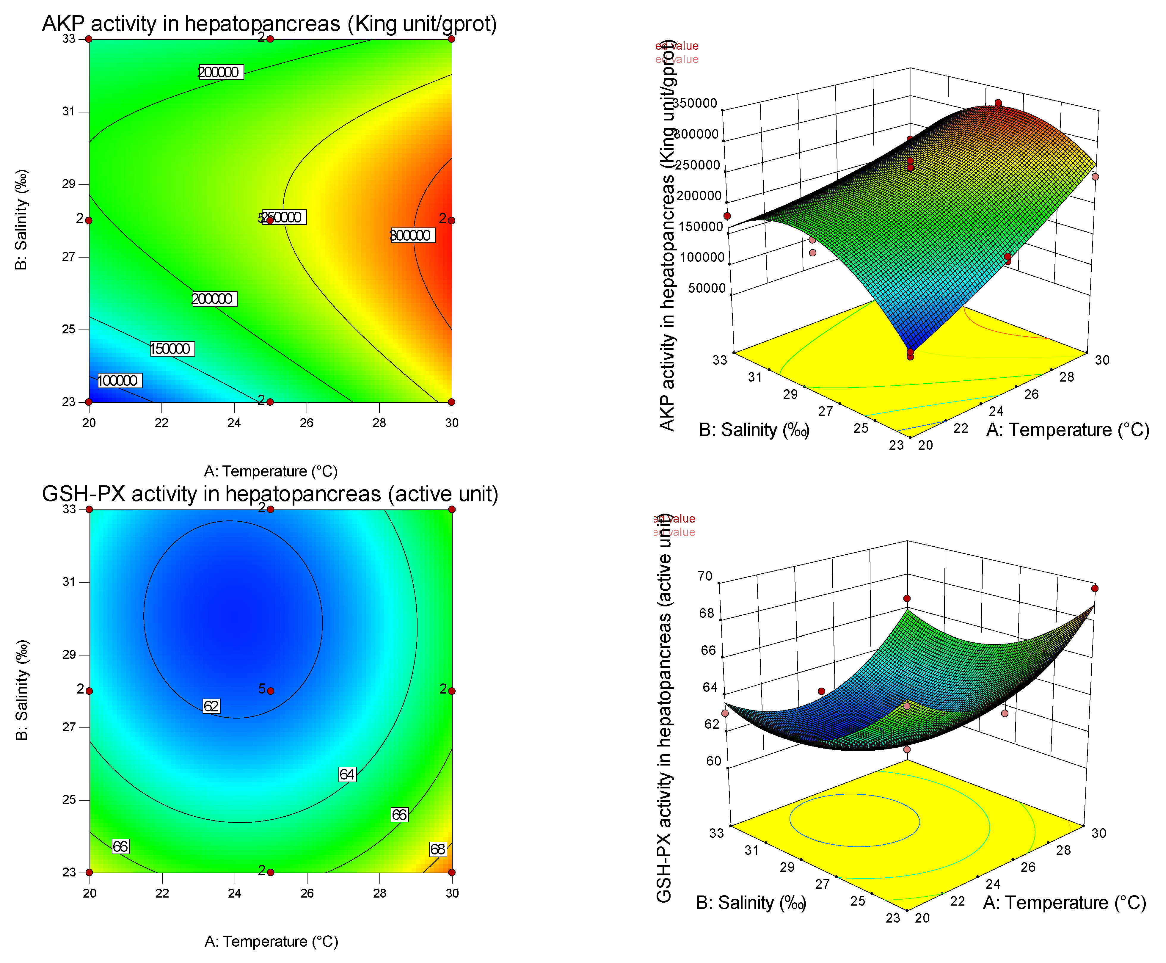
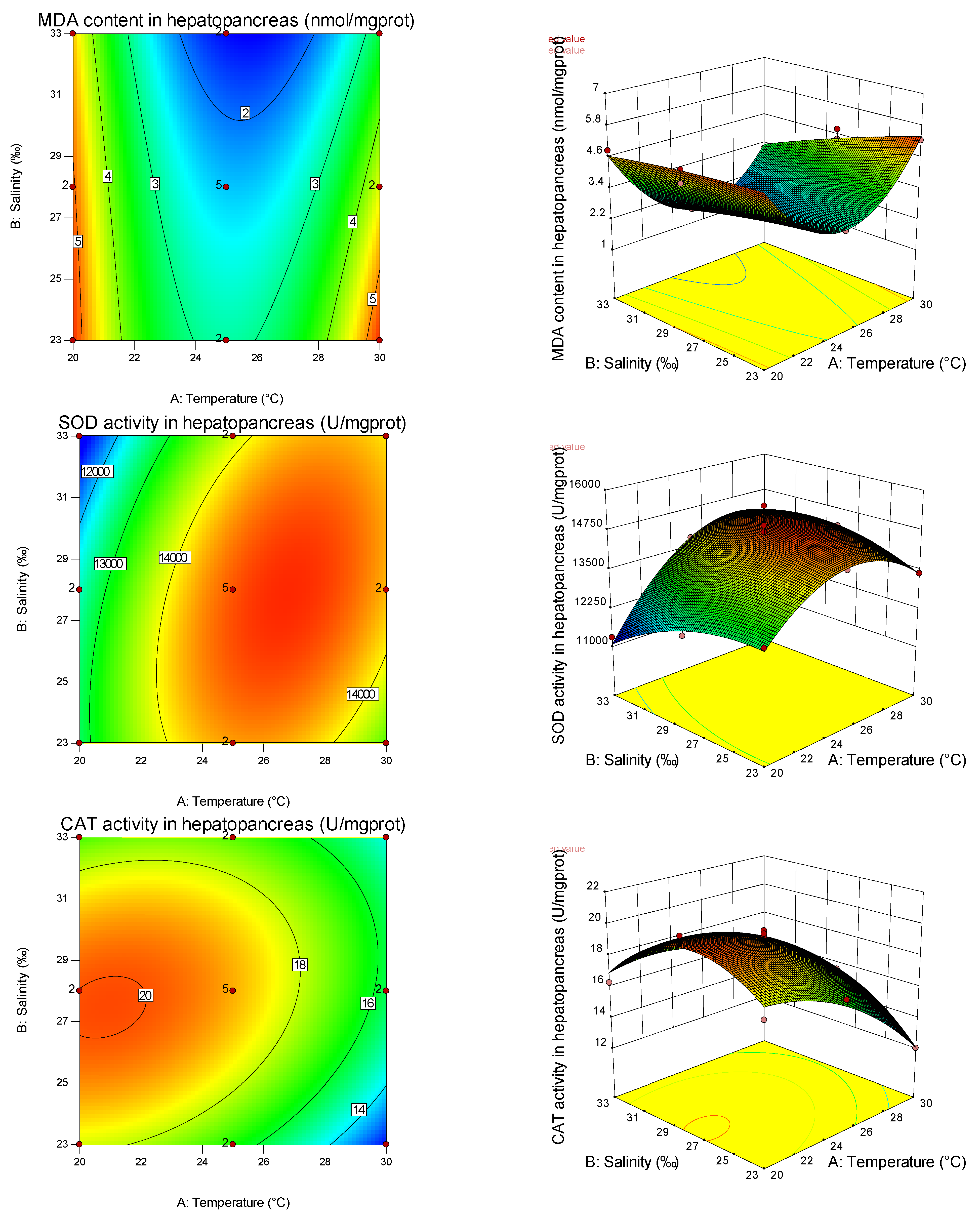
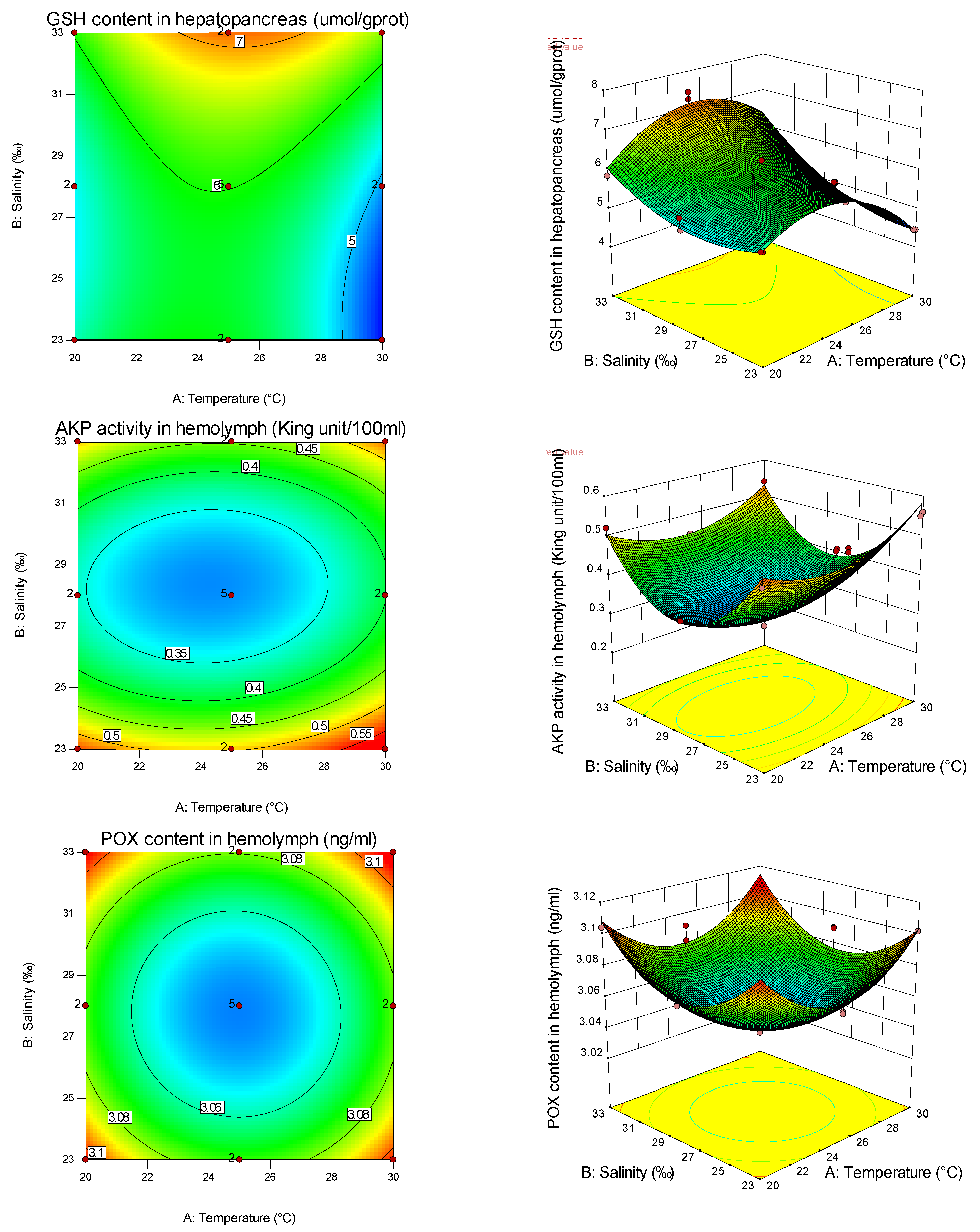
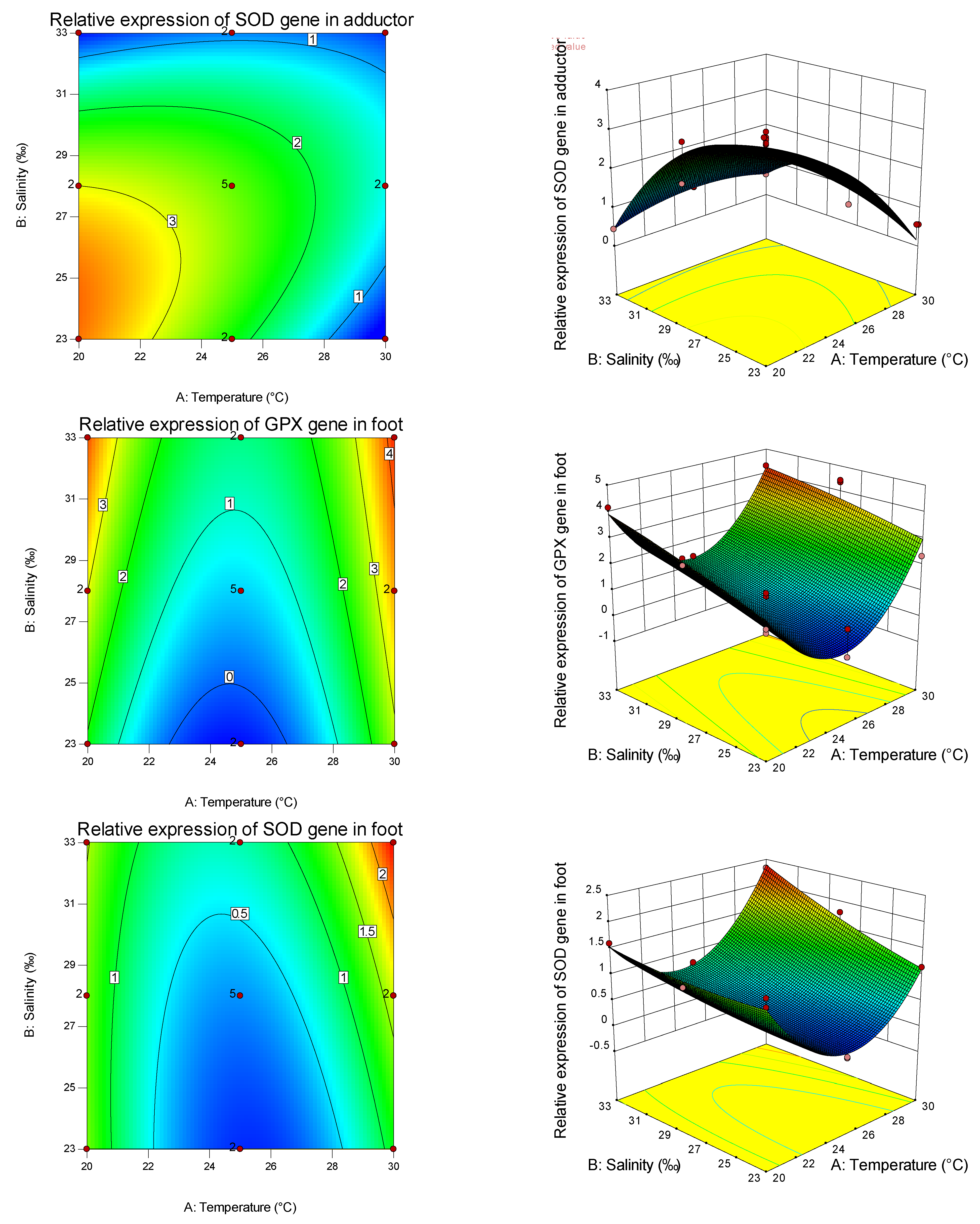
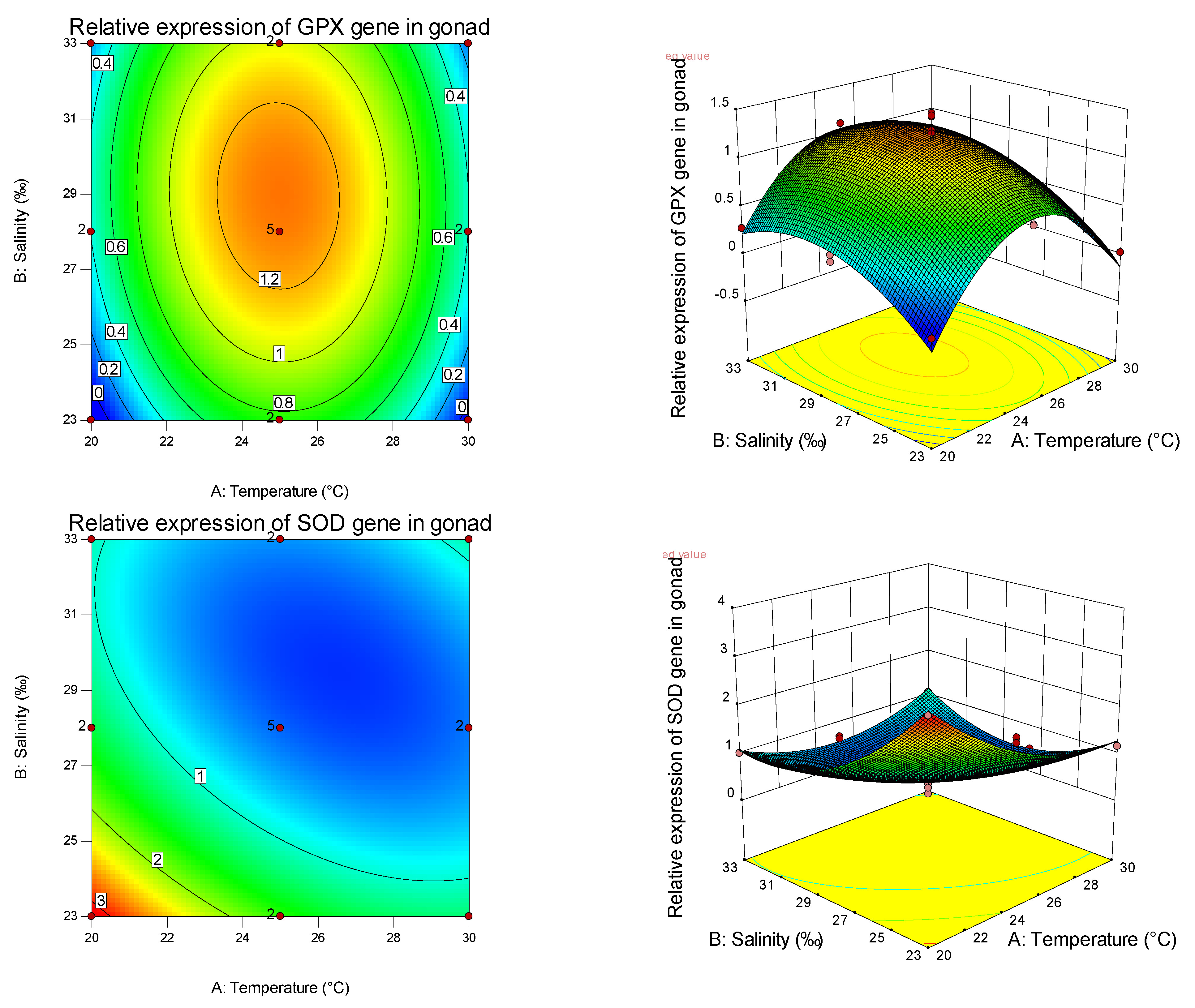
From the present study, it is clear that both temperature and salinity have significant impacts on the AKP, GSH-PX, MDA, SOD, CAT, GSH, POX, and GPX of the pearl oyster in a curvilinear fashion (Figure 1, Figure 2, Table 7 and Table 8). From the magnitude of the linear effect of two coded factors in Table 7 and Table 8, it can be seen that temperature is more important than salinity in affecting AKP, SOD, and CAT. Similar results have been found in other studies on fertilization and hatching in the pearl oyster [1], embryonic development in the coot clam Mulinia lateralis (Say) [38], the brackish water clam Rangia cuneata [15], the northern bay scallop Argopecten irradians irradians [17], and the black-lip pearl oyster Pinctada margaritifera (L.) [39]. From the magnitude of the linear effect of two coded factors in Table 7 and Table 8, it can also be seen that temperature is more important than salinity in affecting the SOD gene, while salinity is more important than temperature in affecting the GPX gene. Thus, different antioxidants are affected by different empositions of temperature and salinity, and have tissue specificity. It can be found from the graphs that the curvature of the response surface is related to whether the first or second effects of temperature and salinity are significant, while the ellipse of the contour is related to the significance of the interaction between temperature and salinity.
The elimination and production of free radicals in aquatic animals are in a dynamic equilibrium state in daily production and life. As a critical (non-biological) factor affecting the physiology and biochemistry of aquatic animals, the salinity change destroys the dynamic balance of free radicals in the body, thereby affecting the body’s antioxidant defense system. In particular, as antioxidant enzymes, CAT and SOD, which can enhance cellular immunity and defense function, are regarded as the first line of defense against oxidative damage [40,41]. SOD plays a crucial role in the body’s immunity, mainly by scavenging free radicals to remove the harmful components left in the body during the oxidation process and reduce damage to the body [42]. CAT is indispensable in living organisms and catalyzes the decomposition of hydrogen peroxide (produced by SOD, metabolized waste, damaging organisms) into oxygen and water (less toxic or harmless) [43]. GSH-PX can catalyze the reduction of peroxides to hydroxyl compounds in aquatic cells, which is the second line of defense in the antioxidant defense system [44,45]. In addition to antioxidant enzymes, antioxidant defense systems include antioxidants, mainly fat-soluble carotenoids and VE, as well as water-soluble VC and GSH [46,47]. GSH is an important antioxidant and hydrogen donor of GSH-PX, which is used to catalyze organic peroxide. It is synthesized by cells and acts on the repair of injury in vivo and eliminating free radicals [43]. In addition, MDA levels can be used to measure the degree of oxidative damage in the body, as it is a product of lipid peroxidation and can also indirectly reflect biological activity, free radical production, and antioxidant capacity [48,49]. In immune defense, AKP in hydrolases mainly exists in serum, which plays a role in removing foreign bodies [50]. POX mainly exists in the hemolymph of invertebrates, as well as in multiple tissues of invertebrates, and plays different main functions in different tissues. POX is an indispensable enzyme in the immunity of mollusks, and is mainly involved in humoral immunity and cellular immunity in animals. Its activity can be directly used to measure the immune function of invertebrates [20].
In this experiment, salinity had a significant effect on SOD and GSH-PX in the primary effect, but had no significant effect on CAT, indicating that SOD and GSH-PX were susceptible to salinity. In the study of the noble scallop [51], it was found that the primary effect of salinity had a significant effect on CAT and SOD activities, but had no significant effect on GSH-PX. This may be because the antioxidant regulation mechanisms of different species on salinity are different, and the interaction effect shields the primary effect of CAT in the pearl oyster [52,53]. The secondary effects of salinity on CAT, SOD, and GSH-PX activities were significant, indicating that salinity had a nonlinear relationship with these antioxidant indexes, and CAT and SOD increased first and then decreased with the increase in salinity, which was consistent with the results of the noble scallop [51]. This may be because (1) the oxygen consumption rate of two kinds of shellfish near the isotonic point was high, and the oxygen consumption rate under the salinity being lower or higher than the isotonic point showed a decreasing trend [54], which inhibited the metabolism and redox reaction of the body, thus reducing the production of oxygen free radicals, and eventually leading to the decrease in antioxidant enzyme activity; (2) under non-isosmotic conditions, in order to adapt to the change of seawater salinity, shellfish need to consume the energy stored in the body, which will affect and change the activity of immune and digestive enzymes in the body; (3) when a large amount of reactive oxygen species far exceeds the scavenging capacity of the antioxidant system in the body, the cell structure will be oxidatively damaged, resulting in the decrease in the activity of antioxidant enzymes in the body [55]. The enzyme activities of the ivory shell Babylonia areolate [55]; SOD activity of the mud crabs Scylla serrata [56]; SOD, CAT, and GSH-PX activity of the Manila clam Ruditapes philippinarum [57]; CAT activity of hepatopancreas of the Nile tilapia Oreochromis niloticus [58]; and SOD activity of the Suminoe oyster Crassostrea rivularis [59] were consistent with the trend of salinity. Therefore, in the process of artificial shellfish culture, the influence of seawater salinity on shellfish cannot be ignored. Salinity higher than 26–28‰ and 27–29‰ reduced the metabolism of the noble scallop [51] and the pearl oyster, respectively, and increased the oxygen free radicals in the body, which could lead to the death of shellfish and cause economic losses.
In this study, the primary effect of salinity had a significant influence on MDA. In contrast, the secondary effect had no significant influence, indicating that salinity had a linear relationship with MDA, and showed a downward trend with the increase in salinity, indicating that low salinity had a greater degree of oxidative damage to shellfish. In this study, the effects of salinity on GSH-PX, GSH, and AKP (hemolymph) were significant, and the effects of secondary effects on POX were significant, indicating that salinity had a nonlinear relationship with these four indicators and its activity had a minimum value. The increase in antioxidant enzyme activity under high and low salinity may be the response of shellfish to maintain the dynamic balance of the antioxidant system under salinity stress. The AKP (hemolymph) of the pearl oyster increased significantly under high and low salinity conditions. Under suitable salinity conditions, the AKP (hemolymph) activity was low, which was the same as the results of Sun et al. [23] on red-shell pearl oysters, Shi et al. [59] on the oyster Ostrea rivularis Gould, and Li et al. [60] on the Zhikong scallop Chlamys farreri and other aquatic organisms [61,62,63]. The results were opposite or inconsistent with those of black-shell pearl oysters [23], AKP in the digestive gland of the razor clam Sinonovacula grandis [64], and AKP in hepatopancreas in this study. This may be related to the differences in species, shell color, tissue site, and experimental conditions. The reason for the changes in AKP, GSH-PX, GSH, and POX may be that shellfish need much energy to regulate the osmotic pressure balance after severe changes in osmotic pressure caused by high and low salt, and the immune level is thus enhanced, so the activity of these four enzymes will increase [60]. However, when the salinity was appropriate, the low activities of the four enzymes might be because shellfish immunity required less energy under suitable environmental conditions, and a large amount of energy was used for the growth of shellfish itself, so the enzyme activities related to antioxidants in the immune system were relatively low [52].
With the change in salinity, although all kinds of immune and antioxidant (non) enzyme indexes of the pearl oyster were significantly affected, there was no consistent rule or obvious linear relationship, and the change rule was specific. It may be because of the disturbance effect of salinity stress on the immune and antioxidant system, but the degree of stress is still within the scope of the pearl oyster, which may also be related to the high adaptation and high resistance of the pearl oyster.
Temperature is an important environmental factor in abiotic factors, which is necessary for the survival of aquatic organisms. It affects the metabolism, biochemical reaction process, and activity of some biological macromolecules [51]. Temperature changes often lead to decreased dissolved oxygen in water, and a large number of oxygen free radicals are generated by respiratory bursts, eventually leading to oxidative damage to shellfish. In this study, the primary and secondary effects of temperature had significant effects on GSH, GSH-PX, SOD, and CAT, indicating that the activities of these four antioxidant enzymes were significantly affected by temperature and had a nonlinear relationship with temperature. Among them, GSH, SOD, and CAT had maximum values. In the study of the Pacific abalone Haliotis discus hannai Ino [65], the Yesso scallop Patinopecten yessoensis [48], and the lwagaki oysters Crassostrea nippona [66], it was found that the changes in CAT and SOD activities in cooling and heating treatments were significantly different, which might be owing to the different regulatory mechanisms of cooling and heating on the antioxidant enzyme activities of different species. Other studies found that salinity’s primary and secondary effects had significant effects on the activities of CAT, GSH-PX, and SOD in the noble scallop [51], and they all had maximum values. The changes in SOD activity in the hepatopancreas of the bivalve Cyclina sinensis [67] were the same, and the results of this study were consistent with it. These changes in SOD and CAT activities in the pearl oyster may be induced by a large amount of reactive oxygen species caused by accelerated metabolism with the increase in temperature under a period of external temperature stress. However, as the temperature exceeds the optimal range, the body’s vitality and metabolic ability will be reduced, affected by the changes in the body environment, eventually leading to the decrease in these two related antioxidant enzyme activities [68,69]. Excessive ambient temperature can make the body produce many oxygen free radicals, leading to death. Combined with the team’s previous experiments, under the corresponding temperature conditions, when the CAT and SOD activities of the pearl oyster were the highest, their activities, feeding, digestion, and metabolism were often better than those of other experimental groups. This indicated that the activities of some antioxidant enzymes in the pearl oyster were usually high under the optimal environmental conditions, which was conducive to reducing individual respiratory burst, scavenging oxygen free radicals in the body, and reducing mortality, which was consistent with the findings of the Pacific abalone [70], the noble scallop [51], the striped venus clam Chamelea gallina [71], the Chinese sturgeon Acipenser sinensis Gray [72], and the infaunal clam Macoma balthica [73]. Both the noble scallop [51] and the pearl oyster belong to warm water shellfish, so their seawater temperatures should be controlled at 26.40 °C and 24.95 °C, respectively, under artificial indoor culture conditions to ensure that the two shellfishes have the strongest antioxidant capacity and the lowest individual mortality. For the sudden death of shellfish in the pool, the change in water temperature can be first checked to maintain it within the normal temperature range to prevent the death caused by respiratory burst, and then other possible factors such as bait or disease can be checked.
The primary effect of temperature significantly influenced AKP, while the secondary effect was not significant, indicating that temperature was linearly correlated with AKP and increased with the increase in temperature within a certain range. When shellfish are subjected to low-temperature stress, the metabolic rate and oxygen consumption are reduced, which inhibits the growth of microorganisms, the production of CO2, ammonia and lactic acid, and the activity of shellfish to a certain extent, and reduces the risk of disease infection. This is also why a low temperature is suitable for in vivo transportation of shellfish [74]. The reaction of AKP under low-temperature stress proves this. However, when shellfish are under high-temperature stress, the physiological disorder, metabolic rate, and oxygen consumption increase in the body under a stress state, which is susceptible to bacterial infection and requires much energy. The activities of nonspecific immune-related enzymes and antioxidant enzymes in shellfish will rise over time to resist changes in the external environment [74]. In this study, the primary and secondary effects of temperature on GSH-PX and AKP in the hemolymph of the pearl oyster were significant, indicating that temperature had a nonlinear relationship with them, and the activity increased at a high temperature, which is consistent with the above rules. The primary effect of temperature and the interaction between temperature and salinity had no significant effect on POX, and the secondary effect was significant, indicating that temperature had a nonlinear relationship with POX and had high activity at a high temperature, consistent with the above rules. Li et al. [60] and Chen et al. [75]’s research on the Zhikong scallop, Liu et al. [76]’ s research on the Zhikong scallop and the bay scallop Argopectehs irradias, and Liu [74]’ s research on the clam Lutraria sieboldii Reeve found that AKP maintained a high activity level at a high temperature, which was consistent with the results of this study and conformed to the above laws. This may be related to the physical and chemical properties of AKP, and shellfish may be more sensitive to high-temperature stress. The change in AKP activity may also be due to the threat of high or low temperature to the normal survival of shellfish, and a high temperature leads to the emergence of many pathogens. Therefore, to resist adverse environments and ensure normal physiological activities, the pearl oyster must enhance its immunity [60]. Hu et al. [66] found that the AKP of the lwagaki oysters also increased significantly after 4 h and 8 h at 35 °C, but it was significantly lower than that of the control group, which was inconsistent with the results of this study, indicating that the lwagaki oysters had poor environmental adaptability, and the regulation of AKP was unbalanced under the temperature mutation of 35 °C. Under high-temperature stress, the AKP of the Manila clam [77] showed a decreasing trend, and finally increased (returned) to a normal salt temperature level. The reason for these different results may be that high-temperature tolerance has species specificity. The primary and secondary effects of temperature had significant effects on MDA, indicating that there was a non-linear relationship between temperature and MDA, and a U-shaped distribution with the increase in temperature, indicating that both high and low temperatures caused oxidative damage to pearl oysters.
In this study, the combined effects of salinity and temperature on antioxidant and immune-related (non) enzyme indexes of the pearl oyster were analyzed by response surface methodology. The response surface method can obtain the model equation with a high fitting degree, optimize the experimental results, and obtain the optimal factor (factor) combination, which is its biggest advantage. However, given the influence of environmental factors on shellfish’s immune and antioxidant indexes, there is no reliable model established in domestic and foreign studies focusing on the analysis of some isolated levels under a single environmental condition. In this paper, based on the response surface analysis, we found that the response surface of SOD and CAT showed a convex curve shape, indicating that the two enzymes had a peak (maximum). By optimizing the established model, the theoretical conditions for SOD and CAT to reach the maximum activity were 24.95 °C/28.11‰, which are close to 26.4 °C and 27.7‰ of the noble scallop [51], indicating that, under this condition, the external environment did not cause stress to shellfish, and the living environment was suitable for shellfish. The results showed that the interaction of salinity and temperature had significant effects on CAT and SOD of the pearl oyster, but had no significant effect on GSH-PX, which was completely consistent with the results of the noble scallop [51]. This indicated that, for CAT and SOD, salinity and temperature had a mutual influence, and for GSH-PX, these two factors were independent without interference or superposition between additive effects. Studies on other aquatic organisms such as the cobia Rachycentron canadum [78] and juvenile GIFT Nile tilapia [58] showed that the interaction between salinity and temperature on CAT and SOD activities were significant, which was consistent with the results of this study. However, there were few studies on the effect of the temperature–salt combination on GSH-PX. From the response surface diagram, the contour lines of AKP in hemolymph, GSH-PX, and POX under temperature and salinity stress were round, indicating that there was no interaction between temperature and salinity on AKP (hemolymph), GSH-PX, and POX. The interaction between salinity and temperature on AKP of the pearl oyster was obvious, which was contrary to the results of AKP (hemolymph) in this paper, and was consistent with the results of Li et al. [60] on AKP, ACP, and GR in the hemolymph of the Zhikong scallop, and Zhu [52] on AKP and ACP in juvenile pearl oysters. This may be because (1) the difference in AKP interaction may be related to the different combined effects of salinity and temperature on antioxidant indexes of different ages, different species, and different tissues; (2) AKP and POX in hemolymph were not affected by the interaction of temperature and salinity, either because the activity of antioxidant and immune enzyme in hemolymph was too low, the kit could not accurately determine its effective activity, or temperature and salinity did not interact with each other; (3) temperature changed the spatial conformation of AKP, and salinity provided the metal ions needed to activate AKP, so as to achieve the synergy between the two and jointly promote the regulation of AKP activity.
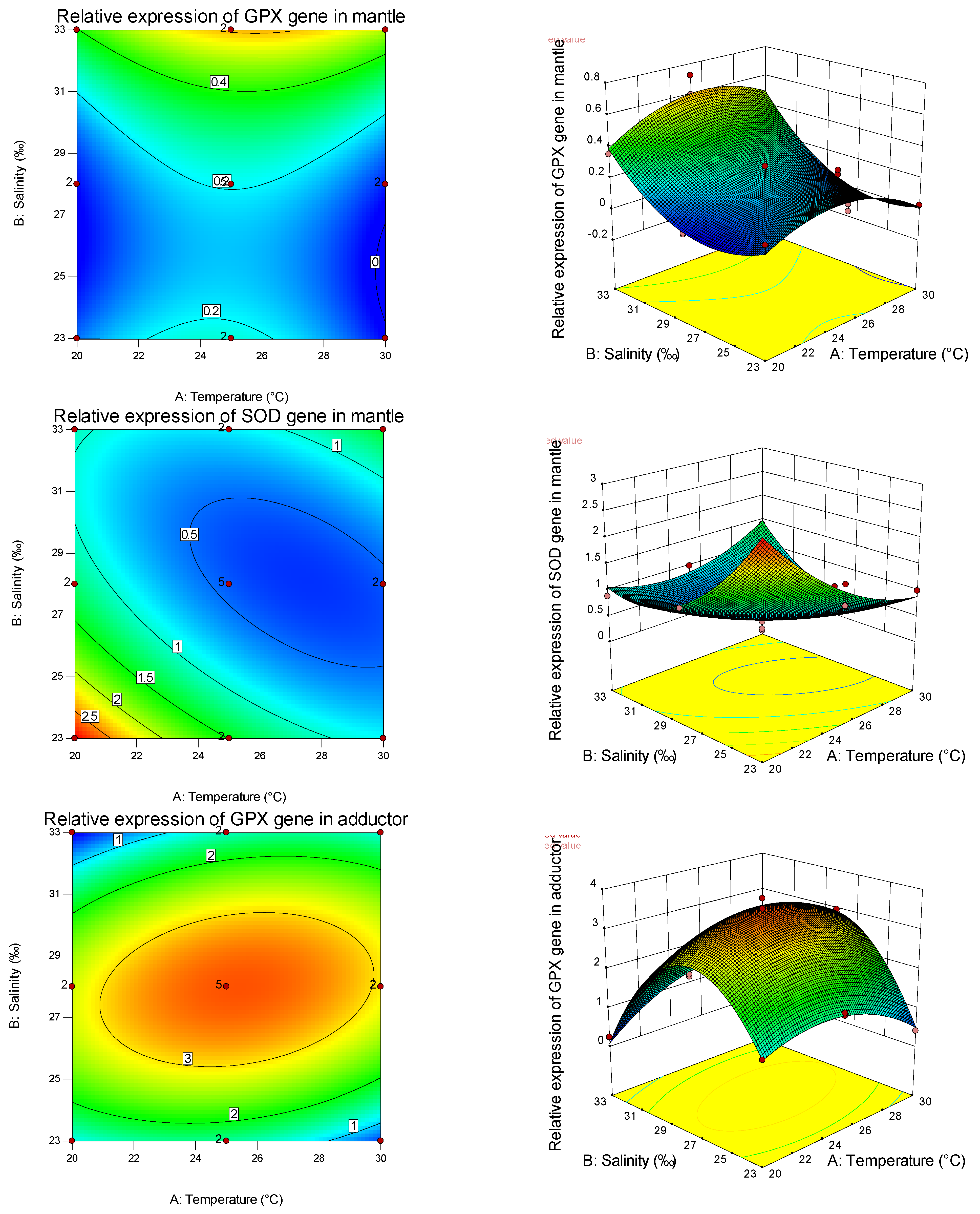
Many domestic and foreign experts and scholars have studied the interaction between marine invertebrate environmental factors and antioxidant enzymes. It is mainly reflected in the spatio-temporal and organizational differences in enzyme activity determination and expression level under various stress factors. So far, GPX, SOD, and GST are the most commonly studied antioxidant enzyme genes related to marine biological stress tolerance [79]. However, there are few studies on the effects of temperature or salinity on the relative expression of SOD and GPX genes in shellfish.
Studies have shown that the shellfish antioxidant enzyme system has high tissue specificity [80,81]. In this study, for the GPX gene, the primary and secondary effects of salinity significantly affected its expression in the mantle, adductor muscle, and gonad, while the primary effect of salinity significantly affected its expression in the foot, and the secondary effect was not significant. This indicated that salinity had a significant influence on the expression of GPX in the mantle, adductor muscle, and gonad and had a non-linear relationship with it, and had a significant influence on the expression of GPX in the foot and had a linear relationship with it. Salinity had the maximum expression of GPX in the obturator muscle and gonad and the minimum expression of GPX in the mantle, while the expression of GPX in the foot increased with salinity. This indicates that salinity has tissue specificity for GPX gene expression. Similarly, the expression of the GPX gene by temperature and the expression of the SOD gene by salinity and temperature also have tissue specificity, and the relative expression of different genes in the same tissue is also different. In the study on salinity stress of the mud crabs [82], with the increase in salinity, GPX activity in the abdominal muscle did not change, and SOD activity gradually decreased. GPX activity in the hepatopancreas decreased with increasing salinity, while SOD activity increased first and then decreased. GPX and SOD activities in gills decreased first and then increased with the increase in salinity, indicating the tissue specificity of salinity stress on the antioxidant defense and oxidative stress in aquatic animals. Through the analysis of the expression of antioxidant genes among different tissues under the effects of salinity and temperature on the primary, secondary, and interaction effects, the expression levels of GPX and SOD are consistent with and different from the trend of antioxidant and immune-related (non-) enzymes mentioned above, indicating that, in the pearl oyster, antioxidant and immune-related (non-) enzymes interacted with the expression of tissue immune genes to enhance the resistance and adaptability of shellfish to the environment.

5. Conclusions

In summary, environmental temperature and salinity may regulate humoral and cellular immunity of the pearl oyster by influencing antioxidant and immune responses and the antioxidant capacity of tissues. The combined effect of salinity and temperature significantly influenced the adaptation of the pearl oyster to the living environment. Therefore, both single-factor and synergistic effects should be considered in artificial breeding and farming. This study examined the effects of salinity and temperature on antioxidant and immune indexes of the pearl oyster, and the effects of other environmental factors such as pH, ammonia, and dissolved oxygen need further study.

Author Contributions

Conceptualization, Z.M. and X.W.; Methodology, G.Y. and Z.F.; Software, J.Y.; Validation, J.Y., Z.M. and X.W.; Formal Analysis, G.Y. and Z.F.; Investigation, J.Y.; Resources, G.Y. and Z.F.; Data Curation, X.W.; Writing—Original Draft Preparation, J.Y.; Writing—Review and Editing, Z.M.; Visualization, Z.F.; Supervision, X.W.; Project Administration, G.Y.; Funding Acquisition, Z.M. All authors have read and agreed to the published version of the manuscript.

Funding

This research was funded by Central Public-Interest Scientific Institution Basal Research Fund, CAFS (NO. 2020TD55).

Institutional Review Board Statement

The animal study was reviewed and approved by the Ethic Committee of Sanya Tropical Fisheries Research (protocol code 2020TD55 and approved on 2020.4.1).

Data Availability Statement

Relevant information has been added in the article.

Acknowledgments

Thanks to Tropical Aquaculture Research and Development Center, South China Sea Fisheries Research Institute, and Chinese Academy of Fishery Sciences Supply of experimental venue and equipment.

Conflicts of Interest

The authors declare no conflict of interest.

References

  1. Wang, H.; Zhu, X.; Wang, Y.; Luo, M.; Liu, Z. Determination of optimum temperature and salinity for fertilization and hatching in the Chinese pearl oyster Pinctada martensii (Dunker). Aquaculture 2012, 358, 292–297. [Google Scholar] [CrossRef]
  2. Wada, K.T. The Pearl Oyster, Pinctada Fucata (Gould) (Family Pteriidae). In Estuarine Marine Bivalve Mollusk Culture; CRC Press: Boca Raton, FL, USA, 2018; Volume 18, pp. 245–260. Available online: https://books.google.com.tw/books?id=jrtHDwAAQBAJ&lpg=PT409&ots=JFuvdX28Ut&dq=Pinctada%20Fucata%20(Gould)(Family%20Pteriidae).%20&lr&hl=zh-CN&pg=PT409#v=onepage&q=Pinctada%20Fucata%20(Gould)(Family%20Pteriidae).&f=false (accessed on 18 January 2018).
  3. Grant, J. The relationship of bioenergetics and the environment to the field growth of cultured bivalves. J. Exp. Mar. Biol. Ecol. 1996, 200, 239–256. [Google Scholar] [CrossRef]
  4. Uemoto, H. Relationship between oxygen consumption by the pearl oyster and its environmental temperature. Bull. Natl. Pearl. Res. Lab. 1968, 13, 1617–1623. [Google Scholar]
  5. Numaguchi, K.; Tanaka, Y. Effects of salinity on mortality and growth of the spat of the pearl oyster, Pinctada fucata martensii. Bull. Natl. Res. Inst. Aquac. 1986, 9, 41–44. [Google Scholar]
  6. Numaguchi, K.; Tanaka, Y. Effects of temperature on mortality and growth of the spat of the pearl oyster, Pinctada fucata martensii. Bull. Natl. Res. Inst. Aquac. 1986, 9, 35–39. [Google Scholar]
  7. Numaguchi, K. Growth and physiological condition of the Japanese pearl oyster, Pinctada fucata martensii (Dunker, 1850) in Ohmura Bay, Japan. J. Shellfish Res. 1994, 13, 93–99. [Google Scholar]
  8. Numaguchi, K. Effect of water temperature on the filtration rate of Japanese pearl oyster, Pinctada fucata martensii. Aquac. Sci. 1994, 42, 1–6. [Google Scholar] [CrossRef]
  9. O’Connor, W.A.; Lawler, N.F. Salinity and temperature tolerance of embryos and juveniles of the pearl oyster, Pinctada imbricata Röding. Aquaculture 2004, 229, 493–506. [Google Scholar] [CrossRef]
  10. Box, G.E. The exploration and exploitation of response surfaces: Some general considerations and examples. Biometrics 1954, 10, 16–60. [Google Scholar] [CrossRef]
  11. Davis, H.C. Combined effects of temperature and salinity on development of eggs and growth of larvae of M. mercenaria and C. virginica. Fish. Bull. 1964, 63, 643–655. [Google Scholar]
  12. Robert, R.; His, E.; Dinet, A. Combined effects of temperature and salinity on fed and starved larvae of the European flat oyster Ostrea edulis. Mar. Biol. 1988, 97, 95–100. [Google Scholar] [CrossRef]
  13. Brenko, M.H.; Calabrese, A. The combined effects of salinity and temperature on larvae of the mussel Mytilus edulis. Mar. Biol. 1969, 4, 224–226. [Google Scholar] [CrossRef]
  14. Lough, R.; Gonor, J. A response-surface approach to the combined effects of temperature and salinity on the larval development of Adula californiensis (Pelecypoda: Mytilidae). II. Long-term larval survival and growth in relation to respiration. Mar. Biol. 1973, 22, 295–305. [Google Scholar] [CrossRef]
  15. Cain, T. The combined effects of temperature and salinity on embryos and larvae of the clam Rangia cuneata. Mar. Biol. 1973, 21, 1–6. [Google Scholar] [CrossRef]
  16. Lough, R.G. A reevaluation of the combined effects of temperature and salinity on survival and growth of bivalve larvae using response surface techniques. Fish. Bull. 1975, 73, 86–92. [Google Scholar]
  17. Tettelbach, S.; Rhodes, E. Combined effects of temperature and salinity on embryos and larvae of the northern bay scallop Argopecten irradians irradians. Mar. Biol. 1981, 63, 249–256. [Google Scholar] [CrossRef]
  18. Lu, W.; Li, W.; Ke, C.; Wang, H. Reproductive success under the joint influences of temperature and salinity in noble scallop, Chlamys nobilis (Reeve). Aquac. Res. 2017, 48, 686–696. [Google Scholar] [CrossRef]
  19. His, E.; Robert, R.; Dinet, A. Combined effects of temperature and salinity on fed and starved larvae of the Mediterranean mussel Mytilus galloprovincialis and the Japanese oyster Crassostrea gigas. Mar. Biol. 1989, 100, 455–463. [Google Scholar] [CrossRef]
  20. Yang, J.; Yang, J.; Chen, M.; Fu, Z.; Sun, J.; Yu, G.; Wang, A.; Ma, Z.; Gu, Z. Physical Responses of Pinctada fucata to Salinity Stress. Front. Mar. Sci. 2022, 8, 792179. [Google Scholar] [CrossRef]
  21. Yang, J. Effects of Salinity and pH on Energy Metabolism and Gene Expression of Meretrix meretrix; Shanghai Ocean University: Shanghai, China, 2016. [Google Scholar]
  22. Liu, Y. Preliminary Study on the Molecular Mechanism of Low Temperature Adaptability of Pinctada fucata martensii; Guangdong Ocean University: Zhanjiang, China, 2018. [Google Scholar]
  23. Sun, J.; Chen, M.; Fu, Z.; Yang, J.; Zhou, S.; Yu, G.; Zhou, W.; Ma, Z. A Comparative Study on Low and High Salinity Tolerance of Two Strains of Pinctada fucata. Front. Mar. Sci. 2021, 8, 1039. [Google Scholar] [CrossRef]
  24. Li, J.; Zhang, Y.; Liu, Y.; Zhang, Y.; Xiao, S.; Yu, Z. Co-expression of heat shock protein (HSP) 40 and HSP70 in Pinctada martensii response to thermal, low salinity and bacterial challenges. Fish Shellfish Immunol. 2016, 48, 239–243. [Google Scholar] [CrossRef] [PubMed]
  25. Hui, W.; Yi, Z.; Long, W.; Chuankun, Z.; Ji, Z.; Nan, W. Growth and survival of Pinctada martensii (Dunker) postlarvae under concurrent variation in temperature, algal ration and stocking density. Aquac. Rep. 2021, 20, 100668. [Google Scholar] [CrossRef]
  26. Wang, T.; Li, Q.; Jiang, X.; Liu, L.; Hu, M.; Wang, Y. Effects of gonadal preoperative treatment on the physiological metabolism of the pearl oyster pinctada martensii: Implications for pearl production. J. Shellfish Res. 2018, 37, 1051–1059. [Google Scholar] [CrossRef]
  27. Li, S.; Huang, J.; Liu, C.; Liu, Y.; Zheng, G.; Xie, L.; Zhang, R. Interactive Effects of Seawater Acidification and Elevated Temperature on the Transcriptome and Biomineralization in the Pearl Oyster Pinctada fucata. Environ. Sci. Technol. 2016, 50, 1157–1165. [Google Scholar] [CrossRef] [PubMed]
  28. Wei, W.; Liu, Q.; Zhang, C.; Hao, R.; Liao, Y.; Wang, Q.; Deng, Y. Changes of Tolerance of Air Exposure and lmmune-related Gene Expression of Pinctada fucata martensii at Different Temperatures. J. Guangdong Ocean Univ. 2021, 41, 69–75. Available online: https://kns.cnki.net/kcms/detail/detail.aspx?FileName=SHDX202102010&DbName=CJFQ2021 (accessed on 22 February 2021).
  29. Gu, Z.; Cao, Y.; Yang, H.; Jiao, Y. Effect of Janus Kinase 3 Inhibitor on the Development of Pearl Sac and the Expression of lmmune-related Genes in Pearl Oyster Pinctada fucata martensii. J. Guangdong Ocean Univ. 2020, 40, 1–6. Available online: https://kns.cnki.net/kcms/detail/detail.aspx?FileName=SHDX202002001&DbName=CJFQ2020 (accessed on 23 October 2019).
  30. Adzigbli, L.; Yu, W.; Li, J.; Yang, C.; Deng, Y. Influence of age on pearl production performance, enzymatic activity, and immune-related gene expression of the pearl oyster Pinctada fucata martensii. N. Am. J. Aquac. 2019, 81, 430–437. [Google Scholar] [CrossRef]
  31. Fu, Z.; Yang, R.; Chen, X.; Qin, J.G.; Gu, Z.; Ma, Z. Dietary non-protein energy source regulates antioxidant status and immune response of barramundi (Lates calcarifer). Fish Shellfish Immunol. 2019, 95, 697–704. [Google Scholar] [CrossRef]
  32. Fu, Z.; Qin, J.G.; Ma, Z.; Yu, G. Acute acidification stress weakens the head kidney immune function of juvenile Lates calcarifer. Ecotoxicol. Environ. Saf. 2021, 225, 112712. [Google Scholar] [CrossRef]
  33. Paria, A.; Dong, J.; Babu, P.; Makesh, M.; Chaudhari, A.; Thirunavukkarasu, A.; Purushothaman, C.; Rajendran, K. Evaluation of candidate reference genes for quantitative expression studies in Asian seabass (Lates calcarifer) during ontogenesis and in tissues of healthy and infected fishes. Ind. J. Exp. Biol. 2016, 54, 597–605. Available online: http://europepmc.org/abstract/MED/28699725 (accessed on 13 July 2017).
  34. Yang, J.; Hong, J.; Fu, Z.; Ma, Z. Effects of Dietary Curcumin on Growth and Digestive Physiology of Seriola dumerili. Front. Mar. Sci. 2022, 9, 435. [Google Scholar] [CrossRef]
  35. Green, M.R.; Sambrook, J. Molecular Cloning: A Laboratory Manual 4th; Cold Spring Harbor Laboratory Press: New York, NY, USA, 2012. [Google Scholar]
  36. Ryan, T.P.; Morgan, J. Modern experimental design. J. Stat. Theory Pract. 2007, 1, 501–506. [Google Scholar] [CrossRef]
  37. Montgomery, D.C. Design and Analysis of Experiments; John Wiley & Sons: New York, NY, USA, 2017. [Google Scholar]
  38. Calabrese, A. Individual and combined effects of salinity and temperature on embryos and larvae of the coot clam, Mulinia lateralis (Say). Biol. Bull. 1969, 137, 417–428. [Google Scholar] [CrossRef] [PubMed]
  39. Doroudi, M.; Southgate, P.C.; Mayer, R. The combined effects of temperature and salinity on embryos and larvae of the black-lip pearl oyster, Pinctada margaritifera (L.). Aquac. Res. 1999, 30, 271–277. [Google Scholar] [CrossRef]
  40. Bhagat, J.; Ingole, B.; Singh, N. Glutathione S-transferase, catalase, superoxide dismutase, glutathione peroxidase, and lipid peroxidation as biomarkers of oxidative stress in snails: A review. Invertebr. Surviv. J. 2016, 13, 336–349. Available online: https://www.isj.unimore.it/index.php/ISJ/article/view/407/321 (accessed on 1 April 1999).
  41. Wang, N.; Yang, J.; Zhang, H.; Soon, T.K.; Liu, H.; Li, S.; Ma, H.; Zheng, H. Differential responses to low salinity on gene expression, physiological and biochemical indexes between the golden and brown noble scallops Chlamys nobilis. Aquac. Res. 2020, 51, 316–325. [Google Scholar] [CrossRef]
  42. McCord, J.M.; Fridovich, I. Superoxide dismutase: An enzymic function for erythrocuprein (hemocuprein). J. Biol. Chem. 1969, 244, 6049–6055. [Google Scholar] [CrossRef]
  43. Liu, M.; Chang, Y.; Yang, Y.; Liu, Y.; Zhang, J.; Zhang, W.; Wang, Z. Effects of Gradual Salinity Changes on lmmune Parameters of Scallop (Patinopecten yessoensis). J. Agric. Sci. Technol. 2011, 13, 129–135. Available online: https://kns.cnki.net/kcms/detail/detail.aspx?FileName=NKDB201103028&DbName=CJFQ2011 (accessed on 15 June 2011).
  44. Cnubben, N.H.; Rietjens, I.M.; Wortelboer, H.; van Zanden, J.; van Bladeren, P.J. The interplay of glutathione-related processes in antioxidant defense. Environ. Toxicol. Pharmacol. 2001, 10, 141–152. [Google Scholar] [CrossRef]
  45. Peña-Llopis, S.; Ferrando, M.D.; Peña, J.B. Fish tolerance to organophosphate-induced oxidative stress is dependent on the glutathione metabolism and enhanced by N-acetylcysteine. Aquat. Toxicol. 2003, 65, 337–360. [Google Scholar] [CrossRef]
  46. Packer, L. Protective role of vitamin E in biological systems. Am. J. Clin. Nutr. 1991, 53, 1050S–1055S. [Google Scholar] [CrossRef] [PubMed]
  47. Sies, H.; Stahl, W.; Sundquist, A.R. Antioxidant functions of vitamins: Vitamins E and C, Beta-Carotene, and other carotenoids a. Ann. N. Y. Acad. Sci. 1992, 669, 7–20. [Google Scholar] [CrossRef] [PubMed]
  48. Jiang, W.; Li, J.; Gao, Y.; Mao, Y.; Jiang, Z.; Du, M.; Zhang, Y.; Fang, J. Effects of temperature change on physiological and biochemical responses of Yesso scallop, Patinopecten yessoensis. Aquaculture 2016, 451, 463–472. [Google Scholar] [CrossRef]
  49. Viarengo, A.; Canesi, L.; Martinez, P.G.; Peters, L.; Livingstone, D. Pro-oxidant processes and antioxidant defence systems in the tissues of the Antarctic scallop (Adamussium colbecki) compared with the Mediterranean scallop (Pecten jacobaeus). Comp. Biochem. Physiol. Part B Biochem. Mol. Biol. 1995, 111, 119–126. [Google Scholar] [CrossRef]
  50. Pipe, R. Hydrolytic enzymes associated with the granular haemocytes of the marine mussel Mytilus edulis. Histochem. J. 1990, 22, 595–603. [Google Scholar] [CrossRef] [PubMed]
  51. Qian, J.; Li, Z.; Shen, Y.; Liu, Z.; Zhang, W. Synergistic effect of temperature and salinity on antioxidant enzymes activities of Chlamys nobilis. South China Fish. Sci. 2015, 11, 49–57. Available online: https://kns.cnki.net/kcms/detail/detail.aspx?FileName=NFSC201506009&DbName=CJFQ2015 (accessed on 5 December 2015).
  52. Zhu, X. Combined Effects of Temperature, Salinity and pH on the Physiological Features of the Juveniles of Pinctada martensii (Dunker); Guangdong Ocean University: Zhanjiang, China, 2012. [Google Scholar]
  53. Qian, J.; Li, Z.; Chen, S. Synergistic Effects of Temperature and Salinity on Activities of Amylase and Na+/K+-ATPase in Juvenile Scallop Chlamys nobilis(Reeve). Fish. Sci. 2015, 34, 269–276. [Google Scholar] [CrossRef]
  54. Lei, S. Effect of salinity and body weight on the oxygen consumption of Oreochromis niloticus x O.mossambicus. Chin. J. Appl. Ecol. 2002, 13, 739–742. Available online: https://kns.cnki.net/kcms/detail/detail.aspx?FileName=YYSB200206022&DbName=CJFQ2002 (accessed on 25 May 2015).
  55. Tan, C.; Zhao, W.; Wu, K.; Wen, W.; Yang, R.; Chen, X.; Yu, G. Effects of Salinity Stress on Activities of Enzymes Related to Immune of Snail Babylonia areolata. Fish. Sci. 2020, 39, 677–684. [Google Scholar] [CrossRef]
  56. Chen, Y.; Ai, C.; Lin, Q.; Xu, H.; Shen, Y. Effect of salinity stress on the activities of phenoloxidase and superoxidedismutase of the serum, tissue and organ of mud crab, Scylla serrata. J. Appl. Oceanogr. 2007, 26, 569–575. Available online: https://kns.cnki.net/kcms/detail/detail.aspx?FileName=TWHX200704018&DbName=CJFQ2007 (accessed on 15 November 2007).
  57. Song, X.; Ma, J.; Qin, Y.; Chang, L.; Wang, Y.; Li, X.; Zhao, L. Effects of Abrupt Decline in Salinity on the Antioxidant Enzyme Activities and Histological Structure in Ruditapes philippenarum. J. Agric. 2013, 3, 50–56, 70. Available online: https://kns.cnki.net/kcms/detail/detail.aspx?FileName=XKKJ201301012&DbName=CJFQ2013 (accessed on 20 January 2013).
  58. Qiang, J.; Ren, H.; Xu, P.; He, J.; Li, R. Synergistic effects of water temperature and salinity on the growth and liver antioxidant enzyme activities of juvenile GIFT Oreochromis niloticus. Chin. J. Appl. Ecol. 2012, 23, 255–263. [Google Scholar] [CrossRef]
  59. Shi, S.; Wang, R.; Wang, J.; Jiang, J.; Liu, G.; Yang, R. Effects of salinity stress on immune factors of Crassostrea hongkongensis. South China Fish. Sci. 2013, 9, 26–30. Available online: https://kns.cnki.net/kcms/detail/detail.aspx?FileName=NFSC201303006&DbName=CJFQ2013 (accessed on 5 June 2013).
  60. Li, Z.; Qian, J.; Lao, C.; LIU, Z. Synergistic Effects of Temperature and Salinity on the Activities of lmmune-Related Enzymes of Chlamys nobilis (Reeve). Adv. Mar. Sci. 2015, 33, 227–238. Available online: https://kns.cnki.net/kcms/detail/detail.aspx?FileName=HBHH201502010&DbName=CJFQ2015 (accessed on 15 April 2015).
  61. Cheng, W.; Juang, F.-M.; Chen, J.-C. The immune response of Taiwan abalone Haliotis diversicolor supertexta and its susceptibility to Vibrio parahaemolyticus at different salinity levels. Fish Shellfish Immunol. 2004, 16, 295–306. [Google Scholar] [CrossRef]
  62. Joseph, A.; Philip, R. Acute salinity stress alters the haemolymph metabolic profile of Penaeus monodon and reduces immunocompetence to white spot syndrome virus infection. Aquaculture 2007, 272, 87–97. [Google Scholar] [CrossRef]
  63. Shen, L.; Chen, Z.; Chen, C.; He, X. Growth and Immunities of the Shrimp, Litopenaeus vannamei (Boone) Exposed to Different Salinity Levels. J. Jimei Univ. (Nat. Sci.) 2007, 12, 108–113. [Google Scholar] [CrossRef]
  64. Li, D.; Nie, H.; Liu, G.; Yan, X. Effects of Salinity and Air Exposure on Activities of Respiratory Metabolism-related Enzymes in Grand Jackknife Clam Solen grandis. Fish. Sci. 2019, 38, 55–60. [Google Scholar] [CrossRef]
  65. Shi, S. Studies on the Immune Parameters of Mollusc Stimulated by Environmental Factors; Shanghai Ocean University: Shanghai, China, 2013. [Google Scholar]
  66. Hu, Y.; Li, Q.; Liu, S.; Kong, L. Effects of acute temperature and salinity stress on the survival and immune indexes of lwagaki oysters, Crassostrea nippona. J. Fish. Sci. China 2020, 27, 286–294. Available online: https://kns.cnki.net/kcms/detail/detail.aspx?FileName=ZSCK202003003&DbName=CJFQ2020 (accessed on 12 September 2019).
  67. Li, X.; Shen, R.; Dong, Z.; Yan, B.; Shen, H.; Li, J. Effects of temperature increment and hypoxia stress on antioxidases of Cyclina sinensis. China Feed 2009, 1, 39–42. Available online: https://kns.cnki.net/kcms/detail/detail.aspx?FileName=SLGZ200901017&DbName=CJFQ2009 (accessed on 5 January 2009). [CrossRef]
  68. Liu, W.; Li, M.; Huang, F.; Zhu, J.; Dong, W.; Yang, J. Effects of cadmium stress on xanthine oxidase and antioxidant enzyme activities in Boleophthalmus pectinirostris liver. Ying Yong Sheng Tai Xue Bao = J. Appl. Ecol. 2006, 17, 1310–1314. Available online: https://kns.cnki.net/kcms/detail/detail.aspx?FileName=YYSB200607030&DbName=CJFQ2006 (accessed on 30 July 2006).
  69. Yu, B.P. Cellular defenses against damage from reactive oxygen species. Physiol. Rev. 1994, 74, 139–162. [Google Scholar] [CrossRef] [PubMed]
  70. De Zoysa, M.; Whang, I.; Lee, Y.; Lee, S.; Lee, J.-S.; Lee, J. Transcriptional analysis of antioxidant and immune defense genes in disk abalone (Haliotis discus discus) during thermal, low-salinity and hypoxic stress. Comp. Biochem. Physiol. Part B Biochem. Mol. Biol. 2009, 154, 387–395. [Google Scholar] [CrossRef] [PubMed]
  71. Monari, M.; Matozzo, V.; Foschi, J.; Cattani, O.; Serrazanetti, G.P.; Marin, M.G. Effects of high temperatures on functional responses of haemocytes in the clam Chamelea gallina. Fish Shellfish Immunol. 2007, 22, 98–114. [Google Scholar] [CrossRef] [PubMed]
  72. Li, D.; Liu, S.; Xie, C.; Zhang, X. Effects of water temperature on serum content of reactive oxygen species and antioxidant defense system in chinese sturgeon, Acipenser sinensis. Acta Hydrobiol. Sin. 2008, 32, 327–332. Available online: https://kns.cnki.net/kcms/detail/detail.aspx?FileName=SSWX200803007&DbName=CJFQ2008 (accessed on 15 May 2008). [CrossRef]
  73. Barda, I.; Purina, I.; Rimsa, E.; Balode, M. Seasonal dynamics of biomarkers in infaunal clam Macoma balthica from the Gulf of Riga (Baltic Sea). J. Mar. Syst. 2014, 129, 150–156. [Google Scholar] [CrossRef]
  74. Liu, M. Effects of Temperature and Salinity on Enzymatic Activity of Digestive and Immunity in Lutraria sieboldii Reeve; Guangdong Ocean University: Zhanjiang, China, 2015. [Google Scholar]
  75. Chen, M.-Y.; Yang, H.-S.; Delaporte, M.; Zhao, S.-J.; Xing, K. Immune responses of the scallop Chlamys farreri after air exposure to different temperatures. J. Exp. Mar. Biol. Ecol. 2007, 345, 52–60. [Google Scholar] [CrossRef]
  76. Liu, S.; Jiang, X.; Hu, X.; Gong, J.; Hwang, H.; Mai, K. Effects of temperature on non-specific immune parameters in two scallop species: Argopecten irradians (Lamarck 1819) and Chlamys farreri (Jones & Preston 1904). Aquac. Res. 2004, 35, 678–682. [Google Scholar] [CrossRef]
  77. Yang, D.; Zhang, Y.; Ding, J.; Yang, F.; Huo, Z.; Nie, H.; Yan, X. Synergistic effects of high temperature and low salinity on immunity of Manila clam Ruditapes philippinarum. J. Dalian Ocean Univ. 2017, 32, 302–309. [Google Scholar] [CrossRef]
  78. Yang, J.; Chen, G.; Huang, J.; Zhang, J.; Shi, G.; Tang, B.; Zhou, H. Effects of Temperature and Salinity on the Growth and Activities of Antioxidant Enzymes of Cobia (Rachycentron canadum) Juveniles. J. Guangdong Ocean Univ. 2007, 4, 25–29. Available online: https://kns.cnki.net/kcms/detail/detail.aspx?FileName=SHDX200704007&DbName=CJFQ2007 (accessed on 15 August 2007).
  79. Li, Y.; Zhang, H. Progress in antioxidant enzymes study of marine invertebrates. Mar. Sci. Bull. 2018, 37, 241–253. Available online: https://kns.cnki.net/kcms/detail/detail.aspx?FileName=HUTB201803002&DbName=CJFQ2018 (accessed on 15 June 2018).
  80. Box, A.; Sureda, A.; Deudero, S. Antioxidant response of the bivalve Pinna nobilis colonised by invasive red macroalgae Lophocladia lallemandii. Comp. Biochem. Physiol. Part C Toxicol. Pharmacol. 2009, 149, 456–460. [Google Scholar] [CrossRef] [PubMed]
  81. Regoli, F.; Principato, G.; Bertoli, E.; Nigro, M.; Orlando, E. Biochemical characterization of the antioxidant system in the scallop Adamussium colbecki, a sentinel organism for monitoring the Antarctic environment. Polar Biol. 1997, 17, 251–258. [Google Scholar] [CrossRef]
  82. Paital, B.; Chainy, G. Antioxidant defenses and oxidative stress parameters in tissues of mud crab (Scylla serrata) with reference to changing salinity. Comp. Biochem. Physiol. Part C Toxicol. Pharmacol. 2010, 151, 142–151. [Google Scholar] [CrossRef]
Figure 1. Response surface plots (right) and contour plots (left) of salinity and temperature to antioxidant and immune-related (non-) enzyme activities (contents) of the pearl oyster. The activities (contents) of AKP, GSH-PX, MDA, SOD, CAT, GSH, AKP (hemolymph), and POX are shown from top to bottom.
Fishes 07 00260 g001aFishes 07 00260 g001bFishes 07 00260 g001c
Figure 2. Response surface (right) and contour map (left) of salinity and temperature to each tissue antioxidant gene relative expression of the pearl oyster. The relative expression levels of GPX and SOD genes in the mantle, adductor muscle, foot, and gonad are shown from top to bottom.
Fishes 07 00260 g002aFishes 07 00260 g002bFishes 07 00260 g002c
Table 1. Factors and levels table of the Box–Benhnken design.
FactorLevel
−101
S (‰)232833
T (°C)202530
Table 2. Experimental design and results of biochemical indicators related to immunity and antioxidation response surface.
RunCodedActualAKP-HepatopancreasGSH-PX-HepatopancreasMDA-HepatopancreasSOD-HepatopancreasCAT-HepatopancreasGSH-HepatopancreasAKP-HemolymphPOX-Hemolymph
T (°C)S (‰)T (°C)S (‰)(King unit/gprot)(active unit)(nmol/mgprot)(U/mgprot)(U/mgprot)(µmol/gprot)(King unit/100mL)(ng/mL)
1012533223,400 ± 11,69366.59 ± 2.383.18 ± 0.0914,200 ± 13814.4 ± 0.16.17 ± 0.260.54 ± 0.003.11 ± 0.00
2−102028256,000 ± 11,16662.65 ± 1.182.35 ± 0.0514,400 ± 31019.4 ± 0.26.25 ± 0.450.27 ± 0.033.05 ± 0.00
3002528245,100 ± 866269.72 ± 0.755.26 ± 0.1713,400 ± 97912.0 ± 0.64.45 ± 0.140.56 ± 0.093.10 ± 0.00
4−1−12023233,000 ± 12,23261.05 ± 1.232.36 ± 0.0614,600 ± 78018.0 ± 3.65.87 ± 0.240.32 ± 0.033.04 ± 0.00
5012533181,700 ± 951063.07 ± 2.254.87 ± 0.1411,300 ± 11016.3 ± 0.15.86 ± 0.240.52 ± 0.003.10 ± 0.00
600252874,913.4 ± 327766.87 ± 1.755.51 ± 0.1812,900 ± 94317.5 ± 0.85.43 ± 0.170.51 ± 0.083.11 ± 0.00
71−13023161,200 ± 317964.79 ± 2.002.86 ± 0.0314,300 ± 50416.9 ± 0.65.87 ± 0.180.52 ± 0.063.08 ± 0.00
8−102028194,600 ± 848863.83 ± 1.205.09 ± 0.1212,300 ± 26420.7 ± 0.35.18 ± 0.370.36 ± 0.043.07 ± 0.00
90−12523161,200 ± 522165.79 ± 2.832.86 ± 0.1014,300 ± 51416.9 ± 0.65.87 ± 0.190.53 ± 0.033.07 ± 0.00
10002528174,600 ± 617063.83 ± 1.674.59 ± 0.1512,300 ± 89920.7 ± 1.05.48 ± 0.170.36 ± 0.063.08 ± 0.00
11103028170,200 ± 410662.62 ± 1.761.56 ± 0.0413,500 ± 35917.3 ± 2.07.26 ± 0.170.43 ± 0.043.09 ± 0.00
12002528320,800 ± 11,33764.62 ± 1.694.84 ± 0.0914,100 ± 31815.6 ± 0.55.01 ± 0.070.41 ± 0.063.09 ± 0.00
13113033168,200 ± 883061.92 ± 1.081.56 ± 0.0413,800 ± 54617.3 ± 0.77.45 ± 0.250.45 ± 0.033.08 ± 0.00
140−12523267,000 ± 864862.66 ± 2.692.32 ± 0.0814,700 ± 52819.2 ± 0.75.82 ± 0.190.29 ± 0.023.04 ± 0.00
15−112033228,000 ± 358461.66 ± 1.652.38 ± 0.0414,900 ± 59518.4 ± 0.35.92 ± 0.140.34 ± 0.023.05 ± 0.00
16002528255,000 ± 873761.53 ± 1.612.33 ± 0.0414,900 ± 33619.6 ± 0.65.82 ± 0.090.31 ± 0.003.05 ± 0.00
17103028320,800 ± 773964.62 ± 1.824.44 ± 0.1214,200 ± 37715.6 ± 1.85.03 ± 0.120.41 ± 0.033.08 ± 0.00
Notes: AKP-hepatopancreas: alkaline phosphatase activity in hepatopancreas; GSH-P-hepatopancreas: glutathione peroxidase activity in hepatopancreas; MDA-hepatopancreas: malondialdehyde content in hepatopancreas; SOD-hepatopancreas: superoxide dismutase activity in hepatopancreas; CAT-hepatopancreas: catalase activity in hepatopancreas; GSH-hepatopancreas: glutathione content in hepatopancreas; AKP-hemolymph: alkaline phosphatase activity in hemolymph; POX-hemolymph: phenoloxidase content in hemolymph.
Table 3. Experimental design and results of the relative expression of antioxidant genes’ response surface.
RunCodedActualGPX-MantleSOD-MantleGPX-AdductorSOD-AdductorGPX-FootSOD-FootGPX-GonadSOD-Gonad
T (°C)S (‰)T (°C)S (‰)--------
10125330.42 ± 0.031.41 ± 0.051.26 ± 0.170.59 ± 0.064.31 ± 0.562.32 ± 0.380.15 ± 0.011.06 ± 0.13
2−1020280.28 ± 0.010.59 ± 0.063.52 ± 0.172.67 ± 0.250.82 ± 0.130.21 ± 0.061.46 ± 0.090.26 ± 0.01
30025280.03 ± 0.011.00 ± 0.200.41 ± 0.030.57 ± 0.052.34 ± 0.181.15 ± 0.010.02 ± 0.001.16 ± 0.12
4−1−120230.19 ± 0.010.22 ± 0.033.45 ± 0.442.64 ± 0.040.09 ± 0.020.35 ± 0.081.31 ± 0.060.13 ± 0.02
50125330.36 ± 0.030.89 ± 0.030.24 ± 0.030.45 ± 0.054.16 ± 0.541.60 ± 0.260.28 ± 0.021.01 ± 0.12
60025280.16 ± 0.072.91 ± 0.511.24 ± 0.083.98 ± 0.361.60 ± 0.121.53 ± 0.02−0.03 ± 0.003.11 ± 0.33
71−130230.21 ± 0.031.24 ± 0.121.59 ± 0.201.80 ± 0.22−0.45 ± 0.01−0.03 ± 0.020.64 ± 0.111.81 ± 0.10
8−1020280.03 ± 0.001.20 ± 0.122.68 ± 0.133.30 ± 0.313.19 ± 0.521.25 ± 0.350.35 ± 0.021.43 ± 0.06
90−125230.17 ± 0.011.63 ± 0.131.53 ± 0.021.80 ± 0.040.46 ± 0.07−0.05 ± 0.010.65 ± 0.051.81 ± 0.06
100025280.03 ± 0.011.46 ± 0.262.63 ± 0.162.30 ± 0.212.86 ± 0.221.25 ± 0.010.28 ± 0.011.53 ± 0.16
111030280.72 ± 0.021.00 ± 0.021.17 ± 0.050.88 ± 0.021.40 ± 0.350.78 ± 0.261.10 ± 0.050.68 ± 0.01
120025280.06 ± 0.000.30 ± 0.052.92 ± 0.181.24 ± 0.043.36 ± 0.231.59 ± 0.020.38 ± 0.020.66 ± 0.07
131130330.60 ± 0.341.00 ± 0.021.23 ± 0.200.89 ± 0.101.01 ± 0.130.72 ± 0.260.90 ± 0.090.63 ± 0.32
140−125230.15 ± 0.010.39 ± 0.033.45 ± 0.042.76 ± 0.060.38 ± 0.060.55 ± 0.061.45 ± 0.110.35 ± 0.01
15−1120330.18 ± 0.020.25 ± 0.013.78 ± 0.832.42 ± 0.400.39 ± 0.260.22 ± 0.031.21 ± 0.040.41 ± 0.02
160025280.18 ± 0.010.64 ± 0.113.42 ± 0.212.96 ± 0.090.69 ± 0.050.36 ± 0.061.28 ± 0.040.56 ± 0.06
171030280.09 ± 0.000.57 ± 0.012.98 ± 0.111.14 ± 0.024.36 ± 1.091.79 ± 0.590.28 ± 0.010.53 ± 0.01
Notes: GPX-mantle: relative expression levels of GPX antioxidant genes in mantle; SOD-mantle: relative expression levels of SOD antioxidant genes in mantle; GPX-adductor: relative expression levels of GPX antioxidant genes in adductor; SOD-adductor: relative expression levels of SOD antioxidant genes in adductor; GPX-foot: relative expression levels of GPX antioxidant genes in foot; SOD-foot: relative expression levels of SOD antioxidant genes in foot; GPX-gonad: relative expression levels of GPX antioxidant genes in gonad; SOD-gonad: relative expression levels of SOD antioxidant genes in gonad.
Table 4. Sequences of the qPCR primers.
GenePrimersSequences (5′-3′)Amplicon Size (bp)Accession Number
SODRCCGGAGCACCAT GATTGACTTT144KP689432.1
FTCCACCTGTCTGGGTTTGATGT
GPXRTCAGGCTGGTAGATTCGTCA221GU362541.1
FGCTTGTCATTCTCGGTTTCC
β-ActinRGACCGGATTCATCGTATTCC197-
FCGGTACCACCATGTTCTCAG
Notes: SOD: superoxide dismutase gene; GPX: glutathione peroxidase gene; β-Actin: β-actin housekeeping gene. β-actin was designed based on the transcriptome data from the previous/another study: Changes in tolerance of air exposure and lmmune-related gene expression of Pinctada fucata martensii at different temperatures [28].
Table 5. Model variance analysis of immune and antioxidant indexes.
ResponseSourceQuadratic SumdfMean SquareF-Valuep-Value
AKP-hepatopancreasmodel6.01 × 101051.20 × 101053.17<0.01significant
residual error
lack of fit2.49 × 109112.26 × 108
pure error1.19 × 10933.95 × 1082.430.14not significant
total deviation1.30 × 10981.63 × 108
GSH-PX-hepatopancreasmodel6.26 × 101016
residual error77.48515.5028.49<0.01significant
lack of fit5.98110.54
pure error3.1931.063.040.09not significant
total deviation2.8080.35
MDA-hepatopancreasmodel83.4616
residual error29.9055.98127.32<0.01significant
lack of fit0.52110.05
pure error0.3130.103.980.05not significant
total deviation0.2180.03
SOD-hepatopancreasmodel30.4216
residual error1.65 × 10753.29 × 10674.52<0.01significant
lack of fit4.86 × 1051144,150.72
pure error2.56 × 105385,219.302.960.10not significant
total deviation2.30 × 105828,750.00
CAT-hepatopancreasmodel1.69 × 10716
residual error76.99515.4036.72<0.01significant
lack of fit4.61110.42
pure error2.7230.913.850.06not significant
total deviation1.8980.24
GSH-hepatopancreasmodel81.6016
residual error8.4651.6943.82<0.01significant
lack of fit0.42110.04
pure error0.2330.083.190.08not significant
total deviation0.1980.02
AKP-hemolymphmodel8.8916
residual error0.1450.0342.74<0.01significant
lack of fit7.37 × 10−3116.70 × 10−4
pure error4.20 × 10−331.40 × 10−33.530.07not significant
total deviation3.17 × 10−383.96 × 10⁻⁴
POX-hemolymphmodel0.1516
residual error8.69 × 10−351.74 × 10−327.93<0.01significant
lack of fit6.85 × 10−4116.22 × 10−5
pure error2.60 × 10−438.65 × 10−51.630.26not significant
total deviation4.25 × 10−485.31 × 10⁻⁵
Table 6. Model variance analysis of relative expression indexes of antioxidant genes.
ResponseSourceQuadratic SumdfMean SquareF-Valuep-Value
GPX-mantlemodel0.5850.1232.19<0.01significant
residual error
lack of fit0.04113.62 × 10−3
pure error0.0237.04 × 10−33.010.09not significant
total deviation0.0282.34 × 10−3
SOD-mantlemodel0.6216
residual error6.6351.3336.12<0.01significant
lack of fit0.40110.04
pure error0.1130.040.950.46not significant
total deviation0.3080.04
GPX-adductormodel7.0416
residual error21.954.38279.89<0.01significant
lack of fit0.17110.02
pure error0.0830.032.220.16not significant
total deviation0.0980.01
SOD-adductormodel22.0716
residual error16.8053.3623.04<0.01significant
lack of fit1.60110.15
pure error0.9530.323.830.06not significant
total deviation0.6680.08
GPX-footmodel18.4016
residual error38.7357.7537.91<0.01significant
lack of fit2.25110.20
pure error0.8730.291.700.24not significant
total deviation1.3780.17
SOD-footmodel40.9816
residual error7.7751.5595.83<0.01significant
lack of fit0.18110.02
pure error0.0830.032.180.17not significant
total deviation0.1080.01
GPX-gonadmodel7.9516
residual error4.1850.8450.44<0.01significant
lack of fit0.18110.02
pure error0.1130.043.780.06not significant
total deviation0.0889.42 × 10−3
SOD-gonadmodel4.3616
residual error8.8651.7779.19<0.01significant
lack of fit0.25110.02
pure error0.1330.042.870.10not significant
total deviation0.1280.02
Table 7. Coefficient estimation of the antioxidant and immune indices’ prediction model equation.
ResponseFactorCoefficient Estimationp-ValueStandard Error95% C.I.
LowHigh
AKP-hepatopancreasIntercept2.45 × 1055974.452.32 × 1052.59 × 105
T60,535.82<0.015315.8148,835.8172,235.83
S12,635.820.045315.81935.8124,335.83
T × S−32,121.64<0.017517.69−48,667.96−15,575.33
T210,195.010.197317.18−5909.9926,300.01
S2−77,304.99<0.017317.18−93,409.99−61,199.99
GSH-PX-hepatopancreasIntercept61.810.2961.1662.45
T1.00<0.010.260.421.57
S−1.62<0.010.26−2.20−1.05
T × S0.170.650.37−0.640.98
T22.54<0.010.361.753.33
S22.09<0.010.361.302.88
MDA-hepatopancreasIntercept2.330.092.142.52
T−0.29<0.010.08−0.46−0.12
S−0.67<0.010.08−0.83−0.50
T × S−0.36<0.010.11−0.60−0.12
T22.44<0.010.112.212.67
S2−0.090.420.11−0.320.14
SOD- hepatopancreasIntercept14,605.2683.4914,421.5014,789.03
T887.50<0.0174.29723.991051.01
S−262.50<0.0174.29−426.01−98.99
T × S600.00<0.01105.06368.76831.24
T2−1261.84<0.01102.26−1486.91−1036.77
S2−511.84<0.01102.26−736.91−286.77
CAT-hepatopancreasIntercept19.190.2618.6219.76
T−2.20<0.010.23−2.70−1.70
S0.250.300.23−0.250.75
T × S0.900.020.320.191.61
T2−1.38<0.010.32−2.07−0.68
S2−2.43<0.010.32−3.12−1.73
GSH-hepatopancreasIntercept6.010.085.846.19
T−0.160.040.07−0.31−8.30 × 10⁻³
S0.64<0.010.070.490.79
T × S0.32<0.010.100.110.54
T2−0.94<0.010.10−1.15−0.73
S20.50<0.010.100.290.71
AKP-hemolymphIntercept0.310.010.290.33
T0.0210.049.15 × 10−31.11 × 10−30.04
S−0.020.039.15 × 10−3−0.04−2.36 × 10⁻³
T × S−7.50 × 10−30.570.01−0.040.02
T20.07<0.010.010.040.09
S20.16<0.010.010.140.19
POX-hemolymphIntercept3.053.14 × 10−33.043.05
T1.39 × 10−30.632.79 × 10−3−4.75 × 10−37.53 × 10−3
S3.44 × 10−30.242.79 × 10−3−2.70 × 10−39.58 × 10−3
T × S1.48 × 10−30.723.94 × 10−3−7.21 × 10−30.01
T20.03<0.013.84 × 10−30.020.04
S20.03<0.013.84 × 10−30.02 0.04
Table 8. Coefficient estimation of the antioxidant gene expression index prediction model.
ResponseFactorCoefficient Estimationp-ValueStandard Error95% C.I.
LowHigh
GPX-mantleIntercept0.210.020.150.26
T3.00 × 10−30.890.02−0.040.05
S0.19<0.010.020.140.24
T × S0.050.140.03−0.020.11
T2−0.16<0.010.03−0.23−0.10
S20.21<0.010.030.150.27
SOD-mantleIntercept0.440.080.280.61
T−0.40<0.010.07−0.55−0.25
S−0.31<0.010.07−0.46−0.16
T × S0.61<0.010.100.400.82
T20.40<0.010.090.200.61
GPX-S20.74<0.010.090.530.94
adductorIntercept3.500.053.393.61
T0.0970.050.041.66 × 10−40.19
S−0.110.030.04−0.21−0.01
T × S0.46<0.010.060.320.60
T2−0.66<0.010.06−0.79−0.53
S2−2.08<0.010.06−2.22−1.95
SOD-Intercept2.530.152.202.87
adductorT−0.81<0.010.14−1.11−0.51
S−0.67<0.010.14−0.96−0.37
T × S0.89<0.010.190.471.31
T2−0.340.090.19−0.750.07
S2−0.99<0.010.19−1.40−0.58
GPX-footIntercept0.570.180.180.97
T0.320.070.16−0.030.67
S0.87<0.010.160.511.22
T × S−0.150.530.23−0.640.35
T22.74<0.010.222.263.23
S2−0.090.680.22−0.580.39
SOD-footIntercept0.300.050.190.42
T0.15<0.010.050.050.25
S0.35<0.010.050.250.45
T × S0.27<0.010.060.130.41
T21.21<0.010.061.071.35
S20.0940.160.06−0.040.23
GPX-gonadIntercept1.280.051.171.39
T−5.50 × 10−30.910.05−0.110.10
S0.14<0.010.050.040.24
T × S−0.050.490.06−0.190.10
T2−0.88<0.010.06−1.01−0.74
S2−0.38<0.010.06−0.51−0.24
SOD-gonadIntercept0.410.060.280.55
T−0.46<0.010.05−0.58−0.34
S−0.56<0.010.05−0.68−0.45
T × S0.50<0.010.080.340.66
T20.53<0.010.070.370.69
S20.73<0.010.070.570.89
Publisher’s Note: MDPI stays neutral with regard to jurisdictional claims in published maps and institutional affiliations.

