Article

Molecular Characterization of Group II Interferon, IFNc, in Largemouth Bass (Micropterus salmoides) and Its Enhancement of Cell Viability Following Micropterus salmoides Rhabdovirus (MSRV) Infection


1
School of Marine and Bioengineering, Yancheng Institute of Technology, Yancheng 224051, China
2
Yancheng Fishery Techonology Extension Station, Yancheng 224000, China
3
Jiangsu Key Laboratory for Exploration and Utilization of Marine Wetland Biological Resources, Yancheng Institute of Technology, Yancheng 224051, China
*
Author to whom correspondence should be addressed.
Fishes 2026, 11(7), 376; https://doi.org/10.3390/fishes11070376
Submission received: 19 May 2026 / Revised: 20 June 2026 / Accepted: 22 June 2026 / Published: 24 June 2026
(This article belongs to the Special Issue Advances in the Immunology of Aquatic Animals)

Abstract

Interferons (IFNs) play vital roles in antiviral immunity, yet the functional diversity of type I IFNs in teleosts remains incompletely characterized. In this study, we identified and characterized a group II type I interferon, designated IFNc (MsIFNc), from largemouth bass (Micropterus salmoides). The cDNA sequence of MsIFNc is 660 bp in length, encoding a 184-amino-acid polypeptide containing a signal peptide and four conserved cysteines predicted to form two disulfide bonds. Phylogenetic analysis confirmed its classification within the teleost IFNc subgroup. Tissue expression profiling revealed constitutive MsIFNc transcription in all examined tissues, with the highest levels in the liver, intestine, and spleen. Moreover, MsIFNc expression was significantly upregulated in the spleen following polyinosinic–polycytidylic acid (polyI:C) stimulation. Recombinant MsIFNc (rMsIFNc) was successfully expressed in Pichia pastoris and significantly enhanced the viability of primary hepatocytes infected with Micropterus salmoides rhabdovirus (MSRV). These results demonstrate that IFNc is an important component of the immune response in largemouth bass, providing a basis for understanding the function of fish IFNc.
Key Contribution: 1. A novel group II IFN, IFNc, was identified from largemouth bass. 2. The expression patterns of IFNc were studied. 3. The in vitro antiviral activity of IFNc was analyzed.

1. Introduction

Interferons (IFNs), a class of cytokines, have crucial roles in host immune defense against viral infections [1]. Based on their protein structure and receptor specificity, human IFNs are classified into three major types: type I IFNs (IFN-α, IFN-β, IFN-δ, IFN-ε, IFN-κ, IFN-ζ, IFN-τ and IFN-ω), type II IFNs (IFN-γ) and type III IFNs (IFN-λ 1-4) [2,3]. Type I and III IFNs are virus-inducible and share similar antiviral functions, and both activate the Janus kinase/signal transducers and activators of transcription (JAK/STAT) signaling pathway [4]. Type II IFNs are important for host immunity against a smaller number of viruses (e.g., vaccinia virus and Theiler’s virus), bacteria (e.g., Chlamydia pneumoniae), fungi, and parasites and mainly act as the macrophage-activating factor [5].
In contrast, teleosts possess type I IFNs, type II IFNs (IFN-γ and IFN-γrel) [6], and type IV IFNs (IFN-υ) [7]. Based on the conserved cysteine residues that form disulfide bonds, fish type I IFNs are categorized into two subgroups: two-cysteine-containing group I and four-cysteine-containing group II IFNs [8,9]. Group I IFNs consist of four subgroups, IFNa, d, e, and h, while group II IFNs include IFNb, c, f, and I, according to phylogenetical analyses [10,11]. The functions of IFNs from different groups are distinct. For example, tilapia (Oreochromis niloticus) IFNc (group II IFN), IFNd, and IFNh (group I IFN) can rapidly phosphorylate STAT2, but only IFNc and IFNh can phosphorylate STAT1 [12]. In addition, IFNs in the same group share different functions. Both zebrafish IFNφ1 and IFNφ4 belong to the group I IFNs, but IFNφ4 is less potent in inducing the phosphorylation of STAT1a, STAT1b and STAT2 and the expression of antiviral genes than IFNφ1 [13]. Studying the functions of IFNs across various fish species will contribute to elucidating the antiviral mechanisms of IFNs and facilitate the development of novel antiviral agents based on IFNs.
Mammalian type I IFNs have been developed and clinically used for treating several viral infections and immune diseases [3]. For example, recombinant pegylated type I IFNs, IFN alfa-2a and IFN alfa-2b, have been used to treat chronic viral hepatitis since 2002. However, the application of IFNs in treating fish viral infections is limited. Largemouth bass (Micropterus salmoides) has become an economically important fish species in China, due to its distinct flavor and lack of intermuscular spines, with annual production of 783,000 tons by 2024 in China [14]. Nevertheless, diseases, especially viral diseases caused by rhabdovirus or ranavirus, greatly hinder the sustainable growth of largemouth bass [15]. In the present study, we characterized the largemouth bass IFNc gene (MsIFNc), which belongs to the group II IFNs and contains two disulfide bonds. Then, the expression of MsIFNc in normal tissues and spleen following polyI:C stimulation was detected by real-time qPCR. Furthermore, the recombinant protein of MsIFNc was expressed in Pichia pastoris strain X-33, and its effect on cell viability following MSRV infection was investigated. Our results provide a basis for elucidating the function of IFNc in largemouth bass.

2. Materials and Methods

2.1. Ethics Statement

This study was conducted in accordance with the Declaration of Helsinki and the regulations for the administration of laboratory animals in Jiangsu Province, China (2008 [45]). All surgery was performed under anesthesia using 500 mg/L ethyl-3-aminobenzoate methanesulfonate (MS-222, Sigma, St. Louis, MO, USA). The experimental design adhered to the 3Rs principle (Replacement, Reduction, and Refinement) to minimize animal suffering. In the absence of a formal Institutional Animal Care and Use Committee mandate for Yancheng Institute of Technology, the corresponding authors assume full responsibility for the applied animal welfare standards. This study did not involve endangered or protected species.

2.2. Fish, Cells and Pichia Pastoris

Seventy healthy largemouth bass with an average body weight of 30 g were provided by Sheyang Kangyu Aquatic Technology Co., Ltd., Yancheng City, China and were acclimated in indoor circular plastic tanks (volume 100 L per tank, 15 fish per tank) for at least 2 weeks on commercial diets (Tianma Group, Fuqing, China) provided twice daily under conditions of 26 ± 2 °C, pH 7.2–7.5, dissolved oxygen (DO) of 6 mg/L, and total ammonia nitrogen < 0.5 mg/L. Primary hepatocytes from the largemouth bass were prepared using the trypsin digestion method as previously described [16]. Briefly, 1 mm3 liver fragments from the largemouth bass were digested with 2.5% trypsin for 20 min. The resulting digest was filtered through a 100-mesh cell strainer. The filtrate was centrifuged at 1000 rpm for 5 min, and the cell pellet was resuspended in L-15 complete medium (Gibco, Grand Island, NY, USA) (supplemented with 15% fetal bovine serum and dual antibiotics). A 100 μL aliquot of the cell suspension was taken for counting using a cell counter (BioRad, Hercules, CA, USA). Based on the count, the suspension was diluted to a final concentration of 1 × 10⁵ cells/mL. Then, 2 mL of the cell suspension was seeded per well into a 6-well plate (Corning, Corning, NY, USA), and the cells were cultured in an incubator at 27 °C. The Pichia pastoris strain X33 was purchased from Invitrogen (Carlsbad, CA, USA).

2.3. Gene Clone and Sequence Analysis of MsIFNc

The partial cDNA sequences of MsIFNc were obtained by searching the largemouth bass spleen transcriptomic database using BLAST (https://blast.ncbi.nlm.nih.gov/Blast.cgi, access on 10 June 2026) software [17]. The correction of the obtained sequence was checked by polymerase chain reactions (PCRs) using specific primers (MsIFNc-F1/MsIFNc-R1). Then, the full cDNA sequence of MsIFNc was obtained by rapid amplification of cDNA ends (RACE) PCRs using the SMART RACE cDNA Amplification Kit (Clontech, Mountain View, CA, USA). All primers used for gene cloning are listed in Table S1.
The deduced amino acid sequence of MsIFNc was predicted using the Translate software tool on the ExPASY website (https://www.expasy.org, access on 10 June 2026) [18]. Sequence identities between MsIFNc and type I IFNs of other fish species were analyzed using MatGAT 2.0 software [19]. Sequence alignment was performed by Clustal O and optimized using ESPript 3.0 [20]. The phylogenetic tree was constructed by MEGA 7.0 software using the Neighbor-Joining (NJ) method [21]. The 3-D structure of MsIFNc was generated by the Swiss-Model website [22].

2.4. Expression Analysis of MsIFNc

Seven tissues, the brain, gill, head kidney (HK), intestine, liver, skin and spleen, of three normal largemouth bass were collected. The total RNA of each tissue was extracted using Trizol Reagent (Invitrogen, Carlsbad, CA, USA) and reverse transcribed into cDNA using the PrimeScript™ RT reagent kit with gDNA Eraser (Takara, Kusatsu, Japan). The expression of MsIFNc in each tissue was detected on the CFX96 Touch Real-time PCR system using TB GreenTM Premix Ex TaqTM II (Tli RNaseH Plus) (TaRaKa, Kusatsu, Japan) and normalized to that of β-actin detected by qPCR using β-actin as a housekeeping gene [14,16,23].
To further investigate the function of MsIFNc, a total of 60 healthy largemouth bass were randomly divided into two groups (30 fish per group) (two tanks per group, 15 fish per tank): a polyI:C-stimulated group in which fish were intraperitoneally (i. p) injected with 100 μL polyI:C (1 mg/mL) (Sigma, USA), and a PBS control group in which fish were i. p injected with 100 μL PBS. At 0, 6, 12, 24, 48 and 72 h post injection (hpi), the spleens from five fish in each group were selected [16]. Then, the total RNA of each spleen was extracted using RNAiso Plus (Takara, Japan) and reverse transcribed into cDNA using the PrimeScript™ RT reagent kit with gDNA Eraser (Takara, Japan). The expression changes of MsIFNc were detected by qPCR and analyzed by the 2−ΔΔCt method [24].

2.5. qPCR

The respective specific primers for MsIFNc and β-actin were designed by Primer3 based on their full cDNA sequences. The qPCRs were performed using SYBR Master Mix on a CFX96 Touch™ Real-time PCR Detection System (BioRad, USA). The qPCR products of MsIFNc and β-actin were ligated with pMD18-T vector (TaKaRa, Japan) and sequenced to confirm their accuracy. Then, the qPCR product of each gene was serially diluted at dilution multiples of 2, 4, 8, 16, and 32 to construct a standard curve [24]. The amplification efficiencies of the primers for MsIFNc and β-actin were 96.02% and 96.68%, respectively, with an R2 value of 0.99. The 20 μL reaction mixture contained 1 μL cDNA template, 10 μL SYBR qPCR Master Mix (Vazyme, Nanjing, China), 0.5 μL each of the forward and reverse primers (10 μM), and 8 μL DEPC-treated water. The conditions for qPCR were: an initial denaturation at 95 °C for 30 s, followed by 40 cycles of denaturation at 95 °C for 10 s, and annealing at 60 °C for 30 s. Melting curve analysis was performed at 95 °C for 15 s, 60 °C for 1 min, and 95 °C for 15 s to confirm the specificity of the amplified products. The primers used for qPCR are listed in Table S1.

2.6. Preparation of MsIFNc Protein

To improve the expression level in Pichia pastoris, the cDNA sequence encoding the mature peptide of MsIFNc was codon-optimized by tsingke Co. Ltd. (Beijing, China) based on the original MsIFNc cDNA sequence, synthesized, and then amplified by PCR using primers MsIFNc-F3 and MsIFNc-R3 (with EcoR I and Not I restriction sites at the 5′-end, respectively). The PCR products and pPICZα-A vector were digested with EcoR I and Not I and ligated using T4 DNA ligase (TaKaRa, Japan) to construct the expression plasmid pPiczαA-IFNc. The final expressed sequence encoded the mature MsIFNc peptide fused with a C-terminal 6 × His tag. The calculated molecular mass of the His-tagged rMsIFNc was 17.97 kDa, containing two potential N-linked glycosylation motifs: 36NISF39 and 123NVTA126. After linearization of pPiczαA-IFNc with BstX I, the construct was transformed into P. pastoris X-33 by electroporation. Positive transformants were selected on yeast extract peptone dextrose (YPD) plates containing 100 μg/mL zeocin. A selected positive clone was initially grown in buffered glycerol-complex medium (BMGY) at 30 °C until OD600 reached 2.0, after which the cells were transferred to buffered methanol-complex medium (BMMY) for continuous culture. Methanol was added to a final concentration of 1% (v/v) every 12 h to induce protein expression. The protein expression was analyzed by SDS-PAGE.
The protein purification was done using the ÄKTA™ pure system (Cytiva, Marlborough, MA, USA) with nickel-affinity chromatography. The purity of the eluted rMsIFNc was determined by SDS-PAGE densitometry to be > 95%. The protein concentration was measured using a BCA Protein Assay Kit (Solarbio, Beijing, China). The presence of rMsIFNc in the supernatant was confirmed by Western blot assay. Briefly, the concentrated supernatant was separated by SDS-PAGE and then electro-transferred onto a polyvinylidene difluoride (PVDF) membrane (Millipore, Billerica, MA, USA). The membrane was blocked with 5% (w/v) non-fat dry milk in Tris-buffered saline (TBS) containing 0.1% Tween-20 (TBST) at room temperature for 1 h. After blocking, the membrane was incubated with mouse anti-His tag monoclonal antibody (1:1000 dilution) (Beyotime, Shanghai, China) as the primary antibody at 4 °C overnight, followed by three washes with TBST. The membrane was then incubated with horseradish peroxidase (HRP)-conjugated goat anti-mouse IgG secondary antibody (Beyotime, China) (1:5000 dilution) at room temperature for 1 h. After three additional washes, the protein bands were visualized using an enhanced chemiluminescence (ECL) substrate (Thermo Fisher Scientific, Waltham, MA, USA) and imaged with a chemiluminescence imaging system. The presence of rMsIFNc was confirmed by a specific band at the expected molecular mass.

2.7. Effect of rMsIFNc on Cell Viability Following MSRV Infection

Primary hepatocytes from the largemouth bass were seeded into a 96-well plate (103 cells per well) and cultured overnight at 27 °C. The cells were then infected with 1 × 106.5 TCID50/mL MSRV. For the treatment group, the crude culture supernatant of pPICZαA-IFNc-transformed P. pastoris X-33 containing rMsIFNc protein (total protein concentration: 1 mg/mL) was added to the infected cells. As a negative control, the crude culture supernatant of pPICZαA empty vector-transformed P. pastoris X-33 (processed in parallel, containing no rMsIFNc protein) was used. At 0, 6, 12, 18, 24 and 48 h post-treatment, the cell viability was counted using the cell counting kit-8 (CCK8) (GlpBio, Montclair, CA, USA) by measuring the OD450 with the Multiskan SkyHigh spectrophotometer (Thermo, USA).

2.8. Statistical Analyses

Statistical analyses were performed using GraphPad Prism software (version 8.0). The results of qPCR are expressed as the mean ± standard error (SE). One-way ANOVA or Student’s t-test was used to analyze the significance of data, as indicated by * p < 0.05 and ** p < 0.01.

3. Results

3.1. Sequence Features of MsIFNc

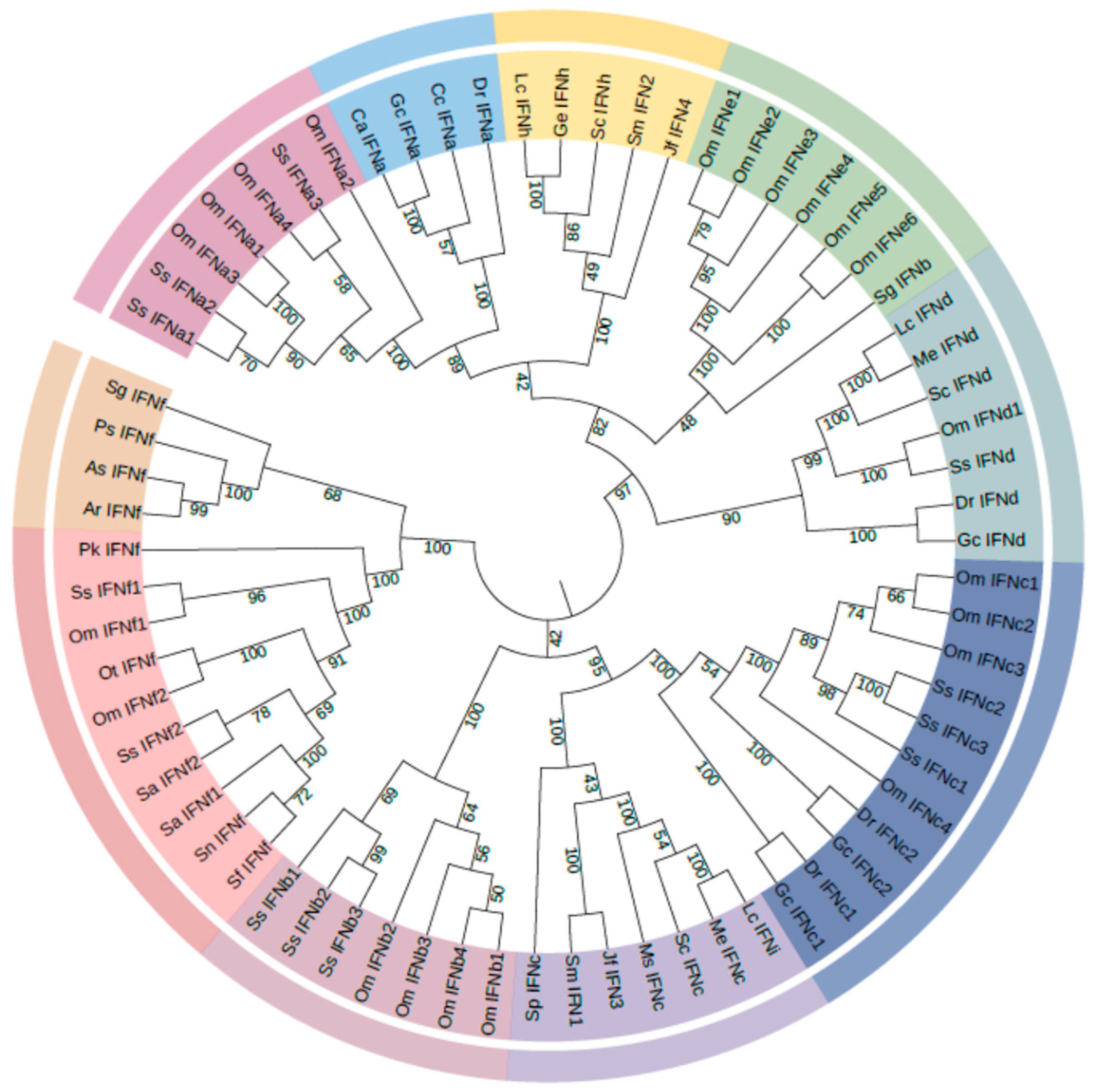
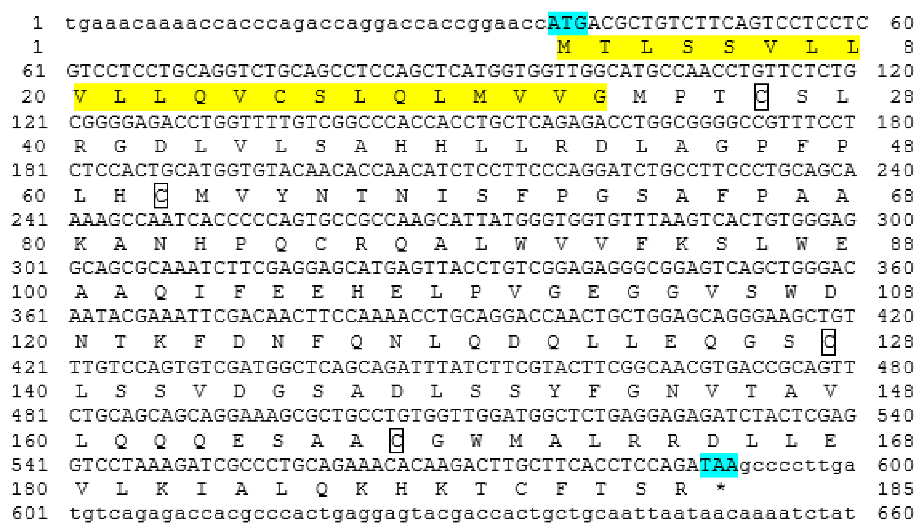
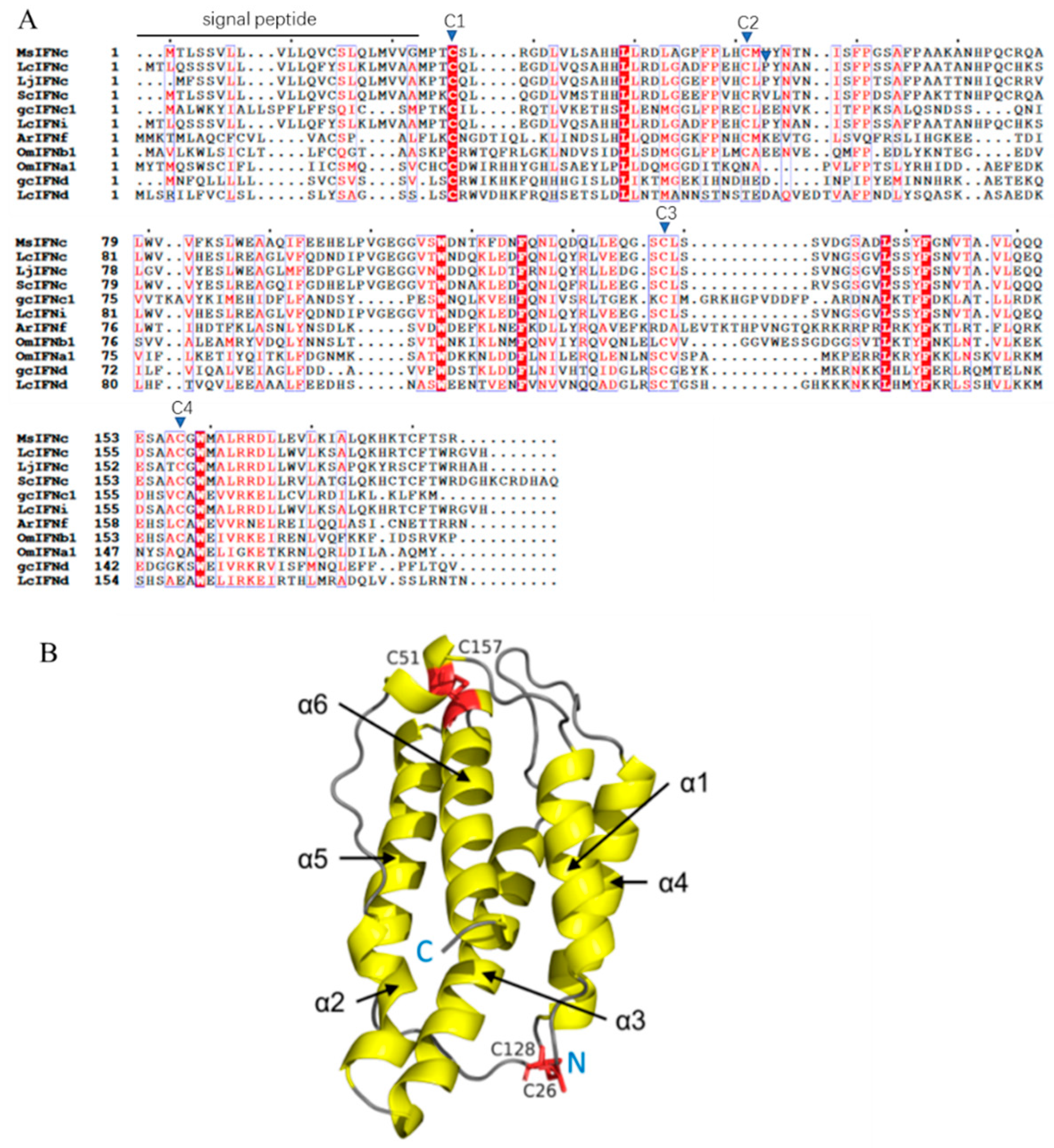
The MsIFNc cDNA was 660 bp in length, including a 36 bp 5′-untranslated region (UTR) and a 69 bp 3′-UTR, and 555 bp ORFs encoding a polypeptide of 184 amino acids (aa). A 22 aa signal peptide was identified in MsIFNc (Figure 1). The molecular weight of MsIFNc was 20.3 kDa, with a theoretical isoelectric point of 5.81. Multiple sequence alignment revealed the presence of four conserved cysteine residues (C26, C51, C128 and C157) in MsIFNc (Figure 2A). A 3-D model analysis showed that MsIFNc comprised six α-helix domains, and the four cysteines formed two disulfide bonds in the patterns C26-C128 and C51-C157 (Figure 2B). Sequence identities indicated that MsIFNc shared high identities with IFNc from Larimichthys crocea (82.5%), followed by those from Lateolabrax japonicus (82.3%) and Siniperca chuatsi (79.4%). Furthermore, MsIFNc exhibited identities ranging from 41.1% to 47.1% with other fish type I IFNs, the highest with Salmo salar IFNb5 (47.1%) and the lowest with Danio rerio IFNa (41.1%) (Table S2). Phylogenetic tree analysis showed that MsIFNc was well clustered with IFNc from teleosts (Figure 3).

3.2. Expression Patterns of MsIFNc

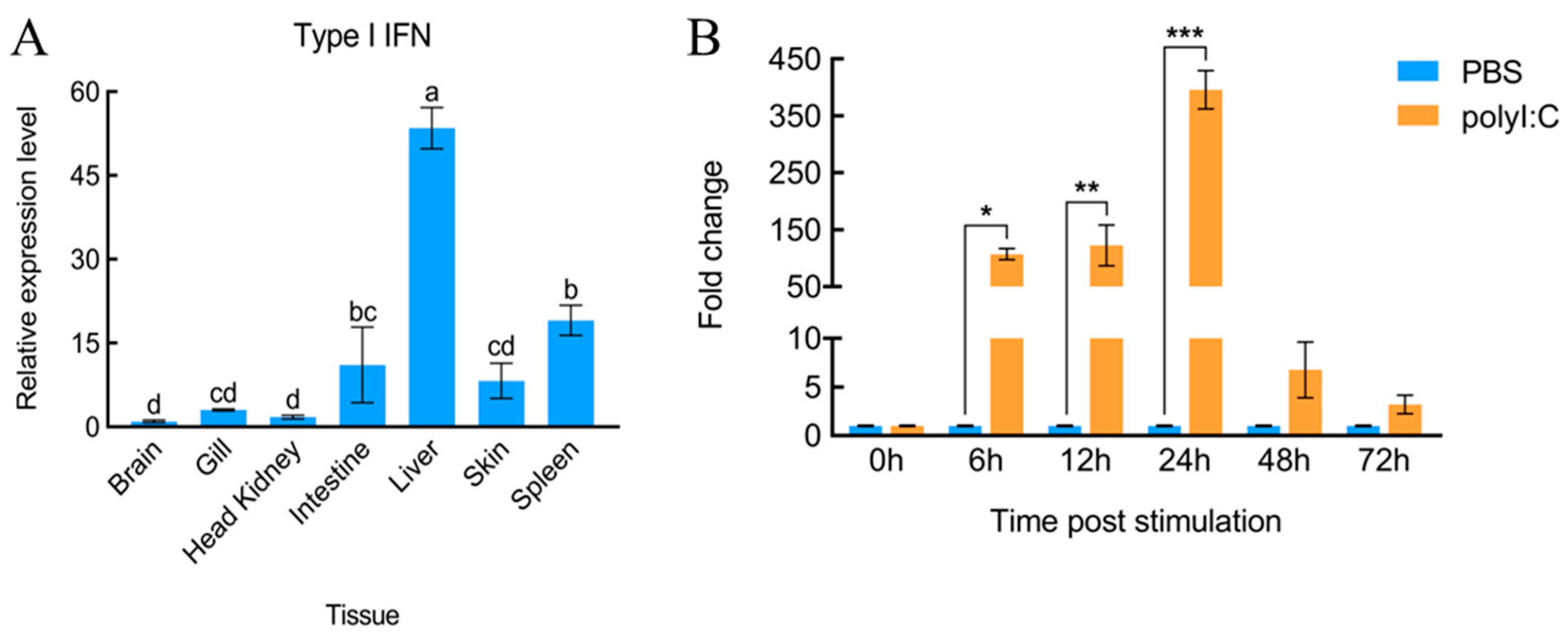
The expression levels of MsIFNc in normal tissues, including brain, gill, HK, intestine, liver, skin and spleen, were examined by qPCR. The results showed that MsIFNc was prominently expressed in liver, followed by intestine and spleen (Figure 4A). Furthermore, MsIFNc was significantly upregulated at 6 hpi to 24 hpi following polyI:C stimulation (Figure 4B).

3.3. Preparation of rMsIFNc

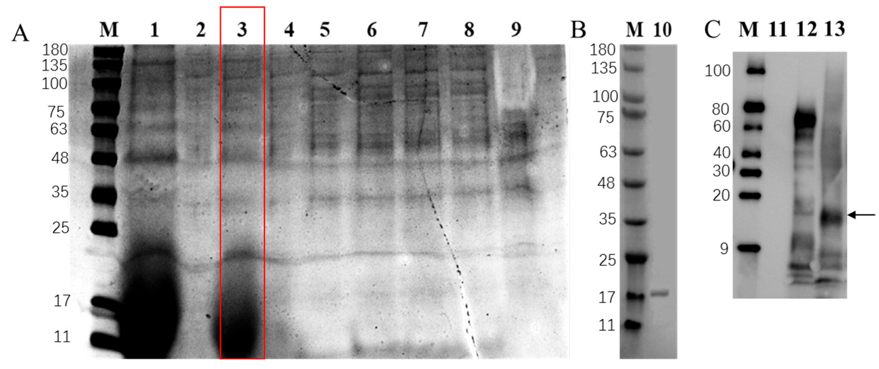
The cDNA encoding mature MsIFNc was constructed into the pPICZα A vector, which successfully constructed the recombinant plasmid pPiczαA-IFNc. Then, the plasmid was transformed into P. pastoris X-33, and rMsIFNc protein was induced by 1% methanol. The culture supernatant induced by methanol every 12 h was analyzed by SDS-PAGE, with the finding that the protein band at about 18 kDa was observed at 24 h post methanol induction (Figure 5A). A specific band at about 18 kDa was obtained by purification with the ÄKTA™ pure system (Figure 5B). These results indicated that no significant N-glycosylation occurred under the expression conditions used in P. pastoris X-33. The total protein in the crude culture supernatant was 1 mg/mL, and rMsIFNc protein was calculated to be 25.2% of this total by Western blotting analysis and BAC assay (Figure 5C). Thus, the concentration of rMsIFNc in the crude supernatant was determined to be 252 μg/mL.

3.4. Effect of rMsIFNc on Cell Viability Following MSRV Infection

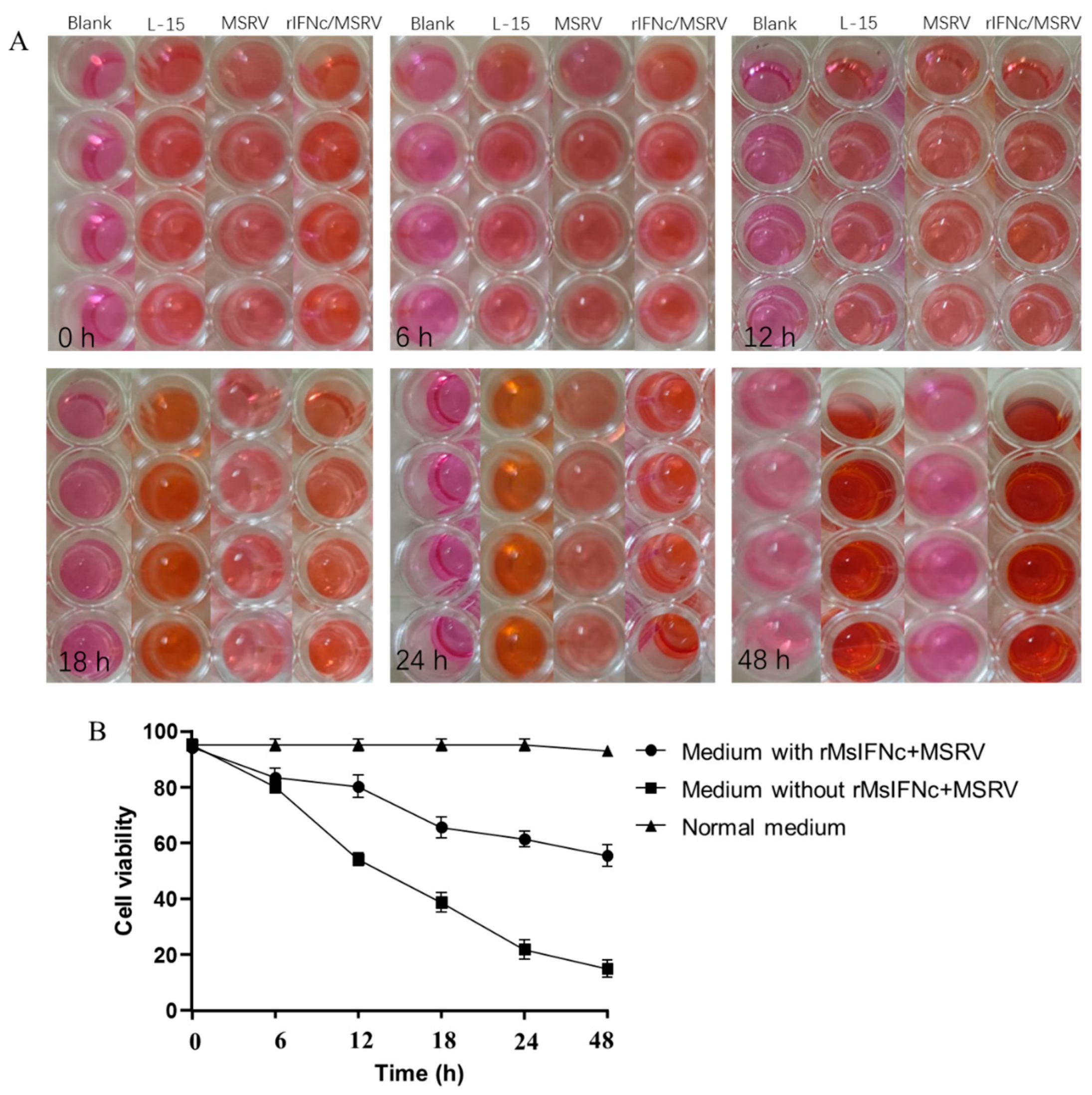
Primary hepatocytes were stimulated with crude supernatant containing rMsIFNc or not containing rMsIFNc and infected with MSRV; then, the cell viability was counted using the CCK8 kit (Figure 6A) and the OD450 was measured. The results showed that the culture supernatant containing rMsIFNc enhanced the protection of cells against MSRV infection (Figure 6B). The cell viability of cells treated with culture supernatant containing rMsIFNc remained 55.58% at 48 h post MSRV infection, which was significantly higher than that of cells treated with culture supernatant not containing rMsIFNc (15.07%) (p < 0.05) (Figure 6B).

4. Discussion

Type I IFNs play crucial roles in the antiviral immune response and immunoregulatory processes of fish [25]. Two groups of type I IFNs had been identified in fish [8]. However, the information about type I IFNs in largemouth bass is limited. In the present study, an interferon gene was characterized from largemouth bass. Based on the following analysis, we named this IFN as IFNc, belonging to the group II IFNs: (i) this IFN possesses the conserved structure of type I IFNs, consisting of six α-helices (Figure 2B); (ii) this IFN contains four conserved cysteines (Figure 1 and Figure 2A); (iii) this IFN clusters with fish IFNc, which is separate from other fish IFNs (Figure 3).
MsIFNc was ubiquitously expressed in all examined tissues (Figure 4A). Similar results were also observed in large yellow croaker (Larimichthys crocea) [26], mandarin fish (Siniperca chuatsi) [27], rock bream (Oplegnathus fasciatus) [28] and zebrafish [13]. However, distinct expression patterns were exhibited in different fish species. Zebrafish IFNφ1 was highly expressed in the spleen, whist IFNφ4 was highly expressed in the brain [13]. IFNd and IFNh in large yellow croaker were most highly expressed in the head kidney (HK) and liver [28]. IFNc, IFNd and IFNh in mandarin fish were highly expressed in the HK [27]. We found that MsIFNc was highly expressed in the liver, followed by the spleen and intestine (Figure 1). These results indicate that the expression of fish type I IFNs in normal tissues might be fish-specific and tissue-specific. The spleen is an important lymphoid organ in fish, and the intestine is crucial for the mucosal immunity of fish [14]. The liver is not only an important metabolic organ but also a crucial immunological organ of fish [29]. High expression of MsIFNc in these tissues indicates its immune roles in largemouth bass. Further, we found that MsIFNc could be upregulated in the spleen following polyI:C stimulation (Figure 4B). Similar results were also observed in other fish species [13,26]. These results indicate that MsIFNc is involved in the antiviral immunity of largemouth bass.
To further analyze the function of MsIFNc, the MsIFNc protein was prepared using a yeast expression system (Figure 5A,B) and verified by Western blot (Figure 5C). Further, the effect of rMsIFNc on the cell viability of hepatocytes following MSRV infection was studied in vitro, with the finding that rMsIFNc protected the primary hepatocytes from MSRV infection (Figure 6). Similar results were also observed in the primary fin cells of Chinese sturgeon treated with rIFNf and rIFNe2 following spring viremia of carp virus (SVCV) infection [30]. These results indicate that type I IFNs might have functions during MSRV infection. More studies should be conducted to elucidate the antiviral activity and mechanism of MsIFNc.
In conclusion, MsIFNc, belonging to group II of the type I IFNs, was identified in largemouth bass. MsIFNc was highly expressed in the liver, intestine and spleen and upregulated in the spleen following polyI:C stimulation. The MsIFNc protein, which was prepared by a yeast expression system, enhanced the cell viability of primary hepatocytes following MSRV infection. We have to admit that we only investigated the effect of MsIFNc on cell stability after MSRV infection, without conducting an in-depth exploration of the role and mechanism of MsIFNc during viral infection, which should be analyzed in our future work.

Supplementary Materials

The following supporting information can be downloaded at https://www.mdpi.com/article/10.3390/fishes11070376/s1, Table S1: Primers in this study; Table S2: Sequence identities between IFNc of largemouth bass and type I IFNs of other fish species; Table S3: Sequence used for this study.

Author Contributions

Q.Z., Conceptualization, Methodology, Formal analysis, Investigation; C.W., Methodology, Formal analysis, Investigation; J.L., Formal analysis, Software, Investigation; Y.W., Fromal analysis, Investigation; J.H., Software, Resources; M.P., Investigation, Data curation; Y.X., Formal analysis, Software; Z.Q., Writing—original draft, Writing—review and editing, Supervision, Funding acquisition. All authors have read and agreed to the published version of the manuscript.

Funding

The Projects for the High-quality Development of Fishery Industry of Yancheng City (Grant No. YCSCYJ20210014 and ycyy2024005) and Natural Science Fund of Shandong Province (Grant No. ZR2023MC208).

Institutional Review Board Statement

This study was conducted in accordance with the Declaration of Helsinki and the regulations for the administration of laboratory animals in Jiangsu Province, China (2008 [45]). All surgery was performed under anesthesia using 500 mg/L gethyl-3-aminobenzoate methanesulfonate (MS-222, Sigma, USA). The experimental design adhered to the 3Rs principle (Replacement, Reduction, and Refinement) to minimize animal suffering. In the absence of a formal Institutional Animal Care and Use Committee mandate for Yancheng Institute of Technology, the corresponding authors assume full responsibility for applied animal welfare standards.

Informed Consent Statement

Not applicable.

Data Availability Statement

The data that support the findings of this study are available from the corresponding author upon reasonable request.

Conflicts of Interest

The authors declare that they have no competing financial interests or personal relationships that could have appeared to influence the work reported in this paper.

References

  1. Stosik, M.; Tokarz-Deptula, B.; Deptula, W. Type I interferons in ray-finned fish (Actinopterygii). Fish Shellfish Immunol. 2021, 110, 35–43. [Google Scholar] [CrossRef] [Scilit] [PubMed]
  2. Lai, J.Y.; Ho, J.X.; Kow, A.S.F.; Liang, G.; Tham, C.L.; Ho, Y.C.; Lee, M.T. Interferon therapy and its association with depressive disorders—A review. Front. Immunol. 2023, 14, 1048592. [Google Scholar] [CrossRef] [Scilit] [PubMed]
  3. Chronopoulou, S.; Tsochantaridis, I. Interferon lambda: The next frontier in antiviral therapy? Pharmaceuticals 2025, 18, 785. [Google Scholar] [CrossRef] [Scilit] [PubMed]
  4. Zhou, Z.; Hmming, O.J.; Ank, N.; Paludan, S.R.; Nielsen, A.L.; Hartmann, R. Type III interferon (IFN) induces a type I IFN-like response in a restricted subset of cells through signaling pathways involving both the Jak-STAT pathway and the mitogen-activated protein kinases. J. Virol. 2007, 81, 7749–7758. [Google Scholar] [CrossRef] [Scilit] [PubMed]
  5. Zhang, S.Y.; Boisson-Dupuis, S.; Chapgier, A.; Yang, K.; Bustamante, J.; Puel, A.; Picard, C.; Abel, L.; Jouanguy, E.; Casanova, J.L. Inborn errors of interferon (IFN)-mediated immunity in humans: Insights into the respective roles of IFN-α/β, IFN-γ and IFN-λ in host defense. Immunol. Rev. 2008, 226, 29–40. [Google Scholar] [CrossRef] [Scilit] [PubMed]
  6. Aggad, D.; Stein, C.; Sieger, D.; Mazel, M.; Boudinot, P.; Herbomel, P.; Levraud, J.-P.; Lutfalla, G.; Leptin, M. In vivo analysis of Ifn-γ1 and Ifn-γ2 signaling in zebrafish. J. Immunol. 2010, 185, 6774–6782. [Google Scholar] [CrossRef] [Scilit] [PubMed]
  7. Chen, S.N.; Gan, Z.; Hou, J.; Yang, Y.C.; Huang, L.; Huang, B.; Wang, S.; Nie, P. Identification and establishment of type IV interferon and the characterization of interferon-υ including its class II cytokine receptors IFN-υR1 and IL-10R2. Nat. Commun. 2022, 13, 999. [Google Scholar] [CrossRef] [Scilit] [PubMed]
  8. Zou, J.; Tafalla, C.; Truckle, J.; Secombes, C.J. Identification of a second group of type I IFNs in fish sheds light on IFN evolution in vertebrates. J. Immunol. 2007, 179, 3859–3871. [Google Scholar] [CrossRef] [Scilit] [PubMed]
  9. Chang, M.X.; Nie, P.; Collet, B.; Secombes, C.J.; Zou, J. Identification of an additional two-cysteine containing type I interferon in rainbow trout Oncorhynchus mykiss provides evidence of a major gene duplication event within this gene family in teleosts. Immunogenetics 2009, 61, 315–325. [Google Scholar] [CrossRef] [Scilit] [PubMed]
  10. Zou, J.; Gorgoglione, B.; Taylor, N.G.; Summathed, T.; Lee, P.T.; Panigrahi, A.; Genet, C.; Chen, Y.M.; Chen, T.Y.; Ul Hassan, M.; et al. Salmonids have an extraordinary complex type I IFN system: Characterization of the IFN locus in rainbow trout Oncorhynchus mykiss reveals two novel IFN subgroups. J. Immunol. 2014, 193, 2273–2286. [Google Scholar] [CrossRef] [Scilit] [PubMed]
  11. Chen, S.N.; Gan, Z.; Huang, B.; Zou, J.; Nie, P. Fish type I and type II interferons: Composition, receptor usage, production and function. Rev. Aquacult. 2020, 12, 773–804. [Google Scholar]
  12. Wen, Q.Q.; Tang, S.S.; Mo, J.Y.; Zhang, M.L.; Long, M.; Lu, Y.S.; Gan, Z. Different activation of STAT1 and STAT2 phosphorylation by IFNc, IFNd, and IFNh in tilapia. Fish Shellfish Immunol. 2024, 152, 109776. [Google Scholar] [CrossRef] [Scilit] [PubMed]
  13. Chen, K.Y.; Tian, J.Y.; Shi, Y.J.; Xie, T.; Huang, W.J.; Jia, Z.; Zhang, Y.W.; Yuan, G.L.; Yan, H.; Wang, J.Y.; et al. Distinct antiviral activities of IFNφ1 and IFNφ4 in zebrafish. Fish Shellfish Immunol. 2024, 146, 109396. [Google Scholar] [CrossRef] [Scilit] [PubMed]
  14. Pi, X.Y.; Yan, D.; Xu, Y.; Pan, M.Z.; Wang, Z.S.; Chang, M.X.; Qi, Z.T. TLRs signaling pathway regulation, antibacterial and apoptotic activity of largemouth bass ECSIT during Edwardsiella piscicida infection. Aquaculture 2025, 595, 741615. [Google Scholar] [CrossRef] [Scilit]
  15. Yang, S.; Zhao, J.; An, N.; Li, D.C.; Huang, M.M.; Fei, H. Updates on infectious diseases of largemouth bass: A major review. Fish Shellfish Immunol. 2024, 154, 109976. [Google Scholar] [CrossRef] [Scilit] [PubMed]
  16. Pi, X.Y.; Pan, M.Z.; Wei, X.; Zhang, Q.H.; Qi, Z.T. Edwardsiella piscicida induces largemouth bass PINK1/Parkin-mediated mitophagy for its survival and virulence. J. Immunol. 2026, 215, vkaf277. [Google Scholar] [PubMed]
  17. Qi, Z.T.; Xu, Y.; Liu, Y.H.; Zhang, Q.H.; Wang, Z.S.; Mei, J.; Wang, D.Z. Transcriptome analysis of largemouth bass (Micropterus salmoides) challenged with LPS and polyI:C. Fish Shellfish Immunol. 2023, 133, 108534. [Google Scholar] [CrossRef] [Scilit] [PubMed]
  18. Artimo, P.; Jonnalagedda, M.; Arnold, K.; Baratin, D.; Csardi, G.; de Castro, E.; Duvaud, S.; Flegel, V.; Fortier, A.; Gasteiger, E.; et al. ExPASy: SIB bioinformatics resource portal. Nucleic Acids Res. 2012, 40, W597–W603. [Google Scholar] [CrossRef] [Scilit] [PubMed]
  19. Sali, A.; Blundell, T.L. Comparative protein modeling by satisfaction of spatial restraints. J. Mol. Biol. 1993, 234, 779–815. [Google Scholar] [CrossRef] [Scilit] [PubMed]
  20. Robert, X.; Guillon, C.; Gouet, P. FoldScript: A web server for the efficient analysis of AI-generated 3D protein models. Nucleic Acids Res. 2025, 53, W277–W282. [Google Scholar] [CrossRef] [Scilit] [PubMed]
  21. Kumar, S.; Stecher, G.; Tamura, K. MEGA 7: Molecular evolutionary genetics analysis version 7.0 for bigger datasets. Mol. Biol. Evol. 2016, 33, 1870–1874. [Google Scholar] [CrossRef] [Scilit] [PubMed]
  22. Waterhouse, A.; Bertoni, M.; Bienert, S.; Studer, G.; Tauriello, G.; Gumienny, R.; Heer, F.T.; de Beer, T.A.P.; Rempfer, C.; Bordoli, L.; et al. SWISS-MODEL: Homology modelling of protein structures and complexes. Nucleic Acids Res. 2018, 46, W296–W303. [Google Scholar] [CrossRef] [Scilit] [PubMed]
  23. Pi, X.Y.; Pan, M.Z.; Zhang, Q.H.; Wang, Z.S.; Qi, Z.T. Molecular characterization, expression, evolutionary analysis and molecular docking of the Janus activated kinase family members of largemouth bass (Micropterus salmoides). Dev. Comp. Immunol. 2025, 167, 105375. [Google Scholar] [CrossRef] [Scilit] [PubMed]
  24. Pfaffl, M.W. A new mathematical model for relative quantification in real-time RT-PCR. Nucleic Acids Res. 2001, 29, e45. [Google Scholar] [CrossRef] [Scilit] [PubMed]
  25. Secombes, C.J.; Zou, J. Evolution of interferon and interferon receptors. Front. Immunol. 2017, 8, 209. [Google Scholar] [CrossRef] [Scilit] [PubMed]
  26. Ding, Y.; Ao, J.; Huang, X.; Chen, X. Identification of two subgroups of type I IFNs in perciforme fish large yellow croaker Larimichthys crocea provides novel insights into function and regulation of fish type I IFNs. Front. Immunol. 2016, 7, 343. [Google Scholar] [CrossRef] [Scilit] [PubMed]
  27. Laghari, Z.A.; Chen, S.N.; Li, L.; Huang, B.; Gan, Z.; Zhou, Y.; Huo, H.J.; Hou, J.; Nie, P. Functional, signalling and transcriptional differences of three distinct type I IFNs in a perciform fish, the Mandarin fish Siniperca chuatsi. Dev. Comp. Immunol. 2018, 84, 94–108. [Google Scholar] [CrossRef] [Scilit] [PubMed]
  28. Wan, Q.; Wicramaarachchi, W.D.; Wang, I.; Lim, B.S.; Oh, M.J.; Jung, S.J.; Kim, H.C.; Yeo, S.-Y.; Lee, J. Molecular cloning and functional characterization of two duplicated two-cysteine containing type I interferon genes in rock bream Oplegnathus fasciatus. Fish Shellfish Immunol. 2012, 33, 886–898. [Google Scholar] [CrossRef] [Scilit] [PubMed]
  29. Gao, B.; Jeong, W.; Tian, Z. Liver: An organ with predominant innate immunity. Hepatology 2008, 47, 729–736. [Google Scholar] [CrossRef] [Scilit] [PubMed]
  30. Ding, G.Y.; Yu, P.P.; Deng, D.; Xie, M.; Luo, K.; Zhang, F.; Xu, D.; Xu, Q.; Guo, H.; Zhang, S. Functional characterization of group II interferon, IFNf in the acipenseriform fish, Chinese sturgeon (Acipenser sinensis). Fish Shellfish Immunol. 2024, 144, 109240. [Google Scholar] [CrossRef] [Scilit] [PubMed]
Figure 1. The nucleotide and putative amino acid sequence of MsIFNc. The start codon (ATG) and stop codon (TAA) are marked in light blue; the signal peptide is marked in yellow; the four conserved cystines are boxed; and the 5′-UTR and 3′-UTR are in lowercase letters.
Fishes 11 00376 g001
Figure 2. Sequence alignment of MsIFNc with fish type I IFNs (A) and the 3-D structure of MsIFNc (B). The sequence alignment was performed by Clustal O and optimized using ESPript 3.0. The four conserved cystines are marked with blue triangles above the sequences. The 3-D structure of MsIFNc was constructed by the Swiss-Model website.
Fishes 11 00376 g002
Figure 3. Phylogenetic tree analysis of fish type I IFNs. The phylogenetic tree was constructed using the Neighbor-Joining (NJ) method in MEGA 7.0 software, with bootstrap values set to 10,000 replicates. The values on the nodes represent bootstrap support. The accession numbers of the IFNs used for the phylogenetic tree construction are listed in Table S3.
Fishes 11 00376 g003
Figure 4. The expression of MsIFNc in normal tissues from largemouth bass (A) and in the spleen following polyI:C stimulation. (A) The relative expression of MsIFNc in each tissue was normalized to that of β-actin. Different letters above the columns indicate a significant difference between the tissues (p < 0.05). (B) Changes in MsIFNc expression in the spleen following polyI:C stimulation were analyzed by the 2−ΔΔCt method and expressed as fold changes relative to time-matched controls. Results are expressed as the mean ± SE. * p < 0.05; ** p < 0.01, *** p < 0.001.
Fishes 11 00376 g004
Figure 5. Preparation of rMsIFNc. (A) SDS-PAGE analysis of the culture supernatant induced by 1% methanol every 12 h. Lane M: protein marker; Lane 1: pPiczαA vector; Lanes 2 to 9: supernatant collected at 0, 12, 24, 36, 48, 60 and 72 h post 1% methanol induction. Methanol (1% v/v) was added every 12 h throughout the induction period. (B) Purification of rMsIFNc. Lane 10: the purified rMsIFNc. (C) Western blot analysis of the supernatant. Lane 11: supernatant without methanol induction; Lane 12: positive control (BSA); Lane 13: supernatant with methanol induction for 24 h. Red box indicates the band at 24 h post methanol induction. The black arrow indicates the results of western blot assay for MsIFNc protein.
Fishes 11 00376 g005
Figure 6. Cell viability of primary hepatocytes treated with culture supernatant containing rMsIFNc or not containing rMsIFNc and infected with MSRV. (A) Cell staining with CCK8. (B) Cell viability derived from OD450 measurement. The error bars represent the standard error (SE) of the mean (SEM) derived from four replicate wells per group.
Fishes 11 00376 g006
Disclaimer/Publisher’s Note: The statements, opinions and data contained in all publications are solely those of the individual author(s) and contributor(s) and not of MDPI and/or the editor(s). MDPI and/or the editor(s) disclaim responsibility for any injury to people or property resulting from any ideas, methods, instructions or products referred to in the content.