Article

Effect of Elevated Temperature and Excess Light on Photosynthetic Efficiency, Pigments, and Proteins in the Field-Grown Sunflower during Afternoon

by
Antonela Markulj Kulundžić
1,*,
Marija Viljevac Vuletić
2,*,
Maja Matoša Kočar
1,
Jasenka Antunović Dunić
3,
Ivana Varga
4,
Zvonimir Zdunić
5,6,
Aleksandra Sudarić
1,6,
Vera Cesar
3,7 and
Hrvoje Lepeduš
7,8
1
Department of Industrial Plants, Agricultural Institute Osijek, 31000 Osijek, Croatia
2
Agrochemical Laboratory, Agricultural Institute Osijek, 31000 Osijek, Croatia
3
Department of Biology, J.J. Strossmayer University of Osijek, 31000 Osijek, Croatia
4
Faculty of Agrobiotechnical Sciences Osijek, J.J. Strossmayer University of Osijek, 31000 Osijek, Croatia
5
Service of General, Legal and Personnel Affairs, Agricultural Institute Osijek, 31000 Osijek, Croatia
6
Centre of Excellence for Biodiversity and Molecular Plant Breeding, Faculty of Agriculture, University of Zagreb, 10000 Zagreb, Croatia
7
Faculty for Dental Medicine and Health, J.J. Strossmayer University of Osijek, 31000 Osijek, Croatia
8
Faculty of Humanities and Social Sciences, J.J. Strossmayer University of Osijek, 31000 Osijek, Croatia
*
Authors to whom correspondence should be addressed.
Horticulturae 2022, 8(5), 392; https://doi.org/10.3390/horticulturae8050392
Submission received: 30 March 2022 / Revised: 25 April 2022 / Accepted: 27 April 2022 / Published: 28 April 2022

Abstract

:
This study examined the photosynthetic responses of two sunflower hybrids to elevated temperatures and excess light intensity in the flowering stage by measuring the chlorophyll a fluorescence (ChlF) under morning and afternoon field conditions to determine the photosynthetic pigment contents and the relative accumulation of photosynthetic proteins. The morning environmental conditions were considered optimal, while the afternoon was characterised by elevated temperatures and excess light intensity. The minimum fluorescence intensity (F0), the electron-flux-reducing end electron acceptors at the photosystem I acceptor side per reaction centre (RE0/RC), and the D1 protein had significant, high, and positive correlations with the environmental conditions, which indicates that they were the most useful in the sunflower-stress-response research. In hybrid 7, the elevated temperatures and the excess light intensity resulted in the inactivation of the oxygen-evolving complex, which was indicated by the positive L, K, and J steps, the increase in the maximum quantum yield of PSII (TR0/ABS), the decrease in the electron transport further than the primary acceptor QA (ET0/(TR0-ET0)), the reduction in the performance index (PIABS), and the higher relative accumulation of the light-harvesting complex of the photosystem (LHCII). Hybrid 4 had smaller changes in the fluorescence curves in phases O–J and J–I, and especially in steps L, K, J, and I, and a higher PIABS, which indicates a more efficient excitation energy under the unfavourable conditions. As the tested parameters were sensitive enough to determine the significant differences between the sunflower hybrids in their photosynthetic responses to the elevated temperatures and excess light intensity in the flowering stage, they can be considered useful selection criteria. The development of more adaptable sunflower hybrids encourages sustainable sunflower production under stressful growing conditions.

1. Introduction

Sunflower (Helianthus annuus L.) is a high-value, widespread oilseed. It is considered to be an important raw material for the production of oil and many other food and nonfood products. Sunflower production is influenced by many different environmental factors that interact, and that often cause plant stress. Because extreme temperature variations, sunlight intensity, quality variations, as well as uneven precipitation patterns that are accompanied by changes in the wind intensity and cloudiness are becoming more frequent [1], studies that investigate the relationship between plants and unfavourable weather conditions are becoming more and more important, and they enable the prediction of plant responses. The environmental factors that play major roles in plant stress are high temperatures, excess light, and drought. According to García-López et al. [2], sunflower is moderately tolerant to drought and heat stress. It is most sensitive to heat from early flowering to grain filling. Growing sunflower at elevated temperatures reduces its growth, which is reflected in the specific leaf mass, the leaf surface, and the soluble protein content [3]. Among the physiological processes, photosynthesis [4] is the most susceptible to the negative impact of heat. In addition to heat, the high light intensity can also negatively affect photosynthesis [5].
At the end of the 20th century, significant progress was made by using modern optical methods and techniques to study photosynthetic processes [6], among which one of the most commonly used methods is the determination of the chlorophyll a fluorescence (ChlF). The ChlF provides insight into the photosynthetic apparatus status and function, the efficiency of photosystem II (PSII), and the electron transport chain function [7]. Many authors have used ChlF parameters to study the effect of stress on plants [5,8,9,10,11].
Acclimatisation to different environments is interrelated to photosynthetic adjustment, which consequently affects the biochemical and physiological processes, the growth, and the yield [12]. Most plants show a significant ability to adjust photosynthesis to temperature and light fluctuations. Elevated temperatures negatively affect cell division and expansion, and they are one of the main stresses that stimulate protein degradation and that cause tissue senescence or death [13]. The decrease in photosynthesis occurs not only because of changes in the flow of energy through PSII, which is extremely sensitive to elevated temperatures, but also because of the reduced content of the pigments in leaves [14]. PSII damage that is caused by elevated temperatures occurs mainly at the oxygen-evolving centre (OEC), and so even slightly increased temperatures cause its deactivation [15]. Although an increase in light intensity can gradually increase the photosynthetic rate, reactive centres absorb more light when the intensity of the light is high (i.e., when there is more light than can be used in photochemistry). The remaining energy is dissipated as heat and fluorescence. If the energy is not utilised or if it is dissipated, it causes photooxidative stress and it increases the level of reactive oxygen species (ROS) [16]. High light intensity is the cause of numerous other disorders in plants besides photodamage, photoactivation, and photoinhibition. One of these disorders is the degradation of photosynthetic proteins. It has been proven that the accumulation of the light-harvesting complex of PSII (LHCII) is related to the chlorophyll a and b ratio, and that it depends on daily weather fluctuations [17]. Protein D1, which is an essential part of the photosynthetic apparatus, is also sensitive to stress [18]. Excess light causes D1 protein phosphorylation, which results in degradation, de novo protein synthesis, and protein incorporation into PSII [19]. The accumulation of the cytochrome f protein is also crucial because the c6f protein complex connects to PSII and to photosystem I (PSI) with cyclic and linear electron transfer [20]. Another significant protein that is directly related to temperature is Rubisco. Its abundance changes under stress [21].
As the global weather changes, extreme weather conditions that occur in the estival afternoons during the most critical sunflower developmental stages are becoming more frequent. Such conditions can often cause short-term temperature and light stress in sunflower plants, which are reflected as changes in the ChlF parameters [10]. Although plants can show a significant ability to adjust photosynthesis to temperature and light fluctuations through daily changes during plant growth, there are those that are less tolerant. Therefore, the timely determination of the stress occurrence and the stress tolerance and the elimination of stress-susceptible genotypes are of great importance for plant production. This study aimed to differentiate genotypes on the basis of their photosynthetic responses to elevated temperatures and excess light intensity in the flowering stage by measuring the ChlF, the photosynthetic pigment contents, and the relative accumulation of photosynthetic proteins in the field (i.e., in their ambient environments). The synergic effect of elevated temperatures and excess light, which are known to be correlated with sunflower leaf stress and to cause the most problems in many production areas, were determined in the afternoon hours during the sunflower flowering stage. Knowing the specific reactions of the individual hybrids to adverse environmental conditions allows breeders to better understand the characteristics of the material during selection, which also increases the breeding programme’s success. Since this study is a part of the sunflower breeding programme, and since it compares the responses of different hybrids to the conditions that are known to affect the metabolism in plants, it is important to select superior material. On the basis of the abovementioned, it was hypothesised that one hybrid would be more adaptable to the elevated temperatures and excess light determined in the chosen afternoons, and that it would show minor daily changes in the tested parameters.

2. Materials and Methods

2.1. Plant Material

The experiment was conducted at the Agricultural Institute Osijek (Osijek, Croatia) on two sunflower hybrids. The hybrids differed in pedigree and in agronomic properties (plant height, head diameter, yield potential), but they had similar maturation times. The hybrids were chosen on the basis of the results of previous trials [22,23]. Hybrid 4 has been recognised and has been widely spread throughout the sunflower production in Croatia in recent years. The producers accept it because of its high seed quality and seed yields under various growing conditions, which shows its stability and wide adaptability. Hybrid 7 is an experimental material that stood out in multiyear microtrials, with good overall agronomic qualities and high oil content. A comparison of the difference in the response mechanisms of these two hybrids to unfavourable environmental conditions (elevated temperature and excess light intensity) will be beneficial for determining the direction of future breeding programmes.
The sowing was performed with manual hand planters (two seeds per hill) at a 4 cm depth in four 5 m-long rows, with a 70 cm distance between the rows, and a 23 cm distance within the rows (45°32′ N, 18°44′ E; 94 m altitude), in four replications. The final density was 6.29 plant/m2. A randomised complete block experimental design was used. The soil analysis determined that the soil texture was silty clay loamy, and it is classified as anthropogenic Eutric Cambisol. The physical and chemical properties in the upper soil layer of the soil were: 64.7% silt; 32.5% clay; 2.8% sand; a pH in K2O of 7.3; a pH in H2O of 7.9; P2O5 > 41 mg/100 g; N: 0.16%; K2O > 40 mg/100 g; CaCO3: 0.9%; Al (mobile): 0.26 mg/100 g; and a humus content of 2.18%. During the experiment, all of the agrotechnical measures were performed by following the recommendations and requirements of sunflower cultivation.

2.2. Weather Conditions

The minimum, maximum, and mean air temperatures, the solar radiation intensity, and the precipitation for the ten days preceding the measurement and sampling are shown in Supplementary Table S1 in order to provide more insight into the environmental conditions that the photosynthetic apparatus needed to adapt to. The temperature, solar radiation, and precipitation measurements were recorded every 10 min (0–24 h), after which the average values per day were calculated.
The measurements were made on a cloudless day (4 August) during the flowering stage, in the morning (7:30–9:00 am) and in the afternoon (12:30–2:00 pm). These two measurement times were selected to quantify the photosynthetic apparatus reaction to and the changes in the physiological parameters under the elevated temperature and excess light conditions that were determined to be characteristic of the early afternoon hours, compared to the lower temperatures and low light conditions that were determined to be characteristic of the early morning hours (optimal conditions). The mean air temperature and the solar radiation during the measurement and sampling in the morning were 27.6 °C and 332.3 W/m2, respectively. In the afternoon, they were 35.7 °C and 830.2 W/m2, respectively. The mentioned afternoon temperature and solar radiation were previously found to be the causes of changes in the photosynthetic efficiency [5,10]. The temperatures, the solar radiation intensities, and the precipitation were recorded by a command and data-acquisition station near the experiment.

2.3. Chlorophyll a Fluorescence (ChlF)

The ChlF was determined during the flowering stage (according to Schneiter and Miller [24], in the R5.5 stage) by a plant efficiency analyser (Handy PEA, Hansatech, Norfolk, UK). The youngest (upper) developed sunflower leaves were used for the ChlF measurements. A leaf is considered developed if it is larger than 4 cm [24]. The measurements were carried out in the middle two rows of each hybrid in the morning (7:30–9:00 am) and afternoon hours (12:30–2:00 pm), and in their ambient environments. The ChlF was determined on 12 leaves per hybrid (three leaves × four replicates) under field conditions. Before measuring the ChlF, the sunflower leaves were adapted to the dark for 30 min and were subjected to dark conditions, during which the electron transfer in the photosynthetic electron transport chain ceases. There is no water oxidation in PSII (i.e., at the OEC), as there is no charge separation at the reaction centre in darkness. After tissue illumination, the obtained information on the intensity of the ChlF during one second is displayed on the OJIP curve. Different environmental conditions can cause the appearance of additional steps in the ChlF OJIP transients [6]. The ChlF transients were induced by using a pulse of saturating red light (peak at 650 nm, 3200 μmol m−2 s−1). The JIP parameters that were calculated from the recorded data are shown in Supplementary Table S2.
A double normalisation of the OJIP transients was made between the O and P steps. The logarithmic time scale was used for presenting the relative variable fluorescence: WOP = (Ft − F0)/(FJ − F0). The K, L, J, and I steps were presented as the variable fluorescence: WOK = (Ft − F0)/(FK − F0), WOJ = (Ft − F0)/(FJ − F0) WOI = (Ft − F0)/(FI − F0), and WO50 = (Ft − F0)/(F50 − F0) plotted with difference kinetics: ΔWOK = WOK − (WOK)ref, ΔWOJ = WOJ − (WOJ)ref, ΔWOI = WOI − (WOI)ref, and ΔWO50 = WO50 − (WO50)ref. The measurements under the morning conditions were used as the reference values of (WOK)ref, (WOJ)ref, (WOI)ref, and (WO50)ref.

2.4. Laboratory Analyses

In total, the eight youngest leaves per hybrid on which the ChlF was determined were sampled for their photosynthetic pigments and for protein analysis. Before the biochemical analyses, the composite sample was homogenised into a powder by using liquid nitrogen.

2.4.1. Photosynthetic Pigment Content Determination

About 0.05–0.1 g of plant tissue, which was previously homogenised by liquid nitrogen with the addition of magnesium hydroxide carbonate, was extracted by 1 mL of cold acetone. The extraction procedure was repeated six times until the plant tissue was completely discoloured. Supernatants were pooled and used for the spectrophotometric measurement of the absorbance at 470, 645, and 662 nm. Acetone was used for the blank. The content of the photosynthetic pigments was calculated by using the appropriate extinction coefficients, according to Lichtenthaler [25]. The chlorophyll a/b and the chlorophyll a + b/Car were calculated as well. Five replicates were performed per the condition of each genotype.

2.4.2. SDS-PAGE and Immunodetection

The relative protein accumulation was determined by sodium dodecyl sulfate-polyacrylamide gel electrophoresis (SDS-PAGE). About 0.5 g of plant tissue was extracted with 1 mL of buffer, heated to 80 °C. The buffer consisted of 0.13 M Tris/HCl (pH = 6.8), 4.6% SDS, 16% glycerol, and 0.01 M dithiothreitol. The protein concentration was determined according to Bradford [26]. The protein extract contained 10 or 30 µg of protein, depending on the protein detected. For the detection of the Rubisco large subunit (LSU), a sample containing 10 µg of protein and 1 µg of loading buffer was applied to the gel, and for the other proteins (Lhcb2, D1, and cytochrome f), 30 µg of protein and 2 µg of buffer were used. After the separation with 12% SDS-PAGE [27], the proteins were transferred from the gel to the nitrocellulose membrane (Bio-Rad) in semidry conditions by using Biometra Fastblot B43 [28]. For the immunodetection, the specific primary antibodies against LHCII (anti-Lhcb2, Agrisera), D1 of Photosystem II (anti-PsbA, Agrisera), cytochrome f (anti-Cyt f, Agrisera), and Rubisco LSU (anti-RbcL, Agrisera), and then secondary antibodies (Donkey anti-rabbit IgG-HRP, Santa Cruz Biotechnology, Dallas, TX, USA), were used. Primary and secondary antibodies were diluted in a buffer in ratios of 1:5000 and 1:10,000, respectively. According to the manufacturer’s instructions, a commercial chemiluminescence detection substrate (Lumi-Light Western Blotting Substrate, Roche, Basel, Switzerland) was used to incubate the membranes. After that, protein bands were detected on ECL films (AGFA, Mortsel, Belgium), according to the standard procedure. ImageJ software was used for the protein band quantification. Three replicates were performed per the condition of each genotype.

2.5. Data Analyses

The ChlF parameters were calculated and visualised in Microsoft Excel, according to Strasser et al. [7]. A one-way ANOVA was used for determining the statistical differences between the hybrids under the morning and afternoon conditions of the ChlF (n = 12) and pigment-content (n = 5) measurements, which were followed by the Tukey’s post hoc honest significant difference (HSD) test at p < 0.05. The correlations among the JIP parameters, the pigment content, the photosynthetic proteins, the sunflower hybrids, and the environmental conditions were explored by principal component analysis (PCA) at p < 0.05. Before the PCA analysis, the data were standardised. The PCA was performed by using a correlation matrix of the average values after autoscaling. The mean values ± standard deviations in the table are used for presenting the data.

3. Results and Discussion

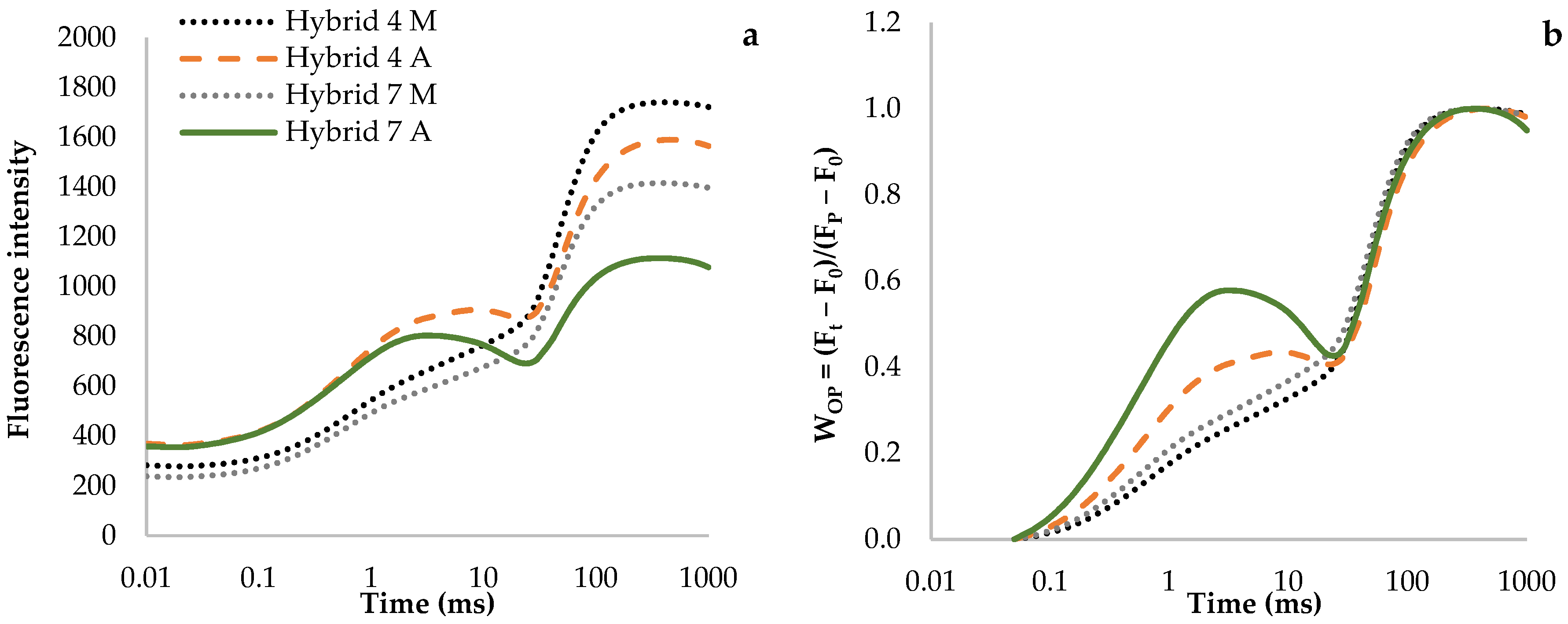
3.1. Fluorescence Transient Curves

The raw fluorescence induction curves showed a high deviation between the hybrids measured in the morning and in the afternoon, where a notable change in the OJIP-curve shape occurred in hybrid 7 (Figure 1a). The double O–P normalised curves, which show the measurement values of hybrids 4 and 7 under the morning conditions, had the typical form of a normalised OJIP curve (Figure 1b), while the curves that were measured in the afternoon altered significantly. A similar shape was found in peach leaves when exposed to high (more than 35 °C) temperatures [29].
The O–J phase, which is also known as the light-dependent phase, represents the 2 ms increase in the OJIP curve. This phase provides information about the excitation energy transfer between the PSII RCs and PSI [30]. It reveals the difference between the measurements under the morning and afternoon conditions, which is seen as the rise in the fluorescence curves for both hybrids. Still, the increase was more pronounced for hybrid 7, which is a consequence of reducing the primary plastoquinone (QA) acceptor [31]. The J–I phase is characterised by a partial reduction in the pool of plastoquinones, which is unlike the I–P phase, which represents the reduction in the PSI’s acceptor side [32]. It is evident from Figure 1b that the curves in the J step are more pronounced for hybrid 7; however, in the I step, the curves are the same for both hybrids and for both conditions. Brestic et al. [33] found a decrease in the fluorescence transient intensity in the J–I phase, followed by an I–P phase increase, which was confirmed in this investigation as well.
A more precise image of the O–P phase’s fluorescence intensity can be obtained from the individual representations of the normalised O–K, O–J, O–I, and O–50 curves. They clearly differentiate between the L, K, J, and I steps among the tested hybrids (Figure 2).
Under the synergic effect of elevated temperatures and excess light, the L step’s appearance at 150 µs reflects the positive transient values in both hybrids (Figure 2a), which signify the weaker energy connectivity and stability of the PSII units [32]. Under the same conditions, the rise in the kinetic fluorescence of the K, J, and I steps has positive curve amplitudes for both hybrids; however, higher amplitudes in all the steps were determined for hybrid 7 (Figure 2b–d). The positive curve amplitudes at step K (300 µs) indicate an impaired PSII antenna function during the electron flow, which was due to an increased reduction rate of the QA of the primary PSII electron acceptor, which indicates impaired OEC function [34]. Furthermore, steps J (2 ms) and I (30 ms) explain the reduction in the plastoquinone pool between PSII and PSI [35]. Although many studies confirm the occurrence of the K and L steps under high temperatures [29,33] and high light intensity [36], numerous authors report their occurrence in other stress conditions as well. The described reactions of sunflower hybrids 4 and 7 in the individual steps concur with the synergic effect of the elevated temperatures and the excess light that was determined in apple cultivars [5].

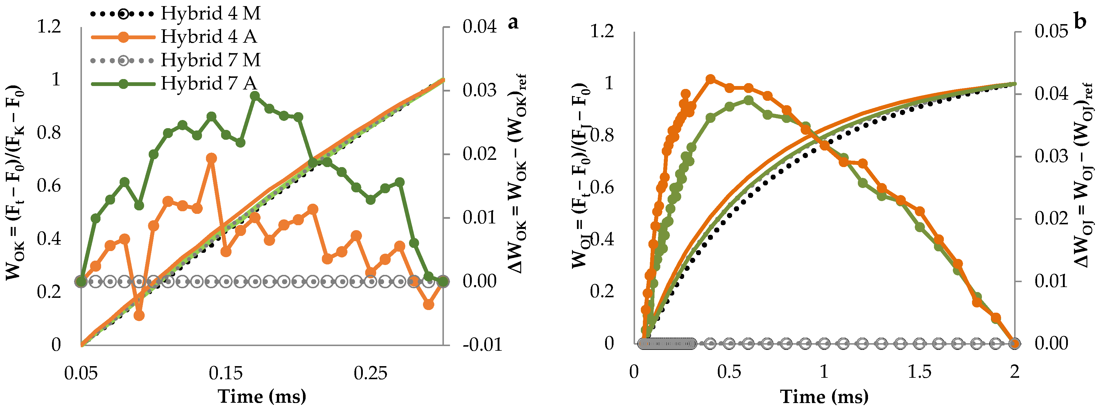
3.2. Chlorophyll JIP-Test Parameters

The results of the JIP-test parameters and the photosynthetic pigments in the morning and afternoon conditions are shown in Table 1.
The afternoon F0 measurements (when all the PSII RCs were open) increased in both hybrids compared to the values under the morning conditions, and with a more pronounced increase for hybrid 7. Contrary to the F0 values, the Fm values decreased because of the synergic effect of the elevated temperatures and excess light for both hybrids, and they represented their maximal intensity when all of the PSII RCs were closed. According to Schansker et al. [37], the fluorescence increase between steps F0 and Fm indicates QA reduction. These two parameters calculate the TR0/ABS, which demonstrates the likelihood that the absorbed photon energy can be trapped by PSII RCs [38]. Under the morning conditions, the TR0/ABS values were similar for hybrids 4 and 7; however, they decreased in the afternoon in both hybrids, which indicates PSII damage [15]. It has been proven that, under heat and light stress, the F0 values typically increase, while the Fm and TR0/ABS values decrease. This result was confirmed in the research by Mihaljević et al. [5]. Misra et al. [39] define the decrease in the maximum quantum efficiency of PSII, which is calculated by the TR0/ABS as photoinhibition.
The values of the VL remained unchanged in hybrid 4, and they increased in hybrid 7, while the VK and the VJ increased in both hybrids when they were measured in the afternoon. However, hybrid 4 had lower baseline values compared to hybrid 7. The elevated VJ in both hybrids indicate that the QA reoxidation was limited, which resulted in reduced QA accumulation and decreased electron transport [7]. The described trend of the VL, the VJ, and the VK under heat and light stress was also observed in common fig leaves [8]. The VI showed no significant change in both hybrids under the afternoon conditions compared to the morning conditions, which indicates that the mentioned parameter is not directly related to the changes in PSII [38]. The RC/CS0 increased in both hybrids when they were measured in the afternoon, which indicates that the inactivation of a particular number of reaction centres did not occur. Hybrid 4 had higher RC/CS0 values than hybrid 7, but the stress still had more impact on hybrid 7. According to other studies that have been conducted on wheat [40] and quinoa [41], stress conditions reduced the RC/CS0 values, which directly affected the TR0/ABS and caused its reduction. The RC/CS0 did not affect the TR0/ABS in this study because the TR0/ABS values decreased despite the increased RC/CS0. All of the above leads to the conclusion that sunflower has a partially different defence reaction of the photosynthetic apparatus to the synergic effect of elevated temperatures and excess light compared to other plant species, at least in terms of the RC/CS0 parameter, which was concluded previously by Çiçek et al. [42] as well. Furthermore, numerous studies have shown that the most sensitive parameters of the JIP-test are the PIABS and the PItotal [5,43]. The PIABS and its components significantly decreased under the synergic effect of elevated temperatures and excess light. Hybrid 4 had a higher PIABS, RC/ABS, and ET0/(TR0-ET0) than Hybrid 7. The hybrids had similar TR0/DI0 and PItotal under the morning conditions. The PItotal parameter includes the PIABS with its components (RC/ABS, TR0/DI0, and ET0/(TR0-ET0)) and the probability of the PSI reducing its end acceptors (RE0/(ET0-RE0)) [32]. The RE0/(ET0-RE0) shows the efficiency of the processes that involve PSI and its ability to reduce its end acceptors. Contrary to the other stress parameters, the PItotal increased in hybrid 4 and decreased in hybrid 7, while the RE0/(ET0-RE0) proved to be the most sensitive component of the PItotal, as it had the largest changes in its values. The sensitivity of the RE0/ET0 was noted earlier by Pavlović et al. [9], who tested brassicas for salt stress, and by Viljevac Vuletić and Španić [43], who investigated leaf senescence in winter wheat. The results described above are similar to those observed in Zoysiagrass leaves that were exposed to cold stress [44], and in nutrient-deficient maise and tomato plants [45]. Çiçek et al. [42] obtained positive and negative PItotal values, and they examined the impact of drought on sensitive and tolerant sunflower hybrids. In this study, the tendency of PSI under the synergic effect of elevated temperatures and excess light is presented through the following parameters: the RE0/ABS, the RE0/ET0, and the RE0/RC. These parameters showed no significant differences between the hybrids under the morning conditions. At the same time, they increased in the afternoon in both hybrids, which reflects the electron flow from the PQH2 to the PSI end electron acceptors. An increase in the RE0/ET0 values under heat stress occurs when fewer electrons are donated to reduce the PQH2 [8,46]. Similarly, Arslan et al. [47] report that the RE0/ABS, the RE0/ET0, and the PItotal were reduced by drought in all sunflower lines. By testing peach leaves for heat stress at three levels (25, 30, and 35 °C), Martinazzo et al. [29] proved that increasing the temperature increases the values of the RE0/RC and the RE0/ABS, while the RE0/ET0 remains the same.

3.3. Photosynthetic Pigment Content

Hybrid 4 had lower chlorophyll a and b (Chl a and b) and carotenoid (Car) contents than hybrid 7 under the morning conditions. The elevated temperatures and the excess light under the afternoon conditions increased the photosynthetic pigment content in hybrid 4, while their amount decreased (except for Chl b) in hybrid 7 (Table 1). Gupta et al. [48] conclude that the decrease in the chlorophyll in wheat seedlings resulted from thylakoid membrane damage that was caused by high temperatures. This study indicates that hybrids 4 and 7 have a different adaptation of the light-harvesting complex (LHC) under the synergic effect of elevated temperatures and excess light. On the other hand, a significant reduction in the Car in hybrid 7 reduced the photosynthetic efficiency because the carotenoids protect the chlorophyll from photooxidative destruction [49]. Therefore, the car-content increase in hybrid 4 as a result of the synergic effect of the elevated temperatures and excess light in the afternoon indicates the initiation of the photoprotective plant defence mechanism for the avoidance of photooxidation, which is further confirmed by the reduction in the ratio of the total chlorophyll content and carotenoids (Chl a + b/Car).

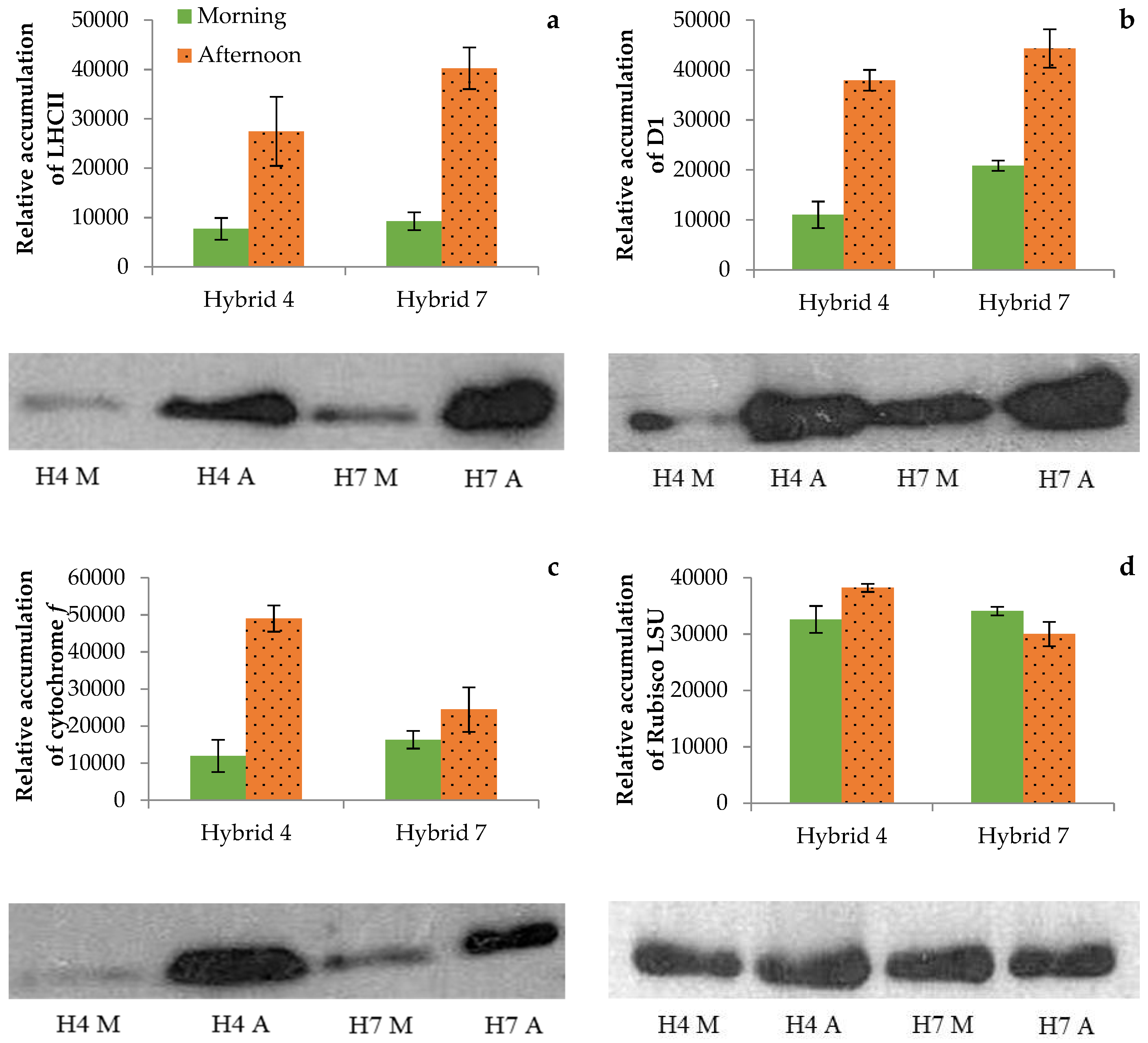
3.4. Photosynthetic Proteins

The LHCII protein is very important for the speed of the adaptation of plants to changes in the light intensity, which results in the relative accumulation of proteins and in changes in the organisation of the antenna position. In this way, plants simultaneously regulate the light absorption and the nonphotochemical dissipation of the excess excitation energy. According to Chen et al. [50], light and heat stress cause the LHCII super-complexes to disassemble, which indicates more susceptibility to stress compared to the other protein complexes in the thylakoid membrane. The antenna complex’s main protein structure for the LCHII is made up of Lhcb2 proteins that bond 45–60% of the pigment molecules (Chl a, Chl b, and carotenoids). In the present experiment, the elevated temperature and the excess light resulted in an accumulation of the LHCII protein in both hybrids, which correspond to the changes in the photosynthetic pigment contents of leaves, as demonstrated by Oguchi et al. [51]. Furthermore, Tanaka and Tanaka [52] conclude that the accumulation of LHCII depends on the content of Chl b. In this research, the accumulation of the LHCII increased during the synergic effect of elevated temperatures and excess light, and more so for hybrid 7 than for hybrid 4 (Figure 3). Mlinarić et al. [8] and Tanaka and Tanaka [52] have determined that higher LHCII accumulation is related to higher Chl b content. This was also confirmed here because hybrid 7 had a higher Chl b content and Chl a/b and higher LHCII accumulation than hybrid 4 (Table 1 and Figure 3) under the synergic effect of elevated temperatures and excess light. In hybrid 7, the Chl b content remained stable despite the stress, but the LHCII increased significantly. A higher LHCII protein accumulation during heat and light stress was also confirmed in wheat [50].
One of the main reaction centres in PSII is transmembrane subunit protein D1 (coded genome psbA), which is the most susceptible to environmental stress of all of the PSII complex components [53]. Su et al. [54] claim that the D1 protein is the target place for heat and light stress action. According to Chan et al. [55], heat and/or light stress stimulate ROS synthesis in the thylakoid membranes, which damages the D1 protein. Although other studies note the decrease in the D1 protein that is due to the synergic effect of elevated temperatures and excess light [54], this was not the case here. The D1 protein was higher in the afternoon than in the morning, and especially in hybrid 4, which indicated that the ROS did not affect the D1 protein (i.e., that the cell oxidation damage was partly prevented). Hybrid 4 showed weaker dynamics between the degradation and the biosynthesis of the D1 protein compared to hybrid 7, as was evidenced by the D1 protein level in the morning, which was much lower in hybrid 4 in the post-stress period (i.e., recovery overnight). The D1 protein accumulation is more affected by the light intensity than by the temperature. According to Guo et al. [56], a higher light intensity causes increased protein phosphorylation. More recent studies indicate that the high D1 protein reactivity under unfavourable high-light-intensity conditions is not just the result of the photoinhibition processes. Its function is to protect PSI from the high flow of the electrons that are generated in PSII that could cause oxidation damage [57]. This is confirmed in the research by Vojta et al. [58]. They report the parallel existence and activity of different electron flow routes in the electron transport chain that prevent excess ROS synthesis.
Besides the D1 protein, the daily fluctuations in the temperatures and the insolation affect cytochrome b6f complex proteins as well. The membrane cytochrome b6f protein complex connects PSI to PSII through electron connections [20]. During the synergic effect of elevated temperatures and excess light, sunflower hybrid 4 accumulated more cyt f proteins than hybrid 7. The relative cyt f protein accumulation during high light intensity is one of the most sensitive components of electron transport [59]. According to Hojka [60], plants adapt to high light intensity by increasing cyt f protein synthesis, but the change amplitudes depend on the species. Higher cyt f accumulation under high-light-intensity conditions was confirmed by Yamori et al. [61] on spruce, and by Pavlovič et al. [62] on tobacco leaves.
The photosynthesis efficiency during heat and light stress depends on the stomatal conductivity and on the CO2 diffusion, which affect the activation of the ribulose-1,5-biphosphate (RuBP) carboxylase/oxygenase enzyme that is known as Rubisco [63]. It is well known that the CO2 diffusion and the Rubisco activity in RuBP carboxylation affect the photosynthetic rate. Rubisco mainly affects the efficiency of PSII and the relative electron transport through CO2 fixation [64]. The same was confirmed in the research by Chen et al. [48], who investigated the effect of simultaneous heat and light stress conditions on wheat. In the afternoon conditions, the Rubisco LSU accumulation increased in hybrid 4, while it decreased in hybrid 7, which may indicate differences in the ROS accumulation, as ROS cause the degradation and fragmentation of the Rubisco LSU. Chen et al. [50] and Zivcak et al. [65] confirm that Rubisco LSU is deactivated under heat and light stress conditions, while Mlinarić et al. [14] report that high temperatures and excess light at noon did not affect the Rubisco LSU accumulation. Lu et al. [66] studied the impact of high temperatures on tomatoes. They confirm that high temperatures promoted the transcription of the Rubisco LSU, which was accompanied by a substantial reduction in the photosynthetic capacity, and by a slight inhibition of the Rubisco activity.

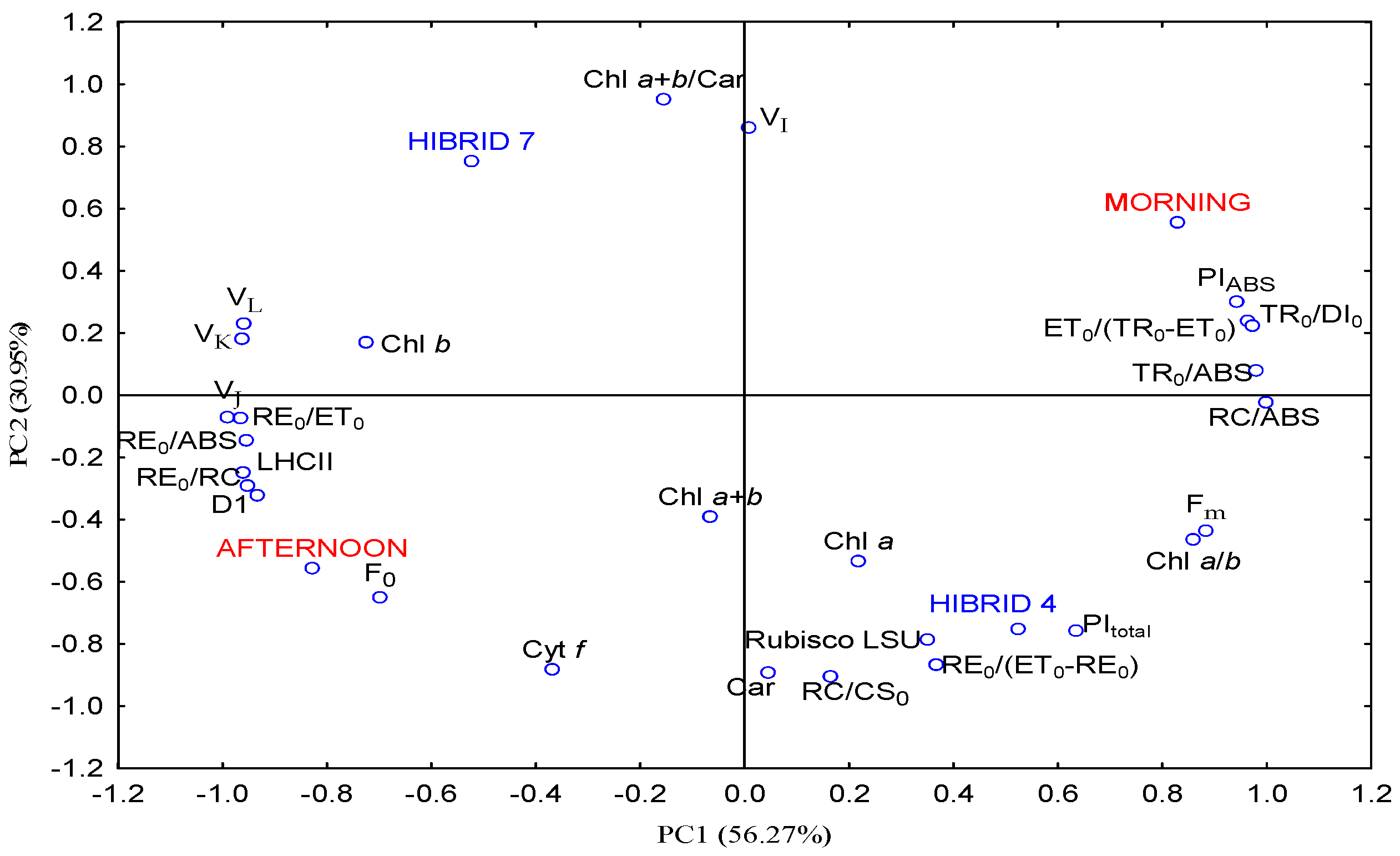
3.5. Principal Component Analysis

The presented and discussed relationships between the environmental conditions, the hybrid sensitivities, and the tested parameters were summarised and visualised with the principal component analysis (PCA) (Figure 4).
The two principal components (PCs) explained 87.22% of the total variability. PC1 and PC2 were responsible for 56.27% and 30.95% of the variability. The RE0/ABS, the TR0/ABS, and the ET0/(TR0-ET0) positively contributed to PC1. A negative contribution to PC1 was determined for the VJ, the RE0/ET0, and the LHCII. The Chl a + b/Car, the VI, and hybrid 7 positively contributed to PC2. According to the correlation coefficients, the synergic effect of the elevated temperatures and excess light was in a highly significant positive correlation with the F0, the RE0/RC, and protein D1, which means that these parameters could be useful as stress indicators for sunflowers. Furthermore, the RE0/RC is in a significant, strong, and positive correlation with the VJ, the RE0/ABS, the RE0/ET0, the LHCII, and D1, while it is in a significant, strong, and negative correlation with the TR0/ABS, the TR0/DI0, the ET0/(TR0-ET0), and the PIABS (Supplementary Table S3), which is evident from the position of the mentioned parameters in the PCA biplot (Figure 4). D1 is in significant, strong, and positive correlation with the RE0/RC and the LHCII, while the correlations with the TR0/DI0, the ET0/(TR0-ET0), and the PIABS were significant, strong, and negative (Figure 4 and Supplementary Table S3). These relationships indicate that increases in the VJ, the RE0/ABS, the RE0/ET0, the RE0/RC, and the LHCII, as well as decreases in the TR0/ABS, the TR0/DI0, the ET0/(TR0-ET0), and the PIABS can be signs that sunflower plants are under stress. Furthermore, the parameters that are in significant correlation can be used interchangeably, which simplifies and speeds up the analysis process. The association of the ChlF parameters with the environmental conditions was previously studied and confirmed by Pavlović et al. [9], Mihaljević et al. [5], and Viljevac Vuletić and Španić [43].

4. Conclusions

Although most of the tested parameters changed as expected during the elevated temperatures and excess light that were determined in the afternoon hours, only the F0, the RE0/RC, and D1 revealed significant, high, and positive correlations with the environmental conditions, which indicates their usefulness in sunflower-stress-response research. Some other chlorophyll fluorescence parameters (VJ, TR0/ABS, TR0/DI0, ET0/(TR0-ET0), RE0/ABS, RE0/ET0, RE0/RC) and photosynthetic proteins (LHCII and D1) can be used as indicators of the physiologic changes that are caused by elevated temperatures and excess light as well, although they are only indirectly associated with environmental conditions.
According to the tested parameters, hybrid 4 appeared more adaptable to the elevated temperatures and excess light that were determined in the afternoon hours than hybrid 7. The better adaptability of hybrid 4 is evident from the smaller changes in the fluorescence curves in phases O–J and J–I, and especially in steps L, K, J, I, and by the higher PIABS values under the afternoon conditions. The photosynthetic apparatus of hybrid 7 can be considered to be more susceptible to the tested unfavourable weather conditions than that of hybrid 4 because of the significant impairment of its functionality, as is indicated by the positive L, K, and J steps, the increase in the TR0/ABS, and the decrease in the ET0/(TR0-ET0), which caused the reduction in the PIABS. The more pronounced stress effect in hybrid 7 was confirmed by the higher relative accumulation of the LHCII potential as well. The determination of the photosynthetic efficiency, pigments, and proteins could be a useful selection criterion for the development of sunflower hybrids that are highly tolerant to elevated temperatures and excess light, which encourages sustainable sunflower production under stressful growing conditions.

Supplementary Materials

The following supporting information can be downloaded at: www.mdpi.com/article/10.3390/horticulturae8050392/s1, Table S1: Temperature and solar radiation ten days before measurements of chlorophyll a fluorescence; Table S2: Measured and calculated chlorophyll a fluorescence parameters according to Strasser et al. [7] and Yusuf et al. [32]; Table S3: Correlation coefficients among analysed traits and environmental conditions in sunflower hybrids.

Author Contributions

Conceptualisation, H.L., V.C. and A.S.; methodology, M.V.V., H.L. and J.A.D.; software, I.V.; validation, H.L. and M.V.V.; formal analysis, A.M.K., M.V.V. and J.A.D.; investigation, A.M.K.; resources, A.S. and Z.Z.; writing—original draft preparation, A.M.K.; writing–review and editing, M.V.V., M.M.K., J.A.D., A.S. and V.C.; visualisation, A.M.K.; supervision, V.C. and A.S. All authors have read and agreed to the published version of the manuscript.

Funding

This research was funded by the Ministry of Science and Education, Croatia, under grant numbers 073-0000000-3538 and 073-0731674-1673.

Institutional Review Board Statement

Not applicable.

Informed Consent Statement

Not applicable.

Data Availability Statement

Not applicable.

Conflicts of Interest

The authors declare no conflict of interest.

References

  1. Jug, D.; Jug, I.; Brozović, B.; Vukadinović, V.; Stipešević, B.; Đurđević, B. The role of conservation agriculture in mitigation and adaptation to climate change. Poljoprivreda 2018, 24, 35–44. [Google Scholar] [CrossRef]
  2. García-López, J.; Lorite, I.J.; García-Ruiz, R.; Domínguez, J. Evaluation of three simulation approaches for assessing yield of rainfed sunflower in a Mediterranean environment for climate change impact modelling. Clim. Chang. 2014, 124, 147–162. [Google Scholar] [CrossRef]
  3. De la Haba, P.; De la Mata, L.; Molina, E.; Aguera, E. High temperature promotes early senescence in primary leaves of sunflower (Helianthus annuus L.) plants. Can. J. Plant Sci. 2014, 94, 659–669. [Google Scholar] [CrossRef]
  4. Greer, D.H. Temperature-dependent responses of the photosynthetic and chlorophyll fluorescence attributes of apple (Malus domestica) leaves during a sustained high temperature event. Plant Physiol. Biochem. 2015, 97, 139–146. [Google Scholar] [CrossRef] [PubMed]
  5. Mihaljević, I.; Lepeduš, H.; Šimić, D.; Viljevac Vuletić, M.; Tomaš, V.; Vuković, D.; Dugalić, K.; Teklić, T.; Babojelić, M.S.; Zdunić, Z. Photochemical efficiency of photosystem II in two apple cultivars affected by elevated temperature and excess light in vivo. S. Afr. J. Bot. 2020, 130, 316–326. [Google Scholar] [CrossRef]
  6. Stirbet, A.; Lazár, D.; Guo, Y.; Govindjee, G. Photosynthesis: Basics, history and modelling (Review: Part of a special issue on functional-structural plant growth modelling). Ann. Bot. 2020, 126, 511–537. [Google Scholar] [CrossRef]
  7. Strasser, R.J.; Tsimilli-Michael, M.; Srivastava, A. Analysis of the fluorescence transient. In Chlorophyll Fluorescence: A Signature of Photosynthesis; Advances in Photosynthesis and Respiration Series; Govindjee, G., Papageorgiou, G., Eds.; Springer: Dordrecht, The Netherlands, 2004; pp. 321–362. [Google Scholar]
  8. Mlinarić, S.; Antunović Dunić, J.; Babojelić, M.S.; Cesar, V. Differential accumulation of photosynthetic proteins regulates diurnal photochemical adjustments of PSII in common fig (Ficus carica L.) leaves. J. Plant Physiol. 2017, 209, 1–10. [Google Scholar] [CrossRef]
  9. Pavlović, I.; Mlinarić, S.; Tarkowská, D.; Oklestkova, J.; Novák, O.; Lepeduš, H.; Bok, V.V.; Brkanac, S.R.; Strnad, M.; Salopek-Sondi, B. Early Brassica Crops Responses to Salinity Stress: A Comparative Analysis Between Chinese Cabbage, White Cabbage, and Kale. Front. Plant Sci. 2019, 10, 450. [Google Scholar] [CrossRef] [Green Version]
  10. Markulj Kulundžić, A.; Viljevac Vuletić, M.; Matoša Kočar, M.; Mijić, A.; Varga, I.; Sudarić, A.; Cesar, V.; Lepeduš, H. The Combination of Increased Temperatures and High Irradiation Causes Changes in Photosynthetic Efficiency. Plants 2021, 10, 2076. [Google Scholar] [CrossRef]
  11. Misra, A.N.; Misra, M.; Singh, R. Chlorophyll fluorescence in plant biology. In Biophysics; Misra, A.N., Ed.; Intech Open: London, UK, 2012; pp. 171–192. Available online: http://www.intechopen.com (accessed on 22 April 2022).
  12. Morales, A.; Kaiser, E. Photosynthetic Acclimation to Fluctuating Irradiance in Plants. Front. Plant Sci. 2020, 11, 268. [Google Scholar] [CrossRef]
  13. Scheurwater, I.; Dunnebacke, M.; Eising, R.; Lambers, H. Respiratory costs and rate of protein turnover in the roots of a fast-growing (Dactylis glomerata L.) and a slow-growing (Festuca ovina L.) grass species. J. Exp. Bot. 2000, 51, 1089–1097. [Google Scholar]
  14. Mlinarić, S.; Antunović Dunić, J.; Štolfa, I.; Cesar, V.; Lepeduš, H. High irradiation and increased temperature induce different strategies for competent photosynthesis in young and mature fig leaves. S. Afr. J. Bot. 2016, 103, 25–31. [Google Scholar] [CrossRef]
  15. Yanhui, C.; Hongrui, W.; Beining, Z.; Shixing, G.; Zihan, W.; Yue, W.; Huihui, Z.; Guangyu, S. Elevated air temperature damage to photosynthetic apparatus alleviated by enhanced cyclic electron flow around photosystem I in tobacco leaves. Ecotoxicol. Environ. Saf. 2020, 204, 111136. [Google Scholar] [CrossRef] [PubMed]
  16. Pospíšil, P. Production of Reactive Oxygen Species by Photosystem II as a Response to Light and Temperature Stress. Front. Plant Sci. 2016, 7, 1950. [Google Scholar] [CrossRef]
  17. Busheva, M.; Garab, G.; Liker, E.; Tóth, Z.; Szèll, M.; Nagy, F. Diurnal fluctuations in the content and functional properties of the light harvesting chlorophyll a/b complex in thylakoid membranes. Plant Physiol. 1991, 95, 997–1003. [Google Scholar] [CrossRef] [Green Version]
  18. Michoux, F.; Ahmad, N.; Wei, Z.Y.; Belgio, E.; Ruban, A.V.; Nixon, P.J. Testing the role of the N-terminal tail of D1 in the maintenance of photosystem II in tobacco chloroplasts. Front. Plant Sci. 2016, 7, 844. [Google Scholar] [CrossRef] [PubMed] [Green Version]
  19. Chen, L.; Jia, H.; Tian, Q.; Du, L.; Gao, Y.; Miao, X.; Liu, Y. Protecting effect of phosphorylation on oxidative damage of D1 protein by down-regulating the production of superoxide anion in photosystem II membranes under high light. Photosynth. Res. 2012, 112, 141–148. [Google Scholar] [CrossRef]
  20. Cramer, W.A.; Baniulus, D.; Yamashita, E.; Zhang, H.; Zatsman, A.I.; Hendrich, M.P. Cytochrome b6f complex, colon structure, spectroscopy, and function of heme cn: N-side electron and proton transfer reactions. In Photosynthetic Protein Complexes; Fromme, P., Ed.; Weily-Blackwell Verlag GmbH&Co.: Weinheim, Germany, 2008; pp. 155–179. [Google Scholar]
  21. Helbling, E.W.; Buma, A.G.J.; Boelen, P.; Strate, H.J.; Giordanino, M.V.F.; Villafane, V.E. Increase in Rubisco activity and gene expression due to elevated temperature partially counteracts ultraviolet radiation-induced photoinhibition in the marine diatom Thalassiosira weissflogii. Limnol. Oceanogr. 2011, 56, 1330–1342. [Google Scholar] [CrossRef] [Green Version]
  22. Markulj Kulundžić, A.; Kovačević, J.; Viljevac Vuletić, M.; Josipović, A.; Liović, I.; Mijić, A.; Lepeduš, H.; Matoša Kočar, M. Impact of abiotic stress on photosynthetic efficiency and leaf temperature in sunflower. Poljoprivreda 2016, 22, 17–22. [Google Scholar] [CrossRef]
  23. Markulj Kulundžić, A.; Kovačević, J.; Viljevac Vuletić, M.; Jocić, S.; Cvejić, S.; Matoša Kočar, M.; Mijić, A.; Liović, I.; Sudarić, A.; Lepeduš, H.; et al. Effect of different soil water content effect on genotype expession in photosynthetic efficiency and leaf temperature in sunflower. Genet.-Belgrade 2016, 48, 971–982. [Google Scholar] [CrossRef] [Green Version]
  24. Schneiter, A.A.; Miller, J.F. Description of sunflower growth stages. Crop Sci. 1981, 21, 901–903. [Google Scholar] [CrossRef]
  25. Lichtenthaler, H.K. Chlorophylls and carotenoids: Pigments of photosynthetic biomembranes. Methods Enzymol. 1987, 148, 350–382. [Google Scholar] [CrossRef]
  26. Bradford, M.M. A Rapid and sensitive method for quantitation of microgram quantities of protein utilising principle of protein-dye binding. Anal. Biochem. 1976, 72, 248–254. [Google Scholar] [CrossRef]
  27. Laemmli, U.K. Cleavage of structural proteins during assembly of head of Bacteriophage-T4. Nature 1970, 227, 680–685. [Google Scholar] [CrossRef]
  28. Towbin, H.; Staehelin, T.; Gordon, J. Electrophoretic transfer of proteins from polyacrylamide gels to nitrocellulose sheets: Procedure and some applications. Proc. Natl. Acad. Sci. USA 1979, 76, 4350–4354. [Google Scholar] [CrossRef] [Green Version]
  29. Martinazzo, E.G.; Ramm, A.; Bacarin, M.A. The chlorophyll a fluorescence as an indicator of the temperature stress in the leaves of Prunus persica. Braz. J. Plant Physil. 2012, 24, 237–246. [Google Scholar] [CrossRef] [Green Version]
  30. Tsimilli-Michael, M.; Strasser, R. The energy flux theory 35 years later: Formulations and applications. Photosynth. Res. 2013, 117, 289–320. [Google Scholar] [CrossRef] [PubMed]
  31. Kalaji, H.M.; Jajoo, A.; Oukarroum, A.; Brestic, M.; Zivcak, M.; Samoborska, I.A.; Cetner, M.D.; Łukasik, I.; Goltsev, V.; Ladle, R.J. Chlorophyll fluorescence as a tool to monitor physiological status of plants under abiotic stress conditions. Acta Physiol. Plant. 2016, 38, 102. [Google Scholar] [CrossRef] [Green Version]
  32. Yusuf, M.A.; Kumar, D.; Rajwanshi, R.; Strasser, R.J.; Tsimilli-Michael, M.; Govindjee; Sarin, N.B. Overexpression of g-tocopherol methyl transferase gene in transgenic Brassica juncea plants alleviates abiotic stress: Physiological and chlorophyll a fluorescence measurements. Biochim. Biophys. Acta 2010, 1797, 1428–1438. [Google Scholar] [CrossRef] [Green Version]
  33. Brestic, M.; Zivcak, M.; Kalaji, H.M.; Carpentier, R.; Allakhverdiev, S.I. Photosystem II thermostability in situ: Environmentally induced acclimation and genotype-specific reactions in Triticum aestivum L. Plant Physiol. Biochem. 2012, 57, 93–105. [Google Scholar] [CrossRef] [PubMed]
  34. Papageorgiou, G.C.; Govindjee. The Non-Photochemical Quenching of the Electronically Excited State of Chlorophyll a in Plants: Definitions, Timelines, Viewpoints, Open Questions. In Non-Photochemical Quenching and Energy Dissipation in Plants, Algae and Cyanobacteria; Demmig-Adams, B., Garab, G., Adams, W., III, Govindjee, Eds.; Springer: Dordrecht, The Netherlands, 2014; p. 44. [Google Scholar] [CrossRef]
  35. Paunov, M.; Koleva, L.; Vassilev, A.; Vangronsveld, J.; Golstev, V. Effects Different Metals on Photosynthesis: Cadmium and Zink Affect Chlorophyll Fluorescence in Durum Wheat. Int. J. Mol. Sci. 2018, 19, 787. [Google Scholar] [CrossRef] [PubMed] [Green Version]
  36. Desotgiu, R.; Pollastrini, M.; Cascio, C.; Gerosa, G.; Marzuoli, R.; Bussotti, F. Responses to ozone on Populus Oxford clone in an open top chamber experiment assessed before sunrise and in full sunlight. Photosynthetica 2013, 51, 267–280. [Google Scholar] [CrossRef]
  37. Schansker, G.; Tóth, S.Z.; Holzwarth, A.R.; Garab, G. Chlorophyll a fluorescence: Beyond the limits of the QA model. Photosyn. Res. 2014, 120, 43–58. [Google Scholar] [CrossRef] [PubMed]
  38. Kalaji, M.H.; Goltsev, V.N.; Żuk-Gołaszewska, K.; Zivcak, M.; Brestic, M. Chlorophyll Fluorescence Understanding Crop Performance—Basics and Applications, 1st ed.; CRC Press: Boca Raton, FL, USA, 2017. [Google Scholar]
  39. Misra, A.N. Chlorophyll fluorescence: A practical approach to study ecophysiology of green plants. In Advances in Plant Ecophysiology Techniques; Sánchez-Moreiras, A.M., Reigosa, M.J., Eds.; Springer: Cham, Switzerland, 2018; Chapter 5; pp. 77–97. [Google Scholar]
  40. Feng, B.; Liu, P.; Li, G.; Dong, S.T.; Wang, F.H.; Kong, L.A.; Zhang, J.W. Effect of Heat Stress on the Photosynthetic Characteristics in Flag Leaves at the Grain-Filling Stage of Different Heat-Resistant Winter Wheat Varieties. J. Agro. Crop Sci. 2014, 200, 143–155. [Google Scholar] [CrossRef]
  41. Fghire, R.; Anaya, F.; Ali, O.I.; Benlhabib, O.; Ragab, R.; Wahbi, S. Physiological and photosynthetic response of quinoa to drought stress. ChileanJAR 2015, 75, 174–183. [Google Scholar] [CrossRef] [Green Version]
  42. Çiçek, N.; Pekcan, V.; Arslan, Ö.; Erdal, S.E.; Balkan Nalçaiyi, A.S.; Çil, A.N.; Şahin, V.; Kaya, Y.; Ekmekçi, Y. Assessing drought tolerance in field-grown sunflower hybrids by chlorophyll fluorescence kinetics. Braz. J. Bot. 2019, 42, 249–260. [Google Scholar] [CrossRef]
  43. Viljevac Vuletić, M.; Španić, V. Special issue in honour of Prof. Reto, J. Strasser–Characterisation of photosynthetic performance during natural leaf senescence in winter wheat: Multivariate analysis as a tool for phenotypic characterisation. Photosynthetica 2020, 58, 301–313. [Google Scholar] [CrossRef] [Green Version]
  44. Gururani, M.A.; Venkatesh, J.; Ganesan, M.; Strasser, R.J.; Han, Y.; Kim, J.-I.; Lee, H.-Y.; Song, P.-S. In Vivo Assessment of Cold Tolerance through Chlorophyll-a Fluorescence in Transgenic Zoysiagrass Expressing Mutant Phytochrome A. PLoS ONE 2015, 10, e0127200. [Google Scholar] [CrossRef] [Green Version]
  45. Kalaji, H.M.; Schansker, G.; Ladle, R.J.; Goltsev, V.; Bosa, K.; Allakhverdiev, S.I.; Brestic, M.; Bussotti, F.; Calatayud, A.; Dąbrowski, P.; et al. Frequently asked questions about in vivo chlorophyll fluorescence: Practical issues. Photosynth. Res. 2014, 122, 121–158. [Google Scholar] [CrossRef] [Green Version]
  46. Yan, K.; Shao, H.; Shao, C.; Zhao, S.; Brestic, M. Dissection of photosynthetic electron transport process in sweet sorghum under heat stress. PLoS ONE 2013, 8, e62100. [Google Scholar] [CrossRef] [Green Version]
  47. Arslan, Ö.; Balkan Nalçaiyi, A.S.; Çulha Erdal, S.; Pekcan, V.; Kaya, Y.; Çiçek, N.; Ekmekçi, Y. Special issue in honour of Prof. Reto J. Strasser–Analysis of drought response of sunflower inbred lines by chlorophyll a fluorescence induction kinetics. Photosynthetica 2020, 58, 348–357. [Google Scholar] [CrossRef] [Green Version]
  48. Gupta, N.K.; Shubhi Agarwal, S.; Agarwal, V.P.; Nathawat, N.S.; Gupta, S.; Singh, G. Effect of short-term heat stress on growth, physiology and antioxidative defence system in wheat seedlings. Acta Physiol. Plant 2013, 35, 1837–1842. [Google Scholar] [CrossRef]
  49. Prasad, S.M.; Dwivedi, R.; Zeeshan, M. Growth, photosynthetic electron transport, and antioxidant responses of young soybean seedlings to simultaneous exposure of nickel and UV-B stress. Photosynthetica 2005, 43, 177–185. [Google Scholar] [CrossRef]
  50. Chen, Y.E.; Zhang, C.M.; Su, Y.Q.; Ma, J.; Zhang, Z.W.; Yuan, M.; Zhang, H.U.; Yuan, S. Responses of photosystem II and antioxidative systems to high light and high temperature co-stress in wheat. Environ. Exp. Bot. 2017, 135, 45–55. [Google Scholar] [CrossRef]
  51. Oguchi, R.; Hikosaka, K.; Hirose, T. Does the photosynthetic light-acclimation need change in leaf anatomy? Plant Cell Environ. 2003, 26, 505–512. [Google Scholar] [CrossRef]
  52. Tanaka, R.; Tanaka, A. Chlorophyll cycle regulates the construction and destruction of the light-harvesting complexes. Biochim. Biophys. Acta 2011, 1807, 968–976. [Google Scholar] [CrossRef] [Green Version]
  53. Li, H.; Xu, H.; Zhang, P.; Gao, M.; Wang, D.; Zhao, H. High temperature effects on D1 protein turnover in three wheat varieties with different heat susceptibility. Plant Growth Regul. 2017, 81, 385–397. [Google Scholar] [CrossRef]
  54. Su, X.Y.; Wu, S.; Yang, L.; Xue, R.L.; Li, H.; Wang, Y.X.; Zhao, H.J. Exogenous progesterone alleviates heat and high light stress-induced inactivation of photosystem II in wheat by enhancing antioxidant defense and D1 protein stability. Plant Growth Regul. 2014, 74, 311–318. [Google Scholar] [CrossRef]
  55. Chan, T.; Shimizu, Y.; Pospisil, P.; Nijo, N.; Fujiwara, A.; Taninaka, Y.; Ishikawa, T.; Hori, H.; Nanba, D.; Imai, A.; et al. Quality Control of Photosystem II: Lipid Peroxidation Accelerates Photoinhibition under Excessive Illumination. PLoS ONE 2013, 8, e52100. [Google Scholar] [CrossRef]
  56. Guo, W.-D.; Guo, Y.-P.; Liu, J.-R.; Mattson, N. Midday depression of photosynthesis is related with carboxylation efficiency decrease and D1 degradation in bayberry (Myrica rubra) plants. Sci. Hortic. 2009, 123, 188–196. [Google Scholar] [CrossRef]
  57. Järvi, S.; Suorsa, M.; Aro, E.-M. Photosystem II repair in plant chloroplasts-Regulation, assisting proteins and shared components with photosystem II biogenesis. Biochim. Biophys. Acta 2015, 1847, 900–909. [Google Scholar] [CrossRef] [Green Version]
  58. Vojta, L.; Carić, D.; Cesar, V.; Antunović Dunić, J.; Lepeduš, H.; Kveder, M.; Fulgosi, H. TROL-FNR interaction reveals alternative parthways of electron partitioning in photosynthesis. Sci. Rep. 2015, 5, 10085. [Google Scholar] [CrossRef] [Green Version]
  59. Björkman, O. Response to different quantum flux densities. In Physiological Plant Ecology I. Responses to the Physical Environment; Lange, O.D., Nobel, P.S., Osmond, C.B., Ziegler, H., Eds.; Springer: Berlin, Germany, 1981; pp. 57–107. [Google Scholar]
  60. Hojka, M.; Thiele, W.; Tóth, S.Z.; Lein, W.; Bock, R.; Schöttler, M.A. Inducible Repression of Nuclear-Encoded Subunits of the Cytochrome b6f Complex in Tobacco Reveals an Extraordinarily Long Lifetime of the Complex. Plant Physiol. 2014, 165, 1632–1646. [Google Scholar] [CrossRef] [Green Version]
  61. Yamori, W.; Evans, J.R.; von Caemmerer, S. Effects of growth and measurement light intensities on temperature dependence of CO2 assimilation rate in tobacco leaves. Plant Cell Environ. 2010, 33, 332–343. [Google Scholar] [CrossRef]
  62. Pavlovič, A.; Stolárik, T.; Nosek, L.; Kouřil, R.; Ilík, P. Light-induced gradual activation of photosystem II in dark-grown Norway spruce seedlings. Biochim. Biophys. Acta 2016, 1857, 799–809. [Google Scholar] [CrossRef]
  63. Yamori, W.; Hikosaka, K.; Way, D.A. Temperature response of photosynthesis in C3, C4, and CAM plants: Temperature acclimation and temperature adaptation. Photosynth. Res. 2014, 119, 101–117. [Google Scholar] [CrossRef]
  64. Cheng, L.; Fuchigami, L.H.; Breen, P.J. The relationship between photosystem II efficiency and quantum yield for CO2 assimilation is not affected by nitrogen content in apple leaves. J. Exp. Bot. 2001, 52, 1865–1872. [Google Scholar] [CrossRef] [Green Version]
  65. Zivcak, M.; Brestic, M.; Kunderlikova, K.; Sytar, O.; Allakhverdiev, S.I. Repetitive light pulse-induced photoinhibition of photosystem I severely affects CO2 assimilation and photoprotection in wheat leaves. Photosynth. Res. 2015, 126, 449–463. [Google Scholar] [CrossRef]
  66. Lu, T.; Meng, Z.; Zhang, G.; Qi, M.; Sun, Z.; Liu, Y.; Li, T. Sub-high Temperature and High Light Intensity Induced Irreversible Inhibition on Photosynthesis System of Tomato Plant (Solanum lycopersicum L.). Front. Plant Sci. 2017, 8, 365. [Google Scholar] [CrossRef] [Green Version]
Figure 1. (a) Raw fluorescence induction curves, and (b) double O–P normalised OJIP curves of chlorophyll a fluorescence kinetics of dark-adapted leaves of sunflower hybrids 4 and 7 under the morning (M) and afternoon (A) conditions. WOP = (Ft − F0)/(FP − F0) represents normalised OJIP transient data between steps O and P. Each curve represents the average kinetics of 12 replicates per condition.
Horticulturae 08 00392 g001
Figure 2. Chlorophyll a fluorescence transient curves in sunflower hybrids 4 and 7 under the morning (M) and afternoon (A) conditions. WOK = (Ft − F0)/(FK − F0) represents normalised transient data between steps O and K (L step); (a) plotted as difference kinetics (ΔWOK = WOK − (WOK)ref) in the 0.05–0.3 ms time range. WOJ = (Ft − F0)/(FJ − F0) represents normalised transient data between steps O and J (K step); (b) plotted as difference kinetics (ΔWOJ = WOJ − (WOJ)ref) in the 0.05–2 ms time range. WOI = (Ft − F0)/(FI − F0) represents normalised transient data between steps O and I (J step); (c) plotted as difference kinetics (ΔWOI = WOI − (WOI)ref) in the 0.05–30 ms time range. WO50 = (Ft − F0)/(F50 − F0) represents normalised transient data between steps O and 50 (I step); (d) plotted as difference kinetics (ΔWO50 = WO50 − (WO50)ref) in the 0.05–50 ms time range. Each curve represents the average kinetics of 12 replicates. Measurements in morning conditions were used as reference values of (WOK)ref, (WOJ)ref, (WOI)ref, and (WO50)ref. Curve lines with marker points show WOK, WOJ, WOI, and WO50 (primary axis), and curve lines without marker points show ΔWOK, ΔWOJ, ΔWOI, and ΔWO50 (secondary axis).
Horticulturae 08 00392 g002aHorticulturae 08 00392 g002b
Figure 3. The relative accumulation of photosynthetic proteins: (a) light-harvesting complex of photosystem II—(LHCII); (b) D1; (c) cytochrome f (cyt f); and (d) Rubisco large subunit—(Rubisco LSU) in sunflower hybrids 4 and 7 (H4 and H7) under the morning (M) and afternoon (A) conditions. Lines in graphs represent mean values ± standard deviations of three replicates (n = 3).
Horticulturae 08 00392 g003
Figure 4. Biplot of principal component analysis of chlorophyll a fluorescence, photosynthetic pigment contents, and photosynthetic proteins in hybrids 4 and 7 (blue colour) under the morning and afternoon conditions (red colour). JIP parameters: minimum fluorescence intensity (F0); maximum fluorescence intensity (Fm); relative variable fluorescence at 150 µs (VL step); relative variable fluorescence at 300 µs (VK step); relative variable fluorescence at 3 ms (VJ step); relative variable fluorescence at 30 ms (VI step); maximum quantum yield of PSII (TR0/ABS); density of active PSII reaction centers (RCs) per cross section (RC/CS0); density of RC on chlorophyll a basis (RC/ABS); flux ratio trapping per dissipation (TR0/DI0); electron transport further than primary acceptor QA (ET0/(TR0-ET0)); performance index (PIABS); quantum yield for reduction in the end electron acceptors at the PSI acceptor side (RE0/ABS); probability that an electron from the electron transport chain is transferred to reduce end electron acceptors at the PSI acceptor side (RE0/ET0); electron-flux-reducing end electron acceptors at the PSI acceptor side per RC (RE0/RC); electron transport from PQH2 to final PSI acceptors (RE0/(ET0-RE0)); and performance index for energy conservation from exciton to the reduction in the PSI end acceptors (PItotal). Photosynthetic pigment content: chlorophyll a (Chl a); chlorophyll b (Chl b); total chlorophyll a + b (Chl a + b); total carotenoids (Car); ratio of chlorophyll a and b (Chl a/b); and ratio of total chlorophyll content and carotenoids (Chl a + b/Car). The relative accumulation of photosynthetic proteins: light-harvesting complex of photosystem II (LHCII); D1; cytochrome f (cyt f); and Rubisco large subunit (Rubisco LSU).
Horticulturae 08 00392 g004
Table 1. Mean values and standard deviations of JIP parameters (n = 12) and photosynthetic pigment contents (n = 5) in sunflower hybrids 4 and 7 in morning and afternoon conditions.
Hybrid 4Hybrid 7
ParametersMorningAfternoonMorningAfternoon
Minimum fluorescence intensity (F0)267.50 ± 8.51 b348.75 ± 25.09 a223.75 ± 7.31 c341.83 ± 28.87 a
Maximum fluorescence intensity (Fm)1740.92 ± 67.50 a1591.83 ± 104.15 b1416.75 ± 99.07 c1115.58 ± 92.91 d
Relative variable fluorescence at 150 µs (VL step)0.54 ± 0.01 b0.54 ± 0.02 b0.54 ± 0.01 b0.56 ± 0.02 a
Relative variable fluorescence at 300 µs (VK step)0.36 ± 0.02 c0.39 ± 0.03 b0.40 ± 0.02 b0.44 ± 0.03 a
Relative variable fluorescence at 3 ms (VJ step)0.24 ± 0.02 d0.40 ± 0.03 b0.28 ± 0.02 c0.58 ± 0.03 a
Relative variable fluorescence at 30 ms (VI step)0.46 ± 0.05 ab0.45 ± 0.03 b0.50 ± 0.05 a0.47 ± 0.03 ab
Maximum quantum yield of PSII (TR0/ABS)0.85 ± 0.01 a0.78 ± 0.01 b0.84 ± 0.01 a0.69 ± 0.03 c
Density of active PSII reaction centres (RCs) per cross section (RC/CS0)157.84 ± 7.44 b173.32 ± 12.43 a119.35 ± 6.53 d135.53 ± 10.54 c
Density of RC on chlorophyll a basis (RC/ABS)0.59 ± 0.03 a0.50 ± 0.04 b0.53 ± 0.03 b0.40 ± 0.04 c
Flux ratio trapping per dissipation (TR0/DI0)5.51 ± 0.19 a3.58 ± 0.30 b5.34 ± 0.45 a2.28 ± 0.31 c
Electron transport further than primary acceptor QA (ET0/(TR0-ET0))3.16 ± 0.26 a1.53 ± 0.20 c2.61 ± 0.26 b0.73 ± 0.10 d
Performance index (PIABS)10.26 ± 0.66 a2.73 ± 0.48 c7.44 ± 1.20 b0.67 ± 0.17 d
Quantum yield for reduction in end electron acceptors at the PSI acceptor side (RE0/ABS)0.60 ± 0.04 c0.72 ± 0.03 b0.58 ± 0.04 c0.87 ± 0.07 a
Probability that an electron from the electron transport chain is transferred to reduce end electron acceptors at the PSI acceptor side (RE0/ET0)0.71 ± 0.05 c0.92 ± 0.04 b0.69 ± 0.06 c1.26 ± 0.11 a
Electron-flux-reducing end electron acceptors at the PSI acceptor side per RC (RE0/RC)0.78 ± 0.09 b0.87 ± 0.10 ab0.79 ± 0.10 b0.92 ± 0.12 a
Electron transport from PQH2 to final PSI acceptors (RE0/(ET0-RE0))2.54 ± 0.51 b15.95 ± 11.20 a2.33 ± 0.60 b−5.83 ± 2.98 c
Performance index for energy conservation from exciton to the reduction in PSI end acceptors (PItotal)26.09 ± 5.89 b40.21 ± 22.01 a16.97 ± 3.80 b−4.22 ± 3.26 c
Chlorophyll a (Chl a)1.38 ± 0.02 b1.49 ± 0.03 a1.46 ± 0.02 a1.37 ± 0.01 b
Chlorophyll b (Chl b)0.32 ± 0.01 c0.35 ± 0.01 b0.37 ± 0.00 ab0.37 ± 0.01 a
Total chlorophyll a + b (Chl a + b)1.70 ± 0.02 c1.85 ± 0.03 a1.83 ± 0.02 a1.74 ± 0.01 b
Total carotenoids (Car)0.40 ± 0.00 bc0.45 ± 0.01 a0.41 ± 0.01 b0.40 ± 0.00 c
Ratio of chlorophyll a and b (Chl a/b)4.37 ± 0.20 a4.21 ± 0.04 a3.99 ± 0.06 b3.70 ± 0.06 c
Ratio of total chlorophyll content and carotenoids (Chl a + b/Car)4.24 ± 0.01 c4.09 ± 0.03 d4.47 ± 0.05 a4.39 ± 0.03 b
According to Tukey’s HSD test, means with the same letters are not significantly different at p < 0.05.
Publisher’s Note: MDPI stays neutral with regard to jurisdictional claims in published maps and institutional affiliations.

