Article

Salt Stress-Induced Ascorbic Acid Accumulation and Its Trade-Off with Mannan Content in Tomato

1
Institute of Life and Environmental Sciences, University of Tsukuba, Tsukuba 305-8572, Ibaraki, Japan
2
Department of Biology, School of Biological Sciences, Tokai University, Sapporo 005-8601, Hokkaido, Japan
*
Author to whom correspondence should be addressed.
Horticulturae 2025, 11(4), 400; https://doi.org/10.3390/horticulturae11040400
Submission received: 4 March 2025 / Revised: 4 April 2025 / Accepted: 7 April 2025 / Published: 9 April 2025

Abstract

:
Salt stress causes osmotic stress and ion toxicity, often inhibiting plant growth and metabolism. However, salt-stressed tomato plants accumulate ascorbic acid, resulting in fruits with high commercial value. However, it was not well understood how mannose, the material for the synthesis of ascorbic acid, and its metabolism are affected under salt stress conditions. In this study, we found that tomatoes grown under salinity stress had increased levels of ascorbic acid, which correlated with decreased levels of mannan in the skin and seeds. Expression analysis of the ascorbic acid synthase gene showed increased expression in early ripening stages under salt stress. In addition, the expression of cellulose synthase-like A (CSLA), genes involved in mannan metabolism, increased significantly during mid-ripening in the control condition. Since ascorbic acid and mannan share mannose as a precursor, they are likely to compete for it. This suggests that salt-stressed tomatoes may be deficient in both ascorbic acid and mannose, thereby affecting mannan synthesis. To investigate this trade-off, we developed a culture system with added mannose. The results showed that in salt-stressed tomatoes supplemented with mannose, ascorbic acid levels in unripe green peels reached those of fully ripe fruit, highlighting the influence of mannose availability on ascorbic acid accumulation.

Graphical Abstract

1. Introduction

Plants are exposed to various environmental stresses during their growth. Environmental stresses include biotic stresses, such as infection by pathogens and damage by animals, and abiotic stresses, such as high and low temperatures, drought, and high salinity [1]. High soil salinity often causes salt stress in two ways: osmotic stress, due to a decrease in water potential in the root zone, and ion toxicity, where salts such as Na+ and Cl are harmful [2,3]. Therefore, salt stress impairs plant growth and metabolism and has a negative effect on germination, plant vigor, and yield [4]. Many studies on tomato plants have also shown that growth inhibition occurs, such as reduced biomass production and fruit size. In addition, fruit texture and histological characteristics are also affected by salt stress, resulting in adverse effects such as increased fruit firmness [5,6,7,8,9]. On the other hand, tomato fruits grown under salinity stress conditions are known to accumulate sugars such as glucose and fructose and amino acids such as proline and gamma-aminobutyric acid, resulting in fruits of high commercial value to humans [10].
Tomato fruits are composed of five major tissues: skin, mesocarp, endocarp, ovary tissue, and seeds, and they can be divided into four growth and maturity stages: mature green (M), breaker (B), turning (T), and red ripe (R) (Figure 1). At the M stage, fruit growth is complete, and fruit size is determined, after which the fruit ripens [11,12,13]. Various morphological and physiological changes occur in tomato fruits during the ripening process, such as seed development, skin color changes, and nutrient accumulation [14]. In particular, fruit softening occurs during ripening, and this softening is thought to be related to structural changes in the cell wall [15,16,17]. Mannan, a type of cell wall component, is a linear homopolysaccharide in which mannose is linked by β-(1,4) linkages. Galactomannans with α-(1,6)-linked galactose side chains to mannose residues also exist, and the presence of galactose side chains reduces the crystallinity of mannan [18,19,20]. In addition, mannan acts as a storage material for energy used in metabolism in the cell wall and plays a structural function, so that cell walls containing a lot of mannan are known to have strong mechanical properties [18].
For plants, AsA is important as an antioxidant that removes reactive oxygen species (ROS) generated during growth and environmental stress, but its synthetic pathway and its relationship with cell wall components remain largely unknown. Since the D-mannose/L-galactose pathway, in which GDP-D-mannose and L-galactose are metabolic intermediates, was proposed, efforts have been made to isolate and analyze the enzyme genes involved in this pathway [21]. Other pathways for AsA synthesis include the myo-inositol pathway, which uses myo-inositol as a substrate, the L-gulose pathway, which uses L-gulose as a substrate, and the galacturonate pathway, which uses D-galacturonate as a substrate. However, most studies on controlling AsA synthesis by manipulating enzyme genes have focused on the D-mannose/L-galactose pathway [22,23]. In particular, it has been shown that tomato fruits overexpressing the GDP-mannose epimerase (GME) gene accumulate AsA and are more resistant to oxidative, cold, and salt stress [23,24]. On the other hand, it has also been shown that the absence of GME alters the structure of RG-II, a type of pectin, and reduces cross-linking between RG-II, resulting in abnormal plant growth [25]. This is consistent with the fact that the D-mannose/L-galactose pathway, which was discovered as a pathway for AsA synthesis, is also involved in cell wall synthesis. In other words, GDP-D-mannose, which is used as a substrate in this pathway, is also used in the synthesis of galactomannan, a cell wall hemicellulose, and RG-II, a pectin, and it can be said that AsA synthesis and cell wall polysaccharide synthesis share a substrate. In this study, we conducted a new cultivation experiment with mannose addition, thinking that it would be possible to compensate for the mannose deficiency caused by salt stress by cultivating tomato fruits under salt stress conditions and adding mannose.
Based on the above, we aimed to clarify how cell wall components change due to metabolic regulation of polysaccharides in tomato fruits under salt stress conditions. We then investigated the metabolic regulation of cell wall polysaccharides and AsA in tomato fruits under salt stress conditions, respectively, and investigated the factors that cause differences in the skin and the mesocarp and endocarp of tomato fruits under salt stress conditions as well as the effect of mannose addition on metabolism under salt stress conditions.

2. Materials and Methods

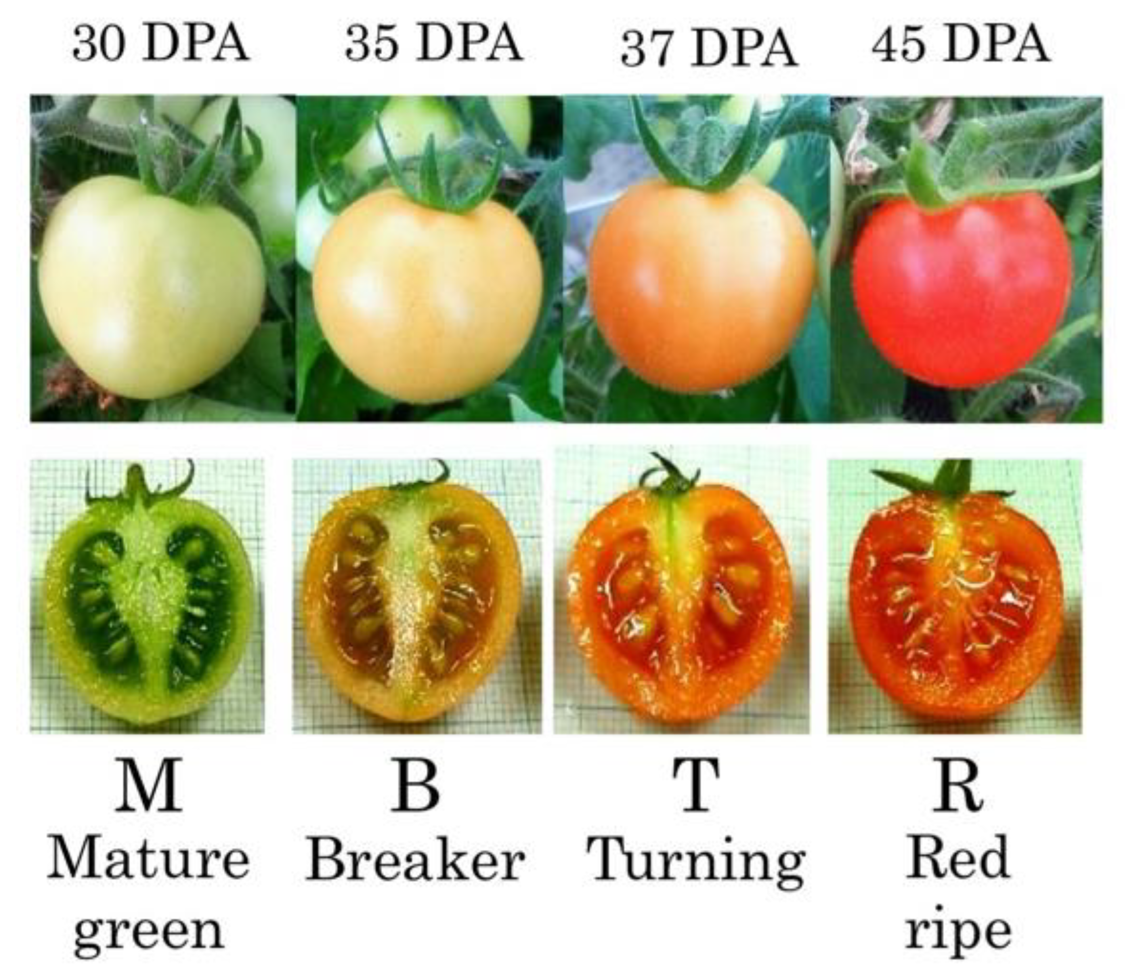
2.1. Plant Materials and Growth Conditions

Tomato seeds (Solanum lycopersicum cv. ‘Micro-Tom’) were germinated at room temperature on moist paper. After one week, seedlings were transplanted into 5 × 5 cm2 rockwool pots and grown in a growth chamber (TOMY CL-301, Tokyo, Japan) under 16 h of light and 8 h of dark at 25 °C for 6 weeks. Plants were grown in plastic trays with a constant volume of 2.0 L. A commercial nutrient solution (Otsuka A; Otsuka Chemical Co., Ltd., Osaka, Japan) with an electrical conductivity (EC) of 2.0 dS m−1 was used. Seven weeks after germination, when the first truss flowered, half of the plants were transferred to a saline nutrient solution with an EC of 15.0 dS m−1, which was achieved by adding NaCl (equivalent to 160 mM NaCl), which was reported to be the appropriate concentration for long-term cultivation under salinity conditions [10]. Plants were maintained in a nutrient solution with an electrical conductivity of 2.0 dS m−1 (0 mM NaCl) as a control. Plants were exposed to two different levels of electrical conductivity (EC) to adapt to salinity stress. Plants were exposed to two different salinity levels, EC 9.0 dS m−1 (80 mM NaCl) and EC 12.0 dS m−1 (120 mM NaCl), for four days each before being transferred to a solution with EC 15.0 dS m−1. Tomato fruits at the corresponding developmental stages were also collected: M (30 days post anthesis (DPA)), B (35 DPA), T (37 DPA), R (45 DPA) (Figure 1).

2.2. Extraction of Cell Walls

Three seeds and 100 mg fresh weight of each tissue were frozen and crushed with a mortar and pestle, and 1.5 mL of 80% EtOH was added. The mixture was centrifuged at 15,000 rpm for 1 min, and the supernatant was removed. Pellets were washed three times with water, three times with 1 mL of methanol: chloroform (MC, 1∶1), and three times with 1 mL of acetone (Wako Pure Chemical Industries, Ltd., Tokyo, Japan). The sample was dried, weighed, and recorded as the amount of cell wall. The extracted cell walls were transferred to a screw-capped test tube (ST-13M, Nichiden Rika Glass, Kobe, Japan), and 250 µL of 2 M trifluoroacetic acid (TFA) was added using a Pasteur pipette (Iwaki Glass Co. Ltd., Chiba, Japan). The tube was then heated at 121 °C for 2 h (EYELA MG-2200, EYELA, Tokyo, Japan) to perform hydrolysis. Then, 300 µL of isopropanol was added, and the tube was dried at room temperature using a spray evaporator. The pellet was mixed with 100 mL of distilled water, agitated, and centrifuged, and the TFA-soluble fraction was extracted from the supernatant. The TFA-soluble fraction contains pectin and hemicellulose, so the amount of mannose can be measured.

2.3. Analysis of Mannan and Cell Wall Sugar Components by Gas Chromatography

The TFA-soluble fraction sample (30 µL) of the seed and the pericarp was lyophilized overnight. A quantity of 250 µL of HCl/MeOH was added, and the tube was sealed and heated at 80 °C for 15 h (EYELA MG-2200). To the sample, 100 µL of t-butyl alcohol was added, and the tube was dried at room temperature with a spray evaporator. The samples were then treated with 100 µL of pyridine, 100 µL of hexamethyldiphenylsilane, and 50 µL of trimethylchlorosilane. The tubes were sealed, and the mixture was heated at 80 °C for 20 min using an EYELA MG-2200. These trimethylsilyl reaction reagents were gently removed using a spray evaporator, and the mixture was dissolved in 1 mL of hexane and centrifuged. The supernatant was carefully transferred to a new test tube and dried again using a spray evaporator. The mixture was dissolved in 5 μL of hexane, and 1 μL of the solution was analyzed by gas chromatography (GC-2010 Plus series, Shimadzu Corporation, Kyoto, Japan) using a DB-1 column. The amount of sugar was measured by the anthrone sulfate method for neutral sugars and the carbazole sulfate method for acidic sugars.

2.4. Expression Analysis of Mannan Synthase Gene and Ascorbic Acid Synthase Gene in Tomato Fruit Under Salinity Conditions

Each tissue from the fruit at each stage of maturity (100 mg) was frozen and crushed using a mortar and pestle. Total RNA was extracted from each tissue using an RNeasy® Plant Mini Kit (Qiagen, Hilden, Germany), and cDNA was synthesized using a QuantiTect Reverse Transcription Kit (QIAGEN) (Gene Amp® PCR System 9700). Go-Taq qPCR Master Mix was used for expression analysis. Gotaq® 5 µL, DNase-free water 4 µL, 10 µM forward and reverse primer (Table S1) 0.2 µL, Carboxy-X-Rhodamine (CXR) reference dye 0.1 µL, and template 0.5 µL were mixed, and PCR was performed under the following conditions (Applied Biosystems 7300 Fast Real-Time PCR System, Applied Biosystems, Tokyo, Japan): 95 °C 2 min → (95 °C 5 s, 63 °C 10 s, 72 °C 31 s) × 50 cycles → dissociation step.

2.5. Measurement of Ascorbic Acid Content Under Salt Stress with Addition of Mannose

Salinity stress treatment with addition of mannose was carried out by adding NaCl and mannose to the culture solution after the first inflorescence flowered, adjusting the NaCl concentration to 15.0 dS m−1 (equivalent to about 160 mM NaCl) and mannose to 1 mM. At the beginning of the stress treatment, the electrical conductivity (EC) was gradually increased every 2 days from NaCl: 1.5 dS m−1 mannose: 0.25 mM to NaCl: 3.0 dS m−1 mannose: 0.5 mM, NaCl: 5.0 dS m−1 mannose: 0.75 mM, NaCl: 8.0 dS m−1 mannose: 1 mM, 12.0 dS m−1 mannose: 1 mM, 15.0 dS m−1 mannose: 1 mM while observing the condition of the plant body, and the plant was allowed to acclimate for about 8 days. Finally, the culture solution concentration was 160 mM NaCl and 1 mM mannose.

2.6. Measurement of Germination Rate

Seeds were collected from fruits at each maturity stage and dried on filter paper for 2 days. The seeds were transferred to a tube, and then 700 µL of sterilized water, 200 µL of sodium hypochlorite (Wako chemicals), and one drop of Tween-20 were added; the seeds were mixed by inversion for 6 min and then suspended in 700 µL of sterilized water and washed. The seeds were sown on moistened autoclaved filter paper, and the germination rate was measured over 7 days.

2.7. Statistical Analysis

The data were expressed as the mean values ± SD taken from 4–9 independent biological experiments. The experimental data of the samples were statistically analyzed through one-way analysis of variance (ANOVA) with Tukey’s post hoc test using Statistica 13.1 software (StatSoft, Inc., Tulsa, OK, USA). The results with a p-value ≤ 0.05 and a p-value ≤ 0.01 were considered statistically significant.

3. Results

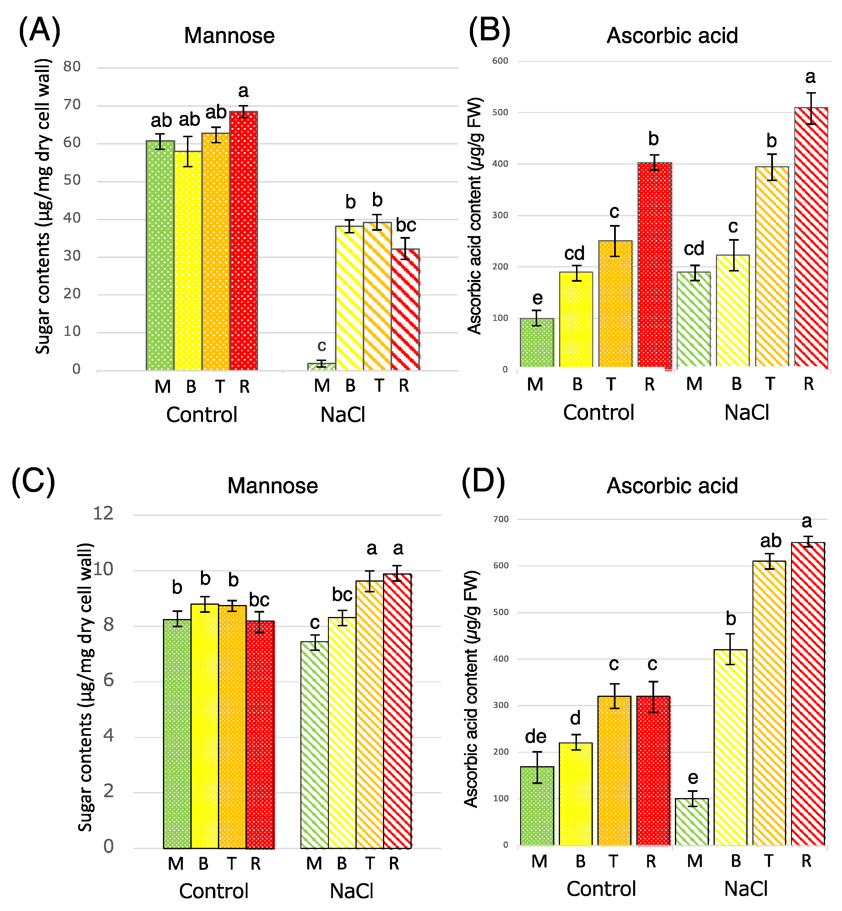
3.1. Measurement of Mannose and Ascorbic Acid Content in Tomato Fruit Under Salt Stress Conditions

The amount of mannose in the seeds (Figure 2A) and the pericarp (Figure 2C) was measured by analyzing the sugars that make up the cell walls using a biochemical method based on gas chromatography. As a result, the amount of mannose in the pericarp did not increase during fruit ripening under the control conditions, whereas the mannose content in the fruits under salt stress conditions increased as they ripened to red (Figure 2C). On the other hand, in the seeds, the amount of mannose was lower at all stages of ripening compared with the control conditions (Figure 2A). Even at the R stage, which is the final red ripened fruit, the seeds contained only about half the amount of mannose. The amount of ascorbic acid was also measured in the pericarp and seeds. As for ascorbic acid, the content of ascorbic acid was higher under salt stress conditions compared with the control conditions, and an increase of about 20% was observed in each tissue (Figure 2B,D). From the above, it was found that under salt stress, the amount of mannose and ascorbic acid both increased in the pericarp, whereas the amount of mannose decreased and the amount of ascorbic acid increased in seeds.

3.2. Expression Analysis of Mannan Synthase Gene and Ascorbic Acid Synthase Gene in Tomato Fruit Under Salt Stress Conditions

In seeds, it has been shown that the amount of mannose decreases and the amount of ascorbic acid increases (Figure 2). Several pathways have been proposed for the biosynthesis of ascorbic acid in plants. Among them, the first reported pathway for ascorbic acid synthesis in higher plants was the D-mannose/L-galactose pathway [8]. GDP-mannose 3′,5′-epimerase, which acts in this pathway, is an enzyme that converts GDP-D-mannose to GDP-L-galactose, and because it is the most highly conserved ascorbic acid biosynthesis-related gene among higher plants, it has been the subject of much research. In this study, the expression of GME was analyzed by qRT-PCR from the M to R stages. Under both the control and the salt stress conditions, GME reached its highest level at the B stage, when the expression level increased by about 45% under salt stress (Figure 3). In response to the finding that the amount of mannose in fruit seeds was reduced under salt stress, we performed an expression analysis of the mannan synthase gene, CSLA. As a result, in the control, the expression of CSLA increased significantly from the M to B stage and then gradually decreased (Figure 3). On the other hand, under salt stress, the peak of expression was delayed to T, not B, and the expression level was also reduced by 55%.

3.3. Changes in Ascorbic Acid Content Under Salt Stress Conditions with Added Mannose

To study the seed germination ability of salt-stressed fruits, germination rates were measured. The results showed that, while there was no significant difference at the R stage, seeds from salt-stressed fruits tended to germinate less at the B stage compared with seeds from the control fruits and that seed development was delayed (Figure 4). However, when mannose was added externally, the germination rate of the control seeds reached 100% on the sixth day when distilled water was used, and, after the increase in mannose concentration, seed germination further accelerated (Figure 4). When distilled water was used for salt-stressed seeds, no germination was observed, but when mannose was added, germination was observed in some seeds. It was shown that mannose tends to be effective for the germination of tomato seeds and that seeds under salt stress conditions were in a state of mannose deficiency.

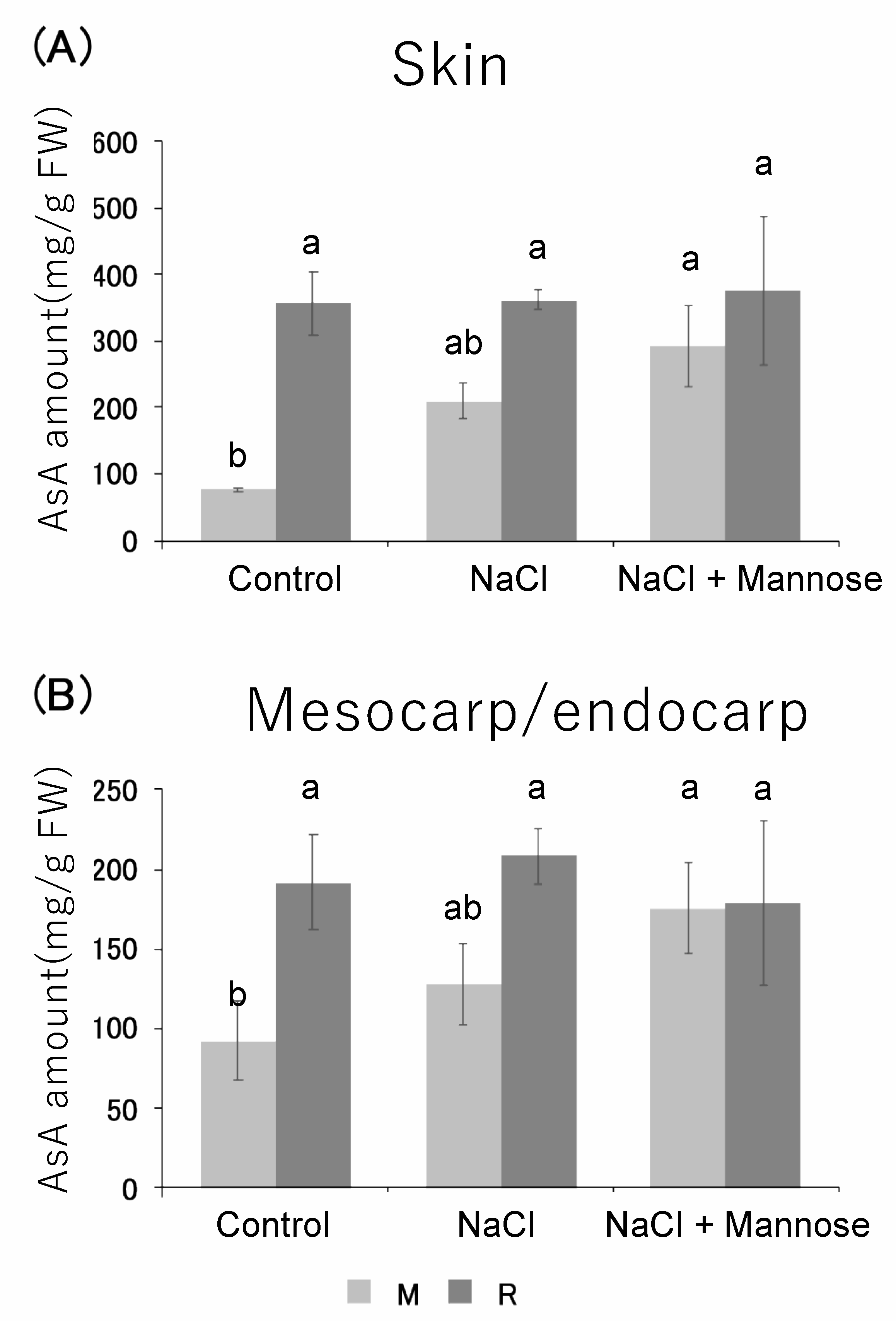
3.4. Measurement of Ascorbic Acid Content Under Salt Stress Conditions with Added Mannose

It has been shown that the ascorbic acid content in tomato fruit is increased by salt stress. In this study, ascorbic acid content was measured in the skin and mesocarp/endocarp tissues. As a result, it was found that ascorbic acid in the exocarp at the M stage, which is the early ripening stage, was significantly increased under salt stress conditions compared with the control. NaCl induced AsA accumulation by 174% in comparison with the control (Figure 5A). Although no significant difference was observed in the mesocarp/endocarp, ascorbic acid in the M stage showed a tendency to increase by about 39% under salt stress conditions as in skin (Figure 5B). The experiment in Figure 4 suggested that saline culture caused a deficiency of ascorbic acid and mannose, which is a material for mannan, so we constructed a culture system in which mannose was added in addition to saline culture. The ascorbic acid content of tomato fruits grown under salinity stress conditions with mannose was measured in each tissue and compared with the ascorbic acid content of tomato fruits under the control and salinity stress conditions. As a result, in tomato fruits grown under salinity stress conditions with mannose, ascorbic acid was significantly increased by about 322% at the M stage of the skin compared with the control condition and showed a tendency to increase compared with salinity stress conditions (Figure 5A). In addition, ascorbic acid at the M stage of the mesocarp/endocarp showed a tendency to increase compared with the other two conditions (Figure 5B). However, the amount of ascorbic acid accumulated at the R stage was similar in all conditions and tissues.

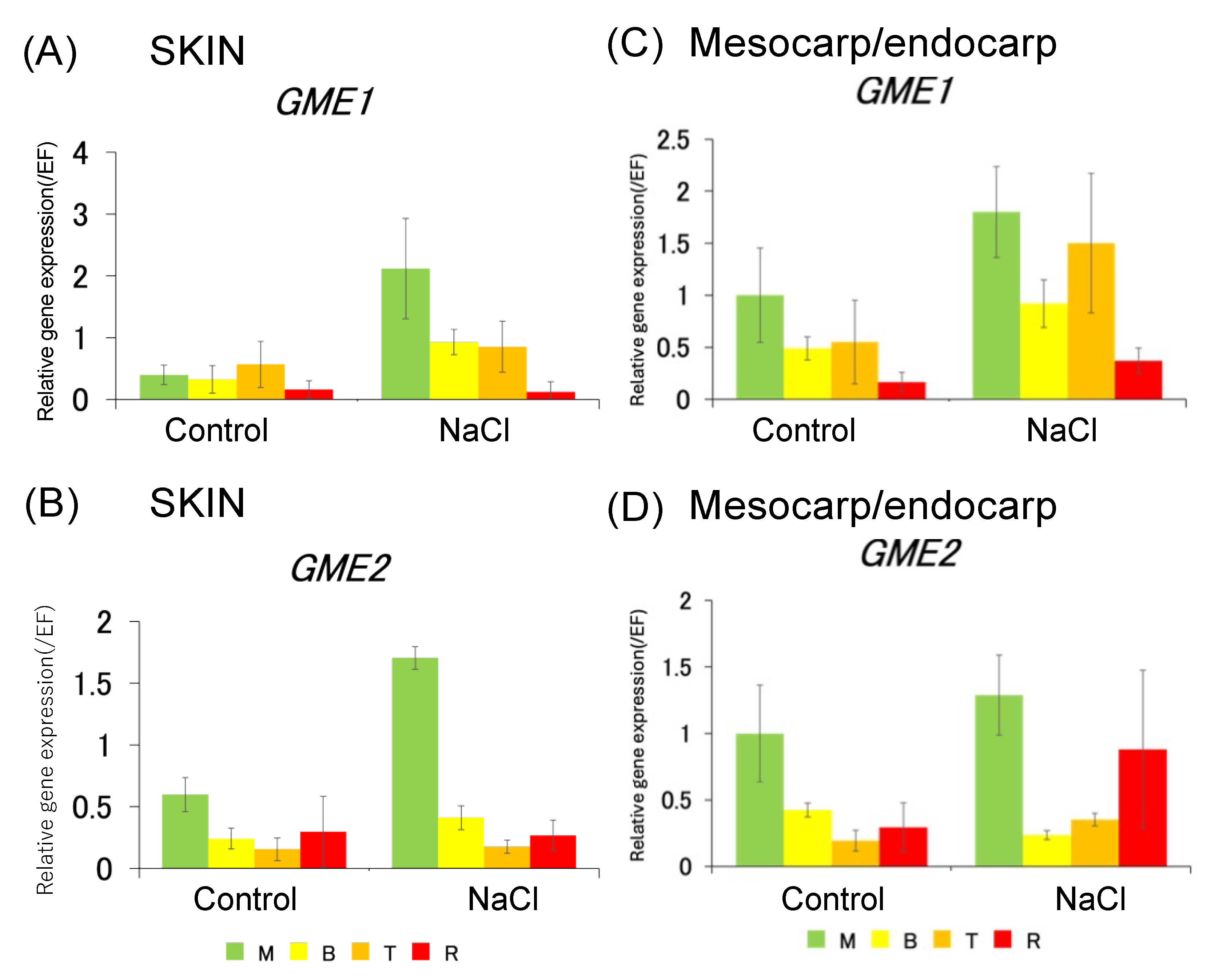
3.5. Expression Analysis of Ascorbic Acid Synthase Gene in Skin and Pericarp

Several pathways have been proposed for the biosynthesis of ascorbic acid in plants. Among them, the first pathway of ascorbic acid synthesis in higher plants was the D-mannose/L-galactose pathway [4]. The GDP-mannose 3′,5′-epimerase (GME) gene, which functions in this pathway, is an enzyme gene that converts GDP-D-mannose to GDP-L-galactose and is the most highly conserved ascorbic acid biosynthesis-related gene among higher plants [5], so it has been extensively studied. GME exists as a single-copy gene in many plants, but two genes, GME1 and GME2, are known to exist in tomato [6]. The primers used for the expression analysis of each gene were designed based on Yin et al. [10]. The expression analysis of GME1 and GME2 was then performed by real-time PCR. As a result, in the skin, the expression of the GME1 gene was significantly increased by about 434% at the M stage and by about 185% at the B stage, and the expression of the GME2 gene was significantly increased by about 185% at the M stage under salt stress conditions compared with the control (Figure 6A,B). In the endocarp, the expression of the GME1 gene was significantly increased by about 88% at the B stage, and the expression of the GME2 gene was significantly increased by about 81% at the T stage under salt stress conditions compared with the control (Figure 6C,D).

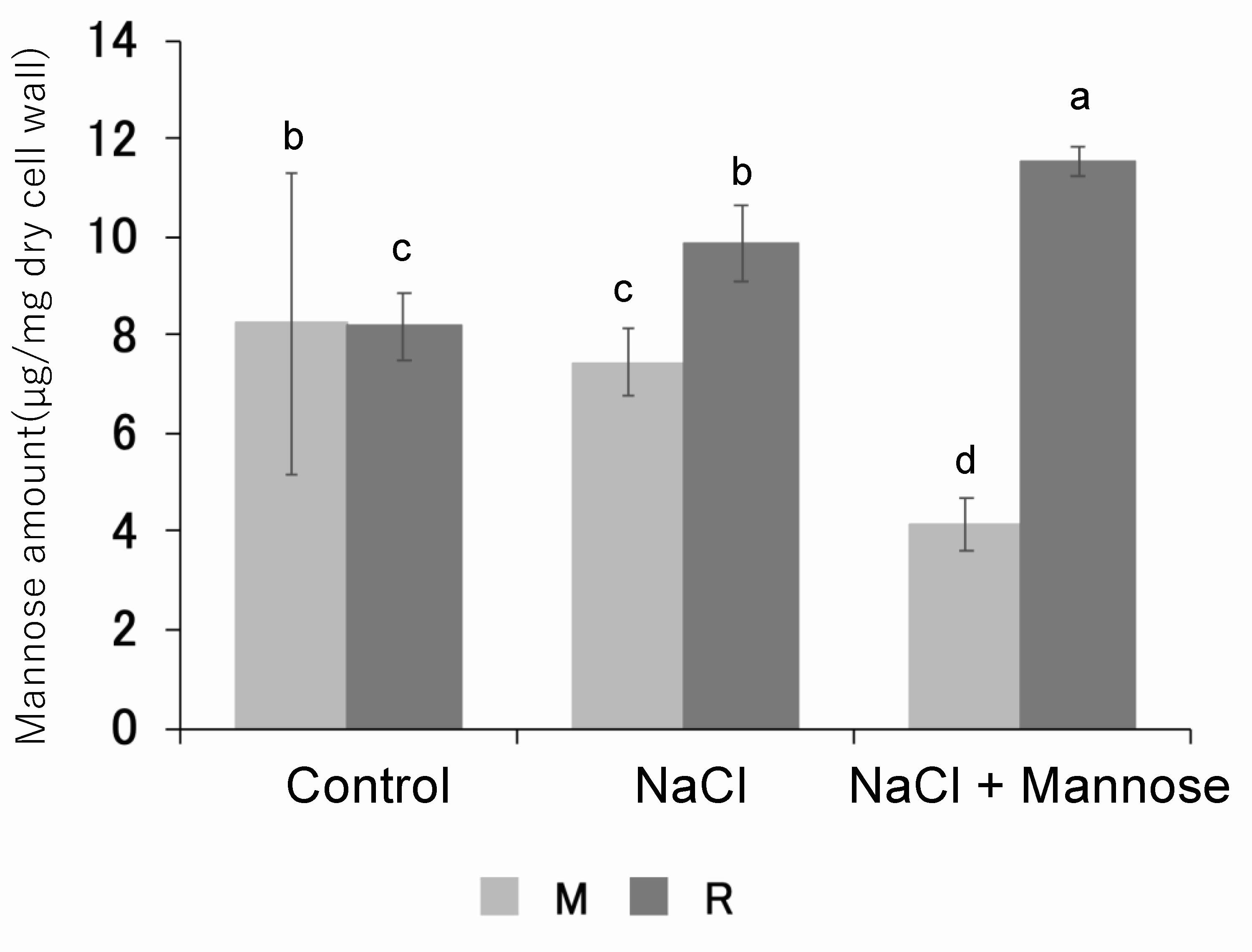
3.6. Measurement of Mannan Content in Skin and Pericarp of Mannose-Supplemented Tomato Fruits

The mannan content in the mesocarp of tomato fruits grown under salinity stress conditions with the addition of mannose was measured and compared with the control and salinity stress conditions. As a result, it was found that the mannan content in the mesocarp of tomato fruits grown under salinity stress conditions with added mannose was significantly reduced by about 45% at the M stage and significantly increased by about 17% at the R stage compared with salinity stress conditions (Figure 7).

4. Discussion

In this study, we measured mannose and ascorbic acid levels in tomato fruits under salinity stress and analyzed the expression of mannan synthase and ascorbic acid synthase genes. Additionally, we investigated how adding mannose affects ascorbic acid levels under salinity stress.
Expression analysis of mannan synthase genes and ascorbic acid synthesis genes in seeds revealed that salinity stress reduced the expression of mannan synthase CSLA (Figure 3). This suggests that under salinity stress, GDP-mannose is preferentially allocated to ascorbic acid synthesis, thereby reducing the expression of mannan synthesis genes. Gene expression data further indicate that seed mannan levels decrease under salinity stress (Figure 2A), resulting in poor seed germination. This supports the hypothesis that there is competition between ascorbic acid and mannan synthesis for GDP-mannose. Mannan, a major cell wall component in tomato seeds, is particularly abundant in the endosperm. Seed mannan of tomato likely originates from the endosperm. This mannan has two functions: (i) to form a crystalline or cross-linked structure that binds to cellulose and (ii) to act as a storage polysaccharide in seed endosperm and vegetative tissues. When mannose was supplied externally, previously non-germinating salt-stressed seeds showed partial germination (Figure 4), reinforcing the idea that seed germination capacity is closely linked to mannan availability.
Conversely, in the pericarp, the main edible part of the fruit, both mannan and ascorbic acid levels increased under salinity stress conditions (Figure 2B,D). Despite the overall mannose deficiency in salinity-stressed tomato fruits, the pericarp maintained active synthesis of both ascorbic acid and mannan. This suggests that the pericarp plays a role in compensating for metabolic shifts that occur under stress conditions. Previous studies have reported an increase in ascorbic acid content in whole tomato fruits under salinity stress. However, our study examined ascorbic acid content specifically in the skin and the endocarp and showed an increase at the early ripening stage in both tissues (Figure 5). Furthermore, GME expression analysis showed a similar trend, indicating that ascorbic acid synthesis is promoted at the early ripening stage under salinity stress (Figure 6).
Ascorbic acid plays a critical role in plants by acting as an antioxidant that mitigates the effects of reactive oxygen species (ROS) generated during growth and environmental stress. Ascorbic acid synthesis is achieved not only by the GDP-mannose pathway, but also by the galacturonic acid pathway using pectin hydrolysis products as substrates. Both pathways share intermediates for ascorbic acid and cell wall polysaccharide synthesis [26,27]. Since the pericarp cell wall mannan remained stable or increased under salinity stress, it is likely that other cell wall components, such as pectin hydrolysis products, contributed to ascorbic acid synthesis. Furthermore, no significant differences in hydrogen peroxide levels were observed between the control and salinity-stressed tomato fruits [28,29], suggesting that ascorbic acid accumulation under salinity stress reflects its active participation in ROS scavenging. In addition, ascorbic acid promotes pectin solubilization, which contributes to fruit softening [30,31]. These findings suggest that salinity stress induces ROS production, which triggers ascorbic acid synthesis to mitigate oxidative stress. This process may also accelerate pectin degradation in the endocarp, leading to cell wall solubilization and fruit softening, as previously proposed by Professor Fry’s group [30].
Since both ascorbic acid and cell wall mannan are synthesized from the same substrate, mannose, salinity stress conditions increase ascorbic acid synthesis while decreasing seed mannan levels. To investigate whether external mannose supplementation could counteract this effect, we added mannose under salinity stress conditions. The results showed that tomato fruits grown with added mannose accumulated higher ascorbic acid levels at the early ripening stage (M) compared with those grown under salinity stress alone (Figure 5). In addition, mannan levels decreased at the M stage but increased at the R stage under mannose supplementation compared with the control and salinity stress conditions (Figure 7). These results suggest that under salinity stress, mannose deficiency suppresses cell wall mannan synthesis, while supplementation promotes ascorbic acid synthesis at early ripening stages. Once sufficient ascorbic acid accumulates, mannose is redirected to cell wall mannan synthesis at later stages of ripening.

5. Conclusions

This study showed increased levels of ascorbic acid in tomatoes grown under salinity stress, which correlated with reduced mannan content in skin and seeds. Expression analysis revealed higher expression of the ascorbic acid synthase gene during early ripening stages under salt stress. In addition, genes related to mannan metabolism showed increased expression during mid-ripening. Since ascorbic acid and mannan both use mannose as a precursor, their synthesis competes under salt stress, potentially leading to deficiencies in both. To investigate this, a mannose-supplemented culture system was developed. In conclusion, tomatoes supplemented with mannose under salt stress conditions exhibited increased ascorbic acid synthesis at the immature M stage compared with the control or salt-stressed tomatoes alone. This suggests that tomatoes with higher ascorbic acid content could be produced for markets where fruit is harvested and shipped at the immature M stage. These results provide insights for optimizing salt-stressed tomato breeding to improve nutritional quality and marketability.

Supplementary Materials

The following supporting information can be downloaded at: https://www.mdpi.com/article/10.3390/horticulturae11040400/s1, Table S1: List of primers used in this study.

Author Contributions

Conceptualization, H.I.; methodology, C.H., K.Y. and N.H.; software, H.I.; validation, C.H. and K.Y.; formal analysis, K.S.; investigation, C.H. and K.Y.; resources, K.S.; data curation, C.H., K.Y. and K.S.; writing—original draft preparation, C.H. and H.I.; writing—review and editing, H.I.; visualization, M.S.; supervision, H.I.; project administration, H.I.; funding acquisition, H.I. All authors have read and agreed to the published version of the manuscript.

Funding

This research was funded by The Salt Science Research Foundation and the 31st, 32nd, 33rd and 34th Botanical Research Grant of ICHIMURA Foundation for New Technology.

Data Availability Statement

Data are contained within the article and Supplementary Materials.

Acknowledgments

Tomato seed was provided by the University of Tsukuba, Tsukuba Plant Innovation Research Center, through the National Bio-Resource Project (NBRP) of the MEXT/AMED, Japan.

Conflicts of Interest

The authors declare no conflicts of interest.

Abbreviations

The following abbreviations are used in this manuscript:
AsAAscorbic Acid
GMEGDP-Mannose Epimerase
CSLACellulose Synthase-like A
ROSReactive Oxygen Species

References

  1. Fujita, M.; Fujita, Y.; Noutoshi, Y.; Takahashi, F.; Narusaka, Y.; Yamaguchi-Shinozaki, K.; Shinozaki, K. Crosstalk between Abiotic and Biotic Stress Responses: A Current View from the Points of Convergence in the Stress Signaling Networks. Curr. Opin. Plant Biol. 2006, 9, 436–442. [Google Scholar] [CrossRef] [PubMed]
  2. Bolarin, M.C.; Estañ, M.T.; Caro, M.; Romero-Aranda, R.; Cuartero, J. Relationship between Tomato Fruit Growth and Fruit Osmotic Potential under Salinity. Plant Sci. 2001, 160, 1153–1159. [Google Scholar] [CrossRef] [PubMed]
  3. Gama, P.B.S.; Inanaga, S.; Tanaka, K.; Nakazawa, R. Physiological Response of Common Bean (Phaseolus Vulgaris L.) Seedlings to Salinity Stress. Afr. J. Biotechnol. 2007, 6, 79–88. [Google Scholar]
  4. Munns, R.; Tester, M. Mechanisms of Salinity Tolerance. Annu. Rev. Plant Biol. 2008, 59, 651–681. [Google Scholar] [CrossRef] [PubMed]
  5. Mohammad, M.; Shibli, R.; Ajlouni, M.; Nimri, L. Tomato Root and Shoot Responses to Salt Stress under Different Levels of Phosphorus Nutrition. J. Plant Nutr. 1998, 21, 1667–1680. [Google Scholar] [CrossRef]
  6. Scholberg, J.M.S.; Locascio, S.J. Growth Response of Snap Bean and Tomato as Affected by Salinity and Irrigation Method. HortScience 1999, 34, 259–264. [Google Scholar] [CrossRef]
  7. Magán, J.J.; Gallardo, M.; Thompson, R.B.; Lorenzo, P. Effects of Salinity on Fruit Yield and Quality of Tomato Grown in Soil-Less Culture in Greenhouses in Mediterranean Climatic Conditions. Agric. Water Manag. 2008, 95, 1041–1055. [Google Scholar] [CrossRef]
  8. Ho, L.C. A Cellular Hypothesis for the Induction of Blossom-End Rot in Tomato Fruit. Ann. Bot. 2005, 95, 571–581. [Google Scholar] [CrossRef]
  9. Saito, T.; Matsukura, C.; Sugiyama, M.; Watahiki, A.; Ohshima, I.; Iijima, Y.; Konishi, C.; Fujii, T.; Inai, S.; Fukuda, N.; et al. Screening for γ-Aminobutyric Acid (GABA)-Rich Tomato Varieties. J. Jpn. Soc. Hortic. Sci. 2008, 77, 242–250. [Google Scholar] [CrossRef]
  10. Yin, Y.-G.; Kobayashi, Y.; Sanuki, A.; Kondo, S.; Fukuda, N.; Ezura, H.; Sugaya, S.; Matsukura, C. Salinity Induces Carbohydrate Accumulation and Sugar-Regulated Starch Biosynthetic Genes in Tomato (Solanum Lycopersicum L. cv. ‘Micro-Tom’) Fruits in an ABA- and Osmotic Stress-Independent Manner. J. Exp. Bot. 2010, 61, 563–574. [Google Scholar] [CrossRef]
  11. Ezura, H.; Hiwasa-Tanase, K. Fruit Development. In Plant Developmental Biology—Biotechnological Perspectives; Pua, E.C., Davey, M.R., Eds.; Springer: Berlin/Heidelberg, Germany, 2010; pp. 301–318. ISBN 978-3-642-02300-2. [Google Scholar]
  12. Gillaspy, G.; Ben-David, H.; Gruissem, W. Fruits: A Developmental Perspective. Plant Cell 1993, 5, 1439–1451. [Google Scholar] [CrossRef] [PubMed]
  13. Terao, A.; Hyodo, H.; Satoh, S.; Iwai, H. Changes in the Distribution of Cell Wall Polysaccharides in Early Fruit Pericarp and Ovule, from Fruit Set to Early Fruit Development, in Tomato (Solanum Lycopersicum). J. Plant Res. 2013, 126, 719–728. [Google Scholar] [CrossRef] [PubMed]
  14. Hyodo, H.; Terao, A.; Furukawa, J.; Sakamoto, N.; Yurimoto, H.; Satoh, S.; Iwai, H. Tissue Specific Localization of Pectin–Ca2+ Cross-Linkages and Pectin Methyl-Esterification during Fruit Ripening in Tomato (Solanum Lycopersicum). PLoS ONE 2013, 8, e78949. [Google Scholar] [CrossRef] [PubMed]
  15. Huber, D.J. The Role of Cell Wall Hydrolases in Fruit Softening. In Horticultural Reviews; John Wiley & Sons, Ltd.: Hoboken, NJ, USA, 1983; pp. 169–219. ISBN 978-1-118-06072-8. [Google Scholar]
  16. Seymour, G.B.; Colquhoun, I.J.; Dupont, M.S.; Parsley, K.R.; Selvendran, R.R. Composition and Structural Features of Cell Wall Polysaccharides from Tomato Fruits. Phytochemistry 1990, 29, 725–731. [Google Scholar] [CrossRef]
  17. Zhang, L.; Wang, P.; Chen, F.; Lai, S.; Yu, H.; Yang, H. Effects of Calcium and Pectin Methylesterase on Quality Attributes and Pectin Morphology of Jujube Fruit under Vacuum Impregnation during Storage. Food Chem. 2019, 289, 40–48. [Google Scholar] [CrossRef]
  18. Moreira, L.R.S.; Filho, E.X.F. An Overview of Mannan Structure and Mannan-Degrading Enzyme Systems. Appl. Microbiol. Biotechnol. 2008, 79, 165–178. [Google Scholar] [CrossRef]
  19. Schröder, R.; Wegrzyn, T.F.; Sharma, N.N.; Atkinson, R.G. LeMAN4 Endo-Beta-Mannanase from Ripe Tomato Fruit Can Act as a Mannan Transglycosylase or Hydrolase. Planta 2006, 224, 1091–1102. [Google Scholar] [CrossRef]
  20. Ishida, K.; Ohba, Y.; Yoshimi, Y.; Wilson, L.F.L.; Echevarría-Poza, A.; Yu, L.; Iwai, H.; Dupree, P. Differing Structures of Galactoglucomannan in Eudicots and Non-Eudicot Angiosperms. PLoS ONE 2023, 18, e0289581. [Google Scholar] [CrossRef]
  21. Wheeler, G.L.; Jones, M.A.; Smirnoff, N. The Biosynthetic Pathway of Vitamin C in Higher Plants. Nature 1998, 393, 365–369. [Google Scholar] [CrossRef]
  22. Hu, T.; Ye, J.; Tao, P.; Li, H.; Zhang, J.; Zhang, Y.; Ye, Z. The Tomato HD-Zip I Transcription Factor SlHZ24 Modulates Ascorbate Accumulation through Positive Regulation of the d-Mannose/l-Galactose Pathway. Plant J. 2016, 85, 16–29. [Google Scholar] [CrossRef]
  23. Zheng, X.; Gong, M.; Zhang, Q.; Tan, H.; Li, L.; Tang, Y.; Li, Z.; Peng, M.; Deng, W. Metabolism and Regulation of Ascorbic Acid in Fruits. Plants 2022, 11, 1602. [Google Scholar] [CrossRef] [PubMed]
  24. Zhang, P.; Senge, M.; Dai, Y. Effects of Salinity Stress on Growth, Yield, Fruit Quality and Water Use Efficiency of Tomato Under Hydroponics System. Rev. Agric. Sci. 2016, 4, 46–55. [Google Scholar] [CrossRef]
  25. Voxeur, A.; Gilbert, L.; Rihouey, C.; Driouich, A.; Rothan, C.; Baldet, P.; Lerouge, P. Silencing of the GDP-D-Mannose 3,5-Epimerase Affects the Structure and Cross-Linking of the Pectic Polysaccharide Rhamnogalacturonan II and Plant Growth in Tomato. J. Biol. Chem. 2011, 286, 8014–8020. [Google Scholar] [CrossRef] [PubMed]
  26. Carpita, N.C.; Gibeaut, D.M. Structural Models of Primary Cell Walls in Flowering Plants: Consistency of Molecular Structure with the Physical Properties of the Walls during Growth. Plant J. Cell Mol. Biol. 1993, 3, 1–30. [Google Scholar] [CrossRef]
  27. Iwai, H. Virtual issue: Cell Wall Functions in Plant Growth and Environmental Responses. J. Plant Res. 2021, 134, 1155–1158. [Google Scholar] [CrossRef]
  28. Zushi, K.; Matsuzoe, N.; Kitano, M. Developmental and Tissue-Specific Changes in Oxidative Parameters and Antioxidant Systems in Tomato Fruits Grown Under Salt Stress. Sci. Hortic. 2009, 122, 362–368. [Google Scholar] [CrossRef]
  29. Soyama, K.; Yano, A.; Miyakoshi, A.; Itano, M.; Sugiyama, H.; Iwai, H. Regulation of Cell Wall Remodeling is an Important Factor in the Reduction of Tomato Fruit Size Immediately after Fruit Set Induced by Salinity Conditions. Hortic. J. 2024, 93, 397–405. [Google Scholar] [CrossRef]
  30. Dumville, J.C.; Fry, S.C. Solubilisation of Tomato Fruit Pectins by Ascorbate: A Possible Non-Enzymic Mechanism of Fruit Softening. Planta 2003, 217, 951–961. [Google Scholar] [CrossRef]
  31. Ford, C.M.; Sweetman, C.; Fry, S.C. Ascorbate Degradation: Pathways, Products, and Possibilities. J. Exp. Bot. 2024, 75, 2733–2739. [Google Scholar] [CrossRef]
Figure 1. Preparation for tissue-specific analysis. The fruit ripening stages of cv. ‘Micro-Tom’. The four stages included: mature green (M), breaker (B), turning (T), and red ripe (R).
Horticulturae 11 00400 g001
Figure 2. Measurements of amounts of mannose and ascorbic acid of red ripe tomato fruit grown under control and saline conditions (160 mM). (A,B): Seed. (C,D): Pericarp. (A,C): Mannose amounts. (B,D): Ascorbic acid amounts. Ripening stages were as follows: mature green (M), breaker (B), turning (T), and red ripe (R). Error bars indicate the SD [n = 5 (Control) and n = 4 (NaCl)]. Different letters in each panel indicate significant differences at p < 0.05 (Tukey’s test).
Horticulturae 11 00400 g002
Figure 3. Gene expression patterns related to mannan and ascorbic acid biosynthesis during fruit ripening. Gene expression was analyzed by qRT-PCR in tomato seeds. Cellulose synthase-like A (CSLA) involved in mannan biosynthesis and GDP-mannose epimerase (GME) involved in ascorbic acid biosynthesis. Expression levels were compared with elongation factor 1α in the same assay. Ripening stages were as follows: mature green (M), breaker (B), turning (T), and red ripe (R). Values are means ± SD (n = 25).
Horticulturae 11 00400 g003
Figure 4. Seed germination rate from breaker-stage tomato fruit grown under control and mannose (1 mM or 10 mM) under salinity conditions. Error bars indicate the SD (n = 5).
Horticulturae 11 00400 g004
Figure 5. The influence of 160 mM NaCl alone and in combination with 1 mM mannose to ascorbic acid (AsA) content in tomato fruit. Ascorbic acids were extracted from tomato fruit skin and mesocarp/endocarp. (A): Skin. (B): Mesocarp and endocarp (Mesocarp/endocarp). Ripening stage: mature green (M) and red ripe (R). Error bars indicate the SD (n = 9). Different letters in each panel indicate significant differences at p < 0.05 (Tukey’s test).
Horticulturae 11 00400 g005
Figure 6. Ascorbic acid biosynthesis-related gene expression patterns during fruit ripening. Gene expression in tomato analyzed by qRT-PCR. GDP-mannose epimerase (GME) 1 and 2 are involved in ascorbic acid biosynthesis. (A,B): Skin. (C,D): Mesocarp and endocarp (Mesocarp/endocarp). Ripening stages were as follows: mature green (M), breaker (B), turning (T), and red ripe (R). Error bars indicate the SD [n = 5 (Control) and n = 4 (NaCl)].
Horticulturae 11 00400 g006
Figure 7. The effect of 160 mM NaCl alone and in combination with 1 mM mannose to mannose content in tomato fruit. Ripening stage: mature green (M) and red ripe (R). Error bars indicate the SD (n = 9). Different letters in each panel indicate significant differences at p < 0.05 (Tukey’s test).
Horticulturae 11 00400 g007
Disclaimer/Publisher’s Note: The statements, opinions and data contained in all publications are solely those of the individual author(s) and contributor(s) and not of MDPI and/or the editor(s). MDPI and/or the editor(s) disclaim responsibility for any injury to people or property resulting from any ideas, methods, instructions or products referred to in the content.

