Article

The Transcription Factor LoTDF1 Plays a Role in Early Anther Development in Lily (Lilium Oriental Hybrids)

1
Engineering Technology Research Center of Anti-Aging Chinese Herbal Medicine, Biology and Food Engineering College, Fuyang Normal University, Fuyang 236037, China
2
College of Architecture, Yantai University, Yantai 264005, China
*
Authors to whom correspondence should be addressed.
Horticulturae 2025, 11(4), 398; https://doi.org/10.3390/horticulturae11040398
Submission received: 5 March 2025 / Revised: 5 April 2025 / Accepted: 7 April 2025 / Published: 9 April 2025

Abstract

:
Lilies are one of the most popular ornamental flowers in the world. However, the abundant pollen produced in their anthers causes significant inconvenience for producers and consumers. Pollen abortion induced by molecular breeding techniques is one of the effective ways to solve this problem. In this study, the LoTDF1 gene, which is involved in regulating lily anther development, was identified and cloned from lily anthers based on transcriptome data. The open reading frame of LoTDF1 is 936 bp and encodes a protein with 311 amino acids. Multiple sequence alignment and phylogenetic tree analysis revealed that the LoTDF1 protein contained a conserved R2R3 domain, belonging to the MYB transcription factor family. Subcellular localization and transcriptional activation assays demonstrated that LoTDF1 localized to the nucleus and functioned as a transcription activator. The transcriptional activation domain was located within the last 195 amino acids (117–311a) of the C-terminus, and there may be more than one transcriptional activation domain in the region. The expression level of the LoTDF1 gene was highest during the pollen mother cell (PMC) stage of lily anther development (2 cm anther), followed by the tetrad stage (4 cm anther). In situ hybridization experiments further confirmed that LoTDF1 transcripts were predominantly localized in PMCs, tapetal cells, middle layer cells, dyads, and tetrads. The experiment data suggest that LoTDF1 plays a critical role in regulating early anther development in lily. LoTDF1 could be a promising candidate gene for molecular breeding strategies aimed at developing pollen-free lily cultivars to enhance commercial and consumer appeal.

1. Introduction

Flowers are important reproductive organs in flowering plants [1]. A typical flower usually consists of four main parts: sepals, petals, stamens, and pistils [1,2]. The stamen is composed of anthers and filaments, and the anthers are important location for pollen production [1,3]. As an essential component for plant sexual reproduction, pollen quantity and quality directly influence pollination success and fertilization rates, ultimately impacting crop and fruit yields [4,5]. However, excessive pollen production presents significant agricultural and horticultural challenges, especially in hybrid breeding where male removal is required [4,6]. The manual removal of stamens is labor-intensive, inefficient, and susceptible to environmental influences [4]. Therefore, causing pollen abortion in plants is one of the effective ways to solve this problem.
Lilies (Lilium oriental hybrids (Liliaceae)), perennial bulbous plants in the Liliaceae family, are prized in floriculture for their vibrant colors, distinctive patterns, and fragrant blooms [7,8]. Widely cultivated as cut flowers and potted plants, lilies also carry significant cultural symbolism. They are often used for festive occasions like weddings and celebrations, symbolizing purity and good wishes for the future. However, lily anthers contain a large amount of pollen, which can lead to various issues, such as contaminating the surrounding environment, staining clothes, causing consumer allergies, and so on [7,8,9,10]. Therefore, people usually remove anthers manually before they open, which is an inefficient process that increases production costs. In recent years, some double lily cultivars have been introduced to the market. However, single-petaled lilies continue to dominate the market with a larger share compared to double-petaled lilies, owing to their classic flower shape and low-maintenance traits. Developing pollen-free lilies that maintain key ornamental characteristics, such as pattern, color, and fragrance, still remains a critical objective in contemporary lily breeding programs.
The regulatory mechanism of pollen abortion involves molecular and cellular processes at multiple levels, primarily including gene expression regulation, cell apoptosis, and metabolic pathway alterations [11,12]. At the molecular level, gene expression regulation plays a crucial role in pollen abortion by controlling the activation or suppression of specific genes. For example, the β-1,3-GLUCANASE10(SlBG10) gene encodes β-1,3-glucanase, which regulates tomato pollen fertility by modulating callose deposition [13]. Knockout of the SlBG10 gene resulted in abnormal callose deposition, which subsequently induced pollen abortion [13]. MORE FLORET1 (MOF1) is an MYB transcription factor that precisely controls the development of the tapetum by negatively regulating the expression of key tapetum-related genes, such as CYP703A3, OsABCG26, and OsABCG15 [14]. The mof1 mutants lead to delayed degradation of the tapetum and pollen abortion [14].
Transcription factors (TFs), a group of protein molecules that specifically bind to particular DNA sequences, ensure the precise spatiotemporal expression of target genes at appropriate intensity levels [15]. Many TFs are expressed in a timely and orderly manner to regulate the development process of anthers, from differentiation to maturity [12,16,17]. Developmental abnormalities at any stage may cause pollen abortion. The tapetum, as the innermost sporophytic tissue of the pollen locule, plays a crucial role in anthers’ development [1,17]. It can provide nutrients for microspore development, secrete callose enzymes to release microspores in a timely manner, and participate in pollen exine synthesis [1,2,12]. In recent years, many studies have focused on the molecular mechanisms of anther development, and some key genes have been identified. Based on the research results, a core transcriptional regulatory pathway, DYT1–TDF1–AMS–MYB80–MS1, related to tapetum development and pollen exine formation, has been established [18,19]. DYT1 (DYSFUNCTIONAL TAPETUM1) is one of the helix–loop–helix (bHLH) transcription factors that is essential for early tapetal development and pollen wall formation [20,21]. It regulates the expression of TDF1 by binding to DNA in a sequence-specific manner [21]. TDF1 (DEFECTIVE in TAPETAL DEVELOPMENT and FUNCTION1), also named MYB35, encodes an R2R3 MYB transcription factor that is critical for early tapetal layer development and microspore maturation [12,22]. It directly regulates the AMS gene via a cis-element (AACCT) and can also form a complex with AMS (TDF1–AMS) to promote the expression of AMS-regulated downstream genes [23]. AMS (ABORTED MICROSPORE) also encodes a bHLH protein [17]. It plays an important role in the development of tapetum cells and pollen wall formation [24,25,26]. MYB80 belongs to the R2R3 MYB family [27]. It can ensure the timely occurrence of tapetal PCD (programmed cell death) [27] and form a complex with AMS to regulate sporopollenin biosynthesis by activating the expression of CYP703A2 [28]. MS1 (MALE STERILITY 1) encodes a PHD-finger transcription factor, which is the direct target of MYB80 [29,30]. It is expressed in the tapetum of the tetrad stage and involved in pollen wall and coat formation [29,30].
Developing pollen-aborted lily varieties through genetic engineering is one of the effective ways to solve the problem of pollen overproduction. To better understand the anther-specific genes that regulate lily anther development, a series of studies were conducted [8,10,31]. Several key regulatory genes involved in lily anther development have been characterized, such as LoMYB80, LoMYB33, and LoAMS [8,10,31]. To further identify anther-specific genes, we conducted an RNA-seq analysis of lily anthers at different developmental stages [32]. Many transcription factors related to tapetum and microspore development have been screened out, such as those from MYB, NAC, bHLH, MADS, and PHD families [32]. In this study, we aimed to identify TFs regulating early-stage lily anther development. Based on the transcriptome data and the core regulatory pathway DYT1-TDF1-AMS-MYB80-MS1 for anther development, TDF1 was screened as a candidate. We studied the protein characteristics and expression characteristics of TDF1, and the result provided a useful reference for lily variety without pollen overproduction.

2. Results

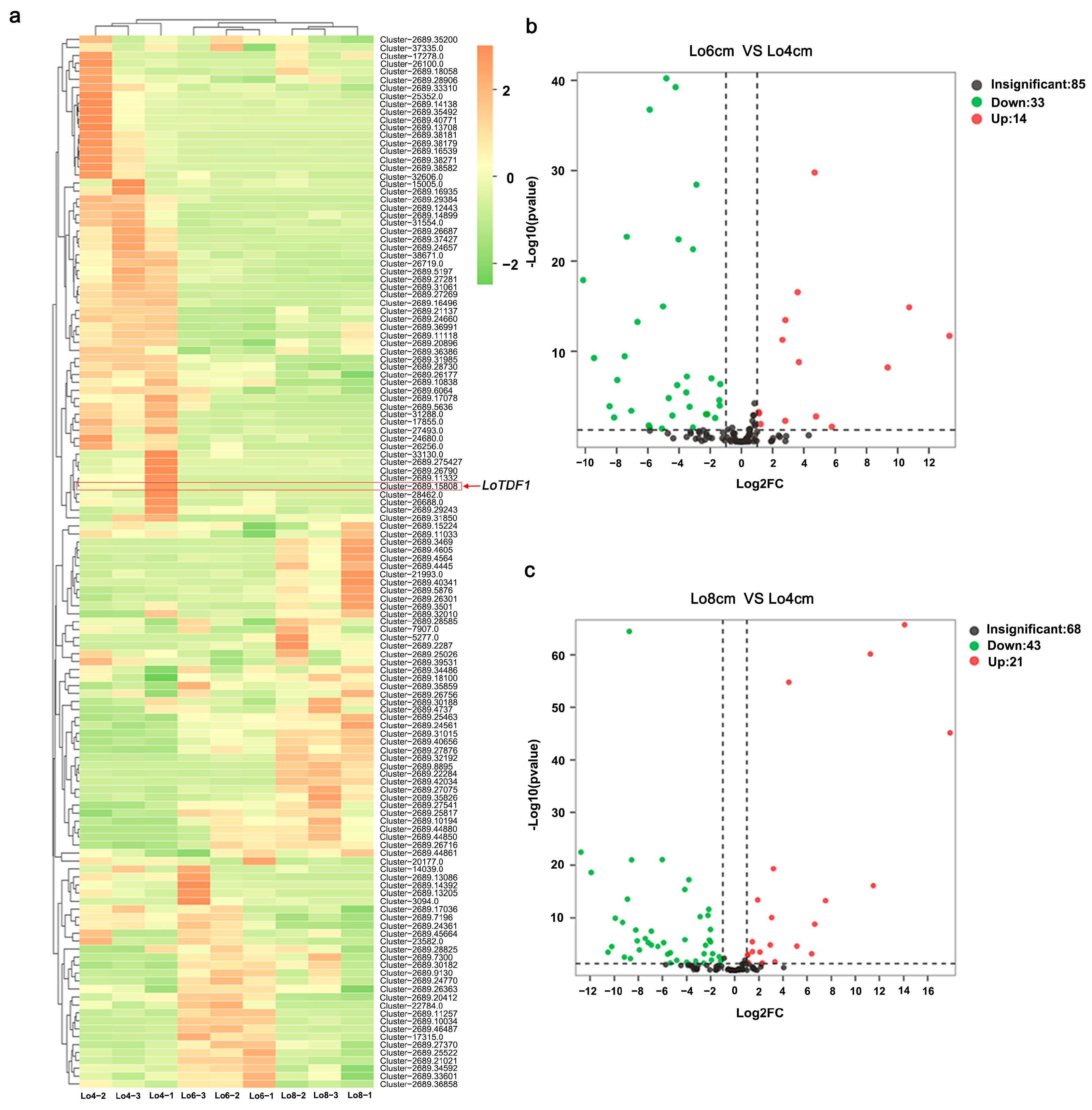
2.1. Screening the LoTDF1 Gene Based on Transcriptome Data

Based on the previous transcriptome data, we focused on the MYB family TFs that regulate lily anther development. The analysis was conducted and 132 TF members of the MYB family were screened out (Figure 1a). According to the volcano plots, we found that the expression of 33 MYB TFs was significantly downregulated during the period from the Lo4 cm to Lo6 cm stages, and 43 were significantly downregulated during the period from the Lo4 cm to Lo8 cm (Figure 1b,c), suggesting stage-specific regulation of MYB family genes during the developmental process. We hoped to screen out the genes that can regulate the early development of lily anthers to prevent the formation of pollen grains through genetic engineering. Thus, LoTDF1/LoMYB35 with high expression in the early stage (Lo4 cm) and low expression in the later stage (Lo6 cm, Lo8 cm) was screened out for further study.

2.2. Molecular Characterization and Phylogenetic Analysis of LoTDF1

The LoTDF1 gene was cloned as a candidate gene for further study. The open reading frame (ORF) of the LoTDF1 gene is 936 bp, encoding a protein with 311 amino acids. Domain analysis revealed that LoTDF1 protein contains a conserved R2R3 domain, which belongs to the R2R3-MYB subfamily (Figure 2a). At the amino acid level, the LoTDF1 protein shared a 55.26% similarity to homologous protein in Elaeis guineensis (XP_010921545.1) and a 44.55% similarity to Arabidopsis (NP_189488.1). A phylogenetic analysis was performed using LoTDF1 and Arabidopsis MYB transcription factors, and it was found that LoTDF1 has the closest genetic relationship with AtMYB35, which further confirmed that LoTDF1 belonged to the transcription factors of the MYB family (Figure 2b). Another phylogenetic tree was performed to further study the phylogenetic relationships among LoTDF1 and other MYB35 homologous proteins from monocots and dicots, respectively, such as Phoenix dactylifera (XP_008804161.1), Elaeis guineensis, and Brassica napus (XP_013707835.1). The result showed that LoTDF1 clustered with monocots and most closely related to Phoenix dactylifera and Elaeis guineensis (Figure 2c). All these results demonstrated that LoTDF1 exhibited hallmark features of MYB transcription factors, including conserved structural domains characteristic of this protein family. This suggests that LoTDF1 likely acts analogously to MYB35 orthologs in angiosperms, potentially regulating anther development through conserved molecular mechanisms.

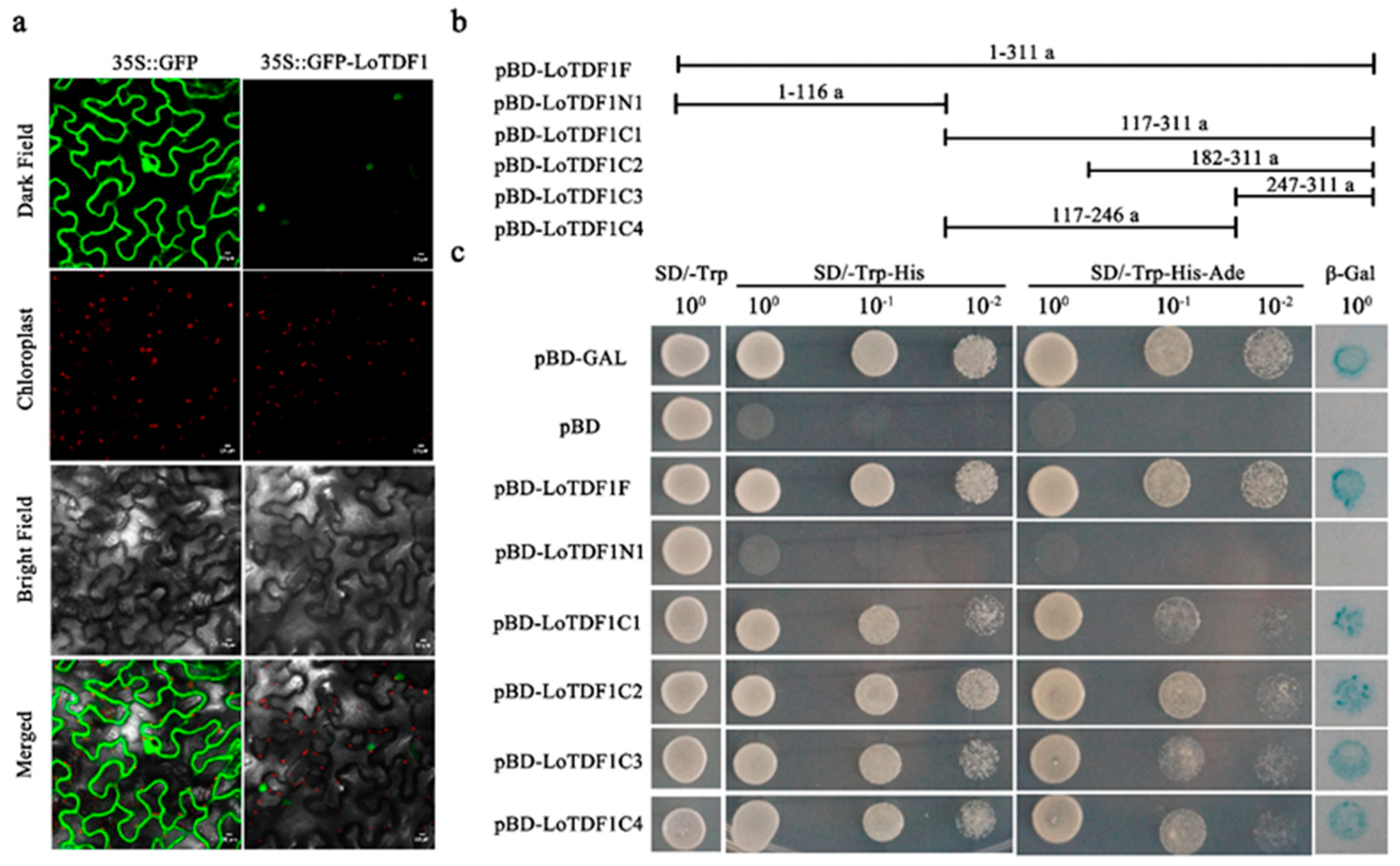
2.3. Subcellular Localization of LoTDF1

The subcellular localization of 35S::GFP-LoTDF1 was determined by transient expression in tobacco leaves’ epidermal cells. The result showed that the green fluorescence signals of 35S::GFP were distributed throughout the entire cell (Figure 3a, left). However, the green fluorescence signals of 35S::GFP-LoTDF1 can only be observed in the nucleus of the cells (Figure 3a, right). The results indicated that LoTDF1 protein is localized in the nucleus with a functional nuclear localization signal, and it possesses the basic characteristics of transcription factors.

2.4. Transcriptional Activity Analysis of LoTDF1

In order to detect the potential transcriptional activity of LoTDF1, a yeast one-hybrid system was applied. The LoTDF1 gene (ORF) was cloned into the pGBKT7 vector to form pBD-LoTDF1F (Figure 3b), and then the recombinant plasmids were transformed into the yeast strain (AH109). The transformants and negative control (pBD) can grow on the selection medium SD/-Trp (Figure 3c). Compared with the negative control, LoTDF1 can grow normally on SD/-Trp-His and SD/-Trp-His-Ade media plates, which meant that LoTDF1 could activate the expression of the reporter in yeast (Figure 3c). The results indicated that LoTDF1 should be a transcription activator that regulates related gene expression during anther development. For further detecting the transcription activation domain, different LoTDF1 truncations were cloned into the pGBKT7 vector and also transferred into the yeast strain. The yeast harboring pBD-LoTDF1C1 (117-311a), pBD-LoTDF1C2 (182-311a), pBD-LoTDF1C3 (247-311a), and pBD-LoTDF1C4 (117-246a) transformants can all grow normally on SD/-Trp-His and SD/-Trp-His-Ade media plates, and they turned blue on X-gal reporter plates, suggesting the presence of transcriptional activation domains in those regions (Figure 3c). However, the yeast harboring the N-terminal portion of pBD-LoTDF1N1 (1-116a) did not grow on SD/-Trp-His and SD/-Trp-His-Ade plates (Figure 3c).
Taken together, these data indicated that LoTDF1 was indeed a transcription activator, with the transcriptional activation domains located within 117-311 amino acids at the C-terminus. Moreover, based on the growth of the yeast harboring different portions, it was speculated that there may be more than one transcriptional activation domain within the region of 117-311 amino acids.

2.5. Histological Observation of Lily Anthers and Expression Analysis of LoTDF1

In order to further investigate the role of LoTDF1 in the development of lily anthers, paraffin sections were made to observe the tissues changes during anther development. The results revealed that many sporogenic cells with large nuclei, dense cytoplasm, and a tight arrangement were present in the locules of anthers in 1 cm flower buds (Figure 4a). At the 2 cm stage, the structures of the fibrous layer, middle layer, and tapetum layer were clearly visible (Figure 4a). The tapetum developed into a layer of long columnar cells, and the cells within the locules were at the PMC stage (Figure 4a). From the 3 to 4 cm stages, dyads and tetrads emerged and were wrapped by callose (Figure 4a). At this stage, the tapetum cells evolved into binucleated or even multinucleated polar secretory cells (Figure 4a). Subsequently, the microspores were released from the callose, and the tapetum further concentrated at the 6 cm stage (Figure 4a). At the 8 cm stage, the microspores within the locules gradually matured into pollen grains. By this stage, the tapetum had degraded, leaving only a portion of the remaining structure (Figure 4a).
The transcript level of LoTDF1 was detected in different organs when the anthers were at the PMC stage. The qRT-PCR result showed that the relative expression level of LoTDF1 in anthers (2 cm) was significantly higher than that in other vegetative organs, including leaves, stems, roots, and bulbs (Figure 4b). This suggested that LoTDF1 is a specific gene for anther development. The transcript level of LoTDF1 was also detected at different anther developmental stages. The result demonstrated that LoTDF1 had the highest expression in the anthers at the 2 cm stage, followed by the 4 cm stage (Figure 4c). This indicates that LoTDF1 mainly functions during the PMC stage of lily anthers.

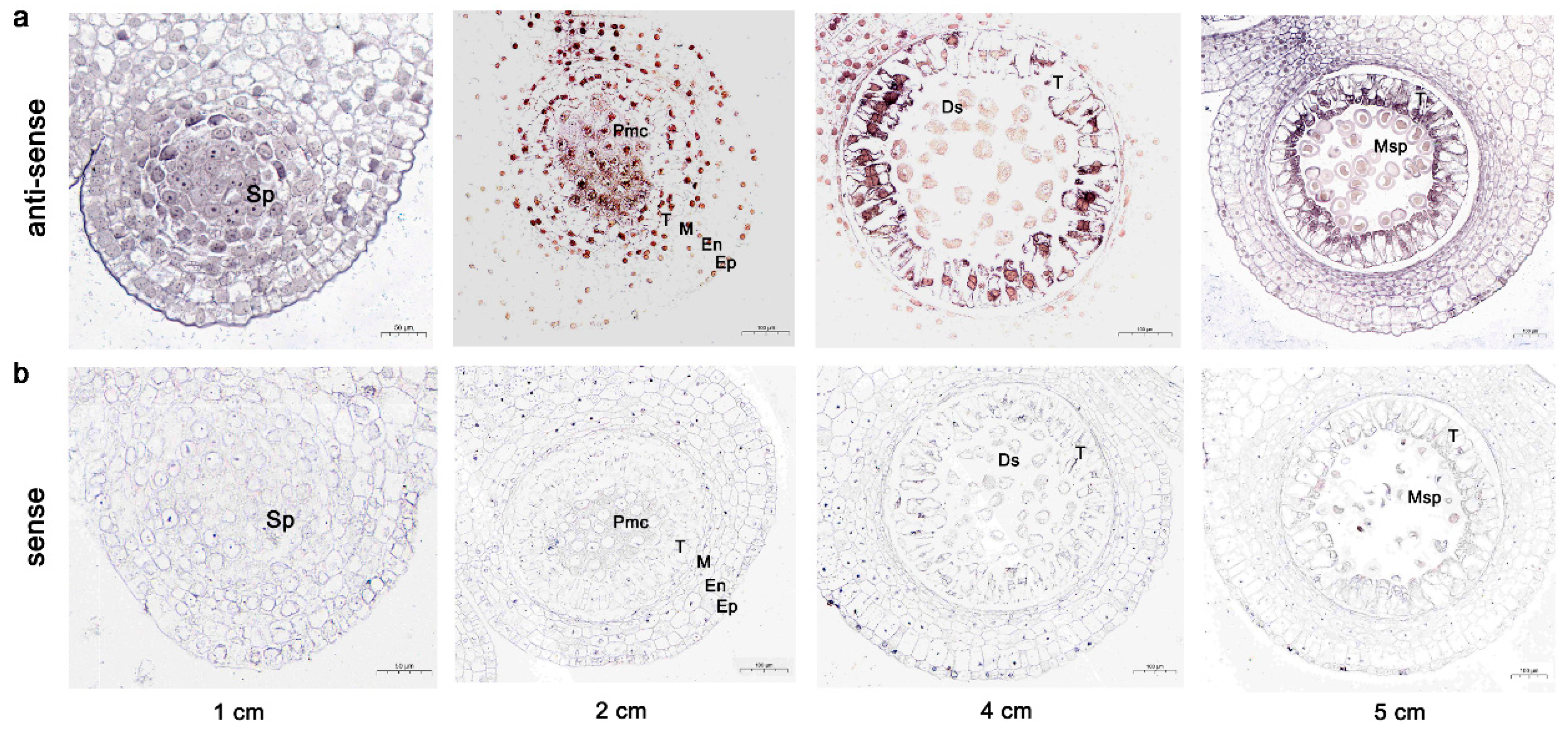
2.6. In Situ Hybridization of LoTDF1

The qRT-PCR analysis indicated that the LoTDF1 expression level was high at the PMC stage of lily anthers. To further explore the transcripts of the LoTDF1 gene in lily anthers, we conducted RNA in situ hybridization experiments at different stages: sporogenous tissue stage (1 cm), PMC stage (2 cm), meiosis stage (4 cm), and prophase of the independent microspore stage (5 cm). The results showed that in situ hybridization signals were detected as distinct brownish-yellow deposits (DAB chromogenic reaction) specifically localized in the PMCs (2 cm), tapetal cells (2 cm and 4 cm), middle layer cells (2 cm and 4 cm), and dyads and tetrads (4 cm) (Figure 5a). After meiosis, the tetrads were released from the encapsulated callose, and the expression of the LoTDF1 gene returned to the initial level (Figure 5a). This result was consistent with the spatial expression pattern of LoTDF1 transcripts (Figure 4c). Hybridization with the LoTDF1 sense probe showed the absence of a signal across all four developmental stages (Figure 5b). The result further demonstrates that the LoTDF1 functions as a key transcriptional regulator during early anther development, particularly in the development of the tapetum and PMCs.

3. Discussion

Lilies are popular all over the world due to their elegant floral morphology and pleasant fragrance [10,31]. Their cut flowers are extensively utilized for various significant occasions. However, the abundant pollen produced in their anthers has led to notable inconveniences in lily applications, including staining clothing and inducing pollen allergies [7,8,9,31]. Cultivating lily varieties without pollen overproduction through genetic engineering is one of the effective solutions to solve this problem. The process of anther development in plants involves intricate molecular and cellular mechanisms that regulate the differentiation and maturation of microsporocytes into pollen grains [25,33]. The process involves the expression and regulation of many genes [12]. In order to further explore the regulatory mechanism of lily anthers’ development, we sequenced the transcriptome of anthers at different developmental stages [32]. The analysis identified multiple differentially expressed genes associated with tapetum degradation and microspore release [32]. Based on the transcriptome data, we focused on the LoTDF1/LoMYB35, a transcription factor belonging to the MYB family.
Male sterility in flowering plants has long been a focal point for breeders, who have conducted extensive research on the genetic regulation of anther development [16,34]. A core transcriptional regulatory pathway, DYT1–TDF1–AMS–MYB80–MS1, has been identified as critical for tapetum development and pollen exine formation [18,19]. Among these, TDF1 plays a pivotal role in anther development [12]. It is involved in regulating the early stages of anther development [12]. It controls the degradation of the tapetum, and the tdf1 mutant exhibited irregular division and tapetum dysfunction [12]. Further research revealed that DYT1 directly regulates the expression of TDF1 by binding to specific cis-elements in the promoter region, which is essential for tapetum development and pollen wall formation [21]. TDF1 can regulate AMS directly and can also form a TDF1-AMS complex to promote the expression of AMS-regulated downstream genes [23].
The function of transcription factor TDF1 has been studied in many plants, such as Arabidopsis thaliana [12], Hordeum vulgare L. [35], and Triticum aestivum L. [16]. In Arabidopsis, AtTDF1 has been proven to be a key factor in regulating tapetal development and microsporogenesis [12]. In lily, a pollen sterility mutation Lfltdf1 was identified in a strain of Lilium × formolongi [36]. Paraffin section observation revealed that the pollen abortion of the Lfltdf1 mutant was caused by the abnormal degradation of PMCs before meiotic cell division [36]. The timing of anther development defects in the Lfltdf1 mutant was different from that in the attdf1 mutant [12,36]. In the attdf1 mutant, tetrads can be formed through meiosis, but due to severe vacuolization and abnormal hypertrophy of the tapetum, microspores cannot be released normally and eventually degrade [12]. However, in the Lfltdf1 mutant, tetrads cannot be formed through meiosis, and PMCs had already degraded before meiosis [36]. The timing of the appearance of defects in tdf1 mutants may vary by species [36]. In order to further investigate the function mechanism of TDF1 in the development of lily anthers, in this study, the LoTDF1 gene, which is also named LoMYB35, was cloned from lily anther based on the transcriptome data. The LoTDF1 protein had a typical R2R3 domain belonging to the MYB family. A series of experiments revealed that the LoTDF1 protein was located in the nucleus (Figure 3a) and possessed the basic features of transcription factors (Figure 2 and Figure 3a).
Transcription factors can regulate the transcription process of genes, thereby affecting gene expression [15]. According to the experiment of transcription activity analysis in yeast, we found that the LoTDF1 protein was a transcription activator. For further detecting the transcription activation domain, different LoTDF1 truncations were constructed, and the results showed that the transcriptional activity depended on a region of amino acids at the end of C-terminus (117-311a) (Figure 3b,c). Interestingly, the regions of 182-311a, 247-311a, and 117-246a all exhibited transcriptional activity (Figure 3c). We speculated that there was more than one transcriptional activation domain within the 117-311a region. Previous studies have shown that the pathway (DYT1–TDF1–AMS–MYB80–MS1) was essential for tapetum and pollen development [18,37]. TDF1 can directly regulate AMS expression via a cis-element and can also form a feed-forward loop (FFL) with AMS (TDF1–AMS) to regulate the expression of downstream genes [21,23]. Its regulatory function may be related to the presence of the transactivation domains which are located within the 117-311a region at the end of the C-terminus.
According to the study in Arabidopsis, AtTDF1 is essential for anther development [12,23]. A series of paraffin sections and qRT-PCR revealed that the relative expression level of the LoTDF1 gene was highest in the anthers from 2 cm flower buds, where it was in the PMC stage (Figure 4c), followed by the anthers from 4 cm flower buds, where it was in the dyads and tetrads stage (Figure 4c). The expression pattern was consistent with previous studies on LoAMS and LoMYB80 [8,10]. RNA in situ hybridization was also used to further identify spatial and temporal LoTDF1 gene expression. The LoTDF1 gene was expressed predominantly in PMCs (2 cm), the tapetal cells, middle layer cells (2 cm and 4 cm), and dyads and tetrads (4 cm) (Figure 5a). The result aligned with the spatial expression pattern of LoTDF1 transcripts in anthers at different developmental stages using qRT-PCR. Furthermore, this result was in accordance with the expression patterns of TDF1 in other plants, such as Arabidopsis and barley (Hordeum vulgare L.) [12,35]. But the reason for the different appearance times of anther development defects in lftdf1 and attdf1 still needs further exploration [36]. However, all tdf1 mutants exhibited sterility without affecting other phenotypes. Therefore, LoTDF1 should be a promising candidate gene for knockout to solve the problem of lily pollen contamination.

4. Materials and Methods

4.1. Plant Materials and Growth Conditions

The material used in this experiment was Oriental Hybrid Lily cultivar ‘Siberia’. The lily was planted in the greenhouse of Fuyang Normal University (Fuyang, Anhui Province, China). The photoperiod of growth was 16 h of light and 8 h of darkness. The anthers from flower buds of different lengths (1–10 cm (±2 mm)) were collected and fixed using FAA (Formalin–Aceto–Alcohol) solution (Servicebio, Wuhan, China). Anthers were frozen using liquid nitrogen and then stored in the ultra-low-temperature freezer (−80 °C) (Thermo Fisher Scientific, Waltham, MA, USA). Different organs were also frozen and stored in the ultra-low-temperature freezer for the subsequent experiments.

4.2. Transcriptome Data Analysis

Transcriptome sequencing was performed on anthers collected from lily flower buds at 4 cm (Lo4 cm), 6 cm (Lo6 cm), and 8 cm (Lo8 cm) developmental stages [32]. Total RNA was extracted from anthers using an RNAprep Pure Kit (Tiangen, Beijing, China). Transcriptome sequencing was performed on the Illumina NextSeq 500 (for RNA-Seq) and PacBio SMRT Sequel (for Iso-Seq) platforms at Novogene Co., Ltd. (Beijing, China). The transcriptome data are available at the National Genomics Data Center of China (project PRJCA008073). The transcriptome analysis was performed using previously published sequencing data [32]. Heatmap and volcano plot analyses were conducted using the Metware Cloud (https://cloud.metware.cn, accessed on 6 November 2024).

4.3. Cloning and Sequence Analysis of LoTDF1

According to the instructions of the RNAprep Pure Kit (Tiangen Biotech, Beijing, China), total RNA was extracted from fresh lily anthers (2 cm). The first-strand cDNA was synthesized using the SweScript RT II First Strand cDNA Synthesis Kit (Servicebio, Wuhan, China). According to the transcriptome data, the specific forward primer LoTDF1-F1 and specific reverse primer LoTDF1-R1 were designed for cloning the ORF of LoTDF1 (Table S1). The phylogenetic trees were constructed using the neighbor joining method by MEGA 5.0 software. Multiple alignment based on the amino acid sequences of TDF1 amino acid sequences from lily and other plant species was constructed by using DNAMAN5.0 software.

4.4. Subcellular Localization

The ORF of LoTDF1 was amplified by PCR with the specific forward primer LoTDF1-F2 and specific reverse primer LoTDF1-R2 (Table S1). The plasmid was fused into the PBWA (V) HS-osgfp vector and then transformed into the Agrobacterium tumefaciens strain (GV3101) (TransGen Biotech, Beijing, China). The Agrobacterium tumefaciens harboring the recombinant plasmids was cultured with liquid LB medium and then injected into tobacco leaves [38]. The formulation of injection buffer includes 100 mM 2-morpholinoethanesulfonic acid, 10 mM MgCl2, and 200 µM acetosyringone, and the PH of the buffer was 5.8 [31,38]. After 2 days, the GFP signal was observed with a confocal microscope (Nikon C2-ER, Tokyo, Japan). The specific operation was carried out according to previous research [31,38].

4.5. Transcription Activation Activity Analysis

The yeast system was used to analyze the transcriptional activity of LoTDF1. LoTDF1 was divided into different fragments by PCR and constructed into the pGBKT7 vector. The fused proteins were as follows: pBD-LoTDF1F (1-311a), pBD-LoMYB35N1 (1-116a), pBD-LoTDF1C1 (117-311a), pBD-LoTDF1C2 (182-311a), pBD-LoTDF1C3 (247-311a), and pBD-LoTDF1C4 (117-246 a). All primers are listed in Table S1. The recombinant plasmids, positive control (pBD-GAL4 vector), and negative control (pBD vector) were, respectively, transformed into the AH109 strain by the PEG/LiOAc method. All the transformants were spread onto SD-Trp solid medium and incubated at 30 °C for 3 days. The transcriptional activities were evaluated according to the growing colonies on SD/-Trp-His and SD/-Trp-His-Ade solid media (Coolaber, Beijing, China). The transformed yeast cells grown on SD-Trp plates were transferred to filter paper for activity detection and treated with X-gal to observe whether blue emerged [8,39].

4.6. Expression Analysis of LoTDF1

Quantitative real-time PCR (qRT-PCR) was used to investigate the expression levels of LoTDF1. Total RNA samples were extracted from different organs including leaves, stems, roots, and bulbs and anthers (from 2, 4, 6, 8, and 10 cm flower buds) with the RNAprep Pure Kit for Plant (Tiangen, Beijing, China). Reverse transcription was performed using the PrimeScript™ RT reagent Kit with gDNA Eraser (TaKaRa, Japan). The qRT-PCR was performed using the kit of SYBR® Premix Ex Taq™ II (Tli RNaseH Plus), ROX plus (TaKaRa, Japan). The specific primers (LoTDF1-F3, forward and LoTDF1-R3, reverse) (Table S1) were used for qRT-PCR. The 18S rRNA of lily, amplified by specific primers (Lo18S-F, forward, and Lo18S-R, reverse), was used as the reference gene. The expression relative level of LoTDF1 was calculated using the 2−ΔΔCt method [40].

4.7. Paraffin Sectioning and Histological Observation

The lily anthers at different developmental stages were vacuum-infiltrated using FAA solution. The fixed anthers were dehydrated in different concentrations of ethanol (50%, 70%, 90%, and 100%) and then embedded in paraffin [8,41]. The slicing machine was used to obtain paraffin sections (8 µm) [41]. The sections were stained with Safranine and Fast Green and observed with a light microscope (Nikon Eclipse E100, Japan).

4.8. In Situ Hybridization

The anthers were carefully taken out from the fresh lily flower buds and fixed in in situ hybridization fixation solution for more than 12 h (4 °C), dehydrated through an ethanol-xylene series, and paraffin-embedded. A slicer was used to make paraffin sections [42]. The sections (4 μm) were mounted on slides, baked (62 °C, 2 h), and then deparaffinized and rehydrated. After antigen retrieval and proteinase K digestion (20 μg/mL), endogenous peroxidase was blocked with 3% methanol–H2O2 [42]. Pre-hybridization (40 °C, 1 h) preceded overnight hybridization with DIG-labeled probes at optimized conditions. Post-hybridization washes included graded SSC solutions (2 × SSC, 40 °C for 10 min; 1 × SSC, 40 °C for 2 × 5 min; 0.5 × SSC, room temperature for 10min) [42,43]. Signals were detected using anti-DIG-HRP (40 °C, 2 h) and DAB development (brown-yellow positivity), followed by hematoxylin counterstaining. Sections were dehydrated through graded alcohols, cleared in xylene, and mounted for microscopic analysis [42,43]. The specific probe was designed according to the unique sequence of LoTDF by Servicebio (Servicebio, Wuhan, China) (Table S1).

5. Conclusions

In this study, an MYB family transcription factor, designated as LoTDF1, was screened out and cloned from anthers of the ‘Siberia’ lily (Lilium spp.) based on the transcriptome data. Transcriptional activity assays demonstrated that LoTDF1 possesses a transcriptional activation capability, potentially through multiple activation domains. Spatial and temporal expression analysis revealed that LoTDF1 is predominantly expressed in 2 cm flower buds at the PMC stage. In situ hybridization further localized LoTDF1 expression in PMCs, tapetal cells, the middle layer, as well as dyads and tetrads. Overall, these findings suggest that LoTDF1 should be a promising candidate gene for addressing pollen contamination during the early stage of lily anther development.

Supplementary Materials

The following supporting information can be downloaded at: https://www.mdpi.com/article/10.3390/horticulturae11040398/s1, Table S1: Primer sequences used in the experiment.

Author Contributions

Conceptualization, J.S. and X.C.; formal analysis, Y.T. and J.Y.; funding acquisition, J.S. and X.C.; investigation, J.S., X.C. and J.Y.; methodology, J.S. and J.Y.; resources, Y.T.; supervision, J.S. and X.C.; writing—original draft, J.S.; writing–review and editing, Y.T. and X.C. All authors have read and agreed to the published version of the manuscript.

Funding

This research was funded by the National Natural Science Foundation of China (31902047); Natural Science Foundation of Anhui Province (2008085MC79); the Visiting Scholar Program for Teaching Staff of Shandong Provincial Undergraduate College; and Biological and Medical Sciences of Applied Summit Nurturing Disciplines in Anhui Province (Anhui Education Secretary Department [2023]13).

Data Availability Statement

The original contributions presented in this study are included in the article/Supplementary Material. Further inquiries can be directed to the corresponding author.

Acknowledgments

We thank Pu Yan (Institute of Tropical Bioscience and Biotechnology, Chinese Academy of Tropical Agriculture Sciences, ITBB/CATAS) for kindly providing the pNC-GBKT7 vector used in this study.

Conflicts of Interest

The authors declare no potential conflicts of interest.

References

  1. Goldberg, R.B.; Beals, T.P.; Sanders, P.M. Anther development: Basic principles and practical applications. Plant Cell 1993, 5, 1217–1229. [Google Scholar] [PubMed]
  2. Sanders, P.M.; Bui, A.Q.; Weterings, K.; Mcintire, K.N.; Goldberg, R.B. Anther developmental defects in Arabidopsis thaliana male-sterile mutants. Sex. Plant Reprod. 1999, 11, 297–322. [Google Scholar] [CrossRef]
  3. Owen, H.A.; Makaroff, C.A. Ultrastructure of microsporogenesis and microgametogenesis in Arabidopsis thaliana (L.) Heynh. ecotype Wassilewskija (Brassicaceae). Protoplasma 1995, 185, 7–21. [Google Scholar] [CrossRef]
  4. Priyadarsini, S.; Singh, S.; Nandi, A. Male sterility systems in the genomics era for expediting vegetable breeding. Sci. Hortic. 2024, 338, 113774. [Google Scholar] [CrossRef]
  5. Siopa, C.; Castro, H.; Loureiro, J.; Castro, S. PolLimCrop, a global dataset of pollen limitation in crops. Sci. Data 2023, 10, 905. [Google Scholar] [CrossRef]
  6. An, X.; Ma, B.; Duan, M.; Dong, Z.; Liu, R.; Yuan, D.; Hou, Q.; Wu, S.; Zhang, D.; Liu, D.; et al. Molecular regulation of ZmMs7 required for maize male fertility and development of a dominant male-sterility system in multiple species. Proc. Natl. Acad. Sci. USA 2020, 117, 23499–23509. [Google Scholar] [CrossRef]
  7. Tong, Z.; Li, Q.; Yang, Y.; Dai, F.; Gao, J.; Hong, B. Isolation and expression analysis of LoPIP2, a lily (Lilium Oriental Hybrids) aquaporin gene involved in desiccation-induced anther dehiscence. Sci. Hortic. 2013, 164, 316–322. [Google Scholar] [CrossRef]
  8. Sui, J.; He, J.; Wu, J.; Gong, B.; Cao, X.; Seng, S.; Wu, Z.; Wu, C.; Liu, C.; Yi, M. Characterization and Functional Analysis of Transcription Factor LoMYB80 Related to Anther Development in Lily (Lilium Oriental Hybrids). J. Plant Growth Regul. 2015, 34, 545–557. [Google Scholar] [CrossRef]
  9. Feng, J.; Wu, Z.; Wang, X.; Zhang, Y.; Teng, N. Analysis of Pollen Allergens in Lily by Transcriptome and Proteome Data. Int. J. Mol. Sci. 2019, 20, 5892. [Google Scholar] [CrossRef]
  10. Sui, J.; Cao, X.; Yi, M.; Wu, J.; He, J. Isolation and characterization of LoAMS gene in anther development of lily (Lilium oriental hybrids). N. Zealand J. Crop Hortic. Sci. 2020, 48, 257–269. [Google Scholar] [CrossRef]
  11. Zhang, S.; Fang, Z.; Zhu, J.; Gao, J.; Yang, Z. OsMYB103 is required for rice anther development by regulating tapetum development and exine formation. Plant Dev. Biol. 2010, 55, 3288–3297. [Google Scholar] [CrossRef]
  12. Zhu, J.; Chen, H.; Gao, J.; Jiang, H.; Wang, C.; Guan, Y.; Yang, Z. Defective in Tapetal development and function 1 is essential for anther development and tapetal function for microspore maturation in Arabidopsis. Plant J. 2008, 55, 266–277. [Google Scholar] [CrossRef]
  13. Pei, Y.; Xue, Q.; Zhang, Z.; Shu, P.; Deng, H.; Bouzayen, M.; Hong, Y.; Liu, M. β-1,3-GLUCANASE10 regulates tomato development and disease resistance by modulating callose deposition. Plant Physiol. 2023, 192, 2785–2802. [Google Scholar] [CrossRef]
  14. Lu, Z.; Zhu, L.; Liang, G.; Li, X.; Li, Q.; Li, Y.; He, S.; Wu, J.; Liu, X.; Zhang, J. MORE FLORET1 controls anther development by negatively regulating key tapetal genes in both diploid and tetraploid rice. Plant Physiol. 2024, 195, 1981–1994. [Google Scholar] [CrossRef] [PubMed]
  15. Verma, N. Transcriptional regulation of anther development in Arabidopsis. Gene 2019, 689, 202–209. [Google Scholar] [CrossRef] [PubMed]
  16. Shan, S.; Tang, P.; Wang, R.; Ren, Y.; Wu, B.; Yan, N.; Zhang, G.; Niu, N.; Song, Y. The characteristic analysis of TaTDF1 reveals its function related to male sterility in wheat (Triticum aestivum L.). BMC Plant Biol. 2024, 24, 746. [Google Scholar] [CrossRef]
  17. Ferguson, A.C.; Pearce, S.; Band, L.R.; Yang, C.; Ferjentsikova, I.; King, J.; Yuan, Z.; Zhang, D.; Wilson, Z.A. Biphasic regulation of the transcription factor ABORTED MICROSPORES (AMS) is essential for tapetum and pollen development in Arabidopsis. New Phytol. 2017, 213, 778–790. [Google Scholar] [CrossRef]
  18. Zhu, J.; Lou, Y.; Xu, X.; Yang, Z.N. A Genetic Pathway for Tapetum Development and Function in Arabidopsis. J. Integr. Plant Biol. 2011, 53, 892–900. [Google Scholar] [CrossRef]
  19. Li, D.D.; Xue, J.S.; Zhu, J.; Yang, Z.N. Gene Regulatory Network for Tapetum Development in Arabidopsis thaliana. Front. Plant Sci. 2017, 8, 1559. [Google Scholar] [CrossRef]
  20. Zhang, W.; Sun, Y.; Timofejeva, L.; Chen, C.; Grossniklaus, U.; Ma, H. Regulation of Arabidopsis tapetum development and function by DYSFUNCTIONAL TAPETUM1 (DYT1) encoding a putative bHLH transcription factor. Development 2006, 133, 3085–3095. [Google Scholar] [CrossRef]
  21. Gu, J.N.; Zhu, J.; Yu, Y.; Teng, X.D.; Lou, Y.; Xu, X.F.; Liu, J.L.; Yang, Z.N. DYT1 directly regulates the expression of TDF1 for tapetum development and pollen wall formation in Arabidopsis. Plant J. 2014, 80, 1005–1013. [Google Scholar] [CrossRef] [PubMed]
  22. Cai, C.F.; Zhu, J.; Lou, Y.; Guo, Z.L.; Xiong, S.X.; Wang, K.; Yang, Z.N. The functional analysis of OsTDF1 reveals a conserved genetic pathway for tapetal development between rice and Arabidopsis. Sci. Bull. 2015, 60, 1073–1082. [Google Scholar] [CrossRef]
  23. Lou, Y.; Zhou, H.S.; Han, Y.; Zeng, Q.Y.; Zhu, J.; Yang, Z.N. Positive regulation of AMS by TDF1 and the formation of a TDF1–AMS complex are required for anther development in Arabidopsis thaliana. New Phytol. 2018, 271, 378–391. [Google Scholar] [CrossRef] [PubMed]
  24. Sorensen, A.M.; Krber, S.; Unte, U.S.; Huijser, P.; Dekker, K.; Saedler, H. The Arabidopsis aborted microspores (ams) gene encodes a MYC class transcription factor. Plant J. 2003, 33, 413–423. [Google Scholar] [CrossRef]
  25. Xu, J.; Yang, C.; Yuan, Z.; Zhang, D.; Gondwe, M.Y.; Ding, Z.; Liang, W.; Zhang, D.; Wilson, Z.A. The ABORTED MICROSPORES Regulatory Network Is Required for Postmeiotic Male Reproductive Development in Arabidopsis thaliana. Plant Cell 2010, 22, 91–107. [Google Scholar] [CrossRef]
  26. Xu, J.; Ding, Z.; Vizcay-Barrena, G.; Shi, J.; Liang, W.; Yuan, Z.; Werck-Reichhart, D.; Schreiber, L.; Wilson, Z.A.; Zhang, D. ABORTED MICROSPORES Acts as a Master Regulator of Pollen Wall Formation in Arabidopsis. Plant Cell 2014, 26, 1544–1556. [Google Scholar] [CrossRef]
  27. Phan, H.A.; Iacuone, S.; Li, S.F.; Parish, R.W. The MYB80 transcription factor is required for pollen development and the regulation of tapetal programmed cell death in Arabidopsis thaliana. Plant Cell 2011, 23, 2209–2224. [Google Scholar] [CrossRef]
  28. Xiong, S.X.; Lu, J.Y.; Lou, Y.; Teng, X.D.; Gu, J.N.; Zhang, C.; Shi, Q.S.; Yang, Z.N.; Zhu, J. The transcription factors MS188 and AMS form a complex to activate the expression of CYP703A2 for sporopollenin biosynthesis in Arabidopsis thaliana. Plant J. Cell Mol. Biol. 2016, 88, 936–946. [Google Scholar] [CrossRef]
  29. Takuya, I.; Noriko, N.; Yoshu, Y.; Masaru, O.T.; Hong, M.; Kazuo, S. Arabidopsis MALE STERILITY1 encodes a PHD-type transcription factor and regulates pollen and tapetum development. Plant Cell 2007, 19, 3549–3562. [Google Scholar]
  30. Lu, J.Y.; Xiong, S.X.; Yin, W.; Teng, X.D.; Lou, Y.; Zhu, Y.; Zhang, C.; Gu, J.N.; Wilson, Z.A.; Yang, Z.N. MS1, a direct target of MS188, regulates the expression of key sporophytic pollen coat protein genes in Arabidopsis. J. Exp. Bot. 2020, 71, 4877–4889. [Google Scholar] [CrossRef]
  31. Liu, X.; Wu, Z.; Feng, J.; Yuan, G.; He, L.; Zhang, D.; Teng, N. A Novel R2R3-MYB Gene LoMYB33 from Lily Is Specifically Expressed in Anthers and Plays a Role in Pollen Development. Front. Plant Sci. 2021, 12, 730007. [Google Scholar] [CrossRef] [PubMed]
  32. Sui, J.; Jia, W.; Xin, Y.; Zhang, Y. Transcriptomics-Based Identification of Genes Related to Tapetum Degradation and Microspore Development in Lily. Genes 2022, 13, 366. [Google Scholar] [CrossRef]
  33. Ko, S.S.; Li, M.J.; Ho, Y.C.; Yu, C.P.; Yang, T.T.; Lin, Y.J.; Hsing, H.C.; Chen, T.K.; Jhong, C.M.; Li, W.H.; et al. Rice transcription factor GAMYB modulates bHLH142 and is homeostatically regulated by TDR during anther tapetal and pollen development. J. Exp. Bot. 2021, 72, 4888–4903. [Google Scholar] [CrossRef] [PubMed]
  34. Ku, S.; Yoon, H.; Suh, H.S.; Chung, Y.Y. Male-sterility of thermosensitive genic male-sterile rice is associated with premature programmed cell death of the tapetum. Planta 2003, 217, 559–565. [Google Scholar] [CrossRef] [PubMed]
  35. Hua, M.; Yin, W.; Gómez, J.F.; Tidy, A.; Xing, G.; Zong, J.; Shi, S.; Wilson, Z.A. Barley TAPETAL DEVELOPMENT and FUNCTION1 (HvTDF1) gene reveals conserved and unique roles in controlling anther tapetum development in dicot and monocot plants. New Phytol. 2023, 240, 173–190. [Google Scholar] [CrossRef]
  36. Moriyama, T.; Shea, D.J.; Yokoi, N.; Imakiire, S.; Saito, T.; Ohshima, H.; Saito, H.; Okamoto, S.; Fukai, E.; Okazaki, K. Identification of a Male Sterile Candidate Gene in Lilium x formolongi and Transfer of the Gene to Easter Lily (L. longiflorum) via Hybridization. Front. Plant Sci. 2022, 13, 914671. [Google Scholar] [CrossRef]
  37. Valentina, C.; Daniela, C.; Nadia, N.; Roberta, G.; Patrizia, B.; Paolo, C.; Maura, C. An auxin maximum in the middle layer controls stamen development and pollen maturation in Arabidopsis. New Phytol. 2017, 213, 1194–1207. [Google Scholar]
  38. Wu, Z.; Liang, J.; Wang, C.; Ding, L.; Zhao, X.; Cao, X.; Xu, S.; Teng, N.; Yi, M. Alternative Splicing Provides a Mechanism to Regulate LlHSFA3 Function in Response to Heat Stress in Lily. Plant Physiol. 2019, 181, 1651–1667. [Google Scholar] [CrossRef]
  39. Ding, L.; Wu, Z.; Teng, R.; Xu, S.; Cao, X.; Yuan, G.; Zhang, D.; Teng, N. LlWRKY39 is involved in thermotolerance by activating LlMBF1c and interacting with LlCaM3 in lily (Lilium longiflorum). Hortic. Res. 2021, 8, 36. [Google Scholar] [CrossRef]
  40. Livak, K.J.; Schmittgen, T.D. Analysis of relative gene expression data using real-time quantitative PCR and the 2−ΔΔCT Method. Methods 2001, 25, 402–408. [Google Scholar] [CrossRef]
  41. Ruzin, S.E. Plant Microtechnique and Microscopy; Oxford University Press: New York, NY, USA, 1999. [Google Scholar]
  42. Wang, H.; Feng, X.; Zhang, Y.; Wei, D.; Zhang, Y.; Jin, Q.; Cai, Y. PbUGT72AJ2-Mediated Glycosylation Plays an Important Role in Lignin Formation and Stone Cell Development in Pears (Pyrus bretschneideri). Int. J. Mol. Sci. 2022, 23, 7893. [Google Scholar] [CrossRef] [PubMed]
  43. Liang, J.; Wu, Z.; Zheng, J.; Koskela, E.A.; Fan, L.; Fan, G.; Gao, D.; Dong, Z.; Hou, S.; Feng, Z.; et al. The GATA factor HANABA TARANU promotes runner formation by regulating axillary bud initiation and outgrowth in cultivated strawberry. Plant J. 2022, 110, 1237–1254. [Google Scholar] [CrossRef] [PubMed]
Figure 1. The analysis of MYB family gene expression based on the transcriptome data. (a) Heatmap analysis showing the relative expression levels of MYB family genes. (b) Volcano chart showing the MYB family genes’ distribution (Lo6 cm vs. Lo4 cm). (c) Volcano chart showing the MYB family genes’ distribution (Lo8 cm vs. Lo4 cm). The red box indicates the candidate gene LoTDF1.
Horticulturae 11 00398 g001
Figure 2. Amino acid sequence alignment and phylogenetic analysis of LoTDF1. (a) Sequences alignment of LoTDF1 with the homologs from other species. Partial amino acid sequence of MYB35 from Dendrobium catenatum (DcMYB35, XP_028553402.1), Elaeis guineensis (EgMYB35, XP_010921545.1), Lilum spp. (LoTDF1), and Phoenix dactylifera (PdMYB35, XP_008804161.1) were used. The R2R3 domain is indicated by red lines. (b) The phylogenetic tree of LoTDF1 protein and Arabidopsis MYB family members from TAIR. (c) The phylogenetic tree of LoTDF1 protein and homologous protein from other plant species including Brassica napus (BnMYB35, XP_013707835.1), Raphanus sativus (RsMYB35, XP_018477416.1), Solanum tuberosum (StMYB35, XP_006342308.1), Solanum lycopersicum (SlMYB35, XP_004234868.1), Arachis ipaensis (AiMYB35, XP_020969154.1), Glycine soja (GsMYB35, XP_028216190.1), Vitis vinifera (VvMYB35,XP_010663429.1), Zingiber officinale (ZoMYB35, XP_042444129.1), Dendrobium catenatum (DcMYB35, XP_028553402.1), Zea mays (ZmMYB35, NP_001150470.1), Dioscorea alata (DaMYB35, KAH7677699.1), Phoenix dactylifera (PdMYB35, XP_008804161.1), and Elaeis guineensis (EgMYB35, XP_010921545.1).
Horticulturae 11 00398 g002
Figure 3. Subcellular localization and transcriptional activity analysis of LoTDF1. (a) The 35S::GFP protein (left) and 35S::GFP-LoTDF1 (right) transiently expressed in the tobacco leaves. (b) The lines indicated the different regions of LoTDF1 that were used in the transcriptional activity analysis. (c) Transactivation activity analysis of different constructs of LoTDF1 in yeast cells. The pBD-GAL was used as positive control; the pBD vector was used as negative control. The SD-Trp medium was used for examining the transformation. The SD/-Trp-His and SD/-Trp-His-Ade media were used to examine the transformants growth. The β-galactosidase activity of transformed yeast cells was detected using X-gal staining. p: plasmid; a: amino acid; Trp: tryptophan; His: histidine; Ade, adenine.
Horticulturae 11 00398 g003
Figure 4. Lily anthers histological observation and LoTDF1 expression analysis. (a) Tissue section of lily anthers from flower buds at different developmental stages (1, 2, 3, 4, 6, and 8 cm). Sp: sporogenous cell; Ep: epidermis; Pmc: pollen mother cell; T: tapetum; M: middle layer; En: endothecium; Ds: dyads; Ts: tetrads; Pg: pollen grain. (b) The expression of LoTDF1 in different organs of lily. (c) The expression of LoTDF1 at different developmental stages in lily anthers. The lowercase letters indicate statistically significant differences as determined by a t-test (p < 0.05).
Horticulturae 11 00398 g004
Figure 5. In situ hybridization of LoTDF1 in lily anthers at different developmental stages. (a) Expression of LoTDF1 gene in lily anthers (1, 2, 4, and 5 cm) detected by in situ hybridization. Anti-sense probe was used. The signal was shown as brownish-yellow coloration. (b) Sense probe was used. No signal was found on hybridization in lily anthers. Sp: sporogenous cell; Pmc: pollen mother cell; Ep: epidermis; En: endothecium; M: middle layer; T: tapetum; Ds: dyads; Pg: pollen grain.
Horticulturae 11 00398 g005
Disclaimer/Publisher’s Note: The statements, opinions and data contained in all publications are solely those of the individual author(s) and contributor(s) and not of MDPI and/or the editor(s). MDPI and/or the editor(s) disclaim responsibility for any injury to people or property resulting from any ideas, methods, instructions or products referred to in the content.

