Review

Penicillium roqueforti Secondary Metabolites: Biosynthetic Pathways, Gene Clusters, and Bioactivities

Department of Food Engineering, Faculty of Engineering and Natural Sciences, Istanbul Sabahattin Zaim University, 34303 Istanbul, Turkey
Fermentation 2023, 9(9), 836; https://doi.org/10.3390/fermentation9090836
Submission received: 28 August 2023 / Revised: 8 September 2023 / Accepted: 11 September 2023 / Published: 13 September 2023
(This article belongs to the Special Issue New Research on Fungal Secondary Metabolites, 2nd Edition)

Abstract

:
Penicillium roqueforti is a fungal starter culture used for the production of blue-veined cheeses, such as Roquefort, Gorgonzola, Stilton, Cabrales, and Danablue. During ripening, this species grows in the veins of the cheese, forming the emblematic blue-green color and establishing the characteristic flavor owin to its biochemical activities. P. roqueforti synthesizes a diverse array of secondary metabolites, including the well-known compounds roquefortine C, clavine alkaloids, such as isofumigaclavine A and B, mycophenolic acid, andrastin A, and PR-toxin. This review provides an in-depth exploration of P. roqueforti’s secondary metabolites, focusing on their biosynthetic pathways, the gene clusters responsible for their production, and their bioactivities. The presence of these compounds in blue cheeses is also reviewed. Furthermore, the silent clusters and the potential of P. roqueforti for producing secondary metabolites were discussed. The review highlights recently identified metabolites, including sesterterpenoids; tetrapeptides, D-Phe-L-Val-D-Val-L-Tyr, and D-Phe-L-Val-D-Val-L-Phe; cis-bis(methylthio)silvatin; and the 1,8-dihydroxynaphthalene (DHN)-melanin precursor, scytalone. Additionally, a gene cluster for DHN–melanin biosynthesis is presented. Finally, a revised cluster for roquefortine C biosynthesis comprising three rather than four genes is proposed.

1. Introduction

Fungal secondary metabolites are chemically diverse natural products with low molecular weights [1]. Because of their biological activities, these compounds have a wide range of pharmacological applications. They are used as antibiotics, such as penicillin and cephalosporins; antifungals, like griseofulvin; immunosuppressants, including mycophenolic acid and cyclosporins; cholesterol-lowering drugs, such as lovastatin; and anti-migraine agents, e.g., ergotamine [1,2,3,4]. They are also important in the food and feed industry mostly because of their toxic properties [5]. Toxic fungal secondary metabolites, mycotoxins, were first described when large numbers of turkeys fed by the Brazilian groundnut meal died in 1960 [6,7]. The responsible contaminating fungus was identified as Aspergillus flavus, and the toxin it produces was named aflatoxin [7]. On the other hand, there are also beneficial secondary metabolites used in the food industry. For example, Monascus spp. pigments have been employed as food colorants in Southeast Asia for a long time [8].
Secondary metabolites serve a wide range of functions for the producing organism [9,10]. In nature, different organisms share the same environment and live in communities. These compounds have roles in the interaction of the producing fungus with the surrounding organisms [2]. The fungus might use these compounds to defend itself, compete with the other organisms, as communication signals, and even regulate symbiotic relationships [9,10]. These compounds might also serve as virulence factors for animal and plant pathogens [9]. Moreover, secondary metabolites have roles in fungal development; for example, proper formation of the fruiting bodies or spore germination depends on the produced metabolites [2].
Secondary metabolites are generally grouped into three major categories: acyl-CoA-derived polyketides, acyl-CoA-derived terpenes, and amino acid-derived peptides [2]. A combination of these is also commonly observed, such as alkaloids, meroterpenoids, and peptide–polyketide hybrids [11]. The genes involved in the biosynthesis of secondary metabolites are generally present as clusters in the genomes and are often co-regulated [2]. A secondary metabolite biosynthetic gene cluster primarily harbors a core gene encoding an enzyme synthesizing the backbone of the secondary metabolite, such as a polyketide synthase, a non-ribosomal peptide synthetase, or a terpene synthase or a terpene cyclase, in addition to genes, products of which, are used in further modification of the backbone [2]. The contiguous configuration of the biosynthetic genes allows for the regulation of the secondary metabolite biosynthesis epigenetically. Epigenetic regulation involves tightly or loosely packed chromatin by histone modifications that change the chromatin’s availability to the transcriptional machinery [12]. Some clusters also harbor a transcription factor with binding sites located at the promoters of the cluster genes [2]. Environmental signals, including temperature, light, pH, solid/liquid medium, heat and oxidative stress, and nutritional factors, regulate the production of secondary metabolites [13].
Penicillium roqueforti is the filamentous fungal species responsible for the characteristic blue-green color of blue cheeses, produced under different names in different countries, such as Roquefort (France), Gorgonzola (Italy), Stilton (UK), Cabrales (Spain), and Danablue (Denmark) [14]. All of these cheeses were given Protected Designation of Origin/Protected Geographical Indication (PDO/PGI) status [15]. In the production of blue cheeses, P. roqueforti spores are either intentionally added to the milk or curd as a secondary starter or ripening culture or grow on the cheese spontaneously in the humid and cool atmosphere of the ripening cellars or caves [16]. After 2–3 weeks, the blue color appears in the cheese veins [17]. P. roqueforti not only gives the characteristic color of blue cheeses but also takes a significant role in aroma development with its biochemical activities [18]. While this species is a starter culture in blue cheeses, it is also a food contaminant in dairy products and rye bread and has been observed in other environments, such as silage and wood [19,20]. In addition, P. roqueforti has been proposed to have a cell factory potential for producing high-value-added molecules with its convenient fermentation properties, such as a wide pH tolerance and ability to metabolize both pentoses and hexoses [21].
P. roqueforti has been reported to produce a variety of secondary metabolites, e.g., roquefortine C, isofumigaclavines, mycophenolic acid, andrastin A, PR-toxin, citreoisocoumarin, and orsellinic acid [22,23,24]. The metabolites of cheese-associated fungi, including P. roqueforti, have been reviewed previously [18,25,26]. There are also reviews specifically on the secondary metabolites of P. roqueforti [27,28]. Garcia-Estrada and Martin [27] concentrated on biosynthesis and the corresponding gene clusters of roquefortine C, PR toxin, and mycophenolic acid. Chavez et al. [28] encompassed a broader spectrum of compounds and, notably, offered valuable insights into regulatory mechanisms.
The current review aims to provide the latest information on the biosynthesis, bioactivities, and toxicities of the secondary metabolites P. roqueforti produces. Additionally, the gene clusters responsible for synthesizing these compounds and their prevalence in blue cheeses were presented. Different from the previous reviews, this study includes the newly determined sesterterpenoids [29,30,31]; tetrapeptides, D-Phe-L-Val-D-Val-L-Tyr, and D-Phe-L-Val-D-Val-L-Phe; cis-bis(methylthio)silvatin; and the DHN–melanin intermediate, scytalone [32,33]. Furthermore, a putative gene cluster responsible for DHN–melanin biosynthesis in P. roqueforti has been introduced. Moreover, recent findings regarding the secondary metabolites were given, such as the subcellular compartmentalization in mycophenolic acid biosynthesis [34]. In addition, it is suggested that the P. roqueforti roquefortine C biosynthetic gene cluster, previously postulated to encompass four genes [35], should be reconsidered to comprise three genes, as per meticulous sequence comparisons.

2. Secondary Metabolites of P. roqueforti
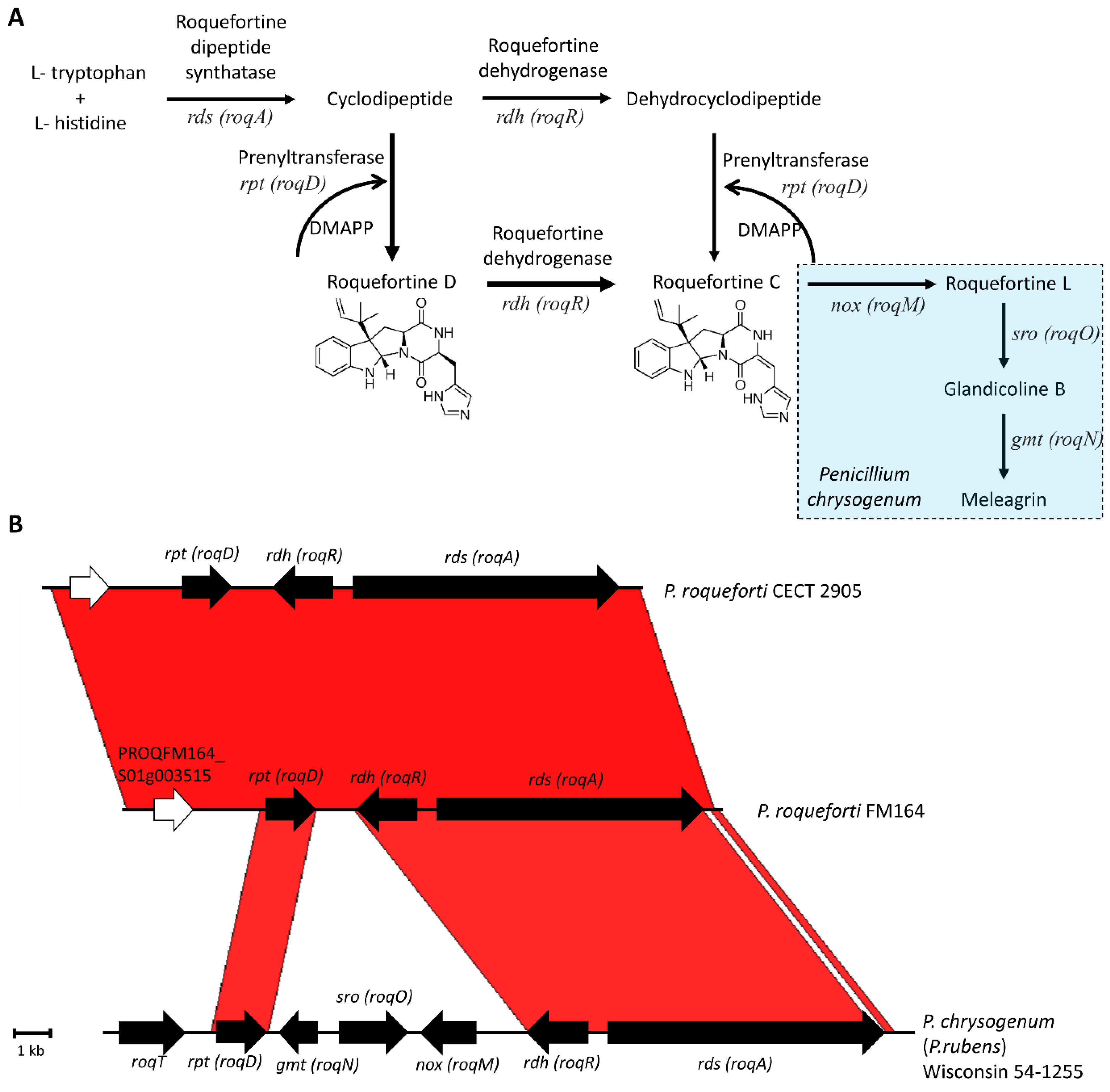
2.1. Roquefortine C

Roquefortine C, first described to be isolated from P. roqueforti in 1975 [36,37], is produced by at least 30 Penicillium species associated with different environments [38]. This compound belongs to a group of secondary metabolites called indole alkaloids [39]. Roquefortine C has neurotoxic properties, as observed in mice and cockerels [37,40]. The compound was also detected in intoxication cases in dogs and cattle [41,42]. While an LD50 value (the dose causing the death of 50% of the tested animals) of 15–20 mg/kg was reported in mice in one study [37], another study reported 169–189 mg/kg in two different mice strains [43]. Cytotoxicity of roquefortine C on Caco-2 cells determined a 50% inhibition concentration (IC50) of 48 µg/mL in one study [44] and >100 µM in another study [45]. The mechanism of toxicity is not precisely known, but the inhibition of rat and human cytochrome P450 enzymes by this compound was reported [46].
In addition to the neurotoxic effects, roquefortine C has antimicrobial properties against Gram-positive bacteria, such as Staphylococcus aureus and some Bacillus species [47]. The mechanism of the antibacterial effect could be the impairment of RNA synthesis [48]. On the other hand, the growth of lactic acid bacteria was only insignificantly impaired in the presence of roquefortine C [47]. During cheese ripening, the production of this metabolite might be useful for controlling unwanted bacterial growth while lactic acid bacteria remain unaffected.
Roquefortine C is produced by almost all of the studied P. roqueforti strains [49,50]. Production on solid substrates is much higher than in cheese [26]. The roquefortine C concentration in cheeses varies considerably; the average concentration was 858 ± 1670 µg/kg, determined using 83 blue cheese samples [51]. Based on this value and the average blue cheese consumption levels in France, a 21-day exposure representing chronic conditions was investigated on Caco-2 cells [45]. Chronic exposure did not change the intestinal barrier, suggesting that the amounts taken with blue cheese consumption do not cause significant toxicological effects [45].
Roquefortine C biosynthesis begins with the condensation of L-tryptophane and L-histidine to form a cyclodipeptide with a diketopiperazine ring by the action of a nonribosomal peptide synthetase (NRPS), named roquefortine dipeptide synthetase (RDS) (Figure 1A) [52]. After the formation of the cyclodipeptide, there is a grid in the biochemical pathway. The resulting dipeptide might first be prenylated to produce roquefortine D and dehydrogenated, or the pathway proceeds with dehydrogenation first and then prenylation to yield roquefortine C [38,53]. The relative amounts of the metabolites in the deletion strains in P. chrysogenum (renamed as P. rubens) indicate that the former path is the predominant one [53]. Some fungal species can convert roquefortine C to metabolites, such as meleagrin, oxaline, and neoxaline [38]. For example, P. chrysogenum produces meleagrin, and the formation of this compound requires three additional steps after roquefortine C is formed [38]. In parallel, the biosynthetic gene clusters responsible for the production of roquefortine C in P. roqueforti and meleagrin in P. chrysogenum are very similar (Figure 1B). However, while the cluster in P. chrysogenum harbors seven genes [53], the P. roqueforti cluster is a shorter version [35]. It has been suggested that the biosynthetic gene cluster for roquefortine C (roq) in P. roqueforti harbors four genes [35]. These genes include gmt (roqN, PROQFM164S01g003515), represented as a white arrow in Figure 1B, rds (roqA), which encodes a nonribosomal peptide synthetase, rdh (roqR), responsible for encoding a dehydrogenase, and rpt (roqD), which codes for a roquefortine prenyltransferase. However, between P. roqueforti FM164 and P. chrysogenum Wisconsin 54-1255 genomes, three stretches of high identity, >85%, are observed; one corresponds to the rpt (roqD) gene, while the other long stretch harbors rdh (roqR) and rds (roqA) (Figure 1B). There is also a tiny stretch at the end of the analyzed regions. The roq clusters in the two P. roqueforti genomes, FM164 and CECT 2905, are 99.2% identical, as indicated by the bright red color of the Artemis Comparison Tool (ACT) in the entire region analyzed (Figure 1B). PROQFM164_S01g003515 (old name: Proq01g022760), shown with a white arrow in Figure 1B, was mentioned to be part of the P. roqueforti cluster as a remnant homolog of the methyltransferase gene, gmt (roqN), in P. chrysogenum [35]. However, the low sequence identity (22%) of this gene with gmt (roqN, Pc21g15440) and its higher sequence identity (79%) with another gene (Pc21g15540) located ~29 kb downstream in the P. chrysogenum genome (Figure S1), suggest that it is not part of the cluster. Therefore, it is likely that the P. roqueforti roquefortine C cluster comprises only three genes. Silencing of rds and rpt genes in P. roqueforti reduced roquefortine C production, indicating these genes’ involvement in biosynthesis [35]. In P. chrysogenum, deletions of the genes in the cluster identified their functions in the pathway. The three genes that are absent in the P. roqueforti gene cluster, nox (roqM), sro (roqO), and gmt (roqN) in P. chrysogenum take part in the formation of meleagrin from roquefortine C [53]. An MFS transporter gene, roqT, in P. chrysogenum is also absent in P. roqueforti. RoqT deletion in P. chrysogenum did not inhibit the production and secretion of meleagrin; however, the concentrations of the intermediate compounds were altered [53]. RoqT in P. chrysogenum might facilitate the transport of the intermediates within the cell or the secretion of the final product, which would otherwise occur by passive transport or by an alternative transporter.

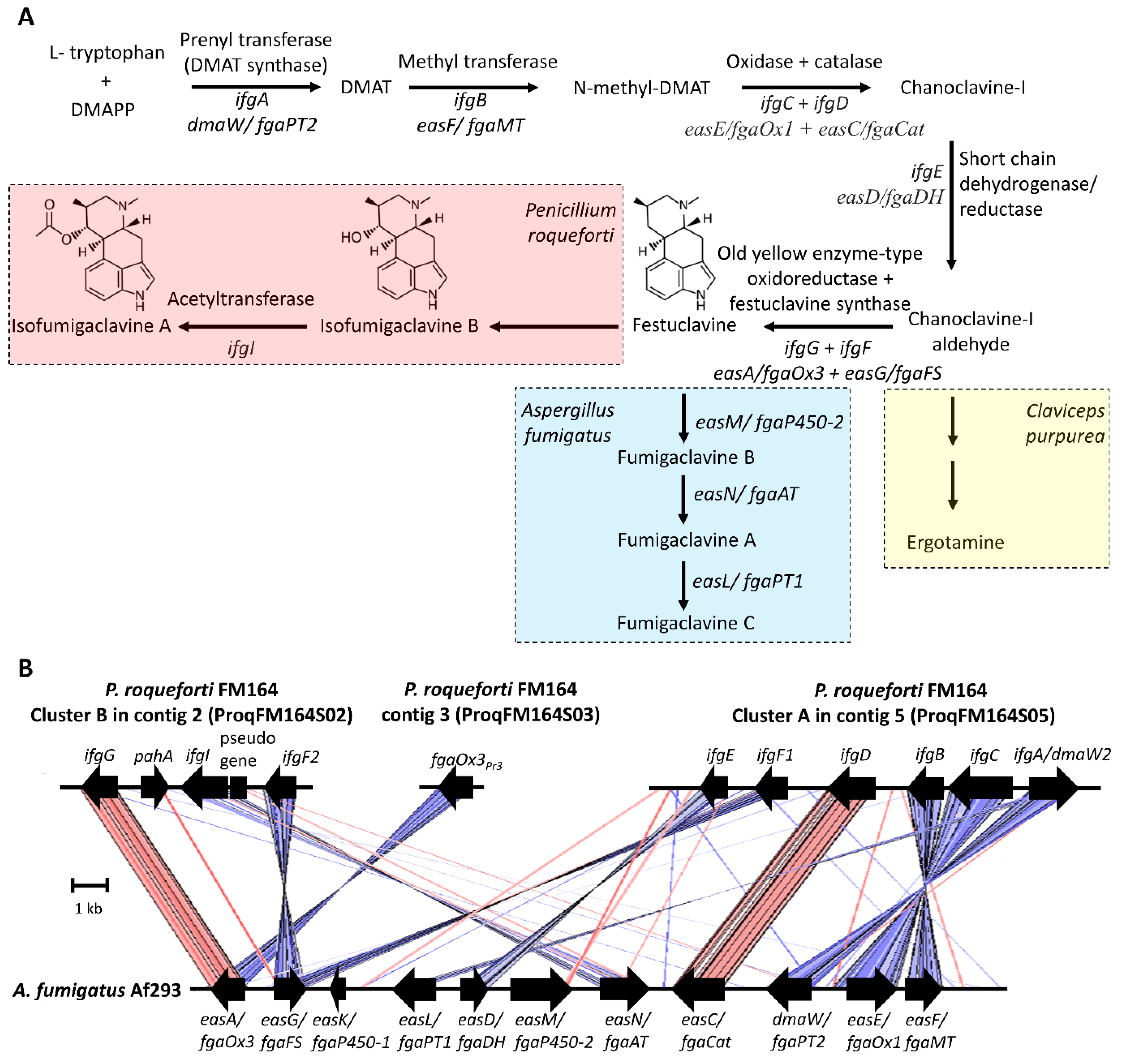
2.2. Clavine Alkaloids

P. roqueforti was reported to produce clavine-type indole alkaloids, isofumigaclavine A [23,49,50,55,56,57], isofumigaclavine B [50,55], festuclavine [24,36], and agroclavine [24,58]. Because of the structural resemblance of clavine alkaloids to the neurotransmitters dopamine, serotonin, and adrenaline, they have diverse pharmacological properties [59]. Agroclavine was reported to enhance natural killer cell activity, an immunostimulatory effect [60]. In blue cheeses, limited amounts of isofumigaclavine A (<5 ppm) and only traces of isofumigaclavine B were reported [55]. The relatively low amounts might be attributed to the low oxygen conditions in cheese that do not allow the high number of oxidation steps in the biosynthetic pathway (Figure 2A) [18].
Clavine alkaloid biosynthesis has been largely elucidated in Aspergillus fumigatus (Neosartorya fumigata), which produces festuclavine and fumigaclavines [65]. The biosynthesis of clavine alkaloids starts with the condensation of tryptophan and dimethylpyrophosphate (DMAPP) by a prenyltransferase enzyme called dimethylallyltryptophan (DMAT) synthase (DmaW/FgaPT2) (Figure 2) [66]. The produced DMAT is methylated by a methyltransferase (EasF/FgaMT) [67], and by the action of at least two enzymes, an oxidase (EasE/fgaOx1), and a catalase (EasC/fgaCat), chanoclavine I, is produced [68,69]. After that, a short-chain dehydrogenase/reductase (EasD/FgaDH) converts chanoclavine I to chanoclavine I aldehyde [70]. From this point on, different fungi produce different metabolites using the intermediate chanoclavine I aldehyde. For example, while Claviceps purpurea produces the ergot alkaloid ergotamine, A. fumigatus produces fumigaclavines via festuclavine (Figure 2A) [61]. P. roqueforti, on the other hand, produces festuclavine similar to A. fumigatus; however, festuclavine is converted into isofumigaclavines. In the production of festuclavine from chanoclavine I aldehyde, two enzymes, one, a member of the old yellow enzyme family of oxidoreductases (EasA/FgaOx3), and the other, festuclavine synthase (EasG/FgaFS), are involved [61,64,71]. In A. fumigatus, festuclavine is hydroxylated by EasM/fgaP450-2) to fumigaclavine B [63], which is later acetylated by EasN/FgaAT to produce fumigaclavine A [72]. This compound is prenylated once more by easL/fgaPT1 and yields fumigaclavine C [73].
The discovery of the clavine alkaloids biosynthetic cluster in P. roqueforti started with the finding of a second prenyltransferase gene (ifgA/dmaW2, proq05g069320 [new ID: PROQFM164_S05g000511]), different from rpt involved in roquefortine biosynthesis [74]. The silencing of ifgA resulted in the suppression of isofumigaclavine A and three other intermediate products in P. roqueforti [62]. Interestingly, ifgA silencing leads to an increase in roquefortine C production [62], which might be the result of the unused precursors, tryptophan and DMAPP, directed to this pathway. IfgA was found in a cluster of six genes (cluster A) (Figure 2B). There is a methyltransferase gene in the cluster, ifgB, the product of which presumably methylates DMAT to produce N-methyl-DMAT [62]. The FAD-binding oxidoreductase (ifgC) and the catalase (ifgD) gene products are the putative enzymes that produce chanoclavine I [62]. The gene ifgE is a short-chain dehydrogenase/reductase that corresponds to the enzyme catalyzing the production of chanoclavine I aldehyde from chanoclavine [62]. The final gene in this cluster is a festuclavine synthase ortholog, named efgF1. In cluster A, only the function of the ifgA/dmaW2 gene in clavine alkaloid biosynthesis has been demonstrated experimentally [62].
Because cluster A in contig 2 did not involve the gene for the old yellow enzyme family necessary for festuclavine production, the authors looked for this gene (ifgG) in the genome. They found one in a second cluster in contig 5 (Figure 2B) [62]. This cluster, cluster B, has three additional genes, another copy of the festuclavine synthase gene, efgF2, and an acetyltransferase gene, ifgI, which putatively catalyzes the acetylation of isofumigaclavine B to isofumigaclavine A, similar to the corresponding enzyme EasN/FgaAT in A. fumigatus that acetylates fumigaclavine B to fumigaclavine A [72]. There is also a phytanoyl-CoA hydroxylase gene, pahA, in cluster B [62,65]. While this gene is not present in the A. fumigatus cluster, it is found in some ergot alkaloid and clavine alkaloid gene clusters in different fungi [65]. For example, in C. purpurea, EasH, a phytanoyl-CoA hydroxylase gene product, functions as a cyclolizing dioxygenase in ergopeptine synthesis [75]. Whether this gene in P. roqueforti has a function in clavine biosynthesis is unknown. In addition, the roles of the cluster B genes in the biosynthesis of clavine alkaloids in P. roqueforti have yet to be shown experimentally.
Meanwhile, Gerhards and Li [61] reported an additional old yellow enzyme gene homolog (CDM33403, named FgaOx3PR3) in contig 3 of the P. roqueforti genome, unlinked to cluster A or B (Figure 2B). They cloned this gene, overexpressed the corresponding protein, and demonstrated the activity of converting chanoclavine I aldehyde to festuclavine in the presence of festuclavine synthase of A. fumigatus. The enzyme also increased the conversion yield of chanoclavine I to chanoclavine aldehyde, the previous step in the biosynthetic pathway, catalyzed by a short-chain dehydrogenase/reductase. The authors reported an amino acid-level sequence identity of 51% between FgaOx3PR3 and IfgG, the old yellow enzyme in cluster B [61]. Meanwhile, A. fumigatus EasA/FgaOx3 amino acid sequence identity is 67.4% and 53.9% to IfgG and FgaOx3PR3, respectively. The higher sequence identity between EasA/FgaOx3 and IfgG suggests that the enzyme responsible for festuclavine synthesis might be IfgG with a better activity towards chanoclavine-I aldehyde, which remains to be explored.
While in A. fumigatus, the genes (n = 11) responsible for the clavin biosynthesis pathway are grouped contiguously in a single cluster, the biosynthesis in P. roqueforti, is apparently associated with more than one gene cluster (Figure 2B) [65]. Generally, for ergot alkaloid biosynthesis in ascomycetes, this is not an uncommon configuration; a single cluster or two unlinked clusters can be involved in biosynthesis [76]. The conservation of the genes responsible for chanoclavine synthesis (ifgA/dmaW2, ifgC, ifgB, and ifgD) as a block between P. roqueforti and A. fumigatus except a single inversion is remarkable (Figure 2B). Eight of the eleven genes in the A. fumigatus cluster have homologs in P. roqueforti cluster A or B. One of the three missing genes is easL/fgaPT1, the product of which prenylates fumigaclavine A to yield fumigaclavine C (Figure 2B). Because in P. roqueforti, the pathway stops at isofumigaclavine A, this gene is not expected. The second missing gene is easK/fgaP450-1, whose function, if any, is unknown in A. fumigatus. The third gene is easM, the product of which is responsible for hydroxylating festuclavine. A homologous gene is not present in the P. roqueforti cluster A or B and might be elsewhere in the genome. When the A. fumigatus EasM amino acid sequence (Genbank ID: Q4WZ65.1) was used to search the genomes of P. roqueforti strains FM164 and LCP96 04111, different hits were obtained with a percent identity between 45–23%. How the hydroxylation reaction is catalyzed in P. roqueforti awaits further research.

2.3. Mycophenolic Acid
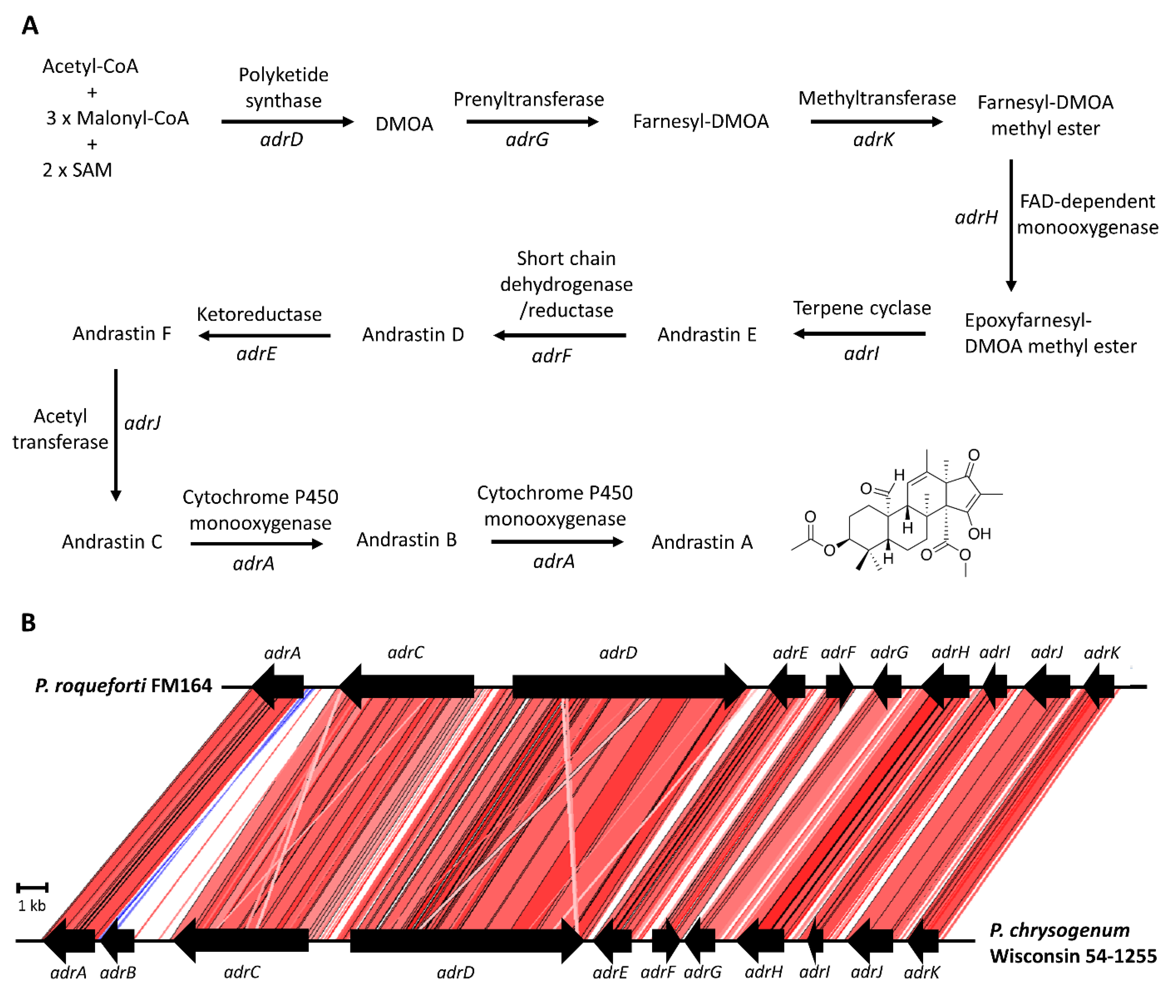
Mycophenolic acid is one of the main secondary metabolites P. roqueforti produces [22]. The compound was discovered by an Italian physician, Bartolomeo Gosio, in 1893 while he was studying spoiled corn to find an agent causing pellagra [77]. At those times, pellagra was believed to be associated with a toxic substance rather than a vitamin deficiency as we know it today. Gosio isolated a fungus from spoiled corn and purified a compound from the culture filtrate of this fungus. This compound had phenolic properties and stopped the growth of “anthrax bacillus” [78,79]. Later, in 1913, Alsberg and Black [80] isolated an acidic compound from a corn-spoilage Penicillium and named it mycophenolic acid. When Clutterbuck et al. conducted extensive studies on the metabolic products of Penicillium brevicompactum in 1932, they concluded that the compounds Gosio, Alsberg, and Black isolated was one and the same [81]. Because of Gosio’s work, mycophenolic acid was prized later as the first fungal antibiotic crystallized [82].
After the first report on its activity against Bacillus anthracis by Gosio [77], mycophenolic acid was later shown to have antibacterial [82,83], antifungal [82,84], antiviral [85], antipsoriasis [86,87], and antitumor [88] properties. However, mycophenolic acid is used in medicine today primarily because of its immunosuppressive activity [79]. Two prodrugs of mycophenolic acid, mycophenolate mofetil (CellCept®, Roche Pharma, Basel, Switzerland) and mycophenolic acid sodium salt (Myfortic®, Novartis Pharma, Basel, Switzerland), are used as immunosuppressants in transplantation to prevent organ rejection and to treat autoimmune diseases [89]. Mycophenolic acid acts as an inhibitor of the inosine 5′-monophosphate dehydrogenase (IMPDH), an enzyme taking the role of purine monophosphate formation during DNA synthesis [90]. The compound inhibits the proliferation of B- and T-lymphocytes, whose DNA synthesis is mainly dependent on this de novo synthesis pathway [90]. Other cells use both de novo synthesis and a salvage pathway that recycles the purines; therefore, the compound explicitly affects the B- and T-lymphocytes, a highly beneficial feature for an immunosuppressant [79,90].
A long-term toxicity study in rabbits given 80 and 320 mg/kg of mycophenolic acid orally for one year showed no apparent toxicity sign [91]. In rats and mice, relatively high concentrations were needed for toxicity. For example, LD50 for rats and mice are 2500 and 700 mg/kg (oral), and 500 and 450 mg/kg (intravenous), respectively [92]. However, using the compound in immunosuppressive therapy revealed some gastrointestinal side effects, such as diarrhea, abdominal pain, mucosal changes, and submucosal inflammation, which indicate gastrointestinal toxicity [93]. The IC50 value of mycophenolic acid on Caco-2 cells representing cytotoxicity was 780 µM, the solubility limit of the compound, indicating that even at this concentration, more than 50% of the cells were viable [45]. In blue cheeses, the average mycophenolic acid concentration was reported to be 841 ± 1271 µg/kg [51]. Chronic exposure (21-day experiment) on Caco-2 cells based on the value attained by ingesting the highest contaminated cheese led to decreased intestinal barrier function [45]. However, this decrease did not result in bacterial passage. Regarding mutagenicity, the Ames test conducted on Salmonella Typhimurium strains using mycophenolic acid up to 400 µg/plate did not result in mutagenic activity [94]. Nevertheless, a study on mouse mammary carcinoma cell line FM3A indicated mycophenolic acid-induced mutations at 0.032 and 0.1 µg/mL and chromosome aberrations at 0.1 µg/mL [95].
Mycophenolic acid is a meroterpenoid, a term first used in 1968 [96], with the prefix mero meaning part, partial, or fragment [97], to describe compounds that are of mixed biosynthetic origin and derived in part from terpenoids [98]. Mycophenolic acid is a polyketide–terpenoid hybrid compound (Figure 3A) [99]. Mycophenolic acid biosynthesis in P. roqueforti is governed by a seven-gene cluster spanning 24.4 kb (Figure 3B) [100], similar in structure to the corresponding cluster in P. brevicompactum [101,102]. The silencing of each of the seven genes in the mycophenolic acid biosynthetic gene cluster of P. roqueforti resulted in significant reductions in mycophenolic acid [100]. The mycophenolic acid biosynthetic pathway has been elucidated with studies involving isotope labeling, gene silencing, and heterologous expression, mainly conducted in P. brevicompactum [34,99,101,102,103,104]. The biosynthesis begins with the formation of the tetraketide 5-methylorsellinic acid (5-MOA) from one acetyl-CoA, three units of malonyl-CoA, and one S-adenosyl-L-methionine (SAM) with the action of a polyketide synthase, coded by mpaC (Figure 3A) [100,101]. 5-MOA is then hydroxylated to produce 4,6-dihydroxy-2-(hydroxymethyl)-3-methylbenzoic acid (DHMB), and the ring is closed to form 5,7-dihydroxy-4-methylphthalide (DHMP). These two reactions are catalyzed by a bifunctional enzyme coded by the mpaDE gene having a P450 monooxygenase and a hydrolase domain [102]. After that, a mpaA-encoded prenyltransferase adds a farnesyl, a 15-carbon isoprenoid, group to DHMP to yield 6-farnesyl-5,7-dihydroxy-4-methylphthalide (FDHMP) [34,99]. Oxidative cleavage of the farnesyl side chain by an oxygenase, coded by mpaB, yields a three-fewer carbon atom containing FDHMP-3C, which is later methylated by an O-methyltranferase, encoded by mpaG, to produce MFDHMP-3C [34]. A recent fascinating discovery demonstrated the compartmentalized nature of mycophenolic acid biosynthesis [101]. While the steps until the production of MFDHMP-3C take place in the cytoplasm, MFDHMP-3C enters into the peroxisome and CoA-ligated, followed by β-oxidation-mediated chain shortening to yield mycophenolic acid-CoA [34]. An acyl-CoA hydrolase, coded by mpaH, hydrolyzes the mycophenolic acid-CoA to prevent further oxidation and yields mycophenolic acid [34,105]. There is also a gene, mpaF, coding for a mycophenolic acid-insensitive IMPDH, which has been suggested to confer self-resistance [100,106].
Mycophenolic acid production in P. roqueforti was reported to be strain-dependent [50,108]. In addition, some P. roqueforti strains were demonstrated to have a 174 bp deletion in the 3′-end of the mpaC gene, leading to a decrease in the protein length by 14 amino acids [107]. The strains with shorter mpaC produced low or no mycophenolic acid, suggesting a correlation between the deletion and the reduction or the absence of mycophenolic acid production [107]. However, even among the strains with no deletion on mpaC, significant differences in mycophenolic acid production were observed [107]; therefore, more factors, such as sequence variations in cluster genes or their regulatory regions or on a regulatory gene outside the cluster, are likely to be involved in the regulation of strain-dependent production. Although the mycophenolic acid cluster does not harbor a regulatory gene, several regulator genes for secondary metabolites were identified in P. roqueforti [109,110,111].
In addition to its variability, the mycophenolic acid production of P. roqueforti is lower than that of P. brevicompactum [112], known to be a good producer, as high as 6700 mg/L [113]. However, using a P. roqueforti mutant strain exposed to Gamma radiation and optimizing the fermentation media, El-Sayed and Zaki were able to increase the yield, which is of relevance in industrial production, up to 2933 mg/L [114].

2.4. Andrastin A

Similar to mycophenolic acid, andrastin A is also a meroterpenoid [115]. Andrastins (andrastin A, B, C) are farnesyl transferase inhibitors that might alter the activity and localization of Ras proteins; therefore, they have the potential to be used as anticancer drugs [116,117]. In addition, andrastin A has been reported to enhance the cytotoxic effect of an anticancer drug, vincristine, in drug-resistant cancer cells [118].
Andrastin A is produced by P. roqueforti in solid and liquid media and has been consistently detected in different blue cheeses [119,120]. The concentration in cheeses depends on the strain used as the starter [119]. Positive or adverse effects of the presence of this metabolite in the human diet involving blue cheeses are unknown. The biological activity as a farnesyl transferase inhibitor might be beneficial for health; however, comprehensive toxicological data is needed to evaluate possible risks.
Isotope labeling studies in the andrastin-producing fungus Penicillium sp. FO-3929 showed that andrastins were synthesized from tetraketide 3,5-dimethylorsellinic acid (DMOA) and farnesyl diphosphate (Figure 4A) [116]. The andrastin biosynthetic pathway (Figure 4A) and the gene cluster with eleven genes, adrA-adrK (Figure 4B), in P. chrysogenum, were studied by Matsuda et al. [121]. The authors hypothesized that similar to austinol [122] and terretonin [123], andrastins were derived from epoxyfarnesyl-DMOA methyl ester based on the fact that the four genes coding for the enzymes catalyzing the formation of this intermediate in austinol and terretonin biosynthesis have high sequence homology to the adrD, adrG, adrK, and adrH in P. chrysogenum [121]. In analogy to austinol and terretonin biosynthesis, Matsuda et al. proposed that andrastin biosynthesis started with the formation of DMOA by an adrD-encoded polyketide synthase [121]. Then, a farnesyl group is added to DMOA by a prenyltransferase, coded by adrG. The resulting farnesyl-DMOA is methylated by an adrK-encoded methyltransferase, and the farnesyl unit is epoxidized by a flavin adenine dinucleotide (FAD)-dependent monooxygenase, AdrH, to produce epoxyfarnesyl-DMOA methyl ester [121], the common intermediate in the biosynthesis of related metabolites, austinol and terretonin. The authors then used a heterologous expression approach to show how the cyclization and the post-cyclization tailoring reactions take place [121]. According to these studies, epoxyfarnesyl-DMOA methyl ester is cyclized by the terpene cyclase, adrI, revealing the tetracyclic intermediate andrastin E. Then, the product of adrF, a short-chain dehydrogenase, oxidizes andrastin E to produce andrastin D, which is reduced by a ketoreductase encoded by adrE [121]. The resulting product, andrastin F, is acetylated by an acetyltransferase, AdrJ, to produce andrastin C [121]. The final steps in the pathway involve two consecutive oxidation reactions catalyzed by a cytochrome P450 monooxygenase, encoded by adrA, to produce andrastin B and then andrastin A [121].
The biosynthetic gene cluster for andrastin A, spanning 29.4 kb, has been defined in P. roqueforti and harbors ten genes (Figure 4B) [115]. The cluster is very similar to the andrastin biosynthetic cluster of P. chrysogenum [121], with the only exception that adrB is a pseudogene in P. roqueforti (Figure 4B) [115]. RNAi silencing of the ten genes in P. roqueforti, including the adrC gene, coding for a major facilitator superfamily (MFS) transporter, significantly reduced andrastin A production [115]. In secondary metabolite biosynthesis, MSF transporters are involved in the trafficking of the precursors, intermediary compounds, or final metabolites to specific locations within the cell [124]. For example, in P. chrysogenum, penicillin production involves at least three MSF transporters [124]. PenV, located in the vacuolar membrane, transports the vacuole-stored precursor L-α-aminoadipic acid into the cytosol; PaaT and PenM, which localize the peroxisomal membrane, translocate the intermediary compounds phenylacetic acid and isopenicillin N, respectively, from the cytosol to the peroxisome, where the final steps of penicillin production take place [125]. The adrC-silenced P. roqueforti strain did not have a difference in the andrastin A quantities in the mycelium and the surrounding agar, indicating that AdrC is not involved in the secretion of the final metabolite [115]. Whether this MSF transporter takes a role in the intracellular transport of intermediates or precursors is an open question that could be addressed in future studies.
An interesting point in the andrastin A biosynthetic cluster is the pseudogenization of one of the genes of the P. chrysogenum cluster, adrB, in P. roqueforti [115]. In P. roqueforti, the region encoding the first ten amino acids is similar to P. chrysogenum; however, deletions after this point on the sequence introduced early stop codons, leaving adrB a residual pseudogene [115]. The role of this gene product in andrastin A biosynthesis was not proposed or analyzed in P. chrysogenum by Matsuda et al. [121]. AdrB-encoded ORF (Pc22g22830) was annotated in the P. chrysogenum genome (accession number: AM920437) as a hypothetical protein with no known functional domains. The mRNA transcript for Pc22g22830 was reported in two studies [126,127], indicating that the gene is active; however, the function, if any, in andrastin A production of P. chrysogenum remains to be explored.

2.5. PR-Toxin

The PR toxin, the most toxic metabolite of P. roqueforti [128], was first described in 1973 as a compound with toxic properties in rats [129]. The compound was extracted from a P. roqueforti strain isolated from contaminated animal feed. Later, the structure of PR toxin was elucidated as an eremophilane-type sesquiterpene [130]. The PR toxin has lethal effects on mice, rats, and cats, significantly damaging internal organs [131]. The toxin inhibits DNA synthesis [132], transcription [133], and protein synthesis [134], and has mutagenic [135] and carcinogenic [136] properties. In addition, it perturbs mitochondrial respiration and oxidative phosphorylation by disturbing the integrity of the mitochondrial membrane and inhibiting the succinate-cytochrome c reductase complex [137]. The cytotoxic effects were also demonstrated on human cell lines, Caco-2 and THP-1, inducing necrosis and inflammatory response in THP-1 cells [138].
Although stored cereals and maize silage are suitable substrates for PR-toxin production [139,140], this toxin was not found in blue cheeses [141]. Cheese production and maturation conditions, such as microaerophilic environment, low pH, and refrigeration temperatures, were determined to be unsuitable for PR-toxin production [142]. In addition, the PR toxin is not stable and is converted to other products, such as less toxic PR imine [43,143], in blue cheeses in the presence of amino compounds [144]. Not only in cheese but also in culture medium, PR toxin is converted to other products, PR imine, PR acid, and PR amide, in time [145,146]. Based on time-course studies, Chang et al. [147] proposed a degradation pathway for PR toxin, in which it is converted into either PR imine or sequentially to PR acid and later PR amide. The major metabolite among these compounds was determined as PR acid. The enzymes PR oxidase and PR amide synthetase, responsible for converting PR toxin to PR acid and PR acid to PR amide, respectively, were purified and characterized [148,149]. Although PR imine was detected in blue cheeses, the presence of PR acid or PR amide remains to be elucidated. The toxicities of PR acid or PR amide are unknown; however, they are not expected to be toxic because the aldehyde group responsible for the biological activity [143] is converted into an acid and an amide in these compounds.
PR toxin biosynthesis starts with the formation of the sesquiterpene aristolochene from farnesyl diphosphate by a terpene cyclase, aristolochene synthase (Figure 5A) [150]. In the biosynthetic pathway, the parent hydrocarbon aristolochene was suggested to be oxidized to 7-epi-neopetasone, a compound detected in culture extracts [151,152]. The presence of closely related other compounds, eremofortin A, eremofortin B, and eremofortin C, in culture fluids indicated that these compounds are intermediates in the biosynthetic pathway [153]. However, eremofortin A was also suggested to be a shunt product instead of an intermediate [151]. In a time course study, the production peaks were obtained in the sequential order: eremofortin B, eremofortin C, and later PR toxin, which suggested the biosynthesis pathway followed this sequence [153]. After the formation of 7-epi-neopetasone, a series of further oxidations were suggested to take place in the biosynthetic pathway producing eremofortin B, which could be both acetylated and further oxidized to yield eremofortin C [151]. The alcohol group at the C-12 position in eremofortin C is converted to an aldehyde group with toxic biological activity in PR-toxin [143] by eremofortin C oxidase [154,155]. This enzyme was suggested to be an oxidase rather than an alcohol dehydrogenase because it did not require NAD+ or NADP+ for activity [154].
The gene coding for aristolochene synthase in P. roqueforti was determined to be ari1, revealed by overexpression in E. coli [158,159]. The search of this gene in a genomic library of the P. roqueforti type strain CECT 2905 revealed three additional genes in the vicinity of ari1 (prx2, prx representing PR-toxin), named prx1, prx3, and prx4, coding for a short-chain oxidoreductase/dehydrogenase, an oxidase, and an alcohol dehydrogenase, respectively (Figure 5B) [156]. Silencing of each of these genes resulted in a reduction in PR toxin production, indicating the role of these genes in the biosynthetic pathway [156]. Later, when the P. roqueforti genome became available, more genes were revealed, resulting in a cluster spanning 22.4 kb and harboring 11 genes. In addition to prx1-prx4, which were named ORF1-ORF4 in this later study, seven additional genes, ORF5-ORF11, were proposed to be part of the cluster (Figure 5B) [157]. While ORF5, ORF6, ORF9, and ORF11 have cytochrome P450 domains, ORF8 and ORF10 have transferase and regulatory domains, respectively. There is also a gene (ORF7) with a domain of unknown function (DUF3237). Silencing ORF5, ORF6, and ORF8 reduced PR toxin production levels, indicating that these genes are involved in PR toxin biosynthesis [157]. Silencing these genes also reduced eremofortin A, suggesting that the products of these genes take roles in the biosynthetic pathway before eremofortin A production. On the other hand, while ORF6 silencing reduced the level of eremofortin B production, silencing ORF5 and ORF8 resulted in eremofortin B accumulation. This result indicates that ORF6 has a function preceding eremofortin B production, whereas the roles of ORF5 and ORF8 are in later steps. While ORF8 presumably functions in the transfer of the acetyl group, ORF5 and ORF6 have cytochrome P450 domains responsible for oxidation reactions. ORF10 encodes a regulatory protein, probably functioning in regulating the gene cluster.
PR toxin gene clusters of P. roqueforti and P. chrysogenum are highly syntenic, including the intergenic regions (Figure 5B). The high similarity in gene content, gene directions, and intergenic regions indicates a recent acquisition of this cluster in these two species. The blue stretches between ORF9 and Pc12g06400, and ORF11 and Pc12g06380 represent inverse matches observed due to the similarity between ORF9 and ORF11. They both encode cytochrome P450 monooxygenases, with 37.5% similarity at the amino acid level.
An interesting finding was the effect of the PR toxin on mycophenolic acid production. Prx-silenced strains produced significantly higher amounts of mycophenolic acid, indicating a common control mechanism [156].

2.6. Other Secondary Metabolites of P. roqueforti

2.6.1. Citreoisocoumarin

Citreoisocoumarin (Figure 6A) was produced by 42% of the P. roqueforti silage isolates (n = 79) [23]. This compound has been isolated more consistently from a closely related species, Penicillium paneum, such that all isolates (n = 78) were reported to produce citreoisocoumarin [23]. This metabolite has also been reported in other Penicillium species, such as P. carneum [24], P. buchwaldii [160], P. citreoviride [161], P. nalgiovense [162], and P. vinaceum [163], and other fungal genera, e.g., Nectria [164] and Fusarium [165]. Neither the biosynthetic gene cluster nor the biosynthetic pathway has been identified for P. roqueforti. However, in Fusarium graminearum, the pigment aurafusarin and citreoisocoumarin are synthesized by the same polyketide synthase, and the biosynthetic pathway is redirected from aurafusarin to citreoisocoumarin under nitrogen starvation conditions [165].

2.6.2. Orsellinic Acid

Few strains of P. roqueforti were shown to produce orsellinic acid (Figure 6B) [24]. Methylated versions of orsellinic acid, 5-MOA, and DMOA are produced by the polyketide synthases in mycophenolic acid and andrastin A biosynthesis, respectively. Whether orsellinic acid is produced as a by-product in these pathways or is synthesized by a different biosynthetic pathway is unknown.

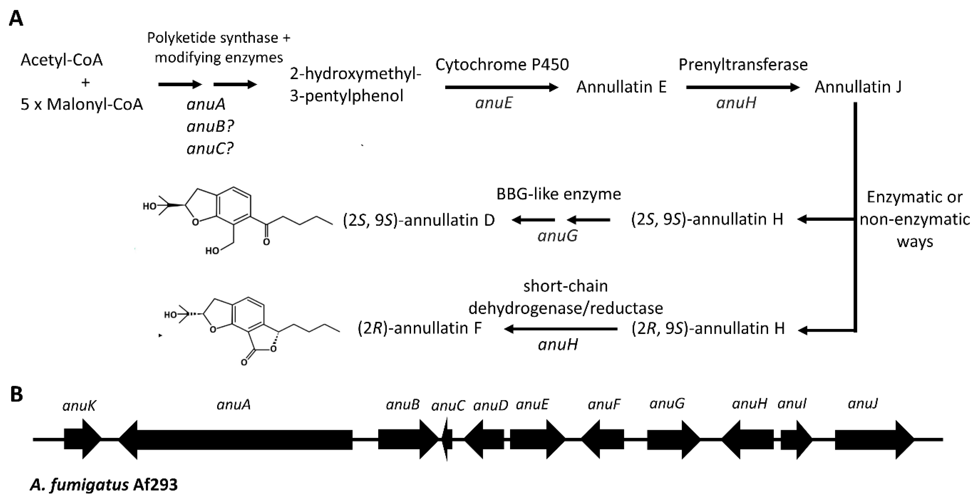
2.6.3. Sesterterpenoids: Peniroquesines A–C and 1–7, and Roquefornine A

P. roqueforti has recently been reported to produce sesterterpenes derived from geranyl farnesyl diphosphate (GFPP), a 25-carbon compound: peniroquesines (A–C, and 1–7), and roquefornine A with pentacyclic ring structures (Figure 6C) [29,30,31]. Some of these compounds demonstrated strong anti-inflammatory properties and/or cytotoxic activities toward human cell lines [29,30,31]. All these compounds were obtained from a plant-associated strain of P. roqueforti, YJ-14, isolated from Aconitum vilmorinianum using solid-state culture conditions. Whether these compounds are also produced by other P. roqueforti strains and under cheese production or maturation conditions are interesting questions to be addressed in future studies.

2.6.4. Tetrapeptides D-Phe-L-Val-D-Val-L-Phe and D-Phe-L-Val-D-Val-L-Tyr

Recently, the tetrapeptides, D-Phe-L-Val-D-Val-L-Phe and D-Phe-L-Val-D-Val-L-Tyr (Figure 6D) were detected in all four P. roqueforti strains tested and one P. paneum isolate [32,33]. These two metabolites were also detected in Penicillium citrinum [166], and one (D-Phe-L-Val-D-Val-L-Tyr) was determined in Penicillium canescens [167]; both of these studies reported antifungal activities for these tetrapeptides. However, biosynthetic routes and the corresponding genes have yet to be determined in P. roqueforti, or other Penicillium species reported to produce these metabolites.

2.6.5. Cis-bis(methylthio)silvatin

Cis-bis(methylthio)silvatin (Figure 6E) was detected from P. roqueforti ATCC10110 (CBS 221.30) in a recent study and demonstrated antibacterial activity for both Gram-positive and Gram-negative organisms [32]. This metabolite is a thiosilvatin, a sulfurous compound originating from the larger class diketopiperazines, cyclic peptides having two amide linkages [168]. Thiosilvatins generally contain two methylated sulfur atoms on the cyclopeptide structure [168]. Cis-bis(methylthio)silvatin, the most commonly observed member, has been detected in several Penicillium species, e.g., P. brevicompactum, P. crustosum, P. commune, and P. bilaiae, in addition to other fungi, such as Fusarium, Trichoderma, and Cordyceps [168]. This compound exerted cytotoxic effects on colon cancer cell lines HCT116 and Caco-2 but with no selectivity toward non-tumoral cell line HCEC [169]. In addition, it exhibited weak cytotoxicity against NS-1 mouse myeloma [170] and P388 murine lymphocytic leukemia cells [171]. The biosynthesis of thiosilvatins is unknown; however, in analogy to sulfide bridge-containing diketopiperazine compounds, such as gliotoxin and sirodesmin, the biosynthesis probably involves nonribosomal peptide synthetases to produce a cyclopeptide followed by sulfurization [172].

2.6.6. Scytalone and Melanin

Scytalone is a known intermediate in DHN–melanin biosynthesis [173] and has been detected in various P. roqueforti isolates [33]. The polyketide synthase gene (corresponding protein ID: Pro_LCP9604111_2|g6432.t1) in P. roqueforti was disrupted by the CRISPR/Cas9 approach using the strain LCP9604111, resulting in white-colored mutants, suggesting the involvement of this gene in melanin production [174]. The study did not mention or analyze a gene cluster around the polyketide synthase gene. While in Aspergillus species, DHN melanin gene clusters are generally conserved in gene content and harbor six genes, pksP/alb1, abr1, abr2, arp1, arp2, and ayg1; these genes may not be found in a single cluster in Penicillium species [173,175]. The pathway is well-established in A. fumigatus (Figure 7A) [176]. The polyketide synthase gene (pksP/alb1) is responsible for the production of the heptaketide naphtopyrone, which is converted to 1,3,6,8-tetrahydroxynaphthalene by the product of the heptaketide hydrolase (ayg1) [176]. After that, scytalone is produced by the action of the arp2-encoded reductase. Later in the biosynthetic pathway, scytalone is dehydrated by the arp1-coded scytalone dehydratase to 1,3,8-trihydroxynaphtalene, which is reduced by the reductase Arp2 again to vermelone. Vermelone is then transformed to DHN by the abr1-encoded multicopper oxidase, and DHN is polymerized into DHN–melanin by the action of the abr2-coded laccase [176].
When the genomes of P. roqueforti FM164 and LCP9604111 were searched for the A. fumigatus DHN–melanin biosynthetic pathway protein sequences using tblastn, five of the six genes were observed to be on the same contig (Figure 7B). The putative abr2 gene (PROQFM164_S02g000511 and LCP9604111_02|g8601.tr1) is located in a separate contig in both genomes. Additionally, the pks/alb1 is ~100 kb away from the other four genes (abr1, arp1, arp2, and ayg1). This configuration is very similar to that in P. chrysogenum (Figure 7B). In addition, different genes (shown as white arrows in Figure 7B) have been recruited between abr1 and arp1 in P. roqueforti and P. chrysogenum.

3. Secondary Metabolite Potential of P. roqueforti: Yet Undiscovered Metabolites and/or Silent Gene Clusters

The genome sequencing of P. roqueforti CECT2905T indicates the presence of 34 biosynthetic gene clusters [177]. Coton et al. [178] reported a similar but variable number of clusters, 34–37, that changed at the strain level. Few clusters (roquefortine C, clavine alkaloids, mycophenolic acid, andrastin A, and PR-toxin) related to the production of secondary metabolites have been uncovered until now, leaving many unstudied. There might be several alternatives for the remaining gene clusters. First, some of these clusters might be responsible for producing the metabolites whose biosynthetic gene cluster is unknown. For instance, P. roqueforti is known to produce citreoisocoumarin, but the associated cluster for this compound has yet to be identified. Second, the remaining clusters might be involved in the production of as-yet-undiscovered metabolites. For example, sesterterpenoids have only recently been discovered in a P. roqueforti strain [29,30,31], and the gene cluster involved in the biosynthesis of these compounds has not been elucidated yet. Third, some clusters identified in the published genomes might be non-functional because modifications, such as gene deletions or acquisition of early stop codons, might have happened; and these changes might prevent the production of the corresponding metabolite. Finally, the cluster might be silent due to an inactive pathway-specific transcription factor or epigenetic regulation [1]. In this case, overexpression of the pathway-specific transcription factor might result in the activation of the cluster. For example, in Penicillium dangeardii, the overexpression of the transcription factor danS resulted in the production of azaphilons that were not detected in the wild-type strain [179]. Another strategy to activate the silent clusters is by inactivating the epigenetic silencing machinery [12]. For example, the deletion of the histone H3 deacetylase of the endophytic fungus Calcarisporium arbuscula activated the transcription of more than 75% of the key metabolite genes, such as polyketide synthases, peptide synthetases, and terpene cyclases, which led to the recovery of new metabolites [180].
An example of the silent clusters in P. roqueforti is the recently discovered annullatin D cluster, harboring 11 genes (anuA-anuK) (Figure 8) [181]. The cluster harbors genes for a polyketide synthase (anuA), short-chain dehydrogenases/reductases (anuB, anuD, anuF, and anuI), an unknown protein (anuC), a cytochrome P450 oxygenase (anuE), a berberine-bridge enzyme (BBG)-like protein (anuG), a prenyltransferase (anuH), a dehydrogenase (anuJ), and a transcription factor (anuK) (Figure 8B). When the genomic region anuA-anuK of P. roqueforti FM164 was heterologously expressed in Aspergillus nidulans under the control of the strong constitutive gpdA promoter, four annullatin derivatives were detected in the A. nidulans culture extract [181]. The fact that annullatins were not observed in P. roqueforti FM164 and were not reported in P. roqueforti before indicates the silent nature of the cluster. As a result of the deletion of the heterologously expressed genes and feeding experiments with single expressed genes in A. nidulans, the authors proposed a biosynthetic pathway for annullatins (Figure 8A) [181]. According to this proposal, the annullatin backbone, 2-hydroxymethyl-3-pentylphenol, is first synthesized by the polyketide synthase anuA and some modification enzymes, which could be anuB and anuC. Then, this intermediate is hydroxylated by the cytochrome P450 AnuE to produce annullatin E, and added an isoprenyl group by the prenyltransferase anuH using a dimethylallyl diphosphate (DMAPP) to yield annullatin J. Afterward, two diastereomers are formed either by an enzymatic or a non-enzymatic way. One of the stereoisomers, (2S, 9S)-annullatin H, is converted to (2S, 9S)-annullatin D by the BBG-like enzyme anuG. Meanwhile, anuF dehydrogenates the other isomer to (2R)-annullatin F. The authors also detected a small amount of an acetylated form of (2R)-annullatin F, which might have been formed via an A. nidulans enzyme because the cluster does not have an acetyltransferase gene [181]. In addition, when they deleted anuE, they discovered a shunt pathway leading to the (R)-configurations of annullatin A and B, the metabolites produced by Cordyceps annullata [182]. These two compounds and annullatin D of C. annullata have potent agonistic or inverse agonistic activities towards cannabinoid receptors, which are promising therapeutic targets [182]. If the annullatins produced by the P. roqueforti cluster have bioactive properties is yet to be analyzed.

4. Conclusions and Future Directions

The blue cheese fungus P. roqueforti produces a variety of secondary metabolites. Some of these have long been known, such as mycophenolic acid, PR-toxin, andrastin A, roquefortine C, and clavine alkaloids. The biosynthetic pathways and the associated gene clusters for these metabolites have been mainly uncovered and presented in this review. However, the functions of most genes in clavine alkaloid and PR toxin biosynthetic pathways remain to be discovered. Other metabolites of P. roqueforti include citreoisocoumarin, orsellinic acid, and scytalone. While the citreoisocoumarin biosynthetic pathway is unknown, the methylated versions of the orsellinic acid are produced in mycophenolic acid and andrastin A biosynthesis. However, whether there is a separate pathway for orsellinic acid needs further research.
Scytalone is an intermediate compound produced during DHN–melanin biosynthesis. This study deduced the P. roqueforti genes putatively taking roles in DHN–melanin biosynthesis using their similarity to their homologs in A. fumigatus. Both P. roqueforti and P. chrysogenum putative DHN–melanin genes are found in two contigs, one harboring the laccase ortholog abr2 and the other having the remaining five genes. However, these remaining genes are not contiguously organized like in A. fumigatus; the polyketide synthase gene, pks/alb1, is about 100 kb away from abr1, arp1, arp2, and ayg1. Moreover, additional genes have been acquired between abr2 and arp1, both in P. roqueforti and P. chrysogenum.
There are also recently described metabolites of P. roqueforti, e.g., sesterterpenes, peniroquesines and roquefornine A; tetrapeptides, D-Phe-L-Val-D-Val-L-Tyr, and D-Phe-L-Val-D-Val-L-Phe; and cis-bis(methylthio)silvatin. The biosynthetic pathways for these compounds remain to be explored.
The secondary metabolites of P. roqueforti hold significance for two distinct industries. The first is the food industry, where P. roqueforti is a fungal starter culture for blue cheeses. Furthermore, this species is a common contaminant in other cheeses and certain food products. The prevalence of secondary metabolites in foods is of significant concern due to their potential impact on toxicological attributes. Fortunately, the most toxic metabolite of P. roqueforti, known as PR toxin, is unstable under cheese production and maturation conditions and is converted into less toxic other products. Moreover, certain metabolites, such as roquefortine C and clavine alkaloids, are in low quantities in cheeses and are not expected to have significant effects. On the other hand, the quantity of mycophenolic acid in cheeses varies considerably depending on the specific strain used as the starter culture. Despite its pharmacological application as an immunosuppressive agent for organ transplant recipients, this compound has been observed to cause gastrointestinal side effects. Consuming an amount equivalent to the most contaminated cheese might affect the gastrointestinal system. Another metabolite, andrastin A, was consistently detected within cheese matrices, and necessitates further investigation to ascertain its potential positive or adverse effects, if any.
The other sector wherein secondary metabolites assume significance is the pharmaceutical industry. The increasing number of identified genomes of P. roqueforti strains underscores a substantial genomic reservoir for the biosynthesis of secondary metabolites. The functional elucidation of one such silent gene cluster, the annullatin biosynthetic gene cluster, was accomplished via heterologous expression in A. nidulans. Overexpression of the pathway-specific or global transcription factors and modulation of epigenetic regulatory mechanisms hold promise to activate additional silent clusters of P. roqueforti. The comprehensive exploration of the latent potential inherent in the secondary metabolism of P. roqueforti carries the prospect of yielding novel pharmaceutical agents.

Supplementary Materials

The following supporting information can be downloaded at: https://www.mdpi.com/article/10.3390/fermentation9090836/s1, Figure S1. Comparison of the roquefortine C gene cluster in P. roqueforti FM164 and larger section harboring the meleagrin cluster in P. chrysogenum Wisconsin 54-1255.

Funding

This research received no external funding.

Data Availability Statement

No new dataset has been created in this study. The sequences available in the NCBI database were used. The accession numbers used in the analyses are given in the text and the Figure legends.

Conflicts of Interest

The author declares no conflict of interest.

References

  1. Brakhage, A.A. Regulation of Fungal Secondary Metabolism. Nat. Rev. Microbiol. 2013, 11, 21–32. [Google Scholar] [CrossRef] [PubMed]
  2. Keller, N.P. Fungal Secondary Metabolism: Regulation, Function and Drug Discovery. Nat. Rev. Microbiol. 2019, 17, 167–180. [Google Scholar] [CrossRef]
  3. Keller, N.P.; Turner, G.; Bennett, J.W. Fungal Secondary Metabolism—From Biochemistry to Genomics. Nat. Rev. Microbiol. 2005, 3, 937–947. [Google Scholar] [CrossRef] [PubMed]
  4. Conrado, R.; Gomes, T.C.; Roque, G.S.; De Souza, A.O. Overview of Bioactive Fungal Secondary Metabolites: Cytotoxic and Antimicrobial Compounds. Antibiotics 2022, 11, 1604. [Google Scholar] [CrossRef] [PubMed]
  5. Wang, W.; Liang, X.; Li, Y.; Wang, P.; Keller, N.P. Genetic Regulation of Mycotoxin Biosynthesis. J. Fungi 2023, 9, 21. [Google Scholar] [CrossRef] [PubMed]
  6. Lancaster, M.C.; Jenkins, F.P.; Philp, J.M. Toxicity Associated with Certain Samples of Groundnuts. Nature 1961, 192, 1095–1096. [Google Scholar] [CrossRef]
  7. Nesbitt, B.F.; O’Kelly, J.; Sargeant, K.; Sheridan, A. Aspergillus flavus and Turkey X Disease: Toxic Metabolites of Aspergillus flavus. Nature 1962, 195, 1062–1063. [Google Scholar] [CrossRef]
  8. Copetti, M.V. Fungi as industrial producers of food ingredients. Curr. Opin. Food Sci. 2019, 25, 52–56. [Google Scholar] [CrossRef]
  9. Macheleidt, J.; Mattern, D.J.; Fischer, J.; Netzker, T.; Weber, J.; Schroeckh, V.; Valiante, V.; Brakhage, A.A. Regulation and Role of Fungal Secondary Metabolites. Annu. Rev. Genet. 2016, 50, 371–392. [Google Scholar] [CrossRef]
  10. Demain, A.L.; Fang, A. The Natural Functions of Secondary Metabolites. In History of Modern Biotechnology I; Fiechter, A., Ed.; Springer: Berlin/Heidelberg, Germany, 2000; pp. 1–39. ISBN 978-3-540-44964-5. [Google Scholar]
  11. Skellam, E. Subcellular Localization of Fungal Specialized Metabolites. Fungal Biol. Biotechnol. 2022, 9, 11. [Google Scholar] [CrossRef]
  12. Pfannenstiel, B.T.; Keller, N.P. On Top of Biosynthetic Gene Clusters: How Epigenetic Machinery Influences Secondary Metabolism in Fungi. Microb. Eng. Biotechnol. 2019, 37, 107345. [Google Scholar] [CrossRef] [PubMed]
  13. Yu, W.; Pei, R.; Zhou, J.; Zeng, B.; Tu, Y.; He, B. Molecular Regulation of Fungal Secondary Metabolism. World J. Microbiol. Biotechnol. 2023, 39, 204. [Google Scholar] [CrossRef] [PubMed]
  14. Metin, B. Filamentous Fungi in Cheese Production. In Microbial Cultures and Enzymes in Dairy Technology; Öztürkoğlu Budak, Ş., Akal, H.C., Eds.; IGI Global: Hershey, PA, USA, 2018; pp. 257–275. ISBN 978-1-522-55363-2. [Google Scholar] [CrossRef]
  15. Morales, M.B.L.; Ardö, Y.; Berthier, F.; Karatzas, K.-A.G.; Bintsis, T. Blue-Veined Cheeses. In Global Cheesemaking Technology: Cheese Quality and Characteristics; Papademas, P., Bintsis, T., Eds.; John Wiley & Sons: Chichester, UK, 2017; pp. 415–435. ISBN 978-1-119-04616-5. [Google Scholar] [CrossRef]
  16. Cantor, M.D.; van den Tempel, T.; Hansen, T.K.; Ardö, Y. Blue Cheese. In Cheese: Chemistry, Physics and Microbiology; Fox, P.F., McSweeney, P.L.H., Cogan, T.M., Guinee, T.P., Eds.; Academic Press: Cambridge, MA, USA, 2004; Volume 2, pp. 175–198. ISBN 978-0-122-63653-0. [Google Scholar] [CrossRef]
  17. Gripon, J.C. Mould-Ripened Cheeses. In Cheese: Chemistry, Physics and Microbiology; Springer: Boston, MA, USA, 1993; pp. 111–136. ISBN 978-1-4613-6137-4. [Google Scholar] [CrossRef]
  18. Martín, J.F.; Coton, M. Chapter 12—Blue Cheese: Microbiota and Fungal Metabolites. In Fermented Foods in Health and Disease Prevention; Frias, J., Martinez-Villaluenga, C., Peñas, E., Eds.; Academic Press: Boston, MA, USA, 2017; pp. 275–303. ISBN 978-0-12-802309-9. [Google Scholar] [CrossRef]
  19. Frisvad, J.C.; Samson, R.A. Polyphasic Taxonomy of Penicillium Subgenus Penicillium. A Guide to Identification of Food and Air-Borne Terverticillate Penicillia and Their Mycotoxins. Stud. Mycol. 2004, 49, C174. [Google Scholar]
  20. Sumarah, M.W.; Miller, J.D.; Blackwell, B.A. Isolation and Metabolite Production by Penicillium roqueforti, P. paneum and P. crustosum Isolated in Canada. Mycopathologia 2005, 159, 571–577. [Google Scholar] [CrossRef] [PubMed]
  21. Mioso, R.; Toledo Marante, F.J.; Herrera Bravo de Laguna, I. Penicillium roqueforti: A Multifunctional Cell Factory of High Value-added Molecules. J. Appl. Microbiol. 2015, 118, 781–791. [Google Scholar] [CrossRef]
  22. Frisvad, J.C.; Smedsgaard, J.; Larsen, T.O.; Samson, R.A. Mycotoxins, Drugs and Other Extrolites Produced by Species in Penicillium Subgenus Penicillium. Stud. Mycol. 2004, 49, 201–241. [Google Scholar]
  23. O’Brien, M.; Nielsen, K.F.; O’Kiely, P.; Forristal, P.D.; Fuller, H.T.; Frisvad, J.C. Mycotoxins and Other Secondary Metabolites Produced in Vitro by Penicillium paneum Frisvad and Penicillium roqueforti Thom Isolated from Baled Grass Silage in Ireland. J. Agric. Food Chem. 2006, 54, 9268–9276. [Google Scholar] [CrossRef]
  24. Nielsen, K.F.; Sumarah, M.W.; Frisvad, J.C.; Miller, J.D. Production of Metabolites from the Penicillium roqueforti Complex. J. Agric. Food Chem. 2006, 54, 3756–3763. [Google Scholar] [CrossRef]
  25. Hymery, N.; Vasseur, V.; Coton, M.; Mounier, J.; Jany, J.L.; Barbier, G.; Coton, E. Filamentous Fungi and Mycotoxins in Cheese: A Review. Compr. Rev. Food Sci. Food Saf. 2014, 13, 437–456. [Google Scholar] [CrossRef]
  26. Martín, J.F.; Liras, P. Secondary Metabolites in Cheese Fungi. In Fungal Metabolites; Mérillon, J.-M., Ramawat, K.G., Eds.; Springer International Publishing: Cham, Switzerland, 2017; pp. 293–315. ISBN 978-3-319-25001-4. [Google Scholar]
  27. García-Estrada, C.; Martín, J.-F. Biosynthetic Gene Clusters for Relevant Secondary Metabolites Produced by Penicillium roqueforti in Blue Cheeses. Appl. Microbiol. Biotechnol. 2016, 100, 8303–8313. [Google Scholar] [CrossRef]
  28. Chávez, R.; Vaca, I.; García-Estrada, C. Secondary Metabolites Produced by the Blue-Cheese Ripening Mold Penicillium roqueforti; Biosynthesis and Regulation Mechanisms. J. Fungi 2023, 9, 459. [Google Scholar] [CrossRef] [PubMed]
  29. Wang, J.-P.; Yu, J.; Shu, Y.; Shi, Y.-X.; Luo, P.; Cai, L.; Ding, Z.-T. Peniroquesines A–C: Sesterterpenoids Possessing a 5-6-5-6-5-Fused Pentacyclic Ring System from Penicillium roqueforti YJ-14. Org. Lett. 2018, 20, 5853–5856. [Google Scholar] [CrossRef] [PubMed]
  30. Wang, J.-P.; Shu, Y.; Hu, J.-T.; Liu, R.; Cai, X.-Y.; Sun, C.-T.; Gan, D.; Zhou, D.-J.; Mei, R.-F.; Ding, H.; et al. Roquefornine A, a Sesterterpenoid with a 5/6/5/5/6-Fused Ring System from the Fungus Penicillium roqueforti YJ-1411. Org. Chem. Front. 2020, 7, 1463–1468. [Google Scholar] [CrossRef]
  31. Wang, J.-P.; Shu, Y.; Liu, R.; Gan, J.-L.; Deng, S.-P.; Cai, X.-Y.; Hu, J.-T.; Cai, L.; Ding, Z.-T. Bioactive Sesterterpenoids from the Fungus Penicillium roqueforti YJ-14. Phytochemistry 2021, 187, 112762. [Google Scholar] [CrossRef] [PubMed]
  32. Hammerl, R.; Frank, O.; Schmittnägel, T.; Ehrmann, M.A.; Hofmann, T. Functional Metabolome Analysis of Penicillium roqueforti by Means of Differential Off-Line LC–NMR. J. Agric. Food Chem. 2019, 67, 5135–5146. [Google Scholar] [CrossRef]
  33. Hammerl, R.; Frank, O.; Dietz, M.; Hirschmann, J.; Hofmann, T. Tyrosine Induced Metabolome Alterations of Penicillium roqueforti and Quantitation of Secondary Key Metabolites in Blue-Mold Cheese. J. Agric. Food Chem. 2019, 67, 8500–8509. [Google Scholar] [CrossRef]
  34. Zhang, W.; Du, L.; Qu, Z.; Zhang, X.; Li, F.; Li, Z.; Qi, F.; Wang, X.; Jiang, Y.; Men, P.; et al. Compartmentalized Biosynthesis of Mycophenolic Acid. Proc. Natl. Acad. Sci. USA 2019, 116, 13305–13310. [Google Scholar] [CrossRef]
  35. Kosalková, K.; Domínguez-Santos, R.; Coton, M.; Coton, E.; García-Estrada, C.; Liras, P.; Martín, J.F. A Natural Short Pathway Synthesizes Roquefortine C but Not Meleagrin in Three Different Penicillium roqueforti Strains. Appl. Microbiol. Biotechnol. 2015, 99, 7601–7612. [Google Scholar] [CrossRef]
  36. Ohmomo, S.; Sato, T.; Utagawa, T.; Abe, M. Isolation of Festuclavine and Three New Indole Alkaloids, Roquefortine A, B and C from the Cultures of Penicillium roqueforti. Agric. Biol. Chem. 1975, 39, 1333–1334. [Google Scholar] [CrossRef]
  37. Scott, P.M.; Merrien, M.-A.; Polonsky, J. Roquefortine and Isofumigaclavine A, Metabolites From Penicillium roqueforti. Experientia 1976, 32, 140–142. [Google Scholar] [CrossRef]
  38. Martín, J.F.; Liras, P. Evolutionary Formation of Gene Clusters by Reorganization: The Meleagrin/Roquefortine Paradigm in Different Fungi. Appl. Microbiol. Biotechnol. 2016, 100, 1579–1587. [Google Scholar] [CrossRef] [PubMed]
  39. Martín, J.-F.; Liras, P.; García-Estrada, C. Roquefortine C and Related Prenylated Indole Alkaloids. In Biosynthesis and Molecular Genetics of Fungal Secondary Metabolites; Martín, J.-F., García-Estrada, C., Zeilinger, S., Eds.; Springer New York: New York, NY, USA, 2014; pp. 111–128. ISBN 978-1-4939-1191-2. [Google Scholar]
  40. Wagener, R.E.; Davis, N.D.; Diener, U.L. Penitrem A and Roquefortine Production by Penicillium commune. Appl. Environ. Microbiol. 1980, 39, 882–887. [Google Scholar] [CrossRef] [PubMed]
  41. Lowes, N.; Smith, R.; Beck, B. Roquefortine in the Stomach Contents of Dogs Suspected of Strychnine Poisoning in Alberta. Can. Vet. J. Rev. Vét. Can. 1992, 33, 535–538. [Google Scholar]
  42. Häggblom, P. Isolation of Roquefortine C from Feed Grain. Appl. Environ. Microbiol. 1990, 56, 2924–2926. [Google Scholar] [CrossRef]
  43. Arnold, D.L.; Scott, P.M.; McGuire, P.F.; Harwig, J.; Nera, E.A. Acute Toxicity Studies on Roquefortine and PR Toxin, Metabolites of Penicillium roqueforti, in the Mouse. Food Cosmet. Toxicol. 1978, 16, 369–371. [Google Scholar] [CrossRef] [PubMed]
  44. Rasmussen, R.R.; Rasmussen, P.H.; Larsen, T.O.; Bladt, T.T.; Binderup, M.L. In Vitro Cytotoxicity of Fungi Spoiling Maize Silage. Food Chem. Toxicol. 2011, 49, 31–44. [Google Scholar] [CrossRef]
  45. Hymery, N.; Mounier, J.; Coton, E. Effect of Penicillium roqueforti Mycotoxins on Caco-2 Cells: Acute and Chronic Exposure. Toxicol. In Vitro 2018, 48, 188–194. [Google Scholar] [CrossRef]
  46. Aninat, C.; Hayashi, Y.; André, F.; Delaforge, M. Molecular Requirements for Inhibition of Cytochrome P450 Activities by Roquefortine. Chem. Res. Toxicol. 2001, 14, 1259–1265. [Google Scholar] [CrossRef]
  47. Kopp, B.; Rehm, H.J. Antimicrobial Action of Roquefortine. Eur. J. Appl. Microbiol. Biotechnol. 1979, 6, 397–401. [Google Scholar] [CrossRef]
  48. Kopp, B.; Rehm, H.J. Studies on the Inhibition of Bacterial Macromolecule Synthesis by Roquefortine, a Mycotoxin from Penicillium roqueforti. Eur. J. Appl. Microbiol. Biotechnol. 1981, 13, 232–235. [Google Scholar] [CrossRef]
  49. Scott, P.M.; Kennedy, B.P.; Harwig, J.; Blanchfield, B.J. Study of Conditions of Production of Roquefortine and Other Metabolites of Penicillium roqueforti. Appl. Environ. Microbiol. 1977, 33, 249–253. [Google Scholar] [CrossRef] [PubMed]
  50. Frisvad, J.C.; Filtenborg, O. Terverticillate Penicillia: Chemotaxonomy and Mycotoxin Production. Mycologia 1989, 81, 837–861. [Google Scholar] [CrossRef]
  51. Fontaine, K.; Passeró, E.; Vallone, L.; Hymery, N.; Coton, M.; Jany, J.-L.; Mounier, J.; Coton, E. Occurrence of Roquefortine C, Mycophenolic Acid and Aflatoxin M1 Mycotoxins in Blue-Veined Cheeses. Food Control 2015, 47, 634–640. [Google Scholar] [CrossRef]
  52. García-Estrada, C.; Ullán, R.V.; Albillos, S.M.; Fernández-Bodega, M.Á.; Durek, P.; von Döhren, H.; Martín, J.F. A Single Cluster of Coregulated Genes Encodes the Biosynthesis of the Mycotoxins Roquefortine C and Meleagrin in Penicillium chrysogenum. Chem. Biol. 2011, 18, 1499–1512. [Google Scholar] [CrossRef]
  53. Ali, H.; Ries, M.I.; Nijland, J.G.; Lankhorst, P.P.; Hankemeier, T.; Bovenberg, R.A.L.; Vreeken, R.J.; Driessen, A.J.M. A Branched Biosynthetic Pathway Is Involved in Production of Roquefortine and Related Compounds in Penicillium chrysogenum. PLoS ONE 2013, 8, e65328. [Google Scholar] [CrossRef] [PubMed]
  54. Carver, T.J.; Rutherford, K.M.; Berriman, M.; Rajandream, M.-A.; Barrell, B.G.; Parkhill, J. ACT: The Artemis Comparison Tool. Bioinformatics 2005, 21, 3422–3423. [Google Scholar] [CrossRef]
  55. Scott, P.M.; Kennedy, B.P.C. Analysis of Blue Cheese for Roquefortine and Other Alkaloids from Penicillium roqueforti. J. Agric. Food Chem. 1976, 24, 865–868. [Google Scholar] [CrossRef]
  56. Boysen, M.; Skouboe, P.; Frisvad, J.; Rossen, L. Reclassification of the Penicillium roqueforti Group into Three Species on the Basis of Molecular Genetic and Biochemical Profiles. Microbiology 1996, 142, 541–549. [Google Scholar] [CrossRef]
  57. Hong, S.L.; Robbers, J.E. Genetics of Ergoline Alkaloid Formation in Penicillium roqueforti. Appl. Environ. Microbiol. 1985, 50, 558–561. [Google Scholar] [CrossRef]
  58. Vinokurova, N.; Boichenko, D.; Baskunov, B.; Zelenkova, N.; Vepritskaya, I.; Arinbasarov, M.; Reshetilova, T. Minor Alkaloids of the Fungus Penicillium roqueforti Thom 1906. Appl. Biochem. Microbiol. 2001, 37, 184–187. [Google Scholar] [CrossRef]
  59. McCabe, S.R.; Wipf, P. Total Synthesis, Biosynthesis and Biological Profiles of Clavine Alkaloids. Org. Biomol. Chem. 2016, 14, 5894–5913. [Google Scholar] [CrossRef] [PubMed]
  60. Starec, M.; Fiserova, A.; Rosina, J.; Malek, J.; Krsiak, M. Effect of Agroclavine on NK Activity In Vivo under Normal and Stress Conditions in Rats. Physiol. Res. Acad. Sci. Bohemoslov. 2001, 50, 513–519. [Google Scholar]
  61. Gerhards, N.; Li, S.-M. A Bifunctional Old Yellow Enzyme from Penicillium roqueforti Is Involved in Ergot Alkaloid Biosynthesis. Org. Biomol. Chem. 2017, 15, 8059–8071. [Google Scholar] [CrossRef] [PubMed]
  62. Fernández-Bodega, Á.; Álvarez-Álvarez, R.; Liras, P.; Martín, J.F. Silencing of a Second Dimethylallyltryptophan Synthase of Penicillium roqueforti Reveals a Novel Clavine Alkaloid Gene Cluster. Appl. Microbiol. Biotechnol. 2017, 101, 6111–6121. [Google Scholar] [CrossRef] [PubMed]
  63. Bilovol, Y.; Panaccione, D.G. Functional Analysis of the Gene Controlling Hydroxylation of Festuclavine in the Ergot Alkaloid Pathway of Neosartorya fumigata. Curr. Genet. 2016, 62, 853–860. [Google Scholar] [CrossRef]
  64. Wallwey, C.; Matuschek, M.; Xie, X.-L.; Li, S.-M. Ergot Alkaloid Biosynthesis in Aspergillus fumigatus: Conversion of Chanoclavine-I Aldehyde to Festuclavine by the Festuclavine Synthase FgaFS in the Presence of the Old Yellow Enzyme FgaOx3. Org. Biomol. Chem. 2010, 8, 3500–3508. [Google Scholar] [CrossRef]
  65. Martín, J.F.; Álvarez-Álvarez, R.; Liras, P. Clavine Alkaloids Gene Clusters of Penicillium and Related Fungi: Evolutionary Combination of Prenyltransferases, Monooxygenases and Dioxygenases. Genes 2017, 8, 342. [Google Scholar] [CrossRef]
  66. Unsöld, I.A.; Li, S.-M. Overproduction, Purification and Characterization of FgaPT2, a Dimethylallyltryptophan Synthase from Aspergillus fumigatus. Microbiology 2005, 151, 1499–1505. [Google Scholar] [CrossRef]
  67. Rigbers, O.; Li, S.-M. Ergot Alkaloid Biosynthesis in Aspergillus fumigatus: Overproduction and Biochemical Characterization of a 4-Dimethylallyltryptophan N-Methyltransferase. J. Biol. Chem. 2008, 283, 26859–26868. [Google Scholar] [CrossRef]
  68. Ryan, K.L.; Moore, C.T.; Panaccione, D.G. Partial Reconstruction of the Ergot Alkaloid Pathway by Heterologous Gene Expression in Aspergillus nidulans. Toxins 2013, 5, 445–455. [Google Scholar] [CrossRef]
  69. Goetz, K.E.; Coyle, C.M.; Cheng, J.Z.; O’Connor, S.E.; Panaccione, D.G. Ergot Cluster-Encoded Catalase Is Required for Synthesis of Chanoclavine-I in Aspergillus fumigatus. Curr. Genet. 2011, 57, 201–211. [Google Scholar] [CrossRef] [PubMed]
  70. Wallwey, C.; Matuschek, M.; Li, S.-M. Ergot Alkaloid Biosynthesis in Aspergillus fumigatus: Conversion of Chanoclavine-I to Chanoclavine-I Aldehyde Catalyzed by a Short-Chain Alcohol Dehydrogenase FgaDH. Arch. Microbiol. 2010, 192, 127–134. [Google Scholar] [CrossRef] [PubMed]
  71. Coyle, C.M.; Cheng, J.Z.; O’Connor, S.E.; Panaccione, D.G. An Old Yellow Enzyme Gene Controls the Branch Point between Aspergillus fumigatus and Claviceps purpurea Ergot Alkaloid Pathways. Appl. Environ. Microbiol. 2010, 76, 3898–3903. [Google Scholar] [CrossRef]
  72. Liu, X.; Wang, L.; Steffan, N.; Yin, W.-B.; Li, S.-M. Ergot Alkaloid Biosynthesis in Aspergillus fumigatus: FgaAT Catalyses the Acetylation of Fumigaclavine B. ChemBioChem 2009, 10, 2325–2328. [Google Scholar] [CrossRef] [PubMed]
  73. Unsöld, I.A.; Li, S.-M. Reverse Prenyltransferase in the Biosynthesis of Fumigaclavine C in Aspergillus fumigatus: Gene Expression, Purification, and Characterization of Fumigaclavine C Synthase FGAPT1. ChemBioChem 2006, 7, 158–164. [Google Scholar] [CrossRef]
  74. Steiner, U.; Ahimsa-Müller, M.A.; Markert, A.; Kucht, S.; Groß, J.; Kauf, N.; Kuzma, M.; Zych, M.; Lamshöft, M.; Furmanowa, M.; et al. Molecular Characterization of a Seed Transmitted Clavicipitaceous Fungus Occurring on Dicotyledoneous Plants (Convolvulaceae). Planta 2006, 224, 533–544. [Google Scholar] [CrossRef]
  75. Havemann, J.; Vogel, D.; Loll, B.; Keller, U. Cyclolization of D-Lysergic Acid Alkaloid Peptides. Chem. Biol. 2014, 21, 146–155. [Google Scholar] [CrossRef]
  76. Young, C.A.; Schardl, C.L.; Panaccione, D.G.; Florea, S.; Takach, J.E.; Charlton, N.D.; Moore, N.; Webb, J.S.; Jaromczyk, J. Genetics, Genomics and Evolution of Ergot Alkaloid Diversity. Toxins 2015, 7, 1273–1302. [Google Scholar] [CrossRef]
  77. Gosio, B. Sperimentate Su Culture Pure Di Bacilli Del Carbonchio Demonstrarano Notevole Potere Antisettica. CR Acad. Med. Torino 1893, 61, 484. [Google Scholar]
  78. Bentley, R. Bartolomeo Gosio, 1863–1944: An Appreciation. In Advances in Applied Microbiology; Academic Press: Cambridge, MA, USA, 2001; Volume 48, pp. 229–250. ISBN 0065-2164. [Google Scholar]
  79. Bentley, R. Mycophenolic Acid:  A One Hundred Year Odyssey from Antibiotic to Immunosuppressant. Chem. Rev. 2000, 100, 3801–3826. [Google Scholar] [CrossRef]
  80. Alsberg, C.; Black, O.F. Contributions to the Study of Maize Deterioration: Biochemical and Toxicological Investigations of Penicillium puberulum and Penicillium stoloniferum; US Government Printing Office: Washington, DC, USA, 1913.
  81. Clutterbuck, P.W.; Oxford, A.E.; Raistrick, H.; Smith, G. Studies in the Biochemistry of Micro-Organisms: The Metabolic Products of the Penicillium brevi-compactum Series. Biochem. J. 1932, 26, 1441. [Google Scholar] [CrossRef] [PubMed]
  82. Florey, H.W.; Jennings, M.A.; Gilliver, K.; Sanders, A.G. Mycophenolic Acid an Antibiotic from Penicillium brevicompactum Dierckx. Lancet 1946, 247, 46–49. [Google Scholar] [CrossRef] [PubMed]
  83. Abraham, E.P. The Effect of Mycophenolic Acid on the Growth of Staphylococcus aureus in Heart Broth. Biochem. J. 1945, 39, 398–408. [Google Scholar] [CrossRef]
  84. Kinoshita, H.; Wongsuntornpoj, S.; Ihara, F.; Nihira, T. Anti-Rhodotorula Activity of Mycophenolic Acid Enhanced in the Presence of Polyene Antibiotic Nystatin. Lett. Appl. Microbiol. 2017, 64, 144–149. [Google Scholar] [CrossRef] [PubMed]
  85. Chan, J.F.W.; Chan, K.-H.; Kao, R.Y.T.; To, K.K.W.; Zheng, B.-J.; Li, C.P.Y.; Li, P.T.W.; Dai, J.; Mok, F.K.Y.; Chen, H.; et al. Broad-Spectrum Antivirals for the Emerging Middle East Respiratory Syndrome Coronavirus. J. Infect. 2013, 67, 606–616. [Google Scholar] [CrossRef]
  86. Silverman Kitchin, J.E.; Pomeranz, M.K.; Pak, G.; Washenik, K.; Shupack, J.L. Rediscovering Mycophenolic Acid: A Review of Its Mechanism, Side Effects, and Potential Uses. J. Am. Acad. Dermatol. 1997, 37, 445–449. [Google Scholar] [CrossRef]
  87. Jones, E.L.; Epinette, W.W.; Hackney, V.C.; Menendez, L.; Frost, P. Treatment of Psoriasis With Oral Mycophenolic Acid. J. Investig. Dermatol. 1975, 65, 537–542. [Google Scholar] [CrossRef]
  88. Domhan, S.; Muschal, S.; Schwager, C.; Morath, C.; Wirkner, U.; Ansorge, W.; Maercker, C.; Zeier, M.; Huber, P.E.; Abdollahi, A. Molecular Mechanisms of the Antiangiogenic and Antitumor Effects of Mycophenolic Acid. Mol. Cancer Ther. 2008, 7, 1656–1668. [Google Scholar] [CrossRef]
  89. Siebert, A.; Cholewiński, G.; Trzonkowski, P.; Rachon, J. Immunosuppressive Properties of Amino Acid and Peptide Derivatives of Mycophenolic Acid. Eur. J. Med. Chem. 2020, 189, 112091. [Google Scholar] [CrossRef]
  90. Allison, A.C.; Eugui, E.M. Mycophenolate Mofetil and Its Mechanisms of Action. Immunopharmacology 2000, 47, 85–118. [Google Scholar] [CrossRef]
  91. Adams, E.; Todd, G.; Gibson, W. Long-Term Toxicity Study of Mycophenolic Acid in Rabbits. Toxicol. Appl. Pharmacol. 1975, 34, 509–512. [Google Scholar] [CrossRef] [PubMed]
  92. Wilson, B.J. Miscellanous Penicillium Toxins. In Microbial Toxins: A Comprehensive Treatise; Ciegler, A., Kadis, S., Ajl, S.J., Eds.; Academic Press: New York, NY, USA; London, UK, 1971; pp. 460–517. [Google Scholar]
  93. Heischmann, S.; Dzieciatkowska, M.; Hansen, K.; Leibfritz, D.; Christians, U. The Immunosuppressant Mycophenolic Acid Alters Nucleotide and Lipid Metabolism in an Intestinal Cell Model. Sci. Rep. 2017, 7, 45088. [Google Scholar] [CrossRef] [PubMed]
  94. Wehner, F.C.; Thiel, P.G.; van Rensburg, S.J.; Demasius, I.P.C. Mutagenicity to Salmonella Typhimurium of Some Aspergillus and Penicillium Mycotoxins. Mutat. Res. Toxicol. 1978, 58, 193–203. [Google Scholar] [CrossRef] [PubMed]
  95. Umeda, M.; Tsutsui, T.; Saito, M. Mutagenicity and Inducibility of DNA Single-Strand Breaks and Chromosome Aberrations by Various Mycotoxins. GANN Jpn. J. Cancer Res. 1977, 68, 619–625. [Google Scholar] [CrossRef]
  96. Cornforth, J. Terpenoid Biosynthesis. Chem. Br. 1968, 4, 102–106. [Google Scholar] [PubMed]
  97. McNaught, A.D.; Wilkinson, A. IUPAC. Compendium of Chemical Terminology, 2nd ed.; Blackwell Scientific Publications: Oxford, UK, 1997. [Google Scholar]
  98. Geris, R.; Simpson, T.J. Meroterpenoids Produced by Fungi. Nat. Prod. Rep. 2009, 26, 1063–1094. [Google Scholar] [CrossRef]
  99. Chen, X.; Wang, L.; Zhang, J.; Jiang, T.; Hu, C.; Li, D.; Zou, Y. Immunosuppressant Mycophenolic Acid Biosynthesis Employs a New Globin-like Enzyme for Prenyl Side Chain Cleavage. Acta Pharm. Sin. B 2019, 9, 1253–1258. [Google Scholar] [CrossRef]
  100. Del-Cid, A.; Gil-Durán, C.; Vaca, I.; Rojas-Aedo, J.F.; García-Rico, R.O.; Levicán, G.; Chávez, R. Identification and Functional Analysis of the Mycophenolic Acid Gene Cluster of Penicillium roqueforti. PLoS ONE 2016, 11, e0147047. [Google Scholar] [CrossRef]
  101. Regueira, T.B.; Kildegaard, K.R.; Hansen, B.G.; Mortensen, U.H.; Hertweck, C.; Nielsen, J. Molecular Basis for Mycophenolic Acid Biosynthesis in Penicillium brevicompactum. Appl. Environ. Microbiol. 2011, 77, 3035–3043. [Google Scholar] [CrossRef]
  102. Hansen, B.G.; Mnich, E.; Nielsen, K.F.; Nielsen, J.B.; Nielsen, M.T.; Mortensen, U.H.; Larsen, T.O.; Patil, K.R. Involvement of a Natural Fusion of a Cytochrome P450 and a Hydrolase in Mycophenolic Acid Biosynthesis. Appl. Environ. Microbiol. 2012, 78, 4908–4913. [Google Scholar] [CrossRef]
  103. Zhang, W.; Cao, S.; Qiu, L.; Qi, F.; Li, Z.; Yang, Y.; Huang, S.; Bai, F.; Liu, C.; Wan, X.; et al. Functional Characterization of MpaG′, the O-Methyltransferase Involved in the Biosynthesis of Mycophenolic Acid. ChemBioChem 2015, 16, 565–569. [Google Scholar] [CrossRef]
  104. Canonica, L.; Kroszczynski, W.; Ranzi, B.M.; Rindone, B.; Santaniello, E.; Scolastico, C. Biosynthesis of Mycophenolic Acid. J. Chem. Soc. Perkin 1 1972, 116, 2639–2643. [Google Scholar] [CrossRef]
  105. You, C.; Li, F.; Zhang, X.; Ma, L.; Zhang, Y.-Z.; Zhang, W.; Li, S. Structural Basis for Substrate Specificity of the Peroxisomal Acyl-CoA Hydrolase MpaH’ Involved in Mycophenolic Acid Biosynthesis. FEBS J. 2021, 288, 5768–5780. [Google Scholar] [CrossRef]
  106. Hansen, B.G.; Genee, H.J.; Kaas, C.S.; Nielsen, J.B.; Regueira, T.B.; Mortensen, U.H.; Frisvad, J.C.; Patil, K.R. A New Class of IMP Dehydrogenase with a Role in Self-Resistance of Mycophenolic Acid Producing Fungi. BMC Microbiol. 2011, 11, 202. [Google Scholar] [CrossRef]
  107. Gillot, G.; Jany, J.-L.; Dominguez-Santos, R.; Poirier, E.; Debaets, S.; Hidalgo, P.I.; Ullán, R.V.; Coton, E.; Coton, M. Genetic Basis for Mycophenolic Acid Production and Strain-Dependent Production Variability in Penicillium roqueforti. Food Microbiol. 2017, 62, 239–250. [Google Scholar] [CrossRef]
  108. Fontaine, K.; Hymery, N.; Lacroix, M.Z.; Puel, S.; Puel, O.; Rigalma, K.; Gaydou, V.; Coton, E.; Mounier, J. Influence of Intraspecific Variability and Abiotic Factors on Mycotoxin Production in Penicillium roqueforti. Int. J. Food Microbiol. 2015, 215, 187–193. [Google Scholar] [CrossRef]
  109. Rojas-Aedo, J.F.; Gil-Durán, C.; Goity, A.; Vaca, I.; Levicán, G.; Larrondo, L.F.; Chávez, R. The Developmental Regulator Pcz1 Affects the Production of Secondary Metabolites in the Filamentous Fungus Penicillium roqueforti. Microbiol. Res. 2018, 212–213, 67–74. [Google Scholar] [CrossRef]
  110. Torrent, C.; Gil-Durán, C.; Rojas-Aedo, J.F.; Medina, E.; Vaca, I.; Castro, P.; García-Rico, R.O.; Cotoras, M.; Mendoza, L.; Levicán, G.; et al. Role of Sfk1 Gene in the Filamentous Fungus Penicillium roqueforti. Front. Microbiol. 2017, 8, 2424. [Google Scholar] [CrossRef]
  111. García-Rico, R.; Chavez, R.; Fierro, F.; Martin, J. Effect of a Heterotrimeric G Protein α Subunit on Conidia Germination, Stress Response, and Roquefortine C Production in Penicillium roqueforti. Int. Microbiol. Off. J. Span. Soc. Microbiol. 2009, 12, 123–129. [Google Scholar] [CrossRef]
  112. Vinokurova, N.G.; Ivanushkina, N.E.; Kochkina, G.A.; Arinbasarov, M.U.; Ozerskaya, S.M. Production of Mycophenolic Acid by Fungi of the Genus Penicillium Link. Appl. Biochem. Microbiol. 2005, 41, 83–86. [Google Scholar] [CrossRef]
  113. Chen, M.; Wang, J.; Lin, L.; Xu, X.; Wei, W.; Shen, Y.; Wei, D. Synergistic Regulation of Metabolism by Ca2+/Reactive Oxygen Species in Penicillium brevicompactum Improves Production of Mycophenolic Acid and Investigation of the Ca2+ Channel. ACS Synth. Biol. 2022, 11, 273–285. [Google Scholar] [CrossRef]
  114. El-Sayed, E.-S.R.; Zaki, A.G. Unlocking the Biosynthetic Potential of Penicillium roqueforti for Hyperproduction of the Immunosuppressant Mycophenolic Acid: Gamma Radiation Mutagenesis and Response Surface Optimization of Fermentation Medium. Biotechnol. Appl. Biochem. 2023, 70, 306–317. [Google Scholar] [CrossRef]
  115. Rojas-Aedo, J.F.; Gil-Durán, C.; Del-Cid, A.; Valdés, N.; Álamos, P.; Vaca, I.; García-Rico, R.O.; Levicán, G.; Tello, M.; Chávez, R. The Biosynthetic Gene Cluster for Andrastin A in Penicillium roqueforti. Front. Microbiol. 2017, 8, 813. [Google Scholar] [CrossRef]
  116. Uchida, R.; Shiomi, K.; Inokoshi, J.; Sunazuka, T.; Tanaka, H.; Iwai, Y.; Takayanagi, H.; Omura, S. Andrastins A-C, New Protein Farnesyltransferase Inhibitors Produced by Penicillium sp. FO-3929. 2. Structure Elucidation and Biosynthesis. J. Antibiot. 1996, 49, 418–424. [Google Scholar] [CrossRef]
  117. Shiomi, K.; Uchida, R.; Inokoshi, J.; Tanaka, H.; Iwai, Y.; Ōmura, S. Andrastins A∼C, New Protein Farnesyltransferase Inhibitors, Produced by Penicillium sp. FO-3929. Tetrahedron Lett. 1996, 37, 1265–1268. [Google Scholar] [CrossRef]
  118. Rho, M.-C.; Toyoshima, M.; Hayashi, M.; Uchida, R.; Shiomi, K.; Komiyama, K.; Ōmura, S. Enhancement of Drug Accumulation by Andrastin A Produced by Penicillium sp. FO-3929 in Vincristine-Resistant KB Cells. J. Antibiot. 1998, 51, 68–72. [Google Scholar] [CrossRef]
  119. Fernández-Bodega, M.A.; Mauriz, E.; Gómez, A.; Martín, J.F. Proteolytic Activity, Mycotoxins and Andrastin A in Penicillium roqueforti Strains Isolated from Cabrales, Valdeón and Bejes–Tresviso Local Varieties of Blue-Veined Cheeses. Int. J. Food Microbiol. 2009, 136, 18–25. [Google Scholar] [CrossRef]
  120. Nielsen, K.F.; Dalsgaard, P.W.; Smedsgaard, J.; Larsen, T.O. Andrastins A−D, Penicillium roqueforti Metabolites Consistently Produced in Blue-Mold-Ripened Cheese. J. Agric. Food Chem. 2005, 53, 2908–2913. [Google Scholar] [CrossRef]
  121. Matsuda, Y.; Awakawa, T.; Abe, I. Reconstituted Biosynthesis of Fungal Meroterpenoid Andrastin A. Tetrahedron 2013, 69, 8199–8204. [Google Scholar] [CrossRef]
  122. Lo, H.-C.; Entwistle, R.; Guo, C.-J.; Ahuja, M.; Szewczyk, E.; Hung, J.-H.; Chiang, Y.-M.; Oakley, B.R.; Wang, C.C.C. Two Separate Gene Clusters Encode the Biosynthetic Pathway for the Meroterpenoids Austinol and Dehydroaustinol in Aspergillus nidulans. J. Am. Chem. Soc. 2012, 134, 4709–4720. [Google Scholar] [CrossRef]
  123. Matsuda, Y.; Awakawa, T.; Itoh, T.; Wakimoto, T.; Kushiro, T.; Fujii, I.; Ebizuka, Y.; Abe, I. Terretonin Biosynthesis Requires Methylation as Essential Step for Cyclization. ChemBioChem 2012, 13, 1738–1741. [Google Scholar] [CrossRef] [PubMed]
  124. Martín, J.F. Vacuolal and Peroxisomal Calcium Ion Transporters in Yeasts and Fungi: Key Role in the Translocation of Intermediates in the Biosynthesis of Fungal Metabolites. Genes 2022, 13, 1450. [Google Scholar] [CrossRef] [PubMed]
  125. Fernández-Aguado, M.; Ullán, R.V.; Teijeira, F.; Rodríguez-Castro, R.; Martín, J.F. The Transport of Phenylacetic Acid across the Peroxisomal Membrane Is Mediated by the PaaT Protein in Penicillium chrysogenum. Appl. Microbiol. Biotechnol. 2013, 97, 3073–3084. [Google Scholar] [CrossRef] [PubMed]
  126. van den Berg, M.A.; Albang, R.; Albermann, K.; Badger, J.H.; Daran, J.-M.; Driessen, A.J.M.; Garcia-Estrada, C.; Fedorova, N.D.; Harris, D.M.; Heijne, W.H.M.; et al. Genome Sequencing and Analysis of the Filamentous Fungus Penicillium chrysogenum. Nat. Biotechnol. 2008, 26, 1161–1168. [Google Scholar] [CrossRef]
  127. Kordula, B.; Sandra, Z.; Katharina, L.; Michael, F.; Ulrich, K. Genome-Wide Chromatin Immunoprecipitation Sequencing Analysis of the Penicillium chrysogenum Velvet Protein PcVelA Identifies Methyltransferase PcLlmA as a Novel Downstream Regulator of Fungal Development. mSphere 2016, 1, e00149-16. [Google Scholar] [CrossRef]
  128. Dubey, M.K.; Aamir, M.; Kaushik, M.S.; Khare, S.; Meena, M.; Singh, S.; Upadhyay, R.S. PR Toxin–Biosynthesis, Genetic Regulation, Toxicological Potential, Prevention and Control Measures: Overview and Challenges. Front. Pharmacol. 2018, 9, 288. [Google Scholar] [CrossRef]
  129. Wei, R.-D.; Still Paul, E.; Smalley, E.B.; Schnoes, H.K.; Strong, F.M. Isolation and Partial Characterization of a Mycotoxin from Penicillium roqueforti. Appl. Microbiol. 1973, 25, 111–114. [Google Scholar] [CrossRef]
  130. Wei, R.; Schnoes, H.K.; Hart, P.A.; Strong, F.M. The Structure of PR Toxin, a Mycotoxin from Penicillium roqueforti. Tetrahedron 1975, 31, 109–114. [Google Scholar] [CrossRef]
  131. Chen, F.-C.; Chen, C.-F.; Wei, R.-D. Acute Toxicity of PR Toxin, a Mycotoxin from Penicillium roqueforti. Toxicon 1982, 20, 433–441. [Google Scholar] [CrossRef]
  132. Lee, Y.-H.; Fang, S.-C.; Wei, R.-D. The Effects of Penicillium roqueforti Toxin on the Activity of Rat Hepatic DNA Polymerases. Toxicology 1984, 33, 43–57. [Google Scholar] [CrossRef]
  133. Moule, Y.; Jemmali, M.; Rousseau, N. Mechanism of the Inhibition of Transcription by Pr Toxin, a Mycotoxin from Penicillium roqueforti. Chem. Biol. Interact. 1976, 14, 207–216. [Google Scholar] [CrossRef] [PubMed]
  134. Moulé, Y.; Jemmali, M.; Darracq, N. Inhibition of Protein Synthesis by PR Toxin, a Mycotoxin from Penicillium roqueforti. FEBS Lett. 1978, 88, 341–344. [Google Scholar] [CrossRef] [PubMed]
  135. Ueno, Y.; Kubota, K.; Ito, T.; Nakamura, Y. Mutagenicity of Carcinogenic Mycotoxins in Salmonella Typhimurium1. Cancer Res. 1978, 38, 536–542. [Google Scholar] [PubMed]
  136. Polonelli, L.; Lauriola, L.; Morace, G. Preliminary Studies on the Carcinogenic Effects of P Penicillium roqueforti Toxin (PR Toxin) on Rats. Mycopathologia 1982, 78, 125–127. [Google Scholar] [CrossRef] [PubMed]
  137. Wei, Y.-H.; Ding, W.-H.; Wei, R.-D. Biochemical Effects of PR Toxin on Rat Liver Mitochondrial Respiration and Oxidative Phosphorylation. Arch. Biochem. Biophys. 1984, 230, 400–411. [Google Scholar] [CrossRef]
  138. Hymery, N.; Puel, O.; Tadrist, S.; Canlet, C.; Scouarnec, H.; Coton, E.; Coton, M. Effect of PR Toxin on THP1 and Caco-2 Cells: An in Vitro Study. World Mycotoxin J. 2017, 10, 375–386. [Google Scholar] [CrossRef]
  139. Chang, S.C.; Wei, Y.H.; Wei, D.L.; Chen, Y.Y.; Jong, S.C. Factors Affecting the Production of Eremofortin C and PR Toxin in Penicillium roqueforti. Appl. Environ. Microbiol. 1991, 57, 2581–2585. [Google Scholar] [CrossRef]
  140. Rasmussen, R.R.; Storm, I.M.L.D.; Rasmussen, P.H.; Smedsgaard, J.; Nielsen, K.F. Multi-Mycotoxin Analysis of Maize Silage by LC-MS/MS. Anal. Bioanal. Chem. 2010, 397, 765–776. [Google Scholar] [CrossRef]
  141. Siemens, K.; Zawistowski, J. Occurrence of PR Imine, a Metabolite of Penicillium roqueforti, in Blue Cheese. J. Food Prot. 1993, 56, 317–320. [Google Scholar] [CrossRef]
  142. Polonelli, L.; Morace, G.; Monache, F.D.; Samson, R.A. Studies on the PR Toxin of Penicillium roqueforti. Mycopathologia 1978, 66, 99–104. [Google Scholar] [CrossRef]
  143. Moulé, Y.; Moreau, S.; Bousquet, J.F. Relationships between the Chemical Structure and the Biological Properties of Some Eremophilane Compounds Related to PR Toxin. Chem. Biol. Interact. 1977, 17, 185–192. [Google Scholar] [CrossRef] [PubMed]
  144. Scott, P.; Kanhere, S. Instability of PR Toxin in Blue Cheese. J. Assoc. Off. Anal. Chem. 1979, 62, 141–147. [Google Scholar] [CrossRef] [PubMed]
  145. Chang, S.C.; Lu, K.L.; Yeh, S.F. Secondary Metabolites Resulting from Degradation of PR Toxin by Penicillium roqueforti. Appl. Environ. Microbiol. 1993, 59, 981–986. [Google Scholar] [CrossRef] [PubMed]
  146. Chang, S.-C.; Yeh, S.-F.; Li, S.-Y.; Lei, W.-Y.; Chen, M.-Y. A Novel Secondary Metabolite Relative to the Degradation of PR Toxin by Penicillium roqueforti. Curr. Microbiol. 1996, 32, 141–146. [Google Scholar] [CrossRef] [PubMed]
  147. Chang, S.-C.; Cheng, M.-K.; Wei, Y.-H. Production of PR-Imine, PR-Acid, and PR-Amide Relative to the Metabolism of PR Toxin by Penicillium roqueforti. Fungal Sci. 2004, 19, 39–46. [Google Scholar] [CrossRef]
  148. Chang, S.-C.; Ho, C.-P.; Cheng, M.-K. Isolation, Purification, and Characterization of the PR-Amide Synthetase from Penicillium roqueforti. Fung Sci. 2004, 19, 117–123. [Google Scholar]
  149. Chang, S.-C.; Lei, W.-Y.; Tsai, Y.-C.; Wei, Y.-H. Isolation, Purification, and Characterization of the PR Oxidase from Penicillium roqueforti. Appl. Environ. Microbiol. 1998, 64, 5012–5015. [Google Scholar] [CrossRef]
  150. Hohn, T.M.; Plattner, R.D. Purification and Characterization of the Sesquiterpene Cyclase Aristolochene Synthase from Penicillium roqueforti. Arch. Biochem. Biophys. 1989, 272, 137–143. [Google Scholar] [CrossRef]
  151. Riclea, R.; Dickschat, J.S. Identification of Intermediates in the Biosynthesis of PR Toxin by Penicillium roqueforti. Angew. Chem. Int. Ed. 2015, 54, 12167–12170. [Google Scholar] [CrossRef]
  152. Brock, N.L.; Dickschat, J.S. PR Toxin Biosynthesis in Penicillium roqueforti. ChemBioChem 2013, 14, 1189–1193. [Google Scholar] [CrossRef]
  153. Moreau, S.; Lablache-Combier, A.; Biguet, J. Production of Eremofortins A, B, and C Relative to Formation of PR Toxin by Penicillium roqueforti. Appl. Environ. Microbiol. 1980, 39, 770–776. [Google Scholar] [CrossRef] [PubMed]
  154. Chang, S.-C.; Wei, Y.-H.; Liu, M.-L.; Wei, R.-D. Isolation and Some Properties of the Enzyme That Transforms Eremofortin C to PR Toxin. Appl. Environ. Microbiol. 1985, 49, 1455–1460. [Google Scholar] [CrossRef] [PubMed]
  155. Moreau, S.; Cacan, M.; Lablache-Combier, A. Eremofortin C, a New Metabolite Obtained from Penicillium roqueforti Cultures and from Biotransformation of PR Toxin. J. Org. Chem. 1977, 42, 2632–2634. [Google Scholar] [CrossRef] [PubMed]
  156. Hidalgo, P.I.; Ullán, R.V.; Albillos, S.M.; Montero, O.; Fernández-Bodega, M.Á.; García-Estrada, C.; Fernández-Aguado, M.; Martín, J.-F. Molecular Characterization of the PR-Toxin Gene Cluster in Penicillium roqueforti and Penicillium chrysogenum: Cross Talk of Secondary Metabolite Pathways. Fungal Genet. Biol. 2014, 62, 11–24. [Google Scholar] [CrossRef] [PubMed]
  157. Hidalgo, P.I.; Poirier, E.; Ullán, R.V.; Piqueras, J.; Meslet-Cladière, L.; Coton, E.; Coton, M. Penicillium roqueforti PR Toxin Gene Cluster Characterization. Appl. Microbiol. Biotechnol. 2017, 101, 2043–2056. [Google Scholar] [CrossRef]
  158. Cane, D.E.; Wu, Z.; Proctor, R.H.; Hohn, T.M. Overexpression in Escherichia coli of Soluble Aristolochene Synthase from Penicillium roqueforti. Arch. Biochem. Biophys. 1993, 304, 415–419. [Google Scholar] [CrossRef]
  159. Proctor, R.H.; Hohn, T.M. Aristolochene Synthase. Isolation, Characterization, and Bacterial Expression of a Sesquiterpenoid Biosynthetic Gene (ari1) from Penicillium roqueforti. J. Biol. Chem. 1993, 268, 4543–4548. [Google Scholar] [CrossRef]
  160. Frisvad, J.C.; Houbraken, J.; Popma, S.; Samson, R.A. Two New Penicillium Species Penicillium buchwaldii and Penicillium spathulatum, Producing the Anticancer Compound Asperphenamate. FEMS Microbiol. Lett. 2013, 339, 77–92. [Google Scholar] [CrossRef]
  161. Lai, S.; Shizuri, Y.; Yamamura, S.; Kawai, K.; Furukawa, H. Three New Phenolic Metalolites from Penicillium Species. Heterocycles 1991, 32, 297–305. [Google Scholar]
  162. Larsen, T.O.; Breinholt, J. Dichlorodiaportin, Diaportinol, and Diaportinic Acid:  Three Novel Isocoumarins from Penicillium nalgiovense. J. Nat. Prod. 1999, 62, 1182–1184. [Google Scholar] [CrossRef]
  163. Asiri, I.A.M.; Badr, J.M.; Youssef, D.T.A. Penicillivinacine, Antimigratory Diketopiperazine Alkaloid from the Marine-Derived Fungus Penicillium vinaceum. Phytochem. Lett. 2015, 13, 53–58. [Google Scholar] [CrossRef]
  164. Cui, H.; Liu, Y.; Nie, Y.; Liu, Z.; Chen, S.; Zhang, Z.; Lu, Y.; He, L.; Huang, X.; She, Z. Polyketides from the Mangrove-Derived Endophytic Fungus Nectria sp. HN001 and Their α-Glucosidase Inhibitory Activity. Mar. Drugs 2016, 14, 86. [Google Scholar] [CrossRef] [PubMed]
  165. Sørensen, J.L.; Nielsen, K.F.; Sondergaard, T.E. Redirection of Pigment Biosynthesis to Isocoumarins in Fusarium. Fungal Genet. Biol. 2012, 49, 613–618. [Google Scholar] [CrossRef] [PubMed]
  166. Costa, J.H.; Wassano, C.I.; Angolini, C.F.F.; Scherlach, K.; Hertweck, C.; Pacheco Fill, T. Antifungal Potential of Secondary Metabolites Involved in the Interaction between Citrus Pathogens. Sci. Rep. 2019, 9, 18647. [Google Scholar] [CrossRef]
  167. Bertinetti, B.V.; Peña, N.I.; Cabrera, G.M. An Antifungal Tetrapeptide from the Culture of Penicillium canescens. Chem. Biodivers. 2009, 6, 1178–1184. [Google Scholar] [CrossRef]
  168. Salvatore, M.M.; Nicoletti, R.; DellaGreca, M.; Andolfi, A. Occurrence and Properties of Thiosilvatins. Mar. Drugs 2019, 17, 664. [Google Scholar] [CrossRef]
  169. Vinale, F.; Salvatore, M.M.; Nicoletti, R.; Staropoli, A.; Manganiello, G.; Venneri, T.; Borrelli, F.; DellaGreca, M.; Salvatore, F.; Andolfi, A. Identification of the Main Metabolites of a Marine-Derived Strain of Penicillium brevicompactum Using LC and GC MS Techniques. Metabolites 2020, 10, 55. [Google Scholar] [CrossRef]
  170. Capon, R.J.; Stewart, M.; Ratnayake, R.; Lacey, E.; Gill, J.H. Citromycetins and Bilains A–C: New Aromatic Polyketides and Diketopiperazines from Australian Marine-Derived and Terrestrial Penicillium spp. J. Nat. Prod. 2007, 70, 1746–1752. [Google Scholar] [CrossRef]
  171. Usami, Y.; Aoki, S.; Hara, T.; Numata, A. New Dioxopiperazine Metabolites from a Fusarium Species Separated from a Marine Alga. J. Antibiot. 2002, 55, 655–659. [Google Scholar] [CrossRef]
  172. Huber, E.M. Epipolythiodioxopiperazine-Based Natural Products: Building Blocks, Biosynthesis and Biological Activities. ChemBioChem 2022, 23, e202200341. [Google Scholar] [CrossRef]
  173. Yang, F.; Cheng, L.; Du, Y.; Xia, L.; Long, C. Functional Identification of the DHN Melanin Synthesis Gene Cluster and Its Role in UV-C Tolerance in Citrus Postharvest Pathogenic Fungus Penicillium digitatum. Fungal Biol. 2022, 126, 566–575. [Google Scholar] [CrossRef] [PubMed]
  174. Seekles, S.J.; Teunisse, P.P.P.; Punt, M.; van den Brule, T.; Dijksterhuis, J.; Houbraken, J.; Wösten, H.A.B.; Ram, A.F.J. Preservation Stress Resistance of Melanin Deficient Conidia from Paecilomyces variotii and Penicillium roqueforti Mutants Generated via CRISPR/Cas9 Genome Editing. Fungal Biol. Biotechnol. 2021, 8, 4. [Google Scholar] [CrossRef] [PubMed]
  175. Woo, P.C.Y.; Tam, E.W.T.; Chong, K.T.K.; Cai, J.J.; Tung, E.T.K.; Ngan, A.H.Y.; Lau, S.K.P.; Yuen, K.-Y. High Diversity of Polyketide Synthase Genes and the Melanin Biosynthesis Gene Cluster in Penicillium marneffei. FEBS J. 2010, 277, 3750–3758. [Google Scholar] [CrossRef] [PubMed]
  176. Perez-Cuesta, U.; Aparicio-Fernandez, L.; Guruceaga, X.; Martin-Souto, L.; Abad-Diaz-de-Cerio, A.; Antoran, A.; Buldain, I.; Hernando, F.L.; Ramirez-Garcia, A.; Rementeria, A. Melanin and Pyomelanin in Aspergillus fumigatus: From Its Genetics to Host Interaction. Int. Microbiol. 2020, 23, 55–63. [Google Scholar] [CrossRef]
  177. van der Nest, M.A.; Chávez, R.; De Vos, L.; Duong, T.A.; Gil-Durán, C.; Ferreira, M.A.; Lane, F.A.; Levicán, G.; Santana, Q.C.; Steenkamp, E.T.; et al. IMA Genome-F14. IMA Fungus 2021, 12, 5. [Google Scholar] [CrossRef]
  178. Coton, E.; Coton, M.; Hymery, N.; Mounier, J.; Jany, J.-L. Penicillium Roqueforti: An Overview of Its Genetics, Physiology, Metabolism and Biotechnological Applications. Fungal Biol. Rev. 2020, 34, 59–73. [Google Scholar] [CrossRef]
  179. Wei, Q.; Bai, J.; Yan, D.; Bao, X.; Li, W.; Liu, B.; Zhang, D.; Qi, X.; Yu, D.; Hu, Y. Genome Mining Combined Metabolic Shunting and OSMAC Strategy of an Endophytic Fungus Leads to the Production of Diverse Natural Products. Acta Pharm. Sin. B 2021, 11, 572–587. [Google Scholar] [CrossRef]
  180. Mao, X.-M.; Xu, W.; Li, D.; Yin, W.-B.; Chooi, Y.-H.; Li, Y.-Q.; Tang, Y.; Hu, Y. Epigenetic Genome Mining of an Endophytic Fungus Leads to the Pleiotropic Biosynthesis of Natural Products. Angew. Chem. Int. Ed. 2015, 54, 7592–7596. [Google Scholar] [CrossRef]
  181. Xiang, P.; Kemmerich, B.; Yang, L.; Li, S.-M. Biosynthesis of Annullatin D in Penicillium roqueforti Implies Oxidative Lactonization between Two Hydroxyl Groups Catalyzed by a BBE-like Enzyme. Org. Lett. 2022, 24, 6072–6077. [Google Scholar] [CrossRef]
  182. Asai, T.; Luo, D.; Obara, Y.; Taniguchi, T.; Monde, K.; Yamashita, K.; Oshima, Y. Dihydrobenzofurans as Cannabinoid Receptor Ligands from Cordyceps annullata, an Entomopathogenic Fungus Cultivated in the Presence of an HDAC Inhibitor. Tetrahedron Lett. 2012, 53, 2239–2243. [Google Scholar] [CrossRef]
Figure 1. Roquefortine C biosynthesis and the associated gene clusters. (A) Roquefortine C biosynthetic pathway. The thicker arrows indicate the predominant path in the metabolic grid in P. chrysogenum. While the pathway stops at roquefortine C in P. roqueforti, the end product is meleagrin in P. chrysogenum, as indicated in the blue rectangle [35]. (B) Roquefortine C biosynthesis gene clusters in P. roqueforti CECT 2905 and FM164, and P. chrysogenum (P. rubens) Wisconsin 54-1255 [35]. The sequence comparison file for the gene clusters was generated using BLAST and used in the Artemis Comparison Tool (ACT) Release 18.2.0 to obtain a visual image [54]. ACT indicates high sequence similarity in red and blue colors representing forward and reverse matches, respectively, with the color intensity proportional to the percent sequence identity. P. roqueforti CECT 2905 roquefortine cluster is in contig 377. P. roqueforti FM164 gene IDs are as follows: rpt (roqD): PROQFM164_S01g003515, rdh (roqR): PROQFM164_S01g003516, rds (roqA): PROQFM164_S01g003517. P. chrysogenum Wisconsin 54-1255 gene IDs: roqT: Pc21g15420, rpt (roqD): Pc21g15430, gmt (roqN): Pc21g15440, sro (roqO): Pc21g15450, nox (roqM): Pc21g15460, rdh (roqR): Pc21g15470, rds (roqA): Pc21g15480. DMAPP: dimethylpyrophosphate.
Fermentation 09 00836 g001
Figure 2. Biosynthesis of clavine alkaloids. (A) Biosynthetic pathway for the production of clavine alkaloids in Penicillium roqueforti and Aspergillus fumigatus and ergotamine in Claviceps purpurea [61,62]. (B) Gene clusters for the production of clavine alkaloids in P. roqueforti and A. fumigatus. The gene IDs in the P. roqueforti FM164 genome are as follows; cluster A genes: ifgA/dmaW2 (PROQFM164_S05g000511), ifgC (PROQFM164_S05g000510), ifgB (PROQFM164_S05g000509), ifgD (PROQFM164_S05g000508), ifgF1 (PROQFM164_S05g000507), ifgE (PROQFM164_S05g000506); cluster B genes: ifgG (PROQFM164_S02g000300), pahA (PROQFM164_S02g000301), ifgI (PROQFM164_S02g000302), pseudogene (PROQFM164_S02g000303), ifgF2 (PROQFM164_S02g000304). The old yellow enzyme gene homolog fgaOx3Pr3 unlinked to cluster A or B: PROQFM164_S03g000127 (CDM33403). The A. fumigatus Af293 cluster was redrawn based on Bilovol et al. [63] and Wallwey et al. [64]. The color intensity created by ACT is proportional to the percent sequence identity. Red and blue colors represent forward and reverse matches, respectively. DMAPP—dimethylpyrophosphate, DMAT—dimethylallyltryptophan.
Fermentation 09 00836 g002
Figure 3. Mycophenolic acid biosynthetic pathway (A) Biosynthesis of mycophenolic acid (B) Mycophenolic acid biosynthetic gene cluster of P. roqueforti CECT 2905 and FM164 [100,107]. The gene IDs in the FM164 genome are as follows: mpaA (PROQFM164_S05g000561), mpaB (PROQFM164_S05g000560), mpaC (PROQFM164_S05g000559), mpaDE (PROQFM164_S05g000557), mpaF (PROQFM164_S05g000556), mpaG (PROQFM164_S05g000555), mpaH (PROQFM164_S05g000554). SAM—S-adenosyl-L-methionine, 5-MOA—5-methylorsellinic acid, DHMB—4,6-dihydroxy-2-(hydroxymethyl)-3-methylbenzoic acid, DHMP—5,7-dihydroxy-4-methylphthalide, FDHMP—6-farnesyl-5,7-dihydroxy-4-methylphthalide, FDHMP-3C—three-fewer carbon atom containing FDHMP, MFDHMP-3C—methylated FDHMP-3C, MPA—mycophenolic acid.
Fermentation 09 00836 g003
Figure 4. Andrastin A biosynthesis. (A) Proposed andrastin A biosynthetic pathway in P. chrysogenum [121]. (B) Biosynthetic gene clusters for andrastin A in P. roqueforti FM164 and P. chrysogenum Wisconsin 54-1255 [115,121]. P. roqueforti gene cluster is present in the genomic scaffold ProqFM164S01, and the gene IDs are as follows: adrA (PROQFM164_S01g001195), adrC (PROQFM164_S01g001194), adrD (PROQFM164_S01g001193), adrE (PROQFM164_S01g001192), adrF (PROQFM164_S01g001191), adrG (PROQFM164_S01g001190), adrH (PROQFM164_S01g001189), adrI (PROQFM164_S01g001188), adrJ (PROQFM164_S01g001187), adrK (PROQFM164_S01g001186). P. chrysogenum genes are in contig Pc00c22, adrA-adrK gene IDs are Pc22g22820- Pc22g22920. SAM—S-adenosyl-L-methionine, DMOA—3,5-dimethylorsellinic acid.
Fermentation 09 00836 g004
Figure 5. Biosynthesis of PR toxin. (A) Proposed biosynthetic pathway for PR-toxin [150,151,152,153] (B) Biosynthetic gene clusters for PR toxin biosynthesis in P. roqueforti and P. chrysogenum [156,157]. Sequence IDs for the P. roqueforti FM164 genes:. ORF1/prx1 (PROQFM164_S02g001464), ari1/ORF2 (PROQFM164_S02g001465),. ORF3/prx3 (PROQFM164_S02g001466), ORF4/prx4 (PROQFM164_S02g001467),. ORF5 (PROQFM164_S02g001468), ORF6 (PROQFM164_S02g001469), ORF7 (PROQFM164_S02g001470), ORF8 (PROQFM164_S02g001471), ORF9 (PROQFM164_S02g001472), ORF10 (PROQFM164_S02g001473), ORF11 (PROQFM164_S02g001474). P. chrysogenum Wisconsin 54-1255 gene IDs are shown in the figure.
Fermentation 09 00836 g005
Figure 6. Other secondary metabolites of P. roqueforti. (A) Citreoisocoumarin, (B) Orsellinic acid, (C) Sesterterpenes: Peniroquesines A–C and 1–7, and Roquefornine A, (D) Tetrapeptides, D-Phe-L-Val-D-Val-L-Phe and D-Phe-L-Val-D-Val-L-Tyr, and (E) Cis-bis(methylthio)silvatin.
Fermentation 09 00836 g006
Figure 7. DHN melanin biosynthesis and associated gene clusters. (A) DHN melanin biosynthesis pathway [176]. (B) DHN melanin biosynthesis genes in P. roqueforti FM164 and LCP960411, P. chrysogenum, and A. fumigatus. Genes shown in white are not found in the A. fumigatus DHN melanin gene cluster. The gene IDs for DHN melanin biosynthesis genes for P. roqueforti FM164 and LCP960411 genomes are as follows: ayg1 (PROQFM164_S01g003413, LCP9604111_6390), arp2 (PROQFM164_S01g003414, LCP9604111_6391), arp1 (PROQFM164_S01g003415, LCP9604111_6392), abr1 (PROQFM164_S01g003418, CP9604111_6395), pks/alb1 (PROQFM164_S01g003466, LCP9604111_6432), abr2 (PROQFM164_S02g000511, LCP9604111_8601). P. chrysogenum Wisconsin 54-1255 gene IDs: pksP/alb1: Pc21g16000, abr1: Pc21g16380, arp1: Pc21g16420, arp2: Pc21g16430, ayg1: Pc21g16440, abr2: Pc22g08420. A. fumigatus Af293 gene IDs for abr2, abr1, ayg1, arp2, arp1, and pksP/alb1 are AFUA_2G17530, AFUA_2G17540, AFUA_2G17550, AFUA_2G17560, AFUA_2G17580, and AFUA_2G17600, respectively. 1,3,6,8-THN—1,3,6,8-tetrahydroxynapthalene, 1,3,8-THN—1,3,8-trihydroxunaphthalene, 1,8-DHN—1,8-dihydroxynaphthalene.
Fermentation 09 00836 g007
Figure 8. Annullatin biosynthesis and the corresponding gene cluster in P. roqueforti (A) Annullatin biosynthetic pathway [181] (B) Annullation biosynthesis gene cluster in P. roqueforti FM 164 [181]. The IDs of the genes in the FM164 genome: anuK (PROQFM164_S03g001173), anuA (PROQFM164_S03g001174), anuB (PROQFM164_S03g001175), anuC (PROQFM164_S03g001176), anuD (PROQFM164_S03g001177), anuE (PROQFM164_S03g001178), anuF (PROQFM164_S03g001179), anuG (PROQFM164_S03g001180), anuH (PROQFM164_S03g001181), anuI (PROQFM164_S03g001182), anuJ (PROQFM164_S03g001183). BBG—Berberine-bridge enzyme.
Fermentation 09 00836 g008
Disclaimer/Publisher’s Note: The statements, opinions and data contained in all publications are solely those of the individual author(s) and contributor(s) and not of MDPI and/or the editor(s). MDPI and/or the editor(s) disclaim responsibility for any injury to people or property resulting from any ideas, methods, instructions or products referred to in the content.

