Article

Fermentation and Microbial Community of Maize Silage Inoculated with Lentilactobacillus buchneri NCIMB 40788 and Contaminated with Bacillus and Clostridium Spore Formers

1
Dairy at Guelph, Department of Food Science, University of Guelph, Guelph, ON N1G 2W1, Canada
2
Independent Researcher, Saint-Jean-Sur-Richelieu, QC, Canada
3
Lallemand Animal Nutrition, 63122 Saint-Genès-Champanelle, France
*
Author to whom correspondence should be addressed.
Fermentation 2023, 9(9), 837; https://doi.org/10.3390/fermentation9090837
Submission received: 24 July 2023 / Revised: 3 September 2023 / Accepted: 6 September 2023 / Published: 13 September 2023
(This article belongs to the Section Industrial Fermentation)

Abstract

:
Spore-forming bacteria in silage may reduce dry matter intake or affect dairy product quality when transferred to milk. The present study investigated the effects of three facultative anaerobes (Bacillus cereus, Bacillus subtilis, and Bacillus licheniformis) and two strict anaerobes (Clostridium tyrobutyricum and Clostridium beijerinckii) commonly found in low-quality silage, milk, and cheese. Maize silage was intentionally contaminated with these spore formers in separate mini silos at 1 × 10⁵ CFU spore former per g and treated with commercial silage inoculant Lentilactobacillus buchneri NCIMB 40788 at 4 × 10⁵ CFU per g or left untreated. The microbial and chemical profiles of maize silage, which were determined using Nuclear Magnetic Resonance (NMR), were measured after fermentation for 100 days, and they were also measured for silage exposed to air for 72 h after opening at 100 days. The effect of the inoculant strain L. buchneri NCIMB 40788 on these contaminated silages was investigated to determine if the inoculant could prevent/limit the negative impacts caused by the spore former contaminants. Overall, inoculation improved silage quality and aerobic stability. Acetic acid content was higher in the INOC samples than in the NIS (p < 0.001), while lactic acid content was lower in INOC than in NIS (p < 0.001). Both lactic and acetic acid levels were higher in the silage samples contaminated with B. cereus. Contamination with the spore formers increased the aerobic and anaerobic spore counts of the samples contaminated with B. subtilis and B. licheniformis compared to the control silage after opening. After 3 days of aerobic exposure, the samples contaminated with B. cereus, B. subtilis, and B. licheniformis showed higher aerobic spore counts than the control. The dominant bacterial population was significantly modified by inoculation. Neither inoculation nor contamination types impacted fungal populations upon opening, but a dominance of Wickerhamomyces was observed after aerobic exposure in the B. cereus silages. The γ-aminobutyrate (GABA) content after aerobic exposure was higher than the uncontaminated control for the silage contaminated with B. licheniformis. The samples contaminated with Clostridium species remained largely unchanged compared to the control samples. Physicochemical and bacterial profiles were mainly affected by inoculation, and a limited impact of spore contaminant was noted. Our results show that when L. buchneri inoculation was carried out, higher aerobic and anaerobic spore counts following contamination with bacterial spore formers were mitigated after reaching aerobic stability.

1. Introduction

Aerobic, anaerobic, and facultative anaerobic spore-forming bacteria present a variety of problems in the fermentation of maize silage used on dairy farms. The main spore formers associated with the spoilage of maize silage are butyric acid bacteria (BAB), namely, species from the genus Clostridium [1]. Clostridium species are strictly anaerobic Gram-positive bacteria that can ferment sugars and lactic acid to produce butyric acid, CO2, and H2. This increased butyrate production through Clostridium fermentation leads to decreased dry matter intake (DMI) of silage by dairy cattle, while the fermentation of key silage sugars can result in ketogenic issues in herds [2]. A reduction in lactic acid can increase the pH of silage and allow proteolytic species to convert amino acids into fatty acids [3]. Furthermore, during aerobic exposure, fungi begin to grow by metabolizing lactic acid, thus increasing the pH in the surrounding area. Clostridium can then propagate in microenvironments close to the surface, where conditions are still anaerobic but the pH is high enough [1]. Clostridium tyrobutyricum is a common species of Clostridium found in silages [1,4], and it is one of the causes of late gas blowing in aged cheeses [5,6]. The growth of Clostridium during silage fermentation can therefore downgrade silage quality and increase the risk of milk contamination.
Bacillus species are common milk spoilage bacteria that can propagate during aerobic stability and survive milk pasteurization [7]. As many as 6 log10 CFU/mL of Bacillus spores have been detected in raw milk on farms with inadequate hygiene practices [8]. These bacteria cause shelf-stability issues including ropiness, bitty cream, sweet curdling, flat sour, and off-flavours of dairy products [9]. Bacillus cereus can be found in milk and silage and can produce either an emetic syndrome via cereulide or diarrheal disease via heat-labile enterotoxins [10,11]. Psychrotrophic strains of Bacillus cereus can affect the shelf life and food safety of pasteurized milk and cream, as they can survive at refrigeration temperatures [12,13]. B. cereus produces extracellular enzymes that negatively affect the organoleptic properties of milk [14]. Bacillus licheniformis is one of the most prevalent species of spore former found in milk products [15] and is the predominant spore former in milk powders [16]. It is not categorized as a human pathogen; however, it does cause issues concerning the shelf-stability of milk by producing spoilage enzymes [17]. The potential milk contamination sources on dairy farms with respect to bacterial spore formers have been identified as silage, soil, feed concentrates, water, bedding, and soiled teats [18,19,20,21,22,23]. For example, bacterial fingerprinting has shown a link between the spore formers that are found in silage and those found in dairy products [8], and increased spore numbers in silage has been linked to greater numbers of spores in raw milk [7].
Inoculants used to obtain high-quality silage have been shown to lead to lower protein degradation levels and higher dry matter intake, thus reducing the potential negative effects of spore formers. Silage inoculants are generally fermentative strains of lactic acid bacteria (LAB) that are meant to rapidly reduce the pH of silage to prevent the growth of unwanted early-fermenting organisms such as Clostridium and Bacillus. One of the most commonly used inoculants in maize silage is Lentilactobacillus buchneri, a heterofermentative acetic acid-producing LAB, which is used to limit the growth of yeasts and moulds and thus prevent rapid aerobic deterioration once the silage has been exposed to air [24]. This usually leads to a higher overall pH [25] and a more gradual drop in pH at the beginning of ensiling because of the conversion of lactic acid to acetic acid and propane-1,2-diol [26]. L. buchneri has also been shown to improve feed efficiency [27] and increase the alfalfa and grass dry matter recovery and milk yield of dairy cattle [24]. Generally, both homofermentative and facultative heterofermentative inoculants should be used together to ensure that silage meets the criteria for both a fast early pH drop and longer aerobic stability [28,29]. Studies have shown that acetic acid has inhibitory effects on Bacillus subtilis and Bacillus licheniformis in vitro [30]; however, these effects have not been widely explored in silage ecology. The production of butyric acid by Clostridium species may increase the aerobic stability of silage because of its antifungal effects [31]. This study aimed to elucidate the impact of aerobic and anaerobic spore formers commonly found in silage on the microbial, physical, and chemical profiles of maize silage after 100 days of fermentation followed by 3 days of exposure to air. Furthermore, we determined how these negative effects were limited by the addition of a heterofermentative inoculant.

2. Materials and Methods

2.1. Silage Preparation and Sampling

The maize used in this study was cut and harvested from a field in Chazy, New York, USA (N 44°53.421′ W 73°28.103′), in the same afternoon at a theoretical length of 23 mm and without conditioning treatment. The maize was loaded into dump trucks and transported to a barn so that it could be packed into 6 L polyethylene pails (Uline, Pleasant Prairie, WI, USA) that would serve as mini silos. It was then thoroughly mixed and placed into 5 kg piles before the application of 10 treatments consisting of contamination by 1 of 5 spore formers (1 × 10⁵ CFU/g)—that is, 3 facultative anaerobes isolated from raw milk, namely, Bacillus cereus GLBC108, Bacillus subtilis GLBS115, and Bacillus licheniformis GLE605, and 2 strict anaerobes, i.e., Clostridium tyrobutyricum ATCC 25755 (reference strain) and Clostridium beijerinckii GLCB103 (isolated from silage)—and a non-contaminated control, with or without inoculation (non-inoculated silage—NIS) with 4 × 10⁵ CFU/g of Lentilactobacillus buchneri NCIMB 40788 (Lallemand Animal Nutrition, Blagnac, France). Spore former contaminants prepared from freeze-dried cells (whose viability was verified using plate counts) and an inoculant diluted in water to reach the proper dosage were sprayed onto the forage and mixed thoroughly (controls were sprayed with the same volume of water as the spore former and inoculant dilutions). The mini silos from each of five replications were prepared sequentially and could be used as a time-related block. Each of the five replicates of mini silos were equipped with U22-001 temperature probes (Onset Comp, Bourne, MA, USA) for recording the temperatures of the silage. The mini silos (3.7 kg of forage; density of 207.9 ± 1.4 kg DM/m3) were compacted, sealed with a polyethylene lid (Uline, Pleasant Prairie, WI, USA), and incubated for 100 days at a temperature of 22 ± 0.5 °C. Upon opening the silos, 400 g of each replicate was removed, bagged, and frozen at −20 °C for DNA extraction and Nuclear Magnetic Resonance (NMR) analysis. A 300 g aliquot was used to quantify the dry matter content of the silage. Another 400 g of each replicate upon opening was placed into a separate one-liter container with air holes for analysis of samples exposed to air for 72 h (AS). After the 72 h period of aerobic exposure, samples were bagged and frozen at −20 °C for further analysis. The remainder of each replicate was monitored with a temperature probe (model TMC6-HD; Onset Comp, Bourne, MA, USA) in an aerobic-stability (AS)-specific container for 10 days (AS10) to determine aerobic stability via a temperature reading once every five minutes using a UX120-006M data logger (Onset Comp, Bourne, MA, USA). The time required to reach 2 °C (AS10+2) or 3 °C (AS10+3) above the ambient temperature was computed using those data. Dry matter content was obtained from fresh forage, opened silage samples, and AS silage samples by incubating 100 g of silage at 55 °C for 24 h.

2.2. DNA Extraction and 16S rRNA Gene Amplicon Sequencing

Maize silage was weighed into 5 g aliquots, placed into a 50 mL centrifugation tube, and filled with 10 mL of sterile water. Tubes were then placed in an ultrasonic bath (FB-11203, Thermo Fisher, Waltham, MA, USA) and sonicated for 10 min at 50 °C, after which they were vortexed for 1 min on high speed. Supernatant was collected in 3 mL aliquots and transferred to two sterile microcentrifuge tubes while avoiding as much plant debris as possible. Microcentrifuge tubes were centrifuged at 30× g for 30 s to precipitate any large plant debris in the supernatant. Avoiding the pellet that contained plant debris, the supernatant was then transferred to a clean microtube and centrifuged for 1 min at 10,000× g. The supernatant was removed, and the pellets were treated using the protocol provided in the Quick-DNA Fecal/Soil Microbe Miniprep DNA extraction kit (Zymo Research, Irvine, CA, USA).
DNA concentrations were measured using Qubit Fluorometric Quantitation (Qubit 4, Invitrogen, Waltham, MA, USA) and then diluted to 5 ng/mL and placed into 96-well microplates. Samples were then submitted as two separate sets to the University of Guelph Genomics Facility for library preparation and sequencing of the 16S V3-V4 rRNA gene region for bacterial amplicons and the ITS1 region for fungal amplicons using the Illumina Miseq platform. Briefly, the 16S V3-V4 rRNA gene and ITS1 regions were amplified separately using a limited-cycle standard PCR. The primers used for the 16S amplicons were S-D-Bact-0341-b-S-17 (5′-TCG TCG GCA GCG TCA GAT GTG TAT AAG AGA CAG CCT ACG GGN GGC WGC AG) and S-D-Bact-0785-a-A-21 (5′-GTC TCG TGG GCT CGG AGA TGT GTA TAA GAG ACA GGA CTA CHV GGG TAT CTA ATC C). This primer set, described by Klindworth et al. [32], was designed to amplify a region no longer than 550 bp. The ITS1 primer set, described by Bellemain et al. [33] and Deshpande et al. [34], consisted of 8 forward and 7 reverse primers to ensure coverage of any taxonomic gaps. The PCR products were then ligated with Illumina adapter primers (forward: 5′-AAT GAT ACG GCG ACC ACC GAG ATC TAC AC and reverse: 5′-CAA GCA GAA GAC GGC ATA CGA GAT) and then again with 8 forward and 12 reverse Illumina index primers to prepare for sequencing.

2.3. Water-Soluble Chemical Compound Quantification via NMR

Water-soluble chemical compounds were quantified using nuclear magnetic resonance at the University of Guelph NMR Center. An amount of 180 mL of distilled water was added to 20 g of corn in a blender and mixed for 1 min. Using a large funnel with a 4 × 4 gauze pad (Medicom Inc., Montréal, QC, Canada), the blended sample was poured into a 250 mL Erlenmeyer flask. After the liquid was filtered using gauze, 50 mL was poured into a conical tube, and the pH was read using a pH meter. The remainder of the liquid was further filtered, using Grade 1 filter paper (Cytiva Global Life Sciences Solutions, Vancouver, Canada) and a funnel, into 15 mL conical tubes. The filtered liquid was then further filtered into microcentrifuge tubes using 0.22 mm syringe filters (MilliporeSigma, Burlington, MA, USA). A volume of 630 µL was then mixed thoroughly with 70 µL of ChenomX DSS NMR internal standard (Chenomx, Edmonton, AB, Canada) and submitted to the University of Guelph NMR Center for analysis. The concentrations of the four main monosaccharides from the NMR results, arabinose, galactose, glucose, and xylose, were grouped together (AGGX) for statistical analysis.

2.4. Microbial Analysis of Spore Former Survival

For each replicate, 5 g of silage was mixed with 45 g of sterile water in a 50 mL centrifugation tube and sonicated for 12 min at 80 °C. A volume of 1 milliliter was then serially diluted into 9 mL of peptone water three separate times to produce three technical replicates. The dilutions were each plated on brain heart infusion plates (BHI; Thermo Fisher Scientific, Ottawa, ON, Canada) for the facultative anaerobic spore formers [35] and reinforced clostridial agar (RCA; Thermo Fisher Scientific, Ottawa, ON, Canada) for the anaerobic spore formers. For LAB counts, MRS (Thermo Fisher Scientific, Ottawa, ON, Canada) was used under anaerobic conditions, and for yeast counts, DRBC (Thermo Fisher Scientific, Ottawa, ON, Canada) was used under aerobic conditions. Plates were incubated at 37 °C for 48–72 h, and the mean of the technical replicates was used for each biological replicate. Incubation under anaerobic conditions was performed in anaerobic jars using BBL Gaspak CO2 generators (BD, Franklin Lakes, NJ, USA).

2.5. Bioinformatics

For taxonomic classification of 16S rRNA gene and ITS amplicons, raw Miseq reads were run through the phyloseq DADA2 pipeline (which entailed filtering, denoising, merging read pairs, and removing chimeras), filtered by read counts paired with accumulation curves, and normalized to the samples with the lowest remaining read count (90,000 for 16S rRNA and 25,000 for ITS). Data were agglomerated to genus level using the Silva136 database and then separated according to time and treatment. Genera were filtered at 0.01% relative abundance in over 89% of samples. Alpha and beta diversity were calculated using the phyloseq pipeline on phyloseq objects using the filtered genus tables described above and classified according to contamination type. Standard alpha diversity metrics for microbial community analysis were determined (Shannon, Simpson, and Chao1), and Shannon was chosen for this study. Beta diversity was determined using the phyloseq pipeline on phyloseq objects using the filtered genus tables described above. Weighted and unweighted UniFrac and Bray–Curtis dissimilarity metrics were selected as beta diversity indices, and PERMANOVA and Adonis post hoc analyses were performed to identify significant differences across experimental groups. Unweighted UniFrac was chosen for the results to highlight the effects of unique taxa present in specific contaminations. For NMR water-soluble chemical compound analysis, Chenomx NMR Mixture Analysis software was used to measure the molar concentration of peaks of 43 of the most common compounds found across silage samples against an internal reference database. Molar concentration was converted into grams/kilogram of dry matter. Correlation across NMR compounds was determined through Spearman analysis (corrplot R package). Correlations between non-agglomerated 16S rRNA, ITS gene amplicons (ASV level), and NMR compounds separated according to time and treatment, were determined using the mixOmics circos pipeline with a correlation coefficient cutoff of 0.8. Genera presented in the results tables were selected according to the significance of the Spearman correlation coefficients as well as the genera of the contaminants and inoculants. Box plots for significant interactions determined via two-way ANOVA were constructed using the R package ggplot2. ASVs of particular interest in the circos correlation plots were subjected to nucleotide BLAST against the NCBI database to determine their query coverage and percent identities.

2.6. Statistical Analysis

Statistical analysis for all comparisons was performed using R [36] with the standard R stats package. All physicochemical data were confirmed to be normally distributed using the Shapiro test. The samples were separated into two sets: opening (36 samples for triplicates) and aerobic exposure (36 samples for triplicates). The two main factors used were inoculation (INOC or NIS; 18 samples for triplicates per inoculation per set) and contaminant (B. cereus, B. subtilis, B. licheniformis, C. tyrobutyricum, C. beijerinckii, and non-contaminated control; three replicates per contaminant per set). Five replicate measurements were carried out for physical and spore count parameters. Two-way ANOVA was performed using both inoculation and contaminant as factors and this was followed by Tukey HSD, which served as a post hoc analysis of all the data that showed significant differences. Comparisons in the two-way ANOVA interaction box plots were performed using Student’s T-tests in R to compare NIS to INOC within individual contaminations.

3. Results

3.1. Physical and Chemical Properties of the Silage

After the opening of the mini silos, the DM content (overall mean of 307.3 g/kg) of the silage was significantly higher in NIS than INOC (Table 1). The time to reach the 2 °C or 3 °C thresholds over ambient temperature following aerobic exposure was significantly higher in INOC (159.47 h and 170.84 h, respectively) than NIS (69.55 h and 73.77 h, respectively; p < 0.001). DM losses and FM losses were significantly higher in INOC (8.49% and 50.45%, respectively) than in NIS (6.4% and 36.34%, respectively). Neither the contamination nor the interaction of contamination x inoculation factors had an impact on any of these parameters.
The inoculation of the maize silage with L. buchneri led to a higher pH (3.95) after the 100 days of storage compared to that observed for NIS (3.79; p < 0.001) (Table 2). Compared to the control, the pH was lower in the BCE (3.83), BSU (3.82), and BLI (3.85) samples. Interactions between the inoculant and contaminants revealed that the pH (Supplementary Material, Figure S1A) was significantly higher in all the contaminations of the INOC samples compared to those of the NIS. The lactic acid content was higher in NIS (39.6 g/kg DM) than in INOC (25.8 g/kg DM; p < 0.001) and higher in BCE (37.0 g/kg DM) than in the control (33.5 g/kg DM; p < 0.001). Acetic acid (Table 2) levels followed the opposite trend and were higher in the INOC (31.35 g/kg DM) compared to the NIS silage (20.47 g/kg DM; p < 0.001), but they were still higher in BCE (28.0 g/kg DM) compared to the control (26.2 g/kg DM; p = 0.022). The propionic acid content was higher in INOC (1.37 g/kg DM) than in NIS (0.09 g/kg DM; p < 0.001). As for butyric acid, INOC (2.3 g/kg DM) had a higher concentration than NIS (0.22 g/kg DM; p < 0.001). Malonic acid levels were higher in the NIS samples (1.0 g/kg DM) than in INOC (0.8 g/kg DM; p = 0.002). All three Bacillus contaminants had higher malonic acid concentrations than the control (p < 0.001). Interactions revealed higher malonic acid concentrations (Supplementary Material, Figure S1B) of CBJ (0.5 g/kg DM) and CON (0.7 g/kg DM) in NIS compared to INOC (0.3 g/kg DM for CBJ, p = 0.018; 0.2 g/kg DM for CON, p < 0.001). Concentrations of total amino acids (AA) were higher in INOC (17.08 g/kg DM) than in NIS (14.81 g/kg DM p < 0.001), while the content of arabinose, galactose, glucose, and xylose (AGGX) was higher in NIS (4.2 g/kg DM) than in INOC (0.89 g/kg DM; p < 0.001). After 3 days of aerobic exposure (Table 2), the pH was higher in INOC (3.72) than in NIS (3.65). Acetic, butyric, and propionic acid levels were significantly higher in the INOC samples than in the NIS, while lactic acid levels were higher in NIS than in INOC. γ-aminobutyric acid levels were higher in the samples contaminated with B. licheniformis (Table 2; 1.6 g/kg DM; p < 0.001) compared to those in the control (0.6 g/kg DM). Interactions showed that γ-aminobutyric (Supplementary Material, Figure S2A) content was higher in the INOC samples of BLI (2.5 g/kg DM) than in NIS (0.7 g/kg DM; p < 0.001) but higher in NIS samples of CTY (1.8 g/kg DM) than those of INOC (0.6 g/kg DM; p = 0.010). AGGX (3.1 g/kg DM) levels were higher in NIS than in INOC (p < 0.001).

3.2. Diversity of the Bacterial and Fungal Communities of Silage

Samples contaminated with B. subtilis had a lower Shannon index than the control in the NIS samples upon opening (p < 0.001) (Figure 1). The Shannon value in the combined INOC was higher than that in the combined NIS but had a much smaller standard deviation at opening (p < 0.001; mean of 2.1 for inoculated samples and 1.8 for NIS). There was no significant difference in overall INOC versus NIS at aerobic stability (p = 0.206). Unweighted UniFrac (Figure 2) showed no significant differences compared to the control for any maize silage samples contaminated with individual spore formers at any time point or for any type of contamination. The INOC samples were statistically different from the NIS samples upon opening (p = 0.041) and after 3 days of aerobic exposure (p = 0.001). Fungal diversity showed no significant differences between NIS and INOC or any contaminations in either alpha diversity or unweighted UniFrac.

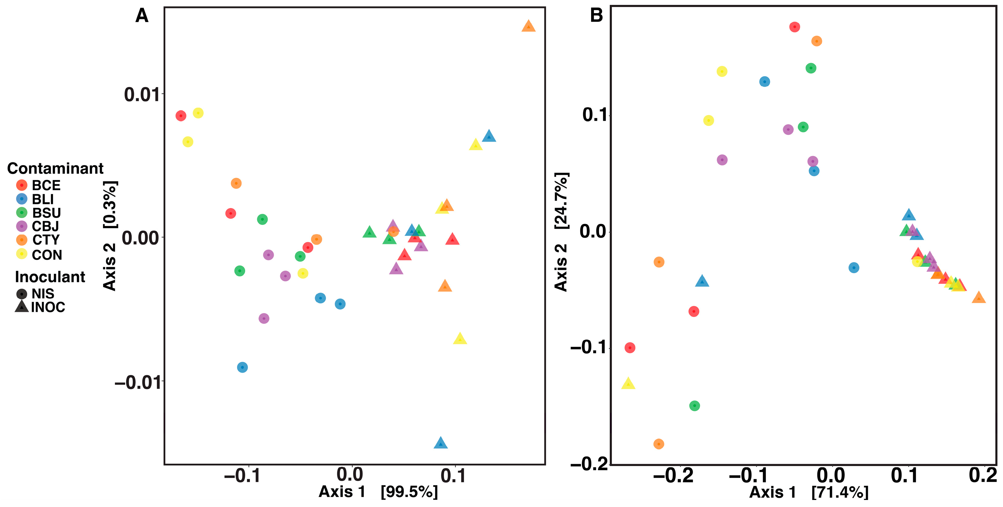
3.3. Impact of Inoculant and Spore Contaminants on the Microbial Communities of Silage

After 100 days of fermentation, important differences in relative abundance (RA) of the main genera of lactic acid bacteria (Table 3) were observed. The most abundant genera were related to the genus Lactobacillus, with higher abundance in NIS than in INOC, (means of 69.82% and 33.64%, respectively; p < 0.001). The RA of Lentilactobacillus was higher in INOC (65.97%) than in NIS (29.87%; p < 0.001). The other lactic acid bacteria genera had much lower RA values. This could be observed with the RA of Loigolactobacillus, which had an overall higher RA in INOC (0.1%) than in NIS (0.04%; p = 0.004. The RA of Levilactobacillus was higher in NIS (0.17 %) than in INOC (<0.01%; p < 0.001). After 3 days of aerobic exposure (Table 3), the mean RA of Lactobacillus was higher in NIS (50.76%) than in INOC (27.97%; p < 0.001). The RA of Lentilactobacillus was higher in INOC (69.54%) than in NIS (39.31%; p < 0.001). The RA of Acetobacter was higher in NIS (8.27%) than in INOC (2.22%; p = 0.012). For the fungi (Table 4), the most abundant mould was Mucor after opening, wherein the RA was numerically higher for INOC (37.24%) than NIS (33.5%). The second most abundant fungus was the yeast Wickerhamomyces, with RAs of 13.62 and 7.63% in the NIS and INOC samples, respectively. However, there were no significantly different RAs for any fungi after opening. Following aerobic exposure, the RA of Kazachstania was higher in INOC (5.04%) than in NIS (2.04%; p = 0.010), and Wickerhamomyces was significantly higher in BCE than in BSU, CTY, and CBJ.

3.4. Changes in Cell and Spore Counts following Inoculation and Contamination

Cell and spore counts were performed following storage and aerobic exposure for aerobic/facultative spore counts (AFSs) and anaerobic spore counts (ANSs). Yeast and LAB counts were only performed following storage (Table 5; two-way ANOVA). After the fermentation of the maize silage, the AFS was overall higher in INOC (3.68 log10 CFU/g) than in NIS (3.49 log10 CFU/g; p = 0.003) and higher in BSU (3.7 log10 CFU/g), BLI (3.8 log10 CFU/g), and CBJ (3.7 log10 CFU/g) than in the control (3.3 log10 CFU/g). Interaction between the contaminant and inoculant revealed that the AFSs in the INOC samples of BLI (4.1 log10 CFU/g; Supplementary Figure S1C) were higher than those of the NIS of BLI (3.6 log10 CFU/g; p = 0.007), while those observed in NIS of CON (3.4 log10 CFU/g) were higher than those in INOC of CON (3.1 log10 CFU/g; p = 0.044). ANSs were higher in INOC (3.19) than in NIS (2.74, p < 0.001) and higher in BSU (3.5 log10 CFU/g) and BLI (3.3 log10 CFU/g) than in the control (2.9 log10 CFU/g). The control samples had higher ANSs than BCE (2.4 log10 CFU/g). Interactions revealed that the ANS (Supplementary Figure S1D) was higher in the INOC samples of BCE (3.6 log10 CFU/g; Supplementary Figure S2D) and CON (3.2 log10 CFU/g) compared to that of NIS (1.3 log10 CFU/g for BCE, p < 0.001; 2.6 log10 CFU/g for CON, p = 0.012). LAB counts were significantly higher in INOC (8.76 log10 CFU/g) than in NIS (8.39 log10 CFU/g; p < 0.001). Yeast counts were significantly higher in NIS (2.34 log10 CFU/g) than in INOC (1.65 log10 CFU/g; p = 0.001). After 3 days of aerobic exposure, the AFS was higher in BCE (4.0 log10 CFU/g), BSU (3.6 log10 CFU/g), and BLI (3.6 log10 CFU/g) than in the control. Interactions revealed that the AFS (Supplementary Figure S2B) was higher in the NIS of CBJ (3.3 log10 CFU/g) and CON (3.5 log10 CFU/g) than those of INOC (3.0 log10 CFU/g for CBJ, p = 0.014; 3.2 log10 CFU/g for CON, p = 0.009) and higher in the INOC sample of CTY (3.7 log10 CFU/g) than in NIS (2.6 log10 CFU/g; p = 0.047). ANS was higher in NIS (3.4 log10 CFU/g) than in INOC (3.24 log10 CFU/g; p = 0.002). ANSs (3.31 log10 CFU/g) were higher in BLI (3.5 log10 CFU/g) than in the control (p = 0.007). Interaction showed that the ANS (Supplementary Figure S2C) was higher in the NIS of CON (3.4 log10 CFU/g) than that of INOC (2.9 log10 CFU/g; p = 0.004).

3.5. Correlations between Microbial and Chemical Variables

NIS contaminated with B. subtilis and C. beijerinckii upon opening favoured a higher RA of eight 16S rRNA gene ASVs of the genus Lentilactobacillus, which were all negatively correlated with three ITS1 ASVs from the genus Mucor, one from Psathyrella, one from Kazachstania, and one from Hanseniaspora (Figure 3A). The Lentilactobacillus ASVs were also negatively correlated with betaine and tyrosine, which were positively correlated with the ITS1 ASVs that were negatively correlated with the 16S rRNA gene ASVs. NIS contaminated with C. tyrobutyricum had an increased RA of 14 16S ASVs from the genus Lentilactobacillus upon opening, which were all negatively correlated with ethane 1,2 diol and arabinose and positively correlated with methylamine, but they had no correlations at a cut-off of 0.8 with any ITS1 ASVs. Inoculated samples contaminated with B. subtilis had a higher RA of eight bacterial 16S ASVs of the genus Lactobacillus upon opening, which were all positively correlated with two fungal ASVs from the genus Mutinus, one from the genus Mucor, and two from unknown genera. All positively correlated ASVs were also positively correlated with asparagine and malonate. One 16S ASV of Lentilactobacillus was negatively correlated with xylose. After aerobic exposure, the NIS samples contaminated with B. licheniformis and the control had a higher RA of eight bacterial 16S ASVs from the genus Lentilactobacillus, while the samples contaminated with B. licheniformis were higher by two additional Lentilactobacillus ASVs. These 16S ASVs were positively correlated with one ITS1 ASV from the genus Mucor and one from the genus Hanseniaspora. Upon aerobic exposure, the NIS samples contaminated with B. cereus had a higher RA of four ASVs from the genus Levilactobacillus, two from the genus Loigolactobacillus, and two from the genus Lactiplantibacillus, which were positively correlated with eight ITS1 ASVs from the genus Wickerhamomyces, which were all positively correlated with methylamine. Under conditions of aerobic exposure, the inoculated samples contaminated with C. beijerinckii had a higher RA in 12 bacterial 16S ASVs from the genus Lentilactobacillus and 8 from the genus Lactobacillus. The Lactobacillus ASVs were negatively correlated with one ITS1 ASV from the genus Aspergillus and one from an unknown genus. These ITS ASVs were positively correlated with tyramine. The inoculated samples contaminated with B. licheniformis after aerobic exposure contained unique ITS1 ASVs: one from the genus Wickerhamomyces, one from Mucor, one from Knufia, two from Kazachstania, one from Candida, and one from an unknown genus. These ASVs were positively correlated with γ-aminobutyrate (GABA).

4. Discussion

4.1. Effects of Inoculation on Silage Quality

The main species of Lactobacillus found in corn silage in this study, based on the closest BLAST matches of the Lactobacillus 16S ASVs against the NCBI database, was Lactobacillus acidophilus. This species has been found as an abundant species in a few previous corn silage studies [37]. L. acidophilus was used as an inoculant to drop pH levels and quickly produce lactic acid for the silage of round-leaf cassia (Chamaecrista rotundifolia), a perennial legume [38]. It is also known to have anti pathogenic effects that are not fully related to acid production, particularly against Clostridium perfringens, suggesting that its antimicrobial activity may be based on the production of bacteriocins [39]. Furthermore, C. rotundifolia fermented with the addition of L. acidophilus has been shown to have a lower relative abundance of Clostridium and Enterobacter [38]. L. acidophilus has been shown to exhibit anti-fungal properties against Aspergillus [38], which could explain the negative correlation between Lactobacillus ASVs and Aspergillus in the inoculated samples after aerobic exposure. There were no detected Aspergillus ASVs in the NIS samples, explaining why the negative correlation was not observable in NIS.
Aerobic exposure leads to the deterioration of silage, as it represents an opportunity for yeasts, moulds, and aerobic/facultative anaerobic bacteria to grow. The presence of these microorganisms, all considered aerobic spoilage organisms of silage, has been associated with reduced dry matter content [40], increased mycotoxin content [26], and an increase in the transfer of spore formers to dairy products [8]. The aerobic stability of the maize silage in our study lasted for much longer in all the INOC samples, likely due to the control of aerobic spoilage organisms via higher levels of acetic acid [41]. The INOC silage had an expected higher RA of Lentilactobacillus following inoculation with L. buchneri, which explains the higher acetic and propionic acid content in the inoculated silage. Acetobacter is a bacterial genus that is often associated with the aerobic deterioration of maize silage upon exposure to air [42]. L. buchneri inoculation resulted in a lower RA of Acetobacter.
The presence of butyric acid in silage is usually an indicator of clostridial fermentation, although it is less present in maize silage than in grass and alfalfa due to its generally lower pH [43]. However, Vissers et al. [1] determined that in the Netherlands, corn had the highest likelihood of clostridial contamination. The presence of butyric acid is not necessarily detrimental to dairy cattle feed intake or silage quality [44]. Aksu et al. [45] reported high levels (5–7 g/kg) of butyric acid in both inoculated and non-inoculated maize silage. The higher content of butyric acid in the INOC samples upon opening was not related to a similar increase in the number of Clostridium ASVs, but there were higher levels of anaerobic spores in these samples. This could indicate early clostridial activity in INOC samples followed by higher levels of sporulation once the fermentation conditions became unsuitable for clostridial growth, such as at a low pH [46]. Butyric acid fermentation generally results in a reduction in DM due to the production of CO2 [47] and could explain the lower DM of INOC maize compared to that of NIS upon opening. Li et al. [48] reported that in alfalfa silage, clostridial fermentation reached a peak after seven days of ensiling and steadily decreased after 56 days. Although our study reports higher butyric acid concentrations with the use of an inoculant, the butyric acid content after aerobic exposure was not significantly different between the NIS and INOC samples and was overall lower than that upon opening. This could be due to the increased presence of butyric-acid-degrading bacteria such as Pseudomonas spp. after exposure to air [49], which were undetectable in the maize samples upon opening but were present at significantly higher levels after 3 days of aerobic exposure. We observed an overall positive correlation between higher butyric acid and higher pH upon opening. Increased butyric acid levels have been previously associated with maize silage samples having a higher pH and can lead to the growth of saccharolytic and proteolytic Clostridium species [48]. Proteolytic clostridia can negatively affect the dry matter intake and performance of dairy cattle [50]. A higher pH was seen in the INOC samples with higher concentrations of butyric acid; however, the pH was not elevated to a level that Clostridium was able to tolerate [2]. Fresh maize forage generally has pH levels high enough to allow Clostridium to grow [51]. This further suggests that the pH drop at the beginning of the mini silo fermentation period was slower in the INOC samples, allowing for more early growth of butyric-acid-producing bacteria, but this growth was inhibited as the pH continued to drop.

4.2. Effects of Bacillus and Clostridium Contaminants on Silage Quality

Malonic acid in silage is usually associated with amino acid synthesis, and its presence could indicate the production of amino acids by certain bacteria [48]. Guo et al. [52], showed that the inoculation of silage with L. buchneri increased the amino acid and malonic acid content of said silage. High amino acid content in silage could signify good fermentation quality, as low amino acid content is an indicator of deamination [52]. The relationship between malonic acid and amino acid content could provide insight into amino acid deamination during silage fermentation, as high amino acid content should be expected with high malonic acid content in a good fermentation profile. In our study, while amino acid levels were higher, malonic acid content was lower in the samples inoculated with L. buchneri. Furthermore, the highest malonic acid content was found in the samples contaminated with B. cereus, B. subtilis, and B. licheniformis. This suggests that it is likely Bacillus that is causing the higher levels of malonic acid. The control samples had lower aerobic spore counts, which may explain the lower levels of malonic acid.
In most of the contamination treatments, the anaerobic spore counts were higher in the samples after aerobic exposure, suggesting that the addition of spore formers affects the spore population after 100 days of fermentation and after 3 days of exposure to air. Although the increased level of anaerobic spore formers did not affect the aerobic stability of the silage, increased quantities of spore formers in silage can pose a risk of higher transfer rates to milk, leading to problems in the resulting dairy products [53]. The level of spore formers in general is not accounted for in the microbial standards for raw milk in the National Dairy Code of Canada [54]. However, ready-to-eat foods of satisfactory quality contain less than 50 CFU/g of B. cereus according to guidelines established by Health Canada [55]. Without additives to control spore germination, levels as low as 200 spores per L of milk can cause spoilage problems in dairy products, particularly for hard cheeses [53]. Considering a study of the rate of transfer from feed to feces to bulk tank, this level in milk could be attained with a count of only 3.5 log10 spores/g in a total mixed ration of corn and alfalfa grass silage [53]. Previous studies have shown that spore-forming bacteria numbers increase during aerobic exposure [56]; however, we only observed this trend in the samples contaminated with B. cereus. This highlights the ability for some Bacillus species to survive throughout fermentation and aerobic exposure with or without an inoculant.
There were no high-value correlations with ASVs associated with any of the genera of the contaminants upon opening or after aerobic exposure, suggesting that their effects occur early on during fermentation or indirectly through the inhibition or stimulation of other specific microorganisms. Instead, the majority of the ASVs with high correlation coefficients were yeasts and dominant LAB that have higher relative abundances in specific contaminations. In many cases, these ASVs were absent from all but one contamination. There were no differences in alpha or beta diversity based on individual contaminants, suggesting that the individual spore formers did not affect the overall diversity of the maize silage but did influence specific strains. Drouin et al. [26] reported that the L. buchneri inoculant used in this study, paired with L. hilgardii, increased the alpha diversity of bacteria in maize silage. The increased alpha diversity was likely the result of a higher RA of heterofermentative LAB ASVs. This contributed to higher aerobic stability by ensuring that no potential spoilage bacteria were able to outcompete. This trend was mimicked in the current study with the use of the L. buchneri inoculant only. An increase in the alpha diversity of the samples after inoculation may have a positive effect on overall silage quality.

4.3. Effects of B. cereus on Silage Quality

Fast fermentation and a high level of lactic acid in maize silage are associated with lower levels of spoilage microorganisms during fermentation [57]. This is exactly the condition observed with the silage contaminated by B. cereus, which had the highest content of lactic acid and acetic acid compared to the other contaminations and the control tested in this trial. The pH of these samples was also significantly lower than that of the control and the Clostridium-contaminated silages, which is linked to the higher lactic acid content [58]. For the silage contaminated by B. cereus, significantly lower anaerobic spore counts were observed compared to the control. All these observations for this specific contamination point to a faster pH drop at the beginning of ensiling. This leaves a lower metabolic potential for clostridial fermentation and results in lower counts of Clostridium [48]. If the higher lactic acid content was due to a higher ratio of homofermentative and/or facultative heterofermentative to heterofermentative LAB, we would expect to see lower levels of acetic acid in the samples contaminated with B. cereus compared to those of other contamination types. However, a higher mean acetic acid level was obtained in these samples. Furthermore, the RA of the genera Lentilactobacillus (heterofermentative) and Lactobacillus (obligate homofermentative) were not significantly different from that of all other contamination types. This may suggest that there was an overall higher availability of fermentable sugars in the maize contaminated with B. cereus. Some Bacillus species are known to produce cellulases and hemicellulases [59,60], which could have beneficial impacts such as increased sugar concentrations in silage and higher dry matter intake (DMI) for dairy cattle [61]. Inoculation with Bacillus subtilis has been shown to lower pH and increase lactic acid concentrations in maize silage [62,63]. When used as silage inoculants, Bacillus licheniformis [64] and Bacillus subtilis [65] have been proven to be effective at reducing yeast and mold quantities and increasing fiber degradation [64]. Using Bacillus as an inoculant has been shown to increase the viable count of LAB in maize silage [65]; however, we did not observe this in our study. We did observe a higher RA of Wickerhamomyces in the silage samples contaminated with B. cereus, specifically after aerobic exposure. Wickerhamomyces is a genus of yeast known to synthesize lactate and acetate into ethyl esters [66], which may be why the lactic acid and acetic acid levels were higher after opening but were reduced to the same levels as other contaminants after aerobic exposure. This suggests that Wickerhamomyces species can confer a negative impact after aerobic exposure by converting volatile fatty acids into esters. Although the use of specific Bacillus strains as silage additives may be beneficial in promoting lower pH and higher organic acid content, without a heterofermentative strain used as an additive, these advantages could be lost upon aerobic exposure. Moreover, the risk for milk contamination and spoilage may pose too great a challenge for the application of this approach to dairy cattle.

4.4. Effects of B. licheniformis on Silage Quality

The numbers of aerobic spore formers in INOC contaminated with B. licheniformis were higher than those of NIS with the same contaminant as well as all other contaminants across both treatments. γ-aminobutyric acid is important to mammalian health and acts as a neurotransmitter inhibitor in the sympathetic nervous system [67]. Higher GABA content using a silage additive has been shown to improve the dry matter intake of ruminants [68]; however, we did not see any significant difference in GABA content dependent on inoculation. Some of the contaminants did show differences in GABA, specifically in the 3-day aerobically exposed samples, suggesting that these contaminants can positively influence the amount of GABA in maize silage. The INOC samples after 3-day aerobic exposure showed a strong negative correlation between GABA and several ASVs from the genus Lentilactobacillus present in all silage 16S profiles except for those of the samples contaminated with B. licheniformis. Tanizawa et al. [69] reported isolating multiple L. buchneri strains from rice silage that were unable to produce GABA. Other reports have shown that L. buchneri and L. hilgardii can be strong GABA producers [70,71], among other members of the Lactobacillaceae family [67]. Furthermore, several strains of yeast have been shown to produce GABA [4,72]. The variation in GABA content between spore former contamination treatments could be due to specific metabolic interactions with high-GABA-producing bacterial or fungal strains [72,73]. Based on our observed trends, it is possible that the addition of B. licheniformis stimulated specific yeasts able to produce GABA during aerobic exposure. For example, in the aerobically exposed INOC samples contaminated with B. licheniformis, there was a positive correlation between ASVs from several genera of yeast (Kazachstania, Wickerhamomyces, Candida, Mucor, and Knufia) and bacteria (Acetobacter) and the GABA content. Furthermore, the Lentilactobacillus species that were more abundant in all contaminations except B. licheniformis might have prevented the growth of GABA-producing yeasts after aerobic exposure.

5. Conclusions

In our study, the use of a heterofermentative inoculant provided a favorable environment for spore former contaminants. This led to a slower pH decrease and clostridial fermentation in the early stages of maize fermentation as indicated by the increased butyric acid concentrations upon opening and higher spore counts. Out of all the contaminants tested, B. licheniformis and B. cereus seemed to have had the most negative effects on the maize silage after its exposure to air, while Clostridium species did not strongly impact any of the physicochemical or microbial parameters studied. Regardless of which spore contaminant was tested, the heterofermentative inoculant dominated the bacterial population and produced the expected metabolites (acetic and propionic acids), leading to strongly improved aerobic stability. However, the combination of both homofermentative and facultative homofermentative inoculants would result in further beneficial effects via a fast pH reduction preventing any early clostridial fermentation while maintaining a strong heterofermentative fermentation to prevent the aerobic deterioration of the maize silage.

Supplementary Materials

The following supporting information can be downloaded at https://www.mdpi.com/article/10.3390/fermentation9090837/s1, Figure S1: Box plots displaying significant differences (* = 0.05; ** = 0.01, *** < 0.001) for silage parameters that showed significant interactions between inoculant and contaminant in the two-way ANOVA after opening.; Figure S2: Box plots displaying significant differences (* = 0.05; ** = 0.01, *** < 0.001) for silage parameters that showed significant interactions between inoculant and contaminant in the two-way ANOVA after aerobic exposure.

Author Contributions

Conceptualization, G.L. and P.D.; methodology, J.H., G.L. and P.D.; validation, J.H., P.D., L.D. and G.L.; formal analysis, J.H. and P.D.; investigation, J.H.; resources, G.L. and P.D.; data curation, J.H. and P.D.; writing—original draft preparation, J.H.; writing—review and editing, G.L., P.D., and L.D.; visualization, J.H.; supervision, G.L. and P.D.; project administration, G.L.; funding acquisition, G.L. All authors have read and agreed to the published version of the manuscript.

Funding

This research was funded by NSERC Collaborative Research and Development Grant (CRDPJ 529498-18) as well as the NSERC/DFO Industrial Research Chair in Dairy Microbiology (490979-15) held by G. LaPointe.

Institutional Review Board Statement

Not applicable.

Informed Consent Statement

Not applicable.

Data Availability Statement

The datasets for this study are available through Borealis in the Agri-Environmental Research Data Repository of the University of Guelph at https://doi.org/10.5683/SP3/RZ2JJK (accessed on 15 August 2023).

Acknowledgments

The authors would like to acknowledge the technical expertise provided by Jeffrey Gross (Advanced Analysis Center: Genomics, University of Guelph) and Sameer Al-Abdul-Wahid (Advanced Analysis Center: Nuclear Magnetic Resonance).

Conflicts of Interest

JH, PD, and GL declare that the research was conducted in the absence of any commercial or financial relationships that could be construed as a potential conflict of interest. LD is employed by Lallemand Inc.; however, her affiliation did not impede the research team’s ability to follow journal guidelines or to remain impartial during the preparation of this manuscript.

References

  1. Vissers, M.M.M.; Te Giffel, M.C.T.; Driehuis, F.; Jong, P.D.; Lankveld, J.M.G. Minimizing the level of Bacillus cereus spores in farm tank milk. J. Dairy Sci. 2007, 90, 3286–3293. [Google Scholar] [CrossRef] [PubMed]
  2. Queiroz, O.C.M.; Ogunade, I.M.; Weinberg, Z.; Adesogan, A.T. Silage Review: Foodborne pathogens in silage and their mitigation by silage additives. J. Dairy Sci. 2018, 101, 4132–4142. [Google Scholar] [CrossRef] [PubMed]
  3. McDonald, P.; Henderson, N.; Heron, S. The Biochemistry of Silage, 2nd ed.; Chalcombe: Marlow, England, 1991. [Google Scholar]
  4. Li, M.M.; White, R.R.; Guan, L.L.; Harthan, L.; Hanigan, M.D. Metatranscriptomic analyses reveal ruminal pH regulates fiber degradation and fermentation by shifting the microbial community and gene expression of carbohydrate-active enzymes. Anim. Microbiome 2021, 3, 32. [Google Scholar] [CrossRef] [PubMed]
  5. D’Incecco, P.; Pellegrino, L.; Hogenboom, J.A.; Cocconcelli, P.S.; Bassi, D. The late blowing defect of hard cheeses: Behaviour of cells and spores of Clostridium tyrobutyricum throughout the cheese manufacturing and ripening. LWT 2018, 87, 134–141. [Google Scholar] [CrossRef]
  6. Klijn, N.; Nieuwenhof, F.F.; Hoolwerf, J.D.; van der Waals, C.B.; Weerkamp, A.H. Identification of Clostridium tyrobutyricum as the causative agent of late blowing in cheese by species-specific PCR amplification. Appl. Environ. Microbiol. 1995, 61, 2919–2924. [Google Scholar] [CrossRef]
  7. Buehner, K.P.; Anand, S.; Djira, G.D.; Garcia, A. Corrigendum to “Prevalence of thermoduric bacteria and spores on 10 midwest dairy farms”. J. Dairy Sci. 2014, 97, 8009–8016. [Google Scholar] [CrossRef]
  8. Te Giffel, M.C.; Wagendorp, A.; Herrewegh, A.; Driehuis, F. Bacterial spores in silage and raw milk. Antonie Van Leeuwenhoek 2002, 81, 625–630. [Google Scholar] [CrossRef]
  9. Heyndrickx, M.; Scheldeman, P. Bacilli associated with spoilage in dairy products and other food. In Applications and Systematics of Bacillus and Relatives; John Wiley & Sons, Ltd.: Hoboken, NJ, USA, 2002; pp. 64–82. ISBN 978-0-470-69674-3. [Google Scholar]
  10. Beecher, D.J.; Schoeni, J.L.; Wong, A.C. Enterotoxic activity of Hemolysin BL from Bacillus cereus. Infect. Immun. 1995, 63, 4423–4428. [Google Scholar] [CrossRef]
  11. Ehling-Schulz, M.; Svensson, B.; Guinebretiere, M.-H.; Lindbäck, T.; Andersson, M.; Schulz, A.; Fricker, M.; Christiansson, A.; Granum, P.E.; Märtlbauer, E.; et al. Emetic toxin formation of Bacillus cereus is restricted to a single evolutionary lineage of closely related strains. Microbiol. Read. Engl. 2005, 151, 183–197. [Google Scholar] [CrossRef]
  12. Griffiths, M. Improving the Safety and Quality of Milk: Milk Production and Processing, 1st ed.; Woodhead Publishing: Thorston, UK, 2010; Volume 1, ISBN 978-1-84569-942-0. [Google Scholar]
  13. Vidic, J.; Chaix, C.; Manzano, M.; Heyndrickx, M. Food sensing: Detection of Bacillus cereus spores in dairy products. Biosensors 2020, 10, 15. [Google Scholar] [CrossRef]
  14. Kumari, S.; Sarkar, P.K. Bacillus cereus hazard and control in industrial dairy processing environment. Food Control 2016, 69, 20–29. [Google Scholar] [CrossRef]
  15. Scheldeman, P.; Herman, L.; Foster, S.; Heyndrickx, M. Bacillus sporothermodurans and other highly heat-resistant spore formers in milk. J. Appl. Microbiol. 2006, 101, 542–555. [Google Scholar] [CrossRef] [PubMed]
  16. Reginensi, S.M.; González, M.J.; Olivera, J.A.; Sosa, M.; Juliano, P.; Bermúdez, J. RAPD-based screening for spore-forming bacterial populations in Uruguayan commercial powdered milk. Int. J. Food Microbiol. 2011, 148, 36–41. [Google Scholar] [CrossRef] [PubMed]
  17. Gopal, N.; Hill, C.; Ross, P.R.; Beresford, T.P.; Fenelon, M.A.; Cotter, P.D. The prevalence and control of Bacillus and related spore-forming bacteria in the dairy industry. Front. Microbiol. 2015, 6, 1418. [Google Scholar] [CrossRef]
  18. Parkinson, T.J. Specific infectious diseases causing infertility and subfertility in cattle. In Veterinary Reproduction and Obstetrics, 10th ed.; Noakes, D.E., Parkinson, T.J., England, G.C.W., Eds.; W.B. Saunders: St. Louis, MO, USA, 2019; pp. 434–466. ISBN 978-0-7020-7233-8. [Google Scholar]
  19. Scheldeman, P.; Pil, A.; Herman, L.; De Vos, P.; Heyndrickx, M. Incidence and diversity of potentially highly heat-resistant spores isolated at dairy farms. Appl. Environ. Microbiol. 2005, 71, 1480–1494. [Google Scholar] [CrossRef]
  20. Vaerewijck, M.J.; De Vos, P.; Lebbe, L.; Scheldeman, P.; Hoste, B.; Heyndrickx, M. Occurrence of Bacillus sporothermodurans and other aerobic spore-forming species in feed concentrate for dairy cattle. J. Appl. Microbiol. 2001, 91, 1074–1084. [Google Scholar] [CrossRef]
  21. Driehuis, F.; Wilkinson, J.M.; Jiang, Y.; Ogunade, I.; Adesogan, A.T. Silage review: Animal and human health risks from silage. J. Dairy Sci. 2018, 101, 4093–4110. [Google Scholar] [CrossRef]
  22. Gleeson, D.; O’Connell, A.; Jordan, K. Review of potential sources and control of thermoduric bacteria in bulk-tank milk. Ir. J. Agric. Food Res. 2013, 52, 217–227. Available online: http://www.jstor.org/stable/23631033 (accessed on 1 May 2023).
  23. Heyndrickx, M. The importance of endospore-forming bacteria originating from soil for contamination of industrial food processing. Appl. Environ. Soil Sci. 2011, 2011, e561975. [Google Scholar] [CrossRef]
  24. Oliveira, A.S.; Weinberg, Z.G.; Ogunade, I.M.; Cervantes, A.A.P.; Arriola, K.G.; Jiang, Y.; Kim, D.; Li, X.; Gonçalves, M.C.M.; Vyas, D.; et al. Meta-analysis of effects of inoculation with homofermentative and facultative heterofermentative lactic acid bacteria on silage fermentation, aerobic stability, and the performance of dairy cows. J. Dairy Sci. 2017, 100, 4587–4603. [Google Scholar] [CrossRef]
  25. Kleinschmit, D.H.; Kung, L. A Meta-analysis of the effects of Lactobacillus buchneri on the fermentation and aerobic stability of corn and grass and small-grain silages. J. Dairy Sci. 2006, 89, 4005–4013. [Google Scholar] [CrossRef]
  26. Drouin, P.; Tremblay, J.; Renaud, J.; Apper, E. Microbiota succession during aerobic stability of maize silage inoculated with Lentilactobacillus buchneri NCIMB 40788 and Lentilactobacillus hilgardii CNCM-I-4785. MicrobiologyOpen 2021, 10, e1153. [Google Scholar] [CrossRef]
  27. Nair, J.; Huaxin, N.; Andrada, E.; Yang, H.-E.; Chevaux, E.; Drouin, P.; McAllister, T.A.; Wang, Y. Effects of inoculation of corn silage with Lactobacillus hilgardii and Lactobacillus buchneri on silage quality, aerobic stability, nutrient digestibility, and growth performance of growing beef cattle. J. Anim. Sci. 2020, 98, skaa267. [Google Scholar] [CrossRef]
  28. Arriola, K.G.; Oliveira, A.S.; Jiang, Y.; Kim, D.; Silva, H.M.; Kim, S.C.; Amaro, F.X.; Ogunade, I.M.; Sultana, H.; Pech Cervantes, A.A.; et al. Meta-analysis of effects of inoculation with Lactobacillus buchneri, with or without other bacteria, on silage fermentation, aerobic stability, and performance of dairy cows. J. Dairy Sci. 2021, 104, 7653–7670. [Google Scholar] [CrossRef] [PubMed]
  29. Huisden, C.M.; Adesogan, A.T.; Kim, S.C.; Ososanya, T. Effect of applying molasses or inoculants containing homofermentative or heterofermentative bacteria at two rates on the fermentation and aerobic stability of corn silage. J. Dairy Sci. 2009, 92, 690–697. [Google Scholar] [CrossRef]
  30. Rosenquist, H.; Hansen, Å. The antimicrobial effect of organic acids, sour dough and nisin against Bacillus subtilis and B. licheniformis isolated from wheat bread. J. Appl. Microbiol. 1998, 85, 621–631. [Google Scholar] [CrossRef]
  31. Adesogan, A.T.; Krueger, N.; Salawu, M.B.; Dean, D.B.; Staples, C.R. The influence of treatment with dual purpose bacterial inoculants or soluble carbohydrates on the fermentation and aerobic stability of bermudagrass. J. Dairy Sci. 2004, 87, 3407–3416. [Google Scholar] [CrossRef] [PubMed]
  32. Klindworth, A.; Pruesse, E.; Schweer, T.; Peplies, J.; Quast, C.; Horn, M.; Glöckner, F.O. Evaluation of general 16S ribosomal RNA gene PCR primers for classical and next-generation sequencing-based diversity studies. Nucleic Acids Res. 2013, 41, e1. [Google Scholar] [CrossRef]
  33. Bellemain, E.; Carlsen, T.; Brochmann, C.; Coissac, E.; Taberlet, P.; Kauserud, H. ITS as an environmental DNA barcode for fungi: An in silico approach reveals potential PCR biases. BMC Microbiol. 2010, 10, 189. [Google Scholar] [CrossRef]
  34. Deshpande, V.; Wang, Q.; Greenfield, P.; Charleston, M.; Porras-Alfaro, A.; Kuske, C.R.; Cole, J.R.; Midgley, D.J.; Tran-Dinh, N. Fungal identification using a bayesian classifier and the Warcup training set of internal transcribed spacer sequences. Mycologia 2016, 108, 1–5. [Google Scholar] [CrossRef]
  35. Løvdal, I.S.; Hovda, M.B.; Granum, P.E.; Rosnes, J.T. Promoting Bacillus cereus spore germination for subsequent inactivation by mild heat treatment. J. Food Prot. 2011, 74, 2079–2089. [Google Scholar] [CrossRef]
  36. R Core Team. A Language and Environment for Statistical Computing. R Foundation for Statistical Computing. Available online: https://www.r-project.org/ (accessed on 1 May 2023).
  37. Santos, A. de O. dos; Ávila, C.L. da S.; Soares, C.; Carvalho, B.F.; Schwan, R.F.; Lima, N. Lactic acid bacteria diversity in corn silage produced in Minas Gerais (Brazil). Ann. Microbiol. 2019, 69, 1445–1459. [Google Scholar] [CrossRef]
  38. Feng, Q.; Shi, W.; Chen, S.; Degen, A.A.; Qi, Y.; Yang, F.; Zhou, J. Addition of organic acids and Lactobacillus acidophilus to the leguminous forage Chamaecrista rotundifolia improved the quality and decreased harmful bacteria of the silage. Animals 2022, 12, 2260. [Google Scholar] [CrossRef] [PubMed]
  39. Gilliland, S.E.; Speck, M.L. Antagonistic action of Lactobacillus acidophilus toward intestinal and foodborne pathogens in associative cultures. J. Food Prot. 1977, 40, 820–823. [Google Scholar] [CrossRef] [PubMed]
  40. Borreani, G.; Tabacco, E.; Schmidt, R.J.; Holmes, B.J.; Muck, R.E. Silage review: Factors affecting dry matter and quality losses in silages. J. Dairy Sci. 2018, 101, 3952–3979. [Google Scholar] [CrossRef] [PubMed]
  41. Danner, H.; Holzer, M.; Mayrhuber, E.; Braun, R. Acetic acid increases stability of silage under aerobic conditions. Appl. Environ. Microbiol. 2003, 69, 562–567. [Google Scholar] [CrossRef]
  42. Guan, H.; Ran, Q.; Li, H.; Zhang, X. Succession of microbial communities of corn silage inoculated with heterofermentative lactic acid bacteria from ensiling to aerobic exposure. Fermentation 2021, 7, 258. [Google Scholar] [CrossRef]
  43. Kung, L.; Shaver, R.D.; Grant, R.J.; Schmidt, R.J. Silage review: Interpretation of chemical, microbial, and organoleptic components of silages. J. Dairy Sci. 2018, 101, 4020–4033. [Google Scholar] [CrossRef]
  44. Vissers, M.M.M.; Driehuis, F.; Te Giffel, M.C.; De Jong, P.; Lankveld, J.M.G. Concentrations of butyric acid bacteria spores in silage and relationships with aerobic deterioration. J. Dairy Sci. 2007, 90, 928–936. [Google Scholar] [CrossRef]
  45. Aksu, T.; Baytok, E.; Bolat, D. Effects of a bacterial silage inoculant on corn silage fermentation and nutrient digestibility. Small Rumin. Res. 2004, 55, 249–252. [Google Scholar] [CrossRef]
  46. Dürre, P. Physiology and sporulation in Clostridium. Microbiol. Spectr. 2014, 2, 315–329. [Google Scholar] [CrossRef]
  47. Fenner, H.; Barnes, H.D. Improved method for determining dry matter in silage. J. Dairy Sci. 1965, 48, 1324–1328. [Google Scholar] [CrossRef]
  48. Li, R.; Jiang, D.; Zheng, M.; Tian, P.; Zheng, M.; Xu, C. Microbial community dynamics during alfalfa silage with or without clostridial fermentation. Sci. Rep. 2020, 10, 17782. [Google Scholar] [CrossRef]
  49. He, J.; Chu, Y.; Li, J.; Meng, Q.; Liu, Y.; Jin, J.; Wang, Y.; Wang, J.; Huang, B.; Shi, L.; et al. Intestinal butyrate-metabolizing species contribute to autoantibody production and bone erosion in rheumatoid arthritis. Sci. Adv. 2022, 8, eabm1511. [Google Scholar] [CrossRef] [PubMed]
  50. Simpson, K.M.; Callan, R.J.; Van Metre, D.C. Clostridial abomasitis and enteritis in ruminants. Vet. Clin. North Am. Food Anim. Pract. 2018, 34, 155–184. [Google Scholar] [CrossRef] [PubMed]
  51. Shaver, R.D.; Erdman, R.A.; O’connor, A.M.; Vandersall, J.H. Effects of silage pH on voluntary intake of corn silage and alfalfa haylage. J. Dairy Sci. 1985, 68, 338–346. [Google Scholar] [CrossRef]
  52. Guo, X.S.; Ke, W.C.; Ding, W.R.; Ding, L.M.; Xu, D.M.; Wang, W.W.; Zhang, P.; Yang, F.Y. Profiling of metabolome and bacterial community dynamics in ensiled Medicago sativa inoculated without or with Lactobacillus plantarum or Lactobacillus buchneri. Sci. Rep. 2018, 8, 357. [Google Scholar] [CrossRef] [PubMed]
  53. Borreani, G.; Ferrero, F.; Nucera, D.; Casale, M.; Piano, S.; Tabacco, E. Dairy farm management practices and the risk of contamination of tank milk from Clostridium spp. and Paenibacillus spp. spores in silage, total mixed ration, dairy cow feces, and raw milk. J. Dairy Sci. 2019, 102, 8273–8289. [Google Scholar] [CrossRef]
  54. Canadian Dairy Information Centre. Acts, Regulations, Codes and Standards. Available online: https://agriculture.canada.ca/en/sector/animal-industry/canadian-dairy-information-centre/acts-regulations-codes-standards. (accessed on 1 May 2023).
  55. Health Canada. Microbial guidelines for ready-to-eat foods–A guide for the conveyance industry and environmental health officers (EHO). Health Canada 2010. Available online: https://publications.gc.ca/site/archivee-archived.html?url=https://publications.gc.ca/collections/collection_2014/sc-hc/H164-167-2013-eng.pdf (accessed on 1 May 2023).
  56. Borreani, G.; Tabacco, E. Improving corn silage quality in the top layer of farm bunker silos through the use of a next-generation barrier film with high impermeability to oxygen. J. Dairy Sci. 2014, 97, 2415–2426. [Google Scholar] [CrossRef]
  57. Muck, R.E.; Nadeau, E.M.G.; McAllister, T.A.; Contreras-Govea, F.E.; Santos, M.C.; Kung, L. Silage review: Recent advances and future uses of silage additives. J. Dairy Sci. 2018, 101, 3980–4000. [Google Scholar] [CrossRef]
  58. Borreani, G.; Tabacco, E. The relationship of silage temperature with the microbiological status of the face of corn silage bunkers. J. Dairy Sci. 2010, 93, 2620–2629. [Google Scholar] [CrossRef]
  59. Nielsen, P.; Sørensen, J. Multi-target and medium-independent fungal antagonism by hydrolytic enzymes in Paenibacillus polymyxa and Bacillus pumilus strains from barley rhizosphere. FEMS Microbiol. Ecol. 1997, 22, 183–192. [Google Scholar] [CrossRef]
  60. Kim, Y.-K.; Lee, S.-C.; Cho, Y.-Y.; Oh, H.-J.; Ko, Y.H. Isolation of cellulolytic Bacillus subtilis strains from agricultural environments. ISRN Microbiol. 2012, 2012, 650563. [Google Scholar] [CrossRef]
  61. Stokes, M.R. Effects of an enzyme mixture, an inoculant, and their interaction on silage fermentation and dairy production. J. Dairy Sci. 1992, 75, 764–773. [Google Scholar] [CrossRef]
  62. Bai, J.; Franco, M.; Ding, Z.; Hao, L.; Ke, W.; Wang, M.; Xie, D.; Li, Z.; Zhang, Y.; Ai, L.; et al. Effect of Bacillus amyloliquefaciens and Bacillus subtilis on fermentation, dynamics of bacterial community and their functional shifts of whole-plant corn silage. J. Anim. Sci. Biotechnol. 2022, 13, 7. [Google Scholar] [CrossRef]
  63. Zhang, F.; Wang, X.; Lu, W.; Li, F.; Ma, C. Improved quality of corn silage when combining cellulose-decomposing bacteria and Lactobacillus buchneri during silage fermentation. BioMed. Res. Int. 2019, 2019, 4361358. [Google Scholar] [CrossRef]
  64. Zhu, Y.; Xiong, H.; Wen, Z.; Tian, H.; Chen, Y.; Wu, L.; Guo, Y.; Sun, B. Effects of different concentrations of Lactobacillus plantarum and Bacillus licheniformis on silage quality, in vitro fermentation and microbial community of hybrid Pennisetum. Animals 2022, 12, 1752. [Google Scholar] [CrossRef]
  65. Bonaldi, D.S.; Carvalho, B.F.; Ávila, C.L.D.S.; Silva, C.F. Effects of Bacillus subtilis and its metabolites on corn silage quality. Lett. Appl. Microbiol. 2021, 73, 46–53. [Google Scholar] [CrossRef]
  66. Wang, W.; Fan, G.; Li, X.; Fu, Z.; Liang, X.; Sun, B. Application of Wickerhamomyces anomalus in simulated solid-state fermentation for baijiu production: Changes of microbial community structure and flavor metabolism. Front. Microbiol. 2020, 11, 598758. [Google Scholar] [CrossRef]
  67. Phuengjayaem, S.; Kuncharoen, N.; Booncharoen, A.; Ongpipattanakul, B.; Tanasupawat, S. Genome analysis and optimization of γ-aminobutyric acid (GABA) production by lactic acid bacteria from plant materials. J. Gen. Appl. Microbiol. 2021, 67, 150–161. [Google Scholar] [CrossRef]
  68. Os, M.V.; Jailler, M.; Dulphy, J.P. The Influence of ammonia, biogenic amines and γ-aminobutyric acid on grass silage intake in sheep. Br. J. Nutr. 1996, 76, 347–358. [Google Scholar] [CrossRef]
  69. Tanizawa, Y.; Kobayashi, H.; Nomura, M.; Sakamoto, M.; Arita, M.; Nakamura, Y.; Ohkuma, M.; Tohno, M. Lactobacillus buchneri subsp. silagei subsp. nov., isolated from rice grain silage. Int. J. Syst. Evol. Microbiol. 2020, 70, 3111–3116. [Google Scholar] [CrossRef]
  70. Cho, Y.R.; Chang, J.Y.; Chang, H.C. Production of gamma-aminobutyric acid (GABA) bSy Lactobacillus buchneri isolated from kimchi and its neuroprotective effect on neuronal cells. J. Microbiol. Biotechnol. 2007, 17, 104–109. [Google Scholar]
  71. Renchinkhand, G.; Magsar, U.; Bae, H.C.; Choi, S.-H.; Nam, M.S. Identification of β-glucosidase activity of Lentilactobacillus buchneri URN103L and its potential to convert ginsenoside Rb1 from panax ginseng. Foods 2022, 11, 529. [Google Scholar] [CrossRef]
  72. Dhakal, R.; Bajpai, V.K.; Baek, K.-H. Production of gaba (γ–aminobutyric acid) by microorganisms: A review. Braz. J. Microbiol. 2012, 43, 1230–1241. [Google Scholar] [CrossRef]
  73. Bach, B.; Meudec, E.; Lepoutre, J.-P.; Rossignol, T.; Blondin, B.; Dequin, S.; Camarasa, C. New insights into γ-aminobutyric acid catabolism: Evidence for γ-hydroxybutyric acid and polyhydroxybutyrate synthesis in Saccharomyces cerevisiae. Appl. Environ. Microbiol. 2009, 75, 4231–4239. [Google Scholar] [CrossRef]
Figure 1. Alpha diversity using the Shannon index of 16S V3-V4 rRNA gene amplicon sequences from maize silage samples upon opening (A,C), after 3 days of aerobic exposure (B,D), and without being inoculated (A,B; NIS) and after being inoculated (C,D; INOC) with L. buchneri (n = 5). Contaminations are coded as BCE (B. cereus), BLI (B. licheniformis), BSU (B. subtilis), CBJ (C. beijerinckii), CTY (C. tyrobutyricum), and CON (non-contaminated control). Significance values are presented as *** for p < 0.001. Only one significant value was found in alpha diversity using the Shannon index and all other indices tested.
Fermentation 09 00837 g001
Figure 2. Results of Principal Coordinate Analysis (PCoA) of beta diversity using Unweighted UniFrac of 16S V3-V4 rRNA gene amplicon sequences from maize silage samples upon opening (A) and after 3-day aerobic exposure (B) for samples either not inoculated (NIS) or inoculated (INOC) with L. buchneri (n = 5). Contaminations are coded as BCE (B. cereus), BLI (B. licheniformis), BSU (B. subtilis), CBJ (C. beijerinckii), CTY (C. tyrobutyricum), and CON (non-contaminated control). There were no significant differences in contamination distances to the control with respect to Unweighted UniFrac or any other index used; however, there were significant differences between overall NIS and INOC maize silage (p = 0.041 after opening and p = 0.001 after 3 days of aerobic exposure). Statistical analysis was performed using PERMANOVA and Adonis post hoc.
Fermentation 09 00837 g002
Figure 3. Pearson correlation plots (Circos) between genera from 16S V3-V4 rRNA and ITS1 gene amplicons, and Nuclear Magnetic Resonance (NMR) water-soluble compounds in maize silage. Plots classified according to time of sampling ((A,B) opening; (C,D) 3-day aerobic exposure) or treatment ((A,C) NIS—non-inoculated; (B,C) INOC—inoculated with L. buchneri) (n = 5). Correlations between genera and compounds are the center lines with either positive (orange) or negative (black) correlations with a cutoff of r = 0.8. The lines on the outside the circles are colour-coded according to contamination type to represent the abundance of each ASV or chemical compound within the contamination type. The 16S rRNA V3-V4 gene amplicon sequences of the 8 Lactobacillus ASVs in (D) were subjected to BLAST analysis against the NCBI database, and they were all found to be a > 99.8% match in query coverage and percent identity to Lactobacillus acidophilus.
Fermentation 09 00837 g003
Table 1. Physical properties after 100 days of fermentation and aerobic stability of the maize silage treated or not with an inoculant and separately contaminated with one of five spore formers (n = 5).
ParameterInoculationContaminant
NISINOCBCEBSUBLICTYCBJCONP (I)P (C)P (I × C)SEM
FM losses (%)38.34 150.4544.5543.4044.7943.9444.3045.41<0.001 20.950 30.671 40.97 5
DM (g/kg)310.4304.3308.9307.8308.9306.1305.4306.9<0.0010.5240.9780.07
DM losses (%)6.408.496.977.296.977.798.037.59<0.0010.5360.9750.23
AS10+2 (hours) 669.55159.47120.10119.74103.38122.31109.18107.10<0.0010.7610.4097.21
AS10+3 (hours) 773.77170.84129.59126.83109.60130.36115.10116.97<0.0010.6940.3177.72
1 Mean of parameters in samples separated according to Inoculation (INO or NIS) and Contamination factors. Contamination types are coded as BCE (B. cereus), BSU (B. subtilis), BLI (B. licheniformis), CTY (C. tyrobutyricum), CBJ (C. beijerinckii), and CON (non-contaminated control). 2 p values from two-way ANOVA test between Inoculation types. 3 p values from two-way ANOVA test between Contamination types. 4 p values from two-way ANOVA test between Inoculation and Contamination types. 5 Standard error of the mean for this parameter in all samples. 6 AS10+2 Time (hours) to reach 2 °C above the ambient temperature during a 240 h aerobic stability assay. 7 AS10+3 Time (hours) to reach 3 °C above the ambient temperature during a 240 h aerobic stability assay. Statistical analysis was performed using two-way ANOVA and a Tukey HSD post hoc test for samples that were significantly different.
Table 2. Fermentation parameters (expressed as g/kg DM) after 100 days of fermentation (upon opening) or after a 72 h aerobic exposure period of maize silage treated or not with an inoculant and separately contaminated with one of five spore formers (n = 5).
FeatureInoculationContaminant
NISINOCBCEBSUBLICTYCBJCONP (I)P (C)P (I × C)SEM
At opening (100 d storage)
pH3.79 13.953.83 cd3.82 d3.85 cd3.93 a3.88 bc3.91 ab<0.001 3<0.001 40.003 50.02 6
Lactic acid39.6125.8437.03 a32.58 ab32.14 ab28.96 ab32.08 ab33.55 b<0.0010.0320.1861.37
Acetic acid20.4731.3527.99 a25.10 ab24.53 ab27.04 ab24.59 ab26.22 b<0.0010.0220.1040.99
Propionic acid0.091.370.670.580.761.100.460.80<0.0010.6140.5890.15
Butyric acid0.222.301.331.291.141.441.011.35<0.0010.3060.2750.19
Malonic acid0.970.771.60 a1.48 a0.81 b0.48 c0.41 c0.43 c0.002<0.001<0.0010.09
Methylamine0.821.031.12 a1.23 a1.22 a1.17 a0.22 b0.57 b0.013<0.0010.0970.08
γ-aminobutyric acid0.891.201.130.820.691.151.221.280.0940.3620.3230.10
Amino acids14.8117.0816.3115.9216.0716.1915.0916.10<0.0010.5740.2910.28
AGGX 24.200.892.592.822.441.812.772.87<0.0010.0700.3060.30
Aerobic exposure (3 d)
pH3.653.713.693.733.673.703.683.73<0.0010.5600.7920.01
Lactic acid30.9124.3524.0727.6129.8025.3829.2129.720.0150.6880.8201.28
Acetic acid18.4825.1623.6921.0420.1123.4721.1521.46<0.0010.3380.3930.78
Butyric acid0.100.490.260.240.420.340.220.300.0250.9800.9990.08
Propionic acid0.131.000.590.560.400.810.380.66<0.0010.2740.4020.11
Malonic acid1.221.211.211.181.231.231.151.320.5710.8330.6870.02
Methylamine0.430.600.920.580.620.320.410.260.0890.2100.5140.07
γ-aminobutyric acid1.020.900.40 c0.80 bc1.59 a1.18 ab1.45 abc0.63 bc0.418<0.001<0.0010.13
Amino acids12.4112.9713.2612.4712.7812.6612.2312.710.7630.1720.8050.18
AGGX4.611.603.132.814.123.212.792.55<0.0010.6420.0830.39
1 Mean of parameters in samples separated according to Inoculation (INO or NIS) and Contamination factors. Contamination types are coded as BCE (B. cereus), BSU (B. subtilis), BLI (B. licheniformis), CTY (C. tyrobutyricum), CBJ (C. beijerinckii), and CON (non-contaminated control). All chemical concentration values other than pH are in grams per kilogram of dry matter (g/kg DM). 2 AGGX: Arabinose, glucose, galactose, and xylose values combined into one parameter (all sugars measured). 3 p values from two-way ANOVA test between Inoculation types. 4 p values from two-way ANOVA test between Contamination types. 5 p values from two-way ANOVA test between Inoculation and Contamination types. 6 Standard error of the mean for this parameter in all samples. Statistical analysis was performed using two-way ANOVA. Post hoc analysis was performed using Tukey HSD for samples that were significantly different.
Table 3. Relative abundance (% of total reads) after the opening of mini silos after 100 days of fermentation and a 72 h aerobic exposure period for selected bacterial genera from 16S rRNA gene amplicon sequences in maize silage treated or not with an inoculant and separately contaminated with one of five spore former strains (n = 5).
GenusInoculationContaminant
NISINOCBCEBSUBLICTYCBJCONP (I)P (C)P (I × C)SEM
At opening (100 d storage)
Lactobacillus69.82 133.6455.4456.2449.5441.9554.7752.43<0.001 20.138 30.365 43.50 5
Lentilactobacillus29.8765.9744.1543.4949.9957.7944.9147.13<0.0010.1260.3603.48
Levilactobacillus0.17<0.010.070.110.12<0.010.100.140.0040.7650.7650.03
Loigolactobacillus0.040.100.070.060.110.080.040.060.0100.3610.0310.01
Acetobacter0.020.01<0.010.030.03<0.010.02<0.010.6670.6890.3560.01
Aerobic exposure (3 d)
Lactobacillus50.7627.9737.3338.3741.6035.3142.5041.08<0.0010.9540.8272.92
Lentilactobacillus39.3169.5452.2957.1053.4057.3654.3652.04<0.0010.9750.2963.40
Levilactobacillus0.730.021.740.050.260.110.060.060.2240.4740.4520.28
Loigolactobacillus0.370.011.03<0.010.010.01<0.01<0.010.2920.4320.4330.17
Acetobacter8.272.226.454.204.347.032.896.600.0120.8620.2001.21
1 Mean of RA (% total reads) in samples separated according to Inoculation (INO or NIS) and Contamination factors. Contamination types are coded as BCE (B. cereus), BSU (B. subtilis), BLI (B. licheniformis), CTY (C. tyrobutyricum), CBJ (C. beijerinckii), and CON (non-contaminated control). 2 p values from two-way ANOVA test between Inoculation types. 3 p values from two-way ANOVA test between Contamination types. 4 p values from two-way ANOVA test between Inoculation and Contamination types. 5 Standard error of the mean for this parameter in all samples. Statistical analysis was performed using two-way ANOVA. Post hoc analysis was performed using Tukey HSD for samples that were significantly different.
Table 4. Relative abundance (% of total reads) after 100 days of fermentation (at opening) or after a 72 h aerobic exposure period for the overall population of selected fungal genera from the ITS amplicons in maize silage treated or not with an inoculant and separately contaminated with one of five spore former strains (n = 5).
GenusInoculationContaminant
NISINOCBCEBSUBLICTYCBJCONP (I)P (C)P (I × C)SEM
At opening
(100 d storage)
Kazachstania5.37 15.113.316.076.196.116.822.950.858 20.470 30.868 40.65 5
Mucor33.5037.2425.8235.8150.2931.4740.6028.210.5390.2230.1943.21
Nakaseomyces0.270.210.100.220.370.280.350.100.5860.6550.4580.06
Torulaspora0.100.110.090.130.040.180.090.160.8070.7390.6960.03
Wickerhamomyces13.627.4314.1916.117.787.833.0014.240.3460.8310.4773.07
Saccharomyces0.560.240.271.270.060.500.230.090.4350.5340.4340.20
Aerobic exposure (3 d)
Kazachstania2.045.040.385.393.994.625.613.660.0100.2980.0870.81
Mucor18.0119.8910.6316.167.4825.6128.4425.400.7400.1860.4382.88
Nakaseomyces0.420.28<0.01<0.011.640.12<0.010.340.7680.2760.7320.22
Torulaspora4.130.30<0.0110.900.340.84<0.011.200.2910.4550.5071.76
Wickerhamomyces33.8619.7875.99 a18.12 b27.49 ab11.28 b1.61 b26.43 ab0.083<0.0010.6225.42
Saccharomyces12.855.931.997.8424.0914.915.771.770.1480.0740.1892.66
1 Mean of RA (% of total reads) in samples separated according to Inoculation (INO or NIS) and Contamination factors. Contamination types are coded as BCE (B. cereus), BSU (B. subtilis), BLI (B. licheniformis), CTY (C. tyrobutyricum), CBJ (C. beijerinckii), and CON (non-contaminated control). 2 p values from two-way ANOVA test between Inoculation types. 3 p values from two-way ANOVA test between Contamination types. 4 p values from two-way ANOVA test between Inoculation and Contamination types. 5 Standard error of the mean for this parameter in all samples. Statistical analysis was performed using two-way ANOVA. Post hoc analysis was performed using Tukey HSD for samples that were significantly different.
Table 5. Cell counts (aerobic spores, anaerobic spores, LAB, and yeast) after 100 days of fermentation upon opening or after a 72 h aerobic exposure period for maize silage treated or not with an inoculant and separately contaminated with one of five spore former strains (n = 5).
Cell CountInoculationContaminant
NISINOCBCEBSUBLICTYCBJCONP (I)P (C)P (I×C)SEM
At opening (100 d storage)
Aerobic spore counts3.49 13.683.56 abc3.68 ab3.83 a3.51 bc3.67 ab3.26 c0.003 2< 0.001 30.002 40.04 5
Anaerobic spore counts2.743.192.42 c3.35 a3.30 a2.84 b3.13 ab2.89 b<0.001<0.001<0.0010.10
LAB8.398.768.598.528.638.588.548.59<0.0010.8870.3060.04
Yeasts2.341.652.461.561.971.722.192.100.0010.1520.0730.12
Aerobic exposure (3 d)
Aerobic spore counts3.493.533.96 a3.67 b3.64 b3.28 c3.14 c3.36 c0.513<0.001<0.0010.05
Anaerobic spore counts3.403.243.37 ab3.29 ab3.47 a3.36 ab3.33 ab3.13 b0.0020.0070.0350.03
1 Mean of cell counts (CFU/g) in samples separated according to Inoculation (INO or NIS) and Contamination factors. Contamination types are coded as BCE (B. cereus), BSU (B. subtilis), BLI (B. licheniformis), CTY (C. tyrobutyricum), CBJ (C. beijerinckii), and CON (non-contaminated control). All cell count values are in log10 colony-forming units per gram (log10 CFU/g). 2 p values from two-way ANOVA test between Inoculation types. 3 p values from two-way ANOVA test between Contamination types. 4 p values from two-way ANOVA test between Inoculation and Contamination types. 5 Standard error of the mean for this parameter in all samples. Statistical analysis was performed using two-way ANOVA. Post hoc analysis was performed using Tukey HSD for samples that were significantly different.
Disclaimer/Publisher’s Note: The statements, opinions and data contained in all publications are solely those of the individual author(s) and contributor(s) and not of MDPI and/or the editor(s). MDPI and/or the editor(s) disclaim responsibility for any injury to people or property resulting from any ideas, methods, instructions or products referred to in the content.

