Article

A Comparative Study of the Antioxidant and Antidiabetic Properties of Fermented Camel (Camelus dromedarius) and Gir Cow (Bos primigenius indicus) Milk and the Production of Bioactive Peptides via In Vitro and In Silico Studies

1
Department of Dairy Microbiology, SMC College of Dairy Science, Kamdhenu University, Anand 388110, Gujarat, India
2
Department of Rural Development and Agricultural Production, North Eastern Hill University, Tura Campus, Chasingre 794001, Meghalaya, India
3
Department of Agriculture Biotechnology, Anand Agricultural University, Anand 388110, Gujarat, India
4
Department of Veterinary Biotechnology, College of Veterinary Science and Animal Husbandry, Kamdhenu University, Anand 388001, Gujarat, India
5
Healthy Gut Research Group, Food & Nutritional Biotechnology Division, National Agri-Food Biotechnology Institute, Knowledge City, Sector 81, SAS Nagar, Mohali 140306, Punjab, India
6
Department of Animal Genetics and Breeding, College of Veterinary Science, Kamdhenu University, Anand 388001, Gujarat, India
7
Department of Food Process Engineering, National Institute of Technology, Rourkela 769008, Odisha, India
*
Author to whom correspondence should be addressed.
Fermentation 2025, 11(7), 391; https://doi.org/10.3390/fermentation11070391
Submission received: 21 May 2025 / Revised: 4 July 2025 / Accepted: 7 July 2025 / Published: 8 July 2025
(This article belongs to the Special Issue Advances in Fermented Foods and Beverages)

Abstract

In this study, camel milk (CM) and Gir cow milk (GCM) were fermented through cofermentation via yeast–lactic cultures, i.e., Lacticaseibacillus rhamnosus (M9, MTCC 25516) and Saccharomyces cerevisiae (WBS2A, MG101828), and their antioxidant and antidiabetic effectiveness were studied. To optimize the growth conditions, the level of proteolysis was evaluated by exploring various inoculation levels (1.5, 2.0 and 2.5%) as well as incubation durations (0, 12, 24, 36 and 48 h). Peptides were extracted and purified through 2D gel electrophoresis as well as SDS–PAGE. Water-soluble extracts (WSEs) of ultrafiltered (UF) peptide fractions were evaluated via reversed-phase high-performance liquid chromatography (RP-HPLC) to identify the peptide segments. By applying the Peakview tool, peptide sequences obtained from liquid chromatography–mass spectrometry (LC/MS) were reviewed by comparison with those in the BIOPEP database. Furthermore, the elevated levels of TNF-α, IL-6, IL-1β and nitric oxide (NO) in RAW 267.4 cells treated with lipopolysaccharide (LPS) are considerably lower than those in cultured CM and GCM. Protein macromolecules in CMs and GCMs have been captured via confocal laser scanning microscopy (CLSM) and Fourier transform infrared (FTIR) spectroscopy both before and after fermentation.

1. Introduction

Milk has long been part of fundamental dietary habits owing to its protein and fat contents and significant mineral profile. It additionally comprises immunoglobulins and biologically functioning substances that assist in shielding adults as well as newborns against illnesses. It is generally accepted that consuming a balanced diet that incorporates milk supports better physical as well as preventative wellness [1].
The rural economy and food security in dry and semiarid areas of Africa and Asian nations rely substantially on CM. It has become a mainstay in the diets of nomads and desert residents, frequently supplying over half of the nutrients needed in some pastoral communities in Africa where camels are mostly raised for milk [2]. The nutritional and therapeutic qualities of CM are superior to those of milk from other mammals. CM, either raw or fermented, encompasses components that are very distinct from those found in cow, sheep and goat milk, which results in special processing and product development behaviors. CMs are employed by nomadic pastoralists to treat several kinds of diseases [3].
A widely recognized breed of dairy cattle, the Gir cow (Bos primigenius indicus), is admired for its long life, excellent milk yield, robust health, fertility, resistance to extreme temperatures, minimal care needs and resistance to infectious diseases. This breed, which originated from the Gir forest in Gujarat, India, is highly regarded for the dairy products it produces. The primary attributes that distinguish Gir cows are their elevated forehead, straight back, well-developed udder, and coat color, which varies from red to white. They are indigenous to Gujarat and are also found in Maharashtra and Rajasthan, India. Certain amino acids present in GCM could alleviate discomfort in the joints, asthma, obesity and restlessness. It has been associated with advantages for those experiencing arthritis and is also abundant in calcium, assisting the integrity of the bones. It might additionally enhance metabolic processes, diminish levels of cholesterol, and minimize acidity through digestion. Renal as well as cardiac wellness may both benefit from the potassium level of the GCM [4].
Bioactive peptides exert a wide range of physiological effects through diverse molecular mechanisms. For example, antihypertensive peptides often function as ACE (angiotensin-converting enzyme) inhibitors, preventing the conversion of angiotensin I to angiotensin II and thereby lowering blood pressure. Antioxidant peptides typically act by scavenging free radicals or chelating pro-oxidative metal ions, often through amino acids with electron-rich side chains such as histidine, tyrosine, and cysteine. Antimicrobial peptides (AMPs) disrupt microbial membranes via pore formation or membrane depolarization via mechanisms such as the carpet model or toroidal pore model. Additionally, opioid-like peptides can modulate neurotransmission by binding to opioid receptors, mimicking endorphins [5].
The contemporary era has seen an evolution of society that has raised living standards with respect to ways of life and food preferences. Food and beverages that are fermented are the products of two processes: the enzyme-mediated modification of dietary components and the use of microbial organisms within regulated circumstances [5]. People admire fermented dairy products for their abundant nutrients and the bioactive substances they generate throughout fermentation stages. These probiotic-rich foods, which contain lactic acid bacteria (LAB), have been shown to increase immune function, relieve various illnesses, promote lactose tolerance, reduce levels of cholesterol, and have antimicrobial qualities. Probiotics are increasingly recognized as beneficial for enhancing human well-being, as demonstrated by the latest research [6]. Despite having nutritional importance, pieces of protein referred to as “bioactive peptides” possess therapeutic properties. Proteins demonstrate a multitude of biological actions, including immune-modulatory, hypertensive, antidiabetic, antioxidant, and mineral-binding properties. It has been postulated that certain peptides, especially those that exist in dairy products, may optimize physiological and metabolic activities, hence improving general well-being [7]. Diabetes is a long-term metabolic disease characterized by excess levels of blood sugar caused by the body’s inefficiency in utilizing or releasing insulin. High blood sugar levels have the potential to cause long-term consequences, such as kidney failure, nerve damage and cardiovascular disease [8]. An imbalance between free radicals and antioxidants causes oxidative stress, which damages cells, proteins and DNA and exacerbates diabetic problems. This imbalance is a major factor in the development of diabetes [9]. Hydrolyzed proteins, including biologically active peptides derived from food, are associated with various health advantages. A number of peptides have been demonstrated to have antidiabetic potential. These peptides may lower blood glucose levels, promote insulin absorption, and restrict important enzymes that lead to the onset and progression of diabetes [10]. Fermented foods consist of biologically active peptides that, through strengthening the body’s antioxidant defense systems, decrease cellular damage and neutralize free radicals, which can help fight oxidative stress. Antioxidant peptides can be helpful in the management of diseases such as diabetes and its consequences because of their capacity to regulate oxidative stress [11].
The purpose of this study was to ascertain the degree to which CM and GCM reveal antidiabetic as well as antioxidant features when fermented with Lacticaseibacillus rhamnosus (M9) along with Saccharomyces cerevisiae (WBS2A) and to evaluate their ability to generate biologically active peptides that have these advantageous characteristics. Using the RAW 264.7 macrophage line, the anti-inflammatory qualities of cultured CM and GCM were also assessed. Protein macromolecules within CMs as well as GCMs have been examined via CLSM both prior to and after microbial fermentation, revealing alterations in the protein network layout. Throughout fermentation, shifts in functional groups have been studied by employing FTIR to assess conformational alterations.

2. Materials and Methods

2.1. Culture

Saccharomyces cerevisiae (WBS2A, MG101828) and the Lacticaseibacillus rhamnosus strain (M9, MTCC 25516) were obtained through the Dairy Microbiology Department, SMC College of Dairy Science, Anand, India.
The strains used in the propagated starter culture were prepared in specific liquid media. M9 strains were propagated in MRS (de Man, Rogosa, and Sharpe) broth, whereas WBS2A strains were propagated in yeast malt (YM) broth. The steps for the propagation of the strains were as follows: pure colonies were inoculated into selective broth and incubated at the appropriate temperature (37 °C for M9 and 25 °C for WBS2A) for 24 to 48 h. After this incubation period, the cultures were transferred into reconstituted skim milk (11% total solids) and incubated again under suitable conditions. These strains were subsequently inoculated into sterilized camel milk and Gir cow milk at a 1:1 ratio (e.g., M9 and WBS2A strains) at a rate of 2%.

2.2. Sample Preparation

A concentration of 11% solids was achieved through reconstituting the powdered CM that had been manufactured by GCMMF Ltd., Anand, India, with water. After that, the mixture was sterilized for 15 min at 121 °C with a pressure of 15 psi and then kept below 5 °C. GCMs purchased from a nearby milk supplier in Kanjari, Anand, India, were equally sterilized and then stored below 5 °C. Both CM and GCM media were inoculated with 2% inoculum to generate pure cultures. After that, the cultures were cultured for 24 h at 37 °C for Lactobacillus and for 3–5 days at 25 °C for yeast. Two percent lactobacilli and yeast were incorporated into both CM and GCM to produce specimens. These mixtures were subsequently allowed to ferment for 0, 12, 24, 36 and 48 h at 30 °C. The cultured milk samples were subsequently centrifuged at 4193× g for 30 min at 4 °C. After collection, the supernatants were filtered through a 0.22 μm syringe filter. The antioxidant and antidiabetic capacities of the filtered supernatants were subsequently assessed.

2.3. Evaluation of the Antioxidant Activity of CM and GCM via the ABTS Assay

The protocol given by [12] was applied to determine the scavenging capacity of ABTS free radicals. A 2.6 mM potassium persulfate stock was prepared, which was blended alongside a 7.4 mM ABTS stock solution with a molar ratio of approximately 1:0.35. Amber-colored containers were chosen for storing this blend, after which it was set aside to activate overnight. Phosphate-buffered saline buffer was used to conduct additional dilutions. To assess the scavenging activity, 2 mL of the ABTS mixture along with 50 μL of the supernatant were blended carefully. A UV/visible spectrophotometer (Spectrophotometer 2202, Systronics, Ahmedabad, India) was used to acquire readings every 30 s for 10 min, and the readings were acquired at 734 nm.
A B T S   A s s a y   a c t i v i t y p e r c e n t = A C o n t r o l A S a m p l e A C o n t r o l × 100
AControl: The absorption rate of the control. ASample: Absorption rate of the sample.

2.4. Assessing Antidiabetic Potentials in Fermented CM and GCM

2.4.1. Evaluation of α-Amylase Inhibition Activity

A combination of 200 μL of culture supernatant, 20 μL of α-amylase enzyme and 400 μL of 100 mM phosphate buffer with a pH of 6.8 was used. Next, with the incorporation of 200 μL of a 1% soluble starch solution, the entire mixture was allowed to stand for 20 min at 37 °C. Two hundred microliters of 3,5-dinitrosalicylic acid was introduced after incubation, and then, the combined solution was left to boil for 10 min. The resulting solution’s absorbance was assessed at 540 nm via the approaches detailed by [13]. A sample of unfermented milk was used as the control. An equation was used to determine the percentage of α-amylase inhibition in the cultured sample.
α - a m y l a s e   i n h i b i t i o n   ( p e r c e n t ) = A C o n t r o l A S a m p l e A C o n t r o l × 100
AControl: The absorption rate of the control. ASample: Absorption rate of the sample.

2.4.2. Evaluation of α-Glucosidase Inhibition Activity

To formulate the resulting blend, 1500 μL of 100 mM phosphate buffer with a pH of 6.8, 2.5 μL of α-glucosidase, and 100 μL of culture supernatant were added. The mixture was subsequently incubated at 37 °C for 20 min soon after 500 μL of 4-nitrophenyl-D-glucopyranoside was introduced. After that, 100 μL of 0.1 M sodium carbonate was added to terminate the process. This approach reviewed the absorbance at 405 nm and adopted changes from [14]. Considering unfermented CM and GCM as controls, the given equation was used to determine the level of α-glucosidase inhibition in the cultured samples.
α - g l u c o s i d a s e   i n h i b i t i o n   ( p e r c e n t ) = A C o n t r o l A S a m p l e A C o n t r o l × 100
AControl: The absorption rate of the control. ASample: Absorption rate of the sample.

2.5. Assessment of Proteolytic Activity

The degree of protein disintegration was assessed via the O-phthalaldehyde (OPA) approach, as described previously [15]. For all CM and GCM samples, different periods (0, 12, 24, 36 and 48 h) along with inoculation percentages (1.5, 2.0 and 2.5%) were applied. To measure the level of peptides, fermented milk was sampled at each time interval. Three milliliters of each sample along with 5 mL of trichloroacetic acid (0.75%) were mixed together for analysis, allowed to stand for a brief period of time and subsequently strained from Whatman No. 42 paper (UK). Following the incorporation of the filtrate (0.4 mL) with 3 mL of OPA reagent, the resulting solution was kept in a dark space at 20 °C for 2 min, and the absorbance at 340 nm was measured.

2.6. Purification and Characterization of Bioactive Peptides with Antioxidant and Antidiabetic Properties

A 2.5% inoculation concentration coupled with a 48 h fermentation period was the most effective for maximizing proteolysis efficiency. At this inoculation degree, cultures were administered CM as well as GCM, after which they were fermented for 48 h at 30 °C. The methodology proposed by [16] was subsequently employed to execute the peptide extraction process. The antioxidant and antidiabetic effects of the UF samples, involving 3 kDa and 10 kDa retentates and penetrates, were also examined.

2.6.1. Protein Profiling of Fermented CM and GCM via SDS–PAGE

To find the molecular weights associated with distinct protein segments, SDS–PAGE was implemented, complying with the steps specified by [17]. In this study, a 12% SDS–PAGE gel was used to assess UF samples from fermented CM and GCM.

2.6.2. 2D Gel Electrophoresis

In accordance with [18], 2D gel electrophoresis was used to separate the peptides formed during the fermentation of CM and GCM with slight electrical amendments.

2.6.3. Isoelectric Focusing

A Ready Prep 2D Clean-Up Kit (Bio-Rad, Hercules, CA, USA, Cat. No. 163-2130) was used to prepare the sample mixture. It was subsequently incorporated with 1000 μL of WSEs. A 7 cm long IPG strip emerged by injecting the sample at a rate close to 125 μg/mL. Under the specified current circumstances, isoelectric focusing took place, according to reports by [19]. After the electrophoresis was finished, the strip was equilibrated in Buffer-I for 10 min and then placed in Buffer-II for another 10 min. Next, the strip was washed off for a min through 1X Tris–glycine sodium dodecyl sulfate buffer. Through the Trypsin Profile IGD Kit (Thermo Fisher, New York, NY, USA), in-gel trypsin digestion was performed after the end of SDS–PAGE.

2.6.4. Peptide Separation and Fractionation Through RP-HPLC

The level of peptide formed during the fermentation of CM and GCM was estimated via RP-HPLC to differentiate among various peptide peaks. A UMlSil C18 (3) column (5 μm, 250 × 4.6 mm) from Thermo Fisher Scientific (Waltham, MA, USA) was used for the binary gradient RP-HPLC configuration. A 20 μL loop microinjector (HAMILTON Bonaduz AG, Bonaduz, Switzerland) was used to introduce the samples. Eluent-B contained 0.01% (v/v) TFA mixed into acetonitrile as well as deionized water; however, Eluent-A contained 0.01% (v/v) TFA in deionized water. With an evacuation rate of 0.25 mL/min, splitting took place at ambient temperature. The following describes the way the amino acid sequences were eluted: 0 to 1 min at 10% B, 1 to 10 min at 20% B, 10 to 15 min at 25% B, 15 to 20 min at 35% B, 20 to 30 min at 50% B, 30 to 33 min at 60% B, 33 to 36 min at 70% B, 36 to 39 min at 80% B and 39 to 50 min at 90%/100% B. The UV/visible detector scaled to 214 nm was employed for detecting the peptides, and then the total quantity of peaks was obtained in accordance with the technique outlined by 20% B.

2.7. Comprehensive Analysis of Isolated Peptides via RPLC/MS

2.7.1. Liquid Chromatography

A liquid chromatography column (2.1 × 100 mm, 1.5 μm, Thermo Fisher, Mumbai, India) was used during the chromatographic study. The operating temperature inside the column was fixed at approximately 40 °C, whereas the temperature of the sample was maintained at 20 °C. Acetonitrile plus 0.1% formic acid served as mobile phase A, whereas pure water was used for mobile phase B. Trypsin was used to disintegrate the protein spots located on the 2D gel. Next, the peptides were allowed to acclimate to the column at an evacuation rate of approximately 0.3 mL/min, making use of a 20 μL injection volume, along with passing through a 0.22 μm filter. The gradient fractionation elements have been modified as per [20].
An ABSCIEX QTRAP 4500 (AB Sciex, Framingham, MA, USA) integrated with an Eksigent Ekspert ultra-LC 100 system sourced from the USA that features an electrospray ionization (ESI) interface was implemented to perform MS assessment. The investigation involved applying RPLC/MS, switching from enhanced mass spectrum (EMS) to enhanced product ion (EPI) scanning. Through this approach, significant peptides could be identified in both the fermented CM and the GCM samples. A groundbreaking EMS scanning system evolved to detect masses with sizes of 350 and 2000 Da. By implementing a rolling energy ramping operation, the electron potential (EP) and declustering potential (DP) were adjusted near 80 and 10, along with 5500 ESI volts, accordingly, for the collision energy (CE). The EPI scan was programmed to perform substantial charged aerosol detection (CAD) while maintaining stable DPs as well as EP levels while recognizing ions within 100 and 2000 Da. Considering a mass allowance of 250 mDa, mass examination was carried out by monitoring from one to three of the steepest peaks in line with information-dependent acquisition (IDA) specifications. Additionally, a greater magnification scanner was used to investigate the isotopic patterns across different masses.

2.7.2. Comprehensive Data Analysis and Peptide Identification

By implementing the protein pilot configuration for interpreting the mass spectra obtained from LC–MS, amino acid sequences have been discovered. The antioxidant and antidiabetic properties of the obtained sequence were subsequently verified by referencing the BIOPEP database (https://biochemia.uwm.edu.pl/biopep/start_biopep.php, accessed on 20 May 2025).

2.8. Evaluation of FTIR

An Alpha spectrometer from Bruker, Ettlingen, Germany, was used, and attenuated total reflectance Fourier transform infrared spectroscopy was employed to identify the functional groups present in the samples. Following the process outlined by [21], the spectra were retrieved via a mean of 24 scans over a wavenumber range of 4000–500 cm−1 at an aperture of 4 cm−1.

2.9. Cellular and Inflammatory Response Assessments

2.9.1. Cell Culture

The National Cell Science Centre in Pune, Maharashtra, India, provided the RAW 264.7 cells that were used in the investigation. We ordered Dulbecco’s modified Eagle’s medium (DMEM) along with phosphate-buffered saline (PBS) through Lonza Bioscience (Basel, Switzerland), a Swiss company. The Thermo Fisher Scientific’s Gibco brand delivers penicillin/streptomycin (P/S), and the MP Biomedicals (Santa Ana, CA, USA) supplies fetal bovine serum (FBS). We obtained LPS from Cusabio Biotech (Wuhan, China), a Chinese company. The levels of TNF-α, IL-6 and IL-1β were measured via enzyme-linked immunosorbent assay kits from the American company Elabscience (Houston, TX, USA).

2.9.2. Cell Viability

For two days, DMEM supplemented with 10% FBS and 1% P/S was administered to nurture RAW 264.7 cells. After that, the MTT (3-(4,5-dimethylthiazolyl-2)-2,5-diphenyltetrazolium bromide) test was conducted via the approach of [22]. A total of 96 wells were equipped alongside 1 × 10⁵ of cells per well, and these cells were grown for 16 h at 37 °C under humid conditions containing 5% carbon dioxide (CO2). The cells were administered cultured milk samples at concentrations of 1, 2 and 0.5 mg/mL throughout a period of 24 h. Afterwards, MTT was introduced to the cells until a final concentration of 0.5 mg/mL was reached, and the cells were allowed to develop for an additional 4 h at 37 °C in the dark. By incorporating 0.1 mL of DMSO, the formazan crystals that had grown throughout this process were dispersed. Afterward, the amount of light absorbed was computed by employing a Tecan M200 PRO microplate reader (Tecan, Salzburg, Austria) at 570 nm.

2.9.3. NO Production

After being plated within 48-well plates at a concentration of 2 × 10⁵ cells per well, the RAW 264.7 cells were allowed to grow for 24 h under customized conditions. Following intersection, the macrophages were administered 1, 2 or 0.5 mg/mL cultured CM or GCM in conjunction with 1 μg/mL LPS. Afterwards, the cell cultures were incubated in a CO2-humidified isolator for 16 h. The nitrite levels were measured, and the 540 nm optical density was reported. The concentrations of cytokines were estimated in cell-free supernatants.

2.9.4. Evaluation of TNF-α, IL-6 and IL-1β Cytokine Levels

The levels of TNF-α, IL-6 and IL-1β in the culture supernatants were measured via ELISA kits from Elabscience, USA, according to the manufacturer’s directions.

2.10. Visualization of Protein Biomarkers via CLSM

A Leica TCS-SP8 confocal laser scanning microscope (Leica Microsystems, Wetzlar, Germany) was used to examine the pattern of molecules in the samples. Fluorescein isothiocyanate (FITC) has been applied to highlight proteins with a 15% w/w level of dimethylsulfoxide. One hundred microlitres of the sample along with 25 μL of the FITC reagent were incorporated to prepare the mixture for inspection. Following a few seconds of stirring, the resulting substance was viewed under a microscope employing the procedure outlined by [21].

2.11. Statistical Analysis

The approach that is used, provided by [23], has been implemented in the evaluation of the acquired data. Five percent was considered the statistically significant level. A one-way analysis of variance was executed for individual inferences, plus Tukey’s post hoc test. GraphPad Prism edition 8.0 (GraphPad Software Inc., La Jolla, CA, USA) was used for data analysis.

3. Results and Discussion

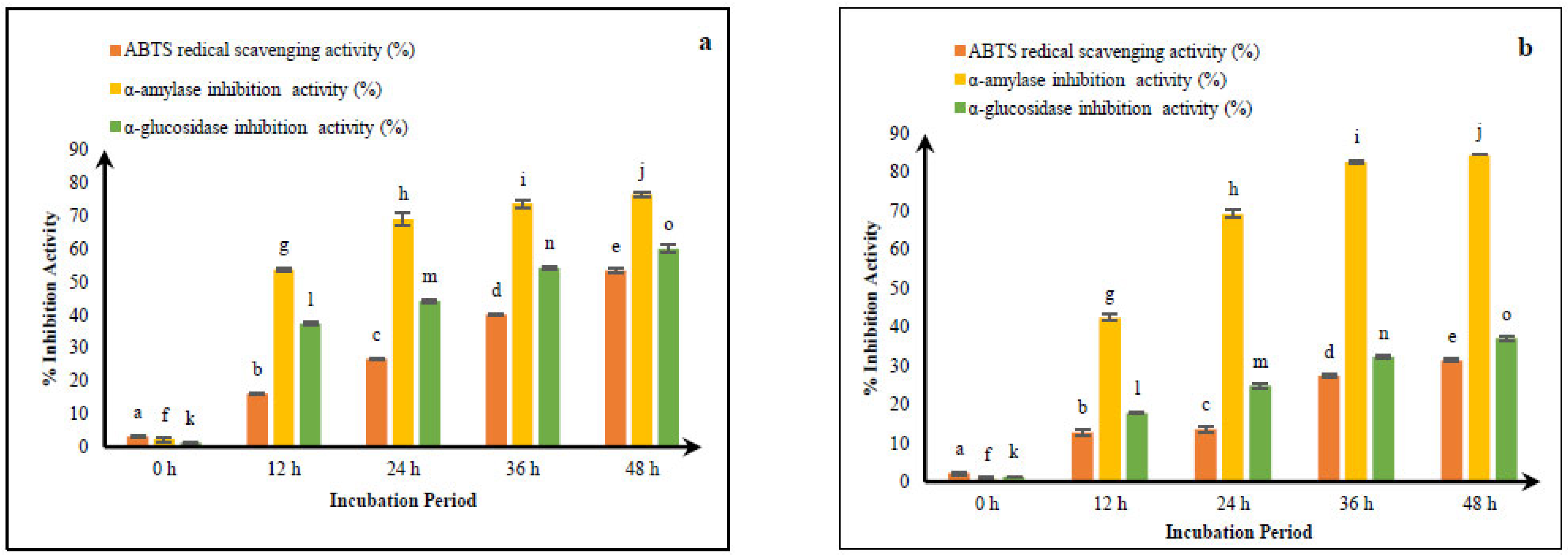
3.1. Comparative ABTS Radical Scavenging of Fermented CM and GCM

Figure 1a,b highlights that M9 strains could neutralize ABTS radicals in both cultured CM and GCM when integrated with WBS2A. There was an apparent pattern revealing that when the fermentation time increased, the activity of removing radicals significantly increased (p < 0.05). Fermented CM and GCM illustrated various degrees of ABTS radical scavenging activity above a 48 h period at 30 °C (3.32 to 53.47% and 2.15 to 31.42%, respectively). The 48 h interval yielded the most intense percentages of ABTS radical scavenging effects, with fermented CM reaching 53.47% and GCM reaching 31.42%.
The data presented in Figure 1a,b indicate that the combination of the M9 and WBS2A strains effectively enhances the antioxidant activity in both CM and GCM through ABTS radical scavenging. A statistically significant increase (p < 0.05) in scavenging activity was observed with longer fermentation times, peaking at 48 h. Fermented CM consistently demonstrated greater antioxidant potential than GCM did, with the maximum ABTS scavenging activity reaching 53.47% in CM versus 31.42% in GCM. These findings suggest that extended fermentation, particularly in CM, enhances the generation of antioxidant peptides or compounds.
According to [24], the ability of CM to scavenge free radicals significantly increased (p < 0.05) with increasing fermentation duration. According to their results, CM fermented with the KGL4 strain presented the maximum activity (64.03%) following the ABTS assay. This finding is consistent with our findings, particularly implying that fermentation improves antioxidant capacity. Moreover, an array of studies suggest that increasing the incubation time significantly improves the antioxidant effect. As reported by [25], Lactobacillus lactis spp. cremoris microbial fermentation greatly increased the antioxidant capacity of cow milk. The antioxidant profile of cow milk increased considerably after 10 h of fermentation, ranging from an initial average of 1.57 ± 0.001 to 18.46 ± 0.013%. Reference [26] developed a follow-on formula using fermented whey inoculated with Lactobacillus helveticus (CNRZ32) and evaluated its antioxidant activity using the DPPH radical scavenging assay. Fermentation was carried out for 2, 4, 6, and 12 h. The highest radical scavenging activity was observed in camel milk whey after 12 h of fermentation, reaching 1.01 μmol Trolox equivalents mL−1. However, no significant difference (p > 0.05) was found between the antioxidant activity at 6 and 12 h of fermentation.

3.2. Antidiabetic Activity of Fermented CM and GCM

The antidiabetic effects of cultured CM and GCM are presented in Figure 1a,b. Our findings revealed that the duration of incubation significantly (p < 0.05) influenced the antidiabetic activity. The antidiabetic activity increased from 2.42 to 76.54% in CM and from 0.76 to 84.41% in GCM. After 48 h of fermentation at 30 °C, cultured CM displayed significant antidiabetic potential, particularly in its capacity to block α-amylase (76.54%) and α-glucosidase (60.06%). Similarly, following the same fermentation period, fermented GCM exhibited antidiabetic benefits, such as α-amylase inhibition (84.41%) and α-glucosidase inhibition (37.10%). Our findings revealed that the antidiabetic qualities increased with increasing incubation time.
Fermentation time significantly (p < 0.05) enhanced the antidiabetic properties of both cultured CM and GCM. The inhibition of α-amylase and α-glucosidase increased progressively with prolonged incubation, indicating time-dependent bioactivity. After 48 h of fermentation at 30 °C, CM strongly inhibited α-amylase (76.54%) and α-glucosidase (60.06%), whereas GCM exhibited even greater α-amylase inhibition (84.41%) but comparatively lower α-glucosidase inhibition (37.10%). These results suggest that bioactive compounds generated during fermentation contribute significantly to the antidiabetic potential, with GCMs excelling in α-amylase inhibition and CMs showing more balanced dual-enzyme inhibition.
As shown by [20], after 48 h, CM fermented with Lacticaseibacillus paracasei (M11) along with WBS2A had an α-amylase inhibition value of 81.66%. The maximal inhibitory capacity of α-glucosidase, which reached 64.55%, was likewise recorded after 48 h of fermentation at 37 °C with the same strains. These outcomes are consistent with our investigation, which demonstrated that fermentation strengthens antidiabetic effects. The inhibition properties also improved with prolonged incubation periods. Fermented camel milk prepared by [27] using a combination of Lactobacillus and yeast strains (KGL4 and WBS2A) to evaluate its anti-diabetic properties. The fermentation process was conducted at 37 °C, and samples were analyzed at time intervals of 12, 24, 36, and 48 h. The highest anti-diabetic activity was observed after 48 h of incubation at 37 °C. The fermented camel milk exhibited maximum inhibitory activities against lipase (73.85 ± 1.19%), α-glucosidase (85.37 ± 2.15%), and α-amylase (70.86 ± 1.02%). In comparison, fermented buffalo milk showed lipase inhibitory activity of 75.25 ± 1.72%, α-glucosidase inhibitory activity of 61.79 ± 2.14%, and α-amylase inhibitory activities of 80.09 ± 0.51% and 67.29 ± 1.75%, indicating superior α-glucosidase inhibition in camel milk.
Camel milk yogurt formulated by [28] supplemented with varying concentrations of carao (Cassia grandis) pulp (0, 1.3, 2.65, and 5.3 g/L) to assess its potential to enhance antioxidant activity. Antioxidant properties were evaluated using both DPPH and ABTS radical scavenging assays. The control sample (0 g/L carao) exhibited the lowest DPPH radical scavenging activity (22.6%), while the yogurt containing 5.3 g/L of carao demonstrated the highest activity (39.08%). This increase in antioxidant capacity is attributed to the bioactive compounds present in carao, including alkaloids, flavonoids, coumarins, phenolic compounds, tannins, free amino acids, amines, saponins, resins, triterpenes, and reducing sugars. The trend observed in the ABTS assay mirrored the results obtained from the DPPH assay, confirming the antioxidant-enhancing effect of carao supplementation in camel milk yogurt.

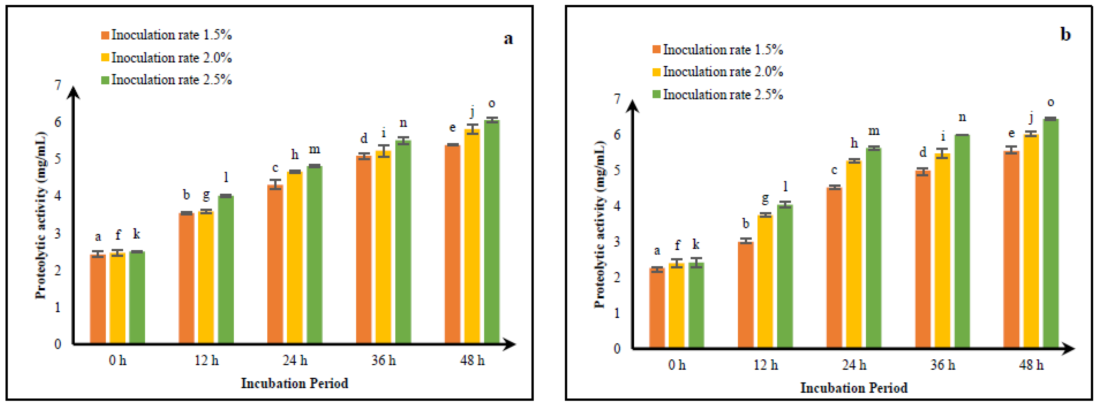
3.3. Optimizing Fermentation Processes of CM and GCM for Improving Peptide Yield

The proteolytic properties of CMs and GCMs cultured with WBS2A along with M9 are presented in Figure 2a and Figure 2b separately. The range of proteolytic activity of cultured CM ranged between 2.43 mg/mL (at 1.5% culture and 0 h) and 6.07 mg/mL (2.5% culture and 48 h). The proteolytic activity of fermented GCM varied from 2.25 mg/mL at 1.5% culture and 0 h to 6.44 mg/mL at 2.5% culture and 48 h. Significant increases in the culture concentration (1.5, 2.0 and 2.5%) as well as the fermentation interval (0, 12, 24, 36 and 48 h) led to changes in the proteolytic activity of both CM and GCM. After 48 h of fermentation with a 2.5% inoculum concentration, the maximum levels of proteolysis were calculated at 6.07 mg/mL for fermented CM and 6.44 mg/mL for fermented GCM, relative to the lower inoculation amounts of 1.5 and 2.0%, respectively.
The proteolytic rate of CM cultured with M11 + WBS2A increased from 7.44 mg/mL (1.5% culture at 12 h) to 9.09 mg/mL (2.5% culture at 48 h), according to a previous study [20]. Proteolytic activity increased considerably (p < 0.05) with increasing fermentation time (12, 24, 36 and 48 h) and culture concentration (1.5, 2 and 2.5%). Following 48 h of fermentation, 2.5% of the cultures yielded the maximum proteolytic activity assessed (9.09 mg/mL) compared with lower inoculum concentrations of 1.5 and 2.0%.
The proteolytic activity of the M9 + WBS2A strains in both camel milk and Gir cow milk progressively increased with increasing incubation time (0, 12, 24, 36 and 48 h) and increasing inoculation level (1.5%, 2.0% and 2.5%). The peak proteolytic activity was recorded after 48 h of incubation with 2.5% inoculum. Therefore, these conditions (48 h of incubation and a 2.5% inoculation rate) were selected for subsequent experiments because of their effectiveness in maximizing proteolytic activity in both milk types.

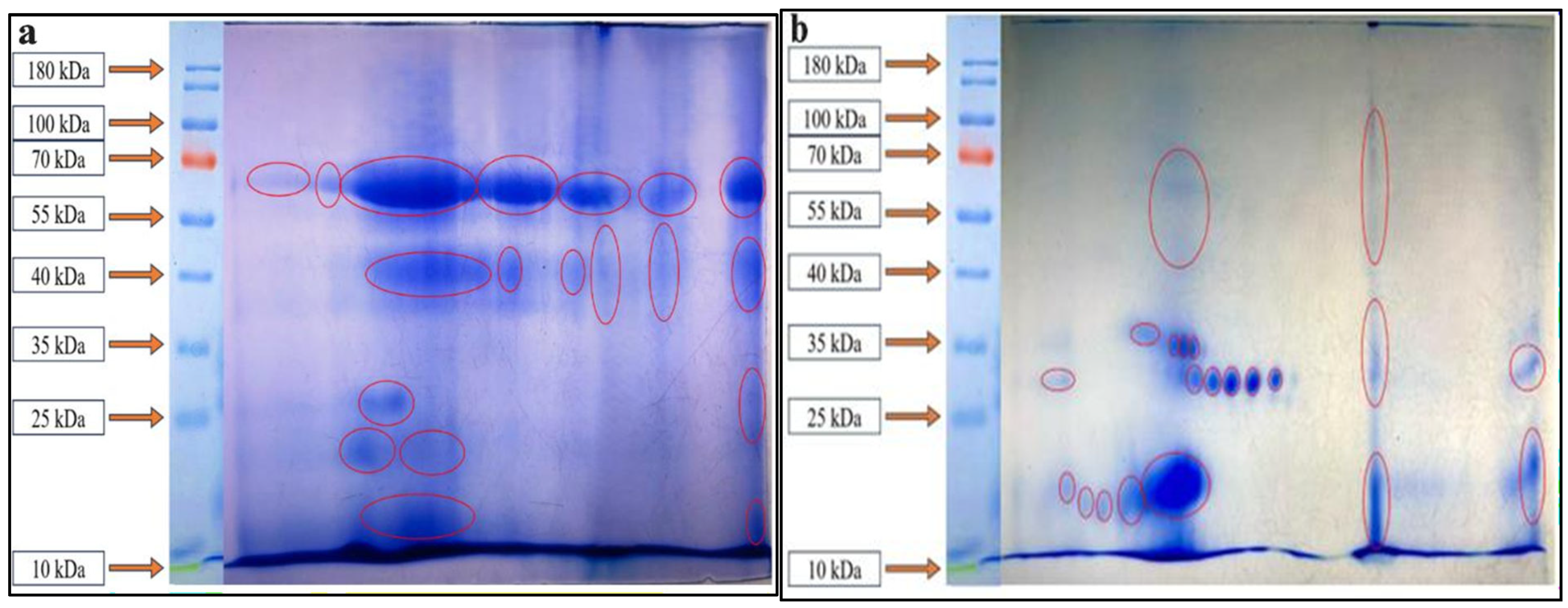
3.4. SDS–PAGE Profiling of Proteins from Fermented CM and GCM

WSEs of fermented CM and GCM were subjected to SDS–PAGE examination via a molecular weight protein ladder with a range of 10–180 kDa (Figure 3a,b). Compared with the unfermented samples, the cultured CM and GCM samples presented more pronounced protein bands. This observation implies that a considerable amount of proteolytic activity exists in bacteria, along with yeast, which ferment CMs and GCMs. Protein bands have been reported to range in size from 10 to 75 kDa in unfermented CM and from 15 to 100 kDa in unfermented GCM. However, all the samples presented peptide bands in the 10–75 kDa range after fermentation. Remarkably, as shown in Figure 3a,b, no peptide bands were observed among the permeate samples.
In [20], WSEs from buffalo milk (BM) and CM that had been cultured utilizing the M11 + WBS2A strains were analyzed, and their effects were examined via SDS–PAGE. Both cultured BM and CM revealed many peptide bands, indicating that these strains had effective proteolytic capabilities during fermentation. While the unfermented BM and CM samples presented bands between 10 and 100 kDa, the fermented samples presented bands between approximately 10 and 75 kDa. However, there were no distinct protein bands among the permeate samples.
An investigation by [29] involved the M11, KGL3A and KGL4 strains to examine WSEs from cultured CM. Abundant peptide bands were evident in the cultured CM via SDS–PAGE, revealing that Lactobacillus had a substantial amount of proteolytic activity in the milk. The unfermented CM had protein bands ranging from 10 to 130 kDa, whereas the fermented milk had bands ranging from 10 to 100 kDa. Interestingly, the permeate from the KGL4 sample had several visible bands; however, no peptide bands were observed among the permeate from the KGL3A or M11 samples.

3.5. Characterization of Fermented CM and GCM via 2D Gel Electrophoresis

Thirty-eight peptide spots appeared in the cultured CM, and thirty-seven spots in the GCM were detected via 2D gel electrophoresis. As illustrated in Figure 4a,b, the masses of the peptides that were identified spanned from 10 to 70 kDa.
Fermented CM and BM were analyzed by [20] through 2D-PAGE, followed by trypsin digestion and RP-LC/MS. On the 2D gel from the fermented milk samples, the evaluation revealed 26 spots in BM (M11 + WBS2A), 15 in BM (KGL4 + WBS2A), 25 in CM (M11 + WBS2A) and 20 in CM (KGL4 + WBS2A). The antihypertensive segments LGP, GPV and YQEPVL from BM cultured with M11 + WBS2A were aligned with the peptide sequence DMPIQAFLLYQEPVLGPVR. Fragments VAAA, LLAP and LPLLR complemented the sequence LLILTCLVALARPK for antidiabetic action. The antihypertensive segments LAHKAL, YANPAVVRP and FFVAP aligned with the CM peptide FFIFTCLLAVVLAK, whereas the antidiabetic fragments MP, VPYPQ and LPQNIPP were associated with TDVMPQWW.

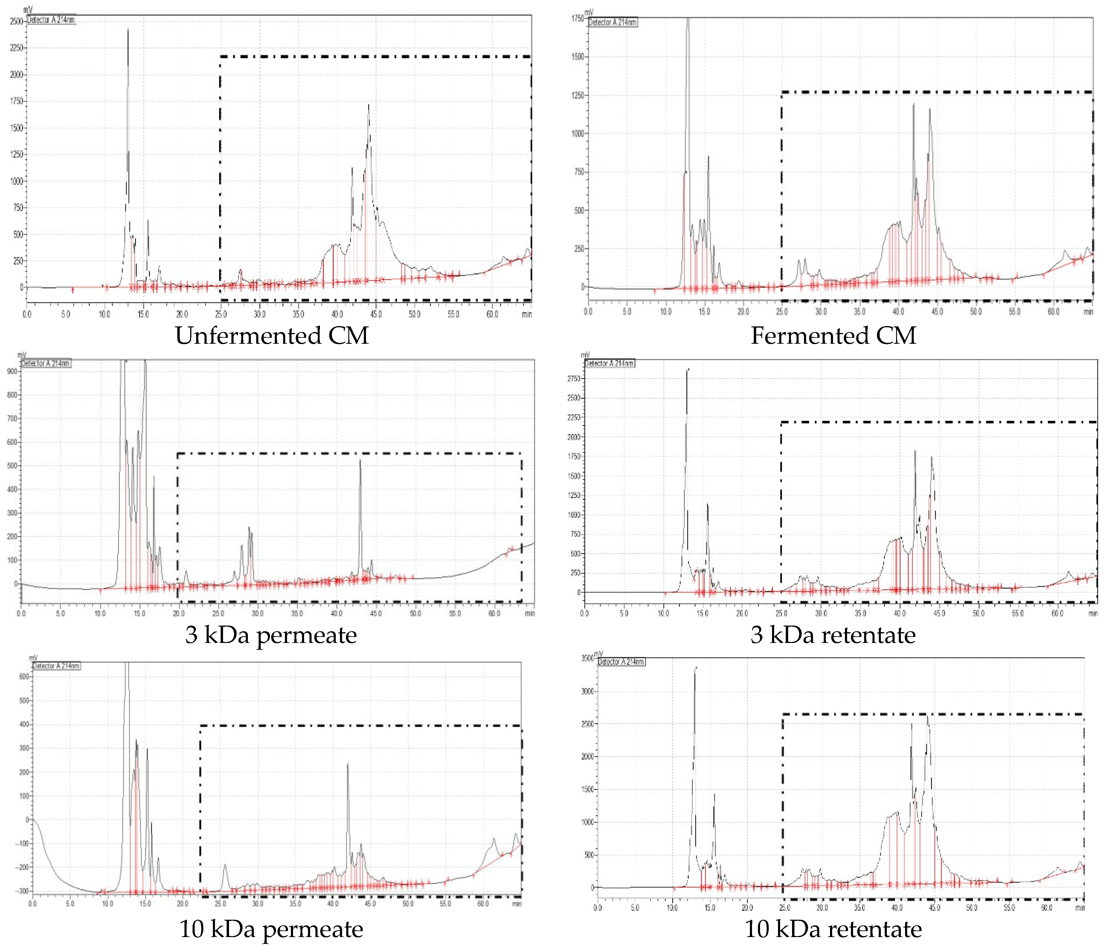
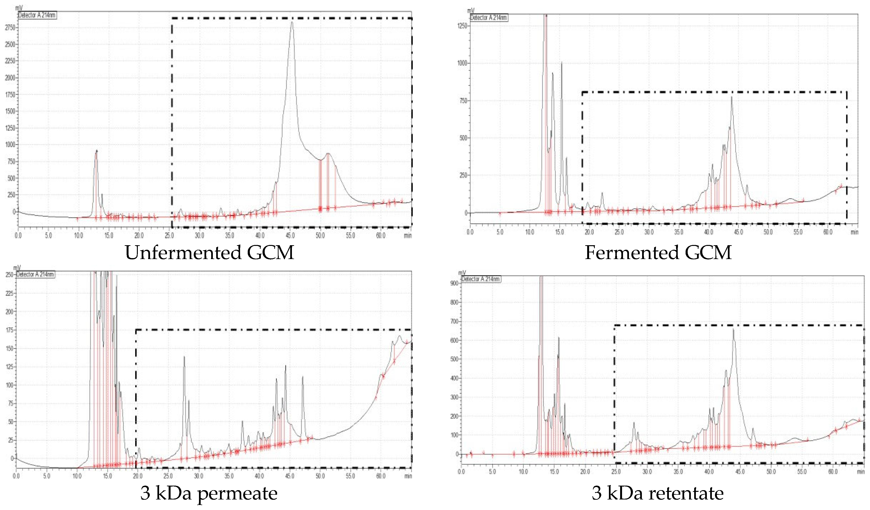
3.6. Evaluation of the Antioxidant and Antidiabetic Activities of UF Fractions of Fermented CM and GCM and RP-HPLC Analysis

The UF segments extracted from fermented CM and GCM demonstrated ABTS radical scavenging activity together with antidiabetic characteristics, as shown in Figure 5 and Figure 6. Figure 5 and Figure 6 show the RP-HPLC chromatograms that correspond to the UF portions of fermented CM and GCM, respectively. Likewise, fermented as well as unfermented samples of CM and GCM are represented in these chromatograms. The outcomes unequivocally indicate that fermented milk produces peptides at far greater rates than unfermented milk does. The cultured milk samples contained an array of amino acid fragments, with substantial peaks emerging within 10–20 min and 25–45 min, as summarized in Table 1. Alternatively, the unfermented samples mostly possess unbroken proteins. The 10 kDa retentate from both CM and GCM presented significant ABTS radical scavenging ability. On the other hand, the 3 kDa permeate of both CM and GCM had greater antidiabetic effects. Interestingly, fermented GCM had lower levels of ABTS radical scavenging activity (47.62%) than fermented CM did (63.56%) (Figure 7). Furthermore, the 3 kDa permeate of CM demonstrated greater inhibition of α-amylase (76.06%) and α-glucosidase (63.90%) than did the fermented GCM, which inhibited α-amylase and α-glucosidase by 75.46% and 40.51%, respectively. The bioactive peptide fractions derived from fermented camel milk (CM) and Gir cow milk (GCM) presented distinct functional properties. The >10 kDa retentate fractions presented greater antioxidant activity, whereas the <3 kDa permeate fractions presented superior antidiabetic effects. Notably, compared with fermented CM, fermented CM displayed greater ABTS radical scavenging activity and stronger inhibition of both α-amylase and α-glucosidase, indicating that fermented CM has greater potential as a functional ingredient for managing oxidative stress and postprandial hyperglycemia.
In accordance with [23], CM cultured with the KGL4 strain had a significant activity level (83.66%) according to the ABTS assay for determining the antioxidant capacity of 3 kDa retentates. The 10 kDa retentates were the next most common (83.23%), but the 10 kDa permeates had lower activity (74.71%). The 3 kDa permeate presented the lowest antioxidant capacity (60.72%). Reference [30] reported that the levels of α-amylase and α-glucosidase inhibition resulting from the various strains employed for CM fermentation varied across samples. The strain with the greatest level of inhibition was KGL4. Compared with the other samples, KGL4 had the most significant inhibition rates for α-amylase and α-glucosidase (85.42% and 78.69%, respectively) in the 3 kDa permeate samples. Our results indicating that peptides with smaller sizes have superior antidiabetic effects have been reinforced by this investigation. As reported by [20], the 3 kDa permeate of CM cultured with M11 + WBS2A significantly inhibited α-amylase activity, resulting in an inhibition level of 81.18%. Furthermore, the 3 kDa extract from the same sample had the highest level of α-glucosidase inhibition, with a rate of 63.33%.

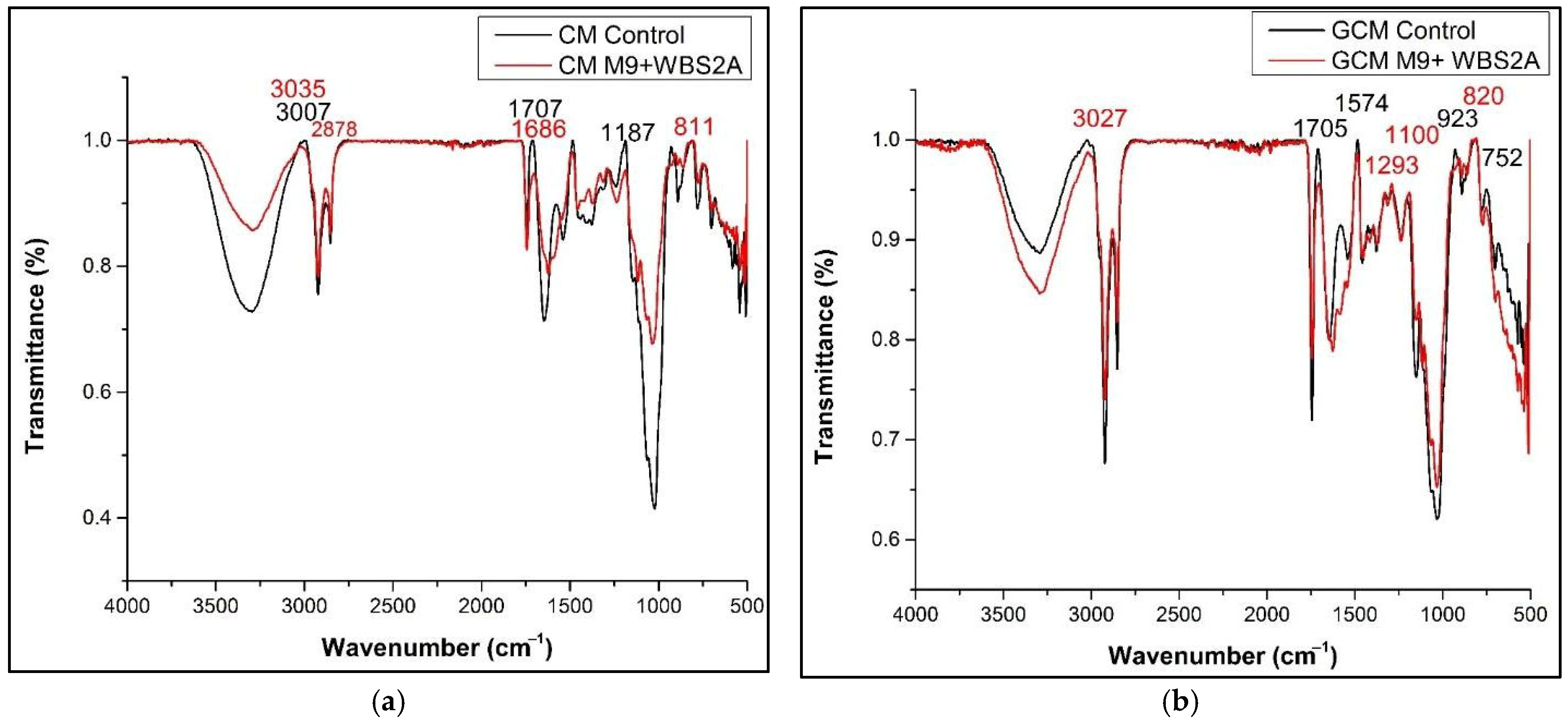
3.7. FTIR Analysis of Fermented CM and GCM

CM and GCM spectral analyses detected several peaks within the 500–4000 cm−1 area. The spectra revealed more pronounced peaks after fermentation with CM and GCM, notably in the 1700 and 500 cm−1 regions. More specifically, the cultured CM FTIR spectra (Figure 8b) displayed discrete peaks at 811, 850, 1697, 1568 and 3035 cm−1. On the other hand, the fermented GCM spectra (Figure 8a) presented prominent peaks at 820, 1100, 1293 and 1492 cm−1.
According to [20], there are multiple peak positions in the 400–2000 cm−1 region in the FTIR spectrum of CM. Interestingly, the cultured CM samples presented more significant peaks between 1800 and 400 cm−1. In particular, the fermented CM presented peaks at 516.75, 822.25, 1036.88, 1120.08 and 1628.81 cm−1. Assessing protein secondary structures requires a grasp of the vibratory stretching of carbonyl (C=O) groups in the bonds of peptides, mostly in the amide-I band (1700–1600 cm−1). In addition to integrating C–N stretching and N–H bending, the amide-II bands (1600–1500 cm−1) yield significant details on hydrogen bonding along with the arrangement of proteins. These amide bands are vital for peptide analysis because they provide valuable insights into the changes in structure that occur during fermentation. The clearest peak in the FTIR spectrum of fermented milk was found at 1640 cm−1 in the amide I region, whereas 10% of the signal was attributed to N–H bending, 10% to C–N stretching, and 80% of the signal was attributable to C=O stretching.

3.8. Comprehensive Purification Peptide Analysis Through RP–LC–MS

To implement PeakView software (https://www.peakviewsoftware.com/, accessed on 20 May 2025) for assessing the spectra, RP–LC–MS analysis was performed to identify the trypsin-digested peptides from the 2D PAGE spots. Antioxidative peptides, along with antidiabetic peptides, were discovered through the BIOPEP database, and the results of mass spectrometry were compared with those of the CM and GCM libraries. The outcomes are outlined in Table 2 and Table 3, whereby amino acid sequences have been categorized according to peptide Ranker scores and evaluated for toxicity. To match the mass spectrometry data with the Swiss-Prot database and corroborate the outputs, BIOPEP was used for peptide identification.
Multiple peptide sequences had scores above 0.45 throughout our investigation. For example, the amino acid PSGFQLFGSPAGQKDLLFK, with a molecular weight of 2037.63 and an isoelectric point (pI) of 8.94, had a ranking score of 0.80. Derived from CM fermentation, this peptide was verified to have antioxidant action and strong compatibility with other peptides, including KD [31], as mentioned in the BIOPEP database (Table 4).
The fermented GCM yielded the peptide sequence ELRAMKVLILACLVALALAR, with a molecular weight of 2168.09, a pI of 9.55, and a peptide ranking score of 0.73. The BIOPEP database’s sequences EL [39], LAC and CLV [40] were determined to coincide with this peptide. Table 5 provides further validation of its antioxidant properties.
The peptide sequence, PSGFQLFGSPAGQKDLLFK (peptide ranking score: 0.80; pI: 8.94; molecular weight: 2037.63), obtained from fermented CM was compared with peptides found in the BIOPEP database, such as GF, AG and FQ [50]. On the basis of this comparison, Table 6 details the peptide’s antidiabetic effects.
The sequences reported in the BIOPEP database, such as EY, FN, MK, MM, ND, NM and NR [50], have been associated with the peptide “NDEYCFNMMKNR” (peptide ranking score: 0.76; pI: 8.53; molecular weight: 1564.93), which was found in fermented GCMs. Table 7 lists the antidiabetic action of this peptide that has been validated.

3.9. Effects of Fermented CM and GCM on the Viability of RAW 264.7 Cells

The MTT protocol has been employed to determine the cytotoxic effects of fermented CM and GCM on RAW 264.7 cells via various quantities of M9 + WBS2A (0.25, 0.5, 1, 2, 4 and 8 mg/mL). The cell viability decreased as the concentration increased from 0.25 to 8 mg/mL. At dosages of 0.25, 0.5, 1 and 2 mg/mL, the samples presented approximately 100% cell viability, indicating a lack of cytotoxicity. However, an increase in cytotoxicity occurred between dosages of 4 and 8 mg/mL, leading to decreased cell viability. Neither CM nor GCM demonstrated cytotoxicity at a concentration of 2 mg/mL (Figure 9a,b). As a result, 2 mg/mL was selected for further inquiry.

3.10. Effects of Fermented CM and GCM on LPS-Induced NO Production in RAW 264.7 Cells

The ingestion of LPS by macrophages provoked an enormous spike in NO levels; nevertheless, when fermented CM and GCM were introduced at 2 mg/mL, that spike was hindered (Figure 9a,b). This implies that M9 + WBS2A could possess anti-inflammatory qualities, as demonstrated by a substantial decrease in NO levels provoked by LPS, in lieu of the cytotoxic effects of the fermented CM and GCM. Additionally, the strains strongly promoted the production of proinflammatory cytokines.

3.11. Examination of Cytokine Levels in Raw264.7 Cells

Compared with the effects of LPS treatment alone, Figure 9a,b reveal that cultured CM and GCM diminished nitrite development in RAW 264.7 cells. The nitrite levels in both treatment groups were low and comparable to those in the control group, confirming that there were no harmful effects on the cells. Furthermore, cultured CM and GCM drastically decreased the increase in TNF-α, IL-6 and IL-1β levels caused by LPS, which was comparable to the control proportions (Figure 9a,b).
Investigated by [28] the anti-inflammatory effects of camel milk yogurt supplemented with varying concentrations of carao (Cassia grandis) pulp (0, 1.3, 2.65, and 5.3 g/L). The results demonstrated that supplementation with 2.65 and 5.3 g/L significantly (ρ < 0.05) reduced the inflammatory marker IL-8, as well as the mRNA expression levels of pro-inflammatory cytokines IL-1β and TNF-α, compared to the control yogurt. Sensory evaluation indicated that the addition of carao at 1.3 and 2.65 g/L did not negatively affect the organoleptic properties of the yogurt. These findings suggest that carao pulp may serve as a functional ingredient for enhancing the nutritional and anti-inflammatory properties of camel milk yogurt.

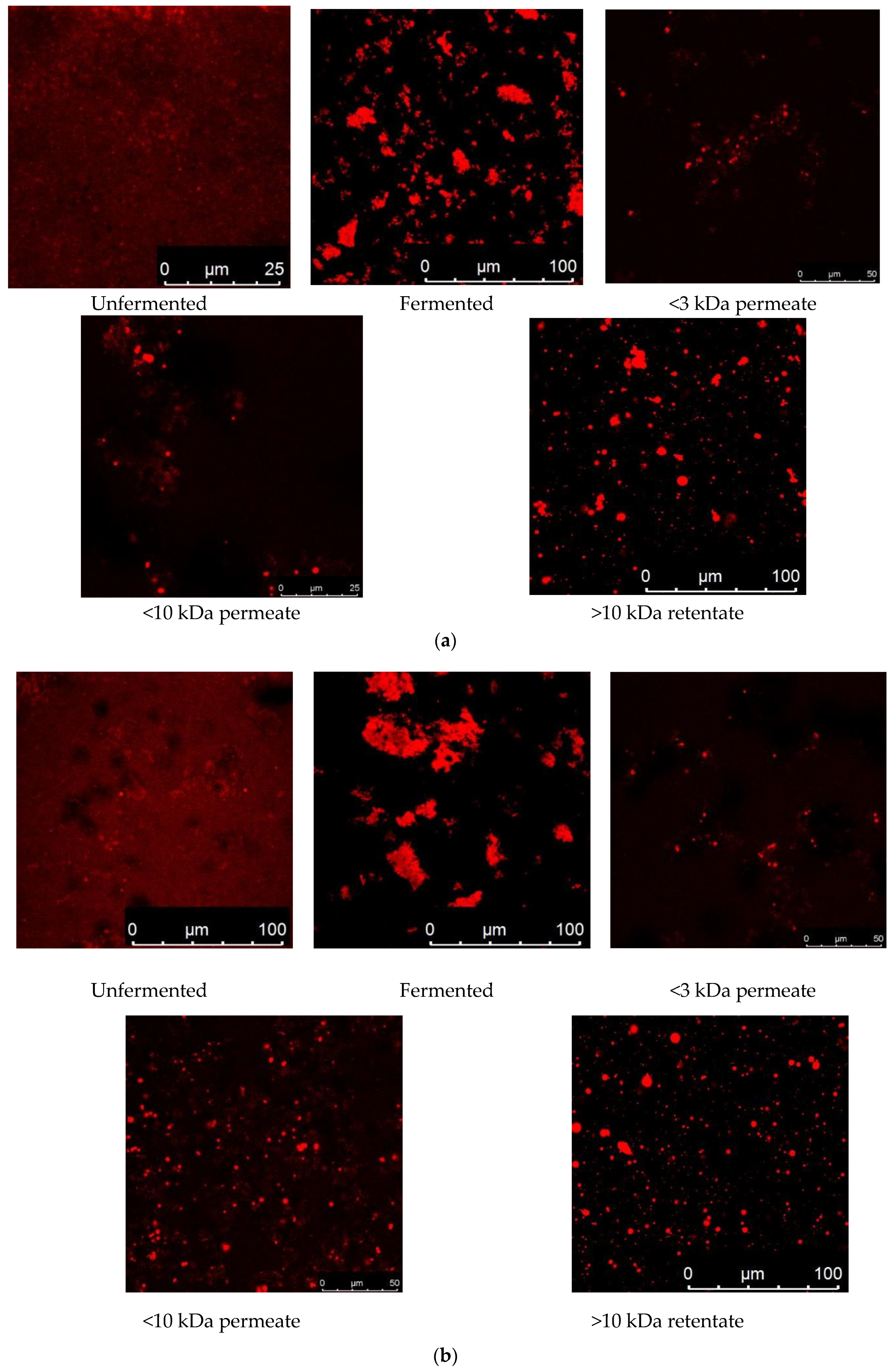
3.12. Visualization of Protein Biomarkers of Fermented CMs and GCMs via CLSM

CLSM was used to investigate the microorganization of the fermented and unfermented CM and GCM (Figure 10a,b). The study emphasized peptide infrastructure and fundamental composition, particularly peptide binding. Proteins were visualized by the use of fluorescein dye, which gave them a red hue. Larger fragments that mimicked native proteins were observed in unfermented samples. Regardless of the proteolytic exertion of the culture, fermentation of the samples resulted in the breakdown of proteins into peptides. GCM and CM fermentation can result in the formation of larger, more intricate protein clusters. Proteins can be shifted into substitute molecules via fermentation. Inspection of fermented milk permeate and retentate treated with 3 kDa and 10 kDa UF membranes revealed that the smaller membrane effectively distinguished smaller pieces of protein because the 3 kDa permeate possessed fewer protein molecules than the 10 kDa permeate and retentate did (Figure 10a,b).
Ref. [21] assessed the influence of fermentation on sheep milk via CLSM. They noted that whereas larger, natural protein complexes appear in untreated milk, tiny pieces of protein emerge after fermentation, which turns proteins into peptides. The M11 culture was recognized for its ability to transform peptides during fermentation, resulting in fermented milk containing increasingly complicated protein structures. Moreover, the 3 kDa permeate had tiny peptides as opposed to the 10 kDa permeate after membrane filtering was carried out. Ref. [58] explored the internal structure of yogurt made from cow, goat and sheep milk via cryo-SEM and CLSM. They concluded that sheep yogurt seemed remarkably uniform and sleek, that goat yogurt had the least dense protein networks and that cow yogurt gels were intermediate. Ref. [59] tested the impacts of variations in β-casein A2 on milk traits. By employing CLSM, they concluded that β-CN A2 A2 milk had thinner protein strands and a more porous matrix, which correlated with poorer gel strength than β-CN A1 A1 milk did.

4. Conclusions

Inoculation of CM and GCM with Lacticaseibacillus rhamnosus (M9) and Saccharomyces cerevisiae (WBS2A) resulted in enhanced ABTS radical scavenging and antidiabetic benefits. The fermented CM had the highest ABTS radical scavenging activity, at 53.47%, while the fermented GCM had the next highest activity, at 31.42%. With respect to their antidiabetic effects, fermented CM and GCM had α-amylase inhibition rates of 76.54% and 84.41%, respectively, and α-glucosidase inhibition rates of 60.06% and 37.10%, respectively. The proteolytic activity was highest in fermented CM and GCM, with values of 6.07 and 6.44 mg/mL, respectively. SDS–PAGE analysis revealed protein bands in both unfermented and fermented samples, with specific bands ranging from 10 to 75 kDa in all fermented milk. At concentrations of 0.25 to 2 mg/mL, fermented CM and GCM showed no cytotoxicity and reduced the levels of proinflammatory cytokines (TNF-α, IL-6 and IL-1β) in RAW 264.7 cells. The microstructure of fermented versus unfermented milk was studied via CLSM in the present study. Fluorescein was used to generate distinct protein structures. Fermented milk contains tiny peptides as well as complicated peptide structures with large clumps, whereas untreated milk contains larger protein molecules. This was caused by the proteolytic activity of strain M9 + WBS2A. The results showed that the 10 kDa retentate as well as the permeate had larger particles, whereas the 3 kDa permeates held tiny peptides. FTIR spectral analysis indicated that fermentation altered the spectral profiles of CM and GCM, highlighting unique peak patterns for each type of milk, which may help in distinguishing between them on the basis of their molecular structure.

Author Contributions

Conceptualization, B.B. (Brijesh Bhuva), A.R., M.B., K.K.K. and S.H.; methodology, B.B. (Brijesh Bhuva), A.A.S., A.R., K.K.K. and S.H.; formal analysis, B.B. (Brijesh Bhuva), A.A.S., P.M.M., A.R., P.S. and S.H.; investigation, A.A.S., A.R., M.B., K.K.K., P.S. and S.H.; writing—original draft, B.B. (Brijesh Bhuva), A.R., M.B., K.K.K., P.S. and S.H.; editing and revising the manuscript, B.B. (Bethsheba Basaiawmoit); supervision, K.K.K. and S.H.; resources, A.P.; visualization, P.M.M. and A.P.; project administration, S.H.; funding acquisition, S.H. All authors have read and agreed to the published version of the manuscript.

Funding

This research received no external funding.

Institutional Review Board Statement

Not applicable.

Informed Consent Statement

Not applicable.

Data Availability Statement

The original contributions presented in this study are included in the article. Further inquiries can be directed to the corresponding author.

Acknowledgments

The authors are grateful to the Dairy Microbiology Department, SMC College of Dairy Science, Kamdhenu University, Anand, Gujarat and the College of Veterinary Science, Kamdhenu University, Anand, Gujarat, for providing support and conducting the entire study.

Conflicts of Interest

The authors declare no conflicts of interest.

References

  1. Guyomarc’h, F.; Héquet, F.; Le Féon, S.; Leconte, N.; Garnier-Lambrouin, F.; Auberger, J.; Malnoë, C.; Pénicaud, C.; Gésan-Guiziou, G. Life cycle assessment to quantify the environmental performance of multiproducts food processing systems such as milk fractionation: Importance of subdivision and allocation. J. Food Eng. 2024, 380, 112147. [Google Scholar]
  2. Seifu, E. Recent advances on camel milk: Nutritional and health benefits and processing implications-A review. AIMS Agric. Food 2022, 7, 777–804. [Google Scholar] [CrossRef]
  3. Vincenzetti, S.; Cammertoni, N.; Rapaccetti, R.; Santini, G.; Klimanova, Y.; Zhang, J.J.; Polidori, P. Nutraceutical and functional properties of camelids’ milk. Beverages 2022, 8, 12. [Google Scholar] [CrossRef]
  4. Vitthalrao, B.K. Gir Cow Milk: The Wealth for Human Health. Acta Sci. Vet. Sci. 2021, 3, 9–12. [Google Scholar]
  5. Gaba, K.; Anand, S. Incorporation of probiotics and other functional ingredients in dairy fat-rich products: Benefits, challenges and opportunities. Dairy 2023, 4, 630–649. [Google Scholar] [CrossRef]
  6. Riane, K.; Sifour, M.; Ouled-Haddar, H.; Idoui, T.; Bounar, S.; Boussebt, S. Probiotic properties and antioxidant efficiency of Lactobacillus plantarum 15 isolated from milk. J. Microbiol. Biotechnol. Food Sci. 2021, 9, 516–520. [Google Scholar] [CrossRef]
  7. Singh, N.; Gaur, S. New insights into multifunctional aspects of milk-derived bioactive peptides: A review. Food Chem. Adv. 2024, 4, 100628. [Google Scholar] [CrossRef]
  8. Chandrasekaran, P.; Weiskirchen, R. The role of obesity in type 2 diabetes mellitus—An overview. Int. J. Mol. Sci. 2024, 25, 1882. [Google Scholar] [CrossRef]
  9. Rais, N.; Ved, A.; Ahmad, R.; Parveen, A. Oxidative Stress and Diabetes Mellitus: Unraveling the Intricate Connection: A Comprehensive Review. J. Pharm. Res. Int. 2024, 36, 13–30. [Google Scholar] [CrossRef]
  10. Wang, C.; Luo, D.; Zheng, L.; Zhao, M. Anti-diabetic mechanism and potential bioactive peptides of casein hydrolysates in STZ/HFD-induced diabetic rats. J. Sci. Food Agric. 2024, 104, 2947–2958. [Google Scholar] [CrossRef]
  11. Zhu, Y.; Wang, K.; Jia, X.; Fu, C.; Yu, H.; Wang, Y. Antioxidant peptides, the guardian of life from oxidative stress. Med. Res. Rev. 2024, 44, 275–364. [Google Scholar] [CrossRef] [PubMed]
  12. Sah, B.N.P.; Vasiljevic, T.; McKechnie, S.; Donkor, O.N. Effect of probiotics on antioxidant and antimutagenic activities of crude peptide extract from yogurt. Food Chem. 2014, 156, 264–270. [Google Scholar] [CrossRef] [PubMed]
  13. Telagari, M.; Hullatti, K. In vitro α-amylase and α-glucosidase inhibitory activity of Adiantum caudatum Linn. and Celosia argentea Linn. extracts and fractions. Indian J. Pharmacol. 2015, 47, 425. [Google Scholar]
  14. Shai, L.J.; Magano, S.R.; Lebelo, S.L.; Mogale, A.M. Inhibitory effects of five medicinal plants on rat alpha-glucosidase: Comparison with their effects on yeast alpha-glucosidase. J. Med. Plants Res. 2011, 5, 2863–2867. [Google Scholar]
  15. Hati, S.; Sreeja, V.; Solanki, D.; Prajapati, J.B. Influence of proteolytic Lactobacilli on ACE inhibitory activity and release of BAPs. Indian J. Dairy Sci. 2015, 68, 1–8. [Google Scholar]
  16. Gibbs, B.F.; Zougman, A.; Masse, R.; Mulligan, C. Production and characterization of bioactive peptides from soy hydrolysate and soy fermented food. Food Res. Int. 2004, 37, 123–131. [Google Scholar] [CrossRef]
  17. Carrasco-Castilla, J.; Hernandez, A.; Alvarez, A.J.; Jimenez-Martinez, C.; Jacinto-Hernandez, C.; Alaiz, M.; Girón-Calle, J.; Vioque, J.; Dávila-Ortiz, G. Antioxidant and metal chelating activities of Phaseolus vulgaris L. var. Jamapa protein isolates, phaseolin and lectin hydrolysates. Food Chem. 2012, 131, 1157–1164. [Google Scholar] [CrossRef]
  18. Yang, Y.; Zheng, N.; Yang, J.; Bu, D.; Wang, J.; Ma, L.; Sun, P. Animal species milk identification by comparison of two-dimensional gel map profile and mass spectrometry approach. Int. Dairy J. 2014, 35, 15–20. [Google Scholar] [CrossRef]
  19. Panchal, G.; Hati, S.; Sakure, A. Characterization and production of novel antioxidative peptides derived from fermented goat milk by L. fermentum. Food Sci. Technol. 2020, 119, 108887. [Google Scholar] [CrossRef]
  20. Khakhariya, R.; Sakure, A.A.; Maurya, R.; Bishnoi, M.; Kondepudi, K.K.; Padhi, S.; Rai, A.K.; Liu, Z.; Patil, G.B.; Mankad, M.; et al. A comparative study of fermented buffalo and camel milk with anti-inflammatory, ACE-inhibitory and anti-diabetic properties and release of bioactive peptides with molecular interactions: In vitro, in silico, and molecular study. Food Biosci. 2023, 52, 102373. [Google Scholar] [CrossRef]
  21. Pipaliya, R.; Basaiawmoit, B.; Sakure, A.A.; Maurya, R.; Bishnoi, M.; Kondepudi, K.K.; Singh, B.P.; Paul, S.; Liu, Z.; Sarkar, P.; et al. Peptidomics-based identification of antihypertensive and antidiabetic peptides from sheep milk fermented using Limosilactobacillus fermentum KGL4 MTCC 25515 with anti-inflammatory activity: In silico, in vitro, and molecular docking studies. Front. Chem. 2024, 12, 1389846. [Google Scholar] [CrossRef] [PubMed]
  22. Khare, P.; Maurya, R.; Bhatia, R.; Mangal, P.; Singh, J.; Podili, K.; Bishnoi, M.; Kondepudi, K.K. Polyphenol-rich extracts of finger millet and kodo millet ameliorate high-fat diet-induced metabolic alterations. Food Funct. 2020, 11, 9833–9847. [Google Scholar] [CrossRef] [PubMed]
  23. Steel, R.G.D.; Torrie, J.H. Principles and Procedure of Statistics—A Biometrical Approach; McGraw Hill Kogakusha Ltd.: Tokyo, Japan, 1980; p. 137. [Google Scholar]
  24. Patel, D.; Sakure, A.; Lodha, D.; Basaiawmoit, B.; Maurya, R.; Das, S.; Bishnoi, M.; Kondepudi, K.K.; Hati, S. Significance of Lactobacillus fermentum on antioxidative and anti-inflammatory activities and ultrafiltration peptide fractions as potential sources of antioxidative peptides from fermented camel milk (Indian breed). J. Am. Nutr. Assoc. 2023, 42, 75–84. [Google Scholar] [CrossRef] [PubMed]
  25. Singh, S.; Bais, B.; Singh, R.; Tak, L.; Gorachiya, P.R.; Kumari, R. Determination of the bioactive potential (antioxidant activity) of camel milk during the fermentation process. J. Camel Pract. Res. 2018, 25, 131–134. [Google Scholar] [CrossRef]
  26. Al-Nassir, N.S.; Sakr, S.S. In Vitro Digestibility Assessment of Whey from Goat and Camel Milk Fermented with Lactobacillus helveticus for Use as a Base in Formulating Follow-On Formula. Foods 2024, 13, 570. [Google Scholar] [CrossRef]
  27. Khakhariya, R.; Basaiawmoit, B.; Sakure, A.A.; Maurya, R.; Bishnoi, M.; Kondepudi, K.K.; Padhi, S.; Rai, A.K.; Liu, Z.; Hati, S. Production and characterization of ACE inhibitory and anti-diabetic peptides from buffalo and camel milk fermented with Lactobacillus and yeast: A comparative analysis with in vitro, in silico, and molecular interaction study. Foods 2023, 12, 2006. [Google Scholar] [CrossRef]
  28. Marcia, J.A.; Aleman, R.S.; Kazemzadeh, S.; Manrique Fernández, V.; Martín Vertedor, D.; Kayanush, A.; Montero Fernández, I. Isolated Fraction of Gastric-Digested Camel Milk Yogurt with Carao (Cassia grandis) Pulp Fortification Enhances the Anti-Inflammatory Properties of HT-29 Human Intestinal Epithelial Cells. Pharmaceuticals 2023, 16, 1032. [Google Scholar] [CrossRef]
  29. Shukla, P.; Sakure, A.; Pipaliya, R.; Basaiawmoit, B.; Maurya, R.; Bishnoi, M.; Kondepudi, K.K.; Hati, S. Exploring the potential of Lacticaseibacillus paracasei M11 on antidiabetic, anti-inflammatory, and ACE inhibitory effects of fermented dromedary camel milk (Camelus dromedarius) and the release of antidiabetic and antihypertensive peptides. J. Food Biochem. 2022, 46, e14449. [Google Scholar] [CrossRef]
  30. Jin, Y.; Yan, J.; Yu, Y.; Qi, Y. Screening and identification of DPP-IV inhibitory peptides from deer skin hydrolysates by an integrated approach of LC–MS/MS and in silico analysis. J. Funct. Foods 2015, 18, 344–357. [Google Scholar] [CrossRef]
  31. Suetsuna, K. Separation and Identification of Antioxidant Peptides from Proteolytic Digest of Dried Bonito. Nippon Suisan Gakkaishi 1999, 65, 92–96. [Google Scholar] [CrossRef]
  32. Saito, K.; Jin, D.H.; Ogawa, T.; Muramoto, K.; Hatakeyama, E.; Yasuhara, T.; Nokihara, K. Antioxidative properties of tripeptide libraries prepared by combinatorial chemistry. J. Agric. Food Chem. 2003, 51, 3668–3674. [Google Scholar] [CrossRef] [PubMed]
  33. Ma, Y.; Xiong, Y.L.; Zhai, J.; Zhu, H.; Dziubla, T. Fractionation and evaluation of radical scavenging peptides from in vitro digests of buckwheat protein. Food Chem. 2010, 118, 582–588. [Google Scholar] [CrossRef] [PubMed]
  34. Anna, T.; Alexey, K.; Anna, B.; Vyacheslav, K.; Mikhail, T.; Ulia, M. Effect of in vitro gastrointestinal digestion on bioactivity of poultry protein hydrolysate. Curr. Res. Nutr. Food Sci. J. 2016, 4, 77–86. [Google Scholar] [CrossRef]
  35. Mine, Y. Egg proteins and peptides in human health-chemistry, bioactivity, and production. Curr. Pharm. Des. 2007, 13, 875–884. [Google Scholar] [CrossRef]
  36. Chen, H.M.; Muramoto, K.; Yamauchi, F. Structural analysis of antioxidative peptides from Soybean β-Conglycinin. J. Agric. Food Chem. 1995, 43, 574–578. [Google Scholar] [CrossRef]
  37. Huang, W.Y.; Majumder, K.; Wu, J. Oxygen radical absorbance capacity of peptides from egg white protein ovotransferrin and their interaction with phytochemicals. Food Chem. 2010, 123, 635–641. [Google Scholar] [CrossRef]
  38. Chen, H.M.; Muramoto, K.; Yamauchi, F.; Nokihara, K. Antioxidant activity of designed peptides based on the antioxidative peptide isolated from digests of a soybean protein. J. Agric. Food Chem. 1996, 44, 2619–2623. [Google Scholar] [CrossRef]
  39. Suetsuna, K.; Ukeda, H.; Ochi, H. Isolation and characterization of free radical scavenging activities peptides derived from casein. J. Nutr. Biochem. 2000, 11, 128–131. [Google Scholar] [CrossRef]
  40. Tian, M.; Fang, B.; Jiang, L.; Guo, H.; Cui, J.; Ren, F. Structure-activity relationship of a series of antioxidant tripeptides derived from β-Lactoglobulin using QSAR modeling. Dairy Sci. Technol. 2015, 95, 451–463. [Google Scholar] [CrossRef]
  41. Liu, W.; Gu, R.; Lin, F.; Lu, J.; Yi, W.; Ma, Y.; Cai, M. Isolation and identification of antioxidative peptides from pilot-scale black-bone silky fowl (Gallus gallus domesticus Brisson) muscle oligopeptides. J. Sci. Food Agric. 2013, 93, 2782–2788. [Google Scholar] [CrossRef]
  42. Wu, J.; Yi, Z.; Chen, X.; Pan, N.; Su, X.; Shi, H.; Liu, Z. Screening and mechanistic study of antioxidant peptides from Bangia fusco-purpurea targeting the Keap1–Nrf2 pathway. Food Biosci. 2024, 59, 104122. [Google Scholar] [CrossRef]
  43. Yokomizo, A.; Takenaka, Y.; Takenaka, T. Antioxidative activity of peptides prepared from okara protein. Food Sci. Technol. Res. 2002, 8, 357–359. [Google Scholar] [CrossRef]
  44. De Gobba, C.; Tompa, G.; Otte, J. Bioactive peptides from caseins released by cold active proteolytic enzymes from Arsukibacterium ikkense. Food Chem. 2014, 165, 205–215. [Google Scholar] [CrossRef]
  45. Tonolo, F.; Fiorese, F.; Moretto, L.; Folda, A.; Scalcon, V.; Grinzato, A.; Rigobello, M.P. Identification of new peptides from fermented milk showing antioxidant properties: Mechanism of action. Antioxidants 2020, 9, 117. [Google Scholar] [CrossRef]
  46. Ren, Q.; Zhao, H.; Hu, H.; Zhou, Z.; Yang, Z.; Yang, Z. Characterization of a novel metalloprotease produced by Bacillus subtilis JQ-2 and casein-derived bioactive peptides by the protease. Int. Dairy J. 2024, 151, 105866. [Google Scholar] [CrossRef]
  47. Beermann, C.; Euler, M.; Herzberg, J.; Stahl, B. Antioxidative capacity of enzymatically released peptides from soybean protein isolate. Eur. Food Res. Technol. 2009, 229, 637–646. [Google Scholar] [CrossRef]
  48. Silva, S.V.; Pihlanto, A.; Malcata, F.X. Bioactive peptides in ovine and caprine cheeselike systems prepared with proteases from Cynara cardunculus. J. Dairy Sci. 2006, 89, 3336–3344. [Google Scholar] [CrossRef]
  49. Sowmya, K.; Mala, D.; Bhat, M.I.; Kumar, N.; Bajaj, R.K.; Kapila, S.; Kapila, R. Bioaccessible milk casein-derived tripeptide (LLY) mediates overlapping anti-inflammatory and antioxidative effects under cellular (Caco-2) and in vivo milieu. J. Nutr. Biochem. 2018, 62, 167–180. [Google Scholar] [CrossRef] [PubMed]
  50. Lan, V.T.T.; Ito, K.; Ohno, M.; Motoyama, T.; Ito, S.; Kawarasaki, Y. Analyzing a dipeptide library to identify human dipeptidyl peptidase IV inhibitor. Food Chem. 2015, 175, 66–73. [Google Scholar] [CrossRef]
  51. Bella, A.M., Jr.; Erickson, R.H.; Kim, Y.S. Rat intestinal brush border membrane dipeptidyl-aminopeptidase IV: Kinetic properties and substrate specificities of the purified enzyme. Arch. Biochem. Biophys. 1982, 218, 156–162. [Google Scholar] [CrossRef]
  52. Hatanaka, T.; Inoue, Y.; Arima, J.; Kumagai, Y.; Usuki, H.; Kawakami, K.; Mukaihara, T. Production of dipeptidyl peptidase IV inhibitory peptides from defatted rice bran. Food Chem. 2012, 134, 797–802. [Google Scholar] [CrossRef] [PubMed]
  53. Nongonierma, A.B.; Mooney, C.; Shields, D.C.; FitzGerald, R.J. Inhibition of dipeptidyl peptidase IV and xanthine oxidase by amino acids and dipeptides. Food Chem. 2013, 141, 644–653. [Google Scholar] [CrossRef] [PubMed]
  54. Gallego, M.; Aristoy, M.C.; Toldrá, F. Dipeptidyl peptidase IV inhibitory peptides generated in Spanish dry-cured ham. Meat Sci. 2014, 96, 757–761. [Google Scholar] [CrossRef]
  55. Neves, A.C.; Harnedy, P.A.; O’Keeffe, M.B.; FitzGerald, R.J. Bioactive peptides from Atlantic salmon (Salmo salar) with angiotensin converting enzyme and dipeptidyl peptidase IV inhibitory and antioxidant activities. Food Chem. 2017, 218, 396–405. [Google Scholar] [CrossRef]
  56. Ren, Y.; Liang, K.; Jin, Y.; Zhang, M.; Chen, Y.; Wu, H.; Lai, F. Identification and characterization of two novel α-glucosidase inhibitory oligopeptides from hemp (Cannabis sativa L.) seed protein. J. Funct. Foods 2016, 26, 439–450. [Google Scholar] [CrossRef]
  57. Nongonierma, A.B.; Mooney, C.; Shields, D.C.; FitzGerald, R.J. In silico approaches to predict the potential of milk protein-derived peptides as dipeptidyl peptidase IV (DPP-IV) inhibitors. Peptides 2014, 57, 43–51. [Google Scholar] [CrossRef]
  58. Nguyen, H.T.; Afsar, S.; Day, L. Differences in the microstructure and rheological properties of low-fat yogurts from goat, sheep, and cow milk. Food Res. Int. 2018, 108, 423–429. [Google Scholar] [CrossRef]
  59. Nguyen, H.T.; Schwendel, H.; Harland, D.; Day, L. Differences in the yogurt gel microstructure and physicochemical properties of bovine milk containing A1A1 and A2A2 β-casein phenotypes. Food Res. Int. 2018, 112, 217–224. [Google Scholar] [CrossRef]
Figure 1. ABTS radical scavenging and antidiabetic activities (%) of fermented CM (a) and GCM (b). Values with different superscripts differ significantly (p < 0.05); ABTS radical scavenging and antidiabetic activities (%); mean ± SD of three replicate experiments (n = 3).
Fermentation 11 00391 g001
Figure 2. Effect of inoculation rates and incubation period on proteolytic activity (mg/mL) of fermented CM (a) and GCM (b). Values with different superscripts differ significantly (p < 0.05); proteolytic activity (mg/mL); mean ± SD of three replicate experiments (n = 3).
Fermentation 11 00391 g002
Figure 3. Protein and peptide profile of fermented CM (a) and GCM (b) revealed by SDS-PAGE (1: protein ladder, 2: unfermented milk, 3: fermented milk, 4: 3 kDa permeate, 5: 3 kDa retentate, 6: 10 kDa permeate, 7: 10 kDa retentate).
Fermentation 11 00391 g003
Figure 4. Two-dimensional gel electrophoresis of fermented CM (a) and GCM (b).
Fermentation 11 00391 g004
Figure 5. RP-HPLC chromatogram of CM.
Fermentation 11 00391 g005
Figure 6. RP-HPLC chromatogram of unfermented GCM.
Fermentation 11 00391 g006aFermentation 11 00391 g006b
Figure 7. ABTS radical scavenging and antidiabetic activities of ultra-filtered fractions (3 kDa and 10 kDa permeate and retentate) from fermented CM (a) and GCM (b). Values with different superscripts differ significantly (p < 0.05); ABTS radical scavenging and antidiabetic activities (%); mean ± SD of three replicate experiments (n = 3).
Fermentation 11 00391 g007
Figure 8. FTIR of fermented CM (a) and GCM (b).
Fermentation 11 00391 g008
Figure 9. (a) Effect of the fermented CM on (A) cell viability (MTT assay) of RAW 264.7 cells. (B) NO production. (C) TNF-α. (D) IL-1β. (E) IL-6 measured in the supernatants of LPS-stimulated RAW 264.7 cells. Data are presented as mean ± SEM; n = 3 and evaluated by one-way ANOVA followed by Tukey’s post hoc test. * relative to the control; # relative to the LPS; LPS: lipopolysaccharide. (b) Effect of the fermented GCM on (A) cell viability (MTT assay) of RAW 264.7 cells. (B) NO production. (C) TNF-α. (D) IL-1β. (E) IL-6 measured in the supernatants of LPS-stimulated RAW 264.7 cells. Data are presented as mean ± SEM; n = 3 and evaluated by one-way ANOVA followed by Tukey’s post hoc test. * relative to the control; # relative to the LPS; LPS: lipopolysaccharide.
Fermentation 11 00391 g009
Figure 10. (a) CLSM images showing the microstructure of fermented CM. (b) CLSM images showing the microstructure of fermented GCM.
Fermentation 11 00391 g010
Table 1. Characterization of <3 kDa, >3 kDa, <10 kDa, >10 kDa peptides generated from fermented camel milk and Gir cow milk by RP-HPLC analysis.
MilkSampleNumber of PeaksRetention Time (min)
Camel milk<3 kDa3824.97 to 48.45
>3 kDa4825.89 to 64.34
<10 kDa5025.60 to 64.30
>10 kDa3927.35 to 64.39
Gir cow milk<3 kDa3126.94 to 47.10
>3 kDa3527.16 to 63.20
<10 kDa3227.12 to 49.15
>10 kDa3723.09 to 63.10
Table 2. Amino acid sequences obtained from fermented camel milk with peptide ranking score.
SequencesPeptide Ranking ScoreMol. wt.PredictionIso-Electric PointNet Charge at pH 7Hydro-PhobicityHydro-PathicityHydro-Philicity
MKLFFPALLSLGALGLCLAASK1.002265.19Non-Toxin9.3620.171.45−0.7
SQPWGLALLLLLLPGTLRAAESHR0.832613.48Non-Toxin9.951.5−0.020.39−0.43
PSGFQLFGSPAGQKDLLFK0.802037.63Non-Toxin8.941−0.04−0.14−0.18
EGIDYWLAHKPLCSEK0.681889.39Non-Toxin5.46−0.5−0.14−0.630.14
LLGHLERGRGNLEWK0.531778.29Non-Toxin9.11.5−0.26−0.90.27
Table 3. Amino acid sequences obtained from fermented Gir cow milk with peptide ranking score.
SequencesPeptide
Ranking Score
Mol. wt.PredictionIso-
Electric Point
Net
Charge at pH 7
Hydro-PhobicityHydro-PathicityHydro-Philicity
MVMVLSPLLLVFILGLGLTPVAPAQDDYR1.003143.31Non-Toxin4.21−10.141.2−0.68
GAMMKSFFLVVTILALTLPFLGAQEQNQEQPIR0.993692.92Non-Toxin6.4900.010.45−0.45
MMSFVSLLLVGILFHATQAEQLTKCEVFR0.983313.45Non-Toxin7.070.50.040.88−0.54
MMSFVSLLLVGILFHATQAEQLTK0.972678.63Non-Toxin7.10.50.11−0.7
MKCLLLALALTCGAQALIVTQTMK0.972535.6Non-Toxin8.9820.091.26−0.65
MKCLLLALALTCGAQALIVTQTMKGLDIQK0.943190.47Non-Toxin8.9420.030.91−0.43
MMSFVSLLLVGILFHATQAEQLTKCEVFRELK0.943683.95Non-Toxin7.070.500.68−0.36
MPGPLRLFPQIK0.791396.94Non-Toxin11.012−0.08−0.03−0.25
WVTFISLLLLFSSAYSRGVFRR0.782619.43Non-Toxin11.72300.83−0.72
NDEYCFNMMKNR0.761564.93Non-Toxin6.380−0.41−1.510.35
GRNDEYCFNMMKNR0.761778.2Non-Toxin8.531−0.47−1.640.51
KWVTFISLLLLFSSAYSRGVFR0.742591.42Non-Toxin11.0130.030.86−0.72
ELRAMKVLILACLVALALAR0.732168.09Non-Toxin9.5520.061.63−0.42
CLLLALALTCGAQALIVTQTMKGLDIQK0.722931.07Non-Toxin8.3610.071.04−0.52
KWVTFISLLLLFSSAYSR0.692131.81Non-Toxin10.0120.060.93−0.83
PKHPIKHQGLPQEVLNENLLR0.662461.21Non-Toxin8.942−0.25−10.2
DDPHACYSTVFDKLKHLVDEPQNLIK0.653026.8Non-Toxin5.31−1−0.2−0.660.26
MKWVTFISLLLLFSSAYSR0.652263.02Non-Toxin10.0120.080.98−0.85
DMPIQAFLLYQEPVLGPVR0.552186.89Non-Toxin4.38−100.31−0.39
KLLILTCLVAVALARPK0.551822.63Non-Toxin10.0730.061.48−0.45
MKLLILTCLVAVALARPK0.521953.84Non-Toxin10.0730.071.51−0.5
NYQEAKDAFLGSFLYEYSRR0.522457.96Non-Toxin6.520−0.27−0.970.13
Table 4. Amino acid sequences with antioxidant activity obtained from fermented camel milk searched on BIOPEP database.
SequencesIDMatched SequencesMolecular MassSourceScavenging ActivityReference
SQPWGLALLLLLLPGTLRAAESHR8042PWG358.39Soybean protein ABTS [32]
8190PW301.34Buckwheat protein ABTS [33]
9082WG261.28Poultry ProteinTRAP[34]
PSGFQLFGSPAGQKDLLFK8134KD261.27Dried bonitoPRAA[31]
EGIDYWLAHKPLCSEK7793AHK354.40Egg ProteinsROS[35]
7886AH226.23SoybeanFerric thiocyanate[36]
8218KP243.30Egg white proteinROS[37]
LLGHLERGRGNLEWK3317HL268.31Soybean proteinFerric thiocyanate[38]
Table 5. Amino acid sequences with antioxidant activity obtained from fermented Gir cow milk searched on BIOPEP database.
SequencesIDMatched SequencesMolecular MassSourceScavenging ActivityReference
MVMVLSPLLLVFILGLGLTPVAPAQDDYR10749LT232.27Black-bone silky fowlDPPH[41]
GAMMKSFFLVVTILALTLPFLGAQEQNQEQPIR8215IR287.35Egg white proteinROS[37]
9086MM280.4Poultry ProteinTRAP[34]
10648TIL345.43Bangia fusco-purpureaROS[42]
10749LT232.27Black-bone silky fowlDPPH[41]
MKCLLLALALTCGAQALIVTQTMK9162KCL362.48β-lactoglobulin FRAP [40]
9163LTC335.41β-lactoglobulin FRAP [40]
9164CGA249.28β-lactoglobulin FRAP [40]
10749LT232.27Black-bone silky fowlDPPH[41]
MMSFVSLLLVGILFHATQAEQLTKCEVFRELK7888EL260.28CaseinSOSA[39]
8217LK259.34Egg white proteinROS[37]
9086MM280.4Poultry ProteinTRAP[34]
10749LT232.27Black-bone silky fowlDPPH[41]
WVTFISLLLLFSSAYSRGVFRR7866AY252.26Okara proteinFerric thiocyanate[43]
NDEYCFNMMKNR9086MM280.4Poultry ProteinTRAP[34]
KWVTFISLLLLFSSAYSRGVFR7866AY252.26Okara proteinFerric thiocyanate[43]
ELRAMKVLILACLVALALAR7888EL260.28CaseinSOSA[39]
9355LAC305.39β-lactoglobulin FRAP [39]
9359CLV333.44β-lactoglobulin FRAP [40]
CLLLALALTCGAQALIVTQTMKGLDIQK9163LTC335.41β-lactoglobulin FRAP [40]
9164CGA249.28β-lactoglobulin FRAP [40]
10749LT232.27Black-bone silky fowlDPPH[41]
KWVTFISLLLLFSSAYSR7866AY252.26Okara proteinFerric thiocyanate[43]
PKHPIKHQGLPQEVLNENLLR8484LLR400.51Casein ABTS [44]
9363NEN375.33β-lactoglobulinFRAP[40]
9901KHQGLPQEVLNENLL1731.94Milk proteinROS[45]
10505GLPQEVLN868.97CaseinDPPH[46]
DDPHACYSTVFDKLKHLVDEPQNLIK3317HL268.31Soybean proteinFerric thiocyanate[38]
8022PHA323.34Soybean proteinABTS[32]
8217LK259.34Egg white proteinROS[37]
MKWVTFISLLLLFSSAYSR7866AY252.26Okara proteinFerric thiocyanate[43]
DMPIQAFLLYQEPVLGPVR7872LY294.34Soybean protein isolateABTS[47]
7879YQEPVLGP901.99Ovine cheese, caprine cheeseABTS[48]
10168LLY407.50Milk caseinABTS[49]
MKLLILTCLVAVALARPK9163LTC335.41β-lactoglobulin FRAP [40]
9359CLV333.44β-lactoglobulin FRAP [40]
10749LT232.27Black-bone silky fowlDPPH[41]
NYQEAKDAFLGSFLYEYSRR7872LY294.34Soybean protein isolateABTS[47]
7928YEY473.47Soybean proteinABTS[32]
8130EAK346.37Dried bonitoPRAA[31]
8134KD261.27Dried bonitoPRAA[31]
Table 6. Amino acid sequences with antidiabetic activity obtained from fermented camel milk searched on BIOPEP database.
SequencesIDMatched SequencesMolecular MassSourceInhibition PropertiesReference
MKLFFPALLSLGALGLCLAASK3175LA202.25Rat intestinal brush border membraneDipeptidyl-aminopeptidase IV[51]
3179PA186.20Rat intestinal brush border membraneDipeptidyl-aminopeptidase IV[51]
3182LL244.32Rat intestinal brush border membraneDipeptidyl-aminopeptidase IV[51]
8506FP262.30Rice branDipeptidyl-aminopeptidase IV[52]
8559AL202.25Milk proteinα-glucosidase[53]
8560SL218.24Milk proteinα-glucosidase[53]
8561GL188.22Milk proteinα-glucosidase[53]
8637AA160.17Spanish dry-cured hamDipeptidyl-aminopeptidase IV[54]
8762AS176.17Soy protein hydrolysatesDipeptidyl-aminopeptidase IV[50]
8831MK277.38Soy protein hydrolysatesDipeptidyl-aminopeptidase IV[50]
8894SK233.26Soy protein hydrolysatesDipeptidyl-aminopeptidase IV[50]
10028FF312.36Atlantic salmonDipeptidyl-aminopeptidase IV[55]
SQPWGLALLLLLLPGTLRAAESHR10360LR287.35Hemp seed proteinα-glucosidase[56]
3175LA202.25Rat intestinal brush border membraneDipeptidyl-aminopeptidase IV[51]
3180LP228.28Rice branDipeptidyl-aminopeptidase IV[52]
3182LL244.32Rat intestinal brush border membraneDipeptidyl-aminopeptidase IV[51]
8559AL202.25Milk proteinDipeptidyl-aminopeptidase IV[53]
8561GL188.22Milk proteinDipeptidyl-aminopeptidase IV[53]
8637AA160.17Spanish dry-cured hamDipeptidyl-aminopeptidase IV[54]
8697WG261.27Milk proteinDipeptidyl-aminopeptidase IV[57]
8758AE218.20Soy protein hydrolysatesDipeptidyl-aminopeptidase IV[50]
8773ES234.20Soy protein hydrolysatesDipeptidyl-aminopeptidase IV[50]
8794HR311.33Soy protein hydrolysatesDipeptidyl-aminopeptidase IV[50]
8855PG172.18Soy protein hydrolysatesDipeptidyl-aminopeptidase IV[50]
8865PW301.33Soy protein hydrolysatesDipeptidyl-aminopeptidase IV[50]
8892SH242.23Soy protein hydrolysatesDipeptidyl-aminopeptidase IV[50]
8905TL232.27Soy protein hydrolysatesDipeptidyl-aminopeptidase IV[50]
PSGFQLFGSPAGQKDLLFK3179PA186.20Rat intestinal brush border membraneDipeptidyl-aminopeptidase IV[51]
3182LL244.32Rat intestinal brush border membraneDipeptidyl-aminopeptidase IV[51]
8505SP202.20Rice branDipeptidyl-aminopeptidase IV[52]
8760AG146.14Soy protein hydrolysatesDipeptidyl-aminopeptidase IV[50]
8779FQ293.31Soy protein hydrolysatesDipeptidyl-aminopeptidase IV[50]
8782GF222.23Soy protein hydrolysatesDipeptidyl-aminopeptidase IV[50]
8862PS202.20Soy protein hydrolysatesDipeptidyl-aminopeptidase IV[50]
8874QL259.30Soy protein hydrolysatesDipeptidyl-aminopeptidase IV[50]
EGIDYWLAHKPLCSEK3175LA202.25Rat intestinal brush border membraneDipeptidyl-aminopeptidase IV[51]
8519KP243.30Rice branDipeptidyl-aminopeptidase IV[52]
8558EK275.30Milk proteinα-glucosidase[53]
8638PL228.28Spanish dry-cured hamDipeptidyl-aminopeptidase IV[54]
8677WL317.38Milk proteinα-glucosidase[57]
8761AH226.23Soy protein hydrolysatesDipeptidyl-aminopeptidase IV[50]
8770EG204.18Soy protein hydrolysatesDipeptidyl-aminopeptidase IV[50]
8785GI188.22Soy protein hydrolysatesDipeptidyl-aminopeptidase IV[50]
8947YW367.39Soy protein hydrolysatesDipeptidyl-aminopeptidase IV[50]
LLGHLERGRGNLEWK3182LL244.32Rat intestinal brush border membraneDipeptidyl-aminopeptidase IV[51]
8557HL268.31Milk proteinα-glucosidase[53]
8676WK332.39Milk proteinα-glucosidase[57]
8776EW333.33Soy protein hydrolysatesDipeptidyl-aminopeptidase IV[50]
8784GH212.20Soy protein hydrolysatesDipeptidyl-aminopeptidase IV[50]
8845NL245.27Soy protein hydrolysatesDipeptidyl-aminopeptidase IV[50]
8882RG231.25Soy protein hydrolysatesDipeptidyl-aminopeptidase IV[50]
Table 7. Amino acid sequences with antidiabetic activity obtained from fermented Gir cow milk searched on BIOPEP database.
SequencesIDMatched SequencesMolecular MassSourceInhibition PropertiesReference
DDPHACYSTVFDKLKHLVDEPQNLIK3184HA226.23Rat intestinal brush border membraneDipeptidyl-aminopeptidase IV[51]
8557HL268.31Milk proteinα-amylase[53]
8767DP230.21Soy protein hydrolysatesDipeptidyl-aminopeptidase IV[50]
8811KH283.32Soy protein hydrolysatesDipeptidyl-aminopeptidase IV[50]
8821LI244.32Soy protein hydrolysatesDipeptidyl-aminopeptidase IV[50]
8825LV230.30Soy protein hydrolysatesDipeptidyl-aminopeptidase IV[50]
8845NL245.27Soy protein hydrolysatesDipeptidyl-aminopeptidase IV[50]
8856PH252.26Soy protein hydrolysatesDipeptidyl-aminopeptidase IV[50]
8861PQ243.25Soy protein hydrolysatesDipeptidyl-aminopeptidase IV[50]
8875QN260.24Soy protein hydrolysatesDipeptidyl-aminopeptidase IV[50]
8912TV218.24Soy protein hydrolysatesDipeptidyl-aminopeptidase IV[50]
8915VD232.23Soy protein hydrolysatesDipeptidyl-aminopeptidase IV[50]
8917VF264.31Soy protein hydrolysatesDipeptidyl-aminopeptidase IV[50]
8945YS268.26Soy protein hydrolysatesDipeptidyl-aminopeptidase IV[50]
DMPIQAFLLYQEPVLGPVR3169GP172.18Spanish dry-cured hamα-amylase[54]
3171MP246.32Rice branα-amylase[52]
3182LL244.32Rat intestinal brush border membraneDipeptidyl-aminopeptidase IV[51]
8555FL278.34Milk proteinDipeptidyl-aminopeptidase IV[53]
8593VLGP384.46Milk proteinDipeptidyl-aminopeptidase IV[53]
8594VR273.33Milk proteinDipeptidyl-aminopeptidase IV[53]
8759AF236.26Soy protein hydrolysatesDipeptidyl-aminopeptidase IV[50]
8805IQ259.30Soy protein hydrolysatesDipeptidyl-aminopeptidase IV[50]
8857PI228.28Soy protein hydrolysatesDipeptidyl-aminopeptidase IV[50]
8864PV214.26Soy protein hydrolysatesDipeptidyl-aminopeptidase IV[50]
8867QA217.22Soy protein hydrolysatesDipeptidyl-aminopeptidase IV[50]
8869QE275.25Soy protein hydrolysatesDipeptidyl-aminopeptidase IV[50]
8922VL230.30Soy protein hydrolysatesDipeptidyl-aminopeptidase IV[50]
8943YQ309.31Soy protein hydrolysatesDipeptidyl-aminopeptidase IV[50]
9116GPV271.31Deer skin hydrolysatesDipeptidyl-aminopeptidase IV[30]
ELRAMKVLILACLVALALAR3172VA188.22Milk proteinDipeptidyl-aminopeptidase IV[53]
3175LA202.25Rat intestinal brush border membraneDipeptidyl-aminopeptidase IV[51]
8559AL202.25Milk proteinDipeptidyl-aminopeptidase IV[53]
8802IL244.32Soy protein hydrolysatesDipeptidyl-aminopeptidase IV[50]
8821LI244.32Soy protein hydrolysatesDipeptidyl-aminopeptidase IV[50]
8825LV230.30Soy protein hydrolysatesDipeptidyl-aminopeptidase IV[50]
8831MK277.38Soy protein hydrolysatesDipeptidyl-aminopeptidase IV[50]
8922VL230.30Soy protein hydrolysatesDipeptidyl-aminopeptidase IV[50]
KWVTFISLLLLFSSAYSRGVFR3182LL244.32Rat intestinal brush border membraneDipeptidyl-aminopeptidase IV[51]
8556WV303.35Milk proteinDipeptidyl-aminopeptidase IV[53]
8560SL218.24Milk proteinDipeptidyl-aminopeptidase IV[53]
8765AY252.26Soy protein hydrolysatesDipeptidyl-aminopeptidase IV[50]
8780FR321.37Soy protein hydrolysatesDipeptidyl-aminopeptidase IV[50]
8786GV174.19Soy protein hydrolysatesDipeptidyl-aminopeptidase IV[50]
8818KW332.39Soy protein hydrolysatesDipeptidyl-aminopeptidase IV[50]
8882RG231.25Soy protein hydrolysatesDipeptidyl-aminopeptidase IV[50]
8917VF264.31Soy protein hydrolysatesDipeptidyl-aminopeptidase IV[50]
8927VT218.24Soy protein hydrolysatesDipeptidyl-aminopeptidase IV[50]
8945YS268.26Soy protein hydrolysatesDipeptidyl-aminopeptidase IV[50]
NDEYCFNMMKNR8777EY310.30Soy protein hydrolysatesDipeptidyl-aminopeptidase IV[50]
8778FN279.30Soy protein hydrolysatesDipeptidyl-aminopeptidase IV[50]
8831MK277.38Soy protein hydrolysatesDipeptidyl-aminopeptidase IV[50]
8833MM280.40Soy protein hydrolysatesDipeptidyl-aminopeptidase IV[50]
8840ND247.20Soy protein hydrolysatesDipeptidyl-aminopeptidase IV[50]
8846NM263.31Soy protein hydrolysatesDipeptidyl-aminopeptidase IV[50]
8849NR288.30Soy protein hydrolysatesDipeptidyl-aminopeptidase IV[50]
NYQEAKDAFLGSFLYEYSRR8555FL278.34Milk proteinDipeptidyl-aminopeptidase IV[53]
8759AF236.26Soy protein hydrolysatesDipeptidyl-aminopeptidase IV[50]
8777EY310.30Soy protein hydrolysatesDipeptidyl-aminopeptidase IV[50]
8869QE275.25Soy protein hydrolysatesDipeptidyl-aminopeptidase IV[50]
8889RR330.38Soy protein hydrolysatesDipeptidyl-aminopeptidase IV[50]
8943YQ309.31Soy protein hydrolysatesDipeptidyl-aminopeptidase IV[50]
8945YS268.26Soy protein hydrolysatesDipeptidyl-aminopeptidase IV[50]
Disclaimer/Publisher’s Note: The statements, opinions and data contained in all publications are solely those of the individual author(s) and contributor(s) and not of MDPI and/or the editor(s). MDPI and/or the editor(s) disclaim responsibility for any injury to people or property resulting from any ideas, methods, instructions or products referred to in the content.

