Article

Descriptions of Four New Species in Cunninghamellaceae (Mucoromycota) from the Brazilian Savanna Through Integrative Taxonomy


by
Leslie Waren Silva de Freitas
1,†,
Layanne de Oliveira Ferro
2,†,
Andre Rodrigues
3,*,
Camila Santana de Oliveira
2,
Mateus Oliveira da Cruz
3,
Jadson Diogo Pereira Bezerra
2,*,
Hyang Burm Lee
4,*,
Cristina Maria de Souza-Motta
1,
Maria Alice Barbosa dos Santos
1,
Roger Fagner Ribeiro Melo
1 and
André Luiz Cabral Monteiro de Azevedo Santiago
1
1
Departamento de Mycologia, Universidade Federal de Pernambuco, Avenida da Engenharia, s/n, Recife 50740-600, Pernambuco, Brazil
2
Laboratório de Micologia, Instituto de Patologia Tropical e Saúde Pública (IPTSP), Universidade Federal de Goiás, Rua 235, s/n, Goiânia 74605-050, Goiás, Brazil
3
Department of General and Applied Biology, São Paulo State University (UNESP), Av. 24-A, 1515, Bela Vista, Rio Claro 13506-900, São Paulo, Brazil
4
Environmental Microbiology Laboratory, Department of Agricultural Biological Chemistry, College of Agriculture and Life Sciences, Chonnam National University, Yongbong-Dong 300, Buk-Gu, Gwangju 61186, Republic of Korea
*
Authors to whom correspondence should be addressed.
These authors contributed equally to this study.
J. Fungi 2026, 12(5), 329; https://doi.org/10.3390/jof12050329
Submission received: 7 April 2026 / Revised: 24 April 2026 / Accepted: 27 April 2026 / Published: 2 May 2026

Abstract

During a survey on Mucorales fungi from soil in the world’s most biodiverse savanna, the the Brazilian Cerrado, nine specimens belonging to the Cunninghamellace were isolated. Morphological, multiloci analyses (ITS-nLSU-act) and maximum temperature growth data revealed that those specimens represent four new species: two in Absidia and two in Gongronella. Morphological characteristics of the isolates distinguishes them from other species: Absidia rhizoidea sp. nov. forms rhizopodiform rhizoids at the end of stolons, commonly next to the sporangiophores; A. variabilis sp. nov., mostly with slightly dorsiventrally flattened sporangia; Gongronella longapophysata sp. nov., which forms a long apophysis below sporangia; and G. verticilatta sp. nov., with whorled-branched sporangiophores. The maximum temperatures growth (Tmax) of those new species are as follows: A. rhizoidea (33 °C on MEA and 32 °C on PDA), A. variabilis (31 °C on MEA and 32 °C on PDA), G. longapophysata (32 °C on MEA and 33 °C on PDA), and G. verticilatta (31 °C on MEA and PDA). The present study highlights and discusses the micromorphological, physiological (Tmax) and phylogenetic characteristics of the new species.

1. Introduction

The Cerrado biome (Brazilian Savanna), located in the Central Plateau of Brazil, is the second largest biome in extension in the country (over 2,000,000 km2, covering about 23% of the Brazilian territory), only surpassed by the Amazon Rainforest [1,2]. Different types of forest formations are observed in the Cerrado, such as riparian forest, gallery forest, dry forest, “cerradão”, savanna and grassland [1,3]. To date (as of 3 March 2026), 613 fungal species have been reported from the Brazilian Cerrado, which corresponds to 0.43% of circa (ca.) 140,000 known fungal species [4] and to 7.74% of the 7918 fungal species registered in Brazil [5]. Regarding the order Mucorales Dumort., only 17 species have been officially registered in this biome [5], from which Backusella gigacellularis J.I. de Souza, Pires-Zottarelli & Harakava [6]; and Isomucor trufemiae J.I. de Souza, Pires-Zottarelli & Harakava [7] were introduced as new species. As for Cunninghamellaceae Naumov ex R.K. Benj., reports of species in this family in the Cerrado are restricted to five species, namely, Absidia caerulea Bainier, A. cylindrospora Hagem, A. spinosa Lendn., Cunninghamella elegans Lendn., and C. phaeospora Boedijn [5]. Despite being one of the largest Brazilian biomes, our knowledge about fungal diversity of the Cerrado is limited, especially regarding Cunninghamellaceae. Furthermore, this biome suffers from constant deforestation, soil degradation, water depletion and fires [8], which makes its unknown mycodiversity even more threatened, making its discovery urgent.
Species in the Cunninghamellaceae form sporangiophores with a columellate sporangium or sporophores with a terminal vesicle with uni- to multispored pedicellate sporangiola. Zygospores are globose with opposed suspensors [9,10]. Naumov [11] accepted Cunninghamella Matr., Sigmoideomyces Thaxt., and Thamnocephalis Blakeslee in this family, and other genera were added to Cunninghamellaceae later, such as Chaetocladium Fresen. [12,13], Mycotypha Fenner [14,15] and Phascolomyces Boedijn [16]. However, Benny et al. [17] and Voigt and Wöstemeyer [18] considered the family as monogeneric, with only Cunninghamella. On the other hand, Voigt [10] and Wijayawardene et al. [4] accepted Absidia Tiegh., Chlamydoabsidia Hesselt. & J. J. Ellis, Cunninghamella, Gongronella Ribaldi, Halteromyces Shipton & Schipper, and Hesseltinella H.P. Upadhyay in the Cunninghamellaceae. Here we are following the classification of Wijayawardene et al. [4].
Absidia species are commonly isolated from herbivore dung, soil, stored food and plant remains [19]. They form stolons and rhizoids not opposed to the sporangium [20,21]. The unbranched or branched sporangiophores arise isolated or in whorls from the stolon. Sporangia are apophysate, globose, subglobose and pyriform (when considering the apophysis), and the varied-shaped columellae commonly present varied-shaped projections on their surface. Zygosporangia are globose to subglobose and born from opposed suspensors with appendages [22]. Some species of this genus are biotechnologically relevant, such as A. caerulea Bainier and A. glauca Hagem, which can produce chitosan and chitin deacetylase [23] and biotransform flavonoids (chrysin, apigenin, luteolin, and diosmetin) and flavanones (pinocembrin, naringenin, eriodictyol, and hesperetin) [24]. Absidia fusca Linnem. and A. cylindrospora Hagem can degrade polycyclic aromatic hydrocarbon compounds [25]. Furthermore, experiments carried out with A. cylindrospora highlighted its capacity to biosorb heavy metals such as cadmium, copper and lead [26]. The genus currently comprises 94 species [27].
Most of the Gongronella species have been reported from soil samples, with only G. namwonensis Hyang B. Lee, A.L. Santiago & H.J. Lim being isolated from fresh water [28]. Species of this genus show morphological characteristics similar to the ones of Absidia species, such as the formation of stolons and rhizoids (although discrete) not opposed to the sporangium and sporangiophores with apophysate sporangia. The columellae are commonly globose, hemispherical and subglobose, and sterile sporangia are formed in some species [9,21]. Sporangiospores are commonly reniform, but may also be lacrimoid, globose and subglobose, and zygosporangia are born from opposed suspensors, without appendages [21,29,30]. Gongronella butleri (Lendn.) Peyronel & Dal Vesco can produce pectinases [31], chitin deacetylase [32], amylases [33], beta-glucosidases [34], and chitosan [35]. This genus currently comprises 28 species [27].
During a survey of Mucorales fungi from the soils of the Chapada das Mesas, an area of Cerrado located in the state of Maranhão, Northeastern Brazil, we discovered four specimens of Gongronella and five of Absidia (Cunninghamellaceae) that differ morphologically and phylogenetically (ITS-LSU-act) from other species. This paper proposes two new species of Absidia and two of Gongronella, as well as discusses their morphology and phylogeny.

2. Materials and Methods

2.1. Sampling Site and Soil Collection

Soil samples were collected in the Chapada das Mesas region (7°17′51.2″ S 47°28′12.2″ W), which has circa (ca.) 160,000 ha and is located within the municipalities of Carolina, Estreito, and Riachão, in the middle of the Tocantins River basin. The vegetation cover of the Chapada das Mesas is typical of the Cerrado biome with formations of gallery forest; riparian forest; dry forest; herbaceous-shrub physiognomy with spaced shrubs and subshrubs, known as “campo sujo”; and herbaceous physiognomy with few shrubs and no trees, known as “campo limpo” [36,37]. The local soil types are cambisols, red-yellow latosols, quartz sands, and the climate is humid and tropical, with temperatures ranging from 26 to 36 °C [37].
Soil collection was carried out randomly in three different areas near the Parque Nacional da Chapada das Mesas: Cachoeira da Mansinha (7°07′54.0″ S 47°26′55.1″ W), Cachoeira do Dodô (7°05′33.2″ S 47°26′35.5″ W), Encanto Azul (7°14′00.6″ S 46°24′24.3″ W) and São Romão (6°02′09.1″ S 46°34′03.5″ W). The soil samples were collected at 5 cm deep, stored in plastic bags and kept in styrofoam boxes with ice during transportation to the Laboratório de Fungos Zigospóricos of the Universidade Federal de Pernambuco, UFPE.

2.2. Isolation, Purification and Deposit

Five milligrams of soil were added to Petri dishes with wheat germ agar medium [38] with chloramphenicol (80 mg L−1), in triplicate. Colony growth was monitored for 96 h at room temperature (28 ± 2 °C). Fragments of the colonies were transferred to Petri dishes with potato dextrose agar (PDA; HIMEDIA, Vadhani, India) [38] with chloramphenicol (80 mg L−1). The lyophilized holotypes and living cultures were deposited at the URM culture collection of the Universidade Federal de Pernambuco (Recife, Brazil).

2.3. Growth Experiments and Micromorphology

Strains were grown in triplicate on potato dextrose agar (PDA) and malt extract agar (MEA) [39], and incubated at 10, 15, 20, 25, 30, and 35 °C. Colony growth was monitored for 8 days, with colony diameters measured every 24 h. Growth results are shown as the arithmetic mean for all isolates of each species on both PDA and MEA. The maximum growth temperature (Tmax) was determined by incubating all the strains on MEA and PDA at temperatures one degree higher than the last temperature with growth. The Tmax of 50 Absidia species retrieved from literature plus four new species (two of Absidia and two of Gongronella) accessed in this work is shown in Table 1.
For observation, mycelial fragments were removed from the cultures, mounted on microscope slides with 3% KOH and/or Amman blue, and observed under a light microscope Leica DM 500 (Leica Microsystems, Wetzlar, Germany). Images were captured using the Leica DM 2500 microscope (Leica Microsystems, Wetzlar, Germany) equipped with a Leica flexacam C3 and processed using the Leica Application Suite X 3.8.1.2 software. One hundred measurements were made for each fungal structure. Measurements of the structures considered the length × width range, including outliers in parentheses. The colony color designations were determined according to Kornerup and Wanscher [40].
Table 1. Maximum growth temperatures of 52 Absidia and two Gongronella species. New species described in this study are in bold.
SpeciesCountryTmax (°C)
MEAPDASMA
Absidia abundans [41]China31--
Absidia ampullaceae [42]China30--
Absidia arrhiza [43]China-33-
Absidia biappendiculata [42]China35--
Absidia brunnea [42]China35--
Absidia cheongyangensis [20]Korea333333
Absidia chinensis [42]China30--
Absidia cinerea [42]China35--
Absidia collariata [44]China-29-
Absidia crystalloides [45]China-32-
Absidia digitula [42]China32--
Absidia fluvii [20]Korea323232
Absidia frigida [46]China24--
Absidia gemella [46]China29--
Absidia globospora [47]China2828-
Absidia hainanensis [44]China-34-
Absidia healeya [48]Australia-30-
Absidia jiangxiensis [42]China31--
Absidia kunryangriensis [20]Korea3132-
Absidia lobata [41]China26--
Absidia longissima [46]China36--
Absidia medula [47]China3232-
Absidia menglianensis [49]China-36-
Absidia nigra [42]China31--
Absidia oblongispora [42]China32--
Absidia ovalispora [50]China--37
Absidia pacifica [45]China-35-
Absidia paracylindrospora [20]Korea313231
Absidia pararepens [20]Korea323232
Absidia pateriformis [45]China-30-
Absidia purpurea [42]China30--
Absidia pyriformis [44]China-33-
Absidia radiata [41]China32--
Absidia rhizoideaBrazil3331-
Absidia sphaerica [43]China-33-
Absidia sichuanensis [41]China28--
Absidia simplex [43]China-29-
Absidia sympodialis [42]China33--
Absidia tarda [20]Brazil363636
Absidia tardiva [44]China-27-
Absidia terrestris [51]Mexico27--
Absidia thailandica [52]Thailand-27-
Absidia tibetensis [44]China-30-
Absidia turgida [47]China3232-
Absidia variabilisBrazil3132-
Absidia varians [20]China29--
Absidia variiprojecta [20]Brazil323233
Absidia variispora [20]Brazil323232
Absidia virescens [42]China33--
Absidia viridis [43]China-29-
Absidia yunnanensis [41]China32--
Absidia zonata [47]China3737-
Gongronella longapophysataBrazil3233-
G. verticilattaBrazil3131-
Legend: PDA—potato dextrose agar; MEA—malt extract agar; SMA—synthetic Mucor agar.

2.4. DNA Extraction, PCR Amplification and Sequencing

Genomic DNA extraction was carried out with the Wizard Genomic DNA Purification Kit (Promega), following the manufacturer’s guidelines. PCR amplifications for the internal transcribed spacer (ITS), part of the nuclear ribosomal large subunit (nLSU), and actin (act) were performed using primers and PCR conditions, as listed in Table 2. The amplification protocol was performed as described by de Freitas et al. [21,53]. Subsequently, bidirectional sequencing was performed with the same primers using the BigDye® Terminator v.3.1 Cycle Sequencing Kit (Applied Biosystems Life Technologies, Carlsbad, CA, USA) at the Laboratório de Ecologia e Sistemática de Fungos—LESF, UNESP, Rio Claro, São Paulo and at the Centro Multiusuário de Pesquisa de Bioinsumos e Tecnologias em Saúde (CMBiotecs, IPTSP, UFG), Goiânia, Goiás. The sequences obtained in this study are available in the NCBI GenBank database (Table 3).

2.5. Phylogenetic Analysis

Phylogenetic analyses were performed using sequences generated in this study and reference sequences obtained from the GenBank database, based on markermultiloci concatenated analysis (ITS, LSU, and act). The sequence datasets used in this study were constructed based on studies on Absidia [20,49] and Gongronella [21,30]. The sequences were aligned using the MAFFT v7 online interface [56,57] and manually edited using MEGA v7 software [58]. The loci were concatenated using Mesquite v3.61 software [59].
The data were first analyzed based on Maximum Likelihood (ML) analysis using IQ-TREE v1.6.12 software [60] and RAxML HPC BlackBox v8.2.12 [61]. The RAxML-HPC BlackBox ML analysis was performed using the default system options in the CIPRES Science Gateway [62] with 5000 bootstrap replicates. The IQ-TREE ML analysis involved 5000 replications, and the ultrafast bootstrap (UFboot2) method was used to calculate the branch support [63]. The ModelFinder software (http://iqtree.cibiv.univie.ac.at, accessed on 3 March 2016) included in IQ-TREE v. 1.6.12 [64] was used to determine the partitioning strategy and models based on the Akaike information criterion. The combined datasets were also analyzed based on Bayesian inference (BI) conducted using MrBayes v3.2.7a [65] in the CIPRES Science Gateway, using the same models and partitions as in the ML analysis (IQ-TREE). The BI analysis was conducted with 5 × 10⁷ generations and a burning value of 25%, with chains sampled every 1000 generations. The resulting phylogenetic trees were visualized using FigTree v1.4.4 [66]. Values ≥ 0.95 BI posterior probability (BPP) and bootstrap support (BS) from IQ-TREE UFboot2-BS and RAxML-BS analyses (both BS ≥ 70%) were plotted near the nodes. The final combined alignment was deposited in Figshare (Study ID 10.6084/m9.figshare.31438165).

3. Results

3.1. Phylogenetic Analyses

3.1.1. Absidia

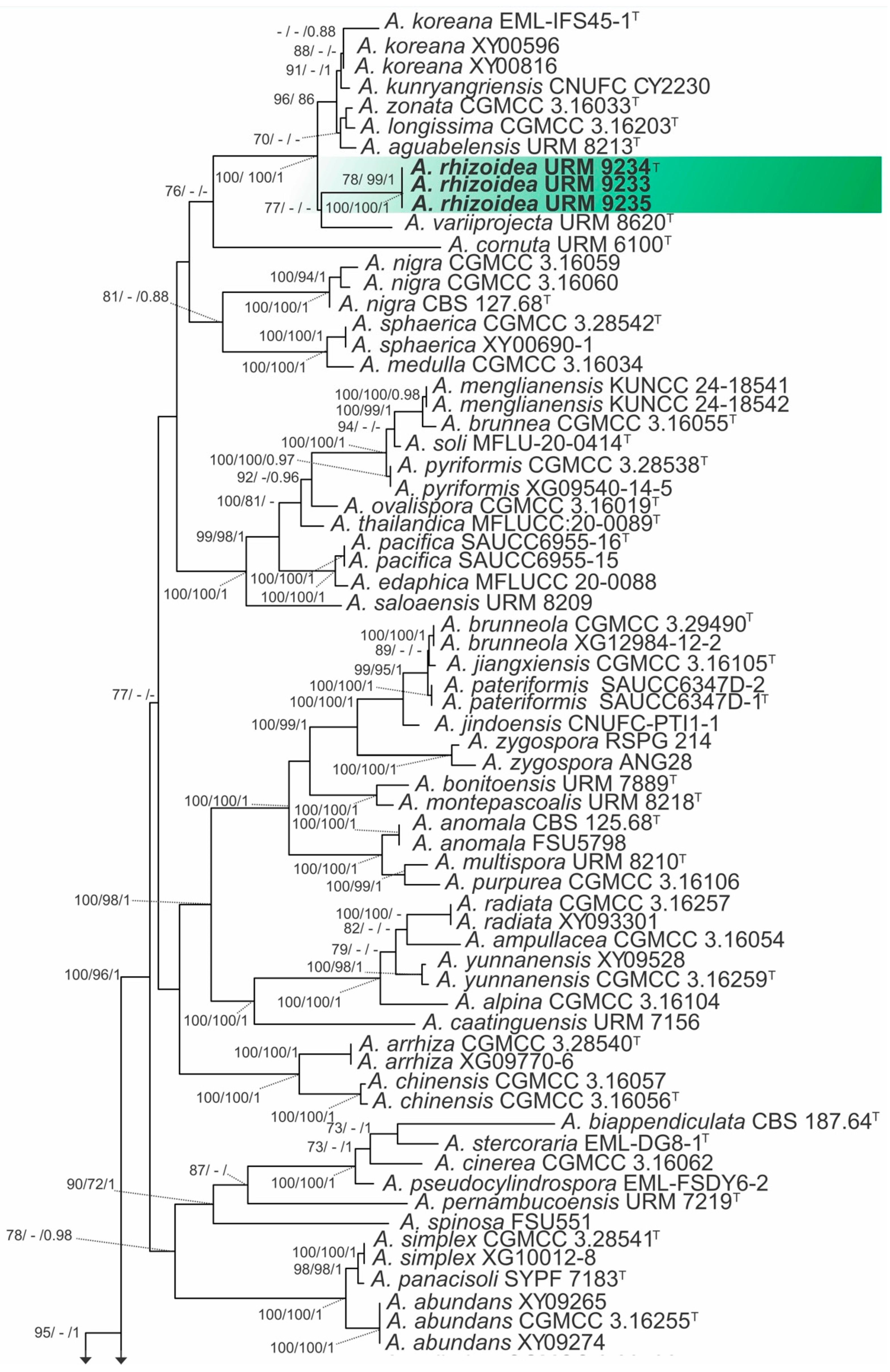
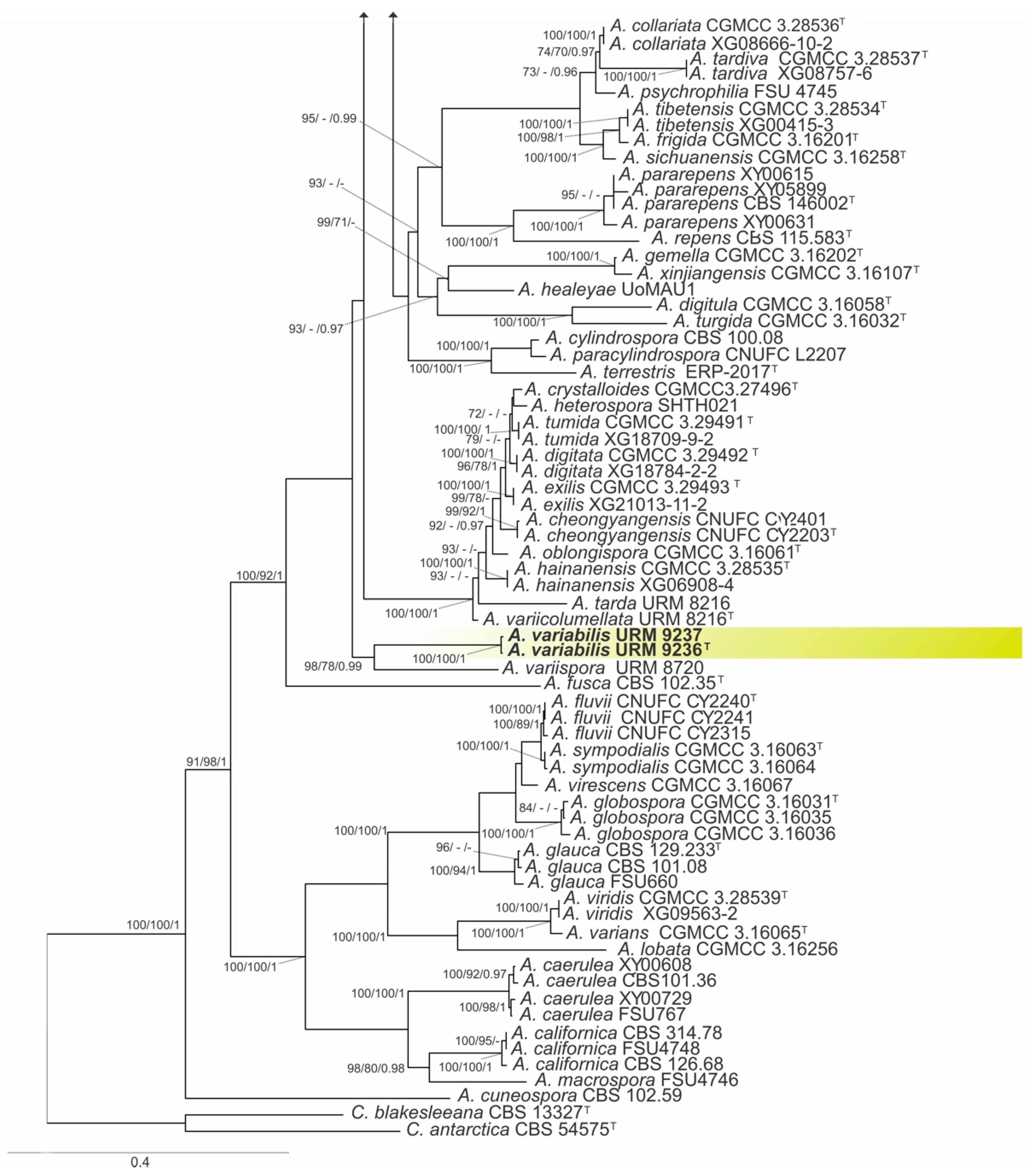
The combined matrix of three markers of Absidia species (ITS, LSU, and act) comprised sequences from 135 strains, including the outgroup Cunninghamella blakesleeana (CBS 133.27) and Cunninghamella antarctica (CBS 545.75), with a total of 2940 characters, including gaps, with 1267 characters for ITS, 823 for LSU, and 850 for act. The models used for IQ-TREE ML and BI were HKY+I+G for ITS, GTR+I+G for LSU and GTR+G for act. For the ML analysis (RAxML-HPC BlackBox) the model GTR+I+G was used and this matrix had 1918 distinct alignment patterns with 49.30% undetermined characters or gaps, and the final ML optimization likelihood value of the best tree was −43,306.257377. The estimated base frequencies were as follows: A = 0.254330, C = 0.209623, G = 0.232160, and T = 0.303887; substitution rates: AC = 1.172145, AG = 2.820942, AT = 1.449869, CG = 0.733730, CT = 4.396602, and GT = 1.000000; gamma distribution shape parameter: α = 0.637676. Based on our multiloci phylogenetic analysis (Figure 1), our isolates were placed as independent lineages with high support values between other species, with one for the isolates URM 9233, URM 9234, and URM 9235 (IQ-TREE-BS = 100%, RAxML-BS = 100%, and BPP = 1) and the other for URM 9236 and URM 9237 (IQ-TREEBS = 100%, RAxML-BS = 100%, and BPP = 1), therefore justifying their statuses as two new species.

3.1.2. Gongronella

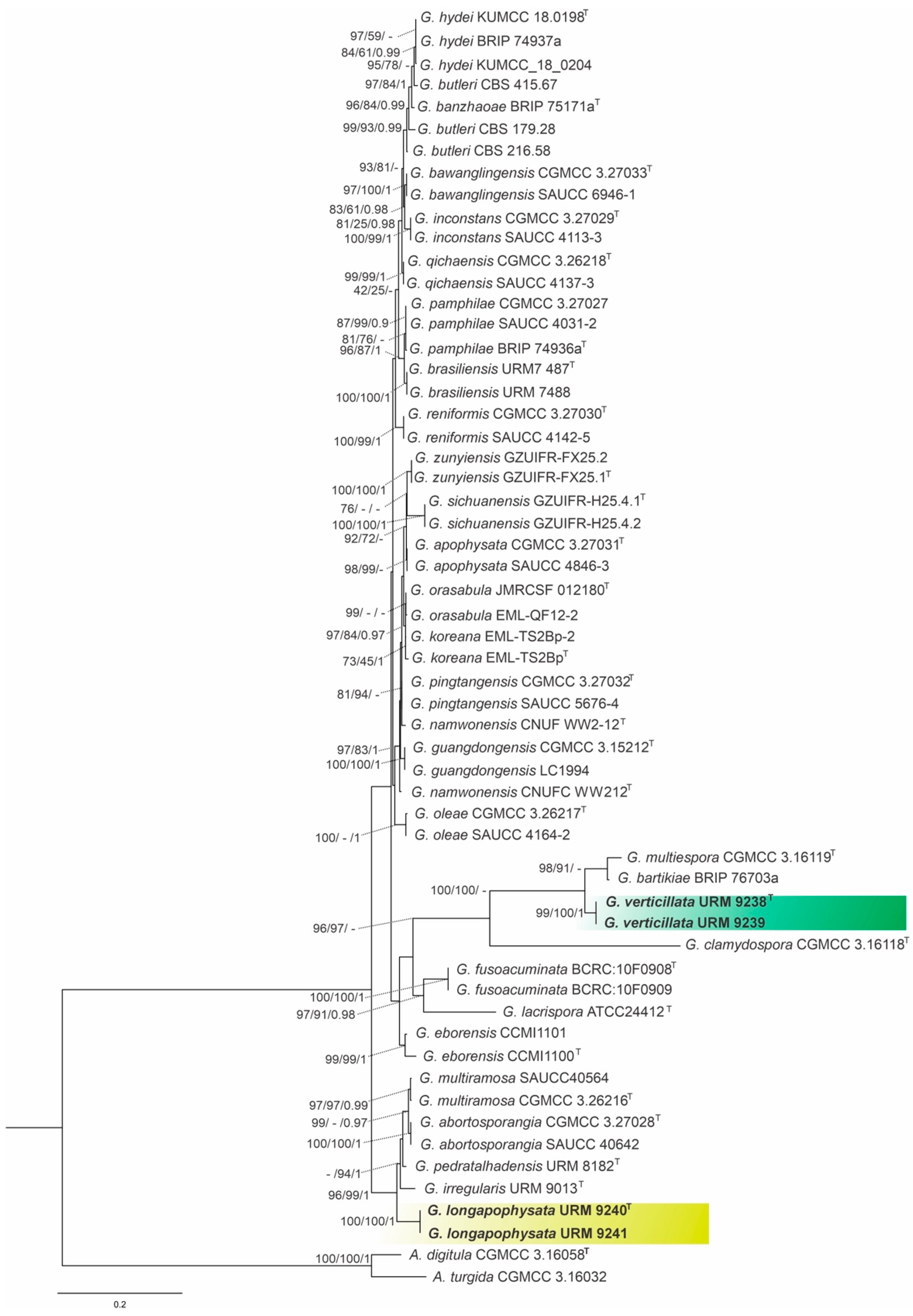
The combined matrix of three markers of Gongronella species (ITS, LSU, and act) comprised sequences from 58 strains, including the outgroup Absidia digitula (CGMCC 3.16058) and Absidia turgida (CGMCC 3.16032), with a total of 2426 characters, including gaps, with 760 characters for ITS, 928 for LSU and 738 for act. The model used for IQ-TREE ML and BI was GTR+G for both genes. For the ML analysis (RAxML-HPC BlackBox), the model GTR+I+G was used; this matrix had 904 distinct alignment patterns with 35.71% undetermined characters or gaps, and the final ML optimization likelihood value of the best tree was -11879.360036. The estimated base frequencies were as follows: A = 0.250425, C = 0.216708, G = 0.245264, and T = 0.287603; substitution rates: AC = 0.872173, AG = 2.309143, AT = 1.587038, CG = 0.516161, CT = 4.286676, and GT = 1.000000; gamma distribution shape parameter: α = 0.508375. Based on our multiloci phylogenetic analysis (Figure 2), our isolates were placed as independent lineages with high support values between other species, with one for the isolates URM 9240, and URM 9241 (IQ-TREE-BS = 100%, RAxML-BS = 100%, and BPP = 1) and the other for URM 9238 and URM 9239 (IQ-TREEBS = 99%, RAxML-BS = 100%, and BPP = 1), therefore justifying their status as two new species.

3.2. Taxonomy

3.2.1. Absidia rhizoidea L.W.S. de Freitas & A.L. Santiago, sp. nov. (Figure 3 and Figure 4)

Mycobank number: MB863003.
Etymology: The epitet rhizoidea (Lat.), referring to the rhizopodiform rhizoids formed at the end of solons, commonly next to the sporangiophores.
Diagnosis: The rhizoids are strongly branched, rhizopodiform, commonly formed next to the sporangiophores. Chlamydospores abundant.
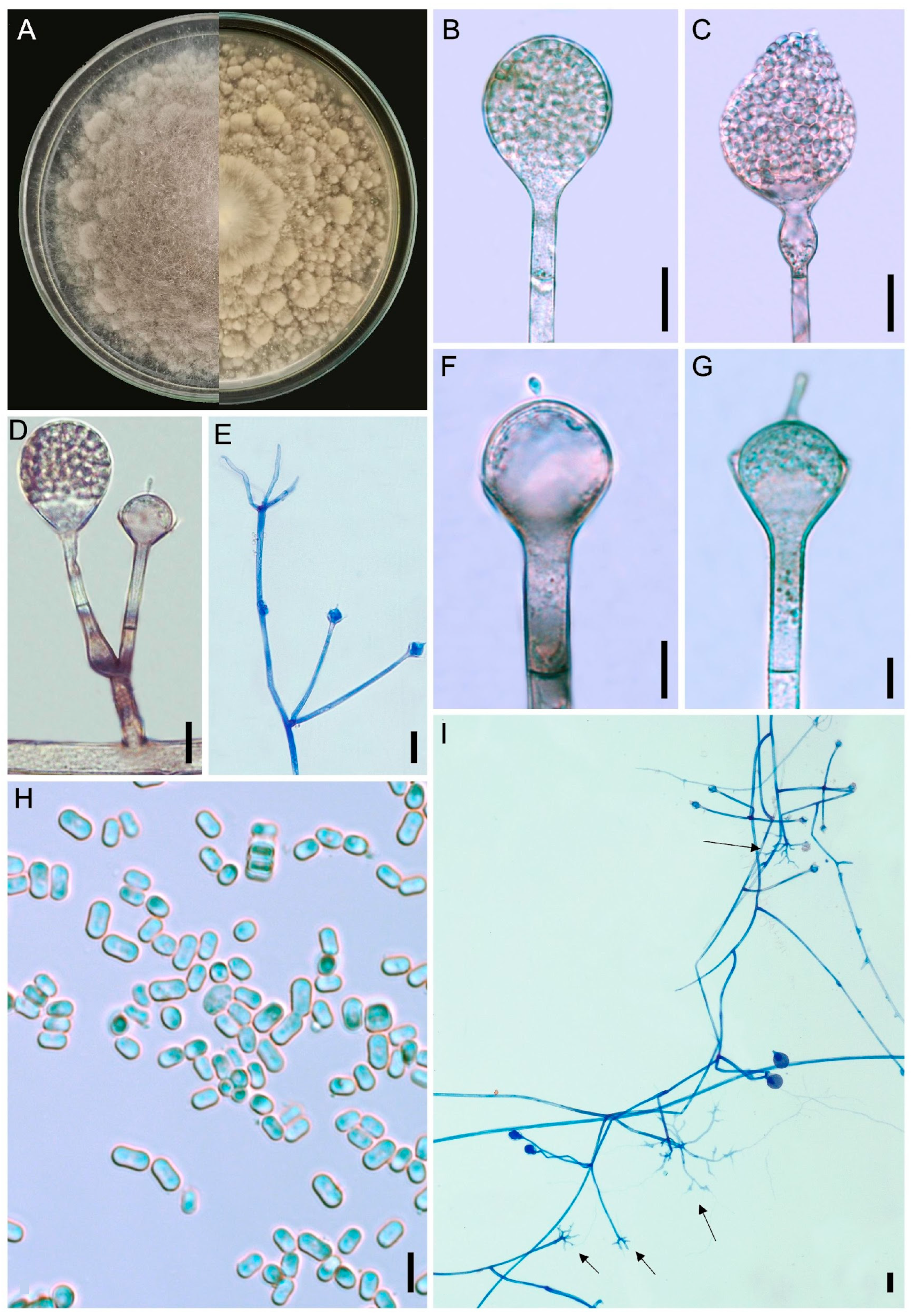
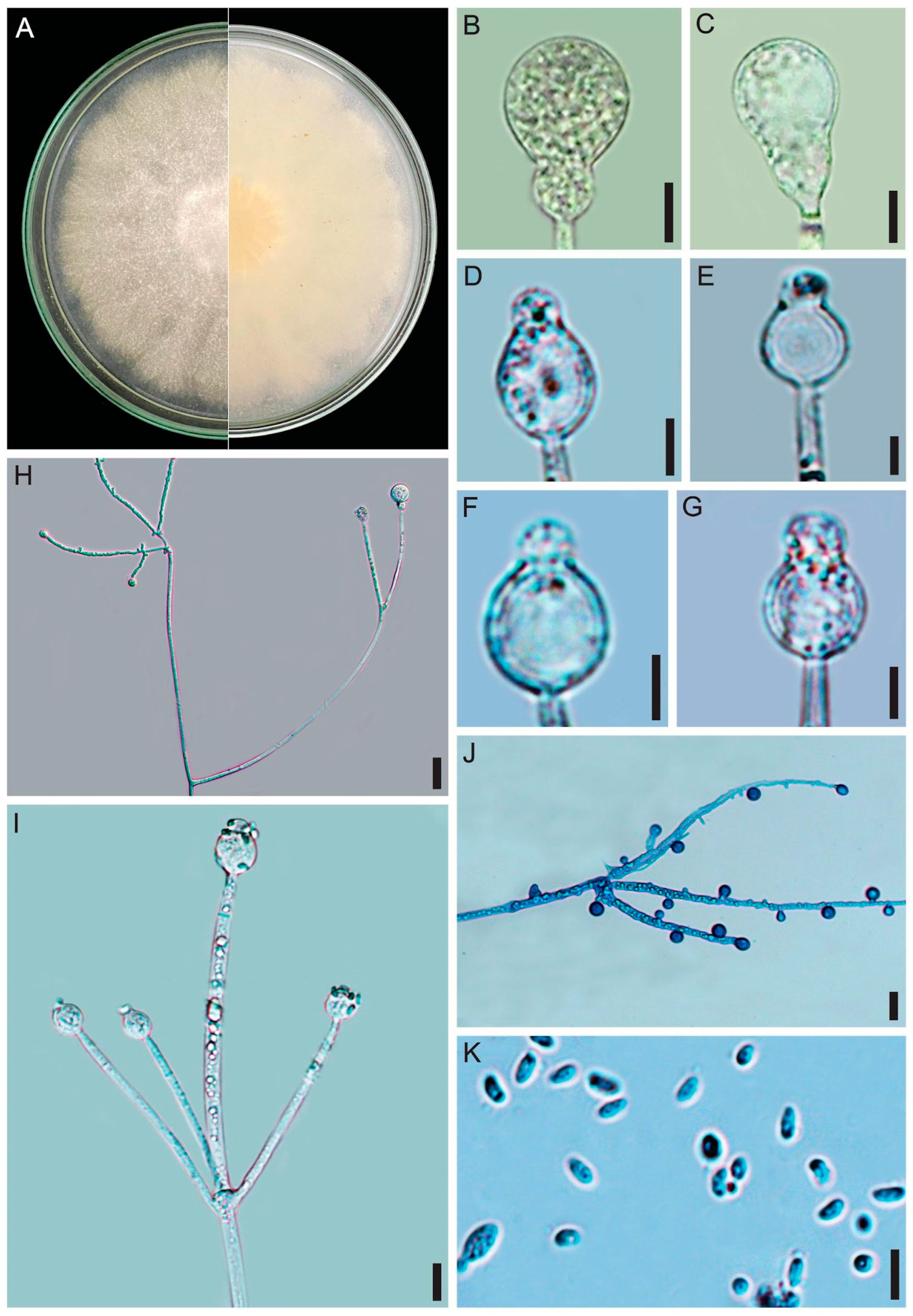
Description: Colonies floccose on PDA, initially white (A1), turning brown (6E8), 8.5 cm in diameter after 8 days of incubation at 25 °C; reverse caramel brown (6C6), irregular. Rhizoids caramel brown, commonly formed at the end of stolons and very close to the sporangiophores, rhizopodiform, strongly branched, 2.5–4.5 µm in diameter, smooth-walled. Sporangiophores brown, arising from stolons, isolated or in whorls of up to seven, unbranched, simple or rarely sympodially branched up to three times, (7–)20–350(–575) × (2.5–)5–12(–20) µm, smooth-walled, occasionally with one swelling after 7 days of incubation. One septum is observed near the apophysis. Sporangia light brown, subglobose, 12–40 × 10–40 µm, smooth and deliquescent-walled. Apophysis brownish, short or long, cup or bell-shaped, 5–30 × 6–30 µm. Columellae light brown, hemispherical or subglobose, 2.5–17 × 7–22 µm, occasionally with up to two needle-shaped or filiform with bulbous tip projection, 3–12 × 2–4 µm. Sporangiospores hyaline, cylindrical, some with a slight constriction in the center, ellipsoid and subglobose, 3–8 × 2–6 µm, smooth-walled. Chlamydospores abundant, hyaline, globose and subglobose, many attached to rhizoids or formed in aerial hyphae, 2.5–7 µm in diameter. Zygosporangia not observed.
Growth experiments (8 days): On PDA: At 5 °C—no growth; at 10 °C—1.5 cm diameter; at 15 °C—5.5 cm diameter; at 20 °C—8 cm diameter; at 25 °C—8.5 cm diameter; at 30 °C—8.3 cm diameter; at 35 °C—no growth. On MEA: At 5 °C—no growth; at 10 °C—0.7 cm diameter; at 15 °C—5.3 cm diameter; at 20 °C—8.8 cm diameter; at 25 °C—9 cm diameter; at 30 °C—8.5 cm diameter; at 35 °C—no growth. The Tmax is 33 °C on MEA and 32 °C on PDA.
Habitat and Distribution: Soil from Maranhão state (Brazil).
Specimen examined: BRAZIL, Maranhão, Carolina city, Chapada das Mesas, Cachoeira do Dodô (7°05′33.2″ S 47°26′35.5″ W), soil, 15 May 2023, L.W.S. de Freitas (Holotype URM 9234H metabolically inactive state, Ex-type living culture URM 9234).
GenBank accession numbers: ITS = PZ234078, LSU = PZ227107, act = PZ227661.
Additional specimens examined: BRAZIL, Maranhão, Carolina, Chapada das Mesas, Cachoeira do Dodô (7°05′33.2″ S 47°26′35.5″ W), soil, 15 May 2023, L.W.S. de Freitas (URM 9233); Encanto azul (7°14′00.6″ S 46°24′24.3″ W), soil, 15 May 2023, L.W.S. de Freitas (URM 9235).
GenBank numbers: URM 9233, ITS = PZ234079, LSU = PZ227108, act = PZ227662; URM 9235, ITS = PZ234080, LSU = PZ227109, act = PZ227663.
Notes: In our phylogenetic analysis, Absidia rhizoidea is a sister species of A. variiprojecta (Figure 1). Morphologically, A. rhizoidea forms constant rhizoids close to the sporangiophores, in addition to sporangiophores arising in whorls of up to seven from stolon, while A. variiprojecta has rhizoids not commonly formed close the sporangiophores and forms sporangiophores arising in whorls of up to five from stolon. Furthermore, columellae of the new species are hemispherical or subglobose, occasionally with up to two needle-shaped or filiform with bulbous tip projection, differing from A. rhizoidea that forms columellae subglobose or subglobose to fig-shaped, with one projection generally needle- or triangle-shaped, some bulbous, very rarely feather-shaped. The Tmax of A. variiprojecta is 32 °C, both on PDA and MEA [20], while the Tmax values of the new species are 33 and 32° C on MEA and PDA, respectively.
Figure 3. Absidia rizhoidea (URM 9234, ex-type) on PDA at 25 °C after eight days. (A) Colony verse (left) and reverse (right). (B) Sporangiophore with sporangium. (C) Sporangiophore with a swallow and sporangium. (D) Sporangiophore short branched with sporangium and projection. (E) Two sporangiophores arising from stolon with rhizoid. (F,G) Sporangiophore with apophysis, columella and projection. (H) Sporangiospores. (I) Stolons with sporangiophores and rhizoids (arrows). Scale bars: (B,C) 20 μm; (D,E,I) 15 μm; (F,G) 10 μm; (H) 10 μm.
Jof 12 00329 g003
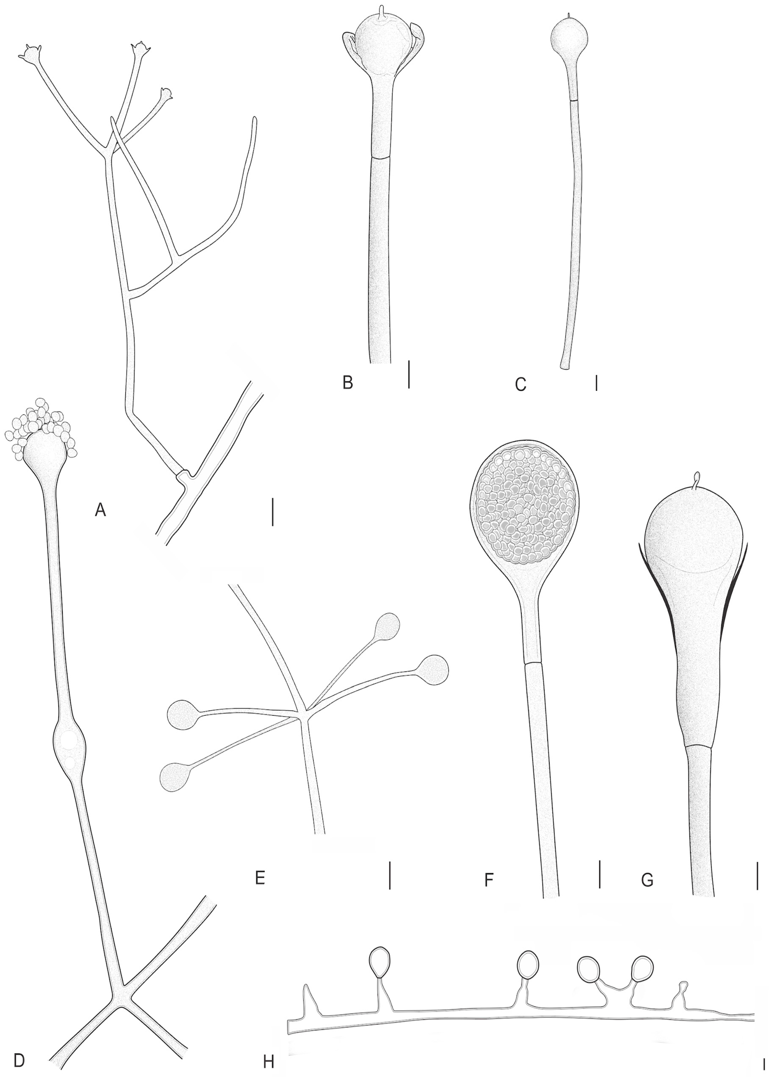
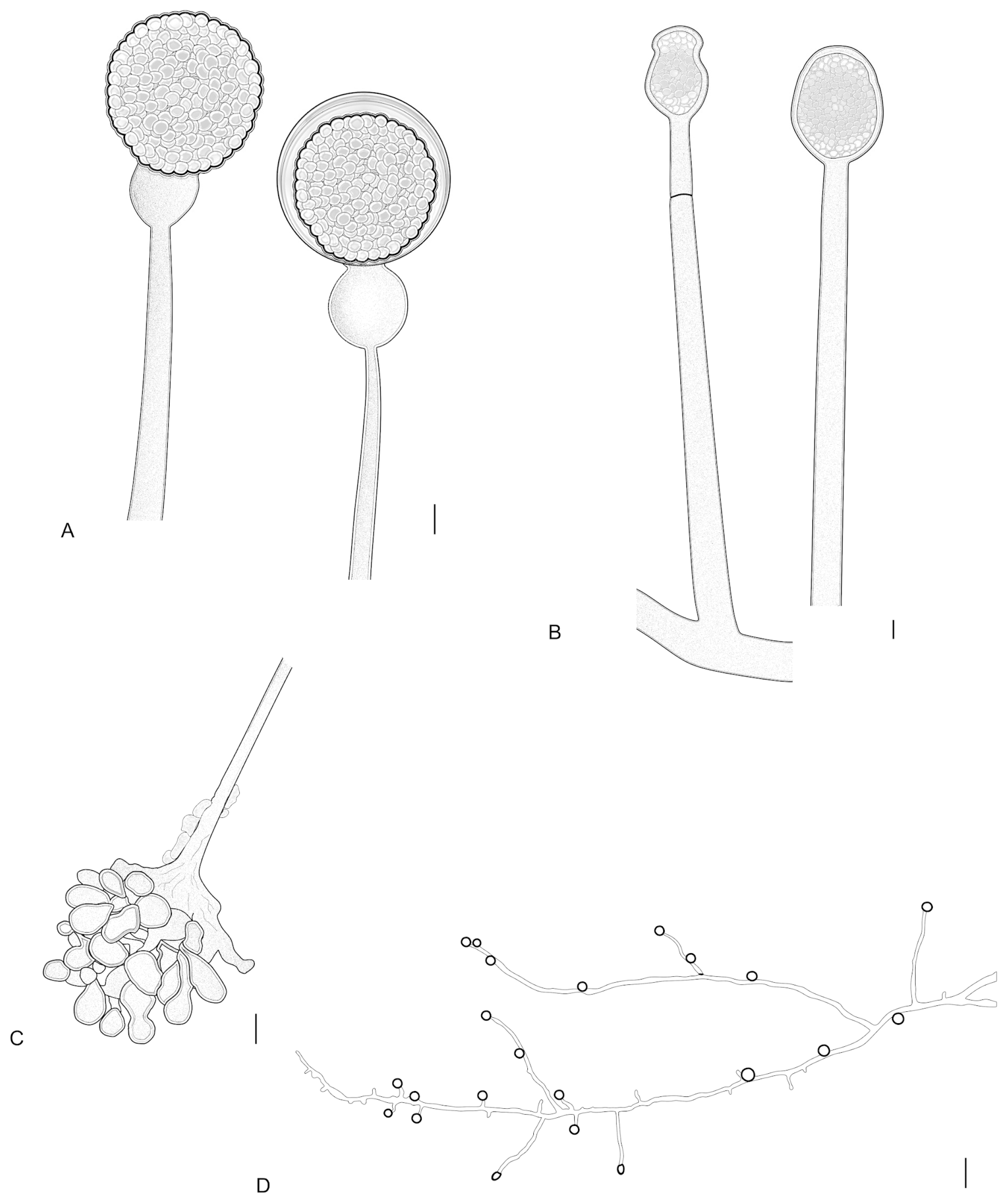
Figure 4. Absidia variabilis (URM 9236, ex-type) on PDA at 25 °C after eight days. (A) Branched sporangiophore. (B,C) Sporangiophore with a septum below the apophysis, columella and projection. Absidia rizhoidea on PDA at 25 °C after eight days. (D) Sporangiophore with swelling. (E) Sporangiophores in a whorl of four arising from stolon. (F) Sporangiophore with sporangium. (G) Sporangiophore with a long apophysis, columella and projection. (H) Chlamydospores. Scale bars: (A) 5 μm; (B,C) 8 μm; (D,E) 20 μm; (FH) 10 μm.
Jof 12 00329 g004

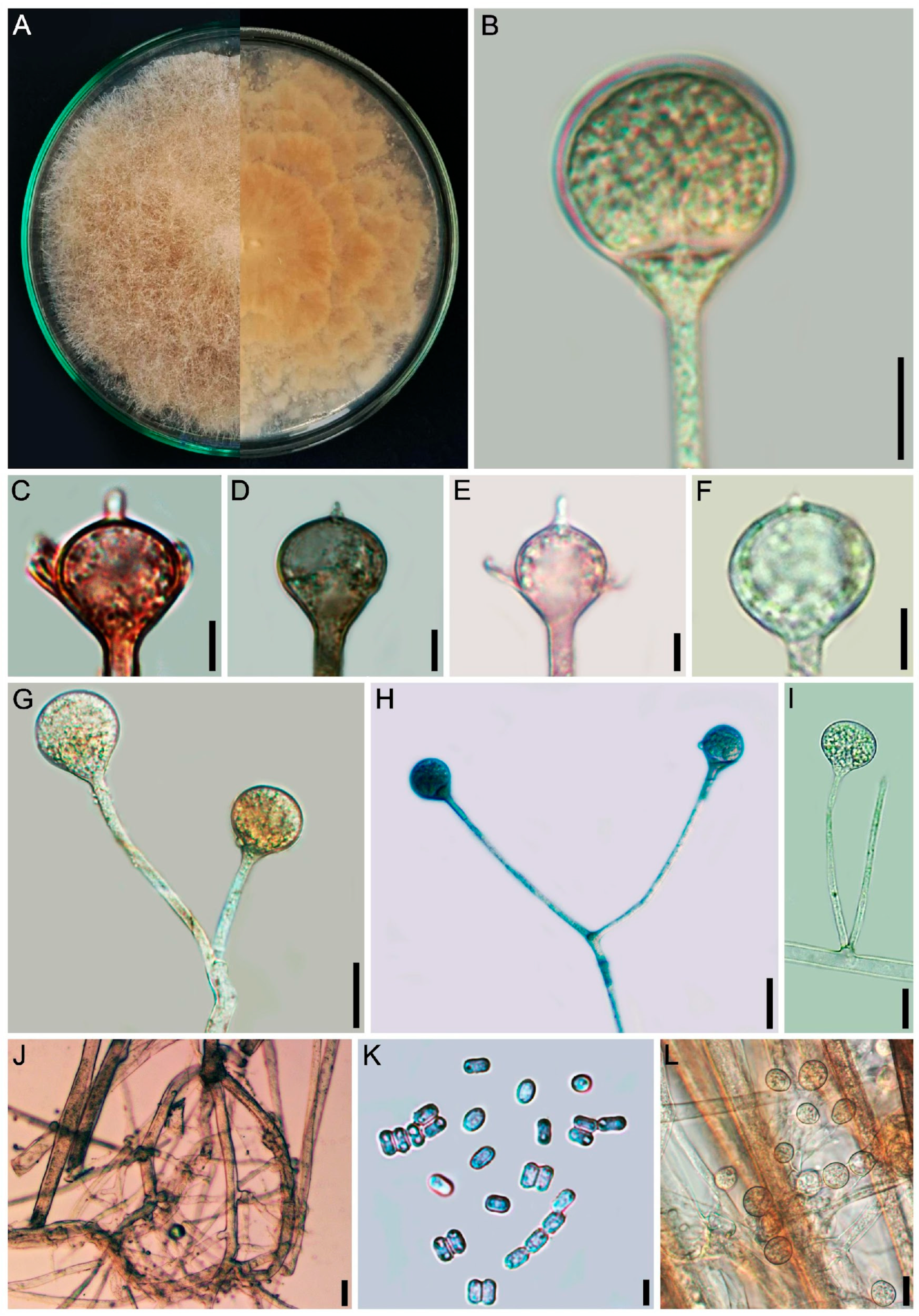
3.2.2. Absidia variabilis L.W.S. de Freitas & A.L. Santiago, sp. nov. (Figure 4 and Figure 5)

Mycobank number: MB863004.
Etymology: The epitet variabilis (lat.) refers to the formation of sporangiophores variable in length.
Diagnosis: Sporangiophores varied in length, arising singular or in whorls of up to five from stolons, commonly unbranched or monopodially branched, never sympodially branched; chlamydospores are abundant.
Description: Colonies floccose on PDA, initially white (A1), becoming light brown (6D8), 8.5 cm in diameter after 8 days of incubation, at 25 °C; reverse light brown (6D8), wave zonate. Rhizoids brown, poorly branched, often with adherent chlamydospores, 2.5–5.5 µm in diameter, with encrusted wall. Sporangiophores erect, light brown, arising from stolons, singular or in whorls of up to five, monopodially, but never sympodially branched, varied in length (15–)60–190(–230) × (2–)3–4(–6) µm, smooth-walled, with no swellings. One septum is observed near the apophysis. Sporangia apophysate, light brown, globose to slightly dorsiventrally flattened, pyriform (when considering the apophysis), (7–)10–20(–30) × (10–)15–30 µm, smooth, with deliquescent wall. Apophysis brownish, short, cup-shaped, (2–)4.5–7(–12) × (3.5–)7.5–15 µm, smooth-walled. Columellae light brown slightly flattened and subglobose (2–)5–17 × (5–)9–22 µm, smooth-walled, collar visible. One filiform, spine-like or occasionally triangular projection, up to 1–4 × 0.5–2.5 µm, can be observed on the columellae surface. Sporangiospores hyaline, mostly cylindrical, constricted in the center, some short cylindrical, 2.5–5.5 × 1.5–2.5 µm, smooth-walled. Chlamydospores abundant, hyaline, globose, subglobose, frequently attached to rhizoids or in aerial hyphae, 2–6 µm in diameter. Zygosporangia not observed.
Growth experiments (8 days): On PDA: At 5 °C—no growth; at 10 °C—1.1 cm diameter; at 15 °C—4.6 cm diameter; at 20 °C—8.6 cm diameter; at 25 °C—9 cm diameter; at 30 °C—8.5 cm diameter; at 35 °C—no growth. On MEA: At 5 °C—no growth; at 10 °C—0.7 cm diameter; at 15 °C—5.1 cm diameter; at 20 °C—8.5 cm diameter; at 25 °C—8.8 cm diameter; at 30 °C—8.6 cm diameter; at 35 °C—no growth. The Tmax is 31 °C on MEA and 32 °C on PDA.
Habitat and Distribution: Soil from Maranhão state (Brazil).
Typification: BRAZIL, Maranhão, Carolina, Chapada das Mesas, São Romão (6°02′09.1′′ S 46°34′03.5′′ W), soil, 15 May 2023, L.W.S de Freitas (Holotype URM 9236H metabolically inactive state, Ex-type living culture URM 9236).
GenBank accession numbers: ITS = PZ234081, LSU = PZ227110, act = PZ227664.
Additional specimen examined: BRAZIL, Maranhão, Carolina, Chapada das Mesas, São Romão (6°02′09.1′′ S 46°34′03.5′′ W), soil, 15 May 2023, L.W.S. de Freitas (URM 9237). GenBank accession numbers: ITS = PZ234082, LSU = PZ227111, act = PZ227665.
Notes: Absidia variabilis is phylogenetically more closely related to A. variispora T.R.L. Cordeiro & A.L. Santiago (Figure 1). The main morphological differences between both species are the formation of sporangiophores arising singular or in whorls of up to six from stolon, as well as the formation of cylindrical, elliptical, globose, and subglobose sporangiospores by A. variispora, while A. variabilis forms sporangiophores single or in whorls of up to five from stolon and produces exclusively cylindrical spores. Furthermore, A. variabilis forms chlamydospores, while A. variispora does not. Absidia variabilis grows up to 31 °C on MEA and 32 °C on PDA, while A. variispora can grow at 32 °C on MEA and PDA [20].
Figure 5. Absidia variabilis (URM 9236, ex-type) on PDA at 25 °C after eight days. (A) Colony verse (left) and reverse (right). (B) Sporangiophore with sporangium. (CF) Different types of columellae, apophyses, and projections. (G,H) Branched sporangiophore. (I) Two sporangiophores arising from stolon. (J) Rhizoid. (K) Sporangiospores. (L) Chlamydospores. Scale bars: (B) 15 μm; (CI) 10 μm; (JL) 5 μm.
Jof 12 00329 g005

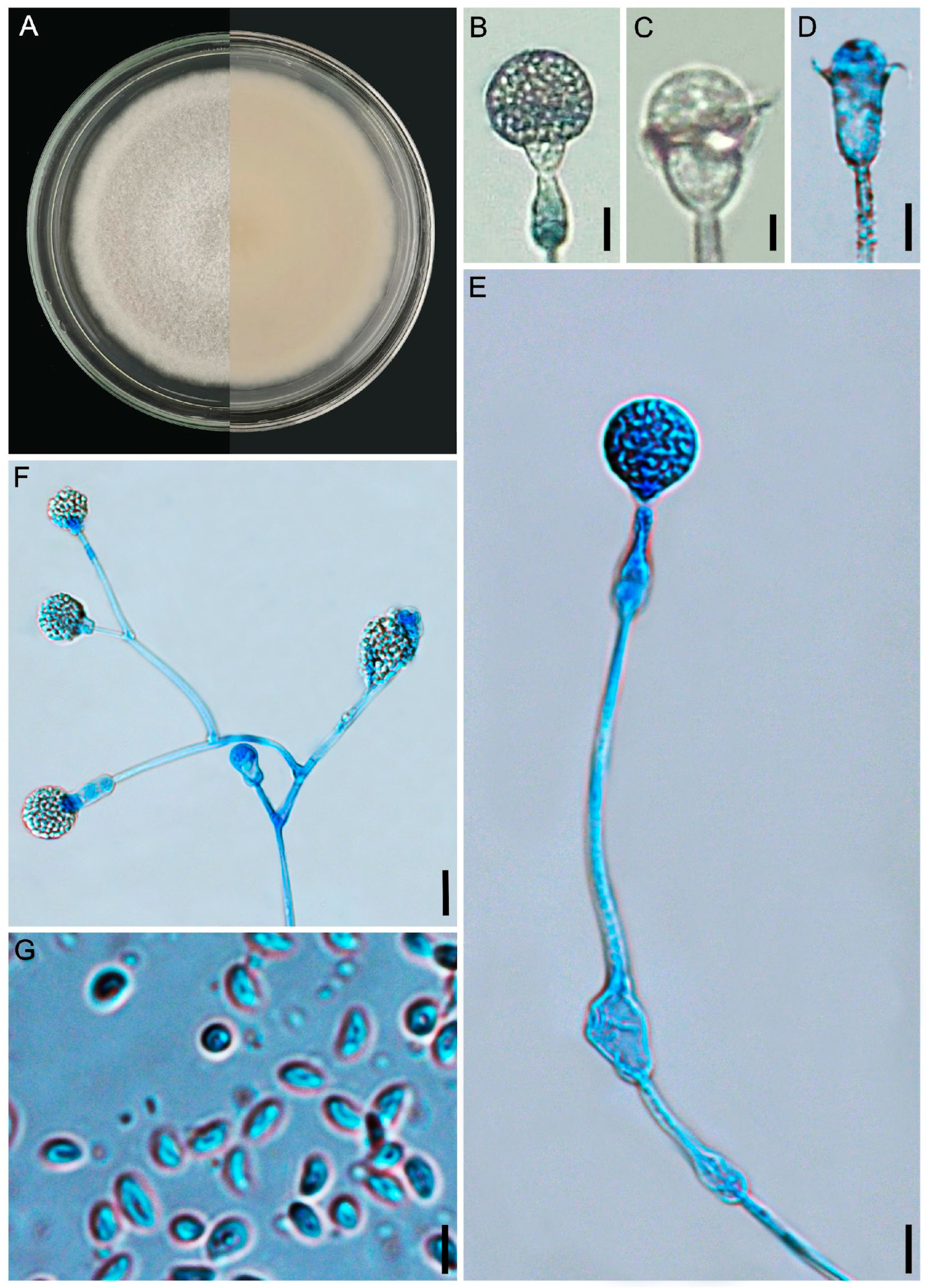
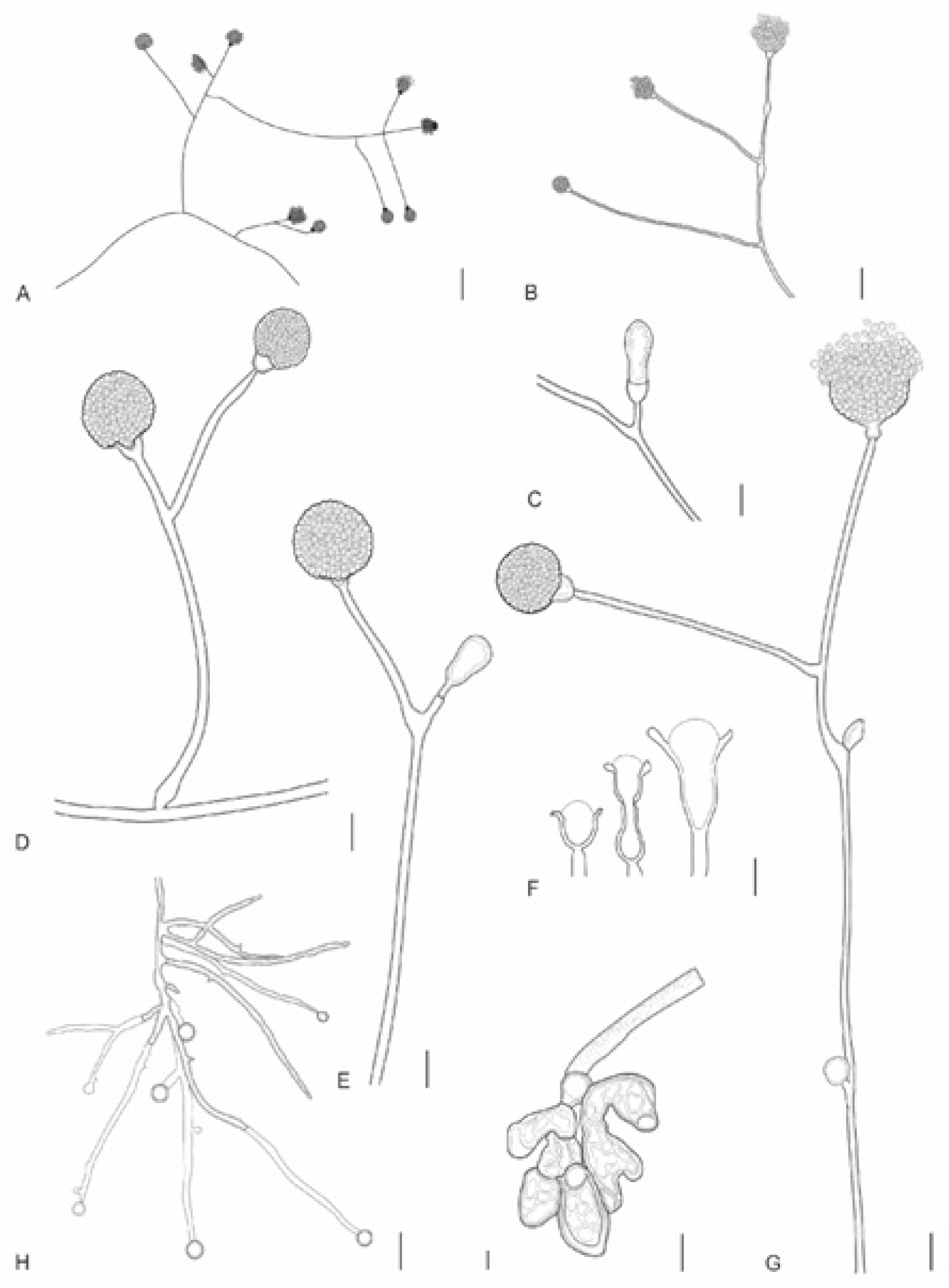
3.2.3. Gongronella longapophysata L.W.S de Freitas & A.L. Santiago, sp. nov. (Figure 6 and Figure 7)

Mycobank number: MB863005.
Etymology: The epithet longapophysata (Lat.) refers to the sporangiophores with elongated apophysis.
Diagnosis: The species forms sporangiophores with some very elongated apophyses, some with an elongated swelling below apophyses, and chains of 3 to 12 sterile sporangia along sporangiophores.
Description: Colonies white (A1), cottony, with regular margin, 6.5 cm in diameter on PDA after 8 days, at 25 °C. Reverse white (A1). Rhizoids branched, long, often with adherent chlamydospores, 2–10 μm in diameter, and bulbous, 5–38 μm in diameter. Stolons hyaline, branched and coenocytic. Sporangiophores hyaline, erect, curved to sinuous, (10–)35–230(–350) × 2.5–7(–12) μm, with one to two septa below the sporangium, simple or sympodially branched up to four times, wall smooth to slightly incrusted, rarely verticillately branched in whorls of up to five. Fertile sporangia hyaline, subglobose (5–)12–20(–25) × (6.5–)15–20(–25) μm, wall with vitreous aspect, smooth and deliquescent, leaving a collar. Sterile sporangia hyaline, ellipsoid, cylindrical to constricted frequently formed on short sporangiophores branches, 3–20 μm in diameter, occasionally so short that the sterile sporangia appear to be sessile; 3–6, rarely, 7–12 sterile sporangia are also formed along the sporangiophore. Columellae hyaline, hemispherical, subglobose (2–)5–10 × (3.5–)7–11(–15) μm. Apophyses bell-shaped, long or short, vasiform, (2–)3–7(–10) × (5–)7–10(–13) μm. Some sporangiophores may show an elongated swelling below the apophysis, 10–25 × 5–7 μm. Sporangiospores hyaline, reniform, some irregularly-shaped, 2–5.5 × 1.5–2 μm, smooth-walled. Chlamydospores abundant, hyaline, globose, 4–15 μm in diameter. Zygosporangia not observed.
Growth experiments (8 days): On PDA: At 10 °C—no growth; at 15 °C—1.8 cm diameter; at 20 °C—3.1 cm diameter; at 25 °C—6.3 cm diameter; at 30 °C—7.5 cm diameter; at 35 °C—no growth. On MEA: At 10 °C—no growth; at 15 °C—1.9 cm diameter; at 20 °C—4.4 cm diameter; at 25 °C—6.0 cm diameter; at 30 °C—6.8 cm diameter; at 35 °C—no growth. The Tmax is 32 °C on MEA and 33 °C on PDA.
Habitat and Distribution: Soil from Maranhão state (Brazil).
Specimen examined: Brazil, Maranhão, Carolina city, Chapada das Mesas, Cachoeira da Mansinha (7°07′54.0″ S 47°26′55.1″ W), soil, 15 May 2023, L.W.S. de Freitas (Holotype URM 9240H metabolically inactive state, Ex-type living culture URM 9240).
GenBank accession numbers: ITS = PZ234083, LSU = PZ227112, act = PZ227666.
Additional specimen examined: BRAZIL, Maranhão, Carolina city, Chapada das Mesas, Cachoeira da Mansinha (7°07′54.0″ S 47°26′55.1″ W), soil, 15 May 2023, L.W.S de Freitas (URM 9241).
GenBank accession numbers: ITS = PZ234084, LSU = PZ227113, act = PZ227667.
Notes: Gongronella longapophysata is phylogenetically closely related to G. multiramosa and G. abortosporangia (Figure 1). Morphologically, G. multiramosa and G. abortosporangia can be easily differentiated from G. longapophysata due to no formed stolons, no verticillately branched sporangiophores (rare in G. longapophysata), no sporangiophores with a swelling below apophysis, and no chains of 3 to 12 sterile sporangia along sporangiophores [30]. The Tmax of G. longapophysata is 32 °C on MEA and 33 °C on PDA, but Tmax of G. abortosporangia and G. multiramosa is not indicated in the protologues nor elsewhere [29,30].
Figure 6. Gongronella longapophysata (URM 9240, ex-type) on PDA at 25 °C after eight days. (A) Colony verse (left) and reverse (right). (BD) Columellae and aphophyses. (E) Sporangiophore with elongated swelling below the apophysis and sterile sporangia. (F) Branched sporangiophore. (G) Sporangiospores. Scale bars: (BE) 10 μm; (F,G) 5 μm.
Jof 12 00329 g006
Figure 7. Gongronella longapophysata (URM 9240, ex-type) on PDA at 25 °C after eight days. (A,B) Branched sporangiophore with fertile sporangia. (C) Sterile sporangium. (D) Simply branched sporangiophore with sporangia. (E) Simply branched sporangiophore with fertile (terminal) and sterile (branch) sporangia. (F) Apophyses and columellae. (G) Sporangiophore with long and short branches. (H) Rhizoid with chlamydospores. (I) Bulbous rhizoid. Scale bars: (A,B) 30 μm; (CF) 5 μm; (GI) 10 μm.
Jof 12 00329 g007

3.2.4. Gongronella verticillata L.W.S. de Freitas & A.L. Santiago, sp. nov. (Figure 8 and Figure 9)

Mycobank number: MB863006.
Etymology: The epithet verticillate (Lat.) refers to the formation of sporangiophores verticillately branched.
Diagnosis: The species commonly forms sporangiophores branched in whorls with up to five branches.
Description: Colonies white (A1), cottony, regular margin, 7 cm in diameter on PDA after 8 days of incubation, at 25 °C; reverse cream (4A3). Rhizoids of two types: root-shaped, hyaline, branched, with spaced septa and chlamydospores attached, and bulbous, without chlamydospores attached, 2.5–8.5 µm in diameter. Stolons hyaline, simple and coenocytic. Sporangiophores simple or sympodially branched up to six times, and commonly verticillately branched, with up to five branches in a whorl, (25–)95–200(–250) × 2.5–5 μm, with one or two septa below the sporangium, rarely forming three septa, smooth-walled. Some sporangiophores are extremely short, 2.5–10 × 2–3 μm, forming sterile apical or lateral sporangia. Fertile sporangia hyaline, globose, (6–)10–17(–22) μm diameter, with vitreous aspect, smooth and deliquescent-walled, leaving a collar. Abortive sporangia ovoid, subglobose and globose 4–18 μm in diameter. Columellae hyaline, mostly globose 1–5 μm in diameter, subglobose, hemispherical, flattened and rounded-topped (1–)4–7(–9) × (2–)6–9(–12) μm, smooth-walled, occasionally so flattened that they seem inconspicuous. Apophyses hyaline, mostly globose, 3–10 μm diameter, subglobose, some bell-shaped and vasiform, (3–)5–10(–13) × (3.5–)6–12(–15) μm, smooth-walled. Sporangiospores hyaline, elliptical to fusiform, irregular or reniform, 2–7.5 × 1.5–4 μm, smooth-walled. Chlamydospores abundant, hyaline, subglobose and ellipsoidal, 4–15 μm diameter. Zygosporangia not observed.
Growth experiments (8 days): On PDA: At 10 °C—no growth; at 15 °C—1.7 cm diameter; at 20 °C—3.2 cm diameter; at 25 °C—6.8 cm diameter; at 30 °C—2.1 cm diameter; at 35 °C—no growth. On MEA: At 10 °C—no growth; at 15 °C—1.9 cm diameter; at 20 °C—4.2 cm diameter; at 25 °C—5.5 cm diameter; at 30 °C—4 cm diameter; at 35 °C—no growth. The Tmax is 31 °C on both MEA and PDA.
Habitat and Distribution: Soil from Maranhão state (Brazil).
Specimen examined: BRAZIL, Maranhão, Carolina city, Chapada das Mesas, Cachoeira da Mansinha (7°07′54.0″ S 47°26′55.1″ W), soil, 15 May 2023, L.W.S. de Freitas (Holotype URM 9238H metabolically inactive state, Ex-type living culture URM 9238).
GenBank accession numbers: ITS = PZ234085, LSU = PZ227114.
Additional specimen examined: BRAZIL, Maranhão, Carolina city, Chapada das Mesas, Cachoeira da Mansinha (7°07′54.0″ S 47°26′55.1″ W), soil, 15 May 2023, L.W.S. de Freitas (URM 9239).
GenBank accession numbers: ITS = PZ234086, LSU = PZ227115.
Notes: Gongronella verticillata is phylogenetically closely related to G. multispora and G. bartikiae (Figure 1). However, G. verticillata forms sporangiophores sympodially and verticillately branched, with up to five branches in a whorl. The apophyses are globose, subglobose, bell-shaped and vasiform; columellae are globose, subglobose, flattened, rounded-topped and inconspicuous, whereas G. multispora forms sporangiophores sympodially and verticillately branched, with 2–3 branches in a whorl. Apophyses are pyriform to subglobose, and the columellae are hemispheric and some inconspicuous. It is curious that both species form sporangiophores branched in whorls, as to the best of our knowledge, this branch pattern has also been reported in G. namwonensis so far [28]. The Tmax of G. verticillata is 31 °C on MEA and PDA, but Tmax of G. multispora has not been given in the protologue [29]. There is no morphological description of G. bartikiae in the literature for comparison [67].
Figure 8. Gongronella verticillata (URM 9238, ex-type) on PDA at 25 °C after eight days. (A) Colony verse (left) and reverse (right). (B,C) Sporangiophore with sporangium. (DG) Sporangiophore with columella. (H) Simply branched sporangiophore arising from stolon with rhizoid. (I) Sporangiophore with terminal columella and lateral whorled branches with columellae. (J) Rhizoid with chlamydospores attached. (K) Sporangiospores. Scale bars: (B,C) 20 μm; (D,F,J,K) 10 μm; (G,H) 15 μm; (E,I) 5 μm.
Jof 12 00329 g008
Figure 9. Gongronella verticillata (URM 9238, ex-type) on PDA at 25 °C after eight days. (A) Sporangiophores with sporangia. (B) Sporangiophores with a septum below the apophysis (left) and columellae. (C) =Bulbous rhizoid. (D) Chlamydospores attached to the mycelium. Scale bars: (A,B) 3 μm; (C) 10 μm; (D) 5 μm.
Jof 12 00329 g009

4. Discussion

Traditionally, new species of Mucorales fungi have been mostly delimited on a morphological basis, leading to further synonymies and generating controversy and doubts among taxonomists. In this context, species of Absidia and Gongronella have been sorted into other genera, as they shared similar morphological characteristics. For instance, G. butleri was first described as A. butleri Lend. in 1926, while Paine [68] proposed A. subpoculata Paine, which was later synonymized with G. butleri. In addition, some species of Absidia have been previously included in Tieghemella, Mycocladus, Proabsidia, Pseudoabsidia, and Protoabsidia [69,70,71]. On the other hand, species of Lichtheimia and Lentamyces have been previously treated as Absidia [72,73].
The advent of the molecular biology allowed the delimitation of new species of Absidia and Gongronella in the last 10–15 years in different countries, such as China, Brazil, South Korea, and Thailand, most of which were proposed based in sequences of ITS and/or LSU rDNA [20,21,29,30,41,42,74,75]. Wang et al. [30] highlighted that the ITS, LSU, Elongation Factor 1-alpha-EF-1α (TEF), act, and RNA Polymerase II Largest Subunit (RPB1) are reliable markers for identification of Gongronella species. Until 1969, only two Gongronella species, namely, G. butleri and G. lacrispora Hesselt. & J. JEllis [76,77], were known, and this number has increased to 28 species (as of 3 March 2026), mostly descried from China. Regarding Absidia, Ji et al. [44] sequenced TEF and act, as well as ITS and LSU, to delimit new species, and 69 new species have been described in the last 15 years [27].
Since Tmax has been successfully used as a discriminative characteristic for delimitation of mucoralean fungi, such as Backusella [39], Cunninghamella [78,79], Lichtheimia [80], Mucor [81], and Rhizopus [82], we believe in the taxonomic relevance of this characteristic for the delimitation of species within Absidia and Gongronella. In this paper, the Tmax of our new Absidia species did not vary much from the phylogenetically close species. However, we retrieved data from the literature on the Tmax of 50 species of Absidia (plus two new species described in this work) and realized that the Tmax varies considerably between species in this genus, from 24 °C (A. frigida) to 37 °C (A. ovalispora and A. zonata) (Table 1); therefore, this feature has taxonomic value. However, as it was also observed in other mucoralean genera [39,78,79,80], some overlapping may occur (e.g., A. menglianensis, A. tarda and A. longissima have a Tmax of 36 °C), which is why this feature should be used along with morphology and phylogeny for delimiting species in Absidia, as well as in other mucoralean genera.
We were unable to compare the Tmax of our new Gongronella species with other species due to the lack of Tmax data in the literature for this genus; therefore, it is still unknown whether this feature is taxonomically informative in Gongronella. Nevertheless, to the best of our knowledge we are providing the first data on Tmax for the genus. Regarding Absidia, Tmax data are available in the literature for only 52% of the known species, mostly for species from China and Korea, with a few species from Brazil and Australia (Table 1). Therefore, studies evaluating the Tmax of all species of both genera should be encouraged.
In this paper, we described, based on an integrated approach, four new species of Cunninghamellaceae from soil samples from the still poorly studied Cerrado biome. As far as we are aware, this is the first report of new Absidia and Gongronella species in this biome. Currently, only 17 species of Absidia have been reported in Brazil, including ten new species [19,20,53,74,75,83,84], whereas only four species of Gongronella were recorded in the country, including three new species [85,86]. All these species were isolated in the Caatinga and Atlantic Forest biomes. This paper increases the number of known species of Absidia and Gongronella to 96 and 30, respectively, as well as the knowledge of these fungi in the Brazilian Savanna. We believe that new inventories of Cunninghamellaceae in other Cerrado areas may unveil additional new species.

Author Contributions

Conceptualization: A.L.C.M.d.A.S. and L.W.S.d.F.; Material collection: L.W.S.d.F.; Methodology: L.W.S.d.F., A.R., A.L.C.M.d.A.S., L.W.S.d.F., C.S.d.O., M.O.d.C., J.D.P.B., M.A.B.d.S., H.B.L., C.M.d.S.-M., R.F.R.M. and L.d.O.F.; writing—preparation of the original draft: L.W.S.d.F., J.D.P.B., A.L.C.M.d.A.S., A.R. and H.B.L.; writing—revision and editing: L.d.O.F., A.R., C.M.d.S.-M., J.D.P.B., R.F.R.M., H.B.L. and A.L.C.M.d.A.S. All authors read and approved the final version of the manuscript.

Funding

This study was supported by the Conselho Nacional de Desenvolvimento Científico e Tecnológico (CNPq) for awarding a doctoral fellowship to de Leslie W.S. de Freitas and a research grant to André L.C.M.A. Santiago (313401/2023-3). Jadson D.P. Bezerra and Layanne O. Ferro thank the CNPq, Coordenação de Aperfeiçoamento de Pessoal de Nível Superior (CAPES) and Fundação de Amparo à Pesquisa do Estado de Goiás (FAPEG) for funding. This research was partially supported by Basic Science Research Program through the National Research Foundation of Korea (NRF) funded by the Ministry of Education (RS-2022-NR075731).

Institutional Review Board Statement

Not applicable.

Informed Consent Statement

Not applicable.

Data Availability Statement

The sequences presented in this study are openly available in https://www.ncbi.nlm.nih.gov/ (see Table 1 for the accession numbers). The alignments and phylogenetic tree files are available in FigShare (https://doi.org/10.6084/m9.figshare.31438165). All new taxa were registered in the Mycobank database (www.mycobank.org). This research was registered in the Brazilian SisGen system (A77E17B).

Acknowledgments

We thank D. Tatiana Baptista Gibertoni for collecting the soil samples.

Conflicts of Interest

The authors declare no conflicts of interest.

References

  1. Ribeiro, J.F.; Walter, B.M.T. Fitofisionomias do bioma Cerrado. In Cerrado: Ambiente e Flora; Sano, S.M., Almeida, S.P., Eds.; Embrapa-CPAC: Planaltina, Brazil, 1998; pp. 89–166. [Google Scholar]
  2. Santos, F.J.M. Checklist of the herpetofauna in an area of the Cerrado Biome, Central Brazil, under strong mining pressure. J. Wildl. Biodivers. 2025, 9, 332–354. [Google Scholar] [CrossRef]
  3. De Jesus, D.L.V.; Aranda, R. Hydromorfological soils drives ant (Hymenoptera: Formicidae) communities in Brazilian wet savanna. Wetl. Ecol. Manag. 2025, 33, 4. [Google Scholar] [CrossRef]
  4. Wijayawardene, N.N.; Hyde, K.D.; Mikhailov, K.V.; Goto, B.T.; Santiago, A.L.C.M.A.; Tokarev, Y.S.; Elshahed, M.S.; Madrid, H.; Pires-Zottarelli, C.L.A.; Pawłowska, J.; et al. Families of non-Dikarya fungi. Mycosphere 2025, 16, 4063–4445. [Google Scholar] [CrossRef]
  5. Flora e Funga do Brasil. Jardim Botânico do Rio de Janeiro. Available online: http://floradobrasil.jbrj.gov.br/reflora/listaBrasil/PrincipalUC/PrincipalUC.do;jsessionid=D8E2833E5478AF52DD9209A15B77387F#CondicaoTaxonCP (accessed on 3 March 2026).
  6. De Souza, J.I.; Marano, A.V.; Pires-Zottarelli, C.L.A.; Chambergo, F.S.; Harakava, R. A new species of Backusella (Mucorales) from a Cerrado reserve in Southeast Brazil. Mycol. Prog. 2014, 13, 975–980. [Google Scholar] [CrossRef]
  7. De Souza, J.I.; Pires-Zottarelli, C.L.A.; Santos, J.F.; Costa, J.P.; Harakava, R. Isomucor (Mucoromycotina): A new genus from a Cerrado reserve in state of São Paulo, Brazil. Mycologia 2012, 104, 232–241. [Google Scholar] [CrossRef] [PubMed]
  8. Cabral, L.; Sauer, S.; Shankland, A. Introduction: Reclaiming the Cerrado—A Territorial Account of a Disputed Frontier. IDS Bull. 2025, 56, 103–117. [Google Scholar] [CrossRef]
  9. Benny, G.L. Zygomycetes. 2005. Available online: http://zygomycetes.org/index.php?id=27 (accessed on 27 February 2026).
  10. Voigt, K. Syllabus of Plant Families, 13th ed.; Borntraeger: Stuttgart, Germany, 2012; pp. 130–156. [Google Scholar]
  11. Naumov, N.A. Opredelitel Mukorovych (Mucorales), 2nd ed.; Botanical Institute Academy of Sciences USSR: Moscow, Russia; Leningrad, Russia, 1935; 140p. [Google Scholar]
  12. Pidoplichko, N.M.; Mil’ko, A.A. Atlas Mukoral’vykh Gribov; Naukova Dumka: Kiev, Ukraine, 1971; 115p. [Google Scholar]
  13. Mil’ko, A.A. Opredelitel’mukoral’nykh Gribov; Naukova Dumka: Kiev, Ukraine, 1974; 303p. [Google Scholar]
  14. Benjamin, R.K. The merosporangiferous Mucorales. Aliso 1959, 4, 321–433. [Google Scholar] [CrossRef]
  15. Hesseltine, C.W. Genera of Mucorales with notes on their synonymy. Mycologia 1955, 47, 344–363. [Google Scholar] [CrossRef]
  16. Hesseltine, C.W.; Ellis, J.J. Mucorales. In The Fungi: An Advanced Treatise. A Taxonomic Review with Keys: Basidiomycetes and Lower Fungi; Ainsworth, G.C., Sparrow, F.K., Sussman, A.S., Eds.; Academic Press: New York, NY, USA, 1973; pp. 187–217. [Google Scholar]
  17. Benny, G.L.; Humber, R.A.; Morton, J.B. Zygomycota: Zygomycetes. In The Mycota. Systematics and Evolution. Part A; McLaughlin, D.G., McLaughlin, E.G., Lemke, P.A., Eds.; Springer: Berlin, Germany, 2001; pp. 113–146. [Google Scholar]
  18. Voigt, K.; Wöstemeyer, J. Phylogeny and origin of 82 Zygomycetes from all 54 genera of the Mucorales and Mortierellales based on combined analysis of actin and translation elongation factor EF-1α genes. Gene 2001, 270, 113–120. [Google Scholar] [CrossRef]
  19. Cordeiro, T.R.L.; Nguyen, T.T.T.; Lima, D.X.; da Silva, S.B.G.; de Lima, C.L.F.; Leitão, J.D.A.; Gurgel, L.M.S.; Lee, H.B.; Santiago, A.L.C.M.d.A. Two new species of the industrially relevant genus Absidia (Mucorales) from soil of the Brazilian Atlantic Forest. Acta Bot. Bras. 2020, 34, 549–558. [Google Scholar] [CrossRef]
  20. Nguyen, T.T.T.; Santiago, A.L.C.M.d.A.; Hallsworth, J.E.; Cordeiro, T.R.L.; Voigt, K.; Crous, P.M.; Júnior, M.A.M.; Elsztein, C.; Lee, H.B. New Mucorales from opposite ends of the world. Stud. Mycol. 2024, 109, 273–321. [Google Scholar] [CrossRef]
  21. De Freitas, L.W.S.; de Oliveira, R.J.V.; Cordeiro, T.R.L.; Nguyen, T.T.T.; Lim, H.J.; Lee, H.B.; Santiago, A.L.C.M.d.A. Gongronella pedratalhadensis, a new species of Mucorales (Mucoromycota) isolated From the Brazilian. Sydowia 2021, 73, 61–68. [Google Scholar] [CrossRef]
  22. Hoffmann, K.; Discher, S.; Voigt, K. Revision of the genus Absidia (Mucorales, Zygomycetes) based on physiological, phylogenetic, and morphological characters; thermotolerant Absidia spp. form a coherent group, Mycocladiaceae fam. nov. Mycol. Res. 2007, 111, 1169–1183. [Google Scholar] [CrossRef]
  23. Stencel, B.M.; Zień, M.; Jaworska, M.M. The energy of activation of intracellular and extracellular chitin deacetylase. Prog. Chem. Appl. Chitin Its Deriv. 2025, 30, 283–289. [Google Scholar] [CrossRef]
  24. Krawczyk-Łebek, A.; Żarowska, B.; Janeczko, T.; Kostrzewa-Susłow, E. Antimicrobial activity of new glycoside derivatives of chloroflavones obtained by fungal biotransformation. Sci. Rep. 2025, 15, 25821. [Google Scholar] [CrossRef]
  25. Guiraud, P.; Villemain, D.; Kadri, M.; Bordjiba, O.; Steiman, R. Biodegradation capability of Absidia fusca Linnemann towards environmental pollutants. Chemosphere 2003, 52, 663–671. [Google Scholar] [CrossRef] [PubMed]
  26. Albert, Q.; Leleyter, L.; Lemoine, M.; Heutte, N.; Rioult, J.-P.; Sage, L.; Garon, D. Comparison of tolerance and biosorption of three trace metals (Cd, Cu, Pb) by the soil fungus Absidia cylindrospora. Chemosphere 2018, 196, 386–392. [Google Scholar] [CrossRef]
  27. Sepcies Fungorum. 2026. Available online: https://www.speciesfungorum.org/ (accessed on 3 March 2026).
  28. Crous, P.W.; Wingfield, M.J.; Chooi, Y.-H.; Gilchrist, C.L.M.; Lacey, E.; Pitt, J.I.; Roets, F.; Swart, W.J.; Cano-Lira, J.F.; Valenzuela-Lopez, N.; et al. Fungal Planet description sheets: 1042-1111. Persoonia 2020, 44, 201–459. [Google Scholar] [CrossRef] [PubMed]
  29. Wang, Y.-X.; Zhao, H.; Ding, Z.-Y.; Ji, X.-Y.; Zhang, Z.-X.; Wang, S.; Zhang, X.-G.; Liu, X.-Y. Three new species of Gongronella (Cunninghamellaceae, Mucorales) from soil in Hainan, China based on morphology and molecular phylogeny. J. Fungi 2023, 12, 1182. [Google Scholar] [CrossRef]
  30. Wang, Y.-X.; Zhao, H.; Jiang, Y.; Liu, X.-Y.; Tao, M.F.; Liu, X.-Y. Unveiling species diversity within early-diverging fungi from China III: Six new species and a new record of Gongronella (Cunninghamellaceae, Mucoromycota). MycoKeys 2024, 110, 287–317. [Google Scholar] [CrossRef] [PubMed]
  31. Braga, H.F.; Baffi, M.A.; Alves, P.H.F. Farinha do caroço de abacate como substrato alternativo para produção de pectinases por Gongronella butleri. J. Biotechnol. Biodivers. 2023, 11, 25–29. [Google Scholar] [CrossRef]
  32. Maw, T.; Tan, T.K.; Khor, E.; Wong, S.M. Complete cDNA sequence of chitin deacetylase from Gongronella butleri and its phylogenetic analysis revealed clusters corresponding to taxonomic classification of fungi. J. Biosci. Bioeng. 2002, 93, 376–381. [Google Scholar] [CrossRef]
  33. Cavalheiro, G.F.; Sanguine, I.S.; Santos, F.R.S.; Costa, A.C.; Fernandes, M.; Paz, M.F.; Fonseca, G.G.; Leite, R.S.R. Catalytic properties of amylolytic enzymes produced by Gongronella butleri using agroindustrial residues on solid-state fermentation. Biomed. Res. Int. 2017, 2017, 7507523. [Google Scholar] [CrossRef]
  34. Santos, F.; Garcia, N.F.L.; da Paz, M.F.; Fonseca, G.G.; Leite, R.S.R. Production and characterization of β-glucosidase from Gongronella butleri by solid-state fermentation. Afr. J. Biotechnol. 2016, 15, 633–641. [Google Scholar] [CrossRef]
  35. Peera, M.; Tayo-Ajimoko, A.; Liao, F. An alternative to conventional antibiotics—The antimicrobial properties of deacetylated chitin extracted from Gongronella butleri: A research protocol. URNCST J. 2024, 8, 1–5. [Google Scholar] [CrossRef]
  36. Veloso, H.P.; Rangel-Filho, A.L.R.; Lima, J.C.A. Classificação da Vegetação Brasileira, Adaptada a um Sistema Universal; IBGE: Rio de Janeiro, Brazil, 1991; pp. 1–124.
  37. Martins, L.P.; Junior, E.C.A.; Martins, A.R.P.; Colins, M.S.; Almeida, G.C.F.; Azevedo, G.G. Butterflies of Amazon and Cerrado remnants of Maranhão, Northeast Brazil. Biota Neotrop. 2017, 17, 1–11. [Google Scholar] [CrossRef]
  38. Benny, G.L. The methods used by Dr. R.K. Benjamin, and other Mycologists to isolate Zygomycetes. Aliso 2008, 26, 37–61. [Google Scholar] [CrossRef]
  39. Cordeiro, T.R.L.; Walther, G.; Lee, H.B.; Nguyen, T.T.T.; de Souza, C.A.F.; Lima, D.X.; de Oliveira, R.J.V.; Góes-Neto, A.; Tomé, L.M.R.; Kurzai, O.; et al. A polyphasic approach to the taxonomy of Backusella reveals two new species. Mycol. Prog. 2023, 22, 16. [Google Scholar] [CrossRef]
  40. Kornerup, A.; Wanscher, J.H. Methuen Handbook of Colour, 3rd ed.; Methuen: London, UK, 1978; pp. 5–252. [Google Scholar]
  41. Zhao, H.; Nie, Y.; Zong, T.K.; Wang, Y.J.; Wang, M.; Dai, Y.C.; Liu, X.Y. Species diversity and ecological habitat of Absidia (Cunninghamellaceae, Mucorales) with emphasis on five new species from forest and grassland soil in China. J. Fungi. 2022, 8, 471. [Google Scholar] [CrossRef]
  42. Zhao, H.; Nie, Y.; Zong, T.-K.; Wang, K.; Lv, M.-L.; Cui, Y.-J.; Tohtirjap, A.; Chen, J.-J.; Zhao, C.-L.; Wu, F.; et al. Species diversity, updated classification and divergence times of the phylum Mucoromycota. Fungal Divers. 2023, 123, 49–157. [Google Scholar] [CrossRef]
  43. Ding, Z.Y.; Ji, X.Y.; Tao, M.F.; Liu, W.X.; Jiang, Y.; Zhao, H.; Meng, Z.; Liu, X.Y. Unveiling species diversity within early-diverging fungi from China IV: Four new species of Absidia (Cunninghamellaceae, Mucoromycota). MycoKeys 2025, 199, 29–46. [Google Scholar] [CrossRef]
  44. Ji, X.-Y.; Ding, Z.-Y.; Nie, Y.; Zhao, H.; Wang, S.; Huang, B.; Liu, X.-Y. Unveiling species diversity within early-diverging fungi from China V: Five new species of Absidia (Cunninghamellaceae, Mucoromycota). MycoKeys 2025, 117, 267–288. [Google Scholar] [CrossRef]
  45. Tao, M.-F.; Ding, Z.-Y.; Wang, Y.-X.; Zhang, Z.-X.; Zhao, H.; Meng, Z.; Liu, X.-Y. Unveiling species diversity within early-diverging fungi from China II: Three new species of Absidia (Cunninghamellaceae, Mucoromycota) from Hainan Province. MycoKeys 2024, 110, 255–272. [Google Scholar] [CrossRef]
  46. Zhao, H.; Nie, Y.; Zong, T.; Dai, Y.; Liu, X. Three New Species of Absidia (Mucoromycota) from China Based on Phylogeny, Morphology and Physiology. Diversity 2022, 14, 132. [Google Scholar] [CrossRef]
  47. Zong, T.K.; Zhao, H.; Liu, X.L.; Ren, L.Y.; Zhao, C.L.; Liu, X.Y. Taxonomy and phylogeny of four new species in Absidia (Cunninghamellaceae, Mucorales) from China. Front. Microbiol. 2021, 12, 677836. [Google Scholar] [CrossRef]
  48. Urquhart, A.S.; Idnurm, A. Absidia healeyae: A new species of Absidia (Mucorales) isolated from Victoria, Australia. Mycoscience 2021, 62, 331–335. [Google Scholar] [CrossRef] [PubMed]
  49. Luo, C.; Chen, F.; Phookamsak, R.; Sun, J.; Jiang, H. Polyphasic taxonomic study of Absidia menglianensis sp. nov. (Cunninghamellaceae, Mucorales) isolated from an avocado plantation in Yunnan, China. Stud. Fungi 2024, 9, e018. [Google Scholar] [CrossRef]
  50. Zhao, H.; Zhu, J.; Zong, T.K.; Liu, X.L.; Ren, L.Y.; Lin, Q.; Qiao, M.; Nie, Y.; Zhang, Z.D.; Liu, X.Y. Two new species in the family Cunninghamellaceae from China. Mycobiology 2021, 49, 142–150. [Google Scholar] [CrossRef]
  51. Crous, P.W.; Luangsa-Ard, J.J.; Wingfield, M.J.; Carnegie, A.J.; Hernández-Restrepo, M.; Lombard, L.; Roux, J.; Barreto, R.W.; Baseia, I.G.; Cano-Lira, J.F.; et al. Fungal Planet description sheets: 785-867. Persoonia 2018, 41, 238–417. [Google Scholar] [CrossRef] [PubMed]
  52. Htet, Y.M.; Yasanthika, W.E.; Hurdeal, V.G.; de Farias, A.R.G. Absidia thailandica sp. nov., an addition to the diversity of soil fungi from Thailand. Phytotaxa 2024, 646, 157–168. [Google Scholar] [CrossRef]
  53. De Freitas, L.W.S.; Cruz, M.O.; Nguyen, T.T.T.; Lee, H.B.; Santos, F.R.S.; Santiago, A.L.C.M.A. Absidia variicolumellata, sp. nov., a new mucoralean fungus isolated from Atlantic Forest in Bahia state (Brazil). Sydowia 2023, 75, 75–86. [Google Scholar] [CrossRef]
  54. White, T.J.; Bruns, T.; Lee, S.; Taylor, J. Amplification and direct sequencing of fungal ribosomal RNA genes for phylogenetics. In PCR Protocols: A Guide to Methods and Applications; Innis, M.A., Gelfand, D.H., Sninsky, J.J., White, T.J., Eds.; Academic Press: New York, NY, USA, 1990; pp. 315–322. [Google Scholar] [CrossRef]
  55. Vilgalys, R.; Hester, M. Rapid genetic identification and mapping of enzymatically amplified ribosomal DNA from several Cryptococcus species. J. Bacteriol. 1990, 172, 4239–4246. [Google Scholar] [CrossRef]
  56. Rehner, S.A.; Samuels, G.J. Taxonomy and phylogeny of Gliocladium analysed from nuclear large subunit ribosomal DNA sequences. Mycol. Res. 1994, 98, 625–634. [Google Scholar] [CrossRef]
  57. Katoh, K.; Standley, D.M. MAFFT multiple sequence alignment software version 7: Improvements in performance and usability. Mol. Biol. Evol. 2013, 30, 772–780. [Google Scholar] [CrossRef] [PubMed]
  58. Kumar, S.; Stecher, G.; Tamura, K. MEGA7: Molecular evolutionary genetics analysis version 7.0 for bigger datasets. Mol. Biol. Evol. 2016, 33, 1870–1874. [Google Scholar] [CrossRef]
  59. Maddison, W.P.; Maddison, D.R. Mesquite: A Modular System for Evolutionary Analysis, Version 3.31. 2017. Available online: http://www.mesquiteproject.org (accessed on 3 March 2016).
  60. Trifinopoulos, J.; Nguyen, L.-T.; Von Haeseler, A.; Minh, B.Q. W-IQ-TREE: A fast online phylogenetic tool for maximum likelihood analysis. Nucleic Acids Res. 2016, 44, W232–W235. [Google Scholar] [CrossRef] [PubMed]
  61. Stamatakis, A. RAxML Version 8: A tool for phylogenetic analysis and post-analysis of large phylogenies. Bioinformatics 2014, 30, 1312–1313. [Google Scholar] [CrossRef]
  62. Miller, M.A.; Pfeiffer, W.; Schwartz, T. Creating the CIPRES science gateway for inference of large phylogenetic trees. In Proceedings of the 2010 Gateway Computing Environments Workshop (GCE), New Orleans, LA, USA, 14 November 2010. [Google Scholar] [CrossRef]
  63. Hoang, D.T.; Chernomor, O.; von Haeseler, A.; Minh, B.Q.; Vinh, L.S. UFBoot2: Improving the ultrafast bootstrap approximation. Mol. Biol. Evol. 2018, 35, 518–522. [Google Scholar] [CrossRef]
  64. Kalyaanamoorthy, S.; Minh, B.Q.; Wong, T.K.F.; von Haeseler, A.; Jermiin, L.S. ModelFinder: Fast model selection for accurate phylogenetic estimates. Nat. Methods 2017, 14, 587–589. [Google Scholar] [CrossRef]
  65. Ronquist, F.; Teslenko, M.; van der Mark, P.; Ayres, D.L.; Darling, A.; Hȯhna, S.; Larget, B.; Liu, L.; Suchard, M.A.; Huelsenbeck, J.P. MrBayes 3.2: Efficient bayesian phylogenetic inference and model choice across a large model space. Syst. Biol. 2012, 61, 539–542. [Google Scholar] [CrossRef]
  66. Rambaut, A. FigTree (Tree Figure Drawing Tool), version 1.4. 3 2006–2016; Institute of Evolutionary Biology, University of Edinburgh: Edinburgh, UK, 2016. [Google Scholar]
  67. Tan, Y.P.; Bishop-Hurley, S.L.; Conroy, J.R.; Shivas, R.G. Index of Australian Fungi No. 50. 2025. Available online: https://zenodo.org/records/14693768 (accessed on 3 March 2016).
  68. Paine, F.S. Studies on fungous flora of virgin soils. Mycologia 1927, 19, 248–267. [Google Scholar] [CrossRef]
  69. Hesseltine, C.W.; Ellis, J.J. An interesting species of Mucor, M. ramosissimus. Sabouraudia 1964, 3, 151–154. [Google Scholar] [CrossRef] [PubMed]
  70. Schipper, M.A.A. Notes on Mucorales—I. Observations on Absidia. Persoonia 1990, 14, 133–149. [Google Scholar]
  71. Kirk, P.M.; Cannon, P.; Stalpers, J.; Minter, D.W. Dictionary of the Fungi, 10th ed.; CAB International: Wallingford, UK, 2008; pp. 1–771. [Google Scholar] [CrossRef]
  72. Hoffmann, K.; Walther, G.; Voigt, K. Mycocladus vs. Lichtheimia: A correction (Lichtheimiaceae fam. nov., Mucorales, Mucoromycotina). Mycol. Res. 2009, 113, 275–278. [Google Scholar] [CrossRef]
  73. Hoffmann, K.; Voigt, K. Absidia parricida plays a dominant role in biotrophic fusion parasitism among mucoralean fungi (Zygomycetes): Lentamyces, a new genus for A. parricida and A. zychae. Plant Biol. 2009, 11, 537–554. [Google Scholar] [CrossRef]
  74. Lima, D.X.; Cordeiro, T.R.; De Souza, C.A.; de Oliveira, R.J.; Lee, H.B.; Souza-Motta, C.M.; Santiago, A.L.D.A. Morphological and molecular evidence for two new species of Absidia from Neotropic soil. Phytotaxa 2020, 446, 61–71. [Google Scholar] [CrossRef]
  75. Leitaão, J.D.; Cordeiro, T.R.; Nguyen, T.T.; Lee, H.B.; Gurgel, L.M.; Santiago, A.L.C.M.d.A. Absidia aguabelensis sp. nov.: A new mucoralean fungi isolated from a semiarid region in Brazil. Phytotaxa 2021, 516, 83–91. [Google Scholar] [CrossRef]
  76. Hesseltine, C.W.; Ellis, J.J. Notes on Mucorales, especially Absidia. Mycologia 1961, 53, 406–426. [Google Scholar] [CrossRef]
  77. Upadhyay, H.P. Soil fungi from north-east and north Brazil—VII. The genus Gongronella. Persoonia 1970, 6, 111–117. [Google Scholar]
  78. Zheng, R.-Y.; Chen, G.-Q. A monograph of Cunninghamella. Mycotaxon 2001, 80, 1–75. [Google Scholar] [CrossRef]
  79. De Lima, C.L.F.; Cordeiro, T.R.L.; dos Santos, M.A.B.; Leão, I.F.; dos Santos, F.R.S.; Muniz, A.W.; Yadav, L.S.; Lee, H.B.; Santiago, A.L.C.M. The third report of Cunninghamellaceae fungi in the Brazilian Amazon forest: Two new species of Cunninghamella isolated from soil. Mycologia 2026, 118, 1–15. [Google Scholar] [CrossRef]
  80. Alastruey-Izquierdo, A.; Hoffmann, K.; de Hoog, G.S.; Rodriguez-Tudela, J.L.; Voigt, K.; Bibashi, E.; Walther, G. Species recognition and clinical relevance of the zygomycetous genus Lichtheimia (syn. Absidia Pro Parte, Mycocladus). J. Clin. Microbiol. 2010, 48, 2154–2170. [Google Scholar] [CrossRef] [PubMed]
  81. Wagner, L.; Stielow, J.B.; de Hoog, G.S.; Bensch, K.; Schwartze, V.U.; Voigt, K.; Alastruey-Izquierdo, A.; Kurzai, O.; Walther, G. A new species concept for the clinically relevant Mucor circinelloides complex. Persoonia 2020, 44, 67–97. [Google Scholar] [CrossRef]
  82. Zheng, R.-Y.; Chen, G.-Q.; Huang, H.; Liu, X.-Y. A monograph of Rhizopus. Sydowia 2007, 59, 273–372. [Google Scholar]
  83. Ariyawansa, H.A.; Hyde, K.D.; Jayasiri, S.C.; Buyck, B.; Chethana, K.W.T.; Dai, D.Q.; Dai, Y.C.; Daranagama, D.A.; Jayawardena, R.S.; Lücking, R.; et al. Fungal diversity notes 111–252—Taxonomic and phylogenetic contributions to fungal taxa. Fungal Divers. 2015, 75, 27–274. [Google Scholar] [CrossRef]
  84. Crous, P.W.; Cowan, D.A.; Maggs-Kölling, G.; Yilmaz, N.; Thangavel, R.; Wingfield, M.J.; Noordeloos, M.E.; Dima, B.; Brandrud, T.E.; Jansen, G.M.; et al. Fungal Planet description sheets: 1182–1283. Persoonia 2021, 46, 313–528. [Google Scholar] [CrossRef]
  85. Tibpromma, S.; Hyde, K.D.; Jeewon, R.; Maharachchikumbura, S.S.N.; Liu, J.K.; Bhat, D.J.; Jones, E.B.G.; Mckenzie, E.H.C.; Camporesi, E.; Bulgakov, T.S.; et al. Fungal diversity notes 491–602: Taxonomic and phylogenetic contributions to fungal taxa. Fungal Divers. 2017, 83, 1–261. [Google Scholar] [CrossRef]
  86. Crous, P.W.; Wingfield, M.J.; Jurjević, Ž.; Balashov, S.; Osieck, E.R.; Marin-Felix, Y.; Luangsa-ard, J.J.; Mejía, L.C.; Cappelli, A.; Parra, L.A.; et al. Fungal Planet description sheets: 1697–1780. Fungal Syst. Evol. 2024, 14, 325–577. [Google Scholar] [CrossRef]
Figure 1. Maximum Likelihood (ML) phylogenetic tree of the combined ITS, LSU and act sequences of Absidia species. The specimens obtained in this study are in bold and highlighted in yellow or green Ex-type strains are marked with (T). Confidence values for ML-BS ≥ 70% (UFboot2/RAxML) and BPP ≥ 0.95 are included near the nodes and “-” indicates statistical support below the threshold values. The tree was rooted to Cunninghamella blakesleeana (CBS 133.27) and Cunninghamella antarctica (CBS 545.75).
Jof 12 00329 g001aJof 12 00329 g001b
Figure 2. Maximum Likelihood (ML) phylogenetic tree of the combined ITS, LSU, and act sequences of Gongronella species. The specimens obtained in this study are in bold and highlighted in yellow or green Ex-type strains are marked with (T). Confidence values for ML-BS ≥ 70% (UFboot2/RAxML) and BPP ≥ 0.95 are included near the nodes and the “-” indicates statistical support below the threshold values. The tree was rooted to Absidia digitula (CGMCC 3.16058) and Absidia turgida (CGMCC 3.16032).
Jof 12 00329 g002
Table 2. Molecular markers used in this study with respective primer pairs and PCR protocols.
MarkersPCR PrimersPCR Conditions *References
ITSITS4/ITS5 95 °C, 5 min; (95 °C, 30 s; 55 °C, 45 s; 72 °C, 1:15 min) × 35 cycles; 72 °C, 10 minWhite et al. [54]
LSULR5/LR0R 95 °C, 5 min; (95 °C, 30 s; 55 °C, 45 s; 72 °C, 1:15 min) × 35 cycles; 72 °C, 10 minVilgalys and Hester [55]; Rehner and
Samuels [56]
actAct-1/Act-4R95 °C, 3 min; (95 °C, 60 s; 55 °C, 60 s; 72 °C, 60 s) × 30 cycles; 72 °C, 10 minVoigt and Wöstemeyer [18]
* Annealing temperatures are in bold.
Table 3. GenBank accession numbers of sequences used in this study. Sequences in boldface were obtained during this study. Ex-type strains are marked with (T).
SpeciesStrainsGenBank Accession Numbers
ITSLSUact
Absidia abundansXY09265ON074697ON074681
Absidia abundansCGMCC 3.16255TNR_182590ON074683
Absidia abundansXY09274ON074696ON074682
Absidia aguabelensisURM 8213TNR_189383NG_241934
Absidia alpinaCGMCC 3.16104OL678133
Absidia ampullaceaCGMCC 3.16054MZ354138MZ350132
Absidia anomalaCBS 125.68TMH859085MH870799
Absidia anomalaFSU5798EF030523EF030535
Absidia arrhizaCGMCC 3.28540T = XG09770-7PQ600860PQ600254PQ753533
Absidia arrhizaXG09770-6PQ600859PQ600253PQ753532
Absidia biappendiculataCBS 187.64MZ354153MZ350147MZ357438
Absidia bonitoensisURM 7889TMN977786MN977805
Absidia brunneaCGMCC 3.16055TMZ354139MZ350133MZ357421
Absidia brunneolaCGMCC 3.29490T = XG12984-12-1NMDCN0009VE 6NMDCN000 9VEMNMDCN0009 TVO
 XG12984-12-2NMDCN0009VE 7NMDCN000 9VENNMDCN0009 TVP
Absidia caatinguensisURM 7156TNR_154704NG_058582
Absidia caeruleaXY00608OL620081
Absidia caeruleaXY00729OL620082
Absidia caeruleaCBS101.36MH855718MH867230
Absidia caeruleaFSU767AY944870
Absidia californicaCBS 314.78JN205816MH872902
Absidia californicaFSU4748AY944873EU736301EU736224
Absidia californicaCBS 126.68 = FSU4747TAY944872EU736300AY944758
Absidia cheongyangensisCNUFC CY2203TPP844904PP852788PP893196
Absidia cheongyangensisCNUFC CY2401PP852702PP893197
Absidia chinensisCGMCC 3.16057MZ354141MZ350135MZ357422
Absidia chinensisCGMCC 3.16056TMZ354140MZ350134
Absidia cinereaCGMCC 3.16062MZ354146MZ350140MZ357427
Absidia collariataCGMCC 3.28536T =
XG08666-10-1
PQ610533PQ605104PQ613279
Absidia collariataXG08666-10-2PQ610534PQ605105PQ613280
Absidia cornutaURM 6100TNR_172976MN625255
Absidia crystalloidesCGMCC3.27496T =
SAUCC6948-15
PP377803PP373736PP790582
Absidia crystalloidesSAUCC693201PP377804PP373736PP790581
Absidia cuneosporaCBS 102.59JN205819
Absidia cylindrosporaCBS 100.08JN205822JN206588
Absidia digitataCGMCC 3.29492T = XG18784-2-1NMDCN0009VE 2NMDCN000 9VEINMDCN0009 TVK
 XG18784-2-2NMDCN0009VE 3NMDCN000 9VEJNMDCN0009 TVL
Absidia digitulaCGMCC 3.16058TMZ354142MZ350136MZ357423
Absidia edaphicaMFLUCC 20-0088NR_172305NG_075367MT410739
Absidia exilisCGMCC 3.29493T = XG21013-11-1NMDCN0009VE 0NMDCN000 9VEGNMDCN0009 TVI
 XG21013-11-2NMDCN0009VE 1NMDCN000 9VEHNMDCN0009 TVJ
Absidia fluviCNUFC CY2240TPP844891PP852703PP893201
Absidia fluviCNUFC CY2241PP844892PP852704PP893202
Absidia fluviCNUFC CY2315PP844893PP852705PP893203
Absidia frigidaCGMCC 3.16201TNR_182565OM030223
Absidia fuscaCBS 102.35TNR_103625NG_058552
Absidia gemellaCGMCC 3.16202TOM108488OM030224
Absidia glaucaCBS 129233MH865253MH876693
Absidia glaucaCBS 101.08TMH854573MH866105
Absidia glaucaFSU660AY944879EU736302EU736225
Absidia globosporaCGMCC 3.16031TNR_189829MW671544MZ357431
Absidia globosporaCGMCC 3.16035MW671538MW671545MZ357432
Absidia globosporaCGMCC 3.16036MW671539MW671546MZ357433
Absidia healeyaeUoMAU1MT436028MT436027MW861731
Absidia heterosporaSHTH021JN942683JN982936NA
Absidia hainanensisCGMCC 3.28535T = XG06908-1PQ610537PQ605108PQ613283
Absidia hainanensisXG06908-4PQ610538PQ605109PQ613284
Absidia jiangxiensisCGMCC 3.16105T = Ab-216OL678134PP780377PP790577
Absidia jindoensisCNUFC-PTI1-1MF926622MF926616MF926510
Absidia koreanaEML-IFS45-1TKR030062KR030056KR030058
Absidia koreanaXY00816OL620083ON123771
Absidia koreanaXY00596OL620084
Absidia kunryangriensisCNUFC CY2230PP844905PP956882PP893198
Absidia lobataCGMCC 3.16256ON074690ON074679
Absidia longissimaCGMCC 3.16203TNR_182566OM030225
Absidia macrosporaFSU4746AY944882EU736303AY944760
Absidia medullaCGMCC 3.16034NR_189832MW671549MZ357436
Absidia menglianensisKUNCC 24-18541 =
root2-17
PQ594927PQ594929
Absidia menglianensisKUNCC 24-18542 = MLAS011PQ594928PQ594930
Absidia montepascoalisURM 8218TNR_172995
Absidia multisporaURM 8210TMN953780MN953782
Absidia nigraCBS 127.68TNR_173068MZ350146MZ357437
Absidia nigraCGMCC 3.16059MZ354143MZ350137MZ357424
Absidia nigraCGMCC 3.16060MZ354144MZ350138MZ357425
Absidia oblongisporaCGMCC 3.16061TMZ354145MZ350139MZ357426
Absidia ovalisporaCGMCC 3.16019TNR_176748MW264131
Absidia pacificaSAUCC6955-16T = CGMCC 3.27497PP377802PP373735PP790579
Absidia pacificaSAUCC6955-15 = SAUCC 413601PP377801PP373734PP790580
Absidia panacisoliSYPF 7183TMF522181MF522180
Absidia paracylindrosporaCNUFC L2207PP844907PP956883PP893204
Absidia pararepensXY00631OL620085ON123774
Absidia pararepensXY00615OL620086
Absidia pararepensXY05899OL620087
Absidia pararepensCBS 146,002 = CCF 6352TMT193669MT192308
Absidia pateriformisCGMCC 3.27495 = SAUCC6347D-1TPP377805PP373738PP790583
Absidia pateriformisSAUCC6347D-2 =
SAUCC 634702
PP377806PP373739PP790584
Absidia pernambucoensisURM 7219TMN635568MN635569
Absidia pseudocylindrosporaEML-FSDY6-2KU923817KU923814KU923815
Absidia psychrophiliaFSU 4745AY944874EU736306AY944762
Absidia purpureaCGMCC 3.16106OL678135
Absidia pyriformisCGMCC 3.28538T =
XG09540-14-1
PQ610531PQ605102PQ613277
Absidia pyriformisXG09540-14-5PQ610532PQ605103PQ613278
Absidia radiataCGMCC 3.16257ON074698ON074684
Absidia radiataXY09330-1ON074699ON074685
Absidia repensCBS 115583TNR_103624NG_058551
Absidia rhizoideaURM 9234TPZ234078PZ227107PZ227661
Absidia rhizoideaURM 9233PZ234079PZ227108PZ227662
Absidia rhizoideaURM 9235PZ234080PZ227109PZ227663
Absidia saloaensisURM 8209TMN953781MN953783
Absidia sichuanensisCGMCC 3.16258TNR_182589ON074688
Absidia simplexCGMCC 3.28541T = XG10012-9PQ600862PQ600256PQ686230
Absidia simplexXG10012-8PQ600861PQ600255PQ686229
Absidia soliMFLU-20-0414TMT396373MT393988
Absidia sphaericaCGMCC 3.28542T = XY00690PQ600866PQ600260PQ777149
Absidia sphaericaXY00690-1PQ600865PQ600259PQ777148
Absidia spinosaFSU551AY944887EU736307EU736227
Absidia stercorariaEML-DG8-1TKU168828KT921998KT922000
Absidia sympodialisCGMCC 3.16063TMZ354147MZ350141
Absidia sympodialisCGMCC 3.16064MZ354148MZ350142
Absidia tardaURM 8412PP844911PP956884PP893199
Absidia tardivaCGMCC 3.28537T = XG08757-4PQ610529PQ605100PQ613275
Absidia tardivaXG08757-6PQ610530PQ605101PQ613276
Absidia terrestrisERP-2017TLT795003LT795005
Absidia tibetensisCGMCC 3.28534T = XG00415-1PQ610535PQ605106PQ613281
Absidia tibetensisXG00415-3PQ610536PQ605107PQ613282
Absidia thailandicaMFLUCC:20-0089 = MFLUCC:23-0073TOR606547OR606546NA
Absidia tumidaCGMCC 3.29491T = XG18709-9-1NMDCN0009VE 4NMDCN000 9VEKNMDCN0009 TVM
Absidia tumidaXG18709-9-2NMDCN0009VE 5NMDCN000 9VELNMDCN0009 TVN
Absidia turgidaCGMCC 3.16032TNR_189830NG_241931MZ357434
Absidia variansCGMCC 3.16065TMZ354149MZ350143MZ357428
Absidia edaphicaMFLUCC 20-0088TNR_172305NG_075367MT410739
Absidia variabilisURM 9236TPZ234081PZ227110PZ227664
Absidia variabilisURM 9237PZ234082PZ227111PZ227665
Absidia variiprojectaURM 8620TPP844913PP956885PP893200
Absidia variisporaURM 8720PP844915PP956886PP893205
Absidia variicolumellataURM 8216T = DXL-2021bMZ331545MZ331547
Absidia virescensCGMCC 3.16067MZ354151MZ350145MZ357430
Absidia viridisCGMCC 3.28539T = XG09563-3PQ600864PQ600258PQ753531
Absidia viridisXG09563-2PQ600863PQ600257PQ753530
Absidia xinjiangensisCGMCC 3.16107TOL678136
Absidia yunnanensisXY09528ON074701ON074686
Absidia yunnanensisCGMCC 3.16259TNR_182591NG_149054
Absidia zonataCGMCC 3.16033TNR_189831MW671548MZ357435
Absidia zygosporaRSPG 214KC478527
Absidia zygosporaANG28DQ914420
Gongronella abortosporangiaCGMCC 3.27028TPP195847PP195948PP933938
Gongronella abortosporangiaCGMCC 3.27028TPP195848PP195949PP933939
Gongronella apophysataSAUCC 4846-3PP195854PP195955PP933948
Gongronella apophysataCGMCC 3.27031TPP195853PP195954PP933947
Gongronella banzhaoaeBRIP 75171aTOR271908OR259049
Gongronella bartikiaeBRIP 76703aPQ882522
Gongronella bawanglingensisCGMCC 3.27033TPP195857PP195958PP933951
Gongronella bawanglingensisSAUCC 6946-1PP195858PP195959PP933952
Gongronella brasiliensisURM 7487TNR_155148KY114932
Gongronella brasiliensisURM 7488KY114931KY114933
Gongronella butleriCBS 216.58JN206285MH869292
Gongronella butleriCBS 179.28JN206286
Gongronella butleriCBS 415.67MH859014MH870714
Gongronella chlamydosporaCGMCC 3.16118TOL678157
Gongronella eborensisCCMI 1100TKT809408MN947301
Gongronella eborensisCCMI 1101GU244500MN947302
Gongronella fusoacuminataBCRC 10F0908TNR_199105NG_244365
Gongronella fusoacuminataBCRC:10F0918PQ496504PQ496507
Gongronella fusoacuminataBCRC:10F0909PQ496503PQ496506
Gongronella guangdongensisCGMCC 3.15212TNR_158464MN947303
Gongronella guangdongensisLC1994KC462740MN947304
Gongronella hydeiKUMCC 18.0198TNR_171964MT907273
Gongronella hydeiBRIP 74937aOR272184OR272185
Gongronella hydeiKUMCC_18_0204MT152335MT907274
Gongronella inconstansCGMCC 3.27029TPP195849PP195950PP933941
Gongronella inconstansSAUCC 4113-3PP195850PP195951PP933942
Gongronella irregularisURM9013TPP923717PP923718
Gongronella koreanaEML-TS2BpTKP636529KP636530KP636527
Gongronella koreanaEML-TS2Bp-2KP835545KP835542KP835543
Gongronella lacrisporaATCC 24412TGU244498JN206609
Gongronella longapophysataURM 9240TPZ234083PZ227112PZ227666
Gongronella longapophysataURM 9241PZ234084PZ227113PZ227667
Gongronella multiramosaCGMCC 3.26216TOR733546OR733611PP933937
Gongronella multiramosaSAUCC 4056-4OR733545OR733610
Gongronella multisporaCGMCC 3.16119TOL678158
Gongronella namwonensisCNUFC WW2-12TNR_175640MN658482
Gongronella namwonensisXY08131OL620098
Gongronella oleaeCGMCC 3.26217TOR742078OR733608PP933945
Gongronella oleaeSAUCC 4164-2OR742079OR733609PP933946
Gongronella orasabulaJMRC SF 012180TNR_148087KT936263KT936265
Gongronella orasabulaEML-QF12-2KT936270KT936264
Gongronella pamphilaeBRIP 74936aTOR271909OR259050
Gongronella pamphilaeCGMCC 3.27027PP195845PP195946PP933935
Gongronella pamphilaeSAUCC 4031-2PP195846PP195947PP933936
Gongronella pedratalhadensisURM 8182TMN912512MN912508
Gongronella pingtangensisCGMCC 3.27032TPP195855PP195956PP933949
Gongronella pingtangensisSAUCC 5676-4PP195856PP195957PP933950
Gongronella qichaensisCGMCC 3.26218TOR733544OR733607
Gongronella qichaensisSAUCC 4137-3OR733543OR733606
Gongronella reniformisCGMCC 3.27030TPP195851PP195952PP933943
Gongronella reniformisSAUCC 4142-5PP195852PP195953PP933944
Gongronella sichuanensisGZUIFR-H25.4.1TMK813373MK813855MK820625
Gongronella sichuanensisGZUIFR-H25.4.2MK813374MK813856MK820626
Gongronella verticillataURM 9238TPZ234085PZ227114
Gongronella verticillataURM 9239PZ234086PZ227115
Gongronella zunyiensisGZUIFR-FX25.1TMN453856MN453853
Gongronella zunyiensisGZUIFR-FX25.2MN453857MN453854
Cunninghamella blakesleeanaCBS 133.27 TNR_119974MH866397KJ156479
Cunninghamella antarcticaCBS 545.75TJN205893JN206597KJ156492
Disclaimer/Publisher’s Note: The statements, opinions and data contained in all publications are solely those of the individual author(s) and contributor(s) and not of MDPI and/or the editor(s). MDPI and/or the editor(s) disclaim responsibility for any injury to people or property resulting from any ideas, methods, instructions or products referred to in the content.