Article

Chemical and Biological Investigation of the Endophytic Aspergillus terreus (SU5) Inhabiting Date Fruits (Phoenix dactylifera)


1
Department of Medical Laboratory Sciences, College of Applied Medical Sciences, Majmaah University, Majmaah 11952, Saudi Arabia
2
Department of Pharmacognosy, Faculty of Pharmacy (Boys), Al-Azhar University, Cairo 11884, Egypt
3
Department of Botany and Microbiology, Faculty of Science, Al-Azhar University, Assiut Branch, Assiut 71524, Egypt
*
Author to whom correspondence should be addressed.
J. Fungi 2026, 12(4), 249; https://doi.org/10.3390/jof12040249
Submission received: 12 February 2026 / Revised: 23 March 2026 / Accepted: 24 March 2026 / Published: 30 March 2026
(This article belongs to the Special Issue Bioactive Secondary Metabolites from Fungi)

Abstract

Endophytic fungi associated with date fruits (Phoenix dactylifera) are mostly under-explored, despite their potential as reservoirs of natural compounds. The aims of this study were to characterize the endophytic fungus Aspergillus terreus (SU5) isolated from date fruits, and to investigate its biological activities and chemical profile for the first time. Morphological and molecular methods were utilized to identify Aspergillus terreus. A liquid chromatography–mass spectrometry analysis (LC/MS/MS) was conducted to determine the chemical profile of the crude extract. Biological properties were investigated through acetylcholine esterase and butyrylcholine esterase inhibition, cytotoxicity assays against MCF-7 and MCF-7/Adr, and antioxidant assays. LC/MS/MS of the fungal extract resulted in the detection of 39 of established secondary metabolites, primarily comprising polyketides, quinones, and phenolic derivatives. The crude extract demonstrated significant antioxidant activity, especially in the ABTS assay (IC50 = 50.18 μg/mL), considerable cytotoxicity against MCF-7 breast cancer cells, diminished efficacy against the drug-resistant MCF-7/Adr cell line, and preferential inhibition of butyrylcholinesterase compared to acetylcholinesterase. While none of the identified compounds are novel, numerous metabolites are documented here for the first time from an endophytic A. terreus associated with date fruits. The findings underscore date fruits as a prospective ecological niche for a chemically varied endophytic fungus with potential pharmaceutical significance.

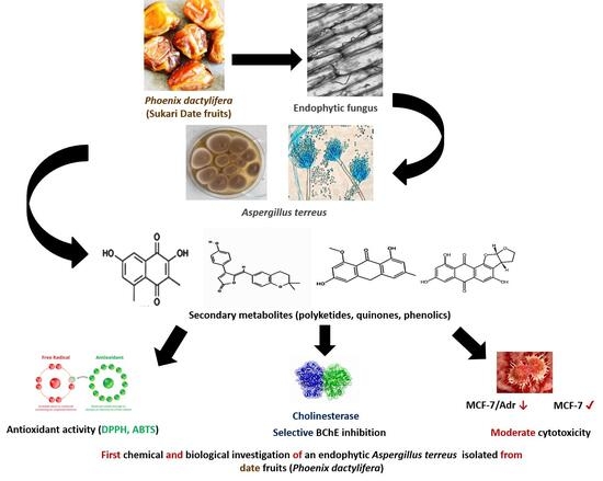
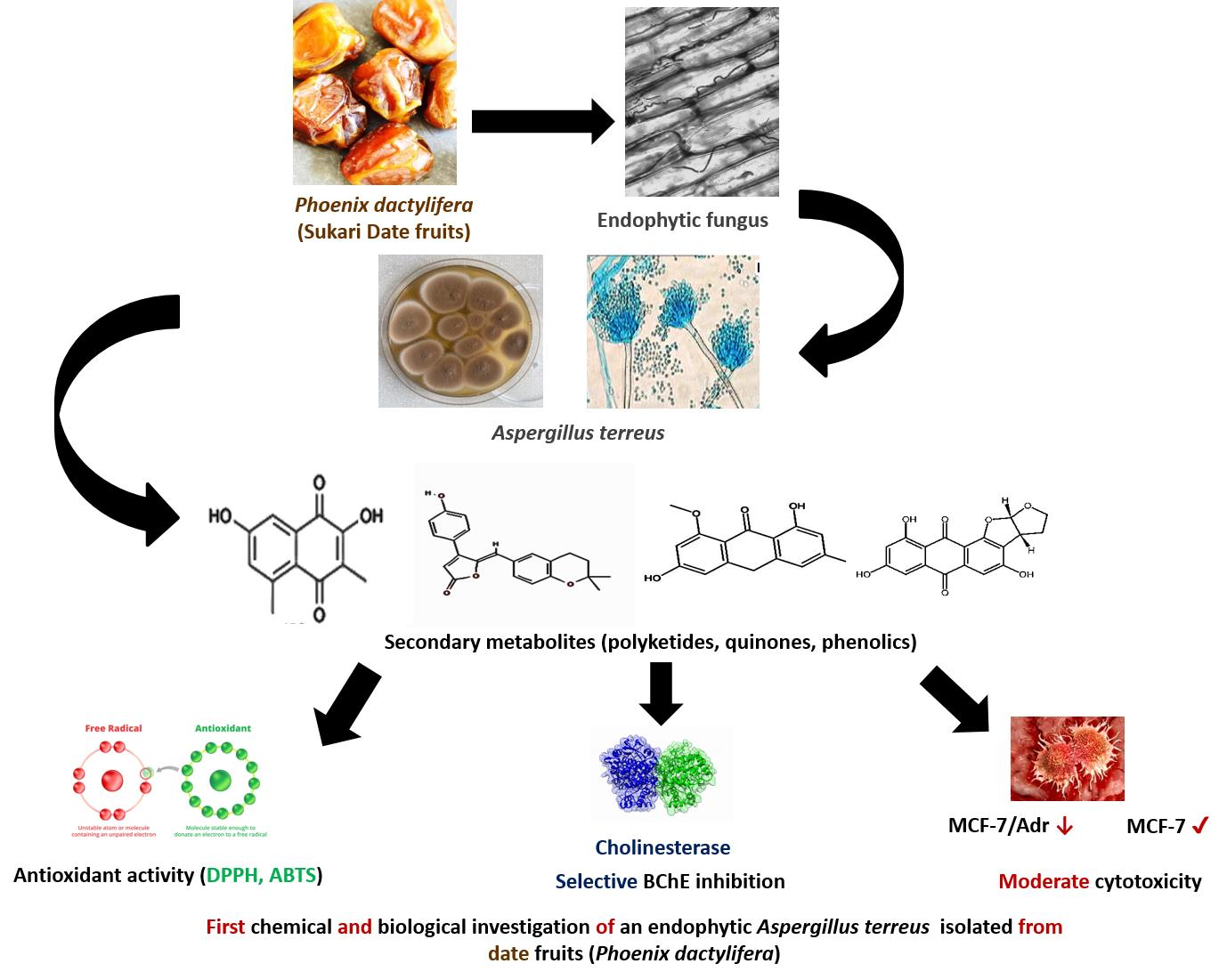
Graphical Abstract

1. Introduction

Fungi produce a wide range of secondary metabolites that possess significant biological activity, many of which can be utilized in medicine, agriculture, and biotechnology. Among them, endophytic fungi are attracting growing interest as valuable sources of bioactive natural products, as they reside symbiotically within plant tissues without causing harm to the host [1,2]. The unique ecological relationships between endophytes and their host plants frequently trigger the synthesis of metabolites that are distinct from those produced by free-living or pathogenic fungi, which makes endophytic fungi a significant source of chemical variation [3].
The date palm (Phoenix dactylifera L.) is the most extensively farmed crop in the Middle East and North Africa. It is utilized extensively and has a significant history of use in traditional medicine. The fruit is rich in nutrients, including dietary fiber, carbohydrates, proteins, minerals, vitamins, flavonoids, and phenolic components which may influence the metabolic profiles of associated endophytic microorganisms [4]. Hamad et al. [5] demonstrated that the Saudi date fruit varieties (Helwat Al Jouf, Al Sagey, and Al Sour) exhibit substantial antioxidant activity, primarily due to their phenolic compounds and flavonoids. Al Sagey cultivar demonstrated the highest antioxidant capabilities and the most substantial phenolic contents. Al Sagey and Helwat Al Jouf displayed analogous glutathione and ascorbate redox statuses, but Al Sour exhibited the lowest glutathione redox state. Despite the long-standing agricultural and cultural importance of date palms, their endophytic fungal communities—particularly those inhabiting date fruits—remain poorly explored from a chemical and biological perspective. Mahmoud et al. [6] examined the community of endophytic root fungi in date palms from three plantations located in Adrar Province (Southeast Algeria). The predominant fungal endophytes were Fusarium sp. (30.9%), followed by Aspergillus terreus (15.4%). Four isolates were evaluated for their capacity to promote wheat growth and the production of secondary metabolites [6]. Two fungi, namely Penicillium citrinum TDPEF34, and Geotrichum candidum TDPEF20, originating from healthy and brittle leaf diseased (BLD) date palm root microbiomes, respectively, were investigated for their potential to serve as cell factories for the production of bioactive secondary metabolites. Extracts from both endophytic fungi demonstrated a high content of polyphenols and flavonoids and, consequently, exhibited potent antioxidant activity [7]. Piombo et al. [8] used metagenomic analysis to compare microbial inhabitants in the pulp and peel of “Medjool” dates at different stages of fruit development. In all fruit sections, Penicillium, Cladosporium, Aspergillus, and Alternaria were the most common genera, but taxonomic distribution varied by time and tissue type. While Penicillium was more numerous in the pulp during green growth (Kimri), Aspergillus was more common in the peel during brown growth (Tamer).
Recent studies highlight A. terreus as a prolific source of structurally diverse secondary metabolites including polyketides, quinones, phenolic derivatives, and butenolides which have been isolated from terrestrial, marine, and endophytic strains [9,10]. Advanced analytical approaches such as LC-MS-based metabolomics, high-resolution mass spectrometry, and NMR spectroscopy have facilitated the identification and structural characterization of these metabolites [11]. Many of the newly discovered compounds have demonstrated significant biological activities including antimicrobial, cytotoxic, phytotoxic, and enzyme inhibitory effects [12,13]. In particular, terrein derivatives, sulfur-containing metabolites, and polyketide-derived compounds have attracted attention due to their potential pharmacological relevance [14,15]. Overall, these studies emphasize the metabolic diversity of A. terreus and support its importance as a promising source of bioactive natural products, while also highlighting the utility of metabolomics and modern natural product discovery tools [16].
Oxidative stress and enzyme dysregulation are key factors involved in the pathogenesis of several chronic diseases, including neurodegenerative disorders and cancer. Antioxidants play a critical role in mitigating oxidative damage [17], while cholinesterase inhibitors—particularly butyrylcholinesterase (BChE) inhibitors—are of growing interest due to the increased role of BChE in the progression of Alzheimer’s disease [18]. In parallel, the search for new anticancer agents remains a major focus of natural product research, and compounds capable of overcoming or bypassing multidrug resistance mechanisms are particularly highly sought. Fungal secondary metabolites, notably phenolic and quinone-containing compounds, have been reported to exhibit antioxidant, cholinesterase inhibitory, and cytotoxic activities, supporting the continued exploration of fungal sources for such bioactivities [19]. Hence, the objectives of this work were to (i) isolate and identify secondary metabolites produced by this endophytic A. terreus isolated from date fruits (Phoenix dactylifera), and (ii) evaluate the antioxidant, cholinesterase inhibitory, and cytotoxic activities of the fungal extract.

2. Materials and Methods

2.1. Isolation, Purification and Characterization of Endophytic Fungus

Fresh date samples from a popular variety (Sukkari) were collected from farms of Zulfi City, Saudi Arabia. The samples were aseptically collected in sterile plastic bags (3M Company, Maplewood, MN, USA), transported to the laboratory within four hours, and maintained at 4 °C until fungal analysis. Surface sterilization of collected samples was performed by immersing them in 70% ethanol for 5 min, followed by 5% sodium hypochlorite for an additional 5 min, and finally rinsing with sterile distilled water [20]. Sterilized dates were cut to small fragments by using a sterile scalpel and forceps, and four fragments were placed on Potato Dextrose Agar medium (PDA) containing antibiotics (streptomycin and chloramphenicol) and incubated at 28 °C for 5–7 days. The grown colonies were purified by repeated subculturing on fresh PDA plates using the hyphal tip isolation method to obtain pure cultures. This process was repeated until morphologically uniform colonies were achieved. Fungal strain was identified based on micro- and macromorphological diagnostic characteristics in culture, combined with subsequent molecular methods [21]. DNA was isolated from a pure culture of the fungal strain using Qiagen kits based on the manufacturer’s instructions (QIAGEN, Valencia, CA, USA). The universal primers ITS1 (5′-TCCGTAGGTGAACCTGCG-3′) and ITS4 (5′-TCCTCCGCTTATTGATATGC-3′) were employed to amplify the ITS regions of the fungal strain. PCR amplification was performed in a total reaction volume of 25 mL, utilizing 1× PCR buffer (DreamTaq™) in a C1000TM Thermo Cycler (Bio-Rad Laboratories GmbH, Feldkirchen, Germany). The initial denaturation phase occurred at 94 °C for 5 min followed by 35 cycles including denaturation at 94 °C for 30 s, annealing at 49 °C for 30 s, and extension at 72 °C for 1 min [22]. The last extension phase at 72 °C for 5 min was performed following the preceding 35 cycles. The DNA amplicons were sequenced utilizing the Gene Analyzer 3121 sequencer with the same primers, ITS1 and ITS4 (Macrogen Co., Seoul, South Korea). The ITS sequences were examined with BioEdit version 7.2.5. The isolate was identified by comparing sequencing data against databases with the BLAST tool from the GenBank database (http://www.ncbi.nlm.nih.gov/BLAST/, accessed on 23 March 2026).

2.2. Fermentation and Extraction of Fungal Metabolites

Fermentation of the isolated fungus was performed utilizing solid state fermentation [21] on 100 g autoclaved rice and 100 mL distilled water in 1 L Erlenmeyer flasks. After cooling, the flasks were inoculated with 2 mL spore suspension (105 cfu/mL) and incubated at 28 ± 2 °C for thirty days. After that, the fermented rice was subjected to extraction of fungal metabolites using ethyl acetate (Analytical Grade, Alpha Chemika, Mumbai, India) for 24 h at room temperature [21]. The extract was filtered, dried with a rotavapor (BÜCHI R-114, Flawil, Switzerland), and kept at −4 °C for further work.

2.3. Estimation of Total Phenolic Content

Total phenolic content determination was assessed using the modified method of Suleria et al. [23]. A half mL of the extract (10 mg/mL) was mixed with the same volume of Folin–Ciocalteu phenol reagent, followed by 1 mL of 10% Na2CO3 (AL-Nasr Chemicals Co., Cairo, Egypt), and then the mixture was incubated under shaking at 180 rpm for 60 min at 25 °C in the dark. Measurement of the absorbance was carried out at 750 nm. Phenolic content was expressed as gallic acid equivalent (GAE) (mg/g) through the standard curve equation: y = 0.0169x–0.1172, R2 = 0.9588. The standard curve of gallic acid was linear between 0.5 and 100 μg/mL.

2.4. Determination of Total Flavonoid Content

The measurement of total flavonoid content was performed following the method reported by Nicolescu et al. [24]. A 0.5 mL quantity of the extract (10 mg/mL) was carefully added to 1.0 mL of a 2% (v/v) AlCl3·6H2O (AL-Nasr Chemicals Co., Cairo, Egypt) ethanolic solution and the absorbance was measured at 430 nm after 10 min. Total flavonoid content was expressed as quercetin equivalent (QE) (mg/g) by employing the standard curve equation: y = 0.0208x–0.2381, R2 = 0.9678. The calibration curve of quercetin (Sigma-Aldrich Chemie GmbH, Taufkirchen, Germany) was linear between 0.5 and 100 μg/mL.

2.5. Antioxidant Activity Assays

2.5.1. DPPH Scavenging Activity Assay

Briefly, 1.8 mL of 0.1 mM DPPH (1,1-Diphenyl-2-picryl hydrazyl) (Sigma-Aldrich, Germany) (4 mg/100 mL of methanol) solution was added to 0.2 mL of the fungal extract in absolute methanol at various concentrations (1, 0.8, 0.6, 0.4, 0.2, 0.1 and 0.05 mg/mL) in addition to the blank. Absorbance was determined by a spectrophotometer ( Bibby Scientific Ltd., Stone, Staffordshire, UK) at 517 nm after 30 min [25]. Butylated hydroxytoluene (BHT) (Chemical Industries Development Company, Cairo, Egypt) was employed as a positive control at concentrations ranging between 100 and 10 μg/mL. The following formula was employed to determine the capacity to scavenge the DPPH radical:
% DPPH radical scavenging = (A0–A1)/A0 × 100
where A0 is the negative control absorbance (methanol (AL-Nasr Chemicals Co., Cairo, Egypt) and DPPH) and A1 is the sample absorbance (DPPH, methanol and sample). The IC50 was calculated as IC50 = (50–b)/a, obtained by interpolation from linear regression analysis where “a” represents the slope and “b” is the Y-intercept.

2.5.2. ABTS Scavenging Assay

The fungal extract was dissolved in dimethyl sulfoxide (DMSO) (MilliporeSigma—St. Louis, MI, USA) and diluted with methanol to a concentration of 10 mg/mL. Subsequently, five serial dilutions were prepared at final concentrations of 7.81, 15.625, 31.25, 62.5, and 125 μg/mL. The assay was conducted in microplates in accordance with the method of Arnao et al. [26], which was subsequently adopted by Elkholy et al. [27]. Briefly, 192 mg of 2,2′-azino-bis(3-ethylbenzothiazoline-6-sulphonic acid) (ABTS) (Sigma-Aldrich, Germany) was dissolved in distilled water and transferred to a 50 mL volumetric flask. The volume was subsequently topped up with distilled water. Then, 17 μL of 140 mM potassium persulphate was combined with 1 mL of the previous solution, and the mixture was incubated in the dark for 24 h. The final ABTS dilution used in the assay was obtained by making 1 mL of the reaction mixture up to 50 mL with methanol. The reaction was incubated at room temperature for 30 min in the dark following the addition of 10 μL of the fungal extract to 190 μL of the freshly prepared ABTS reagent in a 96-well plate. A microplate reader (BMG LABTECH GmbH, Ortenberg, Germany) was employed to measure the decrease in ABTS color intensity at 734 nm at the conclusion of the incubation period. To serve as a standard positive control, Trolox (Sigma-Aldrich, Germany) was prepared at the following final concentrations: 8.75, 6.25, 5, 3.75, and 2.5 μg/mL. The scavenging capacity was determined using the following equation:
Percentage inhibition = ((Absorbance of blank − Absorbance of the test)/(Absorbance of blank)) × 100
After that, the IC50 value was calculated.

2.5.3. Metal Chelation Assay

The fungal extract was dissolved in DMSO and subsequently diluted with methanol to a concentration of 10 mg/mL. Subsequently, five serial dilutions were prepared at concentrations of 4000, 3500, 2500, 1500, and 1000 μg/mL, resulting in final concentrations of 333.33, 500, 833.33, 1166.67, and 1333.33 μg/mL. The assay was conducted in microplates in accordance with the method of Santos et al. [28], with minimal modifications. To prepare the 96-well plate, 20 μL of the freshly prepared ferrous sulphate (Alpha Chemika, Mumbai, India) (0.3 mM) was combined with 50 μL of the fungal extract and 50 μL of acetate buffer with a pH of 6. This was followed by the addition of 30 μL of ferrozine (Sigma-Aldrich, Germany) (0.8 mM) to each well. The reaction mixture was incubated at ambient temperature for a duration of 10 min. The microplate reader was employed to measure the decrease in color intensity at 562 nm at the conclusion of the incubation period. Five serial dilutions were prepared at concentrations of 29.224, 14.612, 10.959, 7.306, and 5.8448 μg/mL from a 292.24 μg/mL EDTA (Alpha Chemika, Mumbai, India) stock solution in water. The chelation capacity was determined using the following equation:
Percentage inhibition = ((Absorbance of blank − Absorbance of the test)/(Absorbance of blank)) × 100
After that, the IC50 value was calculated.

2.5.4. ORAC Assay

The following dilutions were prepared, 1000, 800, 600, 500, 400, 200, 100, and 50 μM, all of which were derived from a 2 mM Trolox stock solution in MeOH. The fungal extract was dissolved in 50% methanol and subsequently diluted with methanol to a concentration of 40 μg/mL. The assay was conducted in accordance with the method of Liang et al. [29], with modifications made by Elkholy et al. [27]. In brief, 10 μL of the prepared sample was incubated with 30 μL of fluoresceine (Sigma-Aldrich, Germany) (100 nM) at 37 °C for 10 min. For background measurement, fluorescence measurements were conducted for three cycles (cycle time, 90 s) at 485 EX and 520 EM nm. Subsequently, 70 μL of freshly prepared 2,2′-Azobis(2-amidinopropane) dihydrochloride (AAPH) (Sigma-Aldrich, Germany) (240 mM) was promptly added to each well. The fluorescence measurement (485 EX, 520 EM, nm) was maintained for 60 min, with a maximum of 40 cycles, each lasting 90 s. μM Trolox equivalents were used to compute the antioxidant effect of the fungal extract through substitution in the linear regression equation:
y = 3526.176x + 527,521.245; R2 = 0.997

2.6. The Brine Shrimp Cytotoxicity Test

The brine shrimp cytotoxicity test (BSCT) is a method used to determine the cytotoxicity of the fungal extract to brine shrimp larvae. The extract was dissolved in DMSO at varying concentrations (100–2.5 mg/mL). Then, 100 μL of each extract concentration was added to vials containing 5 mL of seawater and 10 brine shrimp larvae. After 24 h, the number of viable larvae in each vial was counted. DMSO was used as a negative control, and the percentage mortality was determined. The concentration of the extract that killed 50% of the larvae (LC50) was calculated using probit analysis [30].

2.7. Cell Culture Cytotoxicity Assay

MCF-7 (Breast Adenocarcinoma) and MCF-7-Adr (Doxorubicin-resistant Breast Cancer) were purchased from Nawah Scientific Inc., located in Mokatam, Cairo, Egypt.
Dulbecco’s Modified Eagle’s Medium (DMEM) (Sigma-Aldrich, Germany) supplemented with 100 mg/mL of streptomycin, 100 units/mL of penicillin, and 10% of heat-inactivated fetal bovine serum was used to maintain the cells at 37 °C in a humidified 5% (v/v) CO2 atmosphere. The SRB assay was used to evaluate the viability of the cells. Aliquots of 100 μL cell suspension (5 × 10³ cells) were added to 96-well plates and incubated in complete media for 24 h. The cells were treated with an additional aliquot of 100 μL medium, which contained fungal extract at varying concentrations (100, 10, 1, 0.1, and 0.01 μg/mL). Doxorubicin was produced as a standard positive control at varying concentrations. The cells were fixed by replacing the media with 150 μL of 10% trichloroacetic acid (TCA) and incubating at 4 °C for 1 h after their exposure to the extract. The cells were rinsed five times with distilled water after the TCA solution was removed. Aliquots of 70 μL Sulforhodamine B (SRB) (Sigma-Aldrich, Germany) solution (0.4% w/v) were added and incubated at room temperature for 10 min in a dark place. Plates were rinsed three times with 1% acetic acid and allowed to air-dry overnight. Then, 150 μL of TRIS (10 mM) was added to dissolve the protein-bound SRB stain. The absorbance was measured at 540 nm using an Infinite F50 microplate reader (Tecan Group Ltd., Männedorf, Switzerland), and the IC50 value was calculated [31].

2.8. Acetylcholine Esterase and Butyrylcholine Esterase Inhibition Assay

The samples were initially dissolved in DMSO and subsequently diluted to the desired concentrations with distilled water. The resulting fungal extracts were prepared at the following final concentrations: 500, 250, 125, 62.5, and 50 μg/mL. The final concentrations of standard donepezil in methanol were 1, 0.1, 0.01, 0.001, and 0.0001 μM. These concentrations are equivalent to 0.416, 0.0416, 0.00416, 0.000416 and 0.0000416 μg/mL. The enzyme acetylcholinesterase (Sigma-Aldrich, Germany) with its substrate acetylthiocholine iodide, the enzyme butyrylcholineesterase with its substrate butyrythiolcholine iodide, and the indicator (DTNB Ellman’s reagent) (Sigma-Aldrich, Germany) were utilized in the following assay. The assay was carried out according to the method of Osman et al. [32] with minor modifications. In brief, 10 μL of the indicator solution (0.4 mM in buffer (1): 100 mM tris buffer, pH 7.5) was added to a 96-well plate, followed by 20 μL of enzyme solution (acetylcholine esterase or butyrylcholine esterase at a final concentration of 0.02 U/mL in buffer (2): 50 mM tris buffer, pH 7.5, containing 0.1% bovine serum albumin). Subsequently, 20 μL of the fungal extract solution was introduced, followed by the addition of 140 μL of buffer (1). The mixture was allowed to sit for 15 min at room temperature. Subsequently, 10 μL of the substrate (0.4 mM acetylcholine iodide buffer (1)) was immediately added to all wells. The plate was incubated in a dark place for 20 min at room temperature. At the end of the incubation time, the color was assessed at 412 nm with the FluoStar Omega microplate reader, and the IC50 value was calculated.

2.9. Liquid Chromatography–Mass Spectrometry Analysis (LC/MS/MS)

The sample was analyzed using liquid chromatography–electrospray ionization–tandem mass spectrometry (LC-ESI-MS/MS) (Agilent Technologies, Waldbronn, Germany) with an Exion LC AC system for separation and a SCIEX Triple Quad 5500+ MS/MS system fitted with electrospray ionization (ESI) for detection. The separation was conducted using an Ascentis® C18 column (4.6 × 150 mm, 3 µm) (Merck-Sigma group, Schnelldorf, Germany). The mobile phases were composed of two eluents: A consisted of 0.1% formic acid and B consisted of acetonitrile (LC grade). The mobile phase gradient was programmed as follows: 10% B at 0–1 min, 10–90% B from 1 to 33 min, 90% B from 33 to 37 min, and 10% B from 37.1 to 40 min. The injection volume was 10 µL, and the flow rate was 0.7 mL/min. The scan (EMS-IDA-EPI) was conducted in negative ionization mode for MS/MS analysis [33]. The identification of secondary metabolites was performed using a LC–ESI–MS/MS SCIEX Triple Quad 5500+ MS/MS system for putative dereplication, which is low resolution mass measurement, so tentative identification could be made of the compounds based on both detected mass value and fragmentation patterns comparison with Aspergillus compound database, literature reports and established databases prediction libraries [34,35].

2.10. Data Analysis

Three replicates of each experiment were implemented. The data were analyzed and established using Microsoft Excel® and SPSS software, version 16, and presented as the mean ± SD. Data were established by analysis of variance (one-way ANOVA) as being below the 0.05 level of significance.

3. Results

3.1. Identification of Fungal Strain

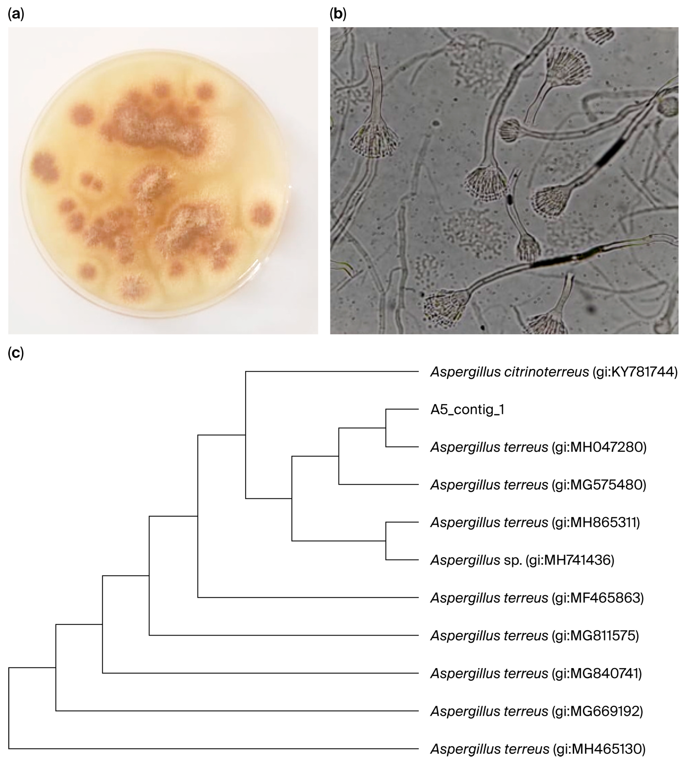
The current investigation utilized an endophytic fungal strain obtained from Sukkari date fruits. This strain was identified using morphological and molecular techniques. The fungal colony cultivated on PDA exhibited rapid growth, characterized by a cinnamon-brown color and a rough texture with cottony mycelium. Microscopic examination revealed elongated, colorless conidiophores with biserrate conidia (Figure 1). High-quality DNA isolated from endophytic fungus utilized ITS1 and ITS4 as primers for PCR. The genomic DNA of the successfully amplified fungus measured 634 bp in length. The results indicated a fungus strain with almost 99% similarity to Aspergillus terreus (gi:MH047280). The fungus strain was designated A. terreus (SU-5) with accession number MH047280.1 (Supplementary Material Table S1).

3.2. Total Phenolic and Flavonoid Content and Antioxidant Activities of A. terreus Extract

Table 1 presents the total concentrations of phenolics and flavonoids in endophytic A. terreus during solid-state fermentation, revealing a phenolic content of 93.13 mg GAE/g and a flavonoid content of 22.70 mg QE/g. The EtOAc extract of A. terreus exhibited a DPPH IC50 value of 575.64 μg/mL, but the IC50 for ABTS radical scavenging activity was reported as 50.18 μg/mL. The fungal extract’s chelating activity was evaluated, showing an IC50 value of 1007.82 μg/mL for ferrous ion chelation. The evaluated fungal extract demonstrated an antioxidant scavenging activity of 3879.42 µmol TE/g against peroxyl radicals in the ORAC testing. The results for BHT, Trolox, and EDTA, serving as standard positive controls in several antioxidant activity assays, are presented in Table 1.

3.3. Anticancer Activities of A. terreus Extract

The ethyl acetate extract of A. terreus was evaluated for its effect on the viability of two cancer cell lines, MCF-7 and MCF-7-Adr, using the SRB assay. The IC50 values were determined from dose–response curves, producing 424.73 μg/mL for MCF-7 and 1140.77 μg/mL for MCF-7-Adr, in comparison to doxorubicin. The Artemia cytotoxicity assay of the fungal extract demonstrated an LC50 value of 1402.92 µg/mL (Table 2).

3.4. Effect of A. terreus Extract on the Inhibition of Acetylcholine and Butyrylcholine Esterases

The results presented in Table 3 indicated that, in comparison with standard donepezil, the EtOAc extract of endophytic A. terreus exhibited IC50 values of 456.90 and 157.10 μg/mL for the inhibition of acetylcholinesterase and butyrylcholinesterase, respectively.

3.5. LC-ESI-MS/MS Analysis of A. terreus Metabolites

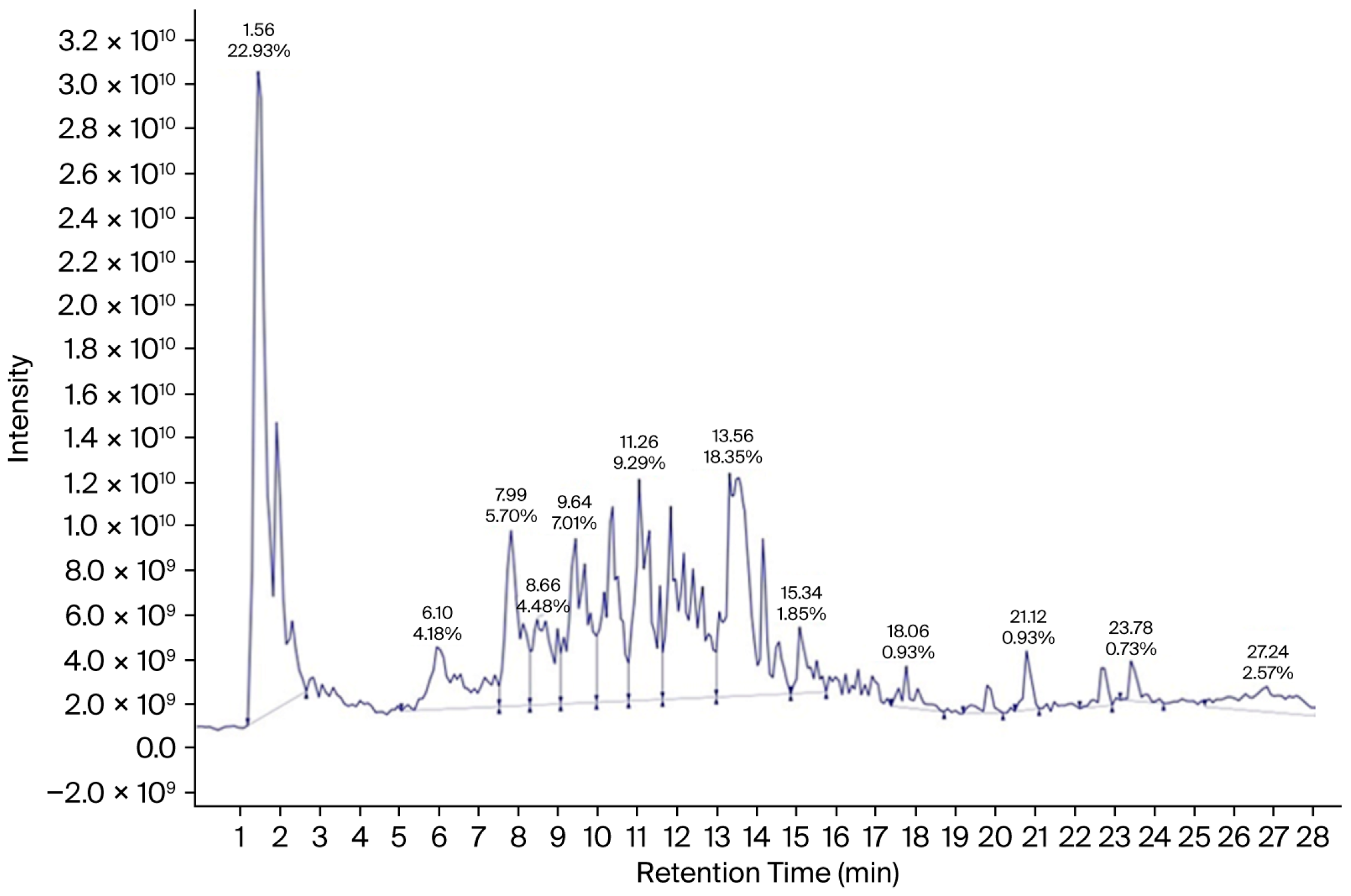
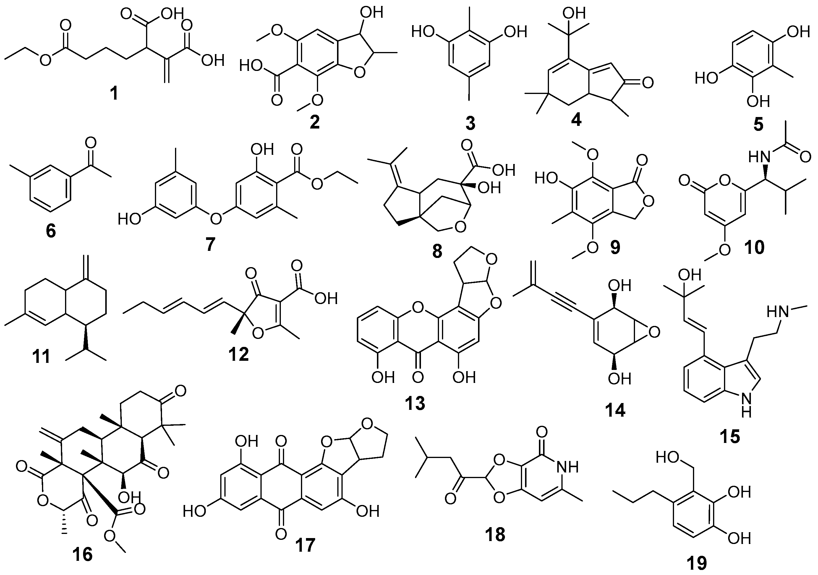
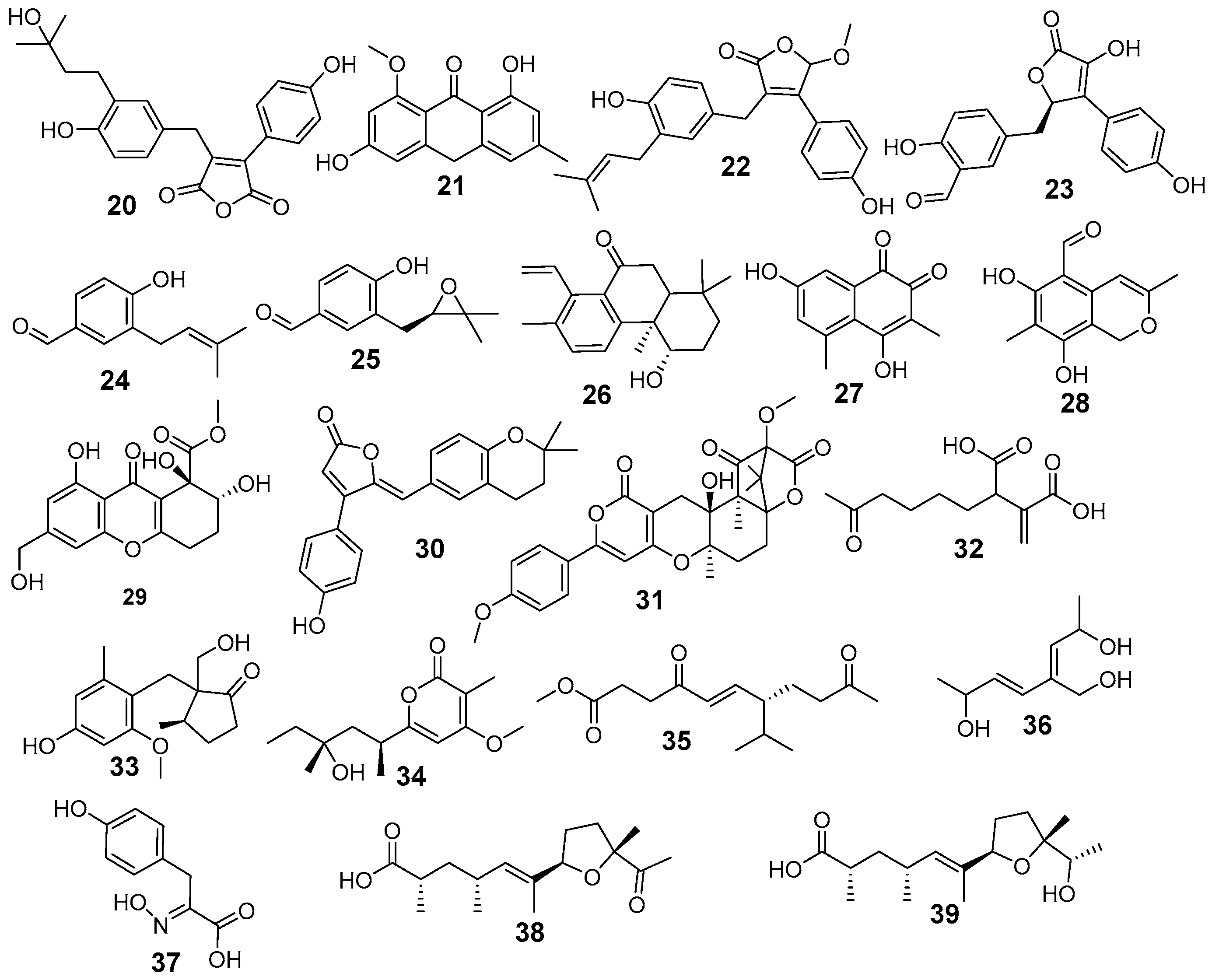
LC-ESI-MS/MS analysis of A. terreus EtOAc extract was carried out for the separation and detection of secondary metabolites. Negative ionization mode was used to characterize the corresponding signals and the total ion current map of the extract was obtained. Figure 2 illustrates the TIC of A. terreus EtAOc extract. Structural and two-stage mass spectrometry analysis was employed to obtain the mass, characteristic fragmentations, and molecular formula of the components. Additionally, previous studies of isolated compounds from the Aspergillus genus and A. terreus were utilized as an identification tool of the detected compounds by comparing constituents’ characteristics with the published data. Analysis of the A. terreus extract revealed that several compounds (39) were proposed in negative ion mode, depending on their retention time, precursor ion, and MS2 fragmentation patterns compared with the Competitive Fragmentation Modeling for Metabolite Identification online database (Table 4, Figure 3 and Figure 4). The detected compounds were arranged according to their retention time.
The molecular ion mass peaked at m/z 253.06, 233.08, 251.11, 203.06, 473.24, 381.08, 379.15, 383.08, 189.06, 205.05, 347.07, 509.21, 277.15, and 267.17 [M − H], fitted with asperterreusine B, brasilanone A, aspterric acid, γ-cadinene, terreustoxin F, aperterone B, (±)-asperteretal D,3-hydroxy-4-(4-hydroxyphenyl)-5-methoxycarbonyl-5-(4-hydroxy-3-formylbenzyl)-2,5-dihydro-2-furanone, 4-hydroxy-3-(3-methylbut-2-enyl)benzaldehyde, terreprenphenol C, rubrolide S, terreulactone A, aspereusin C, and aspereusin E, respectively. Moreover, the mass ion peaks at Rt of 2.10, 3.33, 3.61, 6.16, 6.65, 7.12, 8.70, 8.75, 8.77, 10.67, 10.76, 10.84, 12.05, 14.71, 14.80, 15.24, 15.41, 18.65, 19.78, 23.40, 23.72, and 27.42 min gave hits for tensyuic acid F, 2,5-dimethylresorcinol, versicolin, 1-(3-Methylphenyl)-ethanone, ethericin B, 7-methoxyporriolide, asperfuranone B, dihydrodemethylsterigmatocystin, asperpentyn, isoversicolorin C, isoflavipucine, asperpanoid A, anhydroasperflavin, diterpenoid 6-deoxyaspergiloid C, aspergiodiquinone, anishidiol, aspergillusone B, asperitaconic acid C, nipyrone C, aspinotriol A/B, asperic acid A, and asperic acid B, respectively. The results of negative mode analysis showed the existence of nitrogen-containing compounds at Rt of 7.19, 9.07, and 23.68 min, corresponding to molecular formulae C12H17NO4, C16H22N2O, and C9H9NO4 assigned for campyrone C, ergotryptamine, and 4-hydroxyphenylpyruvic acid oxime, respectively (Supplementary Material; representative MS/MS spectra and additional raw data).

4. Discussion

Endophytic fungi are a source of a variety of secondary metabolites that are important but not extensively investigated, especially those associated with plants adapted to arid and semi-arid environments, such as date palms (Phoenix dactylifera). In this study, eleven fungal strains were isolated from Sukkari date fruits following plating on PDA and subsequent purification. Among them, Aspergillus terreus (SU-5) was selected for detailed analysis based on its metabolite profile and superior bioactivity, which aligns with previous reports highlighting its potential as a producer of bioactive secondary metabolites [9,10,11,12,13,14,15,16]. The chemical analysis of an endophytic A. terreus (SU-5) revealed a diverse range of known fungal metabolites, reflecting the high biosynthetic capacity of this species under endophytic conditions. Thirty-nine metabolites were detected, including polyketides, quinones, and phenolics. Although these compounds have previously been reported from various A. terreus strains [37,42,45,50,54,56,57,58,64,67], this is the first report of metabolites such as versicolin, isoversicolorin C, anhydroasperflavin, aspergiodiquinone, anishidiol, aspergillusone B, and nipyrone C from an endophytic A. terreus inhabiting date fruits [39,51,55,61,62,63,68]. One limitation of the present study is that fermentation was performed using a single substrate, which may influence the metabolic profile of the fungus. Previous studies have shown that variations in culture conditions and nutrient composition can significantly affect fungal secondary metabolite production and diversity [72,73]. Therefore, future studies should investigate the use of alternative substrates and optimized fermentation conditions to explore potential changes in metabolite profiles. Additionally, the LC–MS-based metabolite identification performed in this study provides only tentative assignments based on database comparisons. Consequently, further work should focus on the isolation and purification of the major bioactive compounds followed by structural elucidation using spectroscopic techniques such as NMR and HR-MS to confirm their identities [74]. The extract of A. terreus exhibited a substantial concentration of phenolic compounds (93.13 ± 1.57 mg GAE/g) and flavonoids (22.70 ± 0.57 mg QE/g) in comparison to previous research; Das et al. [75] indicated that the total phenolic content of extracts from eleven fungal endophytes derived from Zingiber nimmonii (J. Graham) Dalzell varied between 10.8 ± 0.7 and 81.6 ± 6.0 mg GAE/g dry extract. Flavonoids were detected in eight extracts, ranging from 5.2 ± 0.5 to 24.3 ± 0.9 mg QE/g of dry extract. Bipolaris specifera extracts had the highest total phenolic content (81.58 ± 6.0 mg GAE/g dry extract), followed by A. terreus. Nectria hematococca demonstrated elevated flavonoid content (24.3 ± 0.9 mg CE/g dry extract), but A. terreus displayed a lower concentration (11.3 ± 0.4 mg CE/g dry extract). In another investigation, Gautam et al. [76] found that the total phenolic content of the endophytic fungus Nigrospora sphaerica, isolated from the pantropical weed Euphorbia hirta L., was highest in EtOAcE fermented in potato dextrose broth (PDB) medium, measuring 77.74 ± 0.046 mg GAE/g. The significant production of phenolic compounds and flavonoids generated by A. terreus (SU-5) may result from its habitat of date fruits, which possess natural components that promote A. terreus metabolism, hence enabling the synthesis of phenolic and flavonoid compounds.
Interestingly, the crude extract of Aspergillus terreus showed a relatively high flavonoid content according to the aluminum chloride colorimetric assay; however, none of the metabolites identified by LC–MS analysis could be classified as flavonoids. This discrepancy may be attributed to the known limitations of the aluminum chloride method, which is not entirely specific for flavonoids [77,78]. Several non-flavonoid phenolic compounds can also form complexes with aluminum ions, leading to an overestimation of flavonoid content [77,79]. In the present extract, compounds such as anthraquinones, xanthones, and polyhydroxylated quinones contain keto-hydroxyl or ortho-dihydroxyl functional groups that may produce measurable responses in this assay, thereby contributing to the values expressed as quercetin equivalents (QE). Furthermore, LC–MS analysis allowed tentative identification of metabolites through comparison with available databases, and no flavonoids previously reported from fungi matched the observed molecular masses and fragmentation patterns. These results suggest that the predominant metabolites in the extract are more likely non-flavonoid polyketide derivatives rather than true flavonoids. Therefore, future studies will focus on the isolation of the major bioactive metabolites, followed by structural confirmation using NMR spectroscopy.
Sugars such as glucose, fructose, and sucrose constitute approximately 25–35% of the dry weight of dates and may serve as carbon sources that can be metabolized into intermediates such as acetyl-CoA and malonyl-CoA, which are commonly involved in polyketide biosynthesis. In addition, aromatic amino acids such as phenylalanine and tyrosine present in dates could potentially contribute to metabolic pathways related to phenolic compound formation through the shikimate pathway. Fatty acids, including oleic and palmitic acids, may also undergo catabolism to generate acetyl-CoA, which can serve as a precursor for polyketide chain elongation. However, these proposed links remain hypothetical and require further biochemical and metabolic investigations for confirmation [4,80]. The antioxidant activity of the EtOAc extract of A. terreus was determined using various in vitro assays: DPPH, ABTS radical scavenging, ferrous ion chelation, and peroxyl radicals in the ORAC assay. The extract exhibited variable antioxidative activity, with an IC50 value of 575.64 μg/mL for DPPH, 50.18 μg/mL for ABTS, 1007.82 μg/mL for ferrous ion chelation, and 3879.42 µmol TE/g against peroxyl radicals in the ORAC assay. All tests, in comparison to the standard antioxidants (BHT, Trolox, and EDTA), demonstrated elevated IC50 values, indicating diminished antioxidant efficacy. It is essential to emphasize that the study assessed crude fungal extract rather than purified components [75]. However, our study showed lower IC50 values when estimated with DPPH and ABTS assays in comparison to Zhou et al. [81], who reported that the fungal endophyte Neopestalotiopsis protearum exhibited an IC50 value of 1240 μg/mL when estimated with ABTS and 1800 μg/mL with the DPPH method. Another study by Elhosari et al. [82] indicated that the extract of Aspergillus sp. (TMP16) isolated from Tabernaemontana pandacaqui leaves showed considerable antioxidant capacity, measuring 881.49 ± 44.6 μM TE/mg with the DPPH assay and 866.86 ± 50.0 μM TE/mg through the ABTS assay. Five endophytic strains, namely Aspergillus niger, Penicillium glabrum, Alternaria alternata, A. tenuissima, and Mucor circinelloides, isolated from the stems of Gundelia tournefortii, were evaluated for antioxidant activity using the DPPH assay. The antioxidant activity of isolates indicated that A. alternata extract (IC50 = 471 ± 29 μg/mL) exhibited the most potent antioxidant activities, followed by A. tenuissima extract (IC50 = 512 ± 19 μg/mL), while the extracts of M. circinelloides and A. niger exhibited no antioxidant activities [83]. Das et al. [75] indicated that the extract of B. specifera exhibited elevated levels of total phenolic compounds and ABTS scavenging ability among all isolates, although minimal DPPH scavenging action was shown (1057.2 ± 122.3 μg/mL). Conversely, A. tenuissima had significant DPPH scavenging ability (96.9 ± 2.4 μg/mL) followed by A. terreus (IC50 123.3 ± 7.6 μg/mL) and N. hematococca (IC50 133.4 ± 5.3 μg/mL), despite its total phenolic contents being lower than those of B. specifera. The numbers of hydroxyl moieties attached to the aromatic ring are favorable for the DPPH radical scavenging activity of phenolic acids. In our study, 13 phenolic compounds were found from a total of 39 compounds. Versicolin and isoversicolorin C possess three hydroxyl groups, whereas asperteretal D, 3-hydroxy-4-(4-hydroxyphenyl)-5-methoxycarbonyl-5-(4-hydroxy-3-formylbenzyl)-2,5-dihydro-2-furanone, and anishidiol includes two hydroxyl groups. The antioxidant efficacy of the extract is attributed to the significant presence of isoversicolorin C, as indicated by the peak area analysis. Yang et al. [51] isolated the mangrove-derived endophytic fungus Aspergillus nidulans MA-143, producing isoversicolorin C, under 0.1% ethanol stress, demonstrating antibacterial efficacy against Gram-negative bacteria such as E. coli, M. luteus, and V. vulnificus. In comparison to the assays that were tested, the ABTS assay demonstrated a high level of antioxidant activity. This was consistent with the fact that the majority of the phenolic compounds identified (versicolin, ethericin B, isoversicolorin C, anhydroasperflavin, aspergiodiquinone, aspereusin C, and aspereusin E) are polyphenolic, quinonoid, or polar. ABTS favors structures with more efficient electron transfer, which is poor for DPPH [84]. The fungal extract exhibited weak cytotoxic activity against MCF-7 cells (IC50 = 424.73 ± 8.29 μg/mL) and markedly reduced the activity against MCF-7/Adr cells (IC50 = 1140.77 ± 21.33 μg/mL), yielding a resistance index of 2.68. This differential response suggests limited effectiveness against multidrug-resistant cancer phenotypes and indicates that the cytotoxic constituents may not be substrates for resistance-associated transporters. The moderate activity observed is consistent with the presence of quinone- and polyketide-based metabolites such as aspergiodiquinone, isoversicolorin C, and aspergillusone B, which have previously been associated with antiproliferative effects in cancer cell models [85]. Although these results indicate potential antiproliferative properties, the observed activity should be interpreted cautiously. The potency of these metabolites remains moderate compared with clinically used anticancer agents, and further studies, including mechanistic investigations and in vivo validation, are required to determine their potential therapeutic relevance. In addition, the fungal extract exhibited weak inhibitory activity against acetylcholinesterase (IC50 = 456.90 µg/mL) and slightly stronger inhibition of butyrylcholinesterase (IC50 = 157.10 µg/mL), indicating a degree of selectivity toward BChE. This selectivity is pharmacologically relevant, as BChE activity is known to increase during the progression of Alzheimer’s disease. Phenolic and quinone-containing metabolites, including rubrolide S, anhydroasperflavin, aspergiodiquinone, and aspereusin derivatives, are likely contributors to this activity due to their ability to interact with the active and peripheral sites of cholinesterase enzymes through hydrogen bonding and π–π interactions [86]. However, the observed inhibition was moderate compared with standard cholinesterase inhibitors. Therefore, these findings should be considered preliminary, and further studies are required to clarify their mechanism of action and potential relevance for cholinesterase-related disorders.

5. Conclusions

For the first time, this study highlighted the potential role of the endophytic fungus Aspergillus terreus isolated in date fruits (Phoenix dactylifera) as a source of a diverse array of secondary metabolites including polyketides, quinones, and phenolics. The fungal extract of A. terreus exhibited a significant concentration of phenolic compounds that correlated with its antioxidant activity, moderate cytotoxic activity against MCF-7 cells, and inhibitory effects against AChE and BuChE enzymes. These findings support further research into the eco-friendly sourcing of antioxidants from endophytes residing in date fruits, potentially yielding sustainable alternatives to synthetic compounds. Metabolites of A. terreus showed multifunctional potential, integrating antioxidant and cytotoxicity effects, which could be advantageous for food preservation or pharmaceuticals.

Supplementary Materials

The following supporting information can be downloaded at: https://www.mdpi.com/article/10.3390/jof12040249/s1, 1. Raw data 2. Representative MS/MS spectra for the major identified metabolites (e.g., Isoversicolorin C, Isoflavipucine, and Rubrolide S). 3. The contig sequences used for phylogenetic analysis (Table S1).

Author Contributions

A.A.-H.: Research Design, identification methodology, writing—review and editing. M.A. (Mohammed Alaidarous): writing—review and editing. A.A.: conceptualization, Biological methodology, and data analysis. Y.M.: data analysis and validation. S.B.: writing—review and editing. M.A. (Mohamed Abouelela): chemical methodology, investigation, and analysis. A.H.: Biological methodology, data analysis, writing—review and editing, validation. All authors have read and agreed to the published version of the manuscript.

Funding

This study was funded by the Deanship of Postgraduate Studies and Scientific Research at Majmaah University under project number (R-2026-122).

Institutional Review Board Statement

Not applicable.

Informed Consent Statement

Not applicable.

Data Availability Statement

Data of the measurement results are available from the authors.

Acknowledgments

The authors extend the appreciation to Deanship of Graduate Studies and Scientific Research at Majmaah University for supporting this work under project number (R-2026-122).

Conflicts of Interest

The authors declare no conflicts of interest.

Abbreviations

The following abbreviations are used in this manuscript:
A.Aspergillus
AAPH2,2′-Azobis(2-amidinopropane) dihydrochloride
ABTS2,2′-azino-bis(3-ethylbenzothiazoline-6-sulfonic acid)
AChEAcetylcholinesterase
BHTButylated hydroxytoluene
BSCTBrine shrimp cytotoxicity test
BuChEButyrylcholinesterase
CE/gCatechin equivalent per gram
DPPH2,2-diphenyl-1-picrylhydrazyl
DMEMDulbecco’s Modified Eagle’s Medium
DMSODimethyl sulfoxide
DTNB5,5′-dithio-bis-(2-nitrobenzoic acid)
EDTAEthylenediaminetetraacetic Acid
EtOAcEEthyl acetate
GAE/gGallic acid equivalent per gram
IC50Half-maximal inhibitory concentration
LC/MS/MSLiquid chromatography-mass spectrometry analysis
MCF-7Breast Adenocarcinoma
MCF-7-AdrDoxorubicin-resistant Breast Cancer
ORACOxygen Radical Absorbance Capacity
PDAPotato Dextrose Agar medium
QE/gQuercetin equivalents per gram
SRBSulforhodamine B
TCATrichloroacetic acid
TE/g Trolox equivalents per gram
TICTotal ion chromatogram
PCRPolymerase chain reaction

References

  1. Hashem, A.H.; Attia, M.S.; Kandil, E.K.; Fawzi, M.M.; Abdelrahman, A.S.; Khader, M.S.; Khodaira, M.A.; Emam, A.E.; Goma, M.A.; Abdelaziz, A.M. Bioactive compounds and biomedical applications of endophytic fungi: A recent review. Microb. Cell Factories 2023, 22, 107. [Google Scholar] [CrossRef] [PubMed]
  2. Wani, Z.A.; Ashraf, N.; Mohiuddin, T.; Riyaz-Ul-Hassan, S. Plant-endophyte symbiosis, an ecological perspective. Appl. Microbiol. Biotechnol. 2015, 99, 2955–2965. [Google Scholar] [CrossRef]
  3. Dhayanithy, G.; Subban, K.; Chelliah, J. Diversity and biological activities of endophytic fungi associated with Catharanthus roseus. BMC Microbiol. 2019, 19, 22. [Google Scholar] [CrossRef]
  4. Al-Shwyeh, H.A. Date palm (Phoenix dactylifera L.) fruit as potential antioxidant and antimicrobial agents. J. Pharm. Bioallied Sci. 2019, 11, 1–11. [Google Scholar] [CrossRef]
  5. Hamad, I. Phenolic profile and antioxidant activity of Saudi date palm (Phoenix dactylifera L.) fruit of various cultivars. Life Sci. J. 2014, 11, 1268–1271. [Google Scholar]
  6. Mahmoud, F.M.; Yekkour, A.; Boudffeur, S.; Errahmani, M.B.; Krimi, Z. Root endophytic fungi from date palm (Phoenix dactylifera L.) grove of Algerian Sahara and screening of their growth promotion activities. Adv. Environ. Biol. 2016, 10, 18–26. [Google Scholar]
  7. Mefteh, F.B.; Daoud, A.; Bouket, A.C.; Alenezi, F.N.; Luptakova, L.; Rateb, M.E.; Kadri, A.; Gharsallah, N.; Belbahri, L. Fungal root microbiome from healthy and brittle leaf diseased date palm trees (Phoenix dactylifera L.) reveals antibacterial and antifungal secondary metabolites. Front. Microbiol. 2017, 8, 307. [Google Scholar] [CrossRef]
  8. Piombo, E.; Abdelfattah, A.; Danino, Y.; Salim, S.; Feygenberg, O.; Spadaro, D.; Wisniewski, M.; Droby, S. Characterizing the fungal microbiome in date (Phoenix dactylifera) fruit pulp and peel from early development to harvest. Microorganisms 2020, 8, 641. [Google Scholar] [CrossRef] [PubMed]
  9. Ying, Y.; Tu, S.; Ni, J.; Lu, X.; Hu, X.; Lei, P.; Li, X.; Wang, Y.; Jin, G.; Wang, H. Secondary metabolites from Aspergillus terreus F6-3, a marine fungus associated with Johnius belengerii. Fitoterapia 2023, 170, 105662. [Google Scholar] [CrossRef]
  10. Huang, X.; Wang, Y.; Li, G.; Xia, J.; Qin, J.; Wang, W. Secondary metabolites from the deep-sea derived fungus Aspergillus terreus MCCC M28183. Front. Microbiol. 2024, 15, 1361550. [Google Scholar] [CrossRef]
  11. Fayek, M.; Ebrahim, H.; Abdel-Aziz, M.; Taha, H.; Moharram, F. Bioactive metabolites identified from Aspergillus terreus derived from soil. AMB Express 2023, 13, 107. [Google Scholar] [CrossRef]
  12. Hamed, A.; Abdel-Razek, A.; Abdelwahab, A.; El-Taweel, A.; GabAllah, M.; Sewald, N.; Shaaban, M. Diverse bioactive secondary metabolites from Aspergillus terreus: Antimicrobial, anticancer, and anti-SARS-CoV-2 activity studies. Z. Naturforsch. C 2024, 79, 361–369. [Google Scholar] [CrossRef]
  13. Peng, B.; Peng, Q.; She, J.; Yang, B.; Zhou, X. Secondary metabolites from the coral-derived fungus Aspergillus terreus SCSIO41404 with pancreatic lipase inhibitory activities. Rec. Nat. Prod. 2022, 16, 639–644. [Google Scholar] [CrossRef]
  14. Wang, W.; Xu, K.-W.; Wang, M.; Wu, P.; Zhang, Z.-R.; Gao, X.; Li, Y.-Q.; Wu, G.-X.; Zhang, C.-S.; Zhao, D.-L. Phytotoxic and antimicrobial terrein derivatives and butenolides isolated from the endophytic fungus Aspergillus terreus HT5. J. Agric. Food Chem. 2023, 71, 20713–20723. [Google Scholar] [CrossRef] [PubMed]
  15. Zhang, G.-Y.; Liu, B.-J.; Pan, H.-L.; Li, H.-J.; Huang, Z.-F.; Mahmud, T.; Ma, W.-Z.; Lan, W.-J. Four sulfur-containing compounds with anti-colon cancer effect from marine-derived fungus Aspergillus terreus. Fitoterapia 2024, 175, 105967. [Google Scholar] [CrossRef] [PubMed]
  16. Zheng, Y.; Yao, G.; Li, J.; Han, N.; Mao, J.; Zhang, Y.; Wang, C. Undescribed alkaloids, peptides and polyketides from marine sediment-derived fungus Aspergillus terreus PPS1. Phytochemistry 2025, 234, 114423. [Google Scholar] [CrossRef] [PubMed]
  17. Stone, J.K.; Polishook, J.D.; White, J.F. Endophytic fungi. In Biodiversity of Fungi: Inventory and Monitoring Methods; Elsevier Academic Press: Burlington, MA, USA, 2004; pp. 241–270. [Google Scholar]
  18. Arora, D.S.; Chandra, P. Antioxidant activity of Aspergillus fumigatus. ISRN Pharmacol. 2011, 2011, 619395. [Google Scholar] [CrossRef]
  19. Hamed, A.A.; El-Shiekh, R.A.; Mohamed, O.G.; Aboutabl, E.A.; Fathy, F.I.; Fawzy, G.A.; Al-Taweel, A.M.; Elsayed, T.R.; Tripathi, A.; Al-Karmalawy, A.A. Cholinesterase inhibitors from an endophytic fungus Aspergillus niveus Fv-er401: Metabolomics, isolation and molecular docking. Molecules 2023, 28, 2559. [Google Scholar] [CrossRef]
  20. dos Reis, J.B.A.; Lorenzi, A.S.; do Vale, H.M.M. Methods used for the study of endophytic fungi: A review on methodologies and challenges, and associated tips. Arch. Microbiol. 2022, 204, 675. [Google Scholar] [CrossRef]
  21. Al Mousa, A.A.; Mohamed, H.; Hassane, A.M.; Abo-Dahab, N.F. Antimicrobial and cytotoxic potential of an endophytic fungus Alternaria tenuissima AUMC14342 isolated from Artemisia judaica L. growing in Saudi Arabia. J. King Saud Univ. Sci. 2021, 33, 101462. [Google Scholar] [CrossRef]
  22. Jayatilake, P.L.; Munasinghe, H. Antimicrobial Activity of Cultivable Endophytic and Rhizosphere Fungi Associated with “Mile-a-Minute,” Mikania cordata (Asteraceae). BioMed Res. Int. 2020, 2020, 5292571. [Google Scholar] [CrossRef]
  23. Suleria, H.A.R.; Butt, M.S.; Anjum, F.M.; Saeed, F.; Batool, R.; Ahmad, A.N. Aqueous garlic extract and its phytochemical profile, special reference to antioxidant status. Int. J. Food Sci. Nutr. 2012, 63, 431–439. [Google Scholar] [CrossRef]
  24. Nicolescu, A.; Ioan Bunea, C.; Mocan, A. Total flavonoid content revised: An overview of past, present, and future determinations in phytochemical analysis. Anal. Biochem. 2025, 700, 115794. [Google Scholar] [CrossRef]
  25. Gulcin, İ. Antioxidants and antioxidant methods: An updated overview. Arch. Toxicol. 2020, 94, 651–715. [Google Scholar] [CrossRef]
  26. Arnao, M.B.; Cano, A.; Acosta, M. The hydrophilic and lipophilic contribution to total antioxidant activity. Food Chem. 2001, 73, 239–244. [Google Scholar] [CrossRef]
  27. Elkholy, N.S.; Hariri, M.L.M.; Mohammed, H.S.; Shafaa, M.W. Lutein and β-carotene characterization in free and nanodispersion forms in terms of antioxidant activity and cytotoxicity. J. Pharm. Innov. 2023, 18, 1727–1744. [Google Scholar] [CrossRef]
  28. Santos, J.S.; Brizola, V.R.A.; Granato, D. High-throughput assay comparison and standardization for metal chelating capacity screening: A proposal and application. Food Chem. 2017, 214, 515–522. [Google Scholar] [CrossRef]
  29. Liang, Z.; Cheng, L.; Zhong, G.-Y.; Liu, R.H. Antioxidant and antiproliferative activities of twenty-four Vitis vinifera grapes. PLoS ONE 2014, 9, e105146. [Google Scholar] [CrossRef] [PubMed]
  30. Baravalia, Y.; Vaghasiya, Y.; Chanda, S. Brine Shrimp Cytotoxicity, Anti-inflammatory and Analgesic Properties of Woodfordia fruticosa Kurz Flowers. Iran. J. Pharm. Res. 2012, 11, 851–861. [Google Scholar]
  31. Allam, R.M.; Al-Abd, A.M.; Khedr, A.; Sharaf, O.A.; Nofal, S.M.; Khalifa, A.E.; Mosli, H.A.; Abdel-Naim, A.B. Fingolimod interrupts the cross talk between estrogen metabolism and sphingolipid metabolism within prostate cancer cells. Toxicol. Lett. 2018, 291, 77–85. [Google Scholar] [CrossRef]
  32. Osman, H.; Kumar, R.S.; Basiri, A.; Murugaiyah, V. Ionic liquid mediated synthesis of mono- and bis-spirooxindole-hexahydropyrrolidines as cholinesterase inhibitors and their molecular docking studies. Bioorg. Med. Chem. 2014, 22, 1318–1328. [Google Scholar]
  33. Allam, A.E.; Abouelela, M.E.; Assaf, H.K.; Sayed, A.M.; Nafady, A.M.; El-Shanawany, M.A.; Takano, F.; Ohta, T. Phytochemical and in silico studies for potential constituents from Centaurium spicatum as candidates against the SARS-CoV-2 main protease and RNA-dependent RNA polymerase. Nat. Prod. Res. 2021, 36, 5724–5731. [Google Scholar] [CrossRef]
  34. Fayed, M.A.A.; Abouelela, M.E.; Refaey, M.S. Heliotropium ramosissimum metabolic profiling, in silico and in vitro evaluation with potent selective cytotoxicity against colorectal carcinoma. Sci. Rep. 2022, 12, 12539. [Google Scholar] [CrossRef]
  35. Al Mousa, A.A.; Abouelela, M.E.; Hassane, A.M.A.; Al-Khattaf, F.S.; Hatamleh, A.A.; Alabdulhadi, H.S.; Dahmash, N.D.; Abo-Dahab, N.F. Cytotoxic potential of Alternaria tenuissima AUMC14342 mycoendophyte extract: A study combined with LC-MS/MS metabolic profiling and molecular docking simulation. Curr. Issues Mol. Biol. 2022, 44, 5067–5085. [Google Scholar] [CrossRef]
  36. Hasegawa, Y.; Fukuda, T.; Hagimori, K.; Tomoda, H.; Ōmura, S. Tensyuic acids, new antibiotics produced by Aspergillus niger FKI-2342. Chem. Pharm. Bull. 2007, 55, 1338–1341. [Google Scholar] [CrossRef]
  37. Wu, Z.; Li, D.; Zeng, F.; Tong, Q.; Zheng, Y.; Liu, J.; Zhou, Q.; Li, X.; Chen, C.; Lai, Y.; et al. Brasilane sesquiterpenoids and dihydrobenzofuran derivatives from Aspergillus terreus CFCC 81836. Phytochemistry 2018, 156, 159–166. [Google Scholar] [CrossRef]
  38. Ahuja, M.; Chiang, Y.-M.; Chang, S.-L.; Praseuth, M.B.; Entwistle, R.; Sanchez, J.F.; Lo, H.-C.; Yeh, H.-H.; Oakley, B.R.; Wang, C.C.C. Illuminating the diversity of aromatic polyketide synthases in Aspergillus nidulans. J. Am. Chem. Soc. 2012, 134, 8212–8221. [Google Scholar] [CrossRef] [PubMed]
  39. Dhar, A.K.; Bose, S.K. A new antifungal antibiotic from Aspergillus versicolor. J. Antibiot. 1968, 21, 156–157. [Google Scholar] [CrossRef] [PubMed]
  40. Fischer, G.; Schwalbe, R.; Möller, M.; Ostrowski, R.; Dott, W. Species-specific production of microbial volatile organic compounds (MVOC) by airborne fungi from a compost facility. Chemosphere 1999, 39, 795–810. [Google Scholar] [CrossRef]
  41. König, W.A.; Krause, R.; Loeffler, W.; Schanz, D. Metabolic products of microorganisms. 196. The structure of ethericin B, a new diphenylether antibiotic. J. Antibiot. 1980, 33, 1270–1273. [Google Scholar] [CrossRef][Green Version]
  42. Shimada, A.; Kusano, M.; Takeuchi, S.; Fujioka, S.; Inokuchi, T.; Kimura, Y. Aspterric acid and 6-hydroxymellein, inhibitors of pollen development in Arabidopsis thaliana, produced by Aspergillus terreus. Z. Naturforsch. C 2002, 57, 459–464. [Google Scholar] [CrossRef]
  43. Lin, H.; Lyu, H.; Zhou, S.; Yu, J.; Keller, N.P.; Chen, L.; Yin, W. Deletion of a global regulator LaeB leads to the discovery of novel polyketides in Aspergillus nidulans. Org. Biomol. Chem. 2018, 16, 4973–4976. [Google Scholar] [CrossRef] [PubMed]
  44. Talontsi, F.M.; Tatong, M.D.K.; Dittrich, B.; Douanla-Meli, C.; Laatsch, H. Structures and absolute configuration of three α-pyrones from an endophytic fungus Aspergillus niger. Tetrahedron 2013, 69, 7147–7151. [Google Scholar] [CrossRef]
  45. Cane, D.E.; Rawlings, B.J.; Yang, C.C. Isolation of (−)-γ-cadinene and aristolochene from Aspergillus terreus. J. Antibiot. 1987, 40, 1331–1334. [Google Scholar] [CrossRef] [PubMed]
  46. Wu, Y.; Yin, G.; Gao, H.; Wang, X.; Yang, M.; Kong, L. Asperfuranones A–C, 3(2H)-furanone derivatives from the fungus Aspergillus sp., and configuration reassignment of their eighteen analogues. Fitoterapia 2019, 134, 196–200. [Google Scholar] [CrossRef]
  47. Hatsuda, Y.; Hamasaki, T.; Ishida, M.; Matsui, K.; Hara, S. Dihydrosterigmatocystin and dihydrodemethylsterigmatocystin, new metabolites from Aspergillus versicolor. Agric. Biol. Chem. 1972, 36, 521–522. [Google Scholar] [CrossRef][Green Version]
  48. Mühlenfeld, A.; Achenbach, H. Asperpentyn, a novel acetylenic cyclohexene epoxide from Aspergillus duricaulis. Phytochemistry 1988, 27, 3853–3855. [Google Scholar] [CrossRef]
  49. Ryan, K.L.; Akhmedov, N.G.; Panaccione, D.G. Identification and structural elucidation of ergotryptamine, a new ergot alkaloid produced by genetically modified Aspergillus nidulans and natural isolates of Epichloë species. J. Agric. Food Chem. 2015, 63, 61–67. [Google Scholar] [CrossRef]
  50. Feng, W.; Chen, C.; Mo, S.; Qi, C.; Gong, J.; Li, X.; Zhou, Q.; Zhou, Y.; Li, D.; Lai, Y.; et al. Highly oxygenated meroterpenoids from the Antarctic fungus Aspergillus terreus. Phytochemistry 2019, 164, 184–191. [Google Scholar] [CrossRef] [PubMed]
  51. Yang, S.-Q.; Li, X.-M.; Xu, G.; Li, X.; An, C.; Wang, B.-G. Antibacterial anthraquinone derivatives isolated from a mangrove-derived endophytic fungus Aspergillus nidulans by ethanol stress strategy. J. Antibiot. 2018, 71, 778–784. [Google Scholar] [CrossRef]
  52. Findlay, J.A.; Krepinsky, J.; Shum, A.; Casinovi, C.G.; Radics, L. The structure of isoflavipucine. Can. J. Chem. 1977, 55, 600–603. [Google Scholar] [CrossRef]
  53. Cai, R.; Jiang, H.; Zang, Z.; Li, C.; She, Z. New benzofuranoids and phenylpropanoids from the mangrove endophytic fungus Aspergillus sp. ZJ-68. Mar. Drugs 2019, 17, 478. [Google Scholar] [CrossRef] [PubMed]
  54. Gu, W.; Qiao, C. Furandiones from an endophytic Aspergillus terreus residing in Malus halliana. Chem. Pharm. Bull. 2012, 60, 1474–1477. [Google Scholar] [CrossRef]
  55. Grove, J.F. New metabolic products of Aspergillus flavus. II. Asperflavin, anhydroasperflavin, and 5,7-dihydroxy-4-methylphthalide. J. Chem. Soc. Perkin Trans. 1972, 2406–2411. [Google Scholar] [CrossRef]
  56. Sun, Y.; Liu, J.; Li, L.; Gong, C.; Wang, S.; Yang, F.; Hua, H.; Lin, H. New butenolide derivatives from the marine sponge-derived fungus Aspergillus terreus. Bioorg. Med. Chem. Lett. 2018, 28, 315–318. [Google Scholar] [CrossRef] [PubMed]
  57. Haroon, M.H.; Premaratne, S.R.; Choudhry, M.I.; Dharmaratne, H.R.W. A new β-glucuronidase inhibiting butyrolactone from the marine endophytic fungus Aspergillus terreus. Nat. Prod. Res. 2013, 27, 1060–1066. [Google Scholar] [CrossRef]
  58. Bunbamrung, N.; Intaraudom, C.; Dramae, A.; Komwijit, S.; Laorob, T.; Khamsaeng, S.; Pittayakhajonwut, P. Antimicrobial, antimalarial and anticholinesterase substances from the marine-derived fungus Aspergillus terreus BCC51799. Tetrahedron 2020, 76, 131496. [Google Scholar] [CrossRef]
  59. Li, H.; Li, X.; Yang, S.; Meng, L.; Li, X.; Wang, B. Prenylated phenol and benzofuran derivatives from Aspergillus terreus EN-539, an endophytic fungus derived from marine red alga Laurencia okamurai. Mar. Drugs 2019, 17, 605. [Google Scholar] [CrossRef]
  60. Han, J.J.; Lu, F.M.; Bao, L.; Wang, H.Y.; Chen, B.S.; Li, E.W.; Wang, Z.D.; Xie, L.P.; Guo, C.B.; Xue, Y.F.; et al. Terphenyl derivatives and terpenoids from a wheat-born mold Aspergillus candidus. J. Antibiot. 2020, 73, 189–193. [Google Scholar] [CrossRef]
  61. Sun, S.; Ji, C.; Gu, Q.; Li, D.; Zhu, T. Three new polyketides from marine-derived fungus Aspergillus glaucus HB1-19. J. Asian Nat. Prod. Res. 2013, 15, 956–961. [Google Scholar] [CrossRef]
  62. Hosoe, T.; Mori, N.; Kamano, K.; Itabashi, T.; Yaguchi, T.; Kawai, K. A new antifungal yellow pigment from Aspergillus nishimurae. J. Antibiot. 2011, 64, 211–212. [Google Scholar] [CrossRef]
  63. Trisuwan, K.; Rukachaisirikul, V.; Kaewpet, M.; Phongpaichit, S.; Hutadilok-Towatana, N.; Preedanon, S.; Sakayaroj, J. Sesquiterpene and xanthone derivatives from the sea fan-derived fungus Aspergillus sydowii PSU-F154. J. Nat. Prod. 2011, 74, 1663–1667. [Google Scholar] [CrossRef]
  64. Zhu, T.; Chen, Z.; Liu, P.; Wang, Y.; Xin, Z.; Zhu, W. New rubrolides from the marine-derived fungus Aspergillus terreus OUCMDZ-1925. J. Antibiot. 2014, 67, 315–318. [Google Scholar] [CrossRef]
  65. Kim, W.; Cho, K.; Lee, C.; Yoo, I. Terreulactone A, a novel meroterpenoid with anti-acetylcholinesterase activity from Aspergillus terreus. Tetrahedron Lett. 2002, 43, 3197–3198. [Google Scholar] [CrossRef]
  66. Ding, L.; Li, T.; Liao, X.; He, S.; Xu, S. Asperitaconic acids A–C, antibacterial itaconic acid derivatives produced by a marine-derived fungus of the genus Aspergillus. J. Antibiot. 2018, 71, 902–904. [Google Scholar] [CrossRef]
  67. Wang, Q.; Yang, Y.; Yang, X.; Miao, C.; Li, Y.; Liu, S.; Luo, N.; Ding, Z.; Zhao, L. Lovastatin analogues and other metabolites from soil-derived Aspergillus terreus YIM PH30711. Phytochemistry 2018, 145, 146–152. [Google Scholar] [CrossRef]
  68. Ding, L.; Ren, L.; Li, S.; Song, J.C.; Han, Z.; He, S.; Xu, S. Production of new antibacterial 4-hydroxy-α-pyrones by a marine fungus Aspergillus niger cultivated in solid medium. Mar. Drugs 2019, 17, 344. [Google Scholar] [CrossRef]
  69. Kito, K.; Ookura, R.; Yoshida, S.; Namikoshi, M.; Ooi, T.; Kusumi, T. Pentaketides relating to aspinonene and dihydroaspyrone from a marine-derived fungus Aspergillus ostianus. J. Nat. Prod. 2007, 70, 2022–2025. [Google Scholar] [CrossRef] [PubMed]
  70. Ingavat, N.; Dobereiner, J.; Wiyakrutta, S.; Mahidol, C.; Ruchirawat, S.; Kittakoop, P. Aspergillusol A, an α-glucosidase inhibitor from the marine-derived fungus Aspergillus aculeatus. J. Nat. Prod. 2009, 72, 2049–2052. [Google Scholar] [CrossRef] [PubMed]
  71. Liu, Y.; Ding, L.; Zhang, Z.; Yan, X.; He, S. New antifungal tetrahydrofuran derivatives from a marine sponge-associated fungus Aspergillus sp. LS78. Fitoterapia 2020, 146, 104677. [Google Scholar] [CrossRef] [PubMed]
  72. Bode, H.B.; Bethe, B.; Höfs, R.; Zeeck, A. Big effects from small changes: Possible ways to explore nature’s chemical diversity. ChemBioChem 2002, 3, 619–627. [Google Scholar] [CrossRef]
  73. Brakhage, A.A.; Schroeckh, V. Fungal secondary metabolites—Strategies to activate silent gene clusters. Fungal Genet. Biol. 2011, 48, 15–22. [Google Scholar]
  74. Wolfender, J.-L.; Nuzillard, J.-M.; van der Hooft, J.J.J.; Renault, J.-H.; Bertrand, S. Accelerating metabolite identification in natural product research: Toward an ideal combination of liquid chromatography–high-resolution tandem mass spectrometry and NMR profiling. Anal. Chem. 2019, 91, 704–742. [Google Scholar]
  75. Das, M.; Prakash, H.S.; Nalini, M.S. Antioxidative properties of phenolic compounds isolated from the fungal endophytes of Zingiber nimmonii (J. Graham) Dalzell. Front. Biol. 2017, 12, 151–162. [Google Scholar] [CrossRef]
  76. Gautam, V.S.; Singh, A.; Kumari, P.; Nishad, J.H.; Kumar, J.; Yadav, M.; Bharti, R.; Prajapati, P.; Kharwar, R.N. Phenolic and flavonoid contents and antioxidant activity of an endophytic fungus Nigrospora sphaerica (EHL2), inhabiting the medicinal plant Euphorbia hirta L. Arch. Microbiol. 2022, 204, 140. [Google Scholar] [CrossRef]
  77. Chang, C.C.; Yang, M.H.; Wen, H.M.; Chern, J.C. Estimation of total flavonoid content in propolis by two complementary colorimetric methods. J. Food Drug Anal. 2002, 10, 178–182. [Google Scholar]
  78. Pękal, A.; Pyrzynska, K. Evaluation of aluminium complexation reaction for flavonoid content assay. Food Anal. Methods 2014, 7, 1776–1782. [Google Scholar] [CrossRef]
  79. Heimler, D.; Vignolini, P.; Dini, M.G.; Romani, A. Rapid tests to assess the antioxidant activity of Phaseolus vulgaris L. dry beans. J. Agric. Food Chem. 2005, 53, 3053–3056. [Google Scholar] [CrossRef] [PubMed]
  80. Chen, L.; Li, E.; Wu, W.; Wang, G.; Zhang, J.; Guo, X.; Xing, F. The secondary metabolites and biosynthetic diversity from Aspergillus ochraceus. Front. Chem. 2022, 10, 938626. [Google Scholar] [CrossRef] [PubMed]
  81. Zhou, J.; Diao, X.; Wang, T.; Chen, G.; Lin, Q.; Yang, X.; Xu, J. Phylogenetic diversity and antioxidant activities of culturable fungal endophytes associated with the mangrove species Rhizophora stylosa and R. mucronata in the South China Sea. PLoS ONE 2018, 13, e0197359. [Google Scholar] [CrossRef]
  82. Elhosari, D.; Abdou, M.; Abdel-Razek, A.S.; Hamed, A.; Shaaban, M.; El-Gindi, M. Preliminary investigation of the fungal endophytic extract isolated from Tabernaemontana pandacaqui leaves and evaluation of its antioxidant and cytotoxic potentials. J. Adv. Pharm. Res. 2022, 6, 68–77. [Google Scholar] [CrossRef]
  83. Ebadi, M.; Ahmadi, F.; Tahmouresi, H.; Pazhang, M.; Mollaei, S. Investigation of the biological activities and metabolite profiles of endophytic fungi isolated from Gundelia tournefortii L. Sci. Rep. 2024, 14, 6810. [Google Scholar] [CrossRef] [PubMed]
  84. Platzer, M.; Kiese, S.; Herfellner, T.; Schweiggert-Weisz, U.; Miesbauer, O.; Eisner, P. Common trends and differences in antioxidant activity analysis of phenolic substances using single electron transfer-based assays. Molecules 2021, 26, 1244. [Google Scholar] [CrossRef]
  85. Li, Z.; Yao, R.; Guo, H.; Jing, W.; Guo, X.; Liu, X.; Pan, Y.; Cao, P.; Zhang, L.; Yang, J.; et al. Research progress on chemical compositions, pharmacological activities, and toxicities of quinone compounds in traditional Chinese medicines. Toxics 2025, 13, 559. [Google Scholar] [CrossRef]
  86. Tamfu, A.N.; Kucukaydin, S.; Yeskaliyeva, B.; Ozturk, M.; Dinica, R.M. Non-alkaloid cholinesterase inhibitory compounds from natural sources. Molecules 2021, 26, 5582. [Google Scholar] [CrossRef]
Figure 1. Characteristics of fungal strain isolated from Sukkari dates fruits: (a) cultural morphology; (b) microscopic image; (c) phylogenetic tree.
Jof 12 00249 g001
Figure 2. LC–MS/MS ESI-TIC chromatogram of ethyl acetate extract of A. terreus negative mode.
Jof 12 00249 g002
Figure 3. Chemical structures of identified compounds 1–19.
Jof 12 00249 g003
Figure 4. Chemical structures of identified compounds 20–39.
Jof 12 00249 g004
Table 1. Total phenolic and flavonoid content and antioxidant activities (IC50 values) of ethyl acetate extract from endophytic A. terreus.
ExtractTotal Phenolics
(mg GAE/g)
Total Flavonoids (mg QE/g)DPPH
IC50
(μg/mL)
ABTS
IC50
(μg/mL)
Metal Chelation
IC50
(μg/mL)
ORAC
(µmol TE/g)
A. terreus93.13 ± 1.5722.70 ± 0.57575.64 ± 3.24 b50.18 ± 0.11 b1007.82 ± 27.38 b3879.42 ± 138.12
BHT--38.07 ± 0.51 a---
Trolox---6.17 ± 0.09 a--
EDTA----12.45 ± 0.09 a-
-: Not tested. Data were presented as the mean of three replicates (mean ± SD). Values associated with superscripts differ significantly, with p < 0.05. a = control, b = variant.
Table 2. IC50 dose–response analysis for MCF-7 and MCF-7-Adr cell lines and brine shrimp lethality assay (LC50 value) using endophytic A. terreus extract.
ExtractMCF7
IC50 (μg/mL)
MCF-7-Adr
IC50 (μg/mL)
LC50 (µg/mL)
A. terreus424.73 ± 8.29 b1140.77 ± 21.331402.92 ± 33.58
Doxorubicin19.78 ± 1.10 aNTNT
NT: Not tested. Data were presented as the mean of three replicates (mean ± SD). Values associated with superscripts differ significantly, with p < 0.05. a = control, b = variant.
Table 3. Acetylcholine and butyrylcholine esterase inhibition (IC50 values) of endophytic A. terreus extract.
ExtractAcetylcholine Esterase Inhibition
IC50 (μg/mL)
Butyrylcholine Esterase Inhibition
IC50 (μg/mL)
A. terreus456.90 ± 34.85 b157.10 ± 9.77 b
Donepezil0.0002632 ± 0.0000019 a0.2418 ± 0.032 a
Data were presented as the mean of three replicates (mean ± SD). Values associated with superscripts differ significantly, with p < 0.05. a = control, b = variant.
Table 4. List of compounds tentatively identified by LC-MS/MS analysis for A. terreus extract.
No.CompoundMolecular FormulaCompoundMass[M − H]Area %MS2 FragmentsSourceRef.
1Tensyuic acid FC11H16O62.10244.09243.050.11199, 197, 171, 157, 153Aspergillus niger FKI-2342[36]
2Asperterreusine BC12H14O6Asperterreusine B254.08253.060.15251, 221, 209, 197, 193, 181, 179, 165, 153Aspergillus terreus [CFCC 81836][37]
32,5-DimethylresorcinolC8H10O22,5-Dimethylresorcinol138.07137.010.37109, 119Aspergillus nidulans[38]
4Brasilanone AC15H22O2Brasilanone A234.16233.080.17217, 215, 175, 173, 159, 149, 147Aspergillus terreus [CFCC 81836][37]
5VersicolinC7H8O3Versicolin140.05139.011.02111, 121Aspergillus versicolor[39]
61-(3-Methylphenyl)-ethanoneC9H10O1-(3-Methylphenyl)-ethanone134.07133.030.0999, 115, 117Aspergillus candidus[40]
7Ethericin BC17H18O5Ethericin B302.12301.110.24273, 257, 255, 229, 193Aspergillus funiculosus Tü 680[41]
8Aspterric acidC14H20O4Aspterric acid252.14251.110.03221, 219, 207, 205, 191, 189, 177, 175, 163Aspergillus terreus[42]
97-MethoxyporriolideC11H12O57-methoxyporriolide224.07223.060.09221, 207, 189, 179,161Aspergillus nidulans[43]
10Campyrone CC12H17NO4Campyrone C239.12238.050.46222, 194, 192, 180, 164, 152Aspergillus niger[44]
11γ-CadineneC15H24γ-Cadinene204.19203.060.30187, 173, 157, 147, 121Aspergillus terreus[45]
12Asperfuranone BC13H16O4Asperfuranone B236.10235.050.30217, 205, 193, 191, 189, 175, 163, 155, 137Aspergillus sp. [46]
13DihydrodemethylsterigmatocystinC17H12O6Dihydrodemethylsterigmatocystin312.06311.082.95309, 293, 281, 267, 255, 251, 243, 227Aspergillus sp. [47]
14AsperpentynC11H12O3Asperpentyn192.08191.072.31189,175, 173,133,119,103Aspergillus duricaulis[48]
15ErgotryptamineC16H22N2OErgotryptamine258.17257.020.22228, 224, 212, 210, 200, 198, 183, 168, 166, 140, 116Aspergillus nidulans[49]
16Terreustoxin FC26H34O8Terreustoxin F474.23473.241.57455, 425, 417, 399Aspergillus terreus[50]
17Isoversicolorin CC18H12O7Isoversicolorin C340.06339.0832.34337, 323, 321, 311, 295, 271Aspergillus nidulans[51]
18IsoflavipucineC12H15NO4Isoflavipucine237.10236.1228.47218, 208, 167, 152, 138, 124Aspergillus flavipes[52]
19Asperpanoid AC10H14O3Asperpanoid A182.09181.020.17163, 151, 137, 135, 123Aspergillus sp. ZJ-68[53]
20Asperterone BC22H22O6Asperterone B382.14381.080.15379, 363, 353, 351, 345, 335, 323, 309, 307, 305, 251, 201Aspergillus terreus MHL-P22[54]
21AnhydroasperflavinC16H14O4Anhydroasperflavin270.09269.010.03253, 239, 229, 227, 225, 223, 213, 197, 133Aspergillus flavus[55]
22(±)-Asperteretal DC23H24O5(±)-asperteretal D380.16379.150.05377, 362, 351, 347, 337, 323, 289Aspergillus terreus[56]
233-Hydroxy-4-(4-hydroxyphenyl)-5-methoxycarbonyl-5-(4-hydroxy-3-formylbenzyl)-2,5-dihydro-2-furanoneC20H16O83-hydroxy-4-(4-hydroxyphenyl)-5-methoxycarbonyl-5-(4-hydroxy-3-formylbenzyl)-2,5-dihydro-2-furanone384.08383.080.45355, 351, 339, 323, 307, 297, 295, 203, 189Aspergillus terreus var. boedijnii (Blochwitz)[57]
244-Hydroxy-3-(3-methylbut-2-enyl)benzaldehydeC12H14O24-Hydroxy-3-(3-methylbut-2-enyl)benzaldehyde190.10189.060.57171, 161, 159, 143, 133, 121Aspergillus terreus BCC51799[58]
25Terreprenphenol CC12H14O3Terreprenphenol C206.09205.050.03203, 187, 177, 173,161,151, 147, 145, 135, 133Aspergillus terreus EN-539[59]
266-Deoxyaspergiloid CC20H26O26-deoxyaspergiloid C298.19297.170.03295, 279, 267, 253, 217, 197Aspergillus candidus[60]
27AspergiodiquinoneC12H10O4Aspergiodiquinone218.06217.050.04201, 189, 175, 173, 135Aspergillus glaucus HB1-19[61]
28AnishidiolC12H12O4Anishidiol220.07219.060.06217, 203, 191, 175, 173, 147Aspergillus nishimurae IFM58441[62]
29Aspergillusone BC16H16O8Aspergillusone B336.08335.090.19317, 305, 301, 289, 287, 265, 257, 245, 217, 203Aspergillus sydowii PSU-F154[63]
30Rubrolide SC22H20O4Rubrolide S348.14347.0710.79331, 329, 319, 313, 311, 303, 291, 289, 277, 211Aspergillus terreus OUCMDZ-1925[64]
31Terreulactone AC28H30O9Terreulactone A510.19509.210.19481, 463, 453, 437, 415, 377Aspergillus terreus[65]
32Asperitaconic acid CC11H16O5Asperitaconic acid C228.10227.150.23209, 183, 167, 165, 163, 139, 137Aspergillus niger[66]
33Aspereusin CC16H22O4Aspereusin C278.15277.150.04275, 247, 233, 231, 221, 215, 193, 191Aspergillus terreus YIM PH30711[67]
34Nipyrone CC14H22O4Nipyrone C254.15253.160.06235, 207, 181, 167, 151, 135, 123Aspergillus niger[68]
35Aspereusin EC15H24O4Aspereusin E268.17267.170.12251, 249, 207, 195, 179Aspergillus terreus YIM PH30711[67]
36Aspinotriol A/BC9H16O3Aspinotriol A/B172.11171.090.32153, 135, 109Aspergillus ostianus[69]
374-Hydroxyphenylpyruvic acid oximeC9H9NO44-Hydroxyphenylpyruvic acid oxime195.05194.050.08176,161, 150, 148, 132, 119, 117Aspergillus aculeatus CRI323-04[70]
38Aspericacid AC16H26O4Aspericacid A282.18281.170.07263, 211, 209, 207Aspergillus sp. LS78[71]
39Aspericacid BC16H28O4Aspericacid B284.20283.190.15265,263, 239, 221, 211, 195, 193Aspergillus sp. LS78[71]
Disclaimer/Publisher’s Note: The statements, opinions and data contained in all publications are solely those of the individual author(s) and contributor(s) and not of MDPI and/or the editor(s). MDPI and/or the editor(s) disclaim responsibility for any injury to people or property resulting from any ideas, methods, instructions or products referred to in the content.