Article

Phenotypic Impact and Multivariable Assessment of Antifungal Susceptibility in Candida auris Survival Using a Galleria mellonella Model

by
Jorge Alvarruiz
1,
Alba Cecilia Ruiz-Gaitán
2,3,*,
Marta Dafne Cabanero-Navalon
1,4,*,
Javier Pemán
2,3,
Rosa Blanes-Hernández
4,5,
Santiago de Cossio
5 and
Victor Garcia-Bustos
2,5,6
1
Department of Internal Medicine, University and Polytechnic Hospital La Fe, 46026 Valencia, Spain
2
Severe Infection Research Group, Health Research Institute La Fe, 46026 Valencia, Spain
3
Department of Microbiology, University and Polytechnic Hospital La Fe, 46026 Valencia, Spain
4
Translational Research Group of Chronic Diseases and HIV Infection, University and Polytechnic Hospital La Fe, 46026 Valencia, Spain
5
Unit of Infectious Diseases, University and Polytechnic Hospital La Fe, 46026 Valencia, Spain
6
Institute of Animal Health and Food Safety, University of Las Palmas de Gran Canaria, 35413 Arucas, Spain
*
Authors to whom correspondence should be addressed.
J. Fungi 2025, 11(6), 406; https://doi.org/10.3390/jof11060406 (registering DOI)
Submission received: 25 March 2025 / Revised: 19 May 2025 / Accepted: 22 May 2025 / Published: 24 May 2025
(This article belongs to the Special Issue Mycological Research in Spain)

Abstract

:
The novel pathogen Candida auris has rapidly become a major health threat due to its high virulence, resistance to multiple antifungal agents, and remarkable environmental persistence. This study evaluated the influence of phenotypic traits and antifungal minimum inhibitory concentrations (MICs) on C. auris virulence using a Galleria mellonella infection model. Ten clinical strains, categorized as aggregative or non-aggregative, were analyzed for antifungal susceptibility and survival outcomes. All strains exhibited fluconazole resistance, with variable susceptibilities to other antifungals. Survival analysis revealed that the non-aggregative phenotype was independently associated with reduced survival in G. mellonella (HR = 2.418, p = 0.015), while antifungal MICs and invasive origin were not significant independent predictors of mortality in an elastic net-adjusted multivariable model. Strong correlations were observed between certain antifungal MICs, suggesting potential cross-resistance patterns; however, no independent association with virulence was identified. These results suggest that C. auris possesses not only an enhanced ability to develop antifungal resistance but also the capacity to do so without incurring fitness costs that could attenuate its virulence.

1. Introduction

Identified for the first time in 2009 from the ear canal of a patient in Japan [1], Candida auris has since been implicated in outbreaks across healthcare facilities worldwide [2,3,4,5]. Its rapid rise to prominence is attributable to a unique combination of traits that include multidrug resistance, high transmissibility, environmental resilience, and significant virulence [5,6,7,8,9]. The addition of such factors has conditioned the emergence of C. auris as a significant global health threat and raised substantial concerns within the medical and scientific communities [10,11].
C. auris is also characterized by a strikingly heterogeneous nature, with its strains classified into six distinct phylogenetic clades, each characterized by unique genetic profiles, antifungal susceptibility patterns, and epidemiological characteristics [6,12]. Furthermore, strains within the same clade can display significant variability in virulence and resistance traits. This complexity complicates efforts to draw generalized conclusions about biology and the pathogenesis of C. auris. Consequently, there is a pressing need to identify genetic and phenotypic patterns that correlate with the pathogen’s pathophysiological behavior, which could provide valuable insights to guide clinical decision-making [13,14].
Among the various factors contributing to the pathophysiology of C. auris, the aggregation phenotype and antifungal resistance have emerged as significant determinants of pathogenicity [5,8,15,16,17]. The aggregative phenotype, characterized by the formation of multicellular clumps, has been associated with altered interactions with the host immune system, distinct resistance profiles, and modulation of biofilm production [16,17,18]. Similarly, antifungal resistance is hypothesized to be a critical driver of C. auris virulence, as resistant strains often persist under therapeutic pressure, allowing for sustained infection and increased transmission [7,8,9]. However, antifungal resistance in pathogenic fungi can often be accompanied by fitness tradeoffs, which can reduce their virulence by impairing growth, biofilm formation, or other traits essential for host colonization and infection [19,20,21,22].
While multiple studies have found non-aggregative C. auris strains to exhibit higher virulence than their aggregative counterparts [16,17,18,23,24], the relationship between antifungal resistance and virulence in C. auris remains complex and incompletely understood. The investigation of this interplay could provide critical insights into the pathophysiology of C. auris, ultimately aiding in the risk stratification of infections and the optimization of therapeutic approaches. This study aims to investigate the virulence determinants of C. auris using a G. mellonella infection model, with a focus on exploring the relationships between antifungal susceptibility, minimum inhibitory concentrations (MICs), and their influence on pathogenicity and survival outcomes.

2. Materials and Methods

2.1. Fungal Strains

We randomly selected 10 Candida auris strains (124819, 182482, 253107, 312755, CJ98, CJ104, CJ173, CJ175, CJ197, and CJ198) from our institutional strain collection at the University and Polytechnic Hospital La Fe (UPHLF). These strains were originally isolated from both blood cultures and epidemiological surveillance samples collected from hospitalized patients. They were classified based on their origin as invasive or non-invasive strains. Patients from whom all three epidemiological surveillance non-invasive samples were obtained never developed candidemia or invasive disease.

2.1.1. Blood Culture Processing and Strain Identification

Blood cultures were processed using the BacT/Alert Virtuo automated system (bioMérieux, Marcy l’Etoile, France). Initial identification of C. auris was performed in the Microbiology Department at UPHLF through sequencing of the internal transcribed spacer (ITS) region. Primers ITS3-ITS4 and ITS2-ITS5 were used with the GenomeLabTM GeXP system (Beckman Coulter, Fullerton, CA, USA). Confirmation was subsequently conducted at the Spanish Mycology Reference Laboratory employing ITS1-ITS4 primers.

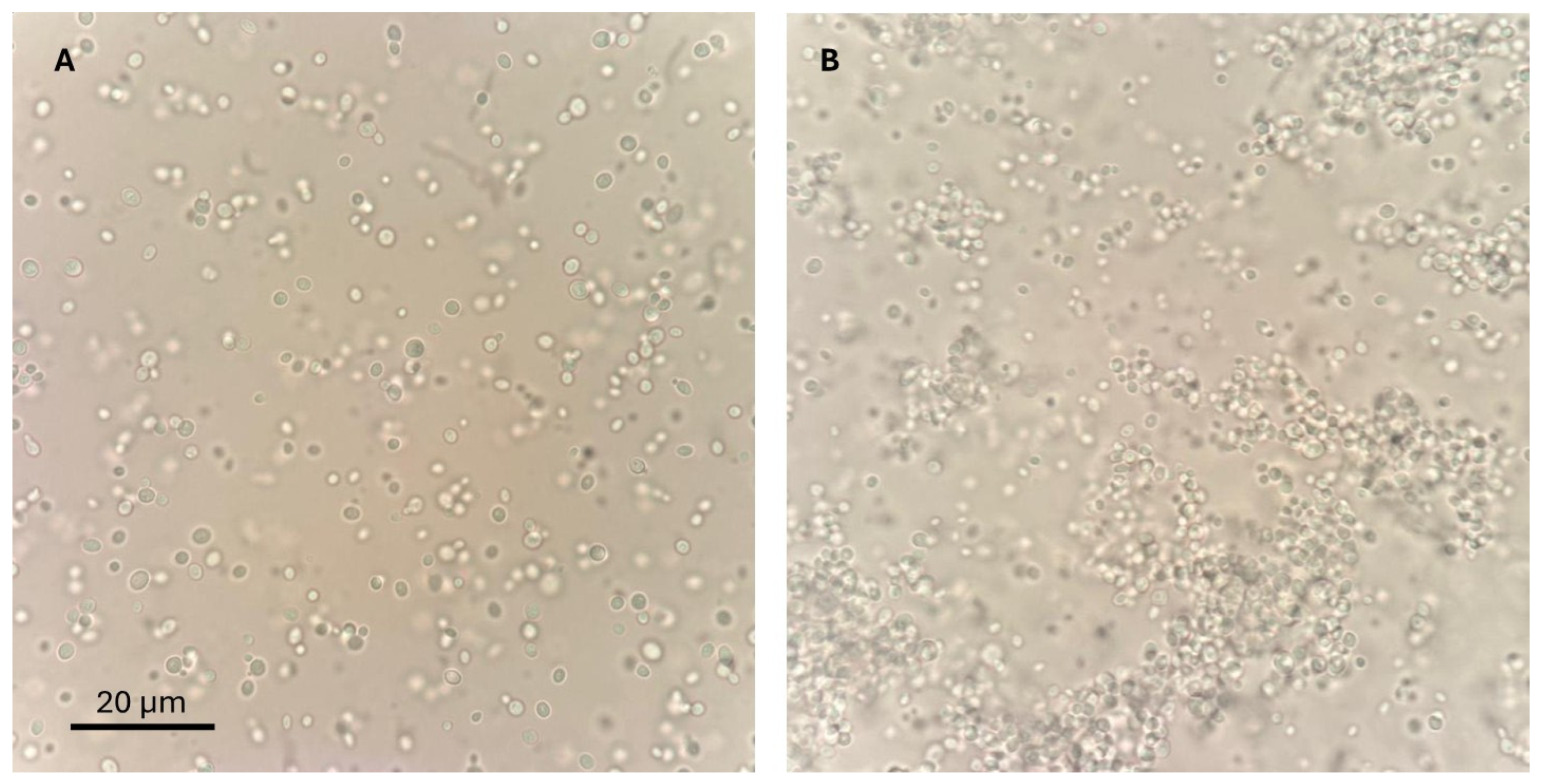
2.1.2. Phenotypic Classification

Strains were phenotypically categorized into aggregative and non-aggregative groups. This was achieved by vortexing 1 mL of sterile saline, containing approximately 108 CFU/mL, for 1 min. A 10 μL aliquot was then examined immediately under ×200 magnification using a TC20 automated cell counter (Bio-Rad Laboratories, Marnes-la-Coquette, France). The phenotype was classified as aggregative when large yeast clusters remained visible despite disaggregation procedures, as described by Borman et al., 2016 [17]. Figure 1 displays representative micrographs of both phenotypes observed in our strains under 100× magnification.

2.1.3. Antifungal Susceptibility Testing

We assessed in vitro antifungal susceptibility following EUCAST standards [25]. The minimum inhibitory concentration (MIC) was defined as the drug concentration that inhibited 50% of fungal growth at 35 °C after 24 h (90% inhibition for amphotericin B). MIC values were determined according to the EUCAST methodology. However, interpretation of MIC values was performed using the tentative breakpoints established by the CDC for C. auris, given the current lack of EUCAST-specific breakpoints for this species at the moment of the experimental procedure.

2.2. Survival Assays in Galleria mellonella

2.2.1. Larvae Handling

We obtained sixth-instar Galleria mellonella larvae from TruLarv (BioSystems Technology Ltd., Worcestershire, UK), a genome-sequenced breeding source. Only active larvae without melanization and weighing between 250 and 350 mg were selected. Initial surface decontamination was performed using 70% ethanol. The larvae were then grouped in sets of ten, placed in Petri dishes, and stored at 15 °C in the dark until they were ready for inoculation for a maximum of 72 h.

2.2.2. Survival Assay Procedures

Survival experiments followed previously described methods [17]. Isolates of C. auris were cultivated on Sabouraud agar plates at 37 °C for 24 h. The resulting colonies were collected using sterile plastic loops, rinsed twice with sterile PBS, and quantified using a TC20 automated cell counter (Bio-Rad Laboratories, France). The final suspension was adjusted to 105 CFU/μL. For C. auris aggregate strains, we prepared homogenous suspensions by allowing initial solutions to settle for 10 min, removing the supernatant with individual yeast cells, and then adjusting to 105 CFU/μL, following the procedure detailed by Borman et al. (2016) [17]. The inoculum concentration (105 CFU/μL, 10 μL per larva) was selected based on the reference method described by Borman et al., 2016 [17], to ensure standardization across experiments and comparability with prior G. mellonella C. auris infection models.
Larvae underwent a second decontamination with 70% ethanol prior to injection. Each larva received an intrahemocelic injection of 10 μL (containing 106 CFU) into the left rear proleg using a 10 μL Hamilton syringe fitted with a 26-gauge blunt needle. Groups of 10–20 larvae per strain were injected with C. auris suspensions for the survival analysis. For each strain, an additional control group of 10 larvae injected with PBS was included. Only larvae that survived the first 12 h post-injection without signs of mechanical injury or early pupation were included in the final analysis.
Following injection, larvae were housed in groups of 10 in Petri dishes and incubated at 37 °C. Each dish was labeled with a unique identifier to ensure blinding during data collection. Mortality and any cocoon formation were recorded daily over a 10-day period, excluding deaths that occurred within the first 12 h post-inoculation.

2.3. Statistical Analysis

The statistical analysis was performed using R software, version 4.4.1.
Firstly, we assessed the relationships between phenotypes and MICs by calculating Spearman correlation coefficients. Spearman correlation was selected due to the categorical nature of the phenotype variable, which is appropriate for capturing monotonic relationships. Additionally, a correlation matrix using Pearson’s correlation coefficients was constructed to investigate potential correlations among different antifungal MICs. Correlations were computed using complete observations to ensure reliability. Variables showing strong correlations with an r coefficient > 0.8 were excluded in the survival analysis due to expected high multicollinearity. p-values were calculated with Bonferroni correction with a threshold of p < 0.05.
To assess the association between covariates and survival outcomes, we employed a two-step approach. Initially, a standard Cox proportional hazards regression was performed to estimate hazard ratios (HRs), coefficients, and p-values for each predictor variable, providing a preliminary evaluation of their associations with the outcome.
Subsequently, a Cox regression model with elastic net regularization was used to further refine the selection of relevant predictors, thereby identifying variables that significantly contributed to the model while managing multicollinearity and potential overfitting. The elastic net approach combines L1 (lasso) and L2 (ridge) penalties, allowing for the selection and shrinkage of coefficients, ultimately yielding a parsimonious model that highlights the most impactful variables. The elastic net model was fitted to optimize hyperparameters and enhance model generalizability. The final model was adjusted with the optimal lambda value, using an alpha value of 0.5 to balance between L1 (Lasso) and L2 (Ridge) regularization. The results of both models were compared to validate the robustness and relevance of the identified predictors.
Median survival times for each phenotype were estimated using the Kaplan–Meier method, and summary statistics of the survival analysis were presented to interpret survival differences across phenotypes.

3. Results

A total of 10 C. auris clinical strains were analyzed in this study, isolated from blood and non-invasive epidemiological surveillance samples obtained from hospitalized patients with diverse diagnoses across multiple departments. Non-invasive isolates were collected as part of a systematic weekly fungal surveillance and colonization monitoring program conducted in the surgical and medical ICUs of our institution. Based on phenotypic traits, three strains were classified as aggregative and seven as non-aggregative. Aggregative strains predominantly originated from polytraumatized patients in surgical ICUs, whereas non-aggregative strains were associated with a broader range of conditions, including febrile neutropenia, status epilepticus, and endocarditis (Table 1).
Antifungal susceptibility testing showed universal resistance to fluconazole (MIC > 256 mg/L) across all strains. Sensitivity to amphotericin B (MIC 0.25–2 mg/L) and echinocandins exhibited variability, with one strain being resistant to amphotericin B. Voriconazole MIC values were diverse, with 60% of isolates displaying MIC ≥ 2 mg/L; however, definitive resistance cannot be determined due to the absence of CDC-established breakpoints for voriconazole and other second-generation triazoles, for which fluconazole resistance serves as a surrogate indicator. Notably, non-aggregative strains demonstrated consistent susceptibility to caspofungin and anidulafungin (Table 2).

3.1. Correlation Analysis of Antifungal MICs Against C. auris: Exploring Associations and Managing Collinearity

The correlation matrix for the MICs of several antifungal agents against C. auris reveals notable associations (Table 3). Amphotericin B MIC shows strong correlations with anidulafungin (r = 0.912) and posaconazole (r = 0.938), indicating a strong positive relationship. Flucytosine MIC is also highly correlated with anidulafungin (r = 0.882) and caspofungin (r = 0.820) MICs, both considered strong correlations.
Itraconazole MIC also shows a strong correlation with flucytosine (r = 0.829) and a moderate correlation with caspofungin (r = 0.789). Voriconazole MIC shows a moderate correlation with micafungin (r = 0.712), suggesting a weaker yet notable association. Posaconazole MIC has a very high correlation with anidulafungin (r = 0.973), indicating an extremely strong positive relationship.
Only weak negative correlations were found between the non-aggregative and MICs to micafungin (r = −0.339), posaconazole (r = −0.363), and anidulafungin (r = −0.326). Negligible correlations were observed between the rest of the antifungal MICs.

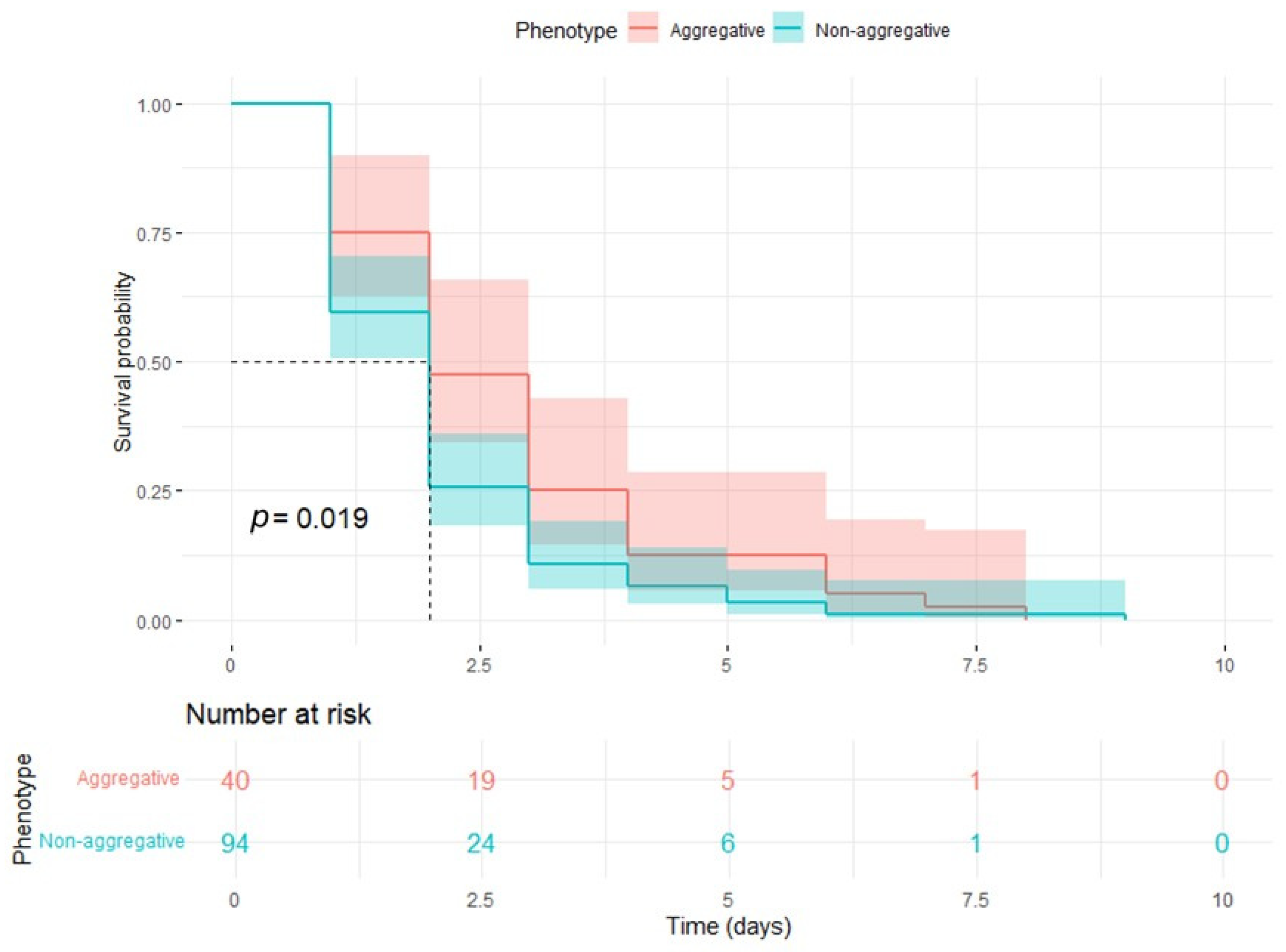
3.2. Survival Assays in G. mellonella: Cox Regression and Elastic Net Analysis of Survival Predictors in a Control Model of C. auris Infection

Finally, 134 inoculated G. mellonella specimens completed in a valid manner the survival assays in the determined follow-up period. The total number of observed events was 134 for C. auris-infected larvae. No deaths or cocoon formation were observed in the control group; therefore, the data were extracted from the cohort and subsequent analyses and graphical representations. The median survival time was 2 days for the whole number of larvae. The initial cohort consisted of 134 individuals at risk, with a survival probability of 64.2% (95% CI: 56.6–72.8%) at 24 h. This probability progressively declined over time, reaching 32.1% (95% CI: 25.1–41.1%) at 48 h and further decreasing to 14.9% (95% CI: 10.0–22.4%) at 72 h. By day 9, survival probability was 0%, with no remaining individuals at risk.
Based on the correlation matrix, we identified high correlations between several variables, suggesting multicollinearity. To minimize this issue, we selected variables with lower correlations while keeping those that were clinically important. The final Cox model included phenotype, invasive origin, amphotericin B MIC, flucytosine MIC, voriconazole MIC, and micafungin MIC. This selection reduced multicollinearity and ensured that the model remained clinically meaningful.
The Cox proportional hazards model revealed that the non-aggregative phenotype of C. auris was significantly associated with reduced survival in G. mellonella (Hazard Ratio [HR] = 2.418, 95% CI: 1.190–4.913, p = 0.015). Invasive origin showed a trend toward an impact on survival, though it did not reach statistical significance (HR = 2.939, 95% CI: 0.830–10.41, p = 0.095). Further data can be seen in Table 4.
Antifungal MICs were not significantly associated with changes in survival. Amphotericin B MIC demonstrated an increased hazard ratio, indirectly related to that of flucytosine, although wide confidence intervals indicate considerable uncertainty about the true impact. Voriconazole MIC had a hazard ratio close to one, indicating minimal impact on survival. Micafungin MIC exhibited an extremely low hazard ratio. For all MICs, the wide confidence intervals reflect considerable uncertainty regarding their precise effects.
To address potential multicollinearity and adjust for variable selection, we subsequently conducted a Cox elastic net regression. The elastic net model confirmed the significant association of the non-aggregative phenotype with decreased survival (HR = 1.117). Variables such as invasive origin, amphotericin B MIC, flucytosine MIC, voriconazole MIC, and micafungin MIC were retained in the final model; however, none demonstrated a meaningful impact on survival (HR = 1.00 for all). This analysis suggests that the non-aggregative phenotype is an independent predictor of reduced survival, while the other variables did not provide additional explanatory power in the model.
Considering that the phenotype was the only significant independent predictor of mortality, Kaplan–Meier survival curves were created, and survival statistics were calculated. In the survival analysis comparing aggregative and non-aggregative C. auris phenotypes, significantly distinct survival trends were observed (Figure 2) (p = 0.019). Aggregative phenotypes demonstrated higher initial survival probabilities, with 75% (95% CI: 62.7–89.7%) survival at time 1, decreasing to 25% (95% CI: 14.6–42.8%) at time 3, and 0% by time 8. Non-aggregative phenotypes exhibited lower survival probabilities from the outset, with 59.6% (95% CI: 50.4–70.4%) survival at time 1, declining to 10.6% (95% CI: 5.9–19.1%) at time 3, and 0% by time 9. Overall, non-aggregative phenotypes showed faster attrition in survival, reflected in steeper declines across time points, suggesting a potentially more severe clinical impact associated with this phenotype. Further data can be seen in Table 5 and Table 6.

4. Discussion

The main findings of this study can be summarized as follows: C. auris phenotype is an independent predictor of mortality in G. mellonella infection models, while other strain traits, such as susceptibility to antifungal drugs or the origin of the clinical isolates, have not been demonstrated to influence the virulence of C. auris infection in our regularized multivariable analysis in the in vivo model. Additionally, while all strains exhibited universal resistance to fluconazole, non-aggregative isolates showed broader antifungal susceptibility to echinocandins or posaconazole. Several strong positive correlations were found between different classes of antifungal drug MICs, suggesting different profiles of higher and lower antifungal susceptibility among the studied strains with potential cross-resistance mechanisms.
Candida auris is unique in many aspects of its pathophysiology. Its sudden and simultaneous emergence as a human pathogen on multiple continents, its high virulence, its environmental endurance, and high transmissibility, together with its increased tendency to develop antifungal resistance, justify the threat this microbe presents [5,7,8,9]. However, according to available data, not all strains of C. auris present the same virulence and resistance traits and mechanisms. Instead, substantial heterogeneity has been documented among the various C. auris clades and even between strains within the same clade [12,13,14]. It is hypothesized that C. auris possesses a remarkable capacity for genetic and phenotypic adaptation, and that this variability impacts its pathogenicity [13,14,26,27,28].
A known heterogenicity trait of C. auris is the capacity of some strains to grow forming aggregates [17]. Several studies have investigated the impact of this phenotype on fungal pathophysiology upon infection. Consistent with our findings, multiple authors have reported that non-aggregative strains exhibit greater virulence than aggregative strains, as demonstrated by the higher mortality rates observed in in vivo experiments [16,17,18,23]. However, C. auris has been shown to be able to form aggregates in mouse infection models regardless of phenotype [29], and Bing et al. retrieved aggregative colonies from the brain of their mouse infection model, which showed increased fitness in this tissue compared to non-aggregative strains [30]. Furthermore, differences between phenotypes in tissue tropism have been described in G. mellonella [16].
These aggregative strains are thought to have an advantage in colonization and environmental endurance [31]. Previous findings from our group indicate that less virulent aggregative strains exhibit a greater tendency to form pseudohyphae compared to non-aggregative strains, suggesting that aggregation and pseudofilamentation are more associated with adaptation than pathogenicity [16]. Additionally, Short et al. reported that aggregative C. auris strains exhibit greater medium survivability and enhanced resistance to sodium hypochlorite [32]. However, these traits may also depend on environmental and host conditions, as Brown et al. observed increased virulence of aggregative C. auris strains in a skin infection model [33].
Decreased antifungal susceptibility is also attributed to the aggregative phenotype [34]. Though contradictory results have been found [23], various authors have reported increased biomass of biofilm produced by aggregative strains in comparison with non-aggregative isolates [24,28,31,32]. In addition to this, in line with our results, other authors have also described higher MICs for aggregative less-virulent strains [32,34]. Furthermore, Louvet et al. identified genetic mechanisms that promote aggregation, enhance biofilm production, and increase surface adhesion [28]. Finally, several studies have documented the formation of aggregates in C. auris colonies following antifungal exposure [35,36,37,38]. Collectively, these findings suggest reduced susceptibility of the aggregative phenotype to certain antifungal drugs and support the associations observed in our results between phenotype and MIC values.
Antifungal susceptibility is one of the main concerns in the management of C. auris infection and colonization due to the tendency of this pathogen to rapidly develop resistance. Historically, the acquisition of antibiotic resistance has been associated with fitness trade-offs due to the evolutionary pressure exerted by the drug, often resulting in less virulent resistant strains. This phenomenon has been extensively documented in Candida species [19,20,21,22], though exceptions to this rule have also been reported [39,40,41,42]. In the case of C. auris, resistant strains have sometimes shown reduced virulence compared to their susceptible counterparts [35,38,43,44,45]. However, Burrack et al. observed no loss of fitness in resistant C. auris strains, as evidenced by the stability of resistance-conferring mutations even in the absence of antifungal drugs [15]. Similarly, Bohner et al. evolved C. auris strains to acquire triazole resistance and assessed their virulence in a murine model. While some resistant strains exhibited hypovirulence, with lower fungal burdens in mouse tissues, others surpassed their parental strains in their ability to colonize brain tissue [46]. Additionally, Carolus et al. reported decreased growth and stress resistance in resistant C. auris strains but demonstrated that these deficits could be reversed through compensatory mutations introduced via molecular engineering [43]. Consistent with these findings, our study found no correlation between antifungal MICs, independently of resistance breakpoints, and virulence, suggesting that C. auris can acquire antifungal resistance without incurring significant fitness costs. The reviewed data highlight the substantial variability in C. auris antifungal susceptibility profiles and suggest the existence of heterogeneous resistance mechanisms, which may underlie the pathogen’s remarkable ability to develop resistance to all currently available antifungal drugs.
Finally, the MIC correlation patterns observed across antifungal classes in C. auris are biologically plausible and reflect this pathogen’s propensity for multidrug resistance. Notably, we found that amphotericin B MICs strongly correlate with anidulafungin MICs (r ≈ 0.91) and posaconazole MICs (r ≈ 0.94), and similarly high correlations exist between azoles and echinocandins (e.g., posaconazole vs. anidulafungin, r ≈ 0.97) and between flucytosine and echinocandins (flucytosine vs. anidulafungin, r ≈ 0.88). At first glance, such cross-class associations seem counterintuitive given the distinct targets of these drugs—amphotericin B binds ergosterol in the fungal membrane, azoles inhibit ergosterol biosynthesis, echinocandins block β-1,3-glucan cell wall synthesis, and flucytosine disrupts DNA/RNA synthesis. Direct cross-resistance between these pathways is not classically expected, as correlations between minimum inhibitory concentrations of azoles and echinocandins, amphotericin and echinocandins, and flucytosine and echinocandins in C. auris are not well-established and are influenced by specific genetic mutations and resistance mechanisms unique to each antifungal class. However, C. auris frequently harbors broad resistance mechanisms that elevate MICs across multiple antifungal classes [47,48]. One of the best documented and understood instances is the appearance of cross-resistance between azoles and polyenes due to EGR gene mutations, as both of these drugs exercise their antifungal function by interfering with ergosterol physiology, which is closely regulated by said genes [43,49,50]. However, other associations have been described without a clear mechanism. For example, exposure to echinocandins can select for mutations in the ergosterol pathway (ERG3) that confer cross-resistance to azoles [47]. Likewise, alterations in cell membrane composition that reduce amphotericin B binding (e.g., via ERG gene mutations) could coincidentally affect cell wall integrity or drug uptake, increasing echinocandin MICs as well [47]. Indeed, C. auris isolates with simultaneous resistance to azoles, polyenes, echinocandins, and even 5-flucytosine, have been documented [48,51], often through the accumulation of multiple resistance determinants (such as ERG11 mutations for azoles, FKS1 hot-spot mutations for echinocandins, ERG3 or ERG6 alterations for amphotericin B, and FUR1 loss-of-function for flucytosine). Furthermore, our data and others suggest that certain phenotypes (e.g., the aggregative phenotype in C. auris) inherently exhibit elevated MICs to a broad range of antifungals, which would naturally produce positive inter-MIC correlations. In light of these factors, it is logical that isolates with high MICs to one drug class often show high MICs to others—not necessarily due to direct target cross-resistance, but due to co-occurring resistance mechanisms and global stress-response adaptations. This co-tolerance underscores the clinical challenge posed by C. auris: resistance to one antifungal agent frequently does not occur in isolation but is part of a multidrug-resistant phenotype [47]. The strong amphotericin B–anidulafungin and azole–echinocandin MIC correlations observed are therefore consistent with known resistance patterns in C. auris, and they highlight the need for vigilance as resistance in this organism can span disparate drug classes.
Despite the robustness of the two-step statistical analysis, which included a multivariable approach using Cox regression with elastic net regularization, several limitations should be acknowledged. On the one hand, the number of strains analyzed was relatively small. Given the heterogeneous nature of the species, a more comprehensive study involving a larger number of isolates and a broader representation of clades, clinical origins, and phenotypes would be valuable. Moreover, although a standardized inoculum of 10⁶ CFU per larva was used based on validated protocols, future studies evaluating a broader range of inocula (e.g., 103 to 108 CFU) could help delineate dose-dependent virulence patterns and assess the potential influence of quorum-sensing mechanisms in C. auris pathogenesis. The absence of a standardized reference C. auris strain could represent a limitation of this study for comparability. However, it is important to note that, to date, there are no universally defined reference strains for the aggregative and non-aggregative phenotypes of C. auris. Solidly establishing these reference models would greatly enhance comparative analyses in future studies. On the other hand, as this study employed an exploratory multivariable virulence animal model, key fungal virulence factors—such as biofilm production, filamentation, and enzyme activity—were not explicitly evaluated. Incorporating genomic or proteomic analyses could provide further insights into the pathophysiology of C. auris during infection. Additionally, this study did not include specific in vitro assays for individual virulence traits such as growth rate, thermotolerance, lipid membrane composition, biofilm formation, or hyphal development. Future studies incorporating these other phenotypic evaluations or genomic determinants are warranted to better explain the mechanisms underlying C. auris pathogenicity and antifungal resistance, and to deepen the understanding of the phenotypic variability observed across strains

5. Conclusions

A major challenge in studying and managing this pathogen is the pronounced genetic and phenotypic heterogeneity among its strains. One such phenotypic trait is the formation of aggregates, which, according to our findings and supporting evidence, impacts both virulence and antifungal susceptibility. Non-aggregative strains appear to exhibit increased virulence in invasive infections, whereas the aggregative phenotype seems to enhance colonization through improved environmental survivability and antifungal resistance. Higher MICs to antifungal drugs, however, do not seem to be independent predictors of mortality in in vivo animal models of infection. Further phenotypic, molecular, and genomic characterization of the differences between C. auris phenotypes could deepen our understanding of its pathophysiology and potentially provide useful markers for the clinical management of both infection and colonization.

Author Contributions

Conceptualization, J.P. and V.G.-B.; methodology, all authors; software, V.G.-B. and M.D.C.-N.; validation, all authors; formal analysis, J.A., A.C.R.-G., M.D.C.-N. and V.G.-B.; investigation, J.A., A.C.R.-G., M.D.C.-N., R.B.-H., S.d.C. and V.G.-B.; resources, A.C.R.-G. and J.P.; data curation, J.A., R.B.-H. and S.d.C.; writing—original draft preparation, J.A. and V.G.-B.; writing—review and editing, all authors; visualization, J.A.; supervision, J.P. and V.G.-B.; project administration, V.G.-B.; funding acquisition, J.P. All authors have read and agreed to the published version of the manuscript.

Funding

This research was funded by the Instituto de Salud Carlos III (ISCIII), Proyectos I+D+I en Salud, Grant Number: PI17/01538.

Institutional Review Board Statement

Not applicable.

Informed Consent Statement

Not applicable.

Data Availability Statement

The raw data supporting the conclusions of this article will be made available by the authors on request.

Conflicts of Interest

The authors declare no conflicts of interest.

References

  1. Satoh, K.; Makimura, K.; Hasumi, Y.; Nishiyama, Y.; Uchida, K.; Yamaguchi, H. Candida auris sp. nov., a Novel Ascomycetous Yeast Isolated from the External Ear Canal of an Inpatient in a Japanese Hospital. Microbiol. Immunol. 2009, 53, 41–44. [Google Scholar] [CrossRef] [PubMed]
  2. Ruiz-Gaitán, A.; Martínez, H.; Moret, A.M.; Calabuig, E.; Tasias, M.; Alastruey-Izquierdo, A.; Zaragoza, Ó.; Mollar, J.; Frasquet, J.; Salavert-Lletí, M.; et al. Detection and Treatment of Candida auris in an Outbreak Situation: Risk Factors for Developing Colonization and Candidemia by This New Species in Critically Ill Patients. Expert Rev. Anti-Infect. Ther. 2019, 17, 295–305. [Google Scholar] [CrossRef] [PubMed]
  3. Ruiz-Gaitán, A.; Moret, A.M.; Tasias-Pitarch, M.; Aleixandre-López, A.I.; Martínez-Morel, H.; Calabuig, E.; Salavert-Lletí, M.; Ramírez, P.; López-Hontangas, J.L.; Hagen, F.; et al. An Outbreak Due to Candida auris with Prolonged Colonisation and Candidaemia in a Tertiary Care European Hospital. Mycoses 2018, 61, 498–505. [Google Scholar] [CrossRef]
  4. Eyre, D.W.; Sheppard, A.E.; Madder, H.; Moir, I.; Moroney, R.; Quan, T.P.; Griffiths, D.; George, S.; Butcher, L.; Morgan, M.; et al. A Candida auris Outbreak and Its Control in an Intensive Care Setting. N. Engl. J. Med. 2018, 379, 1322–1331. [Google Scholar] [CrossRef] [PubMed]
  5. Du, H.; Bing, J.; Hu, T.; Ennis, C.L.; Nobile, C.J.; Huang, G. Candida auris: Epidemiology, Biology, Antifungal Resistance, and Virulence. PLoS Pathog. 2020, 16, e1008921. [Google Scholar] [CrossRef] [PubMed]
  6. Chow, N.A.; Muñoz, J.F.; Gade, L.; Berkow, E.L.; Li, X.; Welsh, R.M.; Forsberg, K.; Lockhart, S.R.; Adam, R.; Alanio, A.; et al. Tracing the Evolutionary History and Global Expansion of Candida auris Using Population Genomic Analyses. mBio 2020, 11, e03364-19. [Google Scholar] [CrossRef]
  7. Lockhart, S.R.; Etienne, K.A.; Vallabhaneni, S.; Farooqi, J.; Chowdhary, A.; Govender, N.P.; Colombo, A.L.; Calvo, B.; Cuomo, C.A.; Desjardins, C.A.; et al. Simultaneous Emergence of Multidrug-Resistant Candida auris on 3 Continents Confirmed by Whole-Genome Sequencing and Epidemiological Analyses. Clin. Infect. Dis. 2017, 64, 134–140. [Google Scholar] [CrossRef]
  8. Chowdhary, A.; Sharma, C.; Meis, J.F. Candida auris: A Rapidly Emerging Cause of Hospital-Acquired Multidrug-Resistant Fungal Infections Globally. PLoS Pathog. 2017, 13, e1006290. [Google Scholar] [CrossRef]
  9. Iguchi, S.; Itakura, Y.; Yoshida, A.; Kamada, K.; Mizushima, R.; Arai, Y.; Uzawa, Y.; Kikuchi, K. Candida auris: A Pathogen Difficult to Identify, Treat, and Eradicate and Its Characteristics in Japanese Strains. J. Infect. Chemother. 2019, 25, 743–749. [Google Scholar] [CrossRef]
  10. Centers for Disease Control and Prevention (U.S.). Antibiotic Resistance Threats in the United States, 2019; Centers for Disease Control and Prevention (U.S.): Atlanta, GA, USA, 2019.
  11. Kadri, S.S. Key Takeaways from the U.S. CDC’s 2019 Antibiotic Resistance Threats Report for Frontline Providers. Crit. Care Med. 2020, 48, 939–945. [Google Scholar] [CrossRef]
  12. Suphavilai, C.; Ko, K.K.K.; Lim, K.M.; Tan, M.G.; Boonsimma, P.; Chu, J.J.K.; Goh, S.S.; Rajandran, P.; Lee, L.C.; Tan, K.Y.; et al. Detection and Characterisation of a Sixth Candida auris Clade in Singapore: A Genomic and Phenotypic Study. Lancet Microbe 2024, 5, 100878. [Google Scholar] [CrossRef] [PubMed]
  13. Fayed, B.; Lazreg, I.K.; AlHumaidi, R.B.; Qasem, M.A.A.A.; Alajmy, B.M.G.N.; Bojbarah, F.M.A.M.; Senok, A.; Husseiny, M.I.; Soliman, S.S.M. Intra-Clade Heterogeneity in Candida auris: Risk of Management. Curr. Microbiol. 2023, 80, 295. [Google Scholar] [CrossRef] [PubMed]
  14. Phan-Canh, T.; Kuchler, K. Do Morphogenetic Switching and Intraspecies Variation Enhance Virulence of Candida auris? PLoS Pathog. 2024, 20, e1012559. [Google Scholar] [CrossRef]
  15. Burrack, L.S.; Todd, R.T.; Soisangwan, N.; Wiederhold, N.P.; Selmecki, A. Genomic Diversity across Candida auris Clinical Isolates Shapes Rapid Development of Antifungal Resistance In Vitro and In Vivo. mBio 2022, 13, e00842-22. [Google Scholar] [CrossRef]
  16. Garcia-Bustos, V.; Pemán, J.; Ruiz-Gaitán, A.; Cabañero-Navalon, M.D.; Cabanilles-Boronat, A.; Fernández-Calduch, M.; Marcilla-Barreda, L.; Sigona-Giangreco, I.A.; Salavert, M.; Tormo-Mas, M.Á.; et al. Host–Pathogen Interactions upon Candida auris Infection: Fungal Behaviour and Immune Response in Galleria mellonella. Emerg. Microbes Infect. 2022, 11, 136–146. [Google Scholar] [CrossRef]
  17. Borman, A.M.; Szekely, A.; Johnson, E.M. Comparative Pathogenicity of United Kingdom Isolates of the Emerging Pathogen Candida auris and Other Key Pathogenic Candida Species. mSphere 2016, 1, e00189-16. [Google Scholar] [CrossRef] [PubMed]
  18. Garcia-Bustos, V.; Ruiz-Saurí, A.; Ruiz-Gaitán, A.; Sigona-Giangreco, I.A.; Cabañero-Navalon, M.D.; Sabalza-Baztán, O.; Salavert-Lletí, M.; Tormo, M.Á.; Pemán, J. Characterization of the Differential Pathogenicity of Candida auris in a Galleria mellonella Infection Model. Microbiol. Spectr. 2021, 9, e00013-21. [Google Scholar] [CrossRef]
  19. Papp, C.; Kocsis, K.; Tóth, R.; Bodai, L.; Willis, J.R.; Ksiezopolska, E.; Lozoya-Pérez, N.E.; Vágvölgyi, C.; Mora Montes, H.; Gabaldón, T.; et al. Echinocandin-Induced Microevolution of Candida parapsilosis Influences Virulence and Abiotic Stress Tolerance. mSphere 2018, 3, e00547-18. [Google Scholar] [CrossRef]
  20. Ben-Ami, R.; Garcia-Effron, G.; Lewis, R.E.; Gamarra, S.; Leventakos, K.; Perlin, D.S.; Kontoyiannis, D.P. Fitness and Virulence Costs of Candida albicans FKS1 Hot Spot Mutations Associated with Echinocandin Resistance. J. Infect. Dis. 2011, 204, 626–635. [Google Scholar] [CrossRef]
  21. Vincent, B.M.; Lancaster, A.K.; Scherz-Shouval, R.; Whitesell, L.; Lindquist, S. Fitness Trade-Offs Restrict the Evolution of Resistance to Amphotericin B. PLoS Biol. 2013, 11, e1001692. [Google Scholar] [CrossRef]
  22. Beyda, N.D.; Lewis, R.E.; Garey, K.W. Echinocandin Resistance in Candida Species: Mechanisms of Reduced Susceptibility and Therapeutic Approaches. Ann. Pharmacother. 2012, 46, 1086–1096. [Google Scholar] [CrossRef] [PubMed]
  23. Sherry, L.; Ramage, G.; Kean, R.; Borman, A.; Johnson, E.M.; Richardson, M.D.; Rautemaa-Richardson, R. Biofilm-Forming Capability of Highly Virulent, Multidrug-Resistant Candida auris. Emerg. Infect. Dis. 2017, 23, 328–331. [Google Scholar] [CrossRef]
  24. Hernando-Ortiz, A.; Mateo, E.; Perez-Rodriguez, A.; De Groot, P.W.J.; Quindós, G.; Eraso, E. Virulence of Candida auris from Different Clinical Origins in Caenorhabditis elegans and Galleria mellonella Host Models. Virulence 2021, 12, 1063–1075. [Google Scholar] [CrossRef]
  25. EUCAST E.Def 7.4 EUCAST Method for Susceptibility Testing of Yeasts (v. 7.4 Valid from 13 October, 2023). European Committee on Antimicrobial Susceptibility Testing. 2023. Available online: https://www.eucast.org/astoffungi/methodsinantifungalsusceptibilitytesting/susceptibility_testing_of_yeasts (accessed on 8 May 2025).
  26. Pelletier, C.; Shaw, S.; Alsayegh, S.; Brown, A.J.P.; Lorenz, A. Candida auris Undergoes Adhesin-Dependent and -Independent Cellular Aggregation. PLoS Pathog. 2024, 20, e1012076. [Google Scholar] [CrossRef] [PubMed]
  27. Bing, J.; Guan, Z.; Zheng, T.; Zhang, Z.; Fan, S.; Ennis, C.L.; Nobile, C.J.; Huang, G. Clinical Isolates of Candida auris with Enhanced Adherence and Biofilm Formation Due to Genomic Amplification of ALS4. PLoS Pathog. 2023, 19, e1011239. [Google Scholar] [CrossRef]
  28. Louvet, M.; Li, J.; Brandalise, D.; Bachmann, D.; Sala De Oyanguren, F.; Labes, D.; Jacquier, N.; Genoud, C.; Mucciolo, A.; Coste, A.T.; et al. Ume6-Dependent Pathways of Morphogenesis and Biofilm Formation in Candida auris. Microbiol. Spectr. 2024, 12, e01531-24. [Google Scholar] [CrossRef]
  29. Forgács, L.; Borman, A.M.; Prépost, E.; Tóth, Z.; Kardos, G.; Kovács, R.; Szekely, A.; Nagy, F.; Kovacs, I.; Majoros, L. Comparison of in Vivo Pathogenicity of Four Candida auris Clades in a Neutropenic Bloodstream Infection Murine Model. Emerg. Microbes Infect. 2020, 9, 1160–1169. [Google Scholar] [CrossRef]
  30. Bing, J.; Guan, Z.; Zheng, T.; Ennis, C.L.; Nobile, C.J.; Chen, C.; Chu, H.; Huang, G. Rapid Evolution of an Adaptive Multicellular Morphology of Candida auris during Systemic Infection. Nat. Commun. 2024, 15, 2381. [Google Scholar] [CrossRef]
  31. Singh, R.; Kaur, M.; Chakrabarti, A.; Shankarnarayan, S.A.; Rudramurthy, S.M. Biofilm Formation by Candida auris Isolated from Colonising Sites and Candidemia Cases. Mycoses 2019, 62, 706–709. [Google Scholar] [CrossRef]
  32. Short, B.; Brown, J.; Delaney, C.; Sherry, L.; Williams, C.; Ramage, G.; Kean, R. Candida auris Exhibits Resilient Biofilm Characteristics In Vitro: Implications for Environmental Persistence. J. Hosp. Infect. 2019, 103, 92–96. [Google Scholar] [CrossRef]
  33. Brown, J.L.; Delaney, C.; Short, B.; Butcher, M.C.; McKloud, E.; Williams, C.; Kean, R.; Ramage, G. Candida auris Phenotypic Heterogeneity Determines Pathogenicity In Vitro. mSphere 2020, 5, e00371-20. [Google Scholar] [CrossRef] [PubMed]
  34. Hernando-Ortiz, A.; Eraso, E.; Jauregizar, N.; De Groot, P.W.J.; Quindós, G.; Mateo, E. Efficacy of the Combination of Amphotericin B and Echinocandins against Candida auris In Vitro and in the Caenorhabditis elegans Host Model. Microbiol. Spectr. 2024, 12, e02086-23. [Google Scholar] [CrossRef]
  35. Das, S.; Singh, S.; Tawde, Y.; Dutta, T.K.; Rudramurthy, S.M.; Kaur, H.; Shaw, T.; Ghosh, A. Comparative Fitness Trade-Offs Associated with Azole Resistance in Candida auris Clinical Isolates. Heliyon 2024, 10, e32386. [Google Scholar] [CrossRef] [PubMed]
  36. Szekely, A.; Borman, A.M.; Johnson, E.M. Candida auris Isolates of the Southern Asian and South African Lineages Exhibit Different Phenotypic and Antifungal Susceptibility Profiles In Vitro. J. Clin. Microbiol. 2019, 57, e02055-18. [Google Scholar] [CrossRef]
  37. Zamith-Miranda, D.; Amatuzzi, R.F.; Munhoz Da Rocha, I.F.; Martins, S.T.; Lucena, A.C.R.; Vieira, A.Z.; Trentin, G.; Almeida, F.; Rodrigues, M.L.; Nakayasu, E.S.; et al. Transcriptional and Translational Landscape of Candida auris in Response to Caspofungin. Comput. Struct. Biotechnol. J. 2021, 19, 5264–5277. [Google Scholar] [CrossRef]
  38. Fayed, B.; Jayakumar, M.N.; Soliman, S.S.M. Caspofungin-Resistance in Candida auris Is Cell Wall-Dependent Phenotype and Potential Prevention by Zinc Oxide Nanoparticles. Med. Mycol. 2021, 59, 1243–1256. [Google Scholar] [CrossRef]
  39. Feng, W.; Yang, J.; Pan, Y.; Xi, Z.; Qiao, Z.; Ma, Y. The Correlation of Virulence, Pathogenicity, and Itraconazole Resistance with SAP Activity in Candida albicans Strains. Can. J. Microbiol. 2016, 62, 173–178. [Google Scholar] [CrossRef]
  40. Yang, Z.; Zhang, F.; Li, D.; Wang, S.; Pang, Z.; Chen, L.; Li, R.; Shi, D. Correlation Between Drug Resistance and Virulence of Candida Isolates from Patients with Candidiasis. Infect. Drug Resist. 2022, 15, 7459–7473. [Google Scholar] [CrossRef]
  41. Vale-Silva, L.A.; Sanglard, D. Tipping the Balance Both Ways: Drug Resistance and Virulence in Candida glabrata. FEMS Yeast Res. 2015, 15, fov025. [Google Scholar] [CrossRef]
  42. Mohammadi, F.; Ghasemi, Z.; Familsatarian, B.; Salehi, E.; Sharifynia, S.; Barikani, A.; Mirzadeh, M.; Hosseini, M.A. Relationship between Antifungal Susceptibility Profile and Virulence Factors in Candida albicans Isolated from Nail Specimens. Rev. Soc. Bras. Med. Trop. 2020, 53, e20190214. [Google Scholar] [CrossRef]
  43. Carolus, H.; Sofras, D.; Boccarella, G.; Sephton-Clark, P.; Biriukov, V.; Cauldron, N.C.; Lobo Romero, C.; Vergauwen, R.; Yazdani, S.; Pierson, S.; et al. Acquired Amphotericin B Resistance Leads to Fitness Trade-Offs That Can Be Mitigated by Compensatory Evolution in Candida auris. Nat. Microbiol. 2024, 9, 3304–3320. [Google Scholar] [CrossRef] [PubMed]
  44. Jenull, S.; Shivarathri, R.; Tsymala, I.; Penninger, P.; Trinh, P.-C.; Nogueira, F.; Chauhan, M.; Singh, A.; Petryshyn, A.; Stoiber, A.; et al. Transcriptomics and Phenotyping Define Genetic Signatures Associated with Echinocandin Resistance in Candida auris. mBio 2022, 13, e00799-22. [Google Scholar] [CrossRef] [PubMed]
  45. Zhang, H.; Niu, Y.; Tan, J.; Liu, W.; Sun, M.; Yang, E.; Wang, Q.; Li, R.; Wang, Y.; Liu, W. Global Screening of Genomic and Transcriptomic Factors Associated with Phenotype Differences between Multidrug-Resistant and -Susceptible Candida haemulonii Strains. mSystems 2019, 4, e00459-19. [Google Scholar] [CrossRef]
  46. Bohner, F.; Papp, C.; Takacs, T.; Varga, M.; Szekeres, A.; Nosanchuk, J.D.; Vágvölgyi, C.; Tóth, R.; Gacser, A. Acquired Triazole Resistance Alters Pathogenicity-Associated Features in Candida auris in an Isolate-Dependent Manner. J. Fungi 2023, 9, 1148. [Google Scholar] [CrossRef]
  47. Lee, Y.; Puumala, E.; Robbins, N.; Cowen, L.E. Antifungal Drug Resistance: Molecular Mechanisms in Candida albicans and Beyond. Chem. Rev. 2021, 121, 3390–3411. [Google Scholar] [CrossRef]
  48. Ramos, L.S.; Barbosa, P.F.; Lorentino, C.M.A.; Lima, J.C.; Braga, A.L.; Lima, R.V.; Giovanini, L.; Casemiro, A.L.; Siqueira, N.L.M.; Costa, S.C.; et al. The Multidrug-Resistant Candida auris, Candida haemulonii Complex and Phylogenetic Related Species: Insights into Antifungal Resistance Mechanisms. Curr. Res. Microb. Sci. 2025, 8, 100354. [Google Scholar] [CrossRef]
  49. Forastiero, A.; Mesa-Arango, A.C.; Alastruey-Izquierdo, A.; Alcazar-Fuoli, L.; Bernal-Martinez, L.; Pelaez, T.; Lopez, J.F.; Grimalt, J.O.; Gomez-Lopez, A.; Cuesta, I.; et al. Candida Tropicalis Antifungal Cross-Resistance Is Related to Different Azole Target (Erg11p) Modifications. Antimicrob. Agents Chemother. 2013, 57, 4769–4781. [Google Scholar] [CrossRef]
  50. Carolus, H.; Pierson, S.; Muñoz, J.F.; Subotic, A.; Cruz, R.B.; Cuomo, C.A.; Dijck, P.V. Genome-Wide Analysis of Experimentally Evolved Candida auris Reveals Multiple Novel Mechanisms of Multidrug Resistance. mBio 2021, 12, e03333-20. [Google Scholar] [CrossRef]
  51. Jacobs, S.E.; Jacobs, J.L.; Dennis, E.K.; Taimur, S.; Rana, M.; Patel, D.; Gitman, M.; Patel, G.; Schaefer, S.; Iyer, K.; et al. Candida auris Pan-Drug-Resistant to Four Classes of Antifungal Agents. Antimicrob. Agents Chemother. 2022, 66, e00053-22. [Google Scholar] [CrossRef]
Figure 1. Representative micrographs of Candida auris phenotypes: (A) Non-aggregative phenotype displaying dispersed yeast cells with minimal clustering (CJ98). (B) Aggregative phenotype exhibiting large multicellular clusters, consistent with the classification criteria described by Borman et al., 2016 [17] (CJ198). 100× magnification. Scale bar: 20 μm.
Jof 11 00406 g001
Figure 2. Kaplan–Meier survival curves of non-aggregative and aggregative C. auris phenotypes.
Jof 11 00406 g002
Table 1. Basic characteristics of C. auris strains used in the study according to isolate origin and phenotype. ICU, intensive care unit.
TimeNumber at RiskNumber of EventsSurvival ProbabilityStandard Error
Invasive samples
CJ104BloodPolytraumatizedSurgical ICUAggregative
CJ173BloodPolytraumatizedSurgical ICUAggregative
Cj198BloodPneumoniaMedical ICUAggregative
CJ98BloodPolytraumatizedSurgical ICUNon-aggregative
CJ175BloodStatus epilepticusMedical ICUNon-aggregative
CJ197BloodFebrile neutropeniaHematologyNon-aggregative
312775BloodEndocarditisMedical ICUNon-aggregative
Non-invasive epidemiological surveillance samples
124819RectalExtracorporeal mechanical oxygenationMedical ICUNon-aggregative
182482InguinalLiver transplantationMedical ICUNon-aggregative
253107PharyngealMultiple myelomaMedical ICUNon-aggregative
Table 2. Data on antifungal susceptibility and MIC values of C. auris strains used in the study according to aggregative and non-aggregative phenotypes. MIC, minimum inhibitory concentration; AMB, amphotericin B; 5FC, flucytosine; FLU, fluconazole; ITR, itraconazole; VOR, voriconazole; POS, posaconazole; CAS, caspofungin; ANI, anidulafungin; MCF, micafungin; S, susceptible; R, resistant; IE, insufficient evidence.
MIC (mg/L)
StrainPhenotypeAMB
MIC
AMB5FC
MIC
5FCFLU
MIC
FLUITR
MIC
ITRVOR
MIC
VORPOS
MIC
POSCAS
MIC
CASANI
MIC
ANIMCF
MIC
MCF
124819Non-aggregative0.5S<0.06S>256R0.06IE0.03IE0.015IE0.03S0.06S0.03S
182482Non-aggregative0.5S<0.06S>256R0.06IE0.03IE0.015IE0.03S0.03S0.03S
253107Non-aggregative0.5S<0.06S>256R0.06IE0.03IE0.015IE0.03S0.125S0.03S
CJ98Non-aggregative0.25S0.12S>256R0.25IE2IE0.03IE0.5S0.06S0.06S
CJ175Non-aggregative0.5S0.06S>256R0.125IE2IE0.06IE0.03S0.125S0.06S
CJ197Non-aggregative2R0.25S>256R0.25IE4IE0.06IE0.5S0.5S0.25S
312775Non-aggregative0.5S0.06S>256R0.125IE8IE0.06IE0.03S0.125S0.06S
CJ104Aggregative0.5S0.06S>256R0.125IE2IE0.06IE0.03S0.125S0.06S
CJ173Aggregative0.5S<0.06S>256R0.06IE2IE0.03IE0.06S0.06S0.06S
CJ198Aggregative0.25S0.06S>256R0.25IE1IE0.03IE0.06S0.125S0.06S
Table 3. Correlation matrix of minimum inhibitory concentration (MIC) values against tested antifungals in C. auris isolates: Pearson test for inter-MIC correlations and Spearman test for MIC-phenotype associations. Bold represents values related to more than >80% correlation. *, p-value below 0.05 with Bonferroni correction.
Amphotericin B MICFlucytosine MICItraconazole MICVoriconazole MICCaspofungin MICAnidulafungin MICMicafungin MICPosaconazole MIC
Amphotericin B MIC1.0000.659 *0.1910.297 *0.346 *0.912 *0.372 *0.938 *
Flucytosine MIC0.659 *1.0000.829 *0.490 *0.820 *0.882 *0.558 *0.815 *
Itraconazole MIC0.1910.829 *1.0000.340 *0.789 *0.551 *0.357 *0.458 *
Voriconazole MIC0.297 *0.490 *0.340 *1.0000.2260.445 *0.712 *0.428 *
Caspofungin MIC0.346 *0.820 *0.789 *0.2261.0000.587 *0.108 *0.427
Anidulafungin MIC0.912 *0.882 *0.551 *0.445 *0.587 *1.0000.526 *0.973 *
Micafungin MIC0.372 *0.558 *0.357 *0.712 *0.108 *0.526 *1.0000.560 *
Posaconazole MIC0.938 *0.815 *0.458 *0.428 *0.4270.973 *0.560 *1.000
Phenotype0.1160.009−0.101−0.167−0.020−0.326−0.339−0.363
Table 4. Summary of Cox proportional hazards regression analysis for factors affecting survival in the G. mellonella C. auris infection model. *: statistical significance below 0.05.
VariableCoefficientHazard RatioLower 95% Confidence IntervalUpper 95% Confidence IntervalZ-Scorep-Value
Non-aggregative phenotype0.8832.4181.1904.9132.4410.015 *
Invasive origin1.0782.9390.83010.411.6710.095
Amphotericin B MIC1.0382.8230.166147.960.7180.473
Flucytosine MIC−0.9910.3713.7 × 10−63.72 × 10⁴−0.1690.866
Voriconazole MIC−0.1160.8910.7711.029−1.5710.116
Micafungin MIC−9.8725.16 × 10−51 × 10−182.6 × 10⁹−0.6130.540
Table 5. Survival analysis in aggregative C. auris phenotypes. NA: not available.
TimeNumber at RiskNumber of EventsSurvival ProbabilityStandard ErrorLower 95% CIUpper 95% CI
140100.750.06850.627130.897
230110.4750.0790.342930.658
31990.250.06850.146160.428
41050.1250.05230.055060.284
6530.050.03450.012950.193
7210.0250.02470.003610.173
8110.0NANANA
Table 6. Survival analysis in non-aggregative C. auris phenotypes. NA: not available.
TimeNumber at RiskNumber of EventsSurvival ProbabilityStandard ErrorLower 95% CIUpper 95% CI
194380.59570.05060.504360.7037
256320.25530.0450.180780.3606
324140.10640.03180.059210.1911
41040.06380.02520.029430.1384
5630.03190.01810.010480.0972
6320.01060.01060.001510.0747
9110.0NANANA
Disclaimer/Publisher’s Note: The statements, opinions and data contained in all publications are solely those of the individual author(s) and contributor(s) and not of MDPI and/or the editor(s). MDPI and/or the editor(s) disclaim responsibility for any injury to people or property resulting from any ideas, methods, instructions or products referred to in the content.

