Review

A Comprehensive Review on Chemical Structures and Bioactivities of Ostropomycetidae Lichens

College of Agriculture and Biology, Liaocheng University, Liaocheng 252059, China
*
Authors to whom correspondence should be addressed.
These authors contributed equally to this work.
J. Fungi 2025, 11(5), 369; https://doi.org/10.3390/jof11050369
Submission received: 10 April 2025 / Revised: 2 May 2025 / Accepted: 6 May 2025 / Published: 9 May 2025
(This article belongs to the Special Issue Bioactive Secondary Metabolites from Fungi)

Abstract

:
Lichenized fungi, recognized as an ecologically vital and pharmaceutically promising resource, hold substantial value in both environmental conservation and medicinal applications. As the second largest subclass within the lichen-forming fungi of Lecanoromycetes, Ostropomycetidae emerged as a critical reservoir of bioactive secondary metabolites. Current research has revealed that these secondary metabolites demonstrate remarkable bioactivities, positioning them as potential sources for novel pharmaceutical compounds. Despite considerable progress in characterizing chemical constituents and evaluating bioactivities within this subclass, a systematic summary of these discoveries remains absent. This review synthesizes the lichenochemical research progress, providing critical evaluations of 202 structurally characterized compounds from Ostropomycetidae lichen species over recent decades. These Ostropomycetidae-derived compounds cover the phenols, polyketides, fatty acids, terpenoids, steroids, and non-ribosomal peptides, and exhibit diverse bioactivities including antitumor, anti-inflammatory, antibacterial, antifungal, antiviral, antioxidant, anti-angiogenic, anti-neurodegenerative diseases, antitubercular, anti-herbivore, and antitrypanosomal, and so on. The aim of this review is to establish a robust chemodiversity framework and to offer strategic guidance for targeted exploration of lichen-derived drug candidates in the biological resources of Ostropomycetidae lichens.

1. Introduction

Lichenized fungi (lichens) have been known for a long time as one type of excellent symbiotic association. More than 19,000 species of lichenized fungi have been reported, making up 17% of the known 110,000 fungal species and 27% of the known Ascomycota. Lichens grow practically on and within rocks, on soil and tree bark, and on almost any inanimate object, and so on. In nature, lichens grow very slowly [1,2]. The unique composition of lichens, containing fungi and algae or cyanobacteria, bestows them with both environmental sensitivity as bioindicators and remarkable stress resistance through physiological adaptation and chemical diversification [3]. The taxon-specific distribution patterns of chemical substances have also become fundamental to lichen taxonomy and systematics [4].
More than 800 unique secondary metabolites are currently known in lichens, with the number of compounds obtained under axenic culture conditions being significantly lower. Major compound types include depsides, depsidones, depsones, dibenzofurans, usnic acid derivatives, terpenoids, xanthones, chromones, and quinones, with structural diversity observed across most lichen species [2]. The photobiont symbiont, commonly comprising Trebouxia and Trentepohlia (chlorophyta) or Nostoc (cyanobacteria) along with associated microbiota, demonstrates the tremendous biosynthetic ability and potential to produce a range of potentially secondary metabolites [5,6,7,8,9]. Recent pharmacological investigations have revealed multifaceted bioactivities in lichen-derived compounds, including antimicrobial, antioxidant, antiviral, and anticancer properties. Notably, several metabolites exhibit selective cytotoxicity against malignant cells, either as standalone agents or chemotherapeutic adjuvants [10]. Secondary metabolites from lichens are worthy of further investigation in terms of their potential therapeutic applicability, including a better understanding of their mechanisms of action, which positions them as promising drug candidates [11]. These research advancements suggest that the lichens can be productively used in pharmaceutical relevance because of their possible activities reported [2]. These chemicals make up a treasure trove of pharmacologically active ingredients that are important for human life and health.
Ostropomycetidae constitutes the second largest subclass within Lecanoromycetes [12]. It includes lineages with mostly non-amyloid asci but often amyloid hamathecium or ascospores. The predominant type of ascoma ontogeny in the subclass is hemiangiocarpous. All the Lecanoromycetes with trentepohlioid photobiont belong to the Ostropomycetidae subclass [13]. The Ostropomycetidae subclass encompasses nine orders, 34 families, 237 genera, and 4810 species according to current phylogenetically informed taxonomy [14]. This taxonomic group of Ostropomycetidae has emerged as a prolific source of bioactive natural products, exemplified by the baeomycesic acid from Baeomyces spp. [15], and depsidone and depside derivatives [16]. Recent investigations of Graphis mycobionts revealed antimicrobial diphenyl ether red pigment graphinone A from Graphis cf. handelii Zahlbr. with efficacy against methicillin-resistant Staphylococcus aureus [17]. These active substances have increasingly drawn widespread attention. However, the systematic characterization of compounds and bioactivities in Ostropomycetidae remains fragmented. According to the databases of PubMed, Web of Science, and CNKI, we comprehensively expanded the phytochemical research from 1935 to 2024, identifying 202 structurally diverse compounds from only 14 studied families in Ostropomycetidae. The maximum likelihood phylogenetic tree was constructed based on the sequences of ITS, mtSSU, and nuLSU of this group (Figure 1a). Morphological characteristics of the lichen thallus of some species in Ostropomycetidae were illustrated (Figure 1b). The lichenized fungi of Ostropomycetidae, a natural source producing a number of valuable compounds, are economically not feasible and profitable due to their slow growth. Their mycobiont cultures are the alternative sources that have become highly attractive for chemists recently. Out of 202 compounds, about half of these metabolites belong to phenols and their derivatives (including 28 phenolic compounds, 34 depsides, 10 tridepsides, and 22 depsidones), polyketides (including seven lactones, 17 phthalides and dibenzofurans, five macrolides, 23 chromones, 16 xanthones, and three others), nine fatty acids, 21 sesquiterpenes, four sterols, as well as three non-ribosomal peptides. The names and producers of these compounds are given in the following content. These compounds are mainly categorized into six classes: phenolic derivatives, polyketides, fatty acids, terpenoids, sterols, and non-ribosomal peptides, and they exhibit multifaceted bioactivities including antitumor, anti-inflammatory, antimicrobial, antiviral, and antioxidant properties, and so on, which underscores the pharmaceutical potential of them (Figure 2a,c). The aim of this review is to enhance our insight into the underexplored reservoir of bioactive compounds produced in the species of lichenized fungi Ostropomycetidae and to facilitate the therapeutic potential of understudied lichen species for novel drug discovery and utilization in the future.

2. Phenolic Compounds and Bioactivities

2.1. Phenols

Phenolic compounds represent the predominant secondary metabolites in lichenized fungi of the Ostropomycetidae subclass and exhibit special chemical diversity. The current study has documented 28 different phenolic derivatives within the subclass, which are systematically grouped into monophenols, diphenols, triphenols, and tetraphenols (Figure 3).

2.1.1. Monophenol Derivatives

The chloroform extracts of Thamnolia vermicularis (Sw.) Schaer. yielded thamnolic acid A (1), β-resorcylic acid (2), and everninic acid (3) [18], while recent work on the Arctic lichen Anamylopsora pakistanica Usman & Khalid (Baeomycetaceae) found haematommic acid (4) featuring a unique 2,4-dihydroxy-6-pentylbenzoate scaffold [19]. Structure–activity relationship studies identified significant pharmacological properties. Compound 1 exhibited antibacterial activity [20], whereas compound 4 demonstrated good antitumour activity [19].
Orsellinic acid (2,4-dihydroxy-6-methylbenzoic acid) and its structural analogs have emerged as versatile synthetic precursors in pharmaceutical chemistry, biosynthesized across diverse taxa, including plants, lichenized fungi, and bacteria. The β-orcinol (5), a methylated analog of orcinol (3,5-dihydroxytoluene), serves as a crucial scaffold in bioactive compound development. Its natural occurrence was first documented in Ochrolechia turneri (Sm.) Zopf [18], with subsequent identification in T. vermicularis [21]. Orsellinic acid (6) was initially characterized in Diploschistes muscorum (Scop.) R. Sant. [22], and was later confirmed in D. scrupossus, D. norman, D. diacapsis [23], and Ochrolechia frigida (Sw.) Lynge [24]. Structural diversification of this core scaffold has yielded derivatives including ethyl orsellinate (7) from G. handelii mycobionts [17], along with prephenic acid (8) and hypoxyphenone (9) from O. frigida [24]. Compounds (6, 89) demonstrated significant antioxidant activity [24]. Recent advances have uncovered novel orsellinic acid analogs in T. vermicularis, including 3-ethoxycarbonyl-2-hydroxy-6-methoxy-4-methylbenzoic acid (10), 4-hydroxy-2-methoxy-6-methylbenzoic acid (11), 2-hydroxy-6-methoxy-4-methylbenzoic acid (12), and 1-(2,3-dihydroxy-5-methoxyphenyl) ethanone (13). All of these compounds exhibited potent anti-amyloidogenic activity, inhibiting hen egg white lysozyme (HEWL) fibril formation with IC50 values of 0.11 μmol/mL and 0.10 μmol/mL, which suggested the therapeutic potential against protein-misfolding disorders [25].
Orsellinic novel biphenyl derivatives of 3,5-dihydroxy-4-methoxybenzoic acid (14) and O-(4-biphenylylcarbonyl)-benzonic acid (15) emerged from D. pruinosum and O. frigida [24,26]. Antarctic lichen Pertusaria dactylina (Ach.) Nyl. (Pertusariaceae) was found to biosynthesize a series of branched-chain methyl benzoates (1624) featuring distinctive pentyl/methoxy substitutions. This chemically diverse series includes the following: chlorinated derivative of methyl-3-chloro-2-hydroxy-4-methoxy-6-pentylbenzoate (16), oxygenated analog of methyl-2-hydroxy-4-methoxy-6-pentylbenzoate (17), polyhydroxylated structure of methyl-3,6-dihydroxy-2,4-dimethylbenzoate (18), variably substituted isomers with distinct hydroxyl/methoxy position of methyl-2,4-dihydroxy-6-methylbenzoate (19), methyl-2,4-dihydroxy-6-methoxybenzoate (20), methyl-2-hydroxy-6-methoxy-4-methylbenzoate (21), methyl-2-hydroxy-4-methoxy-6-(2-oxopentyl)-benzoate (22), biphenyl precursor of 4-methoxy-6-pentyl-1,2-dihydroxybenzene (23), and olivetol derivative of monomethyl olivetol (24) [27,28]. Notably, only the chlorinated analog compound 16 demonstrated measurable antifungal activity against Candida albicans, showing moderate inhibition with an IC50 value of 246 ± 26 μmol/L [27]. This structure–activity correlation suggests that chlorine substitution at C-3 may play a key role in the antifungal activity.

2.1.2. Polyhydroxylated Derivatives

The genus Graphis (Graphidaceae) serves as a prolific source of structurally intricate polyphenolic metabolites. Pioneering studies identified two architecturally complex diphenolic derivatives: acremonidin E (25) and its analog graphisin A (26), characterized by a unique 2,4-dihydroxy-6-pentylbenzoate scaffold [29]. Subsequent research revealed graphisidin (27), a triphenolic compound distinguished by dual methoxycarbonyl substitutions at C-3 and C-5 positions [30]. Only compound 25 exhibited significant pharmacological potential, showing dual bioactivities of antitubercular efficacy with MIC values of 0.157 μmol/mL and cytotoxic effects against breast cancer, oral epidermoid carcinoma KB, and small cell lung cancer cell line NCI-H187 with IC50 values ranging from 0.04–0.085 μmol/mL [29]. Notably, O. frigida yielded tetrafucol A (28), which is a tetraphenolic derivative representing one oxidized aromatic scaffold with antioxidant activity [24].

2.2. Depsides and Their Derivatives

Depsides represent a structurally distinctive class of lichen phenolics. Lichen depsides are classified into three main groups based on the presence or absence of a C-3 alkyl substituent on their aromatic rings: orcinol-type—aromatic rings of both subunits lack a C-3 substituent; β-orcinol-type—aromatic rings of both subunits possess a C-3 substituent; and a hybrid-type—only one aromatic ring bears a C-3 substituent. While biosynthetically characteristic of lichen symbionts, these compounds demonstrate evolutionary convergence through their presence in select angiosperms and non-lichenized Ascomycota [31]. The researches on the pharmacological activity of depsides from Ostropomycetidae lichens has been acquired in the past decades [32]. Recent investigations confirm that Ostropomycetidae lichens remain the predominant producers of depsides, with a total of 34 structurally characterized compounds, including eight orcinol-type depsides, 20 β-orcinol-type depsides, four hybrid-type depsides, and two benzyldepsides (Figure 4).

2.2.1. Orcinol-Type Depsides

Orcinol-type depsides are biosynthesized through exclusive oxidative coupling of orcinol (5-methylresorcinol) monomers. The archetypal member lecanoric acid (29), featuring a para-para’ ester linkage, was first characterized from the crustose lichen Siphula ceratites (Wahlenb.) Fr. (Icmadophilaceae) [33]. Subsequent chemotaxonomic studies have mapped its broad phylogenetic distribution across Ostropomycetidae, including Ochrolechia parella (L.) A. Massal. [34,35], Ochrolechia androgyna (Hoffm.) Arnold [36], Pertusaria mccroryae Björk, Goward & T. Sprib. [37], and Lobothallia alphoplaca (Wahlenb.) Hafellner [38]. Recent advances extended its detection to D. muscorum and Placopsis contortuplicata I.M. Lamb [22], and four Diploschistes species (D. scrupossus, D. cinereocaesius, D. norman, and D. diacapsis) [23]. Compound 29 exhibits dual pharmacological properties: broad-spectrum antifungal efficacy against clinically relevant strains [36] and potent antioxidant capacity via DPPH radical scavenging pattern [24,38]. Structural modifications and derivatives of compound 29 have been identified from the G. handelii mycobiont [17].
Olivetoric acid (30), first reported as a monophenolic secondary metabolite from crustose lichens Ochrolechia spp. (Ochrolechiaceae, Ostropomycetidae) [39], demonstrated dose-dependent anti-angiogenic effects through dual mechanisms: inhibition of rat adipose-derived endothelial cell proliferation and suppression of capillary-like tube formation in vitro [40]. Subsequent studies revealed its differential cytotoxicity against neural cells, showing IC50 values of 0.354 mmol/L in primary rat cerebral cortex cells versus 0.078 mmol/L in U87MG glioblastoma cell lines [41]. Divaricatic acid (31), another representative of this class, was initially isolated from Pertusaria spp. [42] and demonstrated exceptional antifungal potency against seven phytopathogens, including Fusarium oxysporum TR4 [43]. The structurally related compounds of planaic acid (32) and 2′-O-methylperlatolic acid (33) were co-isolated from Pertusaria paramerae A. Crespo & Vězda, which were differentiated by C-2′ methylation in the former [44]. Diploschistesic acid (34) has been identified to be ubiquitous in Diploschistes taxa, including D. muscorum [22], D. diacapsis, D. scrupossus, and D. norman [23]. 4-O-demethylmicrophyllinic acid (35) was isolated from Ochrolechia sp. [39]. The benzyl ether derivative of cyperine (36), as one phytotoxic metabolite, has been identified with antioxidant activity from P. contortuplicata [24].

2.2.2. β-Orcinol-Type Depsides

This subclass comprises depsides biosynthesized through oxidative coupling of β-orcinol precursors. The prototype compound squamatic acid (37), featuring a characteristic ortho-para’ ester linkage, was first isolated from Siphula sp. [45]. Chemotaxonomic studies subsequently identified its occurrence in Baeomyces rufus (Huds.) Rebent. and Baeomyces speciosus (Körb. Ex Stein) Lindau [16], and in T. vermicularis mycobionts [18,46,47], and Thamnolia subuliformis (Ehrh.) W.L. Culb. [20]. Baeomycesic acid (38), the predominant depside in lichen systems, along with thamnolic acid (39), was initially isolated from lichen Siphula spp. [48]. Compounds (3738) had antimicrobial efficacy to inhibit methicillin-resistant S. aureus [20]. Compound 38 also possesses more extensive bioactivities. It exhibited anti-inflammatory activity [47] and showed dose-dependent inhibitory effects on 5-lipoxygenase with an IC50 value of 8.311 μmol/L [49] and weak inhibitory activity on platelet-type 12(S)-lipoxygenase using a cell-based in vitro system in human platelets at a concentration of 267 μmol/L [50]. It exhibited a moderate radical scavenging antioxidant activity with an IC50 value of 1.608 mmol/L and good cytotoxic activity at the human epithelial carcinoma cell line Hela S3 with an IC50 value of 361 μmol/L [15]. In addition, compound 37 has other multitarget activities such as NFKB inhibition, antioxidant, lipoxygenase inhibition, glucosidase inhibition, and estrogen receptor agonism properties [51]. Thamnolic acid (39) has been identified in Pertusaria corallina (L.) Arnold [52] and Graphis cincta (Pers.) Aptroot [53] with general antibacterial activity [52].
Hypothamnolic acid (40), a structural analog of baeomycesic acid, has been identified in multiple taxa, including Siphula sp. [45], Pertusaria sp. [54], and T. vermicularis [47]. Chemotaxonomic studies reveal that the Thamnolia genus produces at least five specialized metabolites. Notably, vermicularin (41) has been consistently isolated from T. vermicularis across multiple studies [18,25,55], while thamnoliadepsides A–E (4246) and barbatinic acid (47) were identified in T. vermicularis, and both exhibit anti-inflammatory activity characterized in the same species [18,47]. Mechanistic investigations demonstrate that compound 40 reduces intracellular reactive oxygen species (ROS) levels in hydrogen-peroxide-induced fibroblasts, suggesting potential cosmeceutical applications [55]. Furthermore, comparative bioactivity assays indicate that compounds (38, 41) inhibit hen egg-white lysozyme (HEWL) fibril formation with IC50 values of 0.051 μmol/mL and 0.048 μmol/mL, respectively, highlighting their therapeutic potential against neurodegenerative pathologies [25]. Compounds (4142) reveal differential antitumor activity with a growth inhibitory effect in prostate cancer cells with IG50 values of 70.06 µmol/mL and 79.37 µmol/mL, respectively [18].
Haemathamnolic acid (48), featuring a rare monoprotonated aromatic system, was isolated from Pertusaria rhodesiaca Vain. [56]. Decarboxythamnolic acid (49) co-occurred in Pertusaria spp. and Siphula spp. [48,54]. Decarboxyhypothamnolic acid (50) and cryptothamnolic acid (51) from Pertusaria, as well as neothamnolic acid (52) and lactothamnolic acid (53) from Siphula ramalinoides Nyl., further exemplified the structural diversification within this type of compound [54]. Moreover, the methylated derivative of 3-O-methyllecanorate (54) was identified in T. vermicularis [47]. Atranorin (55), a ubiquitously distributed lichen metabolite first characterized in Siphula spp. [48], occurs taxonomically across Aspicilia spp. [57], Baeomyces spp. [16], O. parella [35], P. paramerae [44], T. vermicularis [47], and L. alphoplaca [38]. And halogenated analogs of chloroatranorin (56) were isolated from Siphula sp. [48]. Compound 55 appeared to have anti-herbivore activity in observed slug–lichen interactions [57] and exhibited non-specific antioxidant effects as broad-spectrum antioxidants [38]. It also selectively inhibited breast cancer cell lines of MDA MB-231 and MCF-7 with IC50 values of 5.36  ±  0.85 µmol/mL and 7.55  ±  1.2 µmol/L, respectively [58], and showed an inhibitory effect on nitric oxide levels in lipopolysaccharide-stimulated macrophages and a high anti-inflammatory potential (75.99% at 0.067 μmol/mL) [59].

2.2.3. Hybrid-Type Depsides

Hybrid depsides, characterized by the covalent integration of both orcinol and β-orcinol subunits, exhibit enhanced structural and functional diversity. Sekikaic acid (57), the inaugural hybrid β-orcinol/orcinol type depside, was isolated from S. ceratites [60] and subsequently identified in L. alphoplaca [38]. Compound 57 demonstrates multifaceted bioactivities, including antitumor activity and antibacterial properties [61], anti-diabetic and antioxidant potentials [62], and 1,1-diphenyl-2-picryl-hydrazil (DPPH) radical scavenging activity [63]. It also showed potent inhibition towards a recombinant strain of respiratory syncytial virus with an IC50 value of 0.014 μmol/mL and the respiratory syncytial virus A2 strain with an IC50 value of 0.018 μmol/mL [64], and significant α-glucosidase with IC50 values ranging from 7.9 to 149 μmol/L [65]. The methylated derivative 3′-methylevenic acid (58) was identified in T. vermicularis [47]. Three benzyl ether derivatives have been identified, including phytotoxic metabolite handelone (59) and graphinone A (60) from G. handelii [17,66]. Compound 59 showed dual antiviral activity, including anti-SARS-CoV-2 main protease activity with an IC50 value of 5.2 μmol/L and inhibition of HIV at a final concentration of 10 μmol/L [66].

2.2.4. Benzyldepsides

In lichens, benzyldepsides occur much less often than depsides and depsidones. The benzyldepsides of alectorialin (61) and alectorialic acid (62), featuring a unique benzyl-bridged biaryl scaffold, were isolated from arctic lichen A. pakistanica. The former is a decarboxylated product of compound 62. It also does not occur so often in lichens and is not widely distributed. Compound 62 demonstrates selective cytotoxicity against various cancer cell lines, including HeLa, HCT116, MDA-MB-231, and MRC6 [19].

2.3. Tridepsides

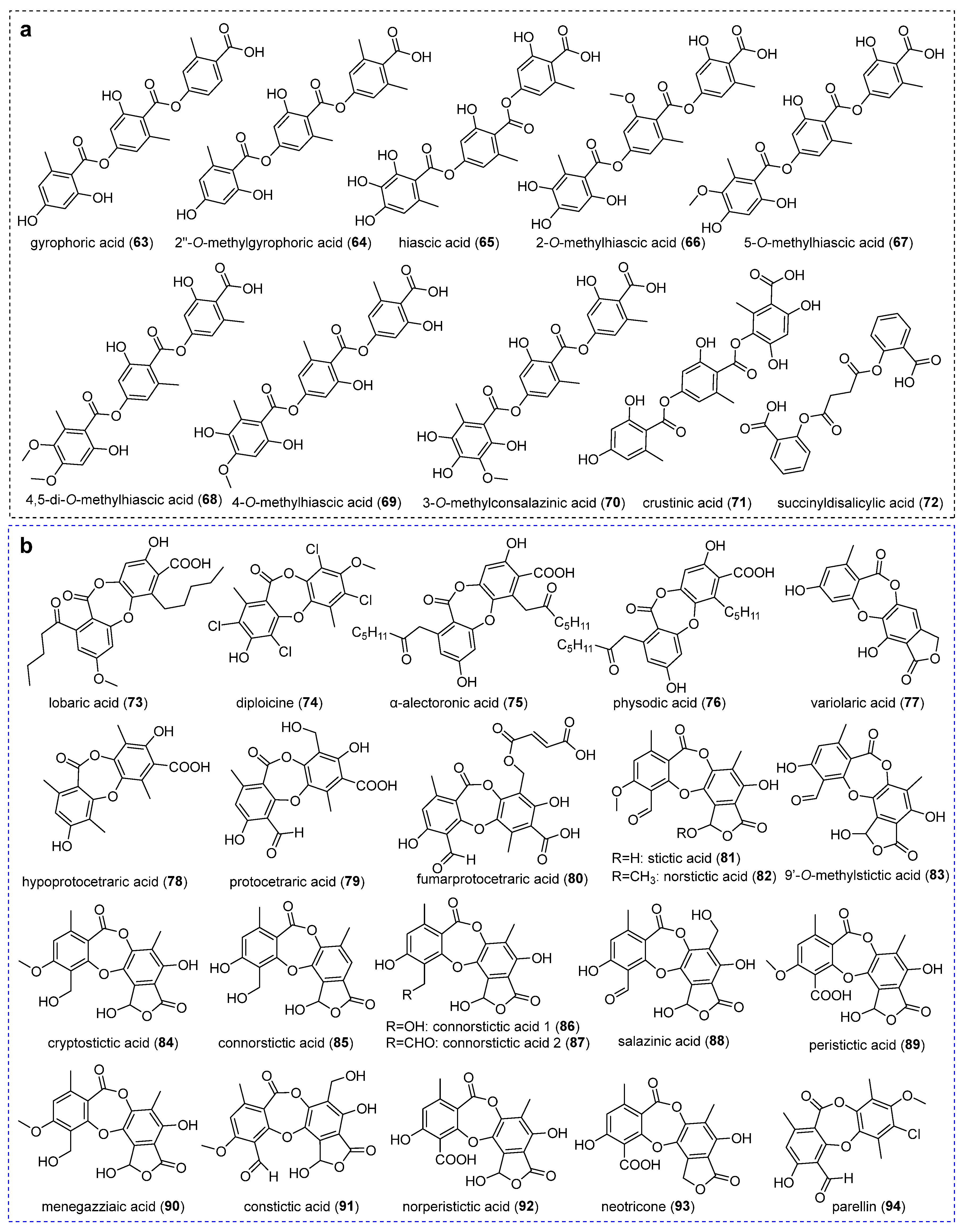
Tridepsides, as fully oxidized polyketides, represent a chemically distinctive class of lichen metabolites with over a century of research history. Recent pharmacological investigations have validated their role as defensive lichenochemicals exhibiting multifunctional bioactivities. Current taxonomic analyses document tridepside production across 37 lichen families (spanning 111 genera and 526 species), with Parmeliaceae, Lobariaceae, and Peltigeraceae accounting for 68% of characterized compounds [67]. Notably, only ten tridepsides have been structurally elucidated from Ostropomycetidae lichens (Figure 5a), among which gyrophoric acid (63), hiascic acid (65), and 5-O-methylhiascic acid (68) demonstrate the broadest phylogenetic distribution.
The archetypal tridepside gyrophoric acid (63) was first isolated from Ochrolechia sp. [39], which was subsequently identified from P. mccroryae, O. androgyna, D. diacapsis, and A. pakistanica [19,23,37]. Compound 63 exhibits distinct pharmacological properties, including anticancer, wound healing, photoprotection, anti-aging, antioxidant, cardiovascular effect, DNA interaction, anti-diabetes, anti-Alzheimer’s, antibacterial, and antifungal [67]. It has minimum inhibitory concentration values ranging from 0.041 μmol/mL to 2.669 μmol/mL against the human colon carcinoma cell line LS174, human lung carcinoma cell line A549, malignant melanoma cell line Fem-x, chronic myelogeneous leukemia cell line K562 [68], melanoma cancer cells [69], anti-proliferative activity against human cervix carcinoma HeLa cell [70], and breast cancer cell MCF-7 [71]. On the mechanism, compound 63 impinges on topoisomerase 1 to cause cell cycle arrest, inhibits cell survival, and promotes apoptosis, so it has been an effective anticancer candidate drug [72]. Compound 63 also inhibited Protein Tyrosine Phosphatase 1B (PTP1B) activity with 50% inhibitory concentration values of 3.6 +/− 0.04 μmol/L in a non-competitive manner, and can be used for treating type 2 diabetes and obesity [73]. It exhibits the largest free radical scavenging activity of 2,2-diphenyl-1-picrylhydrazyl (DPPH) with an IC50 value of 105.75 μmol/L [68] and can be considered an important natural compound with potent anti-aging strategies in the field of cosmetics and healthcare [74]. Compound 63 also has the potential to inhibit SARS-CoV-2 Mpro and act as a lead compound for the development of antiviral drug candidates against SARS-CoV-2 [75].
Methylation-driven structural diversification generates specialized derivatives. Gyrophoric acid’s methylated derivative, 2′’-O-methylgyrophoric acid (64), was specifically isolated from Diploschistes gyrophoricus Lumbsch & Elix [76]. Hiascic acid (65) and its positional isomers demonstrate distinct methylation patterns. 2-O-methylhiascic acid (66) was found in D. diacapsis by LC-MS/MS [23]. 5-O-methylhiascic acid (67) and 4,5-di-O-methylhiascic acid (68) were characterized in Ochrolechia spp. [39], along with compound 67, which was additionally found in P. mccroryae and Rimularia aff. furvella (Nyl. ex Mudd) Hertel & Rambold [37]. 4-O-methylhiascic acid (69) was identified in G. handelii [17]. The other specialized derivatives were isolated, including 3-O-methylconsalazinic acid (70) from P. mccroryae [37] and crustinic acid (71) and succinyldisalicylic acid (72) from P. contortuplicata with antioxidant activity [24].

2.4. Depsidones and Bioactivities

Depsidones, a class of polyphenolic polyketides, are structurally defined by an ether-bridge tricyclic system comprising two aromatic rings linked via a C-7-carbonyl-containing third ring [77]. These lichenized fungal metabolites exhibit remarkable structural diversity and pharmacological potential, particularly as scaffolds for multitarget therapeutic agents [78]. To date, 22 depsidones have been characterized from Ostropomycetidae lichens (Figure 5b).

2.4.1. Orcinol-Type Depsidones

The other remaining key depsidone derivatives were successively characterized, including lobaric acid (73) from T. vermicularis [50] and L. alphoplaca [38], diploicine (74) and α-alectoronic acid (75) from O. parella and Aspicilia radiosa [34,79], α-alectoronic acid (75) from O. parella [35], and physodic acid (76) from D. diacapsis [23]. Notably, these derivatives exhibit distinct pharmacological profiles. Compound 73 demonstrated multiple bioactivities, including antibacterial and antioxidant [80], scavenging free radicals [38], and inhibiting the ability of platelet-type 12(S)-lipoxygenase with an IC50 value of 28.5 mmol/L [50]. Compound 75 exhibited antitumor activity against the B16 murine melanoma cell line with an IC50 value of 10.3 µmol/L [35] and moderate inhibition of recombinant Plk1 kinase with an IC50 value of 1.7 µmol/L [81]. Compound 76 showed anti-inflammatory activity as potent inhibitors of mPGES-1 with an IC50 value of 0.4 µmol/L [82], higher anti-proliferative activity with an IC50 value of 171 µmol/L [83], and cytotoxic, apoptotic, and cell migration inhibitory effects on lung adenocarcinoma A549 cell line [84], and both LNCaP and DU-145 cell lines [85]. Variolaric acid (77) was extracted from both O. parella and A. radiosa, displaying antioxidant properties [79], antitumor activity against murine cancer cell line B16-F1 with an IC50 value of 38.7 µmol/L [35], and antiviral drug candidates against SARS-CoV-2 main protease inhibition [75].

2.4.2. β-Orcinol-Type Depsidones

Hypoprotocetraric acid (78), the first isolated from Siphula sp. in Ostropomycetidae [45] , was subsequently identified in Phaeographis sp. [86]. Structural analogues of protocetraric acid (79) and fumarprotocetraric acid (80) were present in O. androgyna [36]. Compound 79 demonstrated broad-spectrum efficacy against Gram-positive pathogens (Bacillus subtilis and S. aureus), Gram-negative pathogens (Enterobacter cloaceae, Escherichia coli, Klebsiella pneumoniae, and Botrytis cinerea) [36], Salmonella typhi, and the fungus Trichophyton rubrum [87]. It also showed moderate inhibitory antitubercular activity [88], strong anticancer activity toward human melanoma FemX cell lines and the human colon carcinoma LS174 cell line with IC50 values ranging from 0.953 μmol/mL to 0.161 μmol/mL [89], and possessed stronger alpha-glucosidase inhibitory activity with IC50 values ranging from 43.7 to 110.1 μmol/L [90]. Compound 80 exhibited broad-spectrum activity against Gram-positive bacteria (Bacillus mycoides, B. subtilis, and S. aureus) and Gram-negative pathogens (E. cloaceae, E. coli, and K. pneumoniae, with MIC value as low as 0.066 μmol/mL). It was also observed against pathogenic fungi, including Mucor mucedo, Aspergillus flavus, and C. albicans with MIC values ranging from 0.324 to 0.529 μmol/mL [36], and possessed the expectorant and antioxidant properties [91,92].
Stictic acid (81) was initially identified in Huilia albocaerulescens (Wulfen) Hertel [57], Baeomyces spp. [16], G. desquamescens [93], and O. androgyna [36]. Subsequent studies have expanded its taxonomic distribution to include Coccotrema hahriae T. Sprib. & Tønsberg [37], Pertusaria spp. [37,53,94], Aspicilia goettweigensis (Zahlbr.) Hue [95], Leucodecton occultum (Eschw.) Frisch [53], D. norman [23], and D. pruinosum [26]. Stictic acid derivatives were successively identified; norstictic acid (82) was first isolated from Siphula spp. [48] and later detected in Aspicilia spp. [37,79,95], C. hahriae and Phlyctis argena (Spreng.) Flot. [37], D. norman [23], D. pruinosum [26], Graphis spp. [53,93], L. occultum [53], O. parella [80], and Pertusaria pseudocorallina (Lilj.) Arnold [34]. Compound 81 demonstrated moderate antimicrobial activity at MIC values ranging from 0.647 μmol/mL to 1.294 μmol/mL for bacteria and 2.589 μmol/mL for fungi, respectively [36]. Compound 81 also showed moderate anticancer activity with an IC50 value of 0.076 μmol/mL for the cell line HT-29 and a low growth inhibition on the nonmalignant cell line MRC-5 with an IC50 value of 6.415 μmol/mL [96]. Compounds (8182) showed anti-herbivore activity [57] and significant alpha-glucosidase inhibition with IC50 values ranging from 10.4 to 130 µmol/L [97]. Compound 82 also exhibited activity against Mycobacterium tuberculosis with an MIC value of 0.161 μmol/mL and 168 µmol/L [88], and antioxidant activity [34].
The methylated derivatives include 9’-O-methylstictic acid (83) isolated from D. pruinosum [26] and cryptostictic acid (84) identified in P. mccroryae, C. hahriae [37], and D. norman [23]. The other derivatives of compound 81 have been further characterized. For example, connorstictic acid (85) was isolated from C. hahriae and Aspicilia cinerea (L.) Körb. [37], P. pseudocorallina [34], and D. norman [23]. Connorsticitic acid 1 (86), connorsticitic acid 2 (87), and salazinic acid (88) were isolated from P. pseudocorallina [98]. Peristictic acid (89), menegazziaic acid (90), constictic acid (91) and norperistictic acid (92) were isolated from P. mccroryae, C. hahriae [37], Baeomyces spp. [16] and Phaeographis sp. [86], respectively. These compounds demonstrate various pharmacological activities. Compound 88 exhibited significant apoptotic activities [99], strong anticancer activity with IC50 values ranging from 0.92 μmol/mL to 0.155 μmol/mL [89], significant intestinal alpha-glucosidase inhibitory activities with IC50 values of 4.17 ± 0.18 μmol/mL (p < 0.001) [100], and radical scavenging action with IC50 values of 312.14 µmol/L [101]. Compound 90 demonstrated radical scavenging activity [38]. Neotricone (93), a novel C-methylated derivative with hydrogen-bonded hydroxy groups with unique C-methyl/methylene groups and hydrogen-bonded hydroxy, was isolated from Phaeographis sp. [86]. And parellin (94) was isolated from O. parella [35].

3. Aliphatic Acids and Bioactivities

The unique growth conditions of lichens promote the biosynthesis of aliphatic acids (fatty acids) with distinct concentration profiles and structural diversity. While lichen-derived fatty acids share carbon chain similarities with non-lichenized fungi (C16–C24), their biosynthesis primarily occurs via tricarboxylic acid (TCA) cycle modifications, resulting in lower structural complexity and abundance compared to plant systems [2]. To date, nine aliphatic acids have been characterized from Ostropomycetidae lichens (Figure 6a).
The common fatty acid derivatives of stearic acid (95), oleic acid (96), palmitoleic acid (97), and linoleic acid (98) were identified in Aspicilia spp. [102]. Stearic acid (95), linoleic acid (98), 17-octadecynoic acid (99), and hexadecanoic acid (100) were isolated from Platygramme caesiopruinosa (Fée) Fée [103]. Compound 97 showed therapeutic potential for chronic diseases, particularly cardiovascular disorders [104], and compound 98 exhibited multifunctional dermatological effects, including the repair of the skin barrier, wound healing acceleration, and anti-aging properties [105]. Compound 100 demonstrated concentration-dependent antimicrobial activity against human commensal and pathogen S. aureus [106]. In addition, 3,6,9,12-tetraoxapentacosanoic acid (101) and 18-hydroxylinoleic acid (102) were identified from O. frigida, both demonstrating antioxidant activity [24]. Xylarinic acid A (103) was discovered in G. handelii [66], exhibiting broad-spectrum antifungal activity against plant pathogenic fungi including Pythium ultinum, Magnaporthe grisea, Aspergillus niger, Alternaria panax, and Fusarium oxysporium [107].

4. Polyketides and Bioactivities

4.1. Lactone-Type Compounds
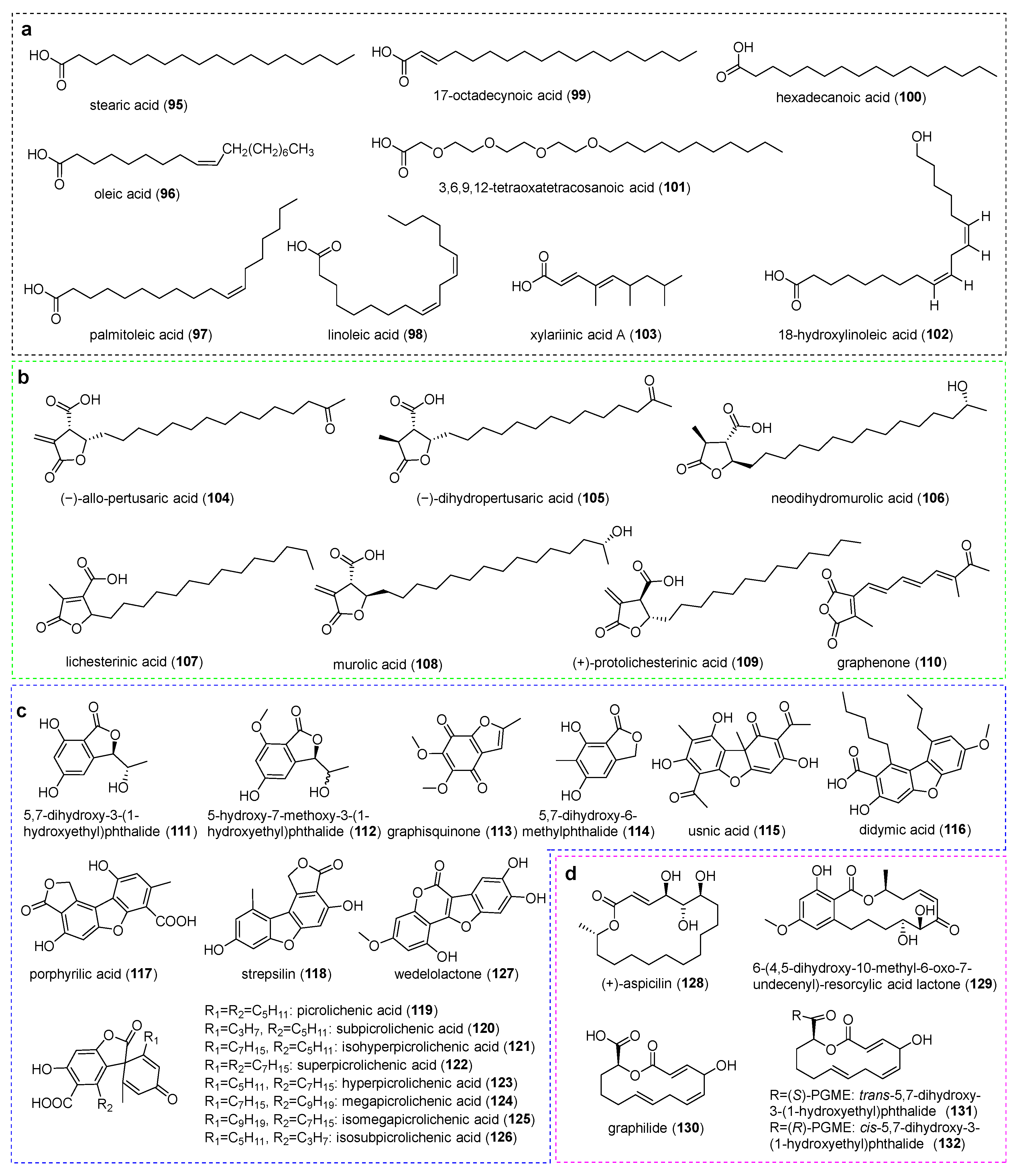
Aliphatic acids undergo enzymatic cyclization to form a γ-lactone characterized by a five-membered lactone ring. The key spectral features are reflected in both a diagnostic 13C NMR signal for the lactonized carbonyl carbon and moderate deshielding of the oxygen-adjacent ring carbon. Structural diversity arises from alkyl chain lengths (C11, C13, or C15), functional group substitutions (ketone, hydroxyl, terminal carboxyl, and acetoxy), as well as electronic effects. Exocyclic methylene groups induce greater shielding versus conjugated endocyclic double bonds [31]. To date, only seven γ-lactones have been identified in Ostropomycetidae lichens (Figure 6b). Among them, (−)-allo-pertusaric acid (104) and (−)-dihydropertusaric acid (105) were isolated from Pertusaria albescens (Huds.) M. Choisy & Werner [108]. Additionally, neodihydromurolic acid (106), lichesterinic acid (107), murolic acid (108), and (+)-protolichesterinic acid (109) were identified from Ochrolechia spp. [39]. Graphenone (110), a compound exhibiting an orangish-yellow hue, was identified in Graphis scripta (L.) Ach. and G. desquamescens [93]. Compounds (107 and 109) exhibit potent antitrypanosomal activity against Trypanosoma brucei with MIC values of 12.5 µmol/L and 6.30 µmol/L, respectively [109]. Compound 109 also acted as the inhibitor of platelet-type 12(S)-lipoxygenase with an IC50 value of 28.5 mmol/L and 77.0 mmol/L [50], and had anti-proliferative activity against HeLa cells [110] and antitumor activity against HER2-overexpressing breast cancer [111].
Figure 6. Chemical structures of compounds (95132). (a) Aliphatic Acids. (b) Lactones. (c) Phthalides and dibenzofurans. (d) Macrolides.
Jof 11 00369 g006

4.2. Phthalides and Dibenzofurans

Phthalides constitute a structurally distinct class of natural products with restricted taxonomic distribution, primarily identified in higher plants and lichen-forming fungi. Mycophenolic acid analogues have emerged as pharmacologically significant representatives of this type of compound [112,113]. Seventeen phthalide derivatives have been isolated from Ostropomycetidae lichens (Figure 6c). These compounds include 5,7-dihydroxy-3-(1-hydroxyethyl)-phthalide (111) and 5-hydroxy-7-methoxy-3-(1-hydroxyethyl) phthalide (112) from Graphis proserpens Vain. [114] and graphisquinone (113) from G. scripta and G. desquamescens [93]. Notably, 5,7-dihydroxy-6-methylphthalide (114), derived biosynthetically from alectorialic acid, was discovered in A. pakistanica with a good antitumour activity against the HeLa cell line from 0.311 ± 0.05 to 1.86 ± 0.155 μmol/mL, against the HCT116 cell line from 0.572 ± 0.089 to 1.099 ± 0.122 μmol/mL, against the MDA-MB-231 cell line from 0.599 ± 0.056 to 1.393 ± 0.155 μmol/mL, and against the MRC6 cell line from 1.038 ± 0.1 to 1.754 ± 0.161 μmol/mL, respectively [19].
Dibenzofurans feature two benzene rings fused to a central furan moiety (positions 2,3 and 4,5), exhibiting three primary structural variations including furan ring annulation patterns, isoprenyl substitutions, and aliphatic side chain modifications. These compounds exhibit diverse biological properties and were traditionally considered to originate primarily from lichens and ascomycetes [115]. Usnic acid (115), the most prominent dibenzofuran derivative, serves as a characteristic yellow pigment in lichens and displays broad pharmacological applications in dietary supplements and pharmaceuticals [113,116,117]. Usnic acid was first isolated from B. rufus and B. speciosus [16], and was then identified from T. vermicularis [118], Aspicilia sp. [102], and A. pakistanica [19]. Compound 115 exhibited broad antibiotic activity [47], antiviral activity [119], and antimicrobial properties and antitumor activity against cell lines with IC50 values of 0.37 μmol/mL [19]. Additional dibenzofuran derivatives include didymic acid (116) isolated from Pertusaria flavens Nyl. [53], porphyrilic acid (117) from Siphula spp. [48] and Dibaeis sp. [37], and strepsilin (118) from Siphula spp. [48]. Compound 116 was active against S. aureus with a minimum inhibitory concentration of 0.024 μmol/mL [120], and compound 117 exhibited noticeable inhibition of the 5-lipoxygenase enzyme and inhibition of xanthine oxidase enzyme with an IC50 value of 0.255 +/− 0.002 μmol/mL [121].
The genus Pertusaria produced a chemically related series of compounds, including picrolichenic acid (119), subpicrolichenic acid (120), and isosubpicrolichenic acid (121) from P. amara and P. truncata [122,123]. Compound 119 also occurred in O. parella [34]. Further derivatives, including superpicrolichenic acid (122), hyperpicrolichenic acid (123), megapicrolichenic acid (124), isomegapicrolichenic acid (125), and isosubpicrolichenic acid (126), were characterized in P. truncata [122,123]. Notably, wedelolactone (127), a pharmacologically active compound typically associated with Asteraceae plants, was recently identified in the lichen O. frigida with antioxidant activity [24]. Compound 127 demonstrated a broad spectrum of therapeutic potential, including anticancer, anti-inflammatory, anti-obesity, anti-myotoxic, antimicrobial, anti-diabetic, and tissue-protective activities [124]; moreover, the compound reduced the pathological damage of the liver, decreased ALT and AST, MMP, ROS, and inflammatory factors, and decreased hepatocyte viability in vitro [125].

4.3. Macrolides

Macrolides, a class of antimicrobial agents with broad clinical applications, have been identified in lichenized fungi of the Ostropomycetidae subclass, with five distinct representatives characterized to date (Figure 6c). Notably, (+)-aspicilin (128) with anti-herbivore activity was isolated from Aspicilia gibbosa (Ach.) Körb. and A. cinerea [57], and Aspicilia contorta (Hoffm.) Körb. [95]. While zearalenone derivatives are predominantly associated with Fusarium species, 6-(4,5-dihydroxy-10-methyl-6-oxo-7-undecenyl)-resorcylic acid lactone (129), which represents the first lichen-derived zearalenone analog, was isolated from both mycobiont cultures and intact thalli of Baeomyces placophyllus Ach.; this compound exhibits topoisomerase I and II inhibitory activities with an IC50 value of 0.275 μmol/mL and apoptosis producing activity on tumor cells with a value of 0.028 μmol/mL [126]. Three 14-membered macrolides were isolated from axenic cultures of Graphis vestitoides (Fink) Staiger, including graphilide (130), trans-5,7-dihydroxy-3-(1-hydroxyethyl)-phthalide (131), and cis-5,7-dihydroxy-3-(1-hydroxyethyl)-phthalide (132) [127].

4.4. Chromones

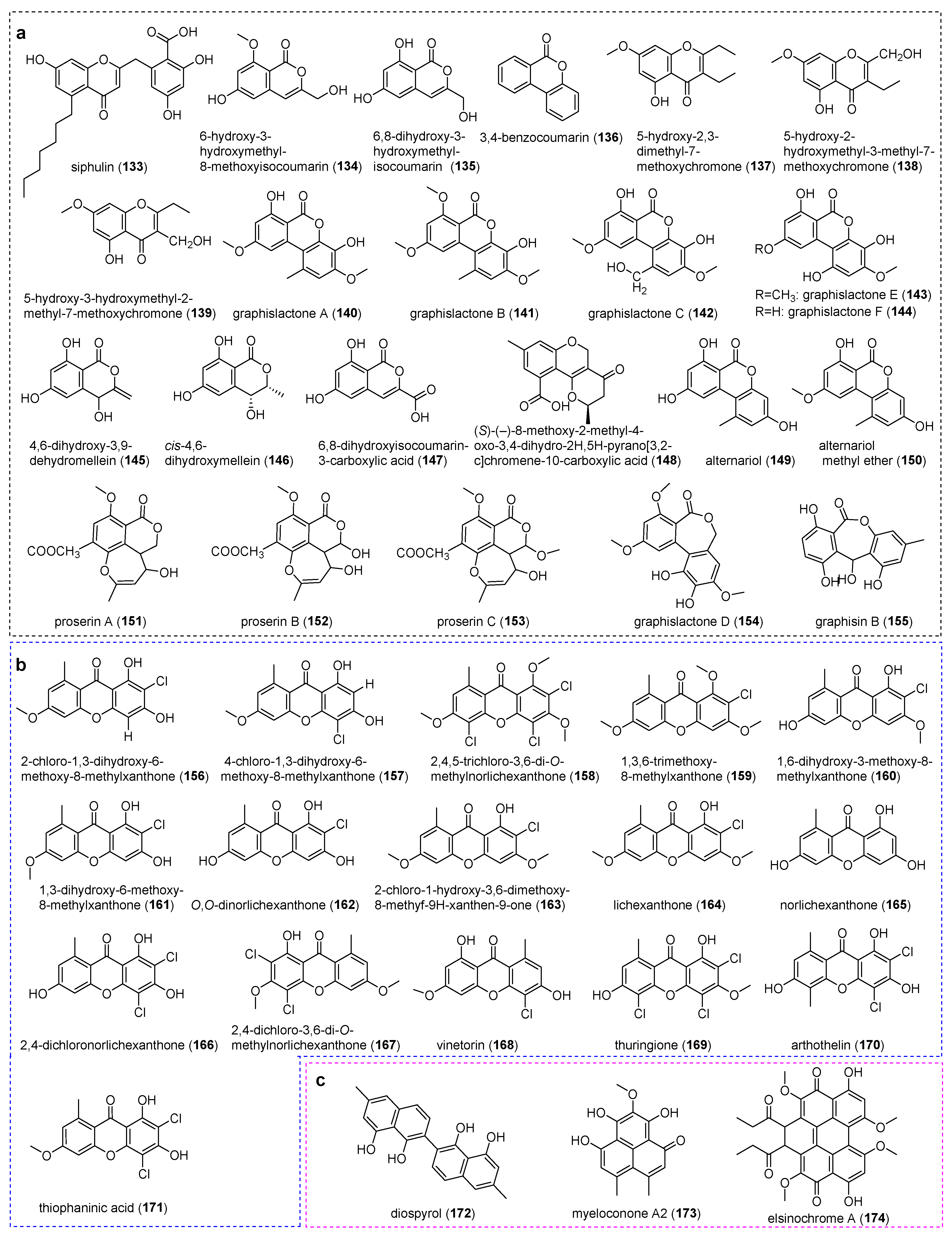
Chromones and their derivatives have emerged as multitarget therapeutic candidates in recent decades [128]. To date, 23 chromones and their derivatives have been characterized in Ostropomycetidae lichens (Figure 7a). Siphulin (133), a chromone-type lichen chromone-like metabolite, was isolated from S. ceratites [33,60]. Both isocoumarin derivatives of 6-hydroxy-3-hydroxymethyl-8-methoxyisocoumarin (134) and 6,8-dihydroxy-3-hydroxymethylisocoumarin (135) were isolated from G. proserpens [114]. Compound 135 featured a 3,6,8-substituted isocoumarin framework [127] and exhibited moderate anti-HSV-1 and antimycobacterial activities with IC50 and MIC values of 0.24 μmol/mL and 0.12 μmol/mL, respectively [129]. 3,4-benzocoumarinaltern (136) was identified in Pertusaria sp. [130]. Three methylated chromones of 5-hydroxy-2,3-dimethyl-7-methoxychromone (137), 5-hydroxy-2-hydroxymethyl-3-methyl-7-methoxychromone (138), and 5-hydroxy-3-hydroxymethyl-2-methyl-7-methoxychromone (139) were isolated from G. scripta mycobiont cultures [131].
Graphislactones A–C (140142) and graphislactones F (144) were isolated from Graphis prunicola Vain., graphislactones A and C were also found in Graphis cognata Müll. Arg., and G. scripta. Graphislactones E (143) was detected in G. scripta and G. prunicola [132]. Compound 140 exhibits cytotoxicity against human lung fibroblasts with a moderate activity towards acetylcholinesterase with an IC50 value of 0.027 μmol/mL [133], antioxidant capacity equivalent to ascorbic acid in hepatocytes, and anti-inflammatory response by lipopolysaccharide in macrophages [134]. Both mellein derivatives of 4,6-dihydroxy-3,9-dehydromellein (145) and cis-4,6-dihydroxymellein (146) were produced by G. proserpens [30], and the latter was also isolated from G. vestitoides. Compound 146 showed different degrees of phytotoxicity towards sunflower leaves and seedlings and may contribute to the severity of the sunflower disease caused by Phomopsis helianthin [135]. 6,8-dihydroxyisocoumarin-3-carboxylic acid (147) was isolated from crustose lichen G. vestitoides [127], and (S)-(–)-8-Methoxy-2-methyl-4-oxo-3,4-dihydro-2H,5H-pyrano[3,2-c] chromene-10-carboxylic acid (148) was characterized by methyl substitution and meta-coupled aromatic protons in Graphis sp. [30].
Alternariol (149), a mycotoxin with genotoxic properties, was found in G. cognata and Pertusaria sp. [132]. And alternariol’s methyl ether (150) was isolated from Pertusaria sp. [130]. Isocoumarin is one chemical compound with a lactonic α-pyranone ring fused to a benzene ring. Compounds (149150) can cause potential acute and chronic human health problems as mycotoxins [136,137]. Proserins A–C (151153), as the isocoumarin derivatives, were produced in G. proserpens [114], with proserin A also detected in Graphis spp. [30]. Graphislactone D (154) has been found in the lichen G. prunicola [132]. Graphisin B (155), a unique tetracyclic chromone, was identified in Graphis tetralocularis C. Bock & M. Hauck [29].

4.5. Xanthones

Xanthones, which are polyketide-derived secondary metabolites extensively studied in free-living fungi, have recently gained prominence in lichen chemistry. Their core structure arises from the linear condensation of seven acetate/malonate units with subsequent orsellinic acid-type cyclization to form two aromatic rings interconnected via ketonic carbonyl groups and ether-oxygen linkages. This unique biosynthetic pathway confers structural diversity and multitarget pharmacological potential, positioning lichen xanthones as valuable candidates for drug discovery [138]. A total of 16 xanthones have been characterized from Ostropomycetidae lichens (Figure 7b).
Figure 7. Chemical structures of compounds (133174). (a) Chromones. (b) Xanthones. (c) Other polyketides.
Jof 11 00369 g007
Pertusaria sulphurata Müll. Arg. yielded eight chlorinated compounds including 2-chloro-1,3-dihydroxy-6-methoxy-8-methylxant (156), 4-chloro-1,3-dihydroxy-6-methoxy-8-methylxanthone (157), 2,4,5-trichloro-3,6-di-O-methylnorlichexanthone (158), 1,3,6-trihydroxy-8-methylxanthone (159), 1,6-dihydroxy-3-methoxy-8-methylxanthone (160), 1,3-dihydroxy-6-methoxy-8-methylxanthone (161), O,O-dinorlichexanthone (162), and 2-chloro-1-hydroxy-3,6-dimethoxy-8-methyf-9H-xanthen-9-one (163) [139]. Lichexanthone (164) was identified from Ochrolechia sp., Pertusaria wui Q. Ren [39]. Norlichexanthone (165) was isolated from Pertusaria laeviganda Nyl. [140] and exhibited the strongest inhibitory activity against 16 cancer-related protein kinases, including aurora-B, PIM1, and VEGF-R2 with IC50 values ranging from 0.40 to 74.0 µmol/L, against these three protein kinases with IC50 values ranging from 0.3 to 11.7 µmol/L [141], and non-specific antioxidant effects with IC50 values greater than 200 µmol/L [140]. 2,4-dichloronorlichexanthone (166) was isolated from P. paramerae [44], which demonstrated antagonistic activities 2,4-dichloronorlichexanthone demonstrated antagonistic activities against Streptococcus agalactiae and S. aureus with MIC values of 10.21 µmol/L and 20.42 µmol/L, respectively [142]. and 2,4-dichloro-3,6-di-O-methylnorlichexanthone (167), vinetorin (168), and thuringione (169) were identified from Pertusaria sp. [143]. Compound 168 exhibited remarkable antagonistic activities against Streptococcus agalactiae and S. aureus with MIC values of 10.21 µmol/L and 20.42 µmol/L, respectively [142]. Arthothelin (170) was identified in Pertusaria sp. [108] and Dibaeis sp. [37]. Thiophaninic acid (171) has been isolated from P. sulghuvata [139] and P. paramerae [44].

4.6. Other Polyketides

Several other polyketide compounds have been identified from the Ostropomycetidae lichens (Figure 7c). Diospyrol (172), an anthelmintic drug from Diospyros mollis griff, was isolated from the lichen O. frigida, and it showed remarkable antioxidant activity [24]. Myeloconone A2 (173), as one new phenalenone of yellow pigment, was first identified from lichen Myeloconis erumpens P.M. McCarthy & Elix [144]. Another perylenequinone pigment, elsinochrome A (174), was identified in Graphis elongata Vain. mycobiont cultures. Elsinochromes, as the perylenequinone toxins from pathogenic fungi, are crucial mediators in fungal–host interactions [145]. And compound 174 displayed photodynamic effects against Gram-positive bacteria Pseudomonas syringae and fungus Alternaria mali [146].

5. Terpenoids and Bioactivities

Sesquiterpenoids, which are crucial components of plant chemical defense systems, exhibit selective bioactivity across antimicrobial, anticancer, and anti-inflammatory domains while maintaining low cytotoxicity toward normal cells [147]. Three structural classes have been characterized in Ostropomycetidae lichens: guaiane-type, sesquiterpene-quinones, and eremophilane-type derivatives.
Guaiane-type sesquiterpenes were characterized by a bicyclo[5.3.0] decane framework; these compounds demonstrate multifaceted pharmacological activities such as antitumor, anti-inflammatory, and antibacterial effects [148]. Six guaiane-type sesquiterpenes have been isolated from Ostropomycetidae lichens (Figure 8a). Pruinosone (175) and hydroxypruinosone (176) were isolated from D. pruinosum, both featuring a 2-methylcyclopent-2-en-1-one moiety. The former possessed antifungal activity and cytotoxic activity, and the latter has shown an inhibitory effect on fungi [149]. Diorygmones A–B (177178) possessed a guaiane-type sesquiterpenoid core and were identified from Diorygma sp. mycobiont cultures with pruinosone (175) and hydroxydiorygmone A (179) [150,151]. Diorygmones C–E (180182) were isolated with diorygmone B from D. pruinosum [26]. Compounds (177178) showed moderate cytotoxic activity against the HepG2 cell line with IC50 values of 0.259 ± 0.009 and 0.136 ± 0.006 μmol/mL, respectively [151], and compounds (178, 180181) were inhibitory to S. aureus [26]. Compound 180 showed moderate α-glucosidase inhibition with an IC50 value of 160 ± 2.2 µmol/L and significant inhibitory activity toward nitric oxide production in LPS-stimulated RAW264.7 cells with an IC50 value of 8.11± 0.21 µmol/L [150].
Sesquiterpene quinones, hybrid metabolites combining a C15-sesquiterpenoid backbone with a C6-benzoquinone/quinol moiety, exhibit broad pharmacological potential including anticancer, anti-inflammatory, antimicrobial, antiviral, and fibrinolytic activities [152]. Three representatives have been characterized in Stictidaceae lichens (Figure 8b). Dasyscyphin C (183), dasyscyphin G (184), and dasyscyphin F (185) were isolated from the lichen Stictidaceae. Compound 183 possesses a 2-cyclohexene-1,4-dione ring [153]. Dasyscyphins showed a variety of biological activities. For example, compound 183 showed cytotoxic activities against human cell lines, including HepG2, Hela S3, U937, Colo-320, and Jurkat with IC50 values from 0.001 to 0.006 μmol/mL [154]. Compounds 183 and 186 have a good antitumor activity and are moderately active against the three cell lines of melanoma MDA-MB-435, breast MDA-MB-231, and ovarian OVCAR3 with IC50 values ranging from 4 to 16 mmol/L [153], and antimicrobial activity to Pseudomonas aeruginosa, Methicillin resistant, S. aureus, and Bacillus anthraci [153]. Compound 183 showed good leishmanicidal activity at 1.982 μmol/mL concentration with an IC50 value of 0.892 μmol/mL against Leishmania major promastigote [155].
Eremophilane sesquiterpenes, characterized by a 6,7-seco-eudesmane skeleton, demonstrate phytotoxic and immunomodulatory properties across both plants and fungi [156]. Nine derivatives were isolated from Sarcographa tricosa (Ach.) Müll. Arg. mycobiont cultures (Figure 8c). The eremophilane compounds, including petasol (186), isopetasol (187), 3-epi-petasol (188), sporogen AO-1 (189), dihydrosporogen AO-1 (190), 8α-OH, dihydropetasol (191), JBIR-27 (192), 1β-hydroxypetasol (193), and sarcographol (195), were characterized from S. tricosa mycobiont cultures [157]. Compounds 189190 and graphilane (194) were additionally detected in Graphis sp. mycobiont axenic cultures [158]. Compounds 186 and 189 exhibited cytotoxicity to HeLa cells and robust growth-restoring activity of Saccharomyces cerevisiae mutant strain [159]; the latter also showed cytotoxicity against human cervical carcinoma cell line HeLa at IC50 values of 8.3 μmol/L [160]. Compounds 189 and 190 caused significant inhibition of radicle growth against Amaranthus hypochondriacus and Echinochloa crus-galli at IC50 values of 0.17 mmol/L and 0.30 mmol/L, respectively [161]. Compound 194 showed moderate cytotoxic activity against the K562 cancer cell line with an IC50 value of 87.20 ± 0.76 μmol/L [158].

6. Steroids and Bioactivities

Sterols, which are essential eukaryotic membrane components, regulate cellular permeability and signaling processes. Phytosterols such as stigmasterol, campesterol, and β-sitosterol are distinguished by C-24 alkyl substituents (methyl for campesterol; ethyl for β-sitosterol) on their side chains [162]. Four steroidal compounds have been characterized from Ostropomycetidae lichens (Figure 8d), with ergosterol derivatives being predominant.
Ergosterol peroxide (196), a C28-sterol widely documented in medicinal mushrooms, was identified in O. parella [35] and S. tricosa [157], and showed anti-inflammatory activity with an IC50 value of 0.016 μmol/mL [163], antimicrobial activity, cytotoxic effects against a wide range of cells, immunosuppressive and anti-inflammatory activities, antiatherosclerotic effects, antifibrotic effects, insecticidal and phytotoxic effects, and cytotoxic effects against different tumor cell lines including MT-1 breast cancer cells, human intrahepatic cholangiocarcinoma cells (HuCCA-1), HeLa cells, prostate cells, colon carcinoma cells, and the myeloma cell line U266 [164]. Three key sterols have been characterized in Ostropomycetidae lichens with stigmasterol (197) and campesterol (198) from Aspicilia sp. [102] and β-Sitosterol (199) from T. vermicularis [55] and D. pruinosum [26]. Stigmasterol (197), biosynthesized through the mevalonate pathway, shares structural homology with β-sitosterol but is distinguished by a trans-configured double bond in its side chain [165]. This phytosterol has garnered significant pharmacological attention for its multifaceted bioactivities, including anti-inflammation, anti-diabetes, antioxidization, lowering blood cholesterol, and antitumor bioactivity (e.g., breast, lung, liver, and ovarian cancers) [166]. Compound 197 indicated potent pharmacological effects such as anticancer, anti-osteoarthritis, anti-inflammatory, anti-diabetic, immunomodulatory, antiparasitic, antifungal, antibacterial, antioxidant, and neuroprotective properties [167]. It also showed pathogen infection and abiotic stress, including cold, salinization, drought, UV radiation, heavy metals, and stress phytohormones in plants [165]. Compounds 198 and 199 exhibited strong antimicrobial activity against pathogenic microbes including B. subtilis, S. pyogenes, S. aureus, K. pneumoniae, E. coli, P. aeruginosa, S. typhii, A. fumigatus, C. albicans, C. krusei, and A. niger [168]. Compound 199 can also increase hyaluronic acid synthases in fibroblasts [55] and showed antibacterial activity inhibitory to S. aureus [26].
Figure 8. Chemical structures of compounds (175202). (a) Guaiane-type sesquiterpenes. (b) Sesquiterpene quinones. (c) Eremophilane-type sesquiterpenes. (d) Sterols. (e) Non-ribosomal peptides.
Jof 11 00369 g008

7. Non-Ribosomal Peptides and Bioactivities

Astins constitute a specialized class of chlorinated cyclic pentapeptides featuring both proteinogenic and non-proteinogenic amino acid residues. Their structural diversity predominantly stems from site-specific chlorination patterns on conserved proline moieties. Three astin congeners have been characterized in Ostropomycetidae lichens, including astin C (200), astin F (201), and astin G (202), all isolated from Cyanodermella asteris L. Jahn & Ludwig-Müll. (Figure 8e) [169,170]. Compound 200 exhibited dual pharmacological properties of anti-inflammatory activity [170], the ability to induce apoptosis in activated T cells, and its potential use in the treatment of colonic inflammation [171]. Both compounds (200, 202) could enhance plant biomass accumulation when individually supplemented in growth media [169].

8. Conclusions and Perspectives

Lichens, representing Earth’s predominant symbiotic flora, exhibit remarkable biological adaptability, physiological diversity, and chemical distinctiveness. These extraordinary organisms constitute a vital reservoir of novel bioactive compounds with multifaceted potential. The advanced techniques over recent decades have tremendously pushed the breakthroughs in pharmacological investigations of lichenized species. Their diverse secondary metabolites have established lichens as indispensable natural resources for pharmaceutical development, nutraceutical applications, and industrial biotechnology [77]. Contemporary reviews on lichen-derived drug discovery and lichen biology have emphasized cutting-edge innovations and breakthroughs in symbiotic biology and biosynthetic pathway elucidation [172]. One artificial key to the lichen genera from China have promoted research of lichen resources in China [173]. Of particular interest, the Ostropomycetidae subclass, the lichenized fungi Lecanoromycetes’ second-largest taxonomic division, serves as a prolific source of structurally unique compounds with broad bioactivity profiles. Despite a systematic investigation of merely 41% of its taxonomic families across nine orders, the exceptional chemodiversity and demonstrated biological efficacy within this subclass underscore its substantial research potential (Figure 2, Table 1). In recent years, a number of new species of this subclass continue to be discovered, such as from Gomphillaceae [174], Graphidaceae [175,176,177,178], Megasporaceae [179,180], and Pertusariaceae [94,181]. However, current exploration limitations primarily stem from the technical challenges in axenic cultivation of symbiotic strains and slow growth, low biomass availability, and factors that have historically deterred the comprehensive phytochemical investigation of lichens.
Rapid development of biotechnological tools is providing a novel opportunity to exploit the compounds from Ostropomycetidae lichens for industrial utilization. Culturing the symbionts and the molecular genetics modulation of lichen gene regulation are recognized to enhance the production of target metabolites. The convergence of multi-omics technologies with synthetic biology facilitates the heterologous expression of lichen-derived biosynthetic gene clusters in the tractable fungal hosts. This paradigm shift addresses critical challenges in sustainable metabolite production by decoupling compound biosynthesis from traditional lichen cultivation constraints [182,183]. Future research directions should prioritize (1) promoting biodiversity expansion via novel species discovery to enhance structural novelty potential; (2) applying advanced cultural strategies for targeted metabolite production through controlled symbiont cultivation; (3) achieving genome sequences to facilitate biosynthetic gene cluster identification and functional annotation for more gene resources; (4) applying synthetic biology to employ fungal chassis systems for heterologous compound biosynthesis; and (5) high-throughput screening platforms for activity-guided isolation of therapeutic candidates. The convergence of metabolic engineering and synthetic biology presents unprecedented opportunities to overcome traditional production bottlenecks. Concurrent advancements in multi-omics technologies and CRISPR-mediated pathway engineering are poised to revolutionize our understanding of lichen-derived biosynthetic pathways, enabling precise manipulation of chemical, physiological, and biotechnological processes in these complex symbiotic systems.

Author Contributions

W.L. and Z.J. conceived and designed the research. Y.W., C.H., S.J. and Y.J. surveyed the scientific literature and analyzed the data. W.L. and Y.W. wrote the draft manuscript. W.L. and Z.J. interpreted the data and reviewed the manuscript. All authors have read and agreed to the published version of the manuscript.

Funding

This work was supported by the National Natural Science Foundation of China (31750001) and the Research Start-up Fund for High-level Talents and Doctors of the Department of Science and Technology, Liaocheng University (318052344).

Data Availability Statement

The data presented in this study are available in this manuscript, and constructs can be requested from the corresponding author.

Acknowledgments

The authors are deeply grateful to Qiang Ren in the School of Life Sciences, Taizhou University, for the valuable pictures of lichen specimens, and to Zhuang Ding in the Institute of Biopharmaceutical Research, Liaocheng University, for the helpful discussions on the structural classification of related compounds.

Conflicts of Interest

The authors declare no conflicts of interest. The funders had no role in the design of the study; in the collection, analyses, or interpretation of data; in the writing of the manuscript, or in the decision to publish the results.

References

  1. Lücking, R.; Hodkinson, B.P.; Leavitt, S.D. Corrections and amendments to the 2016 classification of lichenized fungi in the Ascomycota and Basidiomycota. Bryologist 2017, 120, 58–69. [Google Scholar] [CrossRef]
  2. Ranković, B. Lichen Secondary Metabolites: Bioactive Properties and Pharmaceutical Potential; Springer: Cham, Switzerland, 2019. [Google Scholar]
  3. Pogam, P.L.; Herbette, G.; Boustie, J. Analysis of lichen metabolites, a variety of approaches. In Recent Advances in Lichenology: Modern Methods and Approaches in Biomonitoring and Bioprospection; Springer: New Delhi, India, 2015; Volume 1, p. 273. [Google Scholar]
  4. Nelsen, M.P.; Gargas, A. Phylogenetic distribution and evolution of secondary metabolites in the lichenized fungal genus Lepraria (Lecanorales: Stereocaulaceae). Nova Hedwig. 2008, 86, 115–131. [Google Scholar] [CrossRef]
  5. Ahmad, N.; Ritz, M.; Calchera, A.; Otte, J.; Schmitt, I.; Brueck, T.; Mehlmer, N. Biosynthetic potential of Hypogymnia holobionts: Insights into secondary metabolitepathways. J. Fungi. 2023, 9, 546. [Google Scholar] [CrossRef] [PubMed]
  6. Calcott, M.J.; Ackerley, D.F.; Knight, A.; Keyzers, R.A.; Owen, J.G. Secondary metabolism in the lichen symbiosis. Chem. Soc. Rev. 2018, 47, 1730–1760. [Google Scholar] [CrossRef]
  7. Chen, L.; Zhang, L.; Liu, T. Concurrent production of carotenoids and lipid by a filamentous microalga Trentepohlia Arborum. Bioresour. Technol. 2016, 214, 567–573. [Google Scholar] [CrossRef]
  8. Li, W.; Li, Z.D.; Wu, Y.C.; Cheng, J.; Jia, Z.F. Research progress of Nostoc commune in the age of omics. J. Liaocheng Univ. (Nat. Sci. Ed.) 2025, 38, 286–298. [Google Scholar] [CrossRef]
  9. Weiss, M.B.; Borges, R.M.; Sullivan, P.; Domingues, J.P.B.; da Silva, F.H.S.; Trindade, V.G.S.; Luo, S.; Orjala, J.; Crnkovic, C.M. Chemical diversity of cyanobacterial natural products. Nat. Prod. Rep. 2024, 42, 6–49. [Google Scholar] [CrossRef]
  10. Dar, T.U.H.; Dar, S.A.; Islam, S.U.; Mangral, Z.A.; Dar, R.; Singh, B.P.; Verma, P.; Haque, S. Lichens as a repository of bioactive compounds: An open window for green therapy against diverse cancers. Semin. Cancer. Biol. 2022, 86, 1120–1137. [Google Scholar] [CrossRef]
  11. Adenubi, O.T.; Famuyide, I.M.; McGaw, L.J.; Eloff, J.N. Lichens: An update on their ethnopharmacological uses and potential as sources of drug leads. J. Ethnopharmacol. 2022, 298, 115657. [Google Scholar] [CrossRef]
  12. Reeb, V.; Lutzoni, F.; Roux, C. Contribution of RPB2 to multilocus phylogenetic studies of the euascomycetes (Pezizomycotina, Fungi) with special emphasis on the lichen-forming Acarosporaceae and evolution of polyspory. Mol. Phylogenet Evol. 2004, 32, 1036–1060. [Google Scholar] [CrossRef]
  13. Jaklitsch, W.; Baral, H.O.; Lücking, R.; Lumbsch, H.T.; Frey, W. Syllabus of Plant Families—A. Engler’s Syllabus der Pflanzenfamilien Part 1/2: Ascomycota; Schweizerbart Science Publishers: Stuttgart, Germany, 2016; Volume 13. [Google Scholar]
  14. Wijayawardene, N.N.; Hyde, K.D.; Dai, D.Q.; Sánchez-García, M.; Goto, B.T.; Saxena, R.K.; Erdoğdu, M.; Selçuk, F.; Rajeshkumar, K.C.; Aptroot, A.; et al. Outline of Fungi and fungus-like taxa—2021. Mycosphere 2022, 13, 53–453. [Google Scholar] [CrossRef]
  15. Manojlovic, N.T.; Vasiljevic, P.J.; Nikolic, D.; Bogdanovic-Dusanovic, G.; Markovic, Z.S.; Najman, S. The isolation, analytical characterization by HPLC–UV and NMR spectroscopy, cytotoxic and antioxidant activities of baeomycesic acid from Thamnolia vermicularis var. subuliformis. Hem. Ind. 2011, 65, 591–598. [Google Scholar] [CrossRef]
  16. Christensen, S.N.; Alstrup, V. Chemical and morphological variation in Baeomyces rufus, including B. speciosus, (Baeomycetaceae, Lecanorales, Ascomycotina) with special reference to Denmark. Nova. Hedwig. 1990, 51, 469–474. [Google Scholar]
  17. Do, T.H.; Duong, T.H.; Ho, M.T.; Pham, D.D.; Nguyen, T.H.; Aonbangkhen, C.; Sichaem, J.; Nguyen, C.H.; Vo, T.P.; Nguyen, N.H.; et al. A new diphenyl ether from the cultured lichen mycobiont of Graphis cf. handelii. Nat. Prod. Res. 2024, 1–7, in press. [Google Scholar] [CrossRef]
  18. Guo, J.; Li, Z.L.; Wang, A.L.; Liu, X.Q.; Wang, J.; Guo, X.; Jing, Y.K.; Hua, H.M. Three new phenolic compounds from the lichen Thamnolia vermicularis and their antiproliferative effects in prostate cancer cells. Planta Med. 2011, 77, 2042–2046. [Google Scholar] [CrossRef]
  19. Dukic, V.; Usman, M.; Khalid, A.N.; Manojlovic, A.; Zaric, M.; Canovic, P.; Zivkovic-Zaric, R.; Manojlovic, N. Phytochemical composition and antitumor activity of a new arctic lichen Anamylopsora pakistanica. Nat. Prod. Res. 2024, in press. [Google Scholar] [CrossRef]
  20. Wang, J.; Zhao, H.; Guo, Q.; Ding, H. Identification and antibacterial activity of Thamnolia vermicularis and Thamnolia subuliformis. J. Microbiol. Methods 2022, 203, 106628. [Google Scholar] [CrossRef]
  21. Shaheen, S.; Iqbal, Z.; Hussain, M. First report of dye yielding potential and compounds of lichens; a cultural heritage of Himalayan communities, Pakistan. Pak. J. Bot. 2019, 51, 341–360. [Google Scholar] [CrossRef]
  22. Osyczka, P.; Latkowska, E.; Rola, K. Metabolic processes involved with sugar alcohol and secondary metabolite production in the hyperaccumulator lichen Diploschistes muscorum reveal its complex adaptation strategy against heavy-metal stress. Fungal. Biol. 2021, 125, 999–1008. [Google Scholar] [CrossRef]
  23. Sedrpoushan, A.; Haghi, H.; Sohrabi, M. A new secondary metabolite profiling of the lichen Diploschistes diacapsis using liquid chromatography electrospray ionization tandem mass spectrometry. Inorg. Chem. Commun. 2022, 145, 110006. [Google Scholar] [CrossRef]
  24. Torres-Benitez, A.; Ortega-Valencia, J.E.; Jara-Pinuer, N.; Sanchez, M.; Vargas-Arana, G.; Gomez-Serranillos, M.P.; Simirgiotis, M.J. Antioxidant and antidiabetic activity and phytoconstituents of lichen extracts with temperate and polar distribution. Front. Pharmacol. 2023, 14, 1251856. [Google Scholar] [CrossRef] [PubMed]
  25. Yu, X.; Cai, Y.; Zhao, X.; Wu, C.; Liu, J.; Niu, T.; Shan, X.; Lu, Y.; Ruan, Y.; He, J. Investigation of the chemical structure of anti-amyloidogenic constituents extracted from Thamnolia vermicularis. J. Ethnopharmacol. 2022, 289, 115059. [Google Scholar] [CrossRef] [PubMed]
  26. Le, T.K.; Duong, T.H.; Nguyen, H.T.; Pham, N.K.; Vo, T.P.; Nguyen, N.H.; Niamnont, N.; Sichaem, J.; Tran, T.M. Antimicrobial sesquiterpenes from the cultured mycobiont Diorygma pruinosum against methicillin-resistant Staphylococcus aureus isolated from Vietnamese street foods. RSC Adv. 2024, 14, 4871–4879. [Google Scholar] [CrossRef] [PubMed]
  27. Goo, M.H.; Kim, J.H.; Park, H.; Lee, J.H.; Youn, U.J. Two new phenolic compounds from the Antarctic lichen Pertusaria dactylina. Chem. Nat. Compd. 2020, 56, 27–29. [Google Scholar] [CrossRef]
  28. Koo, M.H.; Kim, M.J.; Seo, J.E.; Kim, J.H.; Han, S.J.; Kim, I.C.; Lee, J.H.; Youn, U.J. A new chlorinated phenolic compound from the Antarctic lichen, Pertusaria dactylina. Nat. Prod. Commun. 2020, 15, 1–4. [Google Scholar] [CrossRef]
  29. Pittayakhajonwut, P.; Sri-Indrasutdhi, V.; Dramae, A.; Lapanun, S.; Suvannakad, R.; Tantichareon, M. Graphisins A and B from the lichen Graphis tetralocularis. Aust. J. Chem. 2009, 62, 389–391. [Google Scholar] [CrossRef]
  30. Takenaka, Y.; Taguchi, S.; Le, D.H.; Hamada, N.; Tanahashi, T. 3,4-dihydro-2,5-pyrano[3,2-]chromene and benzophenone derivatives from cultured lichen mycobionts of Vietnamese Graphis sp. Heterocycles 2013, 87, 2651–2657. [Google Scholar] [CrossRef]
  31. Upreti, D.K.; Divakar, P.K.; Shukla, V.; Bajpai, R. Recent Advances in Lichenology: Modern Methods and Approaches in Biomonitoring and Bioprospection; Springer: New Delhi, India, 2015; Volume 1. [Google Scholar]
  32. Urena-Vacas, I.; Gonzalez-Burgos, E.; Divakar, P.K.; Gomez-Serranillos, M.P. Lichen depsides and tridepsides: Progress in pharmacological approaches. J. Fungi. 2023, 9, 116. [Google Scholar] [CrossRef]
  33. Kjaer, A.; Kjaer, D. Synthesis of siphulin, a naturally-occurring homoflavone. Acta. Chem. Scand. B 1985, 39, 65–68. [Google Scholar] [CrossRef]
  34. Bonny, S.; Paquin, L.; Carrie, D.; Boustie, J.; Tomasi, S. Ionic liquids based microwave-assisted extraction of lichen compounds with quantitative spectrophoto densitometry analysis. Anal. Chim. Acta 2011, 707, 69–75. [Google Scholar] [CrossRef]
  35. Millot, M.; Tomasi, S.; Articus, K.; Rouaud, I.; Bernard, A.; Boustie, J. Metabolites from the lichen Ochrolechia parella growing under two different heliotropic conditions. J. Nat. Prod. 2007, 70, 316–318. [Google Scholar] [CrossRef] [PubMed]
  36. Rankovic, B.; Misic, M. The antimicrobial activity of the lichen substances of the lichens Cladonia furcata, Ochrolechia androgyna, Parmelia caperata and Parmelia conspresa. Biotechnol. Biotec. Eq. 2008, 22, 1013–1016. [Google Scholar] [CrossRef]
  37. Spribille, T.; Pérez Ortega, S.; Tonsberg, T.; Schirokauer, D. Lichens and lichenicolous fungi of the Klondike Gold Rush National Historic Park, Alaska, in a global biodiversity context. Bryologist 2010, 113, 439–515. [Google Scholar] [CrossRef]
  38. Yeash, E.A.; Letwin, L.; Malek, L.; Suntres, Z.; Knudsen, K.; Christopher, L.P. Biological activities of undescribed North American lichen species. J. Sci. Food. Agric. 2017, 97, 4721–4726. [Google Scholar] [CrossRef]
  39. Brodo, I.M. Studies in the lichen genus Ochrolechia. 2. Corticolous species of North America. Can. J. Bot. 1991, 69, 733–772. [Google Scholar] [CrossRef]
  40. Koparal, A.T.; Ulus, G.; Zeytinoglu, M.; Tay, T.; Turk, A.O. Angiogenesis inhibition by a lichen compound olivetoric acid. Phytother. Res. 2010, 24, 754–758. [Google Scholar] [CrossRef]
  41. Emsen, B.; Aslan, A.; Togar, B.; Turkez, H. In vitro antitumor activities of the lichen compounds olivetoric, physodic and psoromic acid in rat neuron and glioblastoma cells. Pharm. Biol. 2016, 54, 1748–1762. [Google Scholar] [CrossRef]
  42. Archer, A.W. A chemical and morphological arrangement of the lichen genus Pertusaria—Additional data and corrections. Mycotaxon 1995, 55, 385–389. [Google Scholar]
  43. Fernández-Pastor, I.; González-Menéndez, V.; Martínez Andrade, K.; Serrano, R.; Mackenzie, T.A.; Benítez, G.; Casares-Porcel, M.; Genilloud, O.; Reyes, F. Xerophytic lichens from Gypsiferous outcrops of arid areas of Andalusia as a source of anti-phytopathogenic depsides. J. Fungi 2023, 9, 887. [Google Scholar] [CrossRef]
  44. Halici, M.G.; Kocakaya, M.; Sweeney, K.; Fankhauser, J.D.; Schmitt, I. Pertusaria paramerae (Pertusariales, Ascomycota), a species with variable secondary chemistry, and a new lichen record for Turkey. Nova Hedwig. 2010, 91, 223–230. [Google Scholar] [CrossRef]
  45. Bendz, G.; Santesson, J.; Wachtmei, C. Studies on chemistry of lichens. 22. the chemistry of genus Siphula. I. Acta. Chem. Scand. 1965, 19, 1250–1252. [Google Scholar] [CrossRef]
  46. Omarsdottir, S.; Freysdottir, J.; Olafsdottir, E.S. Immunomodulating polysaccharides from the lichen Thamnolia vermicularis var. subuliformis. Phytomedicine 2007, 14, 179–184. [Google Scholar] [CrossRef] [PubMed]
  47. Xiang, W.J.; Wang, Q.Q.; Ma, L.; Hu, L.H. β-orcinol-type depsides from the lichen Thamnolia vermicularis. Nat. Prod. Res. 2013, 27, 804–808. [Google Scholar] [CrossRef]
  48. Santesson, J. Chemical studies on lichens.6. chemistry of genus Siphula 2. Acta Chem. Scand. 1967, 21, 1833–1837. [Google Scholar] [CrossRef]
  49. Ingolfsdottir, K.; Wiedemann, B.; Birgisdottir, M.; Nenninger, A.; Jonsdottir, S.; Wagner, H. Inhibitory effects of baeomycesic acid from the lichen Thamnolia subuliformis on 5-lipoxygenase in vitro. Phytomedicine 1997, 4, 125–128. [Google Scholar] [CrossRef]
  50. Bucar, F.; Schneider, I.; Ogmundsdottir, H.; Ingolfsdottir, K. Anti-proliferative lichen compounds with inhibitory activity on 12(S)-HETE production in human platelets. Phytomedicine 2004, 11, 602–606. [Google Scholar] [CrossRef]
  51. Liu, J.; Wu, R.; Yuan, S.; Kelleher, R.; Chen, S.; Chen, R.; Zhang, T.; Obaidi, I.; Sheridan, H. Pharmacogenomic analysis of combined therapies against Glioblastoma based on cell markers from single-cell sequencing. Pharmaceuticals 2023, 16, 1533. [Google Scholar] [CrossRef]
  52. Bjelland, T.; Grube, M.; Hoem, S.; Jorgensen, S.L.; Daae, F.L.; Thorseth, I.H.; Ovreas, L. Microbial metacommunities in the lichen-rock habitat. Environ. Microbiol. Rep. 2011, 3, 434–442. [Google Scholar] [CrossRef]
  53. Barbosa-Silva, A.M.; Silva, A.C.; Pereira, E.C.G.; Buail, M.L.L.; Silva, N.H.; Cáceaes, M.E.S.; Aptroot, A.; Bezerra-Gusmao, M.A. Richness of lichens consumed by in the semi-arid region of Brazil. Sociobiology 2019, 66, 154–160. [Google Scholar] [CrossRef]
  54. Elix, J.A.; Barclay, C.E.; Wardlaw, J.H.; Archer, A.W.; Yu, S.H.; Kantvilas, G. Four new β-orcinol meta-depsides from Pertusaria and Siphula lichens. Aust. J. Chem. 1999, 52, 837–840. [Google Scholar] [CrossRef]
  55. Yu, H.Y.; Shen, X.Q.; Liu, D.; Hong, M.H.; Lu, Y.H. The protective effects of β-sitosterol and vermicularin from Thamnolia vermicularis (Sw.) Ach. against skin aging in vitro. An. Acad. Bras. Cienc. 2019, 91, e20181088. [Google Scholar] [CrossRef]
  56. Harper, S.H.; Letcher, R.M. Chemistry of lichen constituents. Part III. Haemathamnolic acid: A new β-orcinol depside from Pertusaria rhodesiaca Vainio. J. Chem. Soc. C-Org. 1967, 17, 1603–1608. [Google Scholar] [CrossRef]
  57. Lawrey, J.D. Correlations between lichen secondary chemistry and grazing activity by Pallifera varia. Bryologist 1980, 83, 328–334. [Google Scholar] [CrossRef]
  58. Harikrishnan, A.; Veena, V.; Lakshmi, B.; Shanmugavalli, R.; Theres, S.; Prashantha, C.N.; Shah, T.; Oshin, K.; Togam, R.; Nandi, S. Atranorin, an antimicrobial metabolite from lichen Parmotrema rampoddense exhibited in vitro anti-breast cancer activity through interaction with Akt activity. J. Biomol. Struct. Dyn. 2021, 39, 1248–1258. [Google Scholar] [CrossRef]
  59. Mendili, M.; Khadhri, A.; Mediouni-Ben Jemaa, J.; Andolfi, A.; Tufano, I.; Aschi-Smiti, S.; DellaGreca, M. Anti-inflammatory potential of compounds isolated from Tunisian lichens species. Chem. Biodivers. 2022, 19, e202200134. [Google Scholar] [CrossRef]
  60. Bruun, T. Siphulin a chromenone lichen acid. Acta Chem. Scand. 1965, 19, 1677–1693. [Google Scholar] [CrossRef]
  61. Dias, D.A.; Urban, S. Phytochemical investigation of the Australian lichens Ramalina glaucescens and Xanthoria parietina. Nat. Prod. Commun. 2009, 4, 959–964. [Google Scholar] [CrossRef]
  62. Thadhani, V.M.; Karunaratne, V. Potential of lichen compounds as antidiabetic agents with antioxidative properties: A review. Oxid. Med. Cell. Longev. 2017, 2017, 2079697. [Google Scholar] [CrossRef]
  63. Sisodia, R.; Geol, M.; Verma, S.; Rani, A.; Dureja, P. Antibacterial and antioxidant activity of lichen species Ramalina roesleri. Nat. Prod. Res. 2013, 27, 2235–2239. [Google Scholar] [CrossRef]
  64. Lai, D.; Odimegwu, D.C.; Esimone, C.; Grunwald, T.; Proksch, P. Phenolic compounds with in vitro activity against respiratory syncytial virus from the Nigerian lichen Ramalina farinacea. Planta Med. 2013, 79, 1440–1446. [Google Scholar] [CrossRef]
  65. Thuy Le, H.; Vu, Y.T.; Duong, G.H.; Le, T.K.; Dang, M.K.; Pham, D.D.; Pham, N.K.; Sichaem, J.; Nguyen, N.H.; Duong, T.H. Bio-guided isolation of alpha-glucosidase inhibitory compounds from Vietnamese lichen Roccella montagnei. Chem. Biodivers. 2024, 21, e202400438. [Google Scholar] [CrossRef] [PubMed]
  66. Tran, T.M.; Aonbangkhen, C.; Duong, T.H.; Nguyen, T.H.; Ho, M.T.; Chavasiri, W.; Wongsuwan, S.; Chatwichien, J.; Giao Vo, T.P.; Nguyen, N.H.; et al. Diphenyl ethers from the cultured lichen mycobiont of Graphis handelii Zahlbr. Heliyon 2024, 10, e25763. [Google Scholar] [CrossRef] [PubMed]
  67. Norouzi, H.; Sohrabi, M.; Yousefi, M.; Boustie, J. Tridepsides as potential bioactives: A review on their chemistry and the global distribution of their lichenic and non-lichenic natural sources. Front. Fungal Biol. 2023, 4, 1088966. [Google Scholar] [CrossRef] [PubMed]
  68. Kosanic, M.; Rankovic, B.; Stanojkovic, T.; Vasiljevic, P.; Manojlovic, N. Biological activities and chemical composition of lichens from Serbia. Excli. J. 2014, 13, 1226–1238. [Google Scholar]
  69. Cardile, V.; Graziano, A.C.E.; Avola, R.; Piovano, M.; Russo, A. Potential anticancer activity of lichen secondary metabolite physodic acid. Chem. Biol. Interact. 2017, 263, 36–45. [Google Scholar] [CrossRef]
  70. Goga, M.; Kello, M.; Vilkova, M.; Petrova, K.; Backor, M.; Adlassnig, W.; Lang, I. Oxidative stress mediated by gyrophoric acid from the lichen Umbilicaria hirsuta affected apoptosis and stress/survival pathways in HeLa cells. BMC Complem. Altern. Med. 2019, 19, 221. [Google Scholar] [CrossRef]
  71. Mohammadi, M.; Zambare, V.; Suntres, Z.; Christopher, L. Isolation, characterization, and breast cancer cytotoxic activity of gyrophoric acid from the lichen Umbilicaria muhlenbergii. Processes 2022, 10, 1361. [Google Scholar] [CrossRef]
  72. Mohammadi, M.; Bagheri, L.; Badreldin, A.; Fatehi, P.; Pakzad, L.; Suntres, Z.; van Wijnen, A.J. Biological effects of gyrophoric acid and other lichen derived metabolites, on cell proliferation, apoptosis and cell signaling pathways. Chem. Bio. Interact. 2022, 351, 109768. [Google Scholar] [CrossRef]
  73. Seo, C.; Choi, Y.H.; Ahn, J.S.; Yim, J.H.; Lee, H.K.; Oh, H. PTP1B inhibitory effects of tridepside and related metabolites isolated from the Antarctic lichen Umbilicaria antarctica. J. Enzym. Inhib. Med. Ch. 2009, 24, 1133–1137. [Google Scholar] [CrossRef]
  74. Shim, J.H. Anti-aging effects of gyrophoric acid on UVA-irradiated normal human dermal fibroblasts. Nat. Prod. Commun. 2020, 15, 1–8. [Google Scholar] [CrossRef]
  75. Gupta, A.; Sahu, N.; Singh, A.P.; Singh, V.K.; Singh, S.C.; Upadhye, V.J.; Mathew, A.T.; Kumar, R.; Sinha, R.P. Exploration of novel lichen compounds as inhibitors of SARS-CoV-2 Mpro: Ligand-based design, molecular dynamics, and ADMET analyses. Appl. Biochem. Biotechnol. 2022, 194, 6386–6406. [Google Scholar] [CrossRef]
  76. Elix, J.A.; Barbero, M.; Giralt, M.; Lumbsch, H.T.; Mccaffery, L.F. 2″-O-methylgyrophoric acid, a new lichen tridepside. Aust. J. Chem. 1995, 48, 1761–1765. [Google Scholar] [CrossRef]
  77. Das, A.K.; Sharma, A.; Kathuria, D.; Ansari, M.J.; Bhardwaj, G. Chemistry, Biology and Pharmacology of Lichen; John Wiley & Sons Ltd.: Hoboken, NJ, USA, 2024; p. 331. [Google Scholar]
  78. Khayat, M.T.; Ghazawi, K.F.; Samman, W.A.; Alhaddad, A.A.; Mohamed, G.A.; Ibrahim, S.R. Recent advances on natural depsidones: Sources, biosynthesis, structure-activity relationship, and bioactivities. PeerJ 2023, 11, e15394. [Google Scholar] [CrossRef] [PubMed]
  79. Bonny, S.; Hitti, E.; Boustie, J.; Bernard, A.; Tomasi, S. Optimization of a microwave-assisted extraction of secondary metabolites from crustose lichens with quantitative spectrophoto densitometry analysis. J. Chromatogr. A 2009, 1216, 7651–7656. [Google Scholar] [CrossRef] [PubMed]
  80. Lee, H.W.; Kim, J.; Yim, J.H.; Lee, H.K.; Pyo, S. Anti-inflammatory activity of lobaric acid via suppressing NF-kappaB/MAPK pathways or NLRP3 inflammasome activation. Planta Med. 2019, 85, 302–311. [Google Scholar] [CrossRef]
  81. Williams, D.E.; Loganzo, F.; Whitney, L.; Togias, J.; Harrison, R.; Singh, M.P.; McDonald, L.A.; Kathirgamanathar, S.; Karunaratne, V.; Andersen, R.J. Depsides isolated from the Sri Lankan lichen Parmotrema sp. exhibit selective Plk1 inhibitory activity. Pharm. Biol. 2011, 49, 296–301. [Google Scholar] [CrossRef]
  82. Bauer, J.; Waltenberger, B.; Noha, S.M.; Schuster, D.; Rollinger, J.M.; Boustie, J.; Chollet, M.; Stuppner, H.; Werz, O. Discovery of depsides and depsidones from lichen as potent inhibitors of microsomal prostaglandin E2 synthase-1 using pharmacophore models. Chem. Med. Chem. 2012, 7, 2077–2081. [Google Scholar] [CrossRef]
  83. Stojanovic, I.Z.; Najman, S.; Jovanovic, O.; Petrovic, G.; Najdanovic, J.; Vasiljevic, P.; Smelcerovic, A. Effects of depsidones from Hypogymnia physodes on HeLa cell viability and growth. Folia. Biol. 2014, 60, 89–94. [Google Scholar] [CrossRef]
  84. Sahin, E.; Psav, S.D.; Avan, I.; Candan, M.; Sahinturk, V.; Koparal, A.T. Lichen-derived physodic acid exerts cytotoxic and anti-invasive effects in human lung cancer. Rend. Lincei-Sci. Fis. 2021, 32, 511–520. [Google Scholar] [CrossRef]
  85. Cardile, V.; Graziano, A.C.E.; Avola, R.; Madrid, A.; Russo, A. Physodic acid sensitizes LNCaP prostate cancer cells to TRAIL-induced apoptosis. Toxicol. Vitr. 2022, 84, 105432. [Google Scholar] [CrossRef]
  86. Elix, J.A.; Kalb, K.; Wardlaw, J.H. Neotricone and norperistictic acid, two new depsidones from the lichen genus Phaeographis. Aust. J. Chem. 2003, 56, 315–317. [Google Scholar] [CrossRef]
  87. Nishanth, K.S.; Sreerag, R.S.; Deepa, I.; Mohandas, C.; Nambisan, B. Protocetraric acid: An excellent broad spectrum compound from the lichen Usnea albopunctata against medically important microbes. Nat. Prod. Res. 2015, 29, 574–577. [Google Scholar] [CrossRef] [PubMed]
  88. Honda, N.K.; Pavan, F.R.; Coelho, R.G.; de Andrade Leite, S.R.; Micheletti, A.C.; Lopes, T.I.; Misutsu, M.Y.; Beatriz, A.; Brum, R.L.; Leite, C.Q. Antimycobacterial activity of lichen substances. Phytomedicine 2010, 17, 328–332. [Google Scholar] [CrossRef]
  89. Manojlovic, N.; Rankovic, B.; Kosanic, M.; Vasiljevic, P.; Stanojkovic, T. Chemical composition of three Parmelia lichens and antioxidant, antimicrobial and cytotoxic activities of some their major metabolites. Phytomedicine 2012, 19, 1166–1172. [Google Scholar] [CrossRef]
  90. Huynh, B.L.C.; Pham, N.K.T.; Nguyen, T.P. Paresordin A, a new diphenyl cyclic peroxide from the lichen Parmotrema praesorediosum. J. Asian. Nat. Prod. Res. 2022, 24, 190–195. [Google Scholar] [CrossRef]
  91. de Barros Alves, G.M.; de Sousa Maia, M.B.; de Souza Franco, E.; Galvao, A.M.; da Silva, T.G.; Gomes, R.M.; Martins, M.B.; da Silva Falcao, E.P.; de Castro, C.M.; da Silva, N.H. Expectorant and antioxidant activities of purified fumarprotocetraric acid from Cladonia verticillaris lichen in mice. Pulm. Pharmacol. Ther. 2014, 27, 139–143. [Google Scholar] [CrossRef]
  92. Yanez, O.; Osorio, M.I.; Osorio, E.; Tiznado, W.; Ruiz, L.; Garcia, C.; Nagles, O.; Simirgiotis, M.J.; Castaneta, G.; Areche, C.; et al. Antioxidant activity and enzymatic of lichen substances: A study based on cyclic voltammetry and theoretical. Chem. Biol. Interact. 2023, 372, 110357. [Google Scholar] [CrossRef]
  93. Miyagawa, H.; Hamada, N.; Sato, M.; Ueno, T. Pigments from the cultured lichen mycobionts of Graphis scripta and G. desquamescens. Phytochemistry 1994, 36, 1319–1322. [Google Scholar] [CrossRef]
  94. Zhang, J.R.; Liu, L.; Xue, X.D.; Ren, Q. Pertusaria wui sp. nov. on bamboo from Yunnan, China. Mycotaxon 2021, 136, 719–723. [Google Scholar] [CrossRef]
  95. Paukov, A.G.; Teptina, A.Y.; Pushkarev, E.V. Heavy metal uptake by chemically distinct lichens from Aspicilia spp. growing on ultramafic rocks. Aust. J. Bot. 2015, 63, 111–118. [Google Scholar] [CrossRef]
  96. Pejin, B.; Iodice, C.; Bogdanovic, G.; Kojic, V.; Tesevic, V. Stictic acid inhibits cell growth of human colon adenocarcinoma HT-29 cells. Arab. J. Chem. 2017, 10, S1240–S1242. [Google Scholar] [CrossRef]
  97. Do, T.H.; Duong, T.H.; Vu, Y.T.; Tran, H.P.; Nguyen, T.T.; Sichaem, J.; Nguyen, N.H.; Nguyen, H.T.; Pham, D.D. Alpha-glucosidase inhibitory compounds from Vietnamese lichen Usnea baileyi: In vitro and in silico aspects. RSC Adv. 2024, 14, 32624–32636. [Google Scholar] [CrossRef] [PubMed]
  98. Odonovan, D.G.; Roberts, G.; Keogh, M.F. Structure of the β-orcinol depsidones, connorstictic and consalazinic acids. Phytochemistry 1980, 19, 2497–2499. [Google Scholar] [CrossRef]
  99. Correche, E.R.; Enriz, R.D.; Piovano, M.; Garbarino, J.; Gomez-Lechon, M.J. Cytotoxic and apoptotic effects on hepatocytes of secondary metabolites obtained from lichens. Atla-Altern. Lab. Anim. 2004, 32, 605–615. [Google Scholar] [CrossRef]
  100. Kumar, T.K.; Siva, B.; Kiranmai, B.; Alli, V.J.; Jadav, S.S.; Reddy, A.M.; Boustie, J.; Le Devehat, F.; Tiwari, A.K.; Suresh Babu, K. Salazinic acid and norlobaridone from the lichen Hypotrachyna cirrhata: Antioxidant activity, alpha-glucosidase inhibitory and molecular docking studies. Molecules 2023, 28, 7840. [Google Scholar] [CrossRef]
  101. Gaikwad, S.B.; Mapari, S.V.; Sutar, R.R.; Syed, M.; Khare, R.; Behera, B.C. In vitro and in silico studies of lichen compounds atranorin and salazinic acid as potential antioxidant, antibacterial and anticancer agents. Chem. Biodivers. 2023, 20, e202301229. [Google Scholar] [CrossRef]
  102. Esmaeili, A.; Rajaee, S. The preparation of hyaluronic acid nanoparticles from Aspicilia lichens using bifido bacteria for help in the treatment of diabetes in rats in vivo. Phytother. Res. 2017, 31, 1590–1599. [Google Scholar] [CrossRef]
  103. Shanmugam, K.; Srinivasan, M.; Neelakantan, H.G. Insights into in vitro phenotypic plasticity, growth and secondary metabolites of the mycobiont isolated from the lichen Platygramme caesiopruinosa. Arch. Microbiol. 2021, 204, 90. [Google Scholar] [CrossRef]
  104. Zhou, Y.; Sun, M.L.; Lin, L.; Ledesma-Amaro, R.; Wang, K.; Ji, X.J.; Huang, H. Dynamic regulation combined with systematic metabolic engineering for high-level palmitoleic acid accumulation in oleaginous yeast. Metab. Eng. 2025, 89, 33–46. [Google Scholar] [CrossRef]
  105. Wang, X.; Jia, Y.; He, H.M. The role of linoleic acid in skin and hair health: A review. Int. J. Mol. Sci. 2024, 26, 246. [Google Scholar] [CrossRef]
  106. Cartron, M.L.; England, S.R.; Chiriac, A.I.; Josten, M.; Turner, R.; Rauter, Y.; Hurd, A.; Sahl, H.G.; Jones, S.; Foster, S.J. Bactericidal activity of the human skin fatty acid cis-6-hexadecanoic acid on Staphylococcus aureus. Antimicrob. Agents Chemother. 2014, 58, 3599–3609. [Google Scholar] [CrossRef] [PubMed]
  107. Jang, Y.W.; Lee, I.K.; Kim, Y.S.; Lee, S.; Lee, H.J.; Yu, S.H.; Yun, B.S. Xylarinic acids A and B, new antifungal polypropionates from the fruiting body of Xylaria polymorpha. J. Antibiot. 2007, 60, 696–699. [Google Scholar] [CrossRef] [PubMed]
  108. Huneck, S.; Tonsberg, T.; Bohlmann, F. Lichen substances. 144. (−)-allo-pertusaric acid and (−)-dihydropertusaric acid from the lichen Pertusaria albescens. Phytochemistry 1986, 25, 453–459. [Google Scholar] [CrossRef]
  109. Igoli, J.O.; Gray, A.I.; Clements, C.J.; Kantheti, P.; Singla, R.K. Antitrypanosomal activity & docking studies of isolated constituents from the lichen Cetraria islandica: Possibly multifunctional scaffolds. Curr. Top. Med. Chem. 2014, 14, 1014–1021. [Google Scholar] [CrossRef]
  110. Brisdelli, F.; Perilli, M.; Sellitri, D.; Piovano, M.; Garbarino, J.A.; Nicoletti, M.; Bozzi, A.; Amicosante, G.; Celenza, G. Cytotoxic activity and antioxidant capacity of purified lichen metabolites: An in vitro study. Phytother. Res. 2013, 27, 431–437. [Google Scholar] [CrossRef]
  111. Bessadottir, M.; Skuladottir, E.A.; Gowan, S.; Eccles, S.; Ogmundsdottir, S.; Ogmundsdottir, H.M. Effects of anti-proliferative lichen metabolite, protolichesterinic acid on fatty acid synthase, cell signalling and drug response in breast cancer cells. Phytomedicine 2014, 21, 1717–1724. [Google Scholar] [CrossRef]
  112. León, A.; Del-Ángel, M.; Ávila, J.L.; Delgado, G. Phthalides: Distribution in nature, chemical reactivity, synthesis, and biological activity. Prog. Chem. Org. Nat. Prod. 2017, 104, 252. [Google Scholar] [CrossRef]
  113. Wei, X.D.; Zeng, Y.P.; Sun, C.; Meng, F.C.; Wang, Y.B. Recent advances in natural phthalides: Distribution, chemistry, and biological activities. Fitoterapia 2022, 160, 105223. [Google Scholar] [CrossRef]
  114. Takenaka, Y.; Morimoto, N.; Hamada, N.; Tanahashi, T. Phenolic compounds from the cultured mycobionts of Graphis proserpens. Phytochemistry 2011, 72, 1431–1435. [Google Scholar] [CrossRef]
  115. Liang, X.; Chen, W.; Jiang, B.; Xiao, C.J. Dibenzofurans from nature: Biosynthesis, structural diversity, sources, and bioactivities. Bioorg. Chem. 2024, 144, 107107. [Google Scholar] [CrossRef]
  116. Millot, M.; Dieu, A.; Tomasi, S. Dibenzofurans and derivatives from lichens and ascomycetes. Nat. Prod. Rep. 2016, 33, 801–811. [Google Scholar] [CrossRef] [PubMed]
  117. Wang, H.; Xuan, M.; Huang, C.; Wang, C. Advances in research on bioactivity, toxicity, metabolism, and pharmacokinetics of usnic acid in vitro and in vivo. Molecules 2022, 27, 7469. [Google Scholar] [CrossRef]
  118. Luo, H.; Ren, M.; Lim, K.M.; Koh, Y.J.; Wang, L.S.; Hur, J.S. Antioxidative activity of lichen Thamnolia vermicularis in vitro. Mycobiology 2006, 34, 124–127. [Google Scholar] [CrossRef] [PubMed]
  119. Araujo, A.A.; de Melo, M.G.; Rabelo, T.K.; Nunes, P.S.; Santos, S.L.; Serafini, M.R.; Santos, M.R.; Quintans-Junior, L.J.; Gelain, D.P. Review of the biological properties and toxicity of usnic acid. Nat. Prod. Res. 2015, 29, 2167–2180. [Google Scholar] [CrossRef] [PubMed]
  120. Dieu, A.; Millot, M.; Champavier, Y.; Mambu, L.; Chaleix, V.; Sol, V.; Gloaguen, V. Uncommon chlorinated xanthone and other antibacterial compounds from the lichen Cladonia incrassata. Planta Med. 2014, 80, 931–935. [Google Scholar] [CrossRef]
  121. Tatipamula, V.K.; Tatipamula, V.B. Dibenzofurans from Cladonia corniculata Ahti and Kashiw inhibit key enzymes involved in inflammation and gout: An in vitro approach. Indian. J. Chem. B 2021, 60, 715–719. [Google Scholar] [CrossRef]
  122. Elix, J.A.; Calanasan, C.A.; Archer, A.W. Subpicrolichenic acid and superpicrolichenic acid, 2 new depsones from Pertusaria lichens. Aust. J. Chem. 1991, 44, 1487–1493. [Google Scholar] [CrossRef]
  123. Elix, J.A.; Venables, D.A.; Archer, A.W. Further new depsones from the lichen Pertusaria truncata. Aust. J. Chem. 1994, 47, 1345–1353. [Google Scholar] [CrossRef]
  124. Cai, H.; Wu, Y.; Zhang, X. A comprehensive review on wedelolactone: Natural sources, total synthesis, and pharmacological activities. Chin. J. Nat. Med. 2025, 23, 169–181. [Google Scholar] [CrossRef]
  125. Yin, Y.; Mu, F.; Zhang, L.; Zhao, J.; Gong, R.; Yin, Y.; Zheng, L.; Du, Y.; Jin, F.; Wang, J. Wedelolactone activates the PI3K/AKT/NRF2 and SLC7A11/GPX4 signalling pathways to alleviate oxidative stress and ferroptosis and improve sepsis-induced liver injury. J. Ethnopharmacol. 2025, 344, 119557. [Google Scholar] [CrossRef]
  126. Yamamoto, Y.; Kinoshita, Y.; Kinoshita, K.; Koyama, K.; Takahashi, K. A zearalenone derivative from the liquid culture of the lichen, Baeomyces placophyllus. J. Hattori. Bot. Lab. 2002, 285–289. [Google Scholar] [CrossRef]
  127. Le, D.H.; Takenaka, Y.; Hamada, N.; Miyawaki, H.; Tanahashi, T. A 14-membered macrolide and isocoumarin derivatives from the cultured lichen mycobionts of Graphis vestitoides. Chem. Pharm. Bull. 2013, 61, 358–362. [Google Scholar] [CrossRef] [PubMed]
  128. Agarwal, U.; Verma, S.; Tonk, R.K. Chromenone: An emerging scaffold in anti-Alzheimer drug discovery. Bioorg. Med. Chem. Lett. 2024, 111, 129912. [Google Scholar] [CrossRef] [PubMed]
  129. Kornsakulkarn, J.; Thongpanchang, C.; Lapanun, S.; Srichomthong, K. Isocoumarin glucosides from the scale insect fungus Torrubiella tenuis BCC 12732. J. Nat. Prod. 2009, 72, 1341–1343. [Google Scholar] [CrossRef]
  130. Archer, A.W.; Elix, J.A. The lichen genus Pertusaria (lichenised Ascomycotina) in Papua New Guinea: Three new species and two new reports. Mycotaxon 1998, 69, 311–318. [Google Scholar]
  131. Takenaka, Y.; Tanahashi, T.; Nagakura, N.; Hamada, N. 2,3-Dialkylchromones from mycobiont cultures of the lichen Graphis scripta. Heterocycles 2000, 53, 1589–1593. [Google Scholar] [CrossRef]
  132. Tanahashi, T.; Takenaka, Y.; Nagakura, N.; Hamada, N. 6H-Dibenzo[b,d]pyran-6-one derivatives from the cultured lichen mycobionts of Graphis spp. and their biosynthetic origin. Phytochemistry 2003, 62, 71–75. [Google Scholar] [CrossRef]
  133. Hormazabal, E.; Schmeda-Hirschmann, G.; Astudillo, L.; Rodriguez, J.; Theoduloz, C. Metabolites from Microsphaeropsis olivacea, an endophytic fungus of Pilgerodendron uviferum. Z. Naturforsch. C J. Biosci. 2005, 60, 11–21. [Google Scholar] [CrossRef]
  134. Lee, Y.; Jang, H.R.; Lee, D.; Lee, J.; Jung, H.R.; Cho, S.Y.; Lee, H.Y. Graphislactone A, a fungal antioxidant metabolite, reduces lipogenesis and protects against diet-induced hepatic steatosis in mice. Int. J. Mol. Sci. 2024, 25, 1096. [Google Scholar] [CrossRef]
  135. Avantaggiato, G.; Solfrizzo, M.; Tosi, L.; Zazzerini, A.; Fanizzi, F.P.; Visconti, A. Isolation and characterization of phytotoxic compounds produced by Phomopsis helianthi. Nat. Toxins 1999, 7, 119–127. [Google Scholar] [CrossRef]
  136. Bacha, S.A.S.; Li, Y.; Nie, J.; Xu, G.; Han, L.; Farooq, S. Comprehensive review on patulin and Alternaria toxins in fruit and derived products. Front. Plant. Sci. 2023, 14, 1139757. [Google Scholar] [CrossRef] [PubMed]
  137. Solhaug, A.; Eriksen, G.S.; Holme, J.A. Mechanisms of action and toxicity of the mycotoxin alternariol: A review. Basic Clin. Pharmacol. Toxicol. 2016, 119, 533–539. [Google Scholar] [CrossRef] [PubMed]
  138. Badiali, C.; Petruccelli, V.; Brasili, E.; Pasqua, G. Xanthones: Biosynthesis and trafficking in plants, fungi and lichens. Plants 2023, 12, 694. [Google Scholar] [CrossRef] [PubMed]
  139. Elix, J.A.; Musidlak, H.W.; Sala, T.; Sargent, M.V. Structure and synthesis of some lichen xanthones. Aust. J. Chem. 1978, 31, 145–155. [Google Scholar] [CrossRef]
  140. Kawakami, H.; Suzuki, C.; Yamaguchi, H.; Hara, K.; Komine, M.; Yamamoto, Y. Norlichexanthone produced by cultured endolichenic fungus induced from Pertusaria laeviganda and its antioxidant activity. Biosci. Biotechnol. Biochem. 2019, 83, 996–999. [Google Scholar] [CrossRef]
  141. Ebada, S.S.; Schulz, B.; Wray, V.; Totzke, F.; Kubbutat, M.H.; Muller, W.E.; Hamacher, A.; Kassack, M.U.; Lin, W.; Proksch, P. Arthrinins A-D: Novel diterpenoids and further constituents from the sponge derived fungus Arthrinium sp. Bioorg. Med. Chem. 2011, 19, 4644–4651. [Google Scholar] [CrossRef]
  142. Han, L.; Zheng, W.; He, Z.; Qian, S.; Ma, X.; Kang, J.C. Endophytic fungus Biscogniauxia petrensis produces antibacterial substances. PeerJ 2023, 11, e15461. [Google Scholar] [CrossRef]
  143. Huneck, S.; Hofle, G. Study on 13C-NMR and Structure of Chloroxanthones from Lichens. Tetrahedron 1978, 34, 2491–2502. [Google Scholar] [CrossRef]
  144. Ernst-Russell, M.A.; Chai, C.L.L.; Elix, J.A.; McCarthy, P.M. Myeloconone A2, a new phenalenone from the lichen Myeloconis erumpens. Aust. J. Chem. 2000, 53, 1011–1013. [Google Scholar] [CrossRef]
  145. Daub, M.E.; Herrero, S.; Chung, K.R. Reactive oxygen species in plant pathogenesis: The role of perylenequinone photosensitizers. Antioxid. Redox Signal. 2013, 19, 970–989. [Google Scholar] [CrossRef]
  146. Fazio, A.T.; Adler, M.T.; Parnmen, S.; Lücking, R.; Maier, M.S. Production of the bioactive pigment elsinochrome A by a cultured mycobiont strain of the lichen. Mycol. Prog. 2018, 17, 479–487. [Google Scholar] [CrossRef]
  147. Abu-Izneid, T.; Rauf, A.; Shariati, M.A.; Khalil, A.A.; Imran, M.; Rebezov, M.; Uddin, M.S.; Mahomoodally, M.F.; Rengasamy, K.R.R. Sesquiterpenes and their derivatives-natural anticancer compounds: An update. Pharmacol. Res. 2020, 161, 105165. [Google Scholar] [CrossRef] [PubMed]
  148. Ma, G.H.; Chen, K.X.; Zhang, L.Q.; Li, Y.M. Advance in biological activities of natural guaiane-type sesquiterpenes. Med. Chem. Res. 2019, 28, 1339–1358. [Google Scholar] [CrossRef]
  149. Do, T.H.; Aree, T.; Nguyen, H.H.; Nguyen, H.C.; Vo, T.P.G.; Nguyen, N.H.; Duong, T.H. Two new guaiane-sesquiterpenes from the cultured lichen mycobiont of Diorygma pruinosum. Phytochem. Lett. 2022, 52, 59–62. [Google Scholar] [CrossRef]
  150. Nguyen, N.H.; Duong, T.H.; Duong, G.H.; Nguyen, H.T.; Nguyen, T.H.T.; Phan, N.H.N.; Vo, T.P.G.; Tran, T.M.D.; Nguyen, T.P. Hydroxydiorygmone A, a new guaiane-sesquiterpene from the cultured lichen mycobiont of Diorygma sp. Nat. Prod. Commun. 2024, 19, 1–9. [Google Scholar] [CrossRef]
  151. Nguyen, H.H.; Aree, T.; Nguyen, H.T.; Tran, T.M.; Nguyen, T.P.; Vo, T.G.; Nguyen, N.H.; Duong, T.H. Diorygmones A-B, two new guaiane-sesquiterpenes from the cultured lichen mycobiont of Diorygma sp. Nat. Prod. Res. 2024, 38, 2282–2287. [Google Scholar] [CrossRef]
  152. Tian, X.H.; Hong, L.L.; Jiao, W.H.; Lin, H.W. Natural sesquiterpene quinone/quinols: Chemistry, biological activity, and synthesis. Nat. Prod. Rep. 2023, 40, 718–749. [Google Scholar] [CrossRef]
  153. Flores-Bocanegra, L.; Augustinović, M.; Raja, H.A.; Kurina, S.J.; Maldonado, A.C.; Burdette, J.E.; Falkinham, J.O., 3rd; Pearce, C.J.; Oberlies, N.H. Cytotoxic and antimicrobial drimane meroterpenoids from a fungus of the Stictidaceae (Ostropales, Ascomycota). Tetrahedron Lett. 2021, 68, 152896. [Google Scholar] [CrossRef]
  154. Mierau, V.; Rojas de la Parra, V.; Sterner, O.; Anke, T. The dasyscyphins A-C and niveulone, new biologically active compounds from the ascomycete Dasyscyphus niveus. J. Antibiot. 2006, 59, 53–56. [Google Scholar] [CrossRef]
  155. Khanna, V.G.; Kannabiran, K.; Getti, G. Leishmanicidal activity of saponins isolated from the leaves of Eclipta prostrata and Gymnema sylvestre. Indian J. Pharmacol. 2009, 41, 32–35. [Google Scholar] [CrossRef]
  156. Yuyama, K.T.; Fortkamp, D.; Abraham, W.R. Eremophilane-type sesquiterpenes from fungi and their medicinal potential. Biol. Chem. 2017, 399, 13–28. [Google Scholar] [CrossRef] [PubMed]
  157. Le, D.H.; Takenaka, Y.; Hamada, N.; Tanahashi, T. Eremophilane-type sesquiterpenes from cultured lichen mycobionts of Sarcographa tricosa. Phytochemistry 2013, 91, 242–248. [Google Scholar] [CrossRef] [PubMed]
  158. Vo, V.G.; Le, H.D.; Tran, T.N.; Nguyen, N.H.; Vo, T.G.; Sichaem, J.; Nguyen, V.K.; Duong, T.H. A new eremophilane-sesquiterpene from the cultured lichen mycobiont of Graphis sp. Nat. Prod. Res. 2022, 36, 319–325. [Google Scholar] [CrossRef]
  159. Shiono, Y.; Muslihah, N.I.; Suzuki, T.; Ariefta, N.R.; Anwar, C.; Nurjanto, H.H.; Aboshi, T.; Murayama, T.; Tawaraya, K.; Koseki, T.; et al. New eremophilane and dichlororesorcinol derivatives produced by endophytes isolated from Ficus ampelas. J. Antibiot. 2017, 70, 1133–1137. [Google Scholar] [CrossRef] [PubMed]
  160. Motohashi, K.; Hashimoto, J.; Inaba, S.; Khan, S.T.; Komaki, H.; Nagai, A.; Takagi, M.; Shin-ya, K. New sesquiterpenes, JBIR-27 and -28, isolated from a tunicate-derived fungus, Penicillium sp. SS080624SCf1. J. Antibiot. 2009, 62, 247–250. [Google Scholar] [CrossRef]
  161. Del Valle, P.; Figueroa, M.; Mata, R. Phytotoxic eremophilane sesquiterpenes from the coprophilous fungus Penicillium sp. G1-a14. J. Nat. Prod. 2015, 78, 339–342. [Google Scholar] [CrossRef]
  162. Valitova, J.N.; Sulkarnayeva, A.G.; Minibayeva, F.V. Plant sterols: Diversity, biosynthesis, and physiological functions. Biochemistry 2016, 81, 819–834. [Google Scholar] [CrossRef]
  163. Ndifor, A.R.; Dominique, N.; Claude, M.D.; Raoul, K.; Ebouel, F.L.E.; Dongmo, Y.K.M.; Pantaleon, A.; Henoumont, C.; Stanislaus, N.N.; Laurent, S.; et al. Constituents, nutrient content, and in vitro antioxidant and anti-inflammatory activity of Tricholomopsis aurea (Agaricomycetes) from cameroon. Int. J. Med. Mushrooms 2025, 27, 71–85. [Google Scholar] [CrossRef]
  164. Merdivan, S.; Lindequist, U. Ergosterol peroxide: A mushroom-derived compound with promising biological activities—A review. Int. J. Med. Mushrooms 2017, 19, 93–105. [Google Scholar] [CrossRef]
  165. Valitova, J.; Renkova, A.; Beckett, R.; Minibayeva, F. Stigmasterol: An enigmatic plant stress sterol with versatile functions. Int. J. Mol. Sci. 2024, 25, 8122. [Google Scholar] [CrossRef]
  166. Zhang, X.; Wang, J.; Zhu, L.; Wang, X.; Meng, F.; Xia, L.; Zhang, H. Advances in stigmasterol on its anti-tumor effect and mechanism of action. Front. Oncol. 2022, 12, 1101289. [Google Scholar] [CrossRef] [PubMed]
  167. Bakrim, S.; Benkhaira, N.; Bourais, I.; Benali, T.; Lee, L.H.; El Omari, N.; Sheikh, R.A.; Goh, K.W.; Ming, L.C.; Bouyahya, A. Health benefits and pharmacological properties of stigmasterol. Antioxidants 2022, 11, 1912. [Google Scholar] [CrossRef] [PubMed]
  168. Uttu, A.J.; Sallau, M.S.; Iyun, O.R.A.; Ibrahim, H. In vitro antimicrobial studies of some major bioactive compounds isolated from Strychnos innocua (Delile) root bark. Steroids 2023, 195, 109241. [Google Scholar] [CrossRef] [PubMed]
  169. Jahn, L.; Storm-Johannsen, L.; Seidler, D.; Noack, J.; Gao, W.; Schafhauser, T.; Wohlleben, W.; van Berkel, W.J.H.; Jacques, P.; Kar, T.; et al. The endophytic fungus Cyanodermella asteris influences growth of the nonnatural host plant Arabidopsis thaliana. Mol. Plant Microbe Interact. 2022, 35, 49–63. [Google Scholar] [CrossRef]
  170. Vassaux, A.; Tarayre, C.; Arguëlles-Arias, A.; Compère, P.; Delvigne, F.; Fickers, P.; Jahn, L.; Lang, A.; Leclère, V.; Ludwig-Müller, J.; et al. Astin C production by the endophytic fungus Cyanodermella asteris in planktonic and immobilized culture conditions. Biotechnol. J. 2019, 14, e1800624. [Google Scholar] [CrossRef]
  171. Shen, Y.; Luo, Q.; Xu, H.; Gong, F.; Zhou, X.; Sun, Y.; Wu, X.; Liu, W.; Zeng, G.; Tan, N.; et al. Mitochondria-dependent apoptosis of activated T lymphocytes induced by astin C, a plant cyclopeptide, for preventing murine experimental colitis. Biochem. Pharmacol. 2011, 82, 260–268. [Google Scholar] [CrossRef]
  172. Venugopal, A.; Glaudline, S.; Raj, D.P.; Lakshiakanthan, E.; Vijayanand, S.; Sevanan, M. An overview of the therapeutic potential of understudied lichen species for novel drug discovery. J. Appl. Natl. Sci. 2024, 16, 1516–1529. [Google Scholar] [CrossRef]
  173. Ren, Q. Key to the Lichen genera of China. J. Liaocheng Univ. (Nat. Sci. Ed.) 2023, 36, 107–110. [Google Scholar] [CrossRef]
  174. Zhu, M.L.; Chen, L.W.; Zhao, Y.F.; Jia, Z.F. Echinoplaca infuscata sp. nov. and new records of the genus Echinoplaca from Yunnan, China. Mycotaxon 2023, 137, 715–723. [Google Scholar] [CrossRef]
  175. Cui, C.; Li, Y.J.; Xu, J.H.; Zhao, X.; Jia, Z.F. Diorygma tiantaiense sp. nov. and a checklist and key to Diorygma species from China. Diversity 2024, 16, 213. [Google Scholar] [CrossRef]
  176. Dou, M.Z.; Li, M.; Jia, Z.F. New species and records of Chapsa (Graphidaceae) in China. MycoKeys 2021, 85, 73–85. [Google Scholar] [CrossRef] [PubMed]
  177. Jiang, S.H.; Hao, C.Y.; Zhao, X.; Jia, Z.F. New species and record in the genus Carbacanthographis (lichenized Ascomycota, Graphidaceae) from China. Phytotaxa 2024, 670, 285–292. [Google Scholar] [CrossRef]
  178. Shi, K.J.; Guo, T.L.; Jia, Z.F.; Zhao, X. Four new records of the lichen genus Fissurina from China. Herzogia 2023, 36, 488–496. [Google Scholar] [CrossRef]
  179. Liu, L.; Ren, Q. Aspicilia lixianensis, A. nivalis, and A. pycnocarpa spp. nov. from China. Mycotaxon 2021, 136, 739–748. [Google Scholar] [CrossRef]
  180. Ren, Q.; Alexander, G.P.; Sun, Z.S.; Yan, Q.D. Circinaria weii sp. nov., a new lichen species from China. Bryologist 2025, 128, 30–36. [Google Scholar] [CrossRef]
  181. Zhang, J.R.; Xue, X.D.; Liu, L.; Qiu, X.Y.; Ren, Q. Lepra yunlingensis and L. taiwanensis spp. nov. from China. Mycotaxon 2022, 136, 779–784. [Google Scholar] [CrossRef]
  182. Fan, J.; Wei, P.L.; Li, Y.; Zhang, S.; Ren, Z.; Li, W.; Yin, W.B. Developing filamentous fungal chassis for natural product production. Bioresour. Technol. 2025, 415, 131703. [Google Scholar] [CrossRef]
  183. Kalra, R.; Conlan, X.A.; Goel, M. Recent advances in research for potential utilization of unexplored lichen metabolites. Biotechnol. Adv. 2023, 62, 108072. [Google Scholar] [CrossRef]
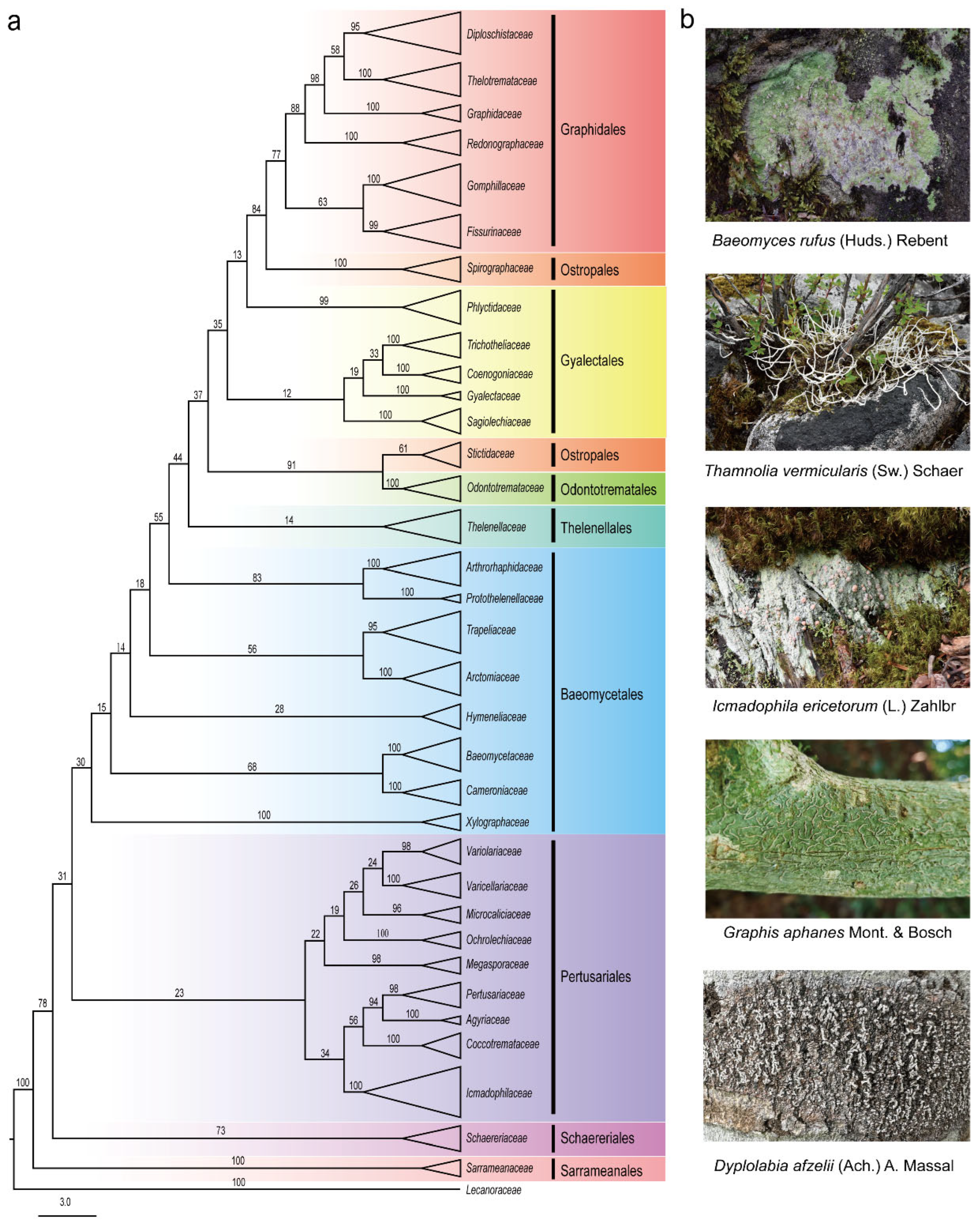
Figure 1. Phylogenetic analysis and the morphology of lichen in the Ostropomycetidae subclass. (a) Phylogenetic tree analysis of taxa at the family level. (b) Morphological characteristics of the lichen thallus of some species in Ostropomycetidae.
Jof 11 00369 g001
Figure 2. Bird’s-eye view of compounds from Ostropomycetidae. (a) The number of families studied to produce compounds in Ostropomycetidae. (b) The number of compounds of different structural types. (c) Different types of compounds with bioactivities.
Jof 11 00369 g002
Figure 3. Chemical structures of phenol compounds (128).
Jof 11 00369 g003
Figure 4. Chemical structures of depside compounds (2962).
Jof 11 00369 g004
Figure 5. Chemical structures of compounds (6394). (a) Tridepsides. (b) Depsidones.
Jof 11 00369 g005
Table 1. Outline of bioactivities of compounds and their producers in Ostropomycetidae.
BioactivityCompoundsLichen SpeciesReferences
AntitumorHaematommic acid (4)A. pakistanica[19]
Olivetoric acid (30)Ochrolechia spp.[39,41]
Baeomycesic acid (38)Siphula spp.[48]
Thamnoliadepside A (42)T. vermicularis[18]
Atranorin (55)Siphula spp., Baeomyces spp.[58]
Sekikaic acid (57)S. ceratites, L. alphoplaca[61]
Alectorialic acid (62)A. pakistanica[19]
Gyrophoric acid (63)P. mccroryae,
O. androgyna, D. diacapsis, A. pakistanica
[69]
α-alectoronic acid (75)O. parella, A. radiosa[35]
Physodic acid (76)D. diacapsis[84,85]
Variolaric acid (77)O. parella, A. radiosa[35]
Protocetraric acid (79)O. androgyna[89]
Stictic acid (81)Baeomyces spp., Pertusaria spp.[96]
Salazinic acid (88)P. pseudocorallina[89]
(+)-protolichesterinic acid (109)Ochrolechia spp.[110,111]
5,7-dihydroxy-6-methylphthalide (114)A. pakistanica[19]
Usnic acid (115)Baeomyces spp., Aspicilia spp.[19]
6-(4,5-dihydroxy-10-methyl-6-oxo-7-undecenyl)-resorcylic acid lactone (129)B. placophyllus[126]
Graphislactone A (140)G. prunicola, G. cognata,
G. scripta
[133]
Norlichexanthone (165)P. laeviganda[141]
Pruinosone (175)D. pruinosum[149]
Diorygmones A–B (177178)Diorygma sp.[151]
Dasyscyphin F (185)Stictidaceae[153]
Petasol (186) and sporogen AO-1 (189)S. tricosa[159]
Graphilane (194)Graphis sp.[158]
Ergosterol peroxide (196)O. parella, S. tricosa[164]
Stigmasterol (197)Aspicilia sp.[167]
Astin C (200)C. asteris[171]
Anti-inflammatoryBarbatinic acid (47)T. vermicularis[47]
Atranorin (55)Siphula spp., Aspicilia spp., Baeomyces spp.[59]
Physodic acid (76)D. diacapsis[82]
Wedelolactone (127)O. frigida[125]
Ergosterol peroxide (196)O. parella, S. tricosa[163]
Stigmasterol (197)Aspicilia sp.[167]
Astin C (200)C. asteris[170]
AntibacterialSquamatic acid (37)Baeomyces spp., Thamnolia spp.[20]
Baeomycesic acid (38)Siphula spp.[117]
Gyrophoric acid (63)Ochrolechia spp.,
P. mccroryae,
[67]
Protocetraric acid (79)O. androgyna[36,87]
Lobaric acid (73)T. vermicularis,
L. alphoplaca
[80]
Fumarprotocetraric acid (80)O. androgyna[36]
Stictic acid (81)Baeomyces spp., Pertusaria spp., Diorygma sp.[36]
Hexadecanoic acid (100)P. caesiopruinosa[106]
Usnic acid (115)Baeomyces spp.,
Aspicilia spp.
[16,19]
Didymic acid (116)P. flavens[120]
Wedelolactone (127)O. frigida[124]
6,8-dihydroxy-3-hydroxymethylisocoumarin (135)G. proserpens[129]
Vinetorin (168)Pertusaria sp.[142]
Elsinochrome A (174)G. elongata[146]
Diorygmone B (178)Diorygma sp.[26]
Diorygmones C–D (180181)D. pruinosum[26]
Dasyscyphin F (185)Stictidaceae[155]
Campesterol (198) Aspicilia sp.[168]
β-sitosterol (199)T. vermicularis,
D. pruinosum
[168]
AntifungalMethyl-3-chloro-2-hydroxy-4-methoxy-6-pentylbenzoate (16)P. dactylina[27]
Lecanoric acid (29)Siphula spp., Diploschistes spp.[36]
Divaricatic acid (31)Pertusaria spp.[43]
Gyrophoric acid (63)Ochrolechia spp.,
P. mccroryae, D. diacapsis, A. pakistanica
[67]
Xylarinic acid A (103)G. handelii[107]
Pruinosone (175) and hydroxypruinosone (176)D. pruinosum[149]
AntiviralSekikaic acid (57)S. ceratites, L. alphoplaca[64]
Handelone (59)G. handelii[66]
Variolaric acid (77)O. parella, A. radiosa[35]
Usnic acid (115)Baeomyces spp., Aspicilia spp.[119]
6,8-dihydroxy-3-hydroxymethylisocoumarin (135)G. proserpens[129]
AntioxidantOrsellinic acid (6)Diploschistes spp.,
O. frigida
[24]
Prephenic acid (8), hypoxyphenone (9), and tetrafucol A (28)O. frigida[24]
Lecanoric acid (29)Siphula spp., Diploschistes spp.[24,38]
Cyperine (36)P. contortuplicata[24]
Atranorin (55)Siphula spp., Baeomyces spp.[38]
Aekikaic acid (57)S. ceratites, L. alphoplaca[62]
Gyrophoric acid (63)Ochrolechia spp.,
P. mccroryae, D. diacapsis, A. pakistanica
[67]
Lobaric acid (73)T. vermicularis,
L. alphoplaca
[80]
Fumarprotocetraric acid (80)O. androgyna[91,92]
Norstictic acid (82)Siphula spp., Aspicilia spp., Graphis spp.[79]
3,6,9,12-tetraoxapentacosanoic acid (101), 18-hydroxylinoleic acid (102), wedelolactone (127), and Diospyrol (173)O. frigida[24]
Stigmasterol (197)Aspicilia sp.[167]
Anti-angiogenicOlivetoric acid (30)Ochrolechia spp.[40]
Anti-neurodegenerative diseases Methylbenzoic acids (10,11,12) and vermicularin (41)T. vermicularis[25]
Baeomycesic acid (38)Siphula spp.[25]
Antitubercular Acremonidin E (25)Graphis sp.[29]
Protocetraric acid (79)O. androgyna[88]
Anti-herbivoreProtocetraric acid (79)O. androgyna[57]
Stictic acid (81)Baeomyces spp., Pertusaria spp., Diorygma sp.[57]
Norstictic acid (82)Siphula spp., Aspicilia spp., Graphis spp.[57]
(+)-aspicilin (128)Aspicilia spp.[57]
Antitrypanosomal Lichesterinic acid (107) and (+)- protolichesterinic acid (109)Ochrolechia spp.[109]
Dasyscyphin C (183)Stictidaceae[155]
Disclaimer/Publisher’s Note: The statements, opinions and data contained in all publications are solely those of the individual author(s) and contributor(s) and not of MDPI and/or the editor(s). MDPI and/or the editor(s) disclaim responsibility for any injury to people or property resulting from any ideas, methods, instructions or products referred to in the content.