Article

Pulse Pressure Variation Can Predict the Hemodynamic Response to Pneumoperitoneum in Dogs: A Retrospective Study

by
Caterina Di Bella
1,*,
Luca Lacitignola
2,
Laura Fracassi
1,
Despoina Skouropoulou
1,
Antonio Crovace
2 and
Francesco Staffieri
2
1
PhD Course in Tissues and Organs Transplantation and Cellular Therapies, Department of Emergency and Organ Transplantation, University of Bari, 70010 Bari, Italy
2
Department of Emergency and Organ Transplantation, Section of Veterinary Clinics and Animal Production; University of Bari, 70010 Bari, Italy
*
Author to whom correspondence should be addressed.
Vet. Sci. 2019, 6(1), 17; https://doi.org/10.3390/vetsci6010017
Submission received: 6 December 2018 / Revised: 7 February 2019 / Accepted: 15 February 2019 / Published: 20 February 2019
(This article belongs to the Special Issue Italian Society of the Veterinary Sciences SISVet 2018)

Abstract

:
Pneumoperitoneum may induce important hemodynamic alterations in healthy subjects. Pulse pressure variation (PPV) is a hemodynamic parameter able to discriminate preload dependent subjects. Anesthesia records of dogs undergoing laparoscopy were retrospectively evaluated. The anesthetic protocol included acepromazine, methadone, propofol and isoflurane administered with oxygen under mechanical ventilation. The hemodynamic parameters were considered five minutes before (BASE) and ten minutes after (P10) the pneumoperitoneum. Based on the cardiac index (CI) variation, at P10, dogs were classified as sensitive (S group, CI ≤ 15%) and non-sensitive (NO-S group). Data were analyzed with the ANOVA test and the ROC curve (p < 0.05). Fifty-five percent of dogs (S) had a reduction of CI ≥ 15% at P10 (2.97 ± 1.4 L/min/m2) compared to BASE (4.32 ± 1.62 L/min/m2) and at P10 in the NO-S group (4.51 ± 1.41 L/min/m2). PPV at BASE was significantly higher in the S group (22.4% ± 6.1%) compared to the NO-S group (10.9% ± 3.3%). The ROC curve showed a threshold of PPV > 16% to distinguish the S and NO-S groups. PPV may be a valid predictor of the hemodynamic response to pneumoperitoneum in dogs. A PPV > 16% can identify patients that may require fluid administration before the creation of pneumoperitoneum.

1. Introduction

During intermittent positive pressure ventilation (IPPV), alterations in intra-thoracic pressure are transmitted to the heart, inducing cyclic changes in the loading conditions of the right and left ventricles (heart lung interaction) [1]. Specifically, positive pressure inspiration decreases preload and increases afterload of the right ventricle (RV), leading to a temporary decrease in stroke volume [2]. Accordingly, the left ventricular (LV) preload reduction induces a decrease in LV filling and a subsequent reduction in stroke volume (SV), which will be evident during expiration due to the lag phase of two to three heart beats because of the pulmonary transit time [2]. The entity of the variation of SV during IPPV is related to the volemic status of the cardiovascular system, being more important in subjects on the step portion of the Frank–Starling curve (preload dependent) [3]. The arterial pulse pressure (the difference between the systolic and diastolic pressures) is directly proportional to SV and its variation in relation to the respiratory cycle (pulse pressure variation, PPV) has been proven to be predictive of the response to the administration of a bolus of fluids (fluid challenge) [3,4]. In human patients, a PPV value equal to or above 13% has been proven to indicate preload dependency; in particular, patients respond with an increase in SV after fluid infusion [5,6]. Recently, evaluation of PPV has gained popularity, including in veterinary medicine to guide volume expansion [7]. In dogs, PPV was shown to be accurate for predicting fluid responsiveness in a model of hemorrhagic shock [8] and in mechanically ventilated isoflurane-anesthetized dogs pre-medicated with acepromazine [9]. These studies showed that changes in PPV and hypovolemic conditions occurred much earlier than changes in classical parameters such as arterial blood pressure, heart rate (HR), or central venous pressure [7,8]. In a clinical study in dogs, Fantoni et al. [10] showed that PPV can predict fluid responsiveness in dogs undergoing orthopedic surgery, and they found a cut-off value of 15% distinguishing responders from non-responders. Drozdzynka et al. [7] used a PPV of 13% as a cut-off for the intraoperative administration of a fluid bolus as part of a goal-directed protocol in dogs undergoing abdominal surgery, showing that a PPV ≥ 13% reliably predicted the cardiovascular response to fluid loading in 82.8% of canine cases undergoing abdominal surgery, in agreement with findings from human studies [2].
The increase of intra-abdominal pressure (IAP) during CO2 pneumoperitoneum (PP) causes several cardiovascular perturbations headed by the reduction of the venous return [11]. From the right ventricle perspective, the rapid decrease of the end diastolic volume, determined by PP, will cause a reduction of the RV stroke volume and of the cardiac output (CO), which will be more important in preload dependent subjects [12,13,14]. The mechanism for the decrease of cardiac output is multifactorial and is related to an increased IAP, which results in caval compression, pooling of blood in the periphery, and an increase in venous resistance [11,15].
Based on this background, the rationale of our study was to evaluate whether the PPV value, before the induction of PP, could be a predictor of the hemodynamic response to laparoscopy in dogs. Our hypothesis is that in preload dependent subjects, higher values of PPV, before the creation of PP, can be predictive of a significant hemodynamic derangement during the procedure, and thus may identify dogs needing cardiovascular stabilization before starting the procedure. To test this hypothesis, the PPV and the hemodynamic values of a series of dogs undergoing laparoscopic ovariectomies were retrospectively evaluated.

2. Materials and Methods

The study was approved by the Ethical Committee for Clinical Study in Animal Patients of the Department of Emergency and Organ Transplantation of the University of Bari (n. 03/2016). In this retrospective study, the anesthesia records of 32 cases of dogs undergoing elective laparoscopic ovariectomies were recruited between January and May 2018 at the Department of Emergency and Organ Transplantation, Section of Veterinary Clinics and Animal Production, “Aldo Moro” University of Bari, Bari, Italy. Dogs affected by systemic or cardiovascular disease were excluded along with cases in which the collection of data was incomplete.

2.1. Anesthetic Protocol

All subjects were premedicated following the standard protocol used for this procedure in healthy dogs (ASA1) at our institution, which included premedication with 10 μg/kg of acepromazine intramuscularly (IM, Prequillan; Fatro, Italy; 10 mg/mL) followed after 15 min by 0.3 mg/kg IM of methadone (Semfortan; Dechra, Italy; 10 mg/mL). The cephalic vein was cannulated for the administration of propofol (Fresenius Kabi Propofol 10 mg/mL) at 5 mg/kg IV, fluids (Ringer Lactate solution; Fresenius Kabi) at 5 mL/kg/h for the entire duration of the procedure, and other drugs, as required. General anesthesia was maintained with inhaled isoflurane in oxygen (FiO2 > 0.8). All dogs were mechanically ventilated in a volume-controlled mode (Servo-I; Maquet, Germany), with a tidal volume (TV) of 15 mL/kg, inspiratory to expiratory ratio of 1:2, inspiratory pause of 25% of inspiratory time, and positive end-expiratory pressure (PEEP) of 0 cmH2O. The respiratory rate (RR) was adjusted for the end-tidal carbon dioxide level (EtCO2), which was maintained between 40 and 55 mmHg. The following respiratory (DatexOhmeda S/5 Anesthesia Monitor, Ohmeda, Soma Technology, Bloomfield, CT, USA) and hemodynamic (PRAM, Most Care®, Vytech, Padova, Italy) parameters, manually collected every five minutes during the procedure, were considered for the study: peripheral capillary oxygen hemoglobin saturation (SpO2, %); TV (mL/kg); EtCO2, (mmHg); peak and plateau airway pressures (Ppeak and Pplat, cmH2O); RR (breaths/minute); static compliance of the respiratory system (Crs, mL/cmH2O/kg); HR (beats/minute); systolic, mean and diastolic arterial pressures (SAP, MAP and DAP respectively; mmHg); SV (mL); CO (L/min); systemic vascular resistances (SVR, dynes*seconds/cm5); and PPV(%). The end-tidal concentration of isoflurane (EtIso, %) and the temperature (T, °C) were also considered. The hemodynamic parameters were collected with a monitoring system based on the pressure recording analytical method technology (PRAM). This uncalibrated pulse contour technique estimates SV and other hemodynamic parameters from the analysis of the arterial pulse waveform, and has been recently validated in dogs [16]. In all cases of the study, PP was created via a Veress needle with a CO2 insufflator (Endoflator; Karl-Storz, Tuttlingen, Germany) at an IAP of 10–11 mmHg.

2.2. Study Protocol

For the purpose of the study, the physiological data registered five minutes before (BASE) and ten minutes after (P10) the induction PP were considered; in particular, the following cardiovascular parameters were recorded: HR, MAP, SV, CO, SVR, and PPV. These data were automatically stored every 3 s by the hemodynamic monitor, which allowed further offline analysis.
The cardiac index (CI, L/minute/m2) was calculated using the formula
CI = CO/BSA
where BSA represents the body surface area (m2).
PPV was automatically calculated by the software of the monitor using the following formula:
PPV(%) = 100*(PPmax − PPmin)/[(PPmax − PPmin)/2]
where PP represents the difference between the systolic and diastolic pressure, and PPmax and PPmin indicate the maximum and minimum value of PP in a single respiratory cycle, respectively.

2.3. Statistical Analysis
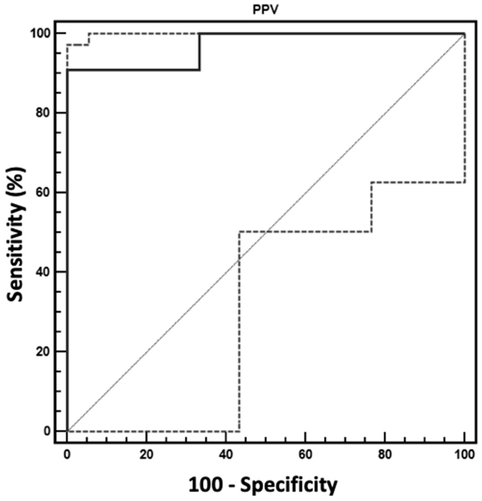
All data were analyzed by MedCalc Software 9.2 (MedCalc, Mariakerke, Belgium). The Shapiro–Wilk test was used to evaluate the normal distribution of the data collected at BASE and P10. All parameters were reported as mean ± standard deviation (SD). The differences between BASE and P10 were tested for the entire population with the paired samples Student’s t-test. Based on the variation of the CI, following the creation of the PP, the cases were divided into two groups. Those presenting a reduction of CI equal to or greater than 15% were considered as sensitive (S). Otherwise, they were considered non-sensitive (NO-S). The comparison between the two groups (S and NO-S) at BASE and P10 was performed with the one-way ANOVA for repeated measurements. A receiver operating characteristic (ROC) curve was generated for PPV at BASE in order to discriminate S from NO-S; the area under the ROC curve was calculated with 95% confidence intervals and the optimal threshold value (the value that maximizes the sum of both sensitivity and specificity) for PPV was determined. Statistical significance was set at p < 0.05.

3. Results

Twelve cases were discarded because either they referred to subjects suffering from systemic or cardiovascular diseases (four cases), or the data collected were not complete (eight cases). Accordingly, 20 cases formed the population of the study with a mean body weight and age of 16.4 ± 8.1 kg and 3.3 ± 1.2 years, respectively. The breeds included were beagle (n = 4), cocker spaniel (n = 3), bull terrier (n = 2) deutscher boxer (n = 2), and mixed breed (n = 9) (Table 1).
The average values of the cardiovascular and respiratory parameters registered before and after PP are reported in Table 2.
After the PP, CI, PPV, and Crs decreased, while MAP, Ppeak and Pplat increased significantly. All other physiological parameters did not exhibit significant differences between the two phases of the study. Eleven (55%) of the 20 subjects were assigned to the S group because they showed a decrease of the CI ≥ 15%; the remaining nine dogs (45%) were ascribed to the NO-S group. Specifically, the mean and SD of the variation of CI after the induction of PP was –32.3% ± 15.2% in the S group, while in the NO-S group it was at 7.18% ± 13.66%. The body weight and age of the two groups were similar (S: 14.6 ± 7.2 kg and 3.2 ± 1.2 years; NO-S: 18.3 ± 9.1 kg and 3.5 ± 1.2 years). The cardiovascular parameters and the EtCO2 recorded in the two groups at the different times of the study are reported in Table 3.
The cardiac index and SVR were similar between the two groups at BASE, but at P10 the CI was lower in group S compared to the NO-S group, and the SVR was lower in the NO-S group compared to the S group (Table 3).
Pulse pressure variation at BASE was greater in the S group compared to the NO-S group (Table 3), while it was similar between the two groups at P10.
Heart rate and MAP were similar between the two groups at the two evaluation times. The mean values of PPV and CI at P10 in the S group were lower than the corresponding values at BASE. The PPV showed a significant area under the ROC curve (0.970 ± 0.039; p = 0.0001). The best cutoff of PPV was 16%, with a sensitivity of 90.91% and a specificity of 100% (confidence interval 0.780–0.987) to distinguish between S and NO-S groups (Figure 1).

4. Discussion

The results of this study demonstrate that PPV could be a valuable parameter to predict the hemodynamic response to PP in dogs. In particular, a PPV value higher than 16% may predict a significant reduction of CO in response to the creation of the pneumoperitoneum; thus, it identify subjects who may benefit from fluids administration before CO2 insufflation.
The hemodynamic perturbations observed during laparoscopy occured mainly at the beginning of the procedure and resulted from the combined effects of pneumoperitoneum, patient position, anesthesia, and hypercapnia from the absorbed CO2. The typical disturbances are characterized by decreases in CO proportional to the increase of the IAP, increased arterial pressure, and elevation of systemic and pulmonary vascular resistances; heart rate remains unchanged or increases slightly [17,18,19]. Cardiac output has also been reported to be increased or unchanged during pneumoperitoneum in human patients and dogs; these discrepancies might be related to differences in rates of CO2 insufflation, IAP, time intervals between insufflation, and differences in data collection and anesthetic technique and drugs [20,21,22]. The absolute or relative intravascular blood volume is an important factor determining the hemodynamic response to PP, and preload-dependent patients usually experience the most severe hemodynamic side effects during laparoscopy [23]. In these patients, the reduction of venous return and CO can be attenuated by increasing circulating volume before the PP is produced [11,24]. The results of this retrospective study showed that 55% (S group) of healthy dogs undergoing laparoscopy might experience an important derangement of the CO 10 min after CO2 insufflation. Interestingly, these dogs did not show any significant hemodynamic differences (CI, MAP, SVR and HR) compared to dogs in the NO-S group before the PP production, except that they had a higher value of PPV. The PPV during positive pressure ventilation is a hemodynamic index able to discriminate preload-dependent subjects that need fluid administration [3]. Thus, we can suppose that the hearts of dogs in the S group were in a compensated preload-dependent condition before PP, and thereafter, the decrease in venous return induced by the PP caused an additional cardiovascular derangement determining an important reduction of the CI. Systemic vascular resistances were similar between the two groups at BASE, but during the PP they were higher in the S group, confirming that these subjects were attempting to compensate for the cardiovascular perturbation created by the PP. All cases included in this study were elective ovariectomy and were healthy. The fact that dogs of the S group resulted as being preload dependent based on the PPV analysis does not necessarily mean they were hypovolemic. Indeed, a preload-dependent condition can also be related to the response of the subjects to the effects of drugs (e.g., acepromazine, isoflurane) or the anesthetic technique (e.g., mechanical ventilation).
The analysis of the ROC curve indicated that a PPV value equal to or higher than 16% before the CO2 insufflation is predictive of an important hemodynamic derangement during the PP, with a high sensibility and specificity. Thus, this threshold of PPV could be useful in clinical cases not only to identify dogs at risk of cardiovascular side effects to PP, but also to guide fluid therapy. Indeed, in these subjects fluids should be administered in order to reduce the PPV to values lower than 16%. Further prospective studies are required to confirm whether this approach could be valid in clinical cases. The literature indicates that in dogs not subjected to laparoscopy, the PPV cut-off value to discriminate “preload dependent” subjects is lower (11%–15%) [7,9,10] than the value found in this study. We can suppose that the difference is related to the impact of PP, which added an additional factor influencing the hemodynamic status compared to dogs not subjected to laparoscopy.
This retrospective study identified a possible monitoring value of PPV in predicting important negative hemodynamic effects of PP. Future prospective studies need to confirm that in these cases (PPV > 16%) fluid administration will prevent the hemodynamic derangement caused by PP. Another limitation of the study is that the dogs evaluated were healthy; thus, it is possible that there are different cut-off values for hemodynamically unstable cases. Moreover, variability on the cardiovascular response can be expected based on breed, age, and sex, but the limited number of cases did not allow this study to consider those factors.

5. Conclusions

Monitoring of PPV is very valuable during laparoscopy in dogs, since it can predict adverse cardiovascular reaction to PP. Moreover, PPV could be used to optimize the fluid therapy of dogs before the production of PP. Values of PPV higher than 16% before the insufflation of CO2 are predictive of cardiovascular side effects to PP, and thus, should suggest prior fluid administration.

Author Contributions

Study execution, data collection, writing up of the first draft of the manuscript, C.D.B.; study execution, data collection, D.S.; study design, analysis and interpretation of data, writing of the first draft of the manuscript, critical revision of the manuscript, F.S.; study execution, data collection, critical revision of the manuscript L.L., L.F. and A.C. All authors read and approved the final version of the manuscript.

Funding

This research received no external funding.

Conflicts of Interest

The authors declare no conflicts of interest.

References

  1. Cheifetz, I.M. Cardiorespiratory interactions: The relationship between mechanical ventilation and hemodynamics. Respir. Care 2014, 59, 1937–1945. [Google Scholar] [CrossRef]
  2. Michard, F. Changes in arterial pressure during mechanical ventilation. Anesthesiology 2005, 103, 419–428. [Google Scholar] [CrossRef] [PubMed]
  3. Cannesson, M. Arterial pressure variation and goal-directed fluid therapy. J. Cardiothorac. Vasc. Anesth. 2010, 24, 487–497. [Google Scholar] [CrossRef] [PubMed]
  4. Yang, X.; Du, B. Does pulse pressure variation predict fluid responsiveness in critically ill patients? A systematic review and meta-analysis. Crit. Care 2014, 18, 650. [Google Scholar] [CrossRef] [PubMed]
  5. Perel, A.; Pizov, R.; Cotev, S. Respiratory variations in the arterial pressure during mechanical ventilation reflect volume status and fluid responsiveness. Intensive Care Med. 2014, 40, 798–807. [Google Scholar] [CrossRef]
  6. Marik, P.E.; Cavallazzi, R.; Vasu, T.; Hirani, A. Dynamic changes in arterial waveform derived variables and fluid responsiveness in mechanically ventilated patients: A systematic review of the literature. Crit. Care Med. 2009, 37, 2642–2647. [Google Scholar] [CrossRef]
  7. Drozdzynska, M.J.; Chang, Y.M.; Stanzani, G.; Pelligand, L. Evaluation of the dynamic predictors of fluid responsiveness in dogs receiving goal-directed fluid therapy. Vet. Anaesth. Analg. 2018, 45, 22–30. [Google Scholar] [CrossRef] [PubMed]
  8. Berkenstadt, H.; Friedman, Z.; Preisman, S.; Keidan, I.; Livingstone, D.; Perel, A. Pulse pressure and stroke volume variations during severe haemorrhage in ventilated dogs. Br. J. Anaesth. 2005, 94, 721–726. [Google Scholar] [CrossRef] [Green Version]
  9. Sano, H.; Seo, J.; Wightman, P.; Cave, N.J.; Gieseg, M.A.; Johnson, C.B.; Chambers, P. Evaluation of pulse pressure variation and pleth variability index to predict fluid responsiveness in mechanically ventilated isoflurane-anesthetized dogs. J. Vet. Emerg. Crit. Care 2018, 28, 301–309. [Google Scholar] [CrossRef]
  10. Fantoni, D.T.; Ida, K.K.; Gimenes, A.M.; Mantovani, M.M.; Castro, J.R.; Patricio, G.C.F.; Ambrosio, A.M.; Otsuki, D.A. Pulse pressure variation as a guide for volume expansion in dogs undergoing orthopedic surgery. Vet. Anaesth. Analg. 2017, 44, 710–718. [Google Scholar] [CrossRef]
  11. Kashtan, J.; Green, J.F.; Parsons, E.Q.; Holcroft, J.W. Hemodynamic effect of increased abdominal pressure. J. Surg. Res. 1981, 30, 249–255. [Google Scholar] [CrossRef]
  12. Zuckerman, R.S.; Heneghan, S. The duration of hemodynamic depression during laparoscopic cholecystectomy. Surg. Endosc. 2002, 16, 1233–1236. [Google Scholar] [CrossRef] [PubMed]
  13. Liu, F.; Zhu, S.; Ji, Q.; Li, W.; Liu, J. The impact of intra-abdominal pressure on the stroke volume variation and plethysmographic variability index in patients undergoing laparoscopic cholecystectomy. Biosci. Trends 2015, 9, 129–133. [Google Scholar] [CrossRef] [PubMed] [Green Version]
  14. Branche, P.E.; Duperret, S.L.; Sagnard, P.E.; Boulez, J.L.; Petit, P.L.; Viale, J.P. Left ventricular loading modifications induced by pneumoperitoneum: A time course echocardiographic study. Anesth. Analg. 1998, 86, 482–487. [Google Scholar] [CrossRef] [PubMed]
  15. Takata, M.; Wise, R.A.; Robotham, J.L. Effects of abdominal pressure on venous return: Abdominal vascular zone conditions. J. Appl. Physiol. 1990, 69, 1961–1972. [Google Scholar] [CrossRef] [PubMed]
  16. Briganti, A.; Evangelista, F.; Centonze, P.; Rizzo, A.; Bentivegna, F.; Crovace, A.; Staffieri, F. A preliminary study evaluating cardiac output measurement using Pressure Recording Analytical Method (PRAM) in anaesthetized dogs. BMC Vet. Res. 2018, 14, 72. [Google Scholar] [CrossRef]
  17. Struthers, A.D.; Cuschieri, A. Cardiovascular consequences of laparoscopic surgery. Lancet 1998, 352, 568–570. [Google Scholar] [CrossRef]
  18. Koivusalo, A.M.; Lindgren, L. Effects of carbon dioxide pneumoperitoneum for laparoscopic cholecystectomy. Acta Anaesthesiol. Scand. 2000, 44, 834–841. [Google Scholar] [CrossRef]
  19. Atkinson, T.M.; Giraud, G.D.; Togioka, B.M.; Jones, D.B.; Cigarroa, J.E. Cardiovascular and Ventilatory Consequences of Laparoscopic Surgery. Circulation 2017, 135, 700–710. [Google Scholar] [CrossRef]
  20. Ivankovich, A.D.; Miletich, D.J.; Albrecht, R.F.; Heyman, H.J.; Bonnet, R.F. Cardiovascular effects of intraperitoneal insufflation with carbon dioxide and nitrous oxide in the dog. Anesthesiology 1975, 42, 281–287. [Google Scholar] [CrossRef]
  21. Duke, T.; Steinacher, S.L.; Remedios, A.M. Cardiopulmonary effects of using carbon dioxide for laparoscopic surgery in dogs. Vet. Surg. 1996, 25, 77–82. [Google Scholar] [CrossRef] [PubMed]
  22. Dexter, S.P.; Vucevic, M.; Gibson, J.; McMahon, M.J. Hemodynamic consequences of high- and low-pressure capnoperitoneum during laparoscopic cholecystectomy. Surg. Endosc. 1999, 13, 376–381. [Google Scholar] [CrossRef] [PubMed]
  23. Safran, D.; Sgambati, S.; Orlando, R., 3rd. Laparoscopy in high-risk cardiac patients. Surg. Gynecol. Obstet. 1993, 176, 548–554. [Google Scholar] [CrossRef] [PubMed]
  24. Ho, H.S.; Saunders, C.J.; Corso, F.A.; Wolfe, B.M. The effects of CO2 pneumoperitoneum on hemodynamics in hemorrhaged animals. Surgery 1993, 114, 381–387, discussion 387–388. [Google Scholar] [PubMed]
Figure 1. Graphical representation of the ROC curve to test the ability of pulse pressure variation (PPV) to discriminate between sensitive (S group = decrease in CI ≥ 15% after PP) and non-sensitive (NO-S) subjects after the creation of the pneumoperitoneum in dogs undergoing laparoscopic surgery.
Vetsci 06 00017 g001
Table 1. Breed, age, heart rate (HR), mean arterial pressure (MAP), cardiac index (CI), pulse pressure variation (PPV), and respiratory system compliance (Crs) of the cases included in the study, immediately before (BASE) and 10 min after the pneumoperitoneum (P10). Dogs that showed a reduction of CI ≥ 15% at P10 compared to BASE were classified as sensitive (S), the rest of the dogs were classified as non-sensitive (NO-S).
DogPhaseBreedAge (Years)HR (beats/min)MAP (mmHg)CI (L/min/m2)PPV (%)Crs (mL/cmH2O/kg)Group
1BASEcocker spaniel287982.9210.9S
P10861171.1110.8
2BASEmixed breed372795.9321.0S
P1096762.6120.8
3BASEmixed breed375792.2181.6S
P10841281.470.9
4BASEbull terrier441766.3221.6S
P10851214.7210.9
5BASEbeagle4108646.513.62.1S
P10109894.5180.6
6BASEbeagle288865.6191S
P10102544.7130.9
7BASEmixed breed4116762.7201.1S
P10131591.671.1
8BASEboxer5128883.4261.1S
P101201002.6160.6
9BASEcocker spaniel272753.6331.8S
P1083822.3120.9
10BASEmixed breed199542.7241.4S
P1078952.17.80.8
11BASEbeagle471815.3181.9S
P1071914.6120.7
12BASEmixed breed3107814.66.33.1NS
P10101874.451.1
13BASEcocker spaniel556707.5112.3NS
P1080997.6101.3
14BASEmixed breed2102833.77.30.9NS
P1073744.1100.5
15BASEmixed breed260763.58.81.4NS
P1091964.671.2
16BASEbeagle584803.2141.9NS
P10901013.791.2
17BASEbull terrier467684.7141.3NS
P10891005.791
18BASEboxer697703.691.4NS
P1089953.3141.4
19BASEmixed breed376704.2111.6NS
P10102764.491.3
20BASEmixed breed291832.8161.7NS
P1076962.671.1
Table 2. Mean ± SD of the cardiovascular and respiratory parameters evaluated 5 min before (BASE) and 10 min after (P10) the pneumoperitoneum (PP) in twenty mechanically ventilated isoflurane-anesthetized dogs. * p < 0.05 between the two evaluation times of the study.
ParameterBASEP10p Value
HR (beats/min)84.8 ± 21.691.8 ± 15.40.111
MV (L/min/kg)0.17 ± 0.030.19 ± 0.040.127
MAP (mmHg)76.8 ± 9.491.8 ± 18.50.003 *
CI (L/min/m2)4.28 ± 1.43.67 ± 1.60.019 *
SVR (dyn*sec/cm5)3117 ± 14853003 ± 13410.775
PPV (%)17.2 ± 7.6 11.1 ± 4.00.001*
EtCO2 (mmHg)45.9 ± 4.952.6 ± 7.40.067
SpO2 (%)98.2 ± 1.197.6 ± 1.40.765
Crs (mL/cmH2O/kg)1.6 ± 0.60.9 ± 0.20.001 *
Ppeak (cmH2O)8.9 ± 1.812.3 ± 2.90.001 *
Pplat (cmH2O)8.7 ± 1.811.6 ± 2.30.001 *
Table 3. Mean ± SD of the cardiovascular parameters evaluated in mechanically ventilated isoflurane-anesthetized dogs, 5 min before (BASE) and 10 min after (P10) the pneumoperitoneum. Based on the variation of CI after the PP, dogs were divided into the groups S (CI decreased ≥ 15%) and NO-S. The p value related to the comparison of the two groups at the same time is also reported in table. * p < 0.05 between the two groups. # p < 0.05 compared to the corresponding basal value.
ParameterPhaseS GroupNO-S Groupp Value
HR (beats/min)BASE86.4 ± 25.181.1 ± 20.10.601
P1096.1 ± 26.894.1 ± 12.50.822
MAP (mmHg)BASE77.9 ± 11.674.6 ± 6.80.471
P1089.1 ± 27.192.1 ± 7.40.742
CI (L/m2)BASE4.32 ± 1.624.22 ± 1.380.88
P102.97 ± 1.4 #4.51 ± 1.410.02 *
SVR (dyn*sec/cm5)BASE3081 ± 10073160 ± 19910.902
P103573 ± 12452305 ±11380.032 *
EtCO2BASE46.2 ± 5.2443.2 ± 7.20.602
P1050.1 ± 7.3248.2 ±5.60.732
PPV (%)BASE22.4 ± 6.110.9 ± 3.30.000 *
P1012.6 ± 4.3 #9.1 ± 2.50.05

