
Isolation and Molecular Characterization of Swine Parainfluenza Virus 5 from Piglets Co-Infected with PEDV
 , and
, and
Simple Summary
Abstract
1. Introduction
2. Materials and Methods
2.1. Cells and Clinical Samples
2.2. Virus Isolation and Identification
2.3. Phylogenetic Analysis and Sequence Alignment
3. Results
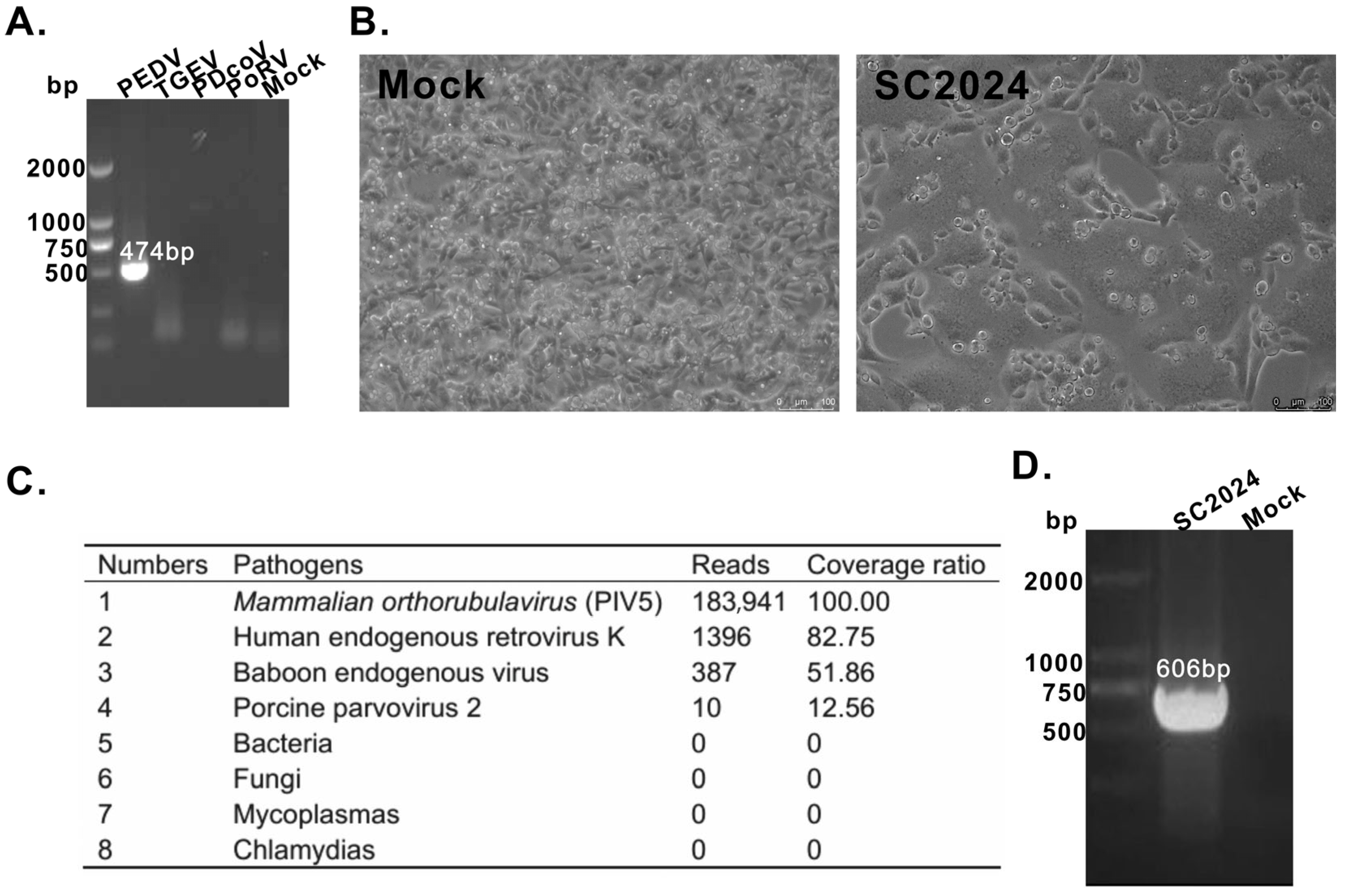
3.1. Isolation and Identification of PIV5
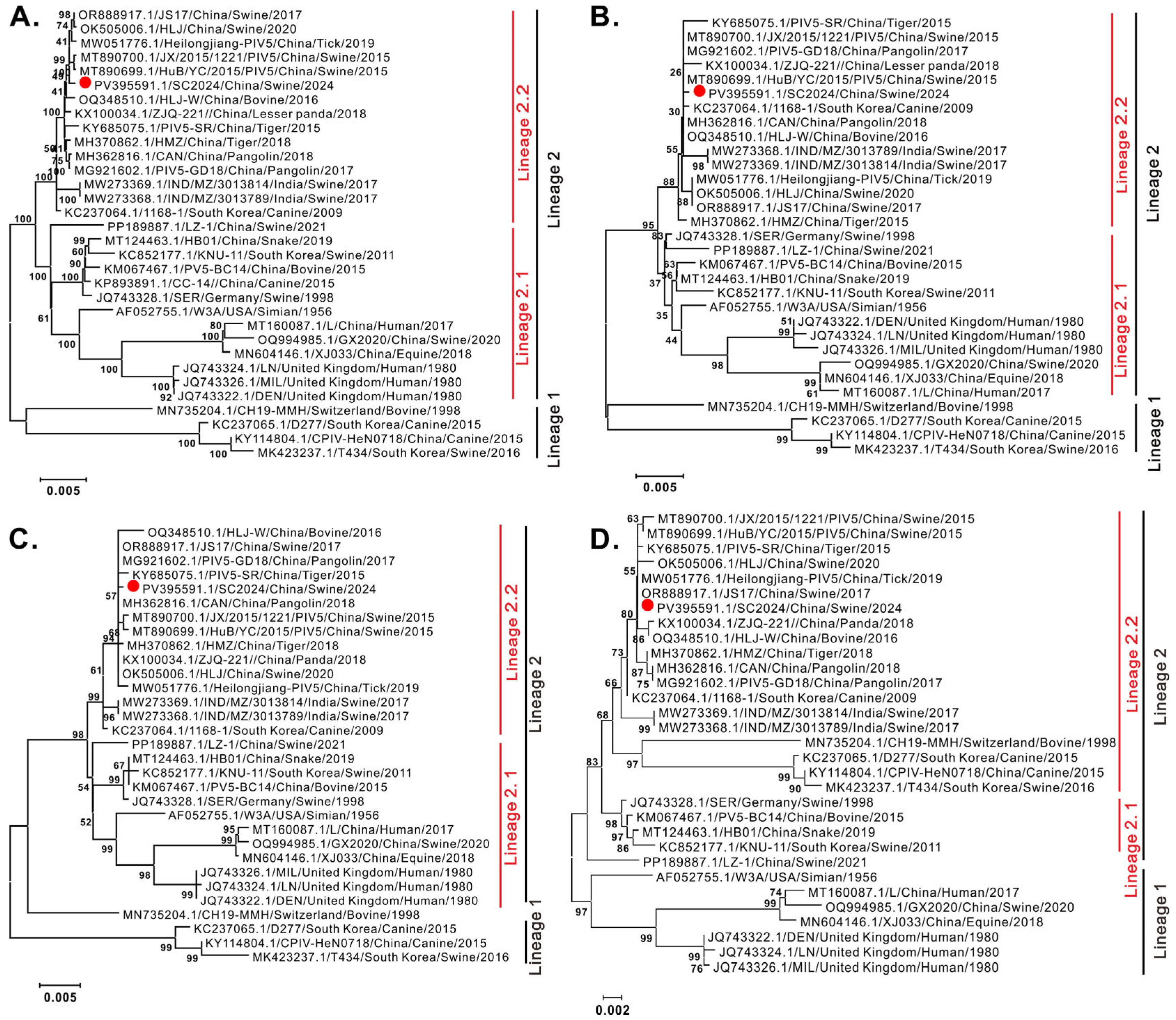
3.2. Phylogenetic Analysis
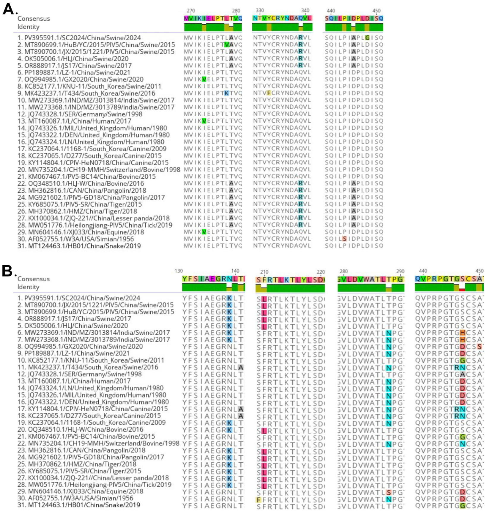
3.3. Sequence Identity and Alignment Analysis
4. Discussion
Supplementary Materials
Author Contributions
Funding
Institutional Review Board Statement
Informed Consent Statement
Data Availability Statement
Conflicts of Interest
References
- Choppin, P.W.; Stoeckenius, W. The Morphology of Sv5 Virus. Virology 1964, 23, 195–202. [Google Scholar] [CrossRef] [PubMed]
- Chatziandreou, N.; Stock, N.; Young, D.; Andrejeva, J.; Hagmaier, K.; McGeoch, D.J.; Randall, R.E. Relationships and host range of human, canine, simian and porcine isolates of simian virus 5 (parainfluenza virus 5). J. Gen. Virol. 2004, 85 Pt 10, 3007–3016. [Google Scholar] [CrossRef] [PubMed]
- Chen, Z. Parainfluenza virus 5-vectored vaccines against human and animal infectious diseases. Rev. Med. Virol. 2018, 28, e1965. [Google Scholar] [CrossRef] [PubMed]
- Walker, P.J.; Siddell, S.G.; Lefkowitz, E.J.; Mushegian, A.R.; Adriaenssens, E.M.; Alfenas-Zerbini, P.; Davison, A.J.; Dempsey, D.M.; Dutilh, B.E.; Garcia, M.L.; et al. Changes to virus taxonomy and to the International Code of Virus Classification and Nomenclature ratified by the International Committee on Taxonomy of Viruses (2021). Arch. Virol. 2021, 166, 2633–2648. [Google Scholar] [CrossRef] [PubMed]
- Vainionpaa, R.; Hyypia, T. Biology of parainfluenza viruses. Clin. Microbiol. Rev. 1994, 7, 265–275. [Google Scholar] [CrossRef] [PubMed]
- Henrickson, K.J. Parainfluenza viruses. Clin. Microbiol. Rev. 2003, 16, 242–264. [Google Scholar] [CrossRef] [PubMed]
- Paterson, R.G.; Leser, G.P.; Shaughnessy, M.A.; Lamb, R.A. The paramyxovirus SV5 V protein binds two atoms of zinc and is a structural component of virions. Virology 1995, 208, 121–131. [Google Scholar] [CrossRef] [PubMed]
- Lin, G.Y.; Paterson, R.G.; Lamb, R.A. The RNA binding region of the paramyxovirus SV5 V and P proteins. Virology 1997, 238, 460–469. [Google Scholar] [CrossRef] [PubMed]
- Thomas, S.M.; Lamb, R.A.; Paterson, R.G. Two mRNAs that differ by two nontemplated nucleotides encode the amino coterminal proteins P and V of the paramyxovirus SV5. Cell 1988, 54, 891–902. [Google Scholar] [CrossRef] [PubMed]
- Hull, R.N.; Minner, J.R.; Smith, J.W. New viral agents recovered from tissue cultures of monkey kidney cells. I. Origin and properties of cytopathogenic agents S.V.1, S.V.2, S.V.4, S.V.5, S.V.6, S.V.11, S.V.12 and S.V.15. Am. J. Hyg. 1956, 63, 204–215. [Google Scholar] [CrossRef] [PubMed]
- Liu, Y.; Li, N.; Zhang, S.; Zhang, F.; Lian, H.; Hu, R. Parainfluenza Virus 5 as Possible Cause of Severe Respiratory Disease in Calves, China. Emerg. Infect. Dis. 2015, 21, 2242–2244. [Google Scholar] [CrossRef] [PubMed]
- Wang, X.; Chen, W.; Xiang, R.; Li, L.; Chen, J.; Zhong, R.; Xiang, H.; Chen, J. Complete Genome Sequence of Parainfluenza Virus 5 (PIV5) from a Sunda Pangolin (Manis javanica) in China. J. Wildl. Dis. 2019, 55, 947–950. [Google Scholar] [CrossRef] [PubMed]
- Xie, J.; Tong, P.; Zhang, A.; Zhang, L.; Song, X.; Kuang, L. Identification and Characterization of the First Equine Parainfluenza Virus 5. Virol. Sin. 2020, 35, 245–247. [Google Scholar] [CrossRef] [PubMed]
- He, W.T.; Hou, X.; Zhao, J.; Sun, J.; He, H.; Si, W.; Wang, J.; Jiang, Z.; Yan, Z.; Xing, G.; et al. Virome characterization of game animals in China reveals a spectrum of emerging pathogens. Cell 2022, 185, 1117–1129.e8. [Google Scholar] [CrossRef] [PubMed]
- Liu, J.; Ren, T.; Wang, X.; Wang, H.; Chen, G.; Qin, Y.; Ouyang, K.; Chen, Y.; Huang, W.; Wei, Z. The emergence, isolation, and phylogenetic analysis of a closely related human strain of parainfluenza virus 5 from a case of porcine reproductive and respiratory syndrome in China. Virology 2024, 597, 110157. [Google Scholar] [CrossRef] [PubMed]
- Huang, M.; Tao, L.; Zha, B.; Zeng, Q.; Hu, Y.; Guo, X.; Liu, P.; Gao, X.; Wu, H. First isolation and phylogenetic analysis of Parainfluenza Virus 5 from Geese. Poult. Sci. 2025, 104, 105177. [Google Scholar] [CrossRef] [PubMed]
- Zhai, J.Q.; Zhai, S.L.; Lin, T.; Liu, J.K.; Wang, H.X.; Li, B.; Zhang, H.; Zou, S.Z.; Zhou, X.; Wu, M.F.; et al. First complete genome sequence of parainfluenza virus 5 isolated from lesser panda. Arch. Virol. 2017, 162, 1413–1418. [Google Scholar] [CrossRef] [PubMed]
- Hsiung, G.D. Parainfluenza-5 virus. Infection of man and animal. Progress. Med. Virol. 1972, 14, 241–274. [Google Scholar]
- Hierweger, M.M.; Werder, S.; Seuberlich, T. Parainfluenza Virus 5 Infection in Neurological Disease and Encephalitis of Cattle. Int. J. Mol. Sci. 2020, 21, 498. [Google Scholar] [CrossRef] [PubMed]
- Oem, J.K.; Kim, S.H.; Kim, Y.H.; Lee, M.H.; Lee, K.K. Molecular characteristics of canine parainfluenza viruses type 5 (CPIV-5) isolated in Korea. Can. J. Vet. Res.-Rev. Can. Rech. Vet. 2015, 79, 64–67. [Google Scholar]
- Liu, C.; Li, X.; Zhang, J.; Yang, L.; Li, F.; Deng, J.; Tan, F.; Sun, M.; Liu, Y.; Tian, K. Isolation and genomic characterization of a canine parainfluenza virus type 5 strain in China. Arch. Virol. 2017, 162, 2337–2344. [Google Scholar] [CrossRef] [PubMed]
- Cornwell, H.J.; McCandlish, I.A.; Thompson, H.; Laird, H.M.; Wright, N.G. Isolation of parainfluenza virus SV5 from dogs with respiratory disease. Vet. Rec. 1976, 98, 301–302. [Google Scholar] [CrossRef] [PubMed]
- Viitanen, S.J.; Lappalainen, A.; Rajamaki, M.M. Co-infections with respiratory viruses in dogs with bacterial pneumonia. J. Vet. Intern. Med. 2015, 29, 544–551. [Google Scholar] [CrossRef] [PubMed]
- Ibrahim, Y.M.; Zhang, W.; Werid, G.M.; Zhang, H.; Pan, Y.; Zhang, L.; Xu, Y.; Li, C.; Chen, H.; Wang, Y. Characterization of parainfluenza virus 5 from diarrheic piglet highlights its zoonotic potential. Transbound. Emerg. Dis. 2022, 69, e1510–e1525. [Google Scholar] [CrossRef] [PubMed]
- Chen, Z.; Xu, P.; Salyards, G.W.; Harvey, S.B.; Rada, B.; Fu, Z.F.; He, B. Evaluating a parainfluenza virus 5-based vaccine in a host with pre-existing immunity against parainfluenza virus 5. PLoS ONE 2012, 7, e50144. [Google Scholar] [CrossRef] [PubMed]
- Yang, M.; Ma, Y.; Jiang, Q.; Song, M.; Kang, H.; Liu, J.; Qu, L. Isolation, identification and pathogenic characteristics of tick-derived parainfluenza virus 5 in northeast China. Transbound. Emerg. Dis. 2022, 69, 3300–3316. [Google Scholar] [CrossRef] [PubMed]
- Durchfeld, B.; Baumgartner, W.; Krakowka, S. Intranasal infection of ferrets (Mustela putorius furo) with canine parainfluenza virus. J. Vet. Med. Ser. B 1991, 38, 505–512. [Google Scholar] [CrossRef] [PubMed]
- Davidson, W.R.; Appel, M.J.; Doster, G.L.; Baker, O.E.; Brown, J.F. Diseases and parasites of red foxes, gray foxes, and coyotes from commercial sources selling to fox-chasing enclosures. J. Wildl. Dis. 1992, 28, 581–589. [Google Scholar] [CrossRef] [PubMed]
- Heinen, E.; Herbst, W.; Schmeer, N. Isolation of a cytopathogenic virus from a case of porcine reproductive and respiratory syndrome (PRRS) and its characterization as parainfluenza virus type 2. Arch. Virol. 1998, 143, 2233–2239. [Google Scholar] [CrossRef] [PubMed]
- Lee, Y.N.; Lee, C. Complete genome sequence of a novel porcine parainfluenza virus 5 isolate in Korea. Arch. Virol. 2013, 158, 1765–1772. [Google Scholar] [CrossRef] [PubMed]
- Jiang, N.; Wang, E.; Guo, D.; Wang, X.; Su, M.; Kong, F.; Yuan, D.; Zhai, J.; Sun, D. Isolation and molecular characterization of parainfluenza virus 5 in diarrhea-affected piglets in China. J. Vet. Med. Sci./Jpn. Soc. Vet. Sci. 2018, 80, 590–593. [Google Scholar] [CrossRef] [PubMed]
- Ni, M.; Lin, S.; Shao, Y.; Tang, J.; Li, S.; Tan, C.; Gong, Z.; Li, H.; Wang, J.; Liu, G.; et al. Enteric pathogenicity characterization of emerging parainfluenza virus 5 in western China. Virology 2025, 604, 110409. [Google Scholar] [CrossRef] [PubMed]
- Truong, H.T.; Nguyen, V.G.; Pham, L.B.; Huynh, T.M.; Lee, J.; Hwang, S.J.; Lee, J.M.; Chung, H.C. PCR-Based Detection and Genetic Characterization of Parainfluenza Virus 5 Detected in Pigs in Korea from 2016 to 2018. Vet. Sci. 2023, 10, 414. [Google Scholar] [CrossRef] [PubMed]
- Ren, J.L.; Tan, S.S.; Chen, X.X.; Yao, J.Y.; Niu, Z.H.; Wang, Y.; Ma, L.; Gao, X.L.; Niu, S.; Liang, L.B.; et al. Genomic Characterization and gE/gI-Deleted Strain Construction of Novel PRV Variants Isolated in Central China. Viruses 2023, 15, 1237. [Google Scholar] [CrossRef] [PubMed]
- Gao, Y.; Kou, Q.; Ge, X.; Zhou, L.; Guo, X.; Yang, H. Phylogenetic analysis of porcine epidemic diarrhea virus field strains prevailing recently in China. Arch. Virol. 2013, 158, 711–715. [Google Scholar] [CrossRef] [PubMed]
- Li, J.; Jin, Z.; Gao, Y.; Zhou, L.; Ge, X.; Guo, X.; Han, J.; Yang, H. Development of the full-length cDNA clones of two porcine epidemic diarrhea disease virus isolates with different virulence. PLoS ONE 2017, 12, e0173998. [Google Scholar] [CrossRef] [PubMed]
- Sheng, W.; Wang, K.; Gui, Y.; Qi, X.; Shen, L.; Zhang, Y.; Tang, C.; Li, X.; Tao, J.; Cao, C.; et al. Molecular characteristics and phylogenetic analysis of pigeon paramyxovirus type 1 isolates from pigeon meat farms in Shanghai (2009–2012). Sci. Rep. 2024, 14, 10741. [Google Scholar] [CrossRef] [PubMed]
- Lee, Y.N.; Park, C.K.; Kim, S.H.; Lee du, S.; Shin, J.H.; Lee, C. Characterization in vitro and in vivo of a novel porcine parainfluenza virus 5 isolate in Korea. Virus Res. 2013, 178, 423–429. [Google Scholar] [CrossRef] [PubMed]
- Wang, T.; Zheng, L.L.; Zhao, Q.J.; Yao, Y.Z.; Zhou, F.; Wei, F.; Cai, Q.L. Parainfluenza virus 5 is a next-generation vaccine vector for human infectious pathogens. J. Med. Virol. 2023, 95, e28622. [Google Scholar] [CrossRef] [PubMed]
- Tompkins, S.M.; Lin, Y.; Leser, G.P.; Kramer, K.A.; Haas, D.L.; Howerth, E.W.; Xu, J.; Kennett, M.J.; Durbin, R.K.; Durbin, J.E.; et al. Recombinant parainfluenza virus 5 (PIV5) expressing the influenza A virus hemagglutinin provides immunity in mice to influenza A virus challenge. Virology 2007, 362, 139–150. [Google Scholar] [CrossRef] [PubMed]
- Clark, K.M.; Johnson, J.B.; Kock, N.D.; Mizel, S.B.; Parks, G.D. Parainfluenza virus 5-based vaccine vectors expressing vaccinia virus (VACV) antigens provide long-term protection in mice from lethal intranasal VACV challenge. Virology 2011, 419, 97–106. [Google Scholar] [CrossRef] [PubMed]
- Chen, Z.; Gupta, T.; Xu, P.; Phan, S.; Pickar, A.; Yau, W.; Karls, R.K.; Quinn, F.D.; Sakamoto, K.; He, B. Efficacy of parainfluenza virus 5 (PIV5)-based tuberculosis vaccines in mice. Vaccine 2015, 33, 7217–7224. [Google Scholar] [CrossRef] [PubMed]
- Xiao, P.; Dienger-Stambaugh, K.; Chen, X.; Wei, H.; Phan, S.; Beavis, A.C.; Singh, K.; Adhikary, N.R.D.; Tiwari, P.; Villinger, F.; et al. Parainfluenza Virus 5 Priming Followed by SIV/HIV Virus-Like-Particle Boosting Induces Potent and Durable Immune Responses in Nonhuman Primates. Front. Immunol. 2021, 12, 623996. [Google Scholar] [CrossRef] [PubMed]
- Spearman, P.; Jin, H.; Knopp, K.; Xiao, P.; Gingerich, M.C.; Kidd, J.; Singh, K.; Tellier, M.; Radziewicz, H.; Wu, S.; et al. Intranasal parainfluenza virus type 5 (PIV5)-vectored RSV vaccine is safe and immunogenic in healthy adults in a phase 1 clinical study. Sci. Adv. 2023, 9, eadj7611. [Google Scholar] [CrossRef] [PubMed]
- Beavis, A.C.; Xiao, P.; Gingerich, M.C.; Briggs, K.; Li, G.; Howerth, E.W.; Najera, M.; An, D.; Huang, J.C.; Mousa, J.; et al. A parainfluenza virus 5 (PIV5)-vectored intranasal SARS-CoV-2 vaccine (CVXGA1) elicits protective and long-lasting immunity in nonhuman primates. J. Virol. 2025, 99, e0199024. [Google Scholar] [CrossRef] [PubMed]
- Beavis, A.C.; Li, Z.; Briggs, K.; Gingerich, M.C.; Wrobel, E.R.; Najera, M.; An, D.; Orr-Burks, N.; Murray, J.; Patil, P.; et al. Efficacy of parainfluenza virus 5 (PIV5)-vectored intranasal COVID-19 vaccine as a single dose primer and booster against SARS-CoV-2 variants. J. Virol. 2025, 99, e0198924. [Google Scholar] [CrossRef] [PubMed]
- Chen, Z.; Zhou, M.; Gao, X.; Zhang, G.; Ren, G.; Gnanadurai, C.W.; Fu, Z.F.; He, B. A novel rabies vaccine based on a recombinant parainfluenza virus 5 expressing rabies virus glycoprotein. J. Virol. 2013, 87, 2986–2993. [Google Scholar] [CrossRef] [PubMed]
- Li, H.; Sun, H.; Tao, M.; Han, Q.; Yu, H.; Li, J.; Lu, X.; Tong, Q.; Pu, J.; Sun, Y.; et al. Recombinant parainfluenza virus 5 expressing clade 2.3.4.4b H5 hemagglutinin protein confers broad protection against H5Ny influenza viruses. J. Virol. 2024, 98, e0112923. [Google Scholar] [CrossRef] [PubMed]
- Branche, A.R.; Falsey, A.R. Parainfluenza Virus Infection. Semin. Respir. Crit. Care Med. 2016, 37, 538–554. [Google Scholar] [CrossRef] [PubMed]
- Wang, Z.; Geng, Z.; Zhou, H.; Chen, P.; Qian, J.; Guo, A. Genetic Characterization, Pathogenicity, and Epidemiology Analysis of Three Sub-Genotype Pigeon Newcastle Disease Virus Strains in China. Microorganisms 2024, 12, 738. [Google Scholar] [CrossRef] [PubMed]
- Miyahara, K.; Kitada, S.; Yoshimoto, M.; Matsumura, H.; Kawano, M.; Komada, H.; Tsurudome, M.; Kusagawa, S.; Nishio, M.; Ito, Y. Molecular evolution of human paramyxoviruses. Nucleotide sequence analyses of the human parainfluenza type 1 virus NP and M protein genes and construction of phylogenetic trees for all the human paramyxoviruses. Arch. Virol. 1992, 124, 255–268. [Google Scholar] [CrossRef] [PubMed]
- Zhu, Y.M.; Shi, H.F.; Gao, Y.R.; Xin, J.Q.; Liu, N.H.; Xiang, W.H.; Ren, X.G.; Feng, J.K.; Zhao, L.P.; Xue, F. Isolation and genetic characterization of bovine parainfluenza virus type 3 from cattle in China. Vet. Microbiol. 2011, 149, 446–451. [Google Scholar] [CrossRef] [PubMed]
- Charoenkul, K.; Nasamran, C.; Janetanakit, T.; Chaiyawong, S.; Bunpapong, N.; Boonyapisitsopa, S.; Tangwangvivat, R.; Amonsin, A. Molecular detection and whole genome characterization of Canine Parainfluenza type 5 in Thailand. Sci. Rep. 2021, 11, 3866. [Google Scholar] [CrossRef] [PubMed]
- Torremorell, M.; Allerson, M.; Corzo, C.; Diaz, A.; Gramer, M. Transmission of influenza A virus in pigs. Transbound. Emerg. Dis. 2012, 59 (Suppl. 1), 68–84. [Google Scholar] [CrossRef] [PubMed]
- Ma, W. Swine influenza virus: Current status and challenge. Virus Res. 2020, 288, 198118. [Google Scholar] [CrossRef] [PubMed]
- McLean, R.K.; Graham, S.P. Vaccine Development for Nipah Virus Infection in Pigs. Front. Vet. Sci. 2019, 6, 16. [Google Scholar] [CrossRef] [PubMed]
- Bruno, L.; Nappo, M.A.; Ferrari, L.; Di Lecce, R.; Guarnieri, C.; Cantoni, A.M.; Corradi, A. Nipah Virus Disease: Epidemiological, Clinical, Diagnostic and Legislative Aspects of This Unpredictable Emerging Zoonosis. Animals 2022, 13, 159. [Google Scholar] [CrossRef] [PubMed]
- Yuan, P.; Thompson, T.B.; Wurzburg, B.A.; Paterson, R.G.; Lamb, R.A.; Jardetzky, T.S. Structural studies of the parainfluenza virus 5 hemagglutinin-neuraminidase tetramer in complex with its receptor, sialyllactose. Structure 2005, 13, 803–815. [Google Scholar] [CrossRef] [PubMed]
- Baty, D.U.; Randall, R.E. Multiple amino acid substitutions in the HN protein of the paramyxovirus, SV5, are selected for in monoclonal antibody resistant mutants. Arch. Virol. 1993, 131, 217–224. [Google Scholar] [CrossRef] [PubMed]

| No. | Strain | GenBank No. | Year | Host | Country | SC2024 | ||||||
|---|---|---|---|---|---|---|---|---|---|---|---|---|
| WGS | NP | F | HN | |||||||||
| nt (%) | nt (%) | aa (%) | nt (%) | aa (%) | nt (%) | aa (%) | ||||||
| GX2020 | OQ994985.1 | 2020 | Swine | China | 97.2 | 97.6 | 98.2 | 97.5 | 97.3 | 96.6 | 96.5 | |
| JX | MT890700.1 | 2015 | Swine | China | 99.8 | 99.9 | 99.8 | 99.8 | 99.5 | 99.8 | 100 | |
| HuB/YC | MT890699.1 | 2015 | Swine | China | 99.8 | 99.8 | 99.8 | 99.8 | 99.5 | 99.9 | 100 | |
| JS17 | OR888917.1 | 2017 | Swine | China | 99.9 | 99.9 | 99.6 | 99.9 | 99.8 | 100 | 100 | |
| LZ-1 | PP189887.1 | 2021 | Swine | China | 98.8 | 99.2 | 98.6 | 99.1 | 98.4 | 98.9 | 97.9 | |
| HLJ | OK505006.1 | 2020 | Swine | China | 99.9 | 99.8 | 99.6 | 99.9 | 99.8 | 99.8 | 99.8 | |
| KNU-11 | KC852177.1 | 2011 | Swine | South Korea | 98.7 | 99.1 | 98.4 | 99.0 | 98.2 | 99.1 | 98.4 | |
| T434 | MK423237.1 | 2016 | Swine | South Korea | 96.6 | 96.6 | 97.6 | 95.8 | 94.9 | 97.5 | 97.3 | |
| IND/MZ/3013814 | MW273369.1 | 2017 | Swine | India | 99.6 | 99.7 | 99.4 | 99.6 | 98.9 | 99.5 | 98.9 | |
| IND/MZ/3013789 | MW273368.1 | 2017 | Swine | India | 99.6 | 99.7 | 99.4 | 99.6 | 98.9 | 99.5 | 98.9 | |
| SER | JQ743328.1 | 1998 | Swine | Germany | 99.0 | 99.5 | 99.4 | 99.1 | 98.4 | 99.4 | 98.9 | |
| L | MT160087.1 | 2017 | Human | China | 97.3 | 97.8 | 98.4 | 97.3 | 96.9 | 96.8 | 96.5 | |
| LN | JQ743324.1 | 1980 | Human | United Kingdom | 98.0 | 98.1 | 98.6 | 98.1 | 97.8 | 97.8 | 97.9 | |
| MIL | JQ743326.1 | 1980 | Human | United Kingdom | 98.0 | 98.0 | 98.6 | 98.2 | 98.0 | 97.8 | 97.9 | |
| DEN | JQ743322.1 | 1980 | Human | United Kingdom | 98.1 | 98.2 | 99.0 | 98.2 | 98.0 | 97.9 | 98.1 | |
| CPIV-HeN0718 | KY114804.1 | 2015 | Canine | China | 96.9 | 96.8 | 98.2 | 96.6 | 96.0 | 97.7 | 97.5 | |
| 1168-1 | KC237064.1 | 2009 | Canine | South Korea | 99.8 | 99.9 | 98.8 | 99.7 | 99.1 | 99.9 | 99.6 | |
| D277 | KC237065.1 | 2015 | Canine | South Korea | 97.1 | 97.0 | 98.0 | 96.6 | 96.2 | 97.8 | 97.5 | |
| HLJ-W | OQ348510.1 | 2016 | Bovine | China | 99.8 | 99.9 | 99.8 | 99.6 | 98.4 | 99.8 | 99.8 | |
| PV5-BC14 | KM067467.1 | 2015 | Bovine | China | 98.9 | 99.3 | 99.2 | 99.1 | 98.4 | 99.3 | 98.6 | |
| CH19-MMH | MN735204.1 | 1998 | Bovine | Switzerland | 97.7 | 98.1 | 97.6 | 97.7 | 97.3 | 97.8 | 97.5 | |
| CAN | MH362816.1 | 2018 | Pangolin | China | 99.8 | 99.9 | 99.8 | 99.9 | 99.8 | 99.8 | 99.6 | |
| PIV5-GD18 | MG921602.1 | 2017 | Pangolin | China | 99.8 | 99.9 | 99.8 | 99.9 | 99.8 | 99.8 | 99.6 | |
| PIV5-SR | KY685075.1 | 2015 | Tiger | China | 99.7 | 99.7 | 99.2 | 99.8 | 99.8 | 99.9 | 99.8 | |
| HMZ | MH370862.1 | 2015 | Tiger | China | 99.8 | 99.8 | 99.6 | 99.9 | 99.6 | 99.9 | 99.8 | |
| XJ033 | MN604146.1 | 2018 | Equine | China | 97.4 | 98.0 | 98.6 | 97.6 | 97.5 | 96.9 | 96.6 | |
| W3A | AF052755.1 | 1956 | Simian | USA | 98.7 | 99.2 | 99.2 | 98.6 | 98.1 | 98.4 | 97.5 | |
| ZJQ-221 | KX100034.1 | 2018 | Lesser panda | China | 99.8 | 99.7 | 99.6 | 99.9 | 99.8 | 99.8 | 99.8 | |
| HB01 | MT124463.1 | 2019 | Snake | China | 98.9 | 99.5 | 99.4 | 99.1 | 98.4 | 99.2 | 98.6 | |
| Heilongjiang-PIV5 | MW051776.1 | 2019 | Tick | China | 99.8 | 99.8 | 99.6 | 99.8 | 99.5 | 100 | 100 | |
Disclaimer/Publisher’s Note: The statements, opinions and data contained in all publications are solely those of the individual author(s) and contributor(s) and not of MDPI and/or the editor(s). MDPI and/or the editor(s) disclaim responsibility for any injury to people or property resulting from any ideas, methods, instructions or products referred to in the content. |
© 2025 by the authors. Licensee MDPI, Basel, Switzerland. This article is an open access article distributed under the terms and conditions of the Creative Commons Attribution (CC BY) license (https://creativecommons.org/licenses/by/4.0/).
Share and Cite
Ma, Y.; Chen, X.; Ma, M.; Gao, X.; Song, R.; Yi, Y.; Wang, Y.; Niu, S.; Zhao, Y.; Tian, W.; et al. Isolation and Molecular Characterization of Swine Parainfluenza Virus 5 from Piglets Co-Infected with PEDV. Vet. Sci. 2025, 12, 676. https://doi.org/10.3390/vetsci12070676
Ma Y, Chen X, Ma M, Gao X, Song R, Yi Y, Wang Y, Niu S, Zhao Y, Tian W, et al. Isolation and Molecular Characterization of Swine Parainfluenza Virus 5 from Piglets Co-Infected with PEDV. Veterinary Sciences. 2025; 12(7):676. https://doi.org/10.3390/vetsci12070676
Chicago/Turabian StyleMa, Yuling, Xinxin Chen, Mengyao Ma, Xiaolong Gao, Ruoqi Song, Yue Yi, Ying Wang, Sheng Niu, Yujun Zhao, Wenxia Tian, and et al. 2025. "Isolation and Molecular Characterization of Swine Parainfluenza Virus 5 from Piglets Co-Infected with PEDV" Veterinary Sciences 12, no. 7: 676. https://doi.org/10.3390/vetsci12070676
APA StyleMa, Y., Chen, X., Ma, M., Gao, X., Song, R., Yi, Y., Wang, Y., Niu, S., Zhao, Y., Tian, W., Ren, J., & Yan, F. (2025). Isolation and Molecular Characterization of Swine Parainfluenza Virus 5 from Piglets Co-Infected with PEDV. Veterinary Sciences, 12(7), 676. https://doi.org/10.3390/vetsci12070676