Review

Wine Polyphenols and Neurodegenerative Diseases: An Update on the Molecular Mechanisms Underpinning Their Protective Effects

1
Laboratory of Histology and Embryology, Institute of Biomedical Sciences Abel Salazar (ICBAS), Rua de Jorge Viterbo Ferreira n 228, 4050-313 Porto, Portugal
2
Norwich Medical School, Faculty of Medicine and Health Sciences, University of East Anglia, Norwich NR4 7UQ, UK
*
Authors to whom correspondence should be addressed.
Beverages 2018, 4(4), 96; https://doi.org/10.3390/beverages4040096
Submission received: 14 October 2018 / Revised: 16 November 2018 / Accepted: 19 November 2018 / Published: 27 November 2018
(This article belongs to the Special Issue Wine Components and Chemical Mechanisms for Health)

Abstract

:
Alzheimer’s and Parkinson’s diseases are the most common age-related and predominantly idiopathic neurodegenerative disorders of unknown pathogenesis. Although these diseases differ in their clinical and neuropathological features, they also share some common aetiologies, such as protein aggregation, mitochondrial dysfunction, oxidative stress, and neuroinflammation. Epidemiological, in vitro and in vivo evidences suggest an inverse correlation between wine consumption and the incidence of neurodegenerative disorders. Wine benefits are, in large part, attributable to the intake of specific polyphenols, which mediate cell function under both normal and pathological conditions. In this review, we aim to provide an overview of the role that wine polyphenols play in delaying neurodegenerative disorders. We discuss animal and in vitro studies in support of these actions and we consider how their biological mechanisms at the cellular level may underpin their physiological effects. Together, these data indicate that polyphenols present in wine may hold neuroprotective potential in delaying the onset of neurodegenerative disorders.

Graphical Abstract

1. Introduction

Given the current ageing population demographics, the prevalence of dementia worldwide will approximately double every 20 years, increasing to 115 million by 2050 [1]. As people age, their cognitive function is threatened by the normal ageing process, as well as increasing risk for a range of neurodegenerative conditions, most notably Alzheimer’s Disease (AD) and Parkinson’s disease (PD) [2]. The precise cause of the neuronal degeneration underlying these disorders remains however elusive, although it is thought that several cellular and molecular events are involved in its progression, including oxidative stress increase, mitochondria function impairment, neuronal apoptosis activation, aggregated proteins deposition and excitotoxicity [2]. Existing drug treatments for neurodegenerative conditions rarely curtail the underlying disease processes, and consequently there is an urgent need to develop alternative strategies to directly prevent, slow, and even stop neurodegeneration. Lifestyle strategies, such as nutritional interventions, have potential to be a safe, cheap, and effective alternative to protect against age-related cognitive decline and neurodegeneration, resulting in significant personal and societal benefits [3].
Although the long-term high consumption of alcoholic drinks has been associated with an increased prevalence of cancer, cardiovascular diseases, cirrhosis, dementia and depression [4], a low-moderate wine intake may reduce cognitive impairment [5,6,7], cardiovascular diseases [8,9,10] and may also decrease certain types of cancer [11,12] and/or diabetes [13,14]. In addition, data from the Personnes Agées Quid [6] study demonstrated that people drinking three to four glasses of wine per day had an 80% decreased incidence of dementia and AD three years later, compared to those who drank less or did not drink at all [7]. Wine polyphenols have been reported as potential preventive agents due to their chemical and biological properties. These compounds derive mainly from the grape seeds and skin and may be present at relatively high concentrations in wine, especially as flavonoids (flavan-3-ols, anthocyanins) and other polyphenols (resveratrol, gallic acid or cinnamates). Red wine is known to contain 10 times more phenolic compounds than white wine, most of which come from the extraction derived from the skin and seeds during the fermentation process. Wine compounds proportion, however, depends on terroir, i.e., on grape cultivar type, soil and climate features in which grapes grow, and human activity [15,16] (see Figure 1). Polyphenols have been ascribed with anti-inflammatory properties and are able to modulate signaling pathways that regulate neuronal survival [17]. As such, there is a great interest in the potential of regular and moderate wine consumption to delay the onset of neurological disorders, such as AD and PD [18].
In this review, we summarize the most recent supporting mechanisms of AD and PD pathogenesis and progression. For both disorders, we also summarise the neuroprotective abilities of the wine compounds in correlation to the pathophysiological mechanisms involved in each neurodegenerative disease.

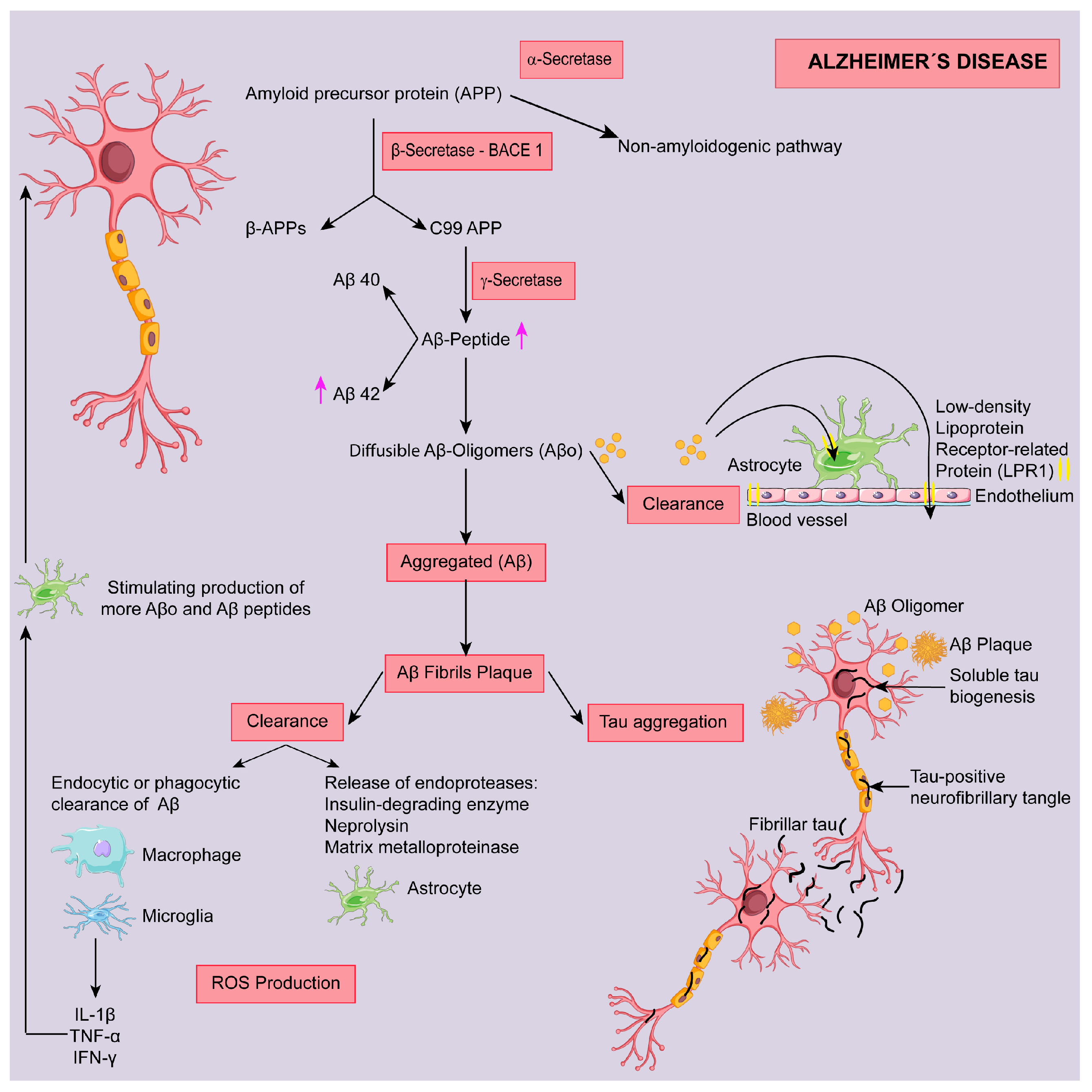
2. Alzheimer Disease (AD)

Alzheimer’s disease is the most common cause of dementia in older adults. In this pathology the damage initially appears to take place in the hippocampus, important for memory formation. As more neurons die, additional parts of the brain, including the cerebral cortex, are affected and begin to shrink. Such changes progressively lead to a loss of mental, behavioral and functional abilities and a decrease in the capacity of learning new things, carry out multistep tasks such as getting dressed, or cope with new situations. Age is by far the main risk factor, although genetic, environmental, and lifestyle factors may also contribute to the disease process. To date, the most prevalent hypothesis for the pathogenesis of AD is the amyloid cascade theory where, insoluble forms of amyloid-β (Aβ) plaques accumulate in extracellular spaces and in blood vessels walls. The process begins in neurons with the cleavage of the amyloid precursor protein (APP), through the activity of the β-secretase (BACE 1) and then followed by the γ-secretase. In the non-pathological pathway (non-amyloidogenic) the γ-secretase precedes by the α-secretase activity. Amyloidogenic cleavage results in production of peptides mainly of 40 and 42 amino acids, known as Aβ40 and Aβ42, respectively [19]. These peptides can be converted into diffusible and soluble oligomers (Aβo) (Figure 2). The neurotoxicity of Aβo is further exacerbated by their capacity to negatively affect long-term potentiation, therefore producing severe damages to synaptic plasticity resulting on impaired memory and learning. When Aβo are not removed by the astrocytic low-density lipoprotein receptor-related protein 1 (LRP 1), their aggregation occurs to form fibrillary structures that accumulate into Aβ plaques. The latter can be cleared from the brain through different pathways [19], including either the intervention of macrophages and microglia cells or involving astrocytes, through endoproteases such as insulin-degrading enzyme, neprilysin and matrix metalloproteinase [20].
Some conformational Aβo, that dissociate from both Aβ fibrils and plaques, may not be removed and are toxic to adjacent synapses and mitochondria. Furthermore, Aβo induce activation of microglia and astrocytes. With microglial activation, proinflammatory cytokines, including interleukin 1 beta (IL-1β), tumor necrosis factor alpha (TNF-α), and interferon gamma (IFN-γ) are produced. These molecules accumulate nearby astrocyte-neuron stimulating the production of more Aβo and Aβ peptides [20]. Moreover, those Aβo induce intraneuronal accumulation of paired helical filaments hyperphosphorylated tau protein abnormalities (neurofibrillary tangles, NFTs) in the brain. The exact biochemical mechanisms of tau aggregation remain however currently unknown. Damaged neurons are the ones with Tau-positive NFTs, which extend to the dendrites, and can be released and taken up by healthy neurons, triggering tau damage in the uptaking cell (Figure 2) [21].
Brain metabolism depends exclusively on glucose utilisation and its metabolism in the mitochondria, via the Krebs cycle and the electron transport chain (ETC). It is estimated that about 2% of the O2 used by the mitochondria is converted into superoxide radical (O2●−), with this figure increasing in damaged or aged mitochondria [22]. Brain has also high amount of polyunsaturated fatty acids, which in the presence of redox-active metals, such as copper (Cu) and iron (Fe), are very susceptible to lipid peroxidation. When the scavenging system of the brain is not able to remove the excess of reactive oxygen species (ROS) produced then oxidative stress (OS) appears. ROS accumulate in brain tissues and cells and interact with lipids, proteins, carbohydrates and nucleic acids leading to cellular oxidative damage (Figure 3). As a result of cellular and molecular deterioration, inflammation can occur by the activation of the immune system [23]. In addition, ROS induced-defects in mitochondrial energy metabolism is linked to the disruption in calcium (Ca2+) metabolism. The mitochondrial Ca2+ sequestering is very important to maintain neurons functionality. In neurogenerative diseases, the hyperactivation of N-methyl-d-aspartate-receptors (NMDA-type receptors) increases the Ca2+ concentration, inducing mitochondrial transition pore to open. Subsequently, pro-apoptotic proteins, such as cytochrome c and the apoptosis inducing factor (AIF), can be released to the cytoplasm therefore activating the apoptotic cascade and neuronal death. Furthermore, the decrease in nuclear factor (erythroid-derived 2)-like 2 (Nrf2) expression observed in neurodegenerative diseases, supports this theory, since Nrf2 protects neurons from the increase in intracellular Ca2+, and other toxic insults such as OS and mitochondrial dysfunction. On the other hand, the NMDA-type receptors hyperactivation also results in glutamate-induced excitotoxicity that can lead to neuronal necrosis [24]. Finally, OS generated through Aβs can also activate stress kinases such as c-Jun N-terminal kinases (JNK), [25]. The high-affinity of Aβs for copper (Cu) and zinc (Zn) may also explain the formation of an oxidative microenvironment. Indeed, at low pH Cu may induce Aβ aggregation and Aβs and APPs can reduce Cu2+ to Cu+, in a reaction that produces hydrogen peroxide (H2O2) [26]. Oligodendrocytes have low amount of glutathione (GSH) and high concentration of iron (Fe), therefore, they have reduced capacity to scavenge ROS, being also very susceptible to OS [27].

2.1. Targeting Aβ Protein

2.1.1. Modulation of Secretase Enzymes

α-Secretase

Cleavage of APP first occurs through the activation of α-secretase or BACE 1. In the first case, it facilitates proteolysis of APP in a non-amyloidogenic pathway and prevents Aβ formation. α-Secretase activation, therefore, is a distinct and a potential therapeutic approach to decrease Aβ generation [28]. Proteases with proposed α-secretase function belong to the ADAM (“a disintegrin and metalloproteinase domain”) family of proteins that include ADAM9, ADAM10 and ADAM17 [29]. All compounds considered for AD must cross the blood–brain–barrier and the plasma membrane [30]. The molecular weight of resveratrol is 228 Da, and therefore has emerged as a potential candidate since its secondary metabolites have lower molecular weight and higher lipophilicity [31]. A diet enriched with resveratrol (1g/Kg) was reported to activate α-secretase by increasing ADAM10 development expression through sirtuin 1 (SIRT1) modulation in SAMP8 mice (senescence-accelerated mice-prone) [32] and in triple transgenic AD mouse model (3xTg-AD) (Figure 4) [33]. α-Secretase activity was also elevated in the brain of mice treated with 6% Cabernet Sauvignon compared to ethanol-treated control Tg2576 mice (model AD-type Aβ neuropathology) or in primary neurons obtained from those mice (Figure 4) [34]. Similar results were also observed in SH-SY5Y cells stably overexpressing the Swedish (APPSwe) mutation when treated with 25–100 nM quercetin [35] or in primary cortical neurons treated with 1–10 µM myricetin (Figure 4) [36] In addition to flavonoids, hydroxycinnamate compounds such as caffeic acid (25 µM) and/or their derivatives (e.g., caffeic acid phenethyl ester) (30 µM) have been reported to activate α-secretase (ADAM-10) in acrolein treated mouse hippocampal HT22 cells, (Figure 4) [37]. However, 3-months treatment with ferulic acid (30 mg/kg) failed to reduce such activity in 12-month old presenilin-amyloid β-protein precursor (PSAPP) transgenic mice [38].

β-Secretase

AD mechanisms analysis (Figure 2) clearly highlights the therapeutic importance of blocking BACE 1 activity to decrease Aβ accumulation. Five resveratrol oligomers isolated from the stembark extract of Vitis vinifera [39] along with eleven resveratrol oligomers, isolated from the seed extract of Paeonia lactiflora (Paeoniaceae) [40], were reported to inhibit baculovirus-expressed BACE 1 activity in a dose dependent manner. Such activity was recently confirmed in a homogeneous time-resolved fluorescence (TRF) assay [41], showing an inhibitory activity with an IC50 of 28 mM. Similarly, gnetin C, a resveratrol dimer (5–100 µM), was reported to suppress BACE 1 expression in SH-SY5Y cells, to reduce Aβ42 production and to ameliorate Aβ42-lowered cell viability [42]. Similarly, miyabenol C, a resveratrol trimer (5–20 μM), inhibited BACE1 and reduced Aβ generation in vitro (SH-SY5Y cells and mouse neuroblastoma N2a naive cells, N2aWT) [43]. In vitro studies also suggest that flavonoids may also inhibit BACE 1 activity. For example, the flavanol (−)-epicatechin (100 nM) [44], the flavanols myricetin [36,45,46,47], quercetin [45,48] and kaempherol (20 µM) [45], all directly inhibited BACE 1 enzymatic activity in a concentration dependent manner (Figure 4). Amin et al. [49], used SH-SY5Y cell lines treated with Aβ1–42, to estimate the biological activity and neuroprotective effect of anthocyanin loaded nanoparticles (An-NPs). The authors suggest that An-NPs are more active and potent than anthocyanin alone as it decreases the level of basic proteins associated with AD such as APP and BACE 1. The inhibitory effect on Aβ-induced activation of BACE 1 was also demonstrated for anthocyanins (malvidin and oenin; 50 µM) in an in vitro study with Neuro2a cell line (Figure 4) [50].
A reduced number of in vivo assays has been carried out with flavonoids to explore their potential as BACE 1 inhibitors. The increase of mice neuronal resistance to age related disease effect of quercetin is explained by its BACE 1 inhibitory effect, suggesting negative regulation of APP amyloidogenesis in old mouse brain (Figure 4) [51]. In a different study, however, quercetin supplementation had no effect on BACE 1 mRNA expression [52]. The experimental design of these studies was very different and may explain the different outcomes. In the former, 16-month-old male C57BL6 strain mice were fed with high-cholesterol diet supplemented with quercetin. In the latter, female C57BL6 mice were fed quercetin-enriched diets (2 mg/g diet) for 6 weeks. Age, gender and diet regimen may explain, at least partially, the differences obtained. More studies are needed to clarify whether BACE 1 can be a molecular target for quercetin (Figure 4).
In addition to flavonoids, some studies have been carried out with both stilbenes and hydroxycinnamates. For example, SAMP8 mice fed with a diet supplemented with 1 g/Kg of trans-resveratrol, from 2 until 9 months of age, showed no alterations in the pro-amyloidogenic BACE 1 enzyme quantified by Western Blot [32], although a decrease in BACE 1 protein expression was observed in the hippocampus of those mice when subjected to a combined intervention of resveratrol (0.03%) and vitamin D (500 IU) [53]. In a recent study, wild type littermates (NoTg) and AD transgenic (3xTg-AD) experimental groups were fed a diet supplemented with 100 mg/kg of trans-resveratrol from 2 months of age for 10 months. Resveratrol treatment protected against memory loss and brain pathology in 3xTg-AD mice and induced cognitive enhancement in healthy wild type mice, an observation that might be related to the modulation of BACE1 [33]. Furthermore, the hydroxycinnamic acid, caffeic acid conjugated to chito-oligosaccharides has the potential to be used as BACE 1 inhibitors to reduce the risk of AD [54]. A combination of caffeic acid/caffeic acid phenethyl ester can also restore acrolein-induced BACE 1 changes in HT22 mouse hippocampal cells [37]. However, ferulic acid (30 mg/kg) had no effect on BACE 1 activity when one-year-old mice with established β-amyloid plaques received daily doses of this non-flavonoid compound [38]. These results disagree with previous data obtained by the same research group, where the administration of the equal amount of ferulic acid inhibited amyloidogenic APP metabolism by reducing BACE 1 expression and β-secretase activity in PSAPP mice [55]. The discrepancy between results could be related with the differences in mice age and treatment duration, which were higher in the latter study.

γ-Secretase

Modulation of γ-secretase can also be a preventive/therapeutic strategy. Resveratrol (0–20 µM), oxy-resveratrol (0–80 µM), and piceatannol (0–20 µM) all decreased γ-secretase processing activity at higher doses in Neuro2a cells [56]. Resveratrol (60 µM) also inhibited γ-secretase activity in autophagy-deteriorated cells [57]. However, Marambaud et al. (2005) using HEK293 cells stably transfected with human APP695 cell line concluded that resveratrol (40 µM) does not inhibit Aβ production, partly because it has no effect on the Aβ-producing enzymes β- and γ-secretases [58].

2.1.2. Targeting Amyloid Clearance

According to the amyloid cascade hypothesis, one of the reasons for Aβo accumulation in the brain is related to the impairment of the clearance mechanisms (Figure 2). These pathways can therefore be targeted as potential preventive/treatments strategies for AD. The neuroprotective mechanism, however, remain unclear with only the activation of AMP-activated protein kinase (AMPK) signaling being proposed as one of the signaling pathways. For example, resveratrol activates AMPK by increasing cytosolic Ca2+ levels and by promoting Ca2+/ Ca2+-calmodulin-dependent protein kinase kinase-β (CaMKKβ) dependent phosphorylation of AMPK. Furthermore, resveratrol reduces Aβ accumulation by activating autophagy and by facilitating the lysosomal degradation of Aβ (Figure 4) [59]. Insulin-like growth factor-I (IGF-I) stimulates β-amyloid release from neurons and promotes brain amyloid clearance (Figure 4) [60]. Resveratrol (20 mg/L) increases hippocampal IGF-I production, thereby improving cognitive function in mice, probably due to an increase in amyloid clearance [61]. Resveratrol (0–20 µM) also induces autophagy in PC12 cells, a process partially mediated through the activation of the tyrosyl transferRNA (tRNA) synthetase (TyrRS)-auto-poly-ADP-ribosylation of poly (ADP-ribose) polymerase 1 (PARP1)-SIRT1 signaling pathway [62]. The flavanol quercetin (20–40 kg/kg) also increases AMPK activity in the APPswe/PS1dE9 transgenic mouse model of AD and, therefore, promotes the clearance of intracellular Aβ (Figure 4) [63].
Increasing the levels of degrading enzymes has been studied as a therapeutic possibility. Resveratrol (0–40 µM) promotes the intracellular degradation of Aβ in cell lines by a mechanism that implicates the proteasome [58]. In an in vivo AD model of Caenorhabditis elegans resveratrol (100 µM) increases the degradation of aged proteins by autophagosomal and proteasomal degradation [64]. Diet supplementation with resveratrol (1 g/kg) promotes an increase in neprilysin levels, one of the most important amyloid-degrading enzymes, in both 3xTg-AD and healthy wild type mice. In this study, and for the first time in vivo, it was also shown that resveratrol promotes enhancement of ubiquitin-proteasome system (UPS), which leads to a reduction of aberrant amyloid and tau proteins. Taken together these results show that resveratrol has a potential in AD prevention by increasing brain resilience against aberrant proteins (Figure 4) [33].

2.1.3. Targeting Amyloid Aggregation

AD is characterised by aggregation of Aβ monomer into multimeric aggregates (Figure 2). Resveratrol has direct effects in Aβ aggregation, both by preventing conversion of lower to higher molecular weight oligomers [65] but also by disrupting Aβ aggregates (Figure 4) [66]. Resveratrol and its glucoside are specific potent inhibitors of Aβ aggregation [67]. Resveratrol inhibits Aβ42 fibril formation in a dose-dependent way but could not prevent Aβ42 oligomerization [68]. Inhibition of Aβ42 fibril formation could be explained by ability of resveratrol to directly bind to monomer and fibril Aβ, as determined by surface plasmon resonance, thioflavin T fluorometric analysis, proton nuclear magnetic resonance, and atomic force microscopy in vitro [69]. The result is the conversion of soluble oligomers, fibrillar intermediates and amyloid fibrils to non-toxic high molecular weight conformation [70]. Moreover, resveratrol can indirectly prevent Aβ aggregation, through improvement of the binding of transthyretin to Aβ oligomers (Figure 4) [71]. In addition to resveratrol, a naturally derived grape seed polyphenolic extract (200 mg/kg/d) inhibits amyloid β-protein aggregation into high-molecular-weight oligomers in vitro. When orally administered to Tg2576 mice, this polyphenolic preparation attenuates AD-type cognitive deterioration coincidentally with reduced high-molecular-weight soluble oligomeric Aβ in the brain [72]. Furthermore, experiments carried out with quercetin (200 µM) show a potent Aβ42 anti-aggregation activity (75.4%) in combined in vitro cell-based/in silico assay approaches [73]. Such data were further confirmed where treatment with quercetin (25 mg/kg) for three months decreased β-amyloidosis in aged 3xTg-AD mice [74]. Furthermore, polyphenolic metabolites identified in the rat brain are after oral dosage of Cabernet Sauvignon red wine were tested for potential AD benefits. It was observed that quercetin-3-O-glucuronide (0–5 µM) reduces the generation of Aβ peptides by primary neuron cultures generated from the Tg2576 AD mouse model. Another brain-targeted metabolite, malvidin-3-O-glucoside (0–50 µM), has no detectable effect on Aβ generation. Quercetin-3-O-glucuronide also interferes with the initial protein-protein interaction of Aβ1–40 and Aβ1–42, which is necessary for the formation of neurotoxic oligomeric Aβ species (Figure 4) [75]. Finally, a preclinical study demonstrated that grape seed proanthocyanidin (GSPA), which consists of catechin, epicatechin and epicatechin gallate, can interrupt the aggregation of neurotoxic Aβ (Figure 4) [76]. Altogether these data suggest that polyphenols may have the potential to reduce protein aggregation, a phenomenon important in AD progression.

2.2. Targeting Tau Protein

Neuroprotective effects of resveratrol also result from the inhibition of tau protein hyperphosphorylation, as it was observed in N2a cells exposed to formaldehyde. Inhibition of glycogen synthase kinase (GSK-3β) and calmodulin-dependent protein kinase II (CaMKII) or the activation of protein phosphatase-2A (PP2A) by resveratrol (2.5–50 µM) protect against the hyperphosphorylation and/or mediates the dephosphorylation of tau protein, respectively (Figure 4) [77]. Resveratrol also inhibits Na3VO4-induced hyperphosphorylation of tau at the Ser396 (p-S396-tau) site, which is upregulated in the hippocampus of AD brains and predominantly linked to AD-associated cognitive dysfunction [78].
Recently, the inhibitory effect of quercetin on okadaic acid (OA)-induced tau protein hyperphosphorylation in HT22 cells was also explored. Western blotting results indicated that quercetin attenuates OA-induced tau protein hyperphosphorylation at the Ser396, Ser199, Thr231 and Thr205 sites. Quercetin also inhibits the activity of cyclin-dependent kinase 5 (CDK5), a key enzyme in the regulation of tau protein, and blocks the Ca2+-calpain-p25-CDK5 signaling pathway. These observations indicate the ability of quercetin to decrease tau protein hyperphosphorylation and thereby to attenuate the associated neuropathology (Figure 4) [79]. Anthocyanins were reported to activate the phosphorylation of anti-phosphorylated GSK3β at Ser 9, thereby possibly preventing GSK-3β-dependent tau hyperphosphorylation at Ser 413 and 404 [80]. GSPA is also able to decrease hyperphosphorylated tau deposition in APP/PS1 male heterozygous mice (Figure 4) [76].
In addition to flavonoids, the hydroxycinnamate caffeic acid (10–20 µg/mL) was reported to protect PC12 cells against Aβ-induced toxicity by decreasing tau phosphorylation through the reduction of GSK-3β activation [81].

2.3. Targeting Oxidative Stress

Resveratrol protects the nervous tissue by reducing ROS production and by affecting several mechanistic pathways. For example, resveratrol reduces OS in the brain by preventing nuclear factor-κB (NF-κB) activation (Figure 4) [82]. AMPK is a key protein in the regulation of cell survival in response to OS insults and resveratrol is a potent AMPK-activator, as shown in Neuro2a cells and primary neuronal in vitro as well as in the brain [83]. Resveratrol also activates SIRT1 in the hippocampus [84], neuronal cells [85], rat cortical primary neurons [86] and thus reduces OS. Different results were observed by Karuppagounder et al. (2009), and they suggested that dietary supplementation with resveratrol protects against Aβ plaque formation and OS without activation of SIRT1 [87]. Resveratrol reduces both lipid peroxidation levels and OS induced by Aβ senile plaques in hippocampal neuronal cells (Figure 4) [88]. Inducible nitric oxide synthase (iNOS) mediates the Aβ-induced lipid peroxidation and HO-1 (heme oxygenase 1) expression, and resveratrol protects adult male rats from Aβ-induced neurotoxicity through the downregulation of iNOS (Figure 4) [89]. Induction of HO-1 by resveratrol is also observed in a dose- and time-dependent manner in cultured mouse cortical neuronal cells, revealing that resveratrol provides neuroprotection from free-radical or excitotoxicity damage [90]. Resveratrol also restores intracellular antioxidant enzymes such as GSH levels in a cell culture model [91,92] or reverses Aβ-induced malondialdehyde (MDA) overproduction derived from the lipid membrane oxidation, which reflect the overall degree of free radical attack (Figure 4) [89]. Resveratrol shows antioxidant proprieties, which may contribute to ameliorate the cognitive deficits and to attenuate oxidative damage in the brain of the SAMP8 mice through scavenging free radicals and modulating activities of antioxidant enzymes (Figure 4) [93]. As aforementioned resveratrol downregulates GSK-3β, a protein that induces OS [91]. Not only resveratrol but also the flavonoids quercetin and (+)-catechin protect hippocampal cells against toxicity induced by nitric oxide (NO). Quercetin can block sodium nitroprusside-stimulated protein kinase C (PKC) activation, pointing to a possible involvement of this enzyme in the protective effects mediated by quercetin (Figure 4) [94]. Recently, effects of solid lipid nanoparticle of quercetin in pentylenetetrazole (PTZ) induced cognitive impairment of Danio rerio species were evaluated. The administration of quercetin nanoparticles (5 and 10 mg/kg) ameliorate the PTZ exposure induces cognitive impairment, due to its potential anti-oxidative, anti-lipid peroxidative and acetylcholinesterase (AChE) inhibitory actions [95]. Epicatechin and myricetin, could inhibit neuronal death, through antioxidative mechanisms in cultured mouse cortical neurons (Figure 4) [96]. Anthocyanins (200 mg/kg) also attenuate memory deficits, protects against oxidative damage in the brain, and restore AChE and ion pump activity in a rat experimental model of sporadic dementia of Alzheimer’s (Figure 4) [97]. Finally, caffeic acid protects PC12 cells against Aβ-induced toxicity through the inhibition of OS and the inhibition of intracellular Ca2+ levels [81].

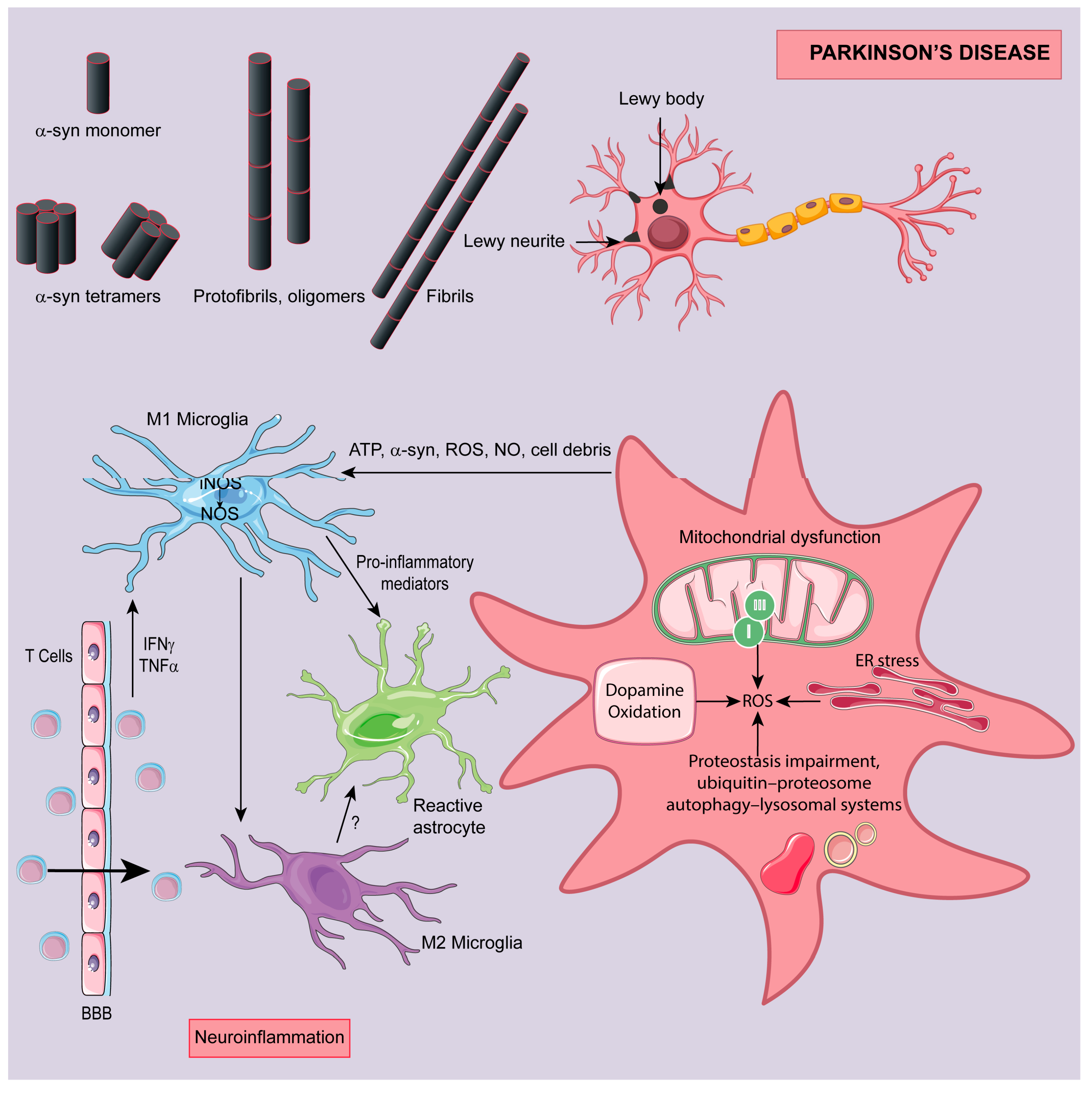
3. Parkinson Disease (PD)

Parkinson disease is the second most common neurodegenerative dementia and the most common neurodegenerative movement disorder. It can be inherited, but most of the cases are sporadic, still both have selective loss of dopaminergic neurons in the substantia nigra that project to the basal ganglia. It is characterized by bradykinesia, tremor, rigidity and postural instability triggered by dopamine decrease [98]. Other regions of the central and peripheral nervous system could be involved and contribute to a range of non-motor symptoms like autonomic dysfunction, depression, and cognitive deficits [99]. Most of the PD cases show Lewy pathology consisting of Lewy bodies and Lewy neurites revealed by histological techniques. α-Synuclein (α-syn) is the major component of the Lewy body (Figure 5) [100]. Natural forms of α-syn protein are the monomeric and possibly tetrameric α-syn ones, which have a physiologic function. They regulate exocytosis and promote microtubule formation through interactions with tubulin. In PD, α-syn changes from its monomer arrangement to protofibrils, fibrils and oligomers, which are involved in toxic processes, protofibril and fibril structures, being probably the most toxic ones. Their production, truncation and maintenance are fundamental to PD pathology (Figure 5). Toxic effects of α-syn include mitochondrial dysfunction, endoplasmic reticulum (ER) stress, impaired proteostasis, enhanced neuroinflammation and pathological aggregation of other proteins [101]. Protein misfolding and aggregation, mitochondrial dysfunction, cell cycle reactivation, apoptosis and excitotoxicity contribute to OS occurrence in PD (Figure 5) [102]. Another oxidative scenario arises when the dopamine is degraded to eliminate cytosolic excess of this neurotransmitter after treatment. Dopamine is easily metabolized via the monoamine oxidase (MAO) or by auto-oxidation. MAO is in outer mitochondrial membrane in neuronal and glial cells and is able of degrade dopamine, which exacerbates degeneration of dopaminergic neurons. 6-Hydroxydopamine (6-OHDA) and H2O2 are products of dopamine degradation by MAO, and contribute to an increase in ROS production and OS damage [103].
As in other neurodegenerative diseases, the mitochondrion plays a role in PD pathogenesis. Mitochondria are the primary source of ROS that may contribute to intracellular OS. The main sites of ROS production in mitochondria are complex I and to a smaller extent complex III in the ETC. The foremost process for ROS production in PD is complex I deficiencies, which is responsible for the majority of unfavorable neural apoptosis generation [104]. Briefly, ETC complex I deficiency, leads to ROS production, reducing levels of the GSH, which induces mitochondrial permeability by transiently opening a pore. Subsequently occurs the depolarization of the mitochondrial membrane potential (Figure 5). These events ultimately lead to neural cell death via the release of pro-apoptotic mitochondrial proteins, including cytochrome c and apoptosis-initiating factor [105].
Neuroinflammation is a characteristic of PD pathology, but it remains unknown whether neuroinflammation promotes or protects from neurodegeneration. Under pathological conditions, such as protein aggregation, gene mutations, environmental factors and cytokines released from infiltrated T cells and microglia become activated and release pro-inflammatory mediators. The latter activate astrocytes, leading to elevated production of proinflammatory factors, NO, O2●−, contributing to degeneration of dopaminergic neurons (Figure 5). Degenerative neurons release molecules that can activate glia and increase inflammatory response. At certain stage of PD, a different microglia subpopulation may become active releasing anti-inflammatory factors, including transforming growth factor beta (TGF-β), and exert a neuroprotective effect in PD (Figure 5) [106].

3.1. Targeting Dopaminergic Neurons Cytotoxicity

Pre-treating SH-SY5Y human neuroblastoma cells [107] and 6-OHDA rat models of PD with resveratrol (20 mg/kg) results in inhibition of dopamine cytotoxicity [108]. Resveratrol reduces intracellular OS in neurotypical SH-SY5Y, through recognized signal pathways of apoptosis. In fact, resveratrol pre-treatment led to a decrease in cleavage of PARP, an increase in the Bcl-2 protein, and activation of caspase-3 [107]. Rotenone is a popular pesticide that could produce 1-methyl-4-phenylpyridine (MPP+) and, consequently, can cause apoptosis in dopaminergic neurons [109]. Resveratrol shows concentration-dependent neuroprotective effects against sodium azide and thrombin, a mitochondrial complex IV inhibitor and a microglia-activating agent, respectively. Moreover, SIRT inhibitors such as nicotinamide and sirtinol, do not attenuate the protective effect of resveratrol against MPP+ cytotoxicity [110]. Resveratrol also alleviates MPP+-induce cytotoxicity and restored MPP+-induced mitochondrial dysfunction in SN4741 cells. These results suggest that resveratrol attenuates MPP+-induced mitochondrial dysfunction and cell apoptosis, which may be achieved through AKT/GSK-3β pathway [111].
Resveratrol (0–50 µM) also protects PC12 cells from 6-OHDA-induced OS and apoptosis via C-X-C chemokine receptor type 4 (CXCR4) signaling pathway [112]. CXCR4 is a common and highly conserved G-protein-coupled receptor (GPCR) with seven transmembrane domains and is a receptor for receptor for CXCL12 that can promote neuronal apoptosis [113]. CXCL12/CXCR4 may be involved in PD occurrence and could be a molecular target for PD [114]. Resveratrol (20 µM) protects SH-SY5Y cells against rotenone-induced neuronal apoptosis, inducing HO-1 expression and preventing dopaminergic cell death by regulating autophagic flux [115]. N-methyl-N′-nitro-N-nitrosoguanidine (MNNG) is a DNA alkylating agent that causes neurotoxicity. Resveratrol (0–100 µM) suppresses MNNG-induced increase in acetylation of p53, a representative target of SIRT deacetylase activity, in midbrain slice culture [110]. Resveratrol (50 µM) inhibits 6-OHDA-induced toxicity and increased the expression of SIRT1 in PC12 cells [116]. Recently, it was observed that resveratrol (0–50 µM) reduces rotenone induced neuronal damage in PC12 cells, and it was suggested that SIRT1/Akt1 (Akt1, serine/threonine-protein kinase) signaling pathway play an essential role in this process [117]. Resveratrol (20 mg/Kg) also activates PGC-1α in dopaminergic cells via the deacetylase SIRT1, and enhanced PGC-1α gene transcription with increases in superoxide dismutase 2 (SOD2) and Trx2 in the MPTP mouse model of Parkinson’s disease [118]. Ferretta et al. (2014) tested in vitro the effect of resveratrol treatment on primary fibroblast cultures from two patients with early-onset PD linked to different Park2 mutations. The authors suggested SIRT1/AMPK/PGC1-α axis as a key neuroprotective pathway [119]. On PD cellular models, resveratrol has neuroprotective effects mediated through activation of the AMPK-SIRT1-autophagy pathway [120]. As suggested by in vitro evidences, activation of SIRT1 could explain the improvement both the motor deficits and pathological changes of PD in mice. It was suggested that activation of SIRT1, led to LC3 deacetylation mediated autophagic degradation of α-syn [121]. In a different in vivo study, resveratrol improved rat’s movement and spatial memory performance by rescuing the reduction of SIRT1 expression and CREB phosphorylation induced by Aβ1-42 [114]. Together, these results indicate that neuroprotective actions of resveratrol against different type of insults is related to SIRT-activating potential.
Quercetin can successfully activate PKD1, the gene that encodes the polycystin 1 protein, pro-survival signaling in dopaminergic cells. Quercetin also induces Akt and CREB phosphorylation and brain-derived neurotrophic factor BDNF expression. Therefore, PKD1/CREB/BDNF axis may at least partially mediate the neuroprotective effects of quercetin. Quercetin treatment increases mitochondrial biogenesis in dopaminergic neurons. Importantly, quercetin protects against dopaminergic neurodegeneration in the MitoPark transgenic mouse model of PD, an animal model of PD with impaired respiratory chain function in dopaminergic neurons [122]. Quercetin binds covalently with α-syn in 1 to 1 ratio. The formed quercetin-α-syn adducts attach to the α-syn oligomers or monomers, increasing the surface hydrophilicity and inhibiting the further fibrillation [123]. Also, caffeic acid proved to be an efficient inhibitor against α-syn fibrillation [124]. Gallic acid inhibits α-syn fibrillation and toxicity by stabilizing the non-toxic oligomeric structure of α-syn through oligomer binding [125].
Pure polyphenols such as gallic acid, ferulic acid, caffeic acid, coumaric acid, propyl gallate, epicatechin, epigallocatechin, and epigallocatechin gallate protect, rescue and, most importantly, restore the impaired movement activity (i.e., climbing capability) induced by paraquat in Drosophila melanogaster, a valid model of PD [126]. Interestingly, ferulic acid and exercise seem to improve the motor performance in a PD rodent model which might be due to the upregulation of Hsp70 and increasing density of dopaminergic neurons in the corpus striatum. Ferulic acid offers more protective effects than exercise and combination of both did not offer more protection than ferulic acid alone [127].

3.2. Targeting Oxidative Stress

As previously mentioned, resveratrol is a very promising antioxidant, therefore several in vitro and in vivo experiments have been carried out to evaluate its potential to ameliorate PD. In neuronal-like cells expressing mutant huntingtin from dopamine toxicity, resveratrol (100 µM) prevents the generation of ROS and restores the level of ATG4, a redox-sensitive autophagy protein that plays a pivotal role in autophagosome formation [128]. Resveratrol also prevents the decrease of dopaminergic neurons and the increase of propidium iodide uptake into midbrain slices induced by an MPP+. In addition, resveratrol was reported to block the accumulation of ROS, the depletion of cellular GSH, and the cellular oxidative damage induced by MPP+, suggesting the involvement of antioxidative properties in the neuroprotective action of this stilbene [110]. A botanical extract (Regrapex-R®, Interpharma Praha, a.s, Modrany, Czech Republic) prepared from whole grape (Vitis vinifera) and Polygonum cuspidatum, which contains polyphenols, including flavans, anthocyanins, emodin, and resveratrol, exhibit dose-dependent scavenging effects on ROS. Extract inhibits ROS and protein carbonyl production and protects enzyme activities of ETC (complexes I and II) and pyruvate dehydrogenase in isolated rat liver mitochondria following exposure to 2,2′-azobis (2-amidino propane) dihydrocholoride (AAPH), a potent lipid oxidant generator. The polyphenolic mixture also protects human neuroblastoma cells (SKN-MC) against AAPH induced oxidation by maintaining cell viability and inhibiting excessive ROS generation. In transgenic Drosophila expressing human α-syn, model for PD, the botanical extract improves climbing ability and extends females lifespan [129].
Resveratrol (20 mg/Kg) ameliorates 6-OHDA-induced rat brain injury [108], upregulating the antioxidant status [108], and suppressing overexpression’s of cyclooxygenase-2 (COX-2), TNF-α mRNA and protein in the substantia nigra [130]. Resveratrol also mediates anti-inflammatory activities by suppressing the activity of NADPH oxidase and, further, decreasing ROS production and inhibiting the activation of mitogen activated protein kinases (MAPKs) and NF-κB cascade signaling pathways, as it was shown in lipopolysaccharide (LPS)-induced dopamine neurons [131]. Resveratrol shows potential therapeutic capacity for rotenone-induced PD in rats through inhibiting endoplasmic reticulum stress-mediated apoptosis. Resveratrol also restores redox balance as evident by suppressing xanthine oxidase activity and protein carbonyls formation; in addition to preservation of intracellular antioxidants status via activating glutathione peroxidase (GPx) and Nrf2 signaling pathway [132]. Resveratrol has neuroprotective effects, through free radical scavenging, in animals in which PD is induced with 1-methyl-4-phenyl-1,2,3,6-tetrahydropyridine (MPTP) [133,134,135]. Resveratrol administration protects mice from MPTP-induced motor coordination impairment, hydroxyl radical overloading, and neuronal loss [134]. Reverses in toxic effects of MPTP were posteriorly confirmed in study with mice, intraperitoneal injected with MPTP (50 mg/kg body weight). It was observed that, resveratrol increases the levels of dopamine, its metabolites, GSH and GPx activities and reduces levels of thiobarbituric acid reactive substances (TBARS), catalase and SOD activities and enhances behavior performance [135].
Quercetin has a dual effect against the 6-OHDA-induced ROS formation in catecholaminergic SH-SY5Y neuroblastoma cells. Small concentrations of quercetin gave some protection, but at high concentrations this protective effect is reduced, or even enhanced toxicity occur [136]. The dual effect of quercetin was observed in other studies using the same [137] and a different in vitro model [138]. Quercetin protection and toxicity effects can be described in a time-based manner. At earlier exposure times, quercetin has strong protection against 6-OHDA-induced cell death in SH-SY5Y cells. In contrast, after a prolonged treatment quercetin do not protect against 6-OHDA toxicity [137]. Also, both quercetin aglycone and quercetin 3-O-β-d-glucuronide suppress the production of H2O2 by scavenging O2•– derived from 6-OHDA in a neuro-2a cell culture system. According with this conjugated quercetin metabolites could scavenge ROS generated from dopamine metabolism, however, their deconjugation to quercetin aglycone is essential to exert protective effects against the neuronal cell death induced by ROS. Re-conjugation is also needed to avoid the inherent cytotoxicity of quercetin aglycone [139]. Moreover, quercetin protects SH-SY5Y cells in which neurotoxicity was induced by lipopolysaccharide plus interferon-γ or interferon-γ released from activated microglia and astrocytes. Quercetin can reduce oxidative/nitrative damage to DNA as well as to the lipids, and proteins of SH-SY5Y cells. Quercetin also increases GSH concentration in SH-SY5Y cells [140]. In a study carried out by Ahn and Jeon (2015), quercetin pre-treatment increased α-syn expression in PC12 cells treated with various toxins. Knocking out α-syn, however, exerted no significant effect on cell survival. This could mean that quercetin is neuroprotective against toxic agents via affecting various mechanisms such as apoptosis, autophagy and aggresome [141].
Regarding quercetin, inconsistent results were observed in vivo, some studies suggest that quercetin has no neuroprotective effect in vivo using 6-OHDA rat models of PD [136,142], while others suggest that quercetin protects striatal dopaminergic neurons against 6-OHDA-induced OS in the rat [143]. Pre-treatment with quercetin has no effects on the number of hydroxylase (TH)-positive cells in the substantia nigra and on dopamine content in the striata [142]. Results from an in vitro and in vivo study revealed a dose-dependency of quercetin effect. Quercetin displays a clear falling of protection at higher concentrations in both viability and caspase-3 like activity in cell cultures and to some extent also in circling behavior in lesioned rats. This dose-dependency of quercetin effect occurs in a thinner range of concentrations, since cytotoxic effects of quercetin begin to take place at concentration only twofold higher than the one that provides the greatest protection [137]. In a posterior study, it was described that quercetin mitigates the loss of complex-I activity induced by rotenone in rats and enhances antioxidant enzyme activity [144]. Using the same animal model of PD, El-Horany et al. (2016) showed that quercetin attenuates rotenone-induced behavioral impairment, increases autophagy, ameliorates endoplasmic reticulum stress-induced apoptosis with attenuated OS [145]. From the current study, quercetin can act as an autophagy enhancer in PD rat model and modulates the microenvironment that leads to neuronal death. Combined strategies could be a better approach as suggested by Haleagrahara et al. (2013) after evaluating the combined treatment with quercetin and desferrioxamine on 6-OHDA-induced neurotoxicity in the striatum of rats. They observed that combined treatment has a more powerful effect in protecting the neurons and increasing the antioxidant enzymes in the striatum [146]. The combination of quercetin with other neuroprotector compounds have been suggested as a therapeutic advantage in the prevention/treatment of OS-mediated neurodegenerative conditions such as PD [147,148,149].
Organic and aqueous Champagne wine extracts exhibit potent neuroprotective activity against peroxynitrite-induced injury at low concentrations (0.1 µg/mL). Results suggest that neuroprotective effect against oxidative neuronal injury is in part due to the cellular actions of individual components found in the organic extracts, particularly tyrosol, caffeic acid, and gallic acid [150]. Neuroprotective effects of tyrosol, caffeic acid, gallic acid, p-coumaric acid, (+)-catechin, (−)-epicatechin and quercetin (0.1–50 μM) and organic champagne extract against CysDA-induced neuronal injury were evaluated in posterior studies, that showed that hydroxycinnamates, phenolic acids and phenolic alcohols induce neuroprotective effects, which are more powerful than that observed for flavonoids [138,151]. Gallic acid has neuroprotective activity against 6-OHDA-induced OS via enhancement of cerebral antioxidant defense, by increasing total thiol and GPx contents and decreasing MDA levels in the rat hippocampus and striatum tissues [152]. Catechin treatment restores GSH levels, and increases dopamine and 3,4-dihydroxyphenylacetic acid content, and tyrosine hydroxylase -immunoreactivity in 6-OHDA-lesioned rats [153]. Kaempferol also shows neuroprotective effects in MPTP-induced PD mice, by scavenging ROS, which results in the survival of more dopamine neurons [154].

4. Conclusions

Epidemiological studies suggest that moderate consumption of wine, an element of the Mediterranean diet, is one of the main factors behind the neuroprotective effects observed in some populations [7,155,156,157,158]. Most of the in vivo and in vitro studies evaluated wine neuroprotection indirectly by analyzing effects of the phenolic compounds of wine, mainly resveratrol. Nonetheless, much of the data obtained on their bioactivity derived from short term basis in vitro or in vivo studies where the dose used were not of nutritional relevance and unlikely achievable through moderate wine drinking. Although at the moment, the balance of evidence does suggest that polyphenol effects contribute to the benefits of a high intake of fruits and vegetables, the extent of their contribution in vivo and at physiological relevant concentrations remains uncertain. More work needs to be done to prove whether this class of compounds is most likely to result in health benefits and to determine their beneficial effects in slowly developing neurodegenerative disorders. These studies remain however fundamental to understand the effects of those compounds in the brain; to highlight that wine is a complex chemical mixture. Analysis of the literature led us to hypothesize that preventive/therapeutic efficacy of wine is more likely due to a combination of neuroprotective effects of wine phenolic compounds. Combined complementary and perhaps synergistic effects could explain the benefits of wine moderate consumption. In fact, the hypothesis that wine could be compared to combined and multi-target therapeutic strategies could explain that drinking lower doses on a “chronic” manner reduces brain damage. It is also very important to continue studying the effects of phenolic metabolites, however, a focus on gut microbiota derived phenolic acids and other metabolites should be addressed. Differences observed among populations in epidemiologic studies could be, in our opinion, attributed to differences in the gut microbiota composition since they are affected by diet. The type and quantity of metabolites produced after a meal will differ according with gut microbiota. In conclusion, wine is a complex mixture which is effective to prevent or slow age-related neurodegenerative diseases because can modulate multiple targets simultaneously involved in these diseases.

Author Contributions

P.S. and D.V. wrote the paper. All authors gave final approval of the version to be published. All authors read and approved the final manuscript.

Funding

This research received no external funding.

Conflicts of Interest

The authors declare no conflict of interest.

References

  1. Prince, M.; Bryce, R.; Albanese, E.; Wimo, A.; Ribeiro, W.; Ferri, C.P. The global prevalence of dementia: A systematic review and metaanalysis. Alzheimer’s Dement. 2013, 9, 63–75. [Google Scholar] [CrossRef] [PubMed]
  2. Nussbaum, R.L.; Ellis, C.E. Alzheimer’s disease and Parkinson’s disease. N. Engl. J. Med. 2003, 348, 1356–1364. [Google Scholar] [CrossRef] [PubMed]
  3. Vauzour, D.; Vafeiadou, K.; Rodriguez-Mateos, A.; Rendeiro, C.; Spencer, J.P. The neuroprotective potential of flavonoids: A multiplicity of effects. Genes Nutr. 2008, 3, 115–126. [Google Scholar] [CrossRef] [PubMed]
  4. Letenneur, L. Risk of dementia and alcohol and wine consumption: A review of recent results. Biol. Res. 2004, 37, 189–193. [Google Scholar] [CrossRef] [PubMed]
  5. Xu, W.; Wang, H.; Wan, Y.; Tan, C.; Li, J.; Tan, L.; Yu, J.T. Alcohol consumption and dementia risk: A dose-response meta-analysis of prospective studies. Eur. J. Epidemiol. 2017, 32, 31–42. [Google Scholar] [CrossRef] [PubMed]
  6. Lemeshow, S.; Letenneur, L.; Dartigues, J.F.; Lafont, S.; Orgogozo, J.M.; Commenges, D. Illustration of analysis taking into account complex survey considerations: The association between wine consumption and dementia in the PAQUID study. Am. J. Epidemiol. 1998, 148, 298–306. [Google Scholar] [CrossRef] [PubMed]
  7. Orgogozo, J.M.; Dartigues, J.F.; Lafont, S.; Letenneur, L.; Commenges, D.; Salamon, R.; Renaud, S.; Breteler, M.B. Wine consumption and dementia in the elderly: A prospective community study in the bordeaux area. Rev. Neurol. (Paris) 1997, 153, 185–192. [Google Scholar] [PubMed]
  8. Costanzo, S.; Di Castelnuovo, A.; Donati, M.B.; Iacoviello, L.; de Gaetano, G. Cardiovascular and Overall Mortality Risk in Relation to Alcohol Consumption in Patients With Cardiovascular Disease. Circulation 2010, 121, 1951–1959. [Google Scholar] [CrossRef] [PubMed] [Green Version]
  9. Klatsky, A.L.; Friedman, G.D.; Armstrong, M.A.; Kipp, H. Wine, liquor, beer, and mortality. Am. J. Epidemiol. 2003, 158, 585–595. [Google Scholar] [CrossRef] [PubMed]
  10. Rehm, J.T.; Bondy, S.J.; Sempos, C.T.; Vuong, C.V. Alcohol consumption and coronary heart disease morbidity and mortality. Am. J. Epidemiol. 1997, 146, 495–501. [Google Scholar] [CrossRef] [PubMed]
  11. Bianchini, F.; Vainio, H. Wine and resveratrol: Mechanisms of cancer prevention? Eur. J. Cancer Prev. 2003, 12, 417–425. [Google Scholar] [CrossRef] [PubMed]
  12. Kunzmann, A.T.; Coleman, H.G.; Huang, W.Y.; Berndt, S.I. The association of lifetime alcohol use with mortality and cancer risk in older adults: A cohort study. PLoS Med. 2018, 15, e1002585. [Google Scholar] [CrossRef] [PubMed]
  13. Napoli, R.; Cozzolino, D.; Guardasole, V.; Angelini, V.; Zarra, E.; Matarazzo, M.; Cittadini, A.; Sacca, L.; Torella, R. Red wine consumption improves insulin resistance but not endothelial function in type 2 diabetic patients. Metabolism 2005, 54, 306–313. [Google Scholar] [CrossRef] [PubMed]
  14. Sluik, D.; Jankovic, N.; Hughes, M.; O’Doherty, M.G.; Schottker, B.; Drygas, W.; Rolandsson, O.; Mannisto, S.; Ordonez-Mena, J.M.; Ferrieres, J.; et al. Alcoholic beverage preference and diabetes incidence across Europe: The Consortium on Health and Ageing Network of Cohorts in Europe and the United States (CHANCES) project. Eur. J. Clin. Nutr. 2017, 71, 659–668. [Google Scholar] [CrossRef] [PubMed]
  15. Prata-Sena, M.; Castro-Carvalho, B.M.; Nunes, S.; Amaral, B.; Silva, P. The terroir of port wine: Two hundred and sixty years of history. Food Chem. 2018, 257, 388–398. [Google Scholar] [CrossRef] [PubMed]
  16. Waterhouse, A.L. Wine phenolics. Ann. N. Y. Acad. Sci. 2002, 957, 21–36. [Google Scholar] [CrossRef] [PubMed]
  17. Flanagan, E.; Muller, M.; Hornberger, M.; Vauzour, D. Impact of flavonoids on cellular and molecular mechanisms underlying age-related cognitive decline and neurodegeneration. Curr. Nutr. Rep. 2018, 7, 49–57. [Google Scholar] [CrossRef] [PubMed]
  18. Caruana, M.; Cauchi, R.; Vassallo, N. Putative role of red wine polyphenols against brain pathology in Alzheimer’s and Parkinson’s disease. Front. Nutr. 2016, 3, 31. [Google Scholar] [CrossRef] [PubMed]
  19. Selkoe, D.J.; Hardy, J. The amyloid hypothesis of Alzheimer’s disease at 25 years. EMBO Mol. Med. 2016, 8, 595–608. [Google Scholar] [CrossRef] [PubMed]
  20. Li, Q.; Barres, B.A. Microglia and macrophages in brain homeostasis and disease. Nat. Rev. Immunol. 2018, 18, 225–242. [Google Scholar] [CrossRef] [PubMed]
  21. Congdon, E.E.; Sigurdsson, E.M. Tau-targeting therapies for Alzheimer disease. Nat. Rev. Neurol. 2018. [Google Scholar] [CrossRef] [PubMed]
  22. Islam, M.T. Oxidative stress and mitochondrial dysfunction-linked neurodegenerative disorders. Neurol. Res. 2017, 39, 73–82. [Google Scholar] [CrossRef] [PubMed]
  23. Salim, S. Oxidative stress and the central nervous system. J. Pharmacol. Exp. Ther. 2017, 360, 201–205. [Google Scholar] [CrossRef] [PubMed]
  24. Pchitskaya, E.; Popugaeva, E.; Bezprozvanny, I. Calcium signaling and molecular mechanisms underlying neurodegenerative diseases. Cell. Calcium. 2018, 70, 87–94. [Google Scholar] [CrossRef] [PubMed]
  25. Oswald, M.C.W.; Garnham, N.; Sweeney, S.T.; Landgraf, M. Regulation of neuronal development and function by ros. FEBS Lett. 2018, 592, 679–691. [Google Scholar] [CrossRef] [PubMed]
  26. Kawahara, M.; Kato-Negishi, M.; Tanaka, K. Cross talk between neurometals and amyloidogenic proteins at the synapse and the pathogenesis of neurodegenerative diseases. Metallomics Integr. Biometal Sci. 2017, 9, 619–633. [Google Scholar] [CrossRef] [PubMed]
  27. Galimberti, D.; Ghezzi, L.; Scarpini, E. Immunotherapy against amyloid pathology in Alzheimer’s disease. J. Neurol. Sci. 2013, 333, 50–54. [Google Scholar] [CrossRef] [PubMed]
  28. Lichtenthaler, S.F. Alpha-secretase in Alzheimer’s disease: Molecular identity, regulation and therapeutic potential. J. Neurochem. 2011, 116, 10–21. [Google Scholar] [CrossRef] [PubMed]
  29. Tanzi, R.E.; Bertram, L. Twenty years of the Alzheimer’s disease amyloid hypothesis: A genetic perspective. Cell. 2005, 120, 545–555. [Google Scholar] [CrossRef] [PubMed]
  30. Sweeney, M.D.; Sagare, A.P.; Zlokovic, B.V. Blood-brain barrier breakdown in Alzheimer disease and other neurodegenerative disorders. Nature Rev. Neurol. 2018, 14, 133–150. [Google Scholar] [CrossRef] [PubMed]
  31. Oh, W.Y.; Shahidi, F. Lipophilization of resveratrol and effects on antioxidant activities. J. Agric. Food. Chem. 2017, 65, 8617–8625. [Google Scholar] [CrossRef] [PubMed]
  32. Porquet, D.; Casadesus, G.; Bayod, S.; Vicente, A.; Canudas, A.M.; Vilaplana, J.; Pelegri, C.; Sanfeliu, C.; Camins, A.; Pallas, M.; et al. Dietary resveratrol prevents Alzheimer’s markers and increases life span in samp8. Age (Dordrecht, Netherlands) 2013, 35, 1851–1865. [Google Scholar] [CrossRef] [PubMed] [Green Version]
  33. Corpas, R.; Grinan-Ferre, C.; Rodriguez-Farre, E.; Pallas, M.; Sanfeliu, C. Resveratrol induces brain resilience against Alzheimer neurodegeneration through proteostasis enhancement. Mol. Neurobiol. 2018. [Google Scholar] [CrossRef] [PubMed]
  34. Wang, J.; Ho, L.; Zhao, Z.; Seror, I.; Humala, N.; Dickstein, D.L.; Thiyagarajan, M.; Percival, S.S.; Talcott, S.T.; Pasinetti, G.M. Moderate consumption of cabernet sauvignon attenuates abeta neuropathology in a mouse model of Alzheimer’s disease. FASEB J. Off. Pub. Fed. Am. Soc. Exp. Biol. 2006, 20, 2313–2320. [Google Scholar]
  35. Martin-Aragon, S.; Jimenez-Aliaga, K.L.; Benedi, J.; Bermejo-Bescos, P. Neurohormetic responses of quercetin and rutin in a cell line over-expressing the amyloid precursor protein (appswe cells). Phytomed. Inter. J. Phytother. Phytopharmacol. 2016, 23, 1285–1294. [Google Scholar] [CrossRef] [PubMed]
  36. Shimmyo, Y.; Kihara, T.; Akaike, A.; Niidome, T.; Sugimoto, H. Multifunction of myricetin on a beta: Neuroprotection via a conformational change of a beta and reduction of a beta via the interference of secretases. J. Neurosci. Res. 2008, 86, 368–377. [Google Scholar] [CrossRef] [PubMed]
  37. Huang, Y.; Jin, M.; Pi, R.; Zhang, J.; Chen, M.; Ouyang, Y.; Liu, A.; Chao, X.; Liu, P.; Liu, J.; et al. Protective effects of caffeic acid and caffeic acid phenethyl ester against acrolein-induced neurotoxicity in ht22 mouse hippocampal cells. Neurosci. Lett. 2013, 535, 146–151. [Google Scholar] [CrossRef] [PubMed]
  38. Mori, T.; Koyama, N.; Tan, J.; Segawa, T.; Maeda, M.; Town, T. Combination therapy with octyl gallate and ferulic acid improves cognition and neurodegeneration in a transgenic mouse model of Alzheimer’s disease. J. Biol. Chem. 2017, 292, 11310–11325. [Google Scholar] [CrossRef] [PubMed]
  39. Choi, Y.H.; Yoo, M.Y.; Choi, C.W.; Cha, M.R.; Yon, G.H.; Kwon, D.Y.; Kim, Y.S.; Park, W.K.; Ryu, S.Y. A new specific bace-1 inhibitor from the stembark extract of vitis vinifera. Planta Med. 2009, 75, 537–540. [Google Scholar] [CrossRef] [PubMed]
  40. Choi, C.W.; Choi, Y.H.; Cha, M.R.; Kim, Y.S.; Yon, G.H.; Hong, K.S.; Park, W.K.; Kim, Y.H.; Ryu, S.Y. In vitro bace-1 inhibitory activity of resveratrol oligomers from the seed extract of paeonia lactiflora. Planta Med. 2011, 77, 374–376. [Google Scholar] [CrossRef] [PubMed]
  41. Martinez, A.; Zahran, M.; Gomez, M.; Cooper, C.; Guevara, J.; Ekengard, E.; Nordlander, E.; Alcendor, R.; Hambleton, S. Novel multi-target compounds in the quest for new chemotherapies against Alzheimer’s disease: An experimental and theoretical study. Bioorg. Med. Chem. 2018. [Google Scholar] [CrossRef] [PubMed]
  42. Seino, S.; Kimoto, T.; Yoshida, H.; Tanji, K.; Matsumiya, T.; Hayakari, R.; Seya, K.; Kawaguchi, S.; Tsuruga, K.; Tanaka, H.; et al. Gnetin c, a resveratrol dimer, reduces amyloid-beta 1-42 (abeta42) production and ameliorates abeta42-lowered cell viability in cultured sh-sy5y human neuroblastoma cells. Biomed. Res. (Tokyo, Japan) 2018, 39, 105–115. [Google Scholar] [CrossRef] [PubMed]
  43. Hu, J.; Lin, T.; Gao, Y.; Xu, J.; Jiang, C.; Wang, G.; Bu, G.; Xu, H.; Chen, H.; Zhang, Y.W. The resveratrol trimer miyabenol c inhibits beta-secretase activity and beta-amyloid generation. PLoS ONE 2015, 10, e0115973. [Google Scholar]
  44. Cox, C.J.; Choudhry, F.; Peacey, E.; Perkinton, M.S.; Richardson, J.C.; Howlett, D.R.; Lichtenthaler, S.F.; Francis, P.T.; Williams, R.J. Dietary (-)-epicatechin as a potent inhibitor of betagamma-secretase amyloid precursor protein processing. Neurobiol. Aging 2015, 36, 178–187. [Google Scholar] [CrossRef] [PubMed] [Green Version]
  45. Shimmyo, Y.; Kihara, T.; Akaike, A.; Niidome, T.; Sugimoto, H. Flavonols and flavones as bace-1 inhibitors: Structure-activity relationship in cell-free, cell-based and in silico studies reveal novel pharmacophore features. Biochim. Biophys. Acta 2008, 1780, 819–825. [Google Scholar] [CrossRef] [PubMed]
  46. De Simone, A.; Mancini, F.; Real Fernandez, F.; Rovero, P.; Bertucci, C.; Andrisano, V. Surface plasmon resonance, fluorescence, and circular dichroism studies for the characterization of the binding of bace-1 inhibitors. Anal. Bioanal.Chem. 2013, 405, 827–835. [Google Scholar] [CrossRef] [PubMed]
  47. Chakraborty, S.; Kumar, S.; Basu, S. Conformational transition in the substrate binding domain of beta-secretase exploited by nma and its implication in inhibitor recognition: Bace1-myricetin a case study. Neurochem. Inter. 2011, 58, 914–923. [Google Scholar] [CrossRef] [PubMed]
  48. Jimenez-Aliaga, K.; Bermejo-Bescos, P.; Benedi, J.; Martin-Aragon, S. Quercetin and rutin exhibit antiamyloidogenic and fibril-disaggregating effects in vitro and potent antioxidant activity in appswe cells. Life Sci. 2011, 89, 939–945. [Google Scholar] [CrossRef] [PubMed]
  49. Amin, F.U.; Shah, S.A.; Badshah, H.; Khan, M.; Kim, M.O. Anthocyanins encapsulated by plga@peg nanoparticles potentially improved its free radical scavenging capabilities via p38/jnk pathway against aβ(1–42)-induced oxidative stress. J. Nanobiotechnol. 2017, 15, 12. [Google Scholar] [CrossRef] [PubMed]
  50. Shih, P.H.; Wu, C.H.; Yeh, C.T.; Yen, G.C. Protective effects of anthocyanins against amyloid beta-peptide-induced damage in neuro-2a cells. J. Agric. Food Chem. 2011, 59, 1683–1689. [Google Scholar] [CrossRef] [PubMed]
  51. Lu, J.; Wu, D.M.; Zheng, Y.L.; Hu, B.; Zhang, Z.F.; Shan, Q.; Zheng, Z.H.; Liu, C.M.; Wang, Y.J. Quercetin activates amp-activated protein kinase by reducing pp2c expression protecting old mouse brain against high cholesterol-induced neurotoxicity. J. Pathol. 2010, 222, 199–212. [Google Scholar] [CrossRef] [PubMed]
  52. Huebbe, P.; Wagner, A.E.; Boesch-Saadatmandi, C.; Sellmer, F.; Wolffram, S.; Rimbach, G. Effect of dietary quercetin on brain quercetin levels and the expression of antioxidant and Alzheimer’s disease relevant genes in mice. Pharmacol. Res. 2010, 61, 242–246. [Google Scholar] [CrossRef] [PubMed]
  53. Cheng, J.; Rui, Y.; Qin, L.; Xu, J.; Han, S.; Yuan, L.; Yin, X.; Wan, Z. Vitamin d combined with resveratrol prevents cognitive decline in samp8 mice. Curr. Alzheimer Res. 2017, 14, 820–833. [Google Scholar] [CrossRef] [PubMed]
  54. Eom, T.K.; Ryu, B.; Lee, J.K.; Byun, H.G.; Park, S.J.; Kim, S.K. Beta-secretase inhibitory activity of phenolic acid conjugated chitooligosaccharides. J. Enzyme Inhib. Med. Chem. 2013, 28, 214–217. [Google Scholar] [CrossRef] [PubMed]
  55. Mori, T.; Koyama, N.; Guillot-Sestier, M.V.; Tan, J.; Town, T. Ferulic acid is a nutraceutical beta-secretase modulator that improves behavioral impairment and Alzheimer-like pathology in transgenic mice. PLoS ONE 2013, 8, e55774. [Google Scholar] [CrossRef] [PubMed]
  56. Choi, B.; Kim, S.; Jang, B.-G.; Kim, M.-J. Piceatannol, a natural analogue of resveratrol, effectively reduces beta-amyloid levels via activation of alpha-secretase and matrix metalloproteinase-9. J. Funct. Foods 2016, 23, 124–134. [Google Scholar] [CrossRef]
  57. Ohta, K.; Mizuno, A.; Ueda, M.; Li, S.; Suzuki, Y.; Hida, Y.; Hayakawa-Yano, Y.; Itoh, M.; Ohta, E.; Kobori, M.; et al. Autophagy impairment stimulates ps1 expression and gamma-secretase activity. Autophagy 2010, 6, 345–352. [Google Scholar] [CrossRef] [PubMed]
  58. Marambaud, P.; Zhao, H.; Davies, P. Resveratrol promotes clearance of Alzheimer’s disease amyloid-beta peptides. J. Biol. Chem. 2005, 280, 37377–37382. [Google Scholar] [CrossRef] [PubMed]
  59. Vingtdeux, V.; Giliberto, L.; Zhao, H.; Chandakkar, P.; Wu, Q.; Simon, J.E.; Janle, E.M.; Lobo, J.; Ferruzzi, M.G.; Davies, P.; et al. Amp-activated protein kinase signaling activation by resveratrol modulates amyloid-β peptide metabolism. J. Biol. Chem. 2010, 285, 9100–9113. [Google Scholar] [CrossRef] [PubMed]
  60. Frater, J.; Lie, D.; Bartlett, P.; McGrath, J.J. Insulin-like growth factor 1 (igf-1) as a marker of cognitive decline in normal ageing: A review. Ageing Res. Rev. 2018, 42, 14–27. [Google Scholar] [CrossRef] [PubMed]
  61. Harada, N.; Zhao, J.; Kurihara, H.; Nakagata, N.; Okajima, K. Resveratrol improves cognitive function in mice by increasing production of insulin-like growth factor-i in the hippocampus. J. Nutr. Biochem. 2011, 22, 1150–1159. [Google Scholar] [CrossRef] [PubMed]
  62. Deng, H.; Mi, M.T. Resveratrol attenuates abeta25-35 caused neurotoxicity by inducing autophagy through the tyrrs-parp1-sirt1 signaling pathway. Neurochem. Res. 2016, 41, 2367–2379. [Google Scholar] [CrossRef] [PubMed]
  63. Wang, D.M.; Li, S.Q.; Wu, W.L.; Zhu, X.Y.; Wang, Y.; Yuan, H.Y. Effects of long-term treatment with quercetin on cognition and mitochondrial function in a mouse model of Alzheimer’s disease. Neurochem. Res. 2014, 39, 1533–1543. [Google Scholar] [CrossRef] [PubMed]
  64. Regitz, C.; Fitzenberger, E.; Mahn, F.L.; Dussling, L.M.; Wenzel, U. Resveratrol reduces amyloid-beta (abeta(1)(-)(4)(2))-induced paralysis through targeting proteostasis in an Alzheimer model of caenorhabditis elegans. Eur. J. Nutr. 2016, 55, 741–747. [Google Scholar] [CrossRef] [PubMed]
  65. Fu, Z.; Aucoin, D.; Ahmed, M.; Ziliox, M.; Van Nostrand, W.E.; Smith, S.O. Capping of abeta42 oligomers by small molecule inhibitors. Biochemistry 2014, 53, 7893–7903. [Google Scholar] [CrossRef] [PubMed]
  66. Ghobeh, M.; Ahmadian, S.; Meratan, A.A.; Ebrahim-Habibi, A.; Ghasemi, A.; Shafizadeh, M.; Nemat-Gorgani, M. Interaction of abeta(25-35) fibrillation products with mitochondria: Effect of small-molecule natural products. Biopolymers 2014, 102, 473–486. [Google Scholar] [CrossRef] [PubMed]
  67. Riviere, C.; Papastamoulis, Y.; Fortin, P.Y.; Delchier, N.; Andriamanarivo, S.; Waffo-Teguo, P.; Kapche, G.D.; Amira-Guebalia, H.; Delaunay, J.C.; Merillon, J.M.; et al. New stilbene dimers against amyloid fibril formation. Bioorg. Med. Chem. Lett. 2010, 20, 3441–3443. [Google Scholar] [CrossRef] [PubMed]
  68. Feng, Y.; Wang, X.P.; Yang, S.G.; Wang, Y.J.; Zhang, X.; Du, X.T.; Sun, X.X.; Zhao, M.; Huang, L.; Liu, R.T. Resveratrol inhibits beta-amyloid oligomeric cytotoxicity but does not prevent oligomer formation. Neurotoxicology 2009, 30, 986–995. [Google Scholar] [CrossRef] [PubMed]
  69. Ge, J.F.; Qiao, J.P.; Qi, C.C.; Wang, C.W.; Zhou, J.N. The binding of resveratrol to monomer and fibril amyloid beta. Neurochem. Inter. 2012, 61, 1192–1201. [Google Scholar] [CrossRef] [PubMed]
  70. Ladiwala, A.R.; Lin, J.C.; Bale, S.S.; Marcelino-Cruz, A.M.; Bhattacharya, M.; Dordick, J.S.; Tessier, P.M. Resveratrol selectively remodels soluble oligomers and fibrils of amyloid abeta into off-pathway conformers. J. Biol. Chem. 2010, 285, 24228–24237. [Google Scholar] [CrossRef] [PubMed]
  71. Ribeiro, C.A.; Saraiva, M.J.; Cardoso, I. Stability of the transthyretin molecule as a key factor in the interaction with a-beta peptide--relevance in Alzheimer’s disease. PLoS ONE 2012, 7, e45368. [Google Scholar] [CrossRef] [PubMed]
  72. Wang, J.; Ho, L.; Zhao, W.; Ono, K.; Rosensweig, C.; Chen, L.; Humala, N.; Teplow, D.B.; Pasinetti, G.M. Grape-derived polyphenolics prevent abeta oligomerization and attenuate cognitive deterioration in a mouse model of Alzheimer’s disease. J. Neurosci. Off. J. Soc. Neurosci. 2008, 28, 6388–6392. [Google Scholar] [CrossRef] [PubMed]
  73. Espargaro, A.; Ginex, T.; Vadell, M.D.; Busquets, M.A.; Estelrich, J.; Munoz-Torrero, D.; Luque, F.J.; Sabate, R. Combined in vitro cell-based/in silico screening of naturally occurring flavonoids and phenolic compounds as potential anti-Alzheimer drugs. J. Nat. Prod. 2017, 80, 278–289. [Google Scholar] [CrossRef] [PubMed]
  74. Sabogal-Guaqueta, A.M.; Munoz-Manco, J.I.; Ramirez-Pineda, J.R.; Lamprea-Rodriguez, M.; Osorio, E.; Cardona-Gomez, G.P. The flavonoid quercetin ameliorates Alzheimer’s disease pathology and protects cognitive and emotional function in aged triple transgenic Alzheimer’s disease model mice. Neuropharmacology 2015, 93, 134–145. [Google Scholar] [CrossRef] [PubMed]
  75. Ho, L.; Ferruzzi, M.G.; Janle, E.M.; Wang, J.; Gong, B.; Chen, T.Y.; Lobo, J.; Cooper, B.; Wu, Q.L.; Talcott, S.T.; et al. Identification of brain-targeted bioactive dietary quercetin-3-o-glucuronide as a novel intervention for Alzheimer’s disease. FASEB J. Off. Pub. Fed. Am. Soc. Exp. Biol. 2013, 27, 769–781. [Google Scholar] [CrossRef] [PubMed]
  76. Lian, Q.; Nie, Y.; Zhang, X.; Tan, B.; Cao, H.; Chen, W.; Gao, W.; Chen, J.; Liang, Z.; Lai, H.; et al. Effects of grape seed proanthocyanidin on Alzheimer’s disease in vitro and in vivo. Exp. Ther. Med. 2016, 12, 1681–1692. [Google Scholar] [CrossRef] [PubMed]
  77. He, X.; Li, Z.; Rizak, J.D.; Wu, S.; Wang, Z.; He, R.; Su, M.; Qin, D.; Wang, J.; Hu, X. Resveratrol attenuates formaldehyde induced hyperphosphorylation of tau protein and cytotoxicity in n2a cells. Front. Neurosci. 2016, 10, 598. [Google Scholar] [CrossRef] [PubMed]
  78. Jhang, K.A.; Park, J.S.; Kim, H.S.; Chong, Y.H. Resveratrol ameliorates tau hyperphosphorylation at ser396 site and oxidative damage in rat hippocampal slices exposed to vanadate: Implication of erk1/2 and gsk-3beta signaling cascades. J. Agric. Food. Chem. 2017, 65, 9626–9634. [Google Scholar] [CrossRef] [PubMed]
  79. Shen, X.Y.; Luo, T.; Li, S.; Ting, O.Y.; He, F.; Xu, J.; Wang, H.Q. Quercetin inhibits okadaic acid-induced tau protein hyperphosphorylation through the ca2+calpainp25cdk5 pathway in ht22 cells. Int. J. Mol. Med. 2018, 41, 1138–1146. [Google Scholar] [PubMed]
  80. Ali, T.; Kim, M.J.; Rehman, S.U.; Ahmad, A.; Kim, M.O. Anthocyanin-loaded peg-gold nanoparticles enhanced the neuroprotection of anthocyanins in an abeta1-42 mouse model of Alzheimer’s disease. Mol. Neurobiol. 2017, 54, 6490–6506. [Google Scholar] [CrossRef] [PubMed]
  81. Sul, D.; Kim, H.S.; Lee, D.; Joo, S.S.; Hwang, K.W.; Park, S.Y. Protective effect of caffeic acid against beta-amyloid-induced neurotoxicity by the inhibition of calcium influx and tau phosphorylation. Life Sci. 2009, 84, 257–262. [Google Scholar] [CrossRef] [PubMed]
  82. Draczynska-Lusiak, B.; Chen, Y.M.; Sun, A.Y. Oxidized lipoproteins activate nf-kappab binding activity and apoptosis in pc12 cells. Neuroreport 1998, 9, 527–532. [Google Scholar] [CrossRef] [PubMed]
  83. Dasgupta, B.; Milbrandt, J. Resveratrol stimulates amp kinase activity in neurons. Proc. Natl. Acad. Sci. USA 2007, 104, 7217–7222. [Google Scholar] [CrossRef] [PubMed]
  84. Li, J.; Feng, L.; Xing, Y.; Wang, Y.; Du, L.; Xu, C.; Cao, J.; Wang, Q.; Fan, S.; Liu, Q.; et al. Radioprotective and antioxidant effect of resveratrol in hippocampus by activating sirt1. Int. J. Mol. Sci. 2014, 15, 5928–5939. [Google Scholar] [CrossRef] [PubMed]
  85. Khan, R.S.; Fonseca-Kelly, Z.; Callinan, C.; Zuo, L.; Sachdeva, M.M.; Shindler, K.S. Sirt1 activating compounds reduce oxidative stress and prevent cell death in neuronal cells. Front. Cell. Neurosci. 2012, 6, 63. [Google Scholar] [CrossRef] [PubMed]
  86. Kim, D.; Nguyen, M.D.; Dobbin, M.M.; Fischer, A.; Sananbenesi, F.; Rodgers, J.T.; Delalle, I.; Baur, J.A.; Sui, G.; Armour, S.M.; et al. Sirt1 deacetylase protects against neurodegeneration in models for Alzheimer’s disease and amyotrophic lateral sclerosis. EMBO J. 2007, 26, 3169–3179. [Google Scholar] [CrossRef] [PubMed]
  87. Karuppagounder, S.S.; Pinto, J.T.; Xu, H.; Chen, H.L.; Beal, M.F.; Gibson, G.E. Dietary supplementation with resveratrol reduces plaque pathology in a transgenic model of Alzheimer’s disease. Neurochem. Inter. 2009, 54, 111–118. [Google Scholar] [CrossRef] [PubMed]
  88. Rege, S.D.; Geetha, T.; Broderick, T.L.; Babu, J.R. Resveratrol protects beta amyloid-induced oxidative damage and memory associated proteins in h19-7 hippocampal neuronal cells. Curr. Alzheimer Res. 2015, 12, 147–156. [Google Scholar] [CrossRef] [PubMed]
  89. Huang, T.C.; Lu, K.T.; Wo, Y.Y.; Wu, Y.J.; Yang, Y.L. Resveratrol protects rats from abeta-induced neurotoxicity by the reduction of inos expression and lipid peroxidation. PLoS ONE 2011, 6, e29102. [Google Scholar] [CrossRef] [PubMed]
  90. Sakata, Y.; Zhuang, H.; Kwansa, H.; Koehler, R.C.; Dore, S. Resveratrol protects against experimental stroke: Putative neuroprotective role of heme oxygenase 1. Exp. Neurol. 2010, 224, 325–329. [Google Scholar] [CrossRef] [PubMed] [Green Version]
  91. Savaskan, E.; Olivieri, G.; Meier, F.; Seifritz, E.; Wirz-Justice, A.; Muller-Spahn, F. Red wine ingredient resveratrol protects from beta-amyloid neurotoxicity. Gerontology 2003, 49, 380–383. [Google Scholar] [CrossRef] [PubMed] [Green Version]
  92. Kwon, K.J.; Kim, H.J.; Shin, C.Y.; Han, S.H. Melatonin potentiates the neuroprotective properties of resveratrol against beta-amyloid-induced neurodegeneration by modulating amp-activated protein kinase pathways. J. Clin. Neurol. 2010, 6, 127–137. [Google Scholar] [CrossRef] [PubMed]
  93. Liu, G.S.; Zhang, Z.S.; Yang, B.; He, W. Resveratrol attenuates oxidative damage and ameliorates cognitive impairment in the brain of senescence-accelerated mice. Life Sci. 2012, 91, 872–877. [Google Scholar] [CrossRef] [PubMed]
  94. Bastianetto, S.; Zheng, W.H.; Quirion, R. Neuroprotective abilities of resveratrol and other red wine constituents against nitric oxide-related toxicity in cultured hippocampal neurons. Br. J. Pharmacol. 2000, 131, 711–720. [Google Scholar] [CrossRef] [PubMed] [Green Version]
  95. Rishitha, N.; Muthuraman, A. Therapeutic evaluation of solid lipid nanoparticle of quercetin in pentylenetetrazole induced cognitive impairment of zebrafish. Life Sci. 2018, 199, 80–87. [Google Scholar] [CrossRef] [PubMed]
  96. Choi, S.M.; Kim, B.C.; Cho, Y.H.; Choi, K.H.; Chang, J.; Park, M.S.; Kim, M.K.; Cho, K.H.; Kim, J.K. Effects of flavonoid compounds on beta-amyloid-peptide-induced neuronal death in cultured mouse cortical neurons. Chonnam Med. J. 2014, 50, 45–51. [Google Scholar] [CrossRef] [PubMed]
  97. Pacheco, S.M.; Soares, M.S.P.; Gutierres, J.M.; Gerzson, M.F.B.; Carvalho, F.B.; Azambuja, J.H.; Schetinger, M.R.C.; Stefanello, F.M.; Spanevello, R.M. Anthocyanins as a potential pharmacological agent to manage memory deficit, oxidative stress and alterations in ion pump activity induced by experimental sporadic dementia of Alzheimer’s type. J. Nut. Biochem. 2018, 56, 193–204. [Google Scholar] [CrossRef] [PubMed]
  98. Poewe, W.; Seppi, K.; Tanner, C.M.; Halliday, G.M.; Brundin, P.; Volkmann, J.; Schrag, A.E.; Lang, A.E. Parkinson disease. Nat. Rev. Dis Primers 2017, 3, 17013. [Google Scholar] [CrossRef] [PubMed] [Green Version]
  99. Pfeiffer, R.F. Non-motor symptoms in Parkinson’s disease. Parkinsonism Relat. Disorders 2016, 22 Suppl 1, S119–S122. [Google Scholar] [CrossRef] [PubMed]
  100. Spillantini, M.G.; Schmidt, M.L.; Lee, V.M.; Trojanowski, J.Q.; Jakes, R.; Goedert, M. Alpha-synuclein in lewy bodies. Nature 1997, 388, 839–840. [Google Scholar] [CrossRef] [PubMed]
  101. Luth, E.S.; Bartels, T.; Dettmer, U.; Kim, N.C.; Selkoe, D.J. Purification of alpha-synuclein from human brain reveals an instability of endogenous multimers as the protein approaches purity. Biochemistry 2015, 54, 279–292. [Google Scholar] [CrossRef] [PubMed]
  102. Blesa, J.; Trigo-Damas, I.; Quiroga-Varela, A.; Jackson-Lewis, V.R. Oxidative stress and Parkinson’s disease. Front. Neuroanat. 2015, 9, 91. [Google Scholar] [CrossRef] [PubMed]
  103. Nagatsu, T.; Sawada, M. Molecular mechanism of the relation of monoamine oxidase b and its inhibitors to Parkinson’s disease: Possible implications of glial cells. J. Neural Transm. Suppl. 2006, 53–65. [Google Scholar]
  104. Subramaniam, S.R.; Chesselet, M.F. Mitochondrial dysfunction and oxidative stress in Parkinson’s disease. Pro. Neurobiol. 2013, 106–107, 17–32. [Google Scholar] [CrossRef] [PubMed]
  105. Keane, P.C.; Kurzawa, M.; Blain, P.G.; Morris, C.M. Mitochondrial dysfunction in Parkinson’s disease. Parkinson’s Dis. 2011, 2011, 716871. [Google Scholar] [CrossRef] [PubMed]
  106. Wang, Q.; Liu, Y.; Zhou, J. Neuroinflammation in Parkinson’s disease and its potential as therapeutic target. Transl. Neurodegener. 2015, 4, 19. [Google Scholar] [CrossRef] [PubMed]
  107. Lee, M.K.; Kang, S.J.; Poncz, M.; Song, K.J.; Park, K.S. Resveratrol protects sh-sy5y neuroblastoma cells from apoptosis induced by dopamine. Exp. Mol. Med. 2007, 39, 376–384. [Google Scholar] [CrossRef] [PubMed]
  108. Khan, M.M.; Ahmad, A.; Ishrat, T.; Khan, M.B.; Hoda, M.N.; Khuwaja, G.; Raza, S.S.; Khan, A.; Javed, H.; Vaibhav, K.; et al. Resveratrol attenuates 6-hydroxydopamine-induced oxidative damage and dopamine depletion in rat model of Parkinson’s disease. Brain Res. 2010, 1328, 139–151. [Google Scholar] [CrossRef] [PubMed]
  109. Lim, M.L.; Mercer, L.D.; Nagley, P.; Beart, P.M. Rotenone and mpp+ preferentially redistribute apoptosis-inducing factor in apoptotic dopamine neurons. Neuroreport 2007, 18, 307–312. [Google Scholar] [CrossRef] [PubMed]
  110. Okawara, M.; Katsuki, H.; Kurimoto, E.; Shibata, H.; Kume, T.; Akaike, A. Resveratrol protects dopaminergic neurons in midbrain slice culture from multiple insults. Biochem. Pharmacol. 2007, 73, 550–560. [Google Scholar] [CrossRef] [PubMed] [Green Version]
  111. Zeng, W.; Zhang, W.; Lu, F.; Gao, L.; Gao, G. Resveratrol attenuates mpp(+)-induced mitochondrial dysfunction and cell apoptosis via akt/gsk-3beta pathway in sn4741 cells. Neurosci. Lett. 2017, 637, 50–56. [Google Scholar] [CrossRef] [PubMed]
  112. Zhang, J.; Fan, W.; Wang, H.; Bao, L.; Li, G.; Li, T.; Song, S.; Li, H.; Hao, J.; Sun, J. Resveratrol protects pc12 cell against 6-ohda damage via cxcr4 signaling pathway. Evidence-Based Complementary Alter. Med. eCAM 2015, 2015, 730121. [Google Scholar] [CrossRef] [PubMed]
  113. Sallusto, F.; Baggiolini, M. Chemokines and leukocyte traffic. Nature Immunol. 2008, 9, 949–952. [Google Scholar] [CrossRef] [PubMed]
  114. Shimoji, M.; Pagan, F.; Healton, E.B.; Mocchetti, I. Cxcr4 and cxcl12 expression is increased in the nigro-striatal system of Parkinson’s disease. Neurotoxicity Res. 2009, 16, 318–328. [Google Scholar] [CrossRef] [PubMed]
  115. Lin, T.K.; Chen, S.D.; Chuang, Y.C.; Lin, H.Y.; Huang, C.R.; Chuang, J.H.; Wang, P.W.; Huang, S.T.; Tiao, M.M.; Chen, J.B.; et al. Resveratrol partially prevents rotenone-induced neurotoxicity in dopaminergic sh-sy5y cells through induction of heme oxygenase-1 dependent autophagy. Int. J. Mol. Sci. 2014, 15, 1625–1646. [Google Scholar] [CrossRef] [PubMed]
  116. Zou, X.D.; Guo, S.Q.; Hu, Z.W.; Li, W.L. Nampt protects against 6-hydroxydopamine-induced neurotoxicity in pc12 cells through modulating sirt1 activity. Mol. Med. Rep. 2016, 13, 4058–4064. [Google Scholar] [CrossRef] [PubMed]
  117. Wang, H.; Dong, X.; Liu, Z.; Zhu, S.; Liu, H.; Fan, W.; Hu, Y.; Hu, T.; Yu, Y.; Li, Y.; et al. Resveratrol suppresses rotenone-induced neurotoxicity through activation of sirt1/akt1 signaling pathway. Anatomical Record 2018, 301, 1115–1125. [Google Scholar] [CrossRef] [PubMed]
  118. Mudo, G.; Makela, J.; Di Liberto, V.; Tselykh, T.V.; Olivieri, M.; Piepponen, P.; Eriksson, O.; Malkia, A.; Bonomo, A.; Kairisalo, M.; et al. Transgenic expression and activation of pgc-1alpha protect dopaminergic neurons in the mptp mouse model of Parkinson’s disease. Cell. Mol. Life Sci. CMLS 2012, 69, 1153–1165. [Google Scholar] [CrossRef] [PubMed]
  119. Ferretta, A.; Gaballo, A.; Tanzarella, P.; Piccoli, C.; Capitanio, N.; Nico, B.; Annese, T.; Di Paola, M.; Dell’aquila, C.; De Mari, M.; et al. Effect of resveratrol on mitochondrial function: Implications in Parkin-associated familiar Parkinson’s disease. Biochim. Biophys. Acta 2014, 1842, 902–915. [Google Scholar] [CrossRef] [PubMed] [Green Version]
  120. Wu, Y.; Li, X.; Zhu, J.X.; Xie, W.; Le, W.; Fan, Z.; Jankovic, J.; Pan, T. Resveratrol-activated ampk/sirt1/autophagy in cellular models of Parkinson’s disease. Neuro-Signals 2011, 19, 163–174. [Google Scholar] [CrossRef] [PubMed]
  121. Guo, Y.J.; Dong, S.Y.; Cui, X.X.; Feng, Y.; Liu, T.; Yin, M.; Kuo, S.H.; Tan, E.K.; Zhao, W.J.; Wu, Y.C. Resveratrol alleviates mptp-induced motor impairments and pathological changes by autophagic degradation of alpha-synuclein via sirt1-deacetylated lc3. Mol. Nutr. Food Res. 2016, 60, 2161–2175. [Google Scholar] [CrossRef] [PubMed]
  122. Ay, M.; Luo, J.; Langley, M.; Jin, H.; Anantharam, V.; Kanthasamy, A.; Kanthasamy, A.G. Molecular mechanisms underlying protective effects of quercetin against mitochondrial dysfunction and progressive dopaminergic neurodegeneration in cell culture and mitopark transgenic mouse models of Parkinson’s disease. J. Neurochem. 2017, 141, 766–782. [Google Scholar] [CrossRef] [PubMed]
  123. Zhu, M.; Han, S.; Fink, A.L. Oxidized quercetin inhibits alpha-synuclein fibrillization. Biochim. Biophys. Acta 2013, 1830, 2872–2881. [Google Scholar] [CrossRef] [PubMed]
  124. Fazili, N.A.; Naeem, A. Anti-fibrillation potency of caffeic acid against an antidepressant induced fibrillogenesis of human alpha-synuclein: Implications for Parkinson’s disease. Biochimie 2015, 108, 178–185. [Google Scholar] [CrossRef] [PubMed]
  125. Ardah, M.T.; Paleologou, K.E.; Lv, G.; Abul Khair, S.B.; Kazim, A.S.; Minhas, S.T.; Al-Tel, T.H.; Al-Hayani, A.A.; Haque, M.E.; Eliezer, D.; et al. Structure activity relationship of phenolic acid inhibitors of alpha-synuclein fibril formation and toxicity. Front. Aging Neurosci. 2014, 6, 197. [Google Scholar] [CrossRef] [PubMed]
  126. Jimenez-Del-Rio, M.; Guzman-Martinez, C.; Velez-Pardo, C. The effects of polyphenols on survival and locomotor activity in drosophila melanogaster exposed to iron and paraquat. Neurochem. Res. 2010, 35, 227–238. [Google Scholar] [CrossRef] [PubMed]
  127. Askar, M.; Hussein, A.M.; Al-Basiony, S.; Meseha, R.; Metias, E.; Salama, M.; Antar, A.; El Sayed, A. Effects of exercise and ferulic acid on alpha synuclein and neuroprotective heat shock protein 70 in an experimental model of Parkinsonism disease. CNS Neurol. Disorders Drug Targets 2018. [Google Scholar] [CrossRef] [PubMed]
  128. Vidoni, C.; Secomandi, E.; Castiglioni, A.; Melone, M.A.B.; Isidoro, C. Resveratrol protects neuronal-like cells expressing mutant huntingtin from dopamine toxicity by rescuing atg4-mediated autophagosome formation. Neurochem. Int. 2018, 117, 174–187. [Google Scholar] [CrossRef] [PubMed]
  129. Long, J.; Gao, H.; Sun, L.; Liu, J.; Zhao-Wilson, X. Grape extract protects mitochondria from oxidative damage and improves locomotor dysfunction and extends lifespan in a drosophila Parkinson’s disease model. Rejuvenation Res. 2009, 12, 321–331. [Google Scholar] [CrossRef] [PubMed]
  130. Jin, F.; Wu, Q.; Lu, Y.F.; Gong, Q.H.; Shi, J.S. Neuroprotective effect of resveratrol on 6-ohda-induced Parkinson’s disease in rats. Eur. J. Pharmacol. 2008, 600, 78–82. [Google Scholar] [CrossRef] [PubMed]
  131. Zhang, F.; Shi, J.S.; Zhou, H.; Wilson, B.; Hong, J.S.; Gao, H.M. Resveratrol protects dopamine neurons against lipopolysaccharide-induced neurotoxicity through its anti-inflammatory actions. Mol. Pharmacol. 2010, 78, 466–477. [Google Scholar] [CrossRef] [PubMed]
  132. Gaballah, H.H.; Zakaria, S.S.; Elbatsh, M.M.; Tahoon, N.M. Modulatory effects of resveratrol on endoplasmic reticulum stress-associated apoptosis and oxido-inflammatory markers in a rat model of rotenone-induced Parkinson’s disease. Chem. Biol. Interact. 2016, 251, 10–16. [Google Scholar] [CrossRef] [PubMed]
  133. Blanchet, J.; Longpre, F.; Bureau, G.; Morissette, M.; DiPaolo, T.; Bronchti, G.; Martinoli, M.G. Resveratrol, a red wine polyphenol, protects dopaminergic neurons in mptp-treated mice. Prog. Neuro-Psychopharmacol. Biol. Psychiatry 2008, 32, 1243–1250. [Google Scholar] [CrossRef] [PubMed]
  134. Lu, K.T.; Ko, M.C.; Chen, B.Y.; Huang, J.C.; Hsieh, C.W.; Lee, M.C.; Chiou, R.Y.; Wung, B.S.; Peng, C.H.; Yang, Y.L. Neuroprotective effects of resveratrol on mptp-induced neuron loss mediated by free radical scavenging. J. Agric. Food Chem. 2008, 56, 6910–6913. [Google Scholar] [CrossRef] [PubMed]
  135. Anandhan, A.; Tamilselvam, K.; Vijayraja, D.; Ashokkumar, N.; Rajasankar, S.; Manivasagam, T. Resveratrol attenuates oxidative stress and improves behaviour in 1 -methyl-4-phenyl-1,2,3,6-tetrahydropyridine (mptp) challenged mice. Annals Neurosci. 2010, 17, 113–119. [Google Scholar] [CrossRef] [PubMed]
  136. Kaariainen, T.M.; Piltonen, M.; Ossola, B.; Kekki, H.; Lehtonen, S.; Nenonen, T.; Lecklin, A.; Raasmaja, A.; Mannisto, P.T. Lack of robust protective effect of quercetin in two types of 6-hydroxydopamine-induced Parkinsonian models in rats and dopaminergic cell cultures. Brain Res. 2008, 1203, 149–159. [Google Scholar] [CrossRef] [PubMed]
  137. Ossola, B.; Kaarainen, T.M.; Raasmaja, A.; Mannisto, P.T. Time-dependent protective and harmful effects of quercetin on 6-ohda-induced toxicity in neuronal sh-sy5y cells. Toxicology 2008, 250, 1–8. [Google Scholar] [CrossRef] [PubMed]
  138. Vauzour, D.; Ravaioli, G.; Vafeiadou, K.; Rodriguez-Mateos, A.; Angeloni, C.; Spencer, J.P. Peroxynitrite induced formation of the neurotoxins 5-s-cysteinyl-dopamine and dhbt-1: Implications for Parkinson’s disease and protection by polyphenols. Arch. Biochem. Biophys. 2008, 476, 145–151. [Google Scholar] [CrossRef] [PubMed]
  139. Mukai, R.; Kawabata, K.; Otsuka, S.; Ishisaka, A.; Kawai, Y.; Ji, Z.S.; Tsuboi, H.; Terao, J. Effect of quercetin and its glucuronide metabolite upon 6-hydroxydopamine-induced oxidative damage in neuro-2a cells. Free Radical Rres. 2012, 46, 1019–1028. [Google Scholar] [CrossRef] [PubMed]
  140. Lee, M.; McGeer, E.G.; McGeer, P.L. Quercetin, not caffeine, is a major neuroprotective component in coffee. Neurobiol. Aging 2016, 46, 113–123. [Google Scholar] [CrossRef] [PubMed]
  141. Ahn, T.B.; Jeon, B.S. The role of quercetin on the survival of neuron-like pc12 cells and the expression of alpha-synuclein. Neural Regener. Res. 2015, 10, 1113–1119. [Google Scholar]
  142. Zbarsky, V.; Datla, K.P.; Parkar, S.; Rai, D.K.; Aruoma, O.I.; Dexter, D.T. Neuroprotective properties of the natural phenolic antioxidants curcumin and naringenin but not quercetin and fisetin in a 6-ohda model of Parkinson’s disease. Free Radical Res. 2005, 39, 1119–1125. [Google Scholar] [CrossRef] [PubMed]
  143. Haleagrahara, N.; Siew, C.J.; Mitra, N.K.; Kumari, M. Neuroprotective effect of bioflavonoid quercetin in 6-hydroxydopamine-induced oxidative stress biomarkers in the rat striatum. Neurosci. Lett. 2011, 500, 139–143. [Google Scholar] [CrossRef] [PubMed]
  144. Karuppagounder, S.S.; Madathil, S.K.; Pandey, M.; Haobam, R.; Rajamma, U.; Mohanakumar, K.P. Quercetin up-regulates mitochondrial complex-i activity to protect against programmed cell death in rotenone model of Parkinson’s disease in rats. Neuroscience 2013, 236, 136–148. [Google Scholar] [CrossRef] [PubMed]
  145. El-Horany, H.E.; El-Latif, R.N.; ElBatsh, M.M.; Emam, M.N. Ameliorative effect of quercetin on neurochemical and behavioral deficits in rotenone rat model of Parkinson’s disease: Modulating autophagy (quercetin on experimental Parkinson’s disease). J. Biochem. Mol. Toxicol. 2016, 30, 360–369. [Google Scholar] [CrossRef] [PubMed]
  146. Haleagrahara, N.; Siew, C.J.; Ponnusamy, K. Effect of quercetin and desferrioxamine on 6-hydroxydopamine (6-ohda) induced neurotoxicity in striatum of rats. J. Toxicol. Sci. 2013, 38, 25–33. [Google Scholar] [CrossRef] [PubMed]
  147. Denny Joseph, K.M.; Muralidhara. Combined oral supplementation of fish oil and quercetin enhances neuroprotection in a chronic rotenone rat model: Relevance to Parkinson’s disease. Neurochem. Res. 2015, 40, 894–905. [Google Scholar] [CrossRef] [PubMed]
  148. Gomez del Rio, M.A.; Sanchez-Reus, M.I.; Iglesias, I.; Pozo, M.A.; Garcia-Arencibia, M.; Fernandez-Ruiz, J.; Garcia-Garcia, L.; Delgado, M.; Benedi, J. Neuroprotective properties of standardized extracts of hypericum perforatum on rotenone model of Parkinson’s disease. CNS Neurol. Disorders Drug Targets 2013, 12, 665–679. [Google Scholar] [CrossRef]
  149. Singh, S.; Jamwal, S.; Kumar, P. Neuroprotective potential of quercetin in combination with piperine against 1-methyl-4-phenyl-1,2,3,6-tetrahydropyridine-induced neurotoxicity. Neural Regener. Res. 2017, 12, 1137–1144. [Google Scholar]
  150. Vauzour, D.; Vafeiadou, K.; Corona, G.; Pollard, S.E.; Tzounis, X.; Spencer, J.P. Champagne wine polyphenols protect primary cortical neurons against peroxynitrite-induced injury. J. Agric. Food Chem. 2007, 55, 2854–2860. [Google Scholar] [CrossRef] [PubMed]
  151. Vauzour, D.; Corona, G.; Spencer, J.P. Caffeic acid, tyrosol and p-coumaric acid are potent inhibitors of 5-s-cysteinyl-dopamine induced neurotoxicity. Arch. Biochem. Biophys. 2010, 501, 106–111. [Google Scholar] [CrossRef] [PubMed]
  152. Mansouri, M.T.; Farbood, Y.; Sameri, M.J.; Sarkaki, A.; Naghizadeh, B.; Rafeirad, M. Neuroprotective effects of oral gallic acid against oxidative stress induced by 6-hydroxydopamine in rats. Food Chem. 2013, 138, 1028–1033. [Google Scholar] [CrossRef] [PubMed]
  153. Teixeira, M.D.; Souza, C.M.; Menezes, A.P.; Carmo, M.R.; Fonteles, A.A.; Gurgel, J.P.; Lima, F.A.; Viana, G.S.; Andrade, G.M. Catechin attenuates behavioral neurotoxicity induced by 6-ohda in rats. Pharmacol. Biochem. Behav. 2013, 110, 1–7. [Google Scholar] [CrossRef] [PubMed]
  154. Li, S.; Pu, X.P. Neuroprotective effect of kaempferol against a 1-methyl-4-phenyl-1,2,3,6-tetrahydropyridine-induced mouse model of Parkinson’s disease. Biol. Pharm. Bull. 2011, 34, 1291–1296. [Google Scholar] [CrossRef] [PubMed]
  155. Espeland, M.A.; Gu, L.; Masaki, K.H.; Langer, R.D.; Coker, L.H.; Stefanick, M.L.; Ockene, J.; Rapp, S.R. Association between reported alcohol intake and cognition: Results from the women’s health initiative memory study. Am. J. Epidemiol. 2005, 161, 228–238. [Google Scholar] [CrossRef] [PubMed]
  156. Stampfer, M.J.; Kang, J.H.; Chen, J.; Cherry, R.; Grodstein, F. Effects of moderate alcohol consumption on cognitive function in women. N. Engl. J. Med. 2005, 352, 245–253. [Google Scholar] [CrossRef] [PubMed]
  157. Ganguli, M.; Vander Bilt, J.; Saxton, J.A.; Shen, C.; Dodge, H.H. Alcohol consumption and cognitive function in late life: A longitudinal community study. Neurology 2005, 65, 1210–1217. [Google Scholar] [CrossRef] [PubMed]
  158. Mehlig, K.; Skoog, I.; Guo, X.; Schutze, M.; Gustafson, D.; Waern, M.; Ostling, S.; Bjorkelund, C.; Lissner, L. Alcoholic beverages and incidence of dementia: 34-year follow-up of the prospective population study of women in goteborg. Am. J. Epidemiol. 2008, 167, 684–691. [Google Scholar] [CrossRef] [PubMed]
Figure 1. Red and white wine phenolic composition. Adapted from Waterhouse A.L. [16].
Beverages 04 00096 g001
Figure 2. Diagrammatic representation of pathogenic mechanisms involved in Alzheimer’s disease. Aβ, amyloid beta; Aβo, amyloid beta oligomers; APP, amyloid precursor protein; IL-1β, interleukin 1 beta; IFN-γ, interferon gamma; LPR1, low-density lipoprotein receptor-related protein 1; ROS, reactive oxygen species; TNF-α, tumor necrosis factor alpha.
Beverages 04 00096 g002
Figure 3. Oxidative stress in neurodegenerative diseases. ROS—reactive oxygen species.
Beverages 04 00096 g003
Figure 4. Effects of wine polyphenols on signaling pathways involved in Alzheimer’s disease pathology.
Beverages 04 00096 g004
Figure 5. Diagrammatic representation of pathogenic mechanisms of Parkinson’s disease. A-syn, alpha synuclein; BBB, blood brain barrier; ER stress, endoplasmic reticulum stress; iNOS, inducible nitric oxide synthetase; NO, nitric oxide; ROS, reactive oxygen species.
Beverages 04 00096 g005

