Article

Poly(3-mercapto-2-methylpropionate), a Novel α-Methylated Bio-Polythioester with Rubber-like Elasticity, and Its Copolymer with 3-hydroxybutyrate: Biosynthesis and Characterization

by
Lucas Vinicius Santini Ceneviva
1,
Maierwufu Mierzati
1,
Yuki Miyahara
1,
Christopher T. Nomura
2,
Seiichi Taguchi
3,
Hideki Abe
4 and
Takeharu Tsuge
1,*
1
Department of Materials Science and Engineering, Tokyo Institute of Technology, 4259 Nagatsuta, Midori-ku, Yokohama 226-8502, Japan
2
Department of Biological Sciences, College of Science, University of Idaho, 875 Perimeter Dr., Moscow, ID 83844-3010, USA
3
Graduate School of Science, Technology and Innovation, Kobe University, 1-1 Rokkodai-cho, Nada, Kobe 657-8501, Japan
4
Bioplastic Research Team, RIKEN Center for Sustainable Resource Science, 2-1 Hirosawa, Wako, Saitama 351-0198, Japan
*
Author to whom correspondence should be addressed.
Bioengineering 2022, 9(5), 228; https://doi.org/10.3390/bioengineering9050228
Submission received: 13 April 2022 / Revised: 16 May 2022 / Accepted: 17 May 2022 / Published: 23 May 2022
(This article belongs to the Special Issue Advances in Polyhydroxyalkanoate (PHA) Production, Volume 3)

Abstract

:
A new polythioester (PTE), poly(3-mercapto-2-methylpropionate) [P(3M2MP)], and its copolymer with 3-hydroxybutyrate (3HB) were successfully biosynthesized from 3-mercapto-2-methylpropionic acid as a structurally-related precursor. This is the fourth PTE of biological origin and the first to be α-methylated. P(3M2MP) was biosynthesized using an engineered Escherichia coli LSBJ, which has a high molecular weight, amorphous structure, and elastomeric properties, reaching 2600% elongation at break. P(3HB-co-3M2MP) copolymers were synthesized by expressing 3HB-supplying enzymes. The copolymers were produced with high content in the cells and showed a high 3M2MP unit incorporation of up to 77.2 wt% and 54.8 mol%, respectively. As the 3M2MP fraction in the copolymer increased, the molecular weight decreased and the polymers became softer, more flexible, and less crystalline, with lower glass transition temperatures and higher elongations at break. The properties of this PTE were distinct from those of previously biosynthesized PTEs, indicating that the range of material properties can be further expanded by introducing α-methylated thioester monomers.

1. Introduction

Global plastic production reached 367 million tons by 2020 [1]. Between 1950 and 2015, only 9% of plastic waste was recycled, with 12% incinerated and 79% accumulated in landfills and the environment [2]. There is increasing concern regarding plastic waste pollution, particularly in oceans, as 80% of these plastics are of land origin [3]. Thus, it is projected that by 2050, there will be more plastics than fish in the oceans [4].
The development of biodegradable and bio-based plastics, known as “bioplastics”, has gained attention [5]. Polyhydroxyalkanoates (PHAs) are a family of aliphatic polyesters naturally produced from sugars, fatty acids, and amino acids by bacteria as intracellular energy reserves in the presence of excess carbon sources and limited nutrients [6,7]. Bioplastics exhibit intrinsic biodegradability and biocompatibility in any medium or marine environment [8,9,10]. PHAs also exhibit extraordinary versatility, as polyesters from more than 160 different monomers can be produced by wild-type or engineered bacteria from structurally related or unrelated carbon sources [11].
Poly(3-hydroxybutyrate) [P(3HB)], the most common PHA in natural PHA-producing bacteria [6], is stiff and brittle [12] and has low thermal stability [13]; numerous methods have been investigated to improve its properties, including thermal processing, blending, fiber inclusion, and copolymerization [14]. Most copolymerization approaches have focused on the biosynthesis of 3HB-based copolymers with medium-chain length monomers, such as 3-hydroxyhexanoate, as their elastomeric characteristics can result in a softer and more flexible structure than those of P(3HB) [15] or PHA monomers containing shorter chain lengths (i.e., 2-hydroxyalkanoate monomers), fluorine, or ring-structures [16,17,18]. Recently, the α-methylated structure of poly(3-hydroxy-2-methylbutyrate) [P(3H2MB)] was used to fabricate PHA materials with high flexibility and superior thermal stability because of its fast crystallization behavior and highest melting temperature (Tm) among PHAs [19].
Polythioesters (PTEs) were first chemically synthesized approximately 70 years ago [20]; however, they have never been produced on an industrial scale and established commercially because of their high production costs, low yields, and use of toxic substances [21]. In 2001, by using structurally related thioester precursors and other carbon sources, Lütke-Eversloh et al. [22] identified that a copolymer of 3HB and 3-mercaptopropionate (3MP) could be biosynthesized by Ralstonia eutropha H16 via its inherent P(3HB) synthesis pathway, making it the eighth class of biological polymers. In sequence, copolymers of 3HB with 3-mercaptobutyrate (3MB) [23] and 3-mercaptovalerate (3MV) [24] were synthesized from their structurally related precursors in R. eutropha. Notably, their homopolymers, P(3MP), P(3MB) and P(3MV), could be biosynthesized by constructing a recombinant strain of Escherichia coli via a non-natural pathway containing butyrate kinase to phosphorylate 3-mercaptoalkanoate precursors, phosphotransbutyrylase to convert 3-mercaptoalkanoate-Pi esters to 3-mercaptoalkanoate-CoA thioesters, and PHA synthase to polymerize these thioesters by releasing CoA [25]. Comparison of the properties of biosynthesized PTE homopolymers and those of their structurally analogous oxyester PHAs indicated that they can have a higher glass transition temperature (Tg) and/or higher Tm than their oxygen analogs [25]. PTE homopolymers may also have higher elongations at break, as copolymers of 3MP with 3HB showed higher elasticity than those of 3HB with an equivalent oxyester [26].
In this study, we biosynthesized a new PTE homopolymer and its copolymers with 3HB by an engineered E. coli and using 3-mercapto-2-methylpropionic acid as the thioester precursor, making it the fourth PTE to be biosynthesized and the first α-methylated PTE. P(3M2MP) and its copolymers had their chemical structure confirmed by nuclear magnetic resonance (NMR), and their thermal and mechanical properties were also investigated by differential scanning calorimetry (DSC) and tensile tests.

2. Materials and Methods

2.1. Bacterial Strain and Plasmids

The host strain, plasmids, and biosynthesis strategies used in this study were the same as in the previous studies [19,27]. The host strain for PHA/PTE accumulation was E. coli LSBJ, a fadB and fadJ knockout mutant of E. coli LS5218, which enables control of the repeating unit composition in PHA biosynthesis [28]. For P(3HB-co-3M2MP) biosynthesis, two plasmids were introduced into the host strains, pTTQ19PCT and pBBR1phaP(D4N)CJAcABRe NSDG. pTTQ19PCT contains the propionyl-CoA transferase (PCT) gene from Megasphaera elsdenii, which supplies 3M2MP-CoA, whereas pBBR1phaP(D4N)CJAcABRe NSDG contains the phasin (PhaPAc) gene from Aeromonas caviae with the point mutation D4N that enables high PHA accumulation by enhancing the expression of the phaPCJ operon [29], PHA synthase (PhaCAc) gene from A. caviae with N149S and D171G point mutations that enhance incorporation of the second monomer unit of the 3HB-based copolymer [30], (R)-specific enoyl-CoA hydratase gene (phaJAc), and genes for the enzymes, 3-ketothiolase (PhaARe) and acetoacetyl-CoA reductase (PhaBRe) from R. eutropha H16 that supply the 3HB precursor for PhaCAc polymerization. For P(3M2MP) homopolymer biosynthesis, the plasmid pTTQ19PCT was also introduced, while the other plasmid had the deletion of the phaABRe and phaJAc genes, becoming the plasmid pBBR1phaP(D4N)CJAc NSDG. To obtain pBBR1phaP(D4N)CAc NSDG, a 7-kb DNA fragment was amplified from pBBR1phaP(D4N)CJAcABRe NSDG using PCR primer sets (5′-gttgggcaggcaaacacggggtt-3′ and 5′-gatccactagttctagagcggcc-3′). The resulting PCR fragment was treated with the Mighty Cloning Reagent Set Blunt End Kit (Takara Bio, Shiga, Japan) and self-ligated using a DNA Ligation Kit (Takara). Figure 1 illustrates the proposed biosynthetic pathways for P(3HB-co-3M2MP) and P(3M2MP).

2.2. P(3HB-co-3M2MP) and P(3M2MP) Biosynthesis, Harvest, and Polymer Content

For copolymer production, recombinant E. coli LSBJ was pre-cultivated for 18 h at 30 °C with reciprocal shaking at 160 rpm in a 50 mL flask containing 20 mL of lysogeny broth (LB) medium (i.e., 10 g NaCl, 10 g tryptone, and 5 g bacto-yeast extract per liter of distilled water) with 50 mg/L kanamycin and 50 mg/L carbenicillin. Thereafter, 10 mL/L of pre-culture was inoculated into 500 mL shake flasks containing approximately 100 mL of M9-modified medium (17.1 g/L Na2HPO4∙12H2O, 3 g/L KH2PO4, 2.5 g/L bacto-yeast extract, 0.5 g/L NaCl) containing 2 mL/L of 1 M MgSO4∙7H2O, 0.1 mL/L of 1 M CaCl2, 50 mg/L kanamycin, 50 mg/L carbenicillin, 20 g/L glucose, and different concentrations of (R,S)-3-mercapto-2-methylpropionic acid (Tokyo Kasei Kogyo Co., Tokyo, Japan; 0.25–2.5 g/L) as a 3M2MP precursor, with the pH adjusted to 7.0 using NaOH. Gene expression was induced by adding 1 mM isopropyl β-D-1-thiogalactopyranoside (IPTG). The culture for P(3HB-co-3M2MP) production was maintained for 72 h at 30 °C with reciprocal shaking at 130 rpm.
Based on the results of Watanabe et al. [27] and Furutate et al. [19], several changes were made in the concentrations of the culture medium used for P(3M2MP) homopolymer biosynthesis. First, an additional pre-cultivation step was performed, in which recombinant E. coli LSBJ was inoculated into 1.7 mL of LB medium containing 50 mg/L kanamycin and 50 mg/L carbenicillin in 5 mL test tubes for 4 h at 37 °C with reciprocal shaking at 160 rpm. This seed culture (0.2 mL) was further inoculated into a 50 mL flask containing 20 mL LB medium, 50 mg/L kanamycin, and 50 mg/L carbenicillin for an additional 18 h at 30 °C with reciprocal shaking at 160 rpm. The pre-culture (5 mL) was inoculated into 500 mL shake flasks containing approximately 100 mL of M9-modified medium with 2 mL/L 1 M MgSO4∙7H2O, 0.1, 1 M CaCl2, 50 mg/L kanamycin, and 50 mg/L carbenicillin, and grown without precursors and IPTG for 4 h at 30 °C with reciprocal shaking at 130 rpm. Thereafter, 3.75 g/L glucose, 1.2 g/L pH-neutralized 3-mercapto-2-methylpropionic acid, and 1 mM IPTG were added to the culture medium, and cultivation was continued until a total biosynthesis time of 76 h was reached.
After cultivation, the cells were harvested by centrifugation, washed twice with distilled water, and lyophilized for 72 h in previously weighed tubes to obtain the dry cell weight. The polymer content was determined by ultrasonication extraction as described previously with some modifications [31]. Approximately 100 mg of lyophilized cells was added to 50 mL plastic tubes and resuspended in 20 mL of distilled water. Subsequently, 13 mL 10% sodium dodecyl sulfate (SDS) was added to a final concentration of approximately 4% SDS. This solution was ultrasonicated continuously for 4 min at an output level of 4 (15 W), harvested by centrifugation with three rinsing steps (20 mL distilled water, 5 mL methanol, and 20 mL distilled water), and lyophilized for 24 h to determine the polymer content.
To produce sufficient polymers for characterization, biosynthesis was performed at a larger scale in 2 L shake flasks containing 1 L M9-modified medium at the same concentrations as described above, except that the reciprocal shaking speed was adjusted to 103 rpm. For the second step of pre-cultivation, the scale was increased to 500 mL shake flasks containing 100 mL of LB medium with 50 mg/L kanamycin and 50 mg/L carbenicillin, and the reciprocal shaking speed was adjusted to 130 rpm.

2.3. Polymer Film Preparation

The polymers produced on a larger scale were harvested by centrifugation, lyophilized for 72 h, extracted with chloroform for 72 h at room temperature, and purified by precipitation with methanol. The purified polymers were dissolved in chloroform to prepare the polymer films. The polymer solution was filtered once or twice with 0.45 µm polytetrafluoroethylene filter membranes and added to a perfluoroalkoxy alkane (PFA) Petri dish with an internal diameter of approximately 7.7 cm. After solvent evaporation, the polymer films were aged for at least three weeks at room temperature before characterizing their thermal and mechanical properties.

2.4. Polymer Structure Characterization

For one-dimensional 1H NMR, two-dimensional 1H-1H correlation spectroscopy (1H-1H COSY), and 1H-13C heteronuclear single quantum coherence (1H-13C HSQC) NMR, 10–20 mg of the purified and filtered polymers was dissolved in 1 mL of CDCl3 and filtered through 0.45 µm polyvinylidene fluoride filter membranes. In contrast, for one-dimensional 13C NMR, 20-40 mg was dissolved in 1 mL of CDCl3 and filtered through 0.45 µm polyvinylidene fluoride filter membranes (NMR; 400 Hz; BioSpin AVANCE III 400A, or 500 MHz AVANCE III HD with CryoProbe, Bruker, Billerica, MA, USA). The chemical structure was determined using 1H and 13C NMR and confirmed using 1H-1H COSY and 1H-13C HSQC. The 3M2MP unit content of the P(3HB-co-3M2MP) copolymers was determined by integrating the methine (>CH−) peaks in the 1H NMR spectra. The sequence distribution was investigated by calculating the D value [32] by integrating the carbonyl groups in the 13C NMR spectra, and the peaks for the four possible combinations of the copolymer units were assigned using 1H-13C heteronuclear multiple bond correlation (1H-13C HMBC). Finally, Fourier transform infrared (FTIR) spectroscopy using an FT/IR-4600 spectrometer (Jasco, Tokyo, Japan) with attenuated total reflection (ATR) (model ATR PRO400-S, Jasco, Tokyo, Japan) was performed to confirm the presence of thioester chemical groups in the polymer molecule.

2.5. Molecular Weight

For molecular weight determination, 2–3 mg of the polymer samples were dissolved in HPLC-grade chloroform at 1 mg/mL, filtered through 0.45 µm polyvinylidene fluoride filter membranes, and analyzed using gel permeation chromatography (GPC) on a Shimadzu Nexera 40 GPC system (Kyoto, Japan) with a Shodex RI-504 refractive index detector (Shanghai, China). The gel permeation chromatography system was operated in a column oven at 40 °C. HPLC-grade chloroform was used as the eluent at a flow rate of 0.3 mL/min. Each sample was analyzed for 28 min. Calibration curves were plotted using polystyrene standards with low polydispersity.

2.6. Thermal Properties

The thermal properties of the polymers were analyzed using DSC 8500 (Perkin-Elmer, Waltham, MA, USA) under a nitrogen atmosphere. Each sample (5–7 mg) was encapsulated in an aluminum pan and subjected to two heating steps. The samples were heated from −50 °C to 200 °C at 20 °C/min (1st heating scan). The melted samples were heated at 200 °C for 1 min, rapidly decreased to −50 °C, and then heated from −50 °C to 200 °C at 20 °C/min (2nd heating scan). The Tm and enthalpy of fusion (ΔHm) were determined from the 1st heating scan curve, whereas the Tg and cold crystallization temperature (Tcc) were determined from the 2nd heating scan curve.

2.7. Mechanical Properties

The tensile strength, yield strength, Young’s modulus, and elongation at break of the polymers were determined using the stress–strain curves measured with a Shimadzu EZ-S 500N testing machine at a strain rate of 5 mm/min. The samples were dumbbell-shaped using a super dumbbell cutter (SDMP-1000, ISO 37-4/ISO 527-2-5B) with a gauge length of 10 mm, width of 2 mm, and thickness of approximately 100 µm.

3. Results

3.1. P(3HB-co-3M2MP) and P(3M2MP) Biosynthesis and Polymer Content

Copolymers of P(3HB-co-3M2MP) were biosynthesized by recombinant E. coli LSBJ containing the plasmids pTTQ19PCT (for pct expression) and pBBR1phaP(D4N)JCAcABRe NSDG (for phaPCJ and phaAB expression) with glucose and increasing concentrations of 3-mercapto-2-methylpropionic acid (0.25–2.5 g/L) as carbon source and 3M2MP precursor, respectively. Use of 0.25–1.5 g/L of 3-mercapto-2-methylpropionic acid led to stable cell growth and a polymer content of approximately 3.5 g/L and 68 wt%, respectively. The 3M2MP content also increased from 5.5 mol% to 53.9 mol%, as summarized in Table 1. However, when the concentrations exceeded 2.0 g/L, cell growth and the polymer content were significantly increased, with ranges of 4.29–5.06 g/L and 72.1–77.2 wt%, respectively. Notably, the 3M2MP content reached a maximum of 54.8% at 2 g/L and then decreased sharply to 10.7 mol% at 2.5 g/L.
The P(3M2MP) homopolymer was biosynthesized by recombinant E. coli LSBJ containing the plasmids pTTQ19PCT and pBBR1phaP(D4N)CAc NSDG cultured with 3.75 g/L of glucose and 1.2 g/L of 3-mercapto-2-methylpropionic acid. As expected, the cell growth and polymer content of P(3M2MP) were significantly lower than those of the 3HB-based copolymer, with 1.28 g/L of cell growth and 8.4 wt% of P(3M2MP) content.

3.2. Chemical Structure and Sequence Distribution Characterization

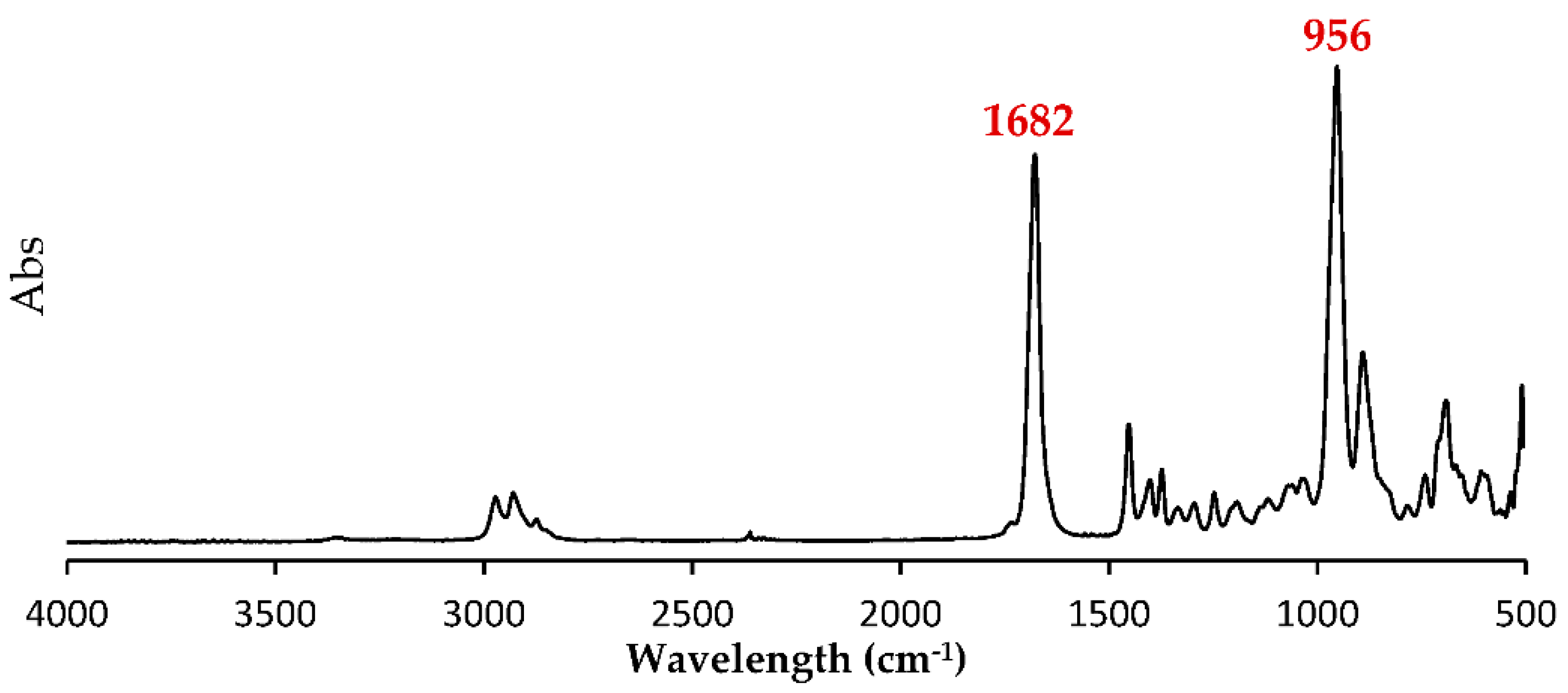
Based on 1H NMR analysis of the P(3M2MP) homopolymer, four peaks directly associated with its structure, A, B, and C, were identified with peak integrations of 0.99–1.00, at 3.14, 3.01, and 2.86 ppm, and D with a peak integration of 3.16 at 1.25 ppm. Other significant peaks at 7.26, 1.58, and 0 ppm corresponded to the solvent d-chloroform, water moisture, and internal standard tetramethylsilane, respectively. 1H-1H COSY NMR revealed correlations between A and B and between C and D. Based on this result and those of 1H NMR integration, A and B were identified as the two protons of the methylene group, C was the proton of the methine group, and D was the proton of the methyl group. Using 13C NMR, peaks E, F, G, and H at 201.3, 48.4, 31.6, and 17.6 ppm, respectively, were found to be directly associated with its structure, which was confirmed using 1H-13C HSQC NMR. This result indicates that A and B correlate with G, serving as the methylene group, C correlates with F as the methine group, D correlates with H as the methyl group, and E serves as the protonless carbonyl group, confirming the expected structure of the homopolymer structurally related to 3-mercapto-2-methylpropionic acid. Figure 2a shows the 1H NMR, Figure 2b 1H-1H COSY NMR, Figure 2c 13C NMR, and Figure 2d 1H-13C HSQC NMR spectra. Moreover, based on the ATR-FTIR results shown in Figure 3, the thioester group was confirmed based on the peaks at 1682 and 956 cm−1, which were associated with dialkyl thioester carbonyl stretching (1700–1680 cm−1) and dialkyl thioester C-S stretching (1035–930 cm−1), respectively [33].
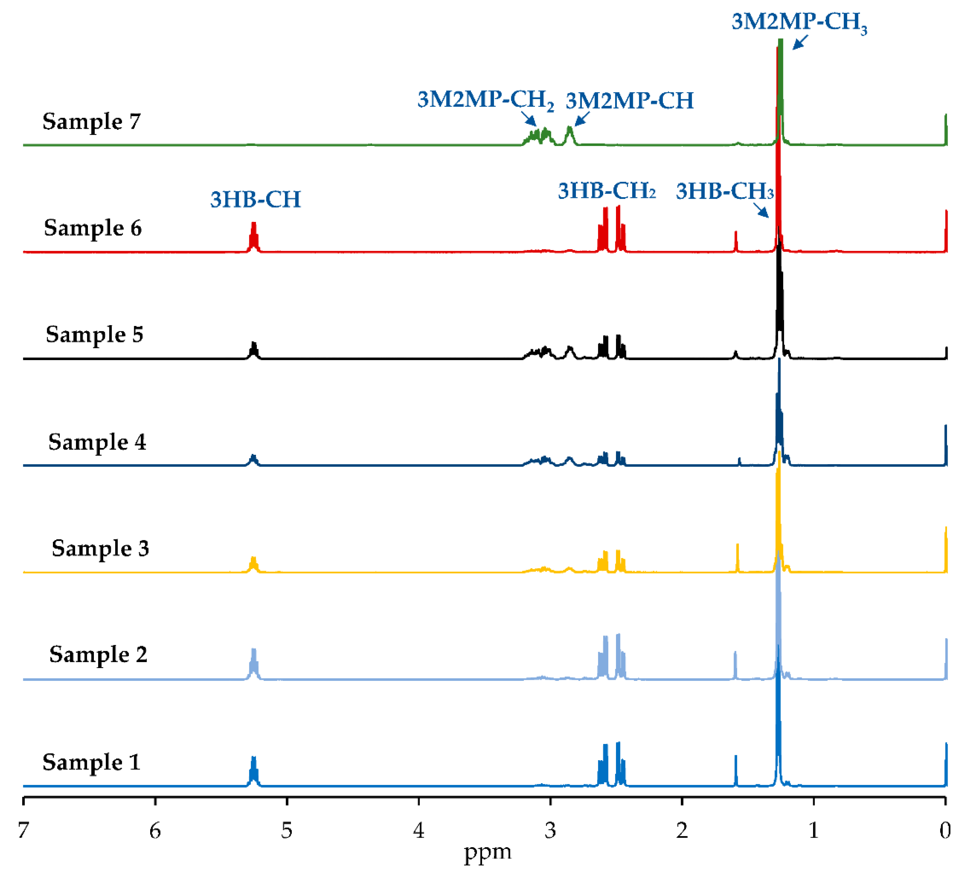
Based on 1H NMR analysis of the copolymers P(3HB-co-3M2MP) in Figure 4, the 3HB chemical shifts were similar to those found by Lütke-Eversloh et al. [22] for the copolymer, P(3HB-co-3MP). However, because of the presence of a methyl group in the 3M2MP unit, overlap with the methyl group of 3HB was observed. Therefore, the comonomer composition was estimated by integrating the methine groups.
Based on the sequence distribution of the polymer, as a copolymer with two different units, four types of unit combinations can occur, 3HB-3M2MP, 3HB-3HB, 3M2MP-3M2MP, and 3M2MP-3HB, with the carbonyl group split into four peaks using a Bernoullian statistical model. The sequence distribution was predicted from the parameter “D” [32] as follows. For simplification, 3HB is expressed as “O” and 3M2MP as “S”.
D = F O O · F S S F O S · F S O  
where FXY is the mole fraction of the carbonyl group in the XY sequence in the 13C NMR spectrum. Therefore, when D = 1, the copolymer is statistically random. However, when D is markedly larger than 1, the copolymer is blocky, and when it is markedly smaller than 1, it is an alternating copolymer [32].
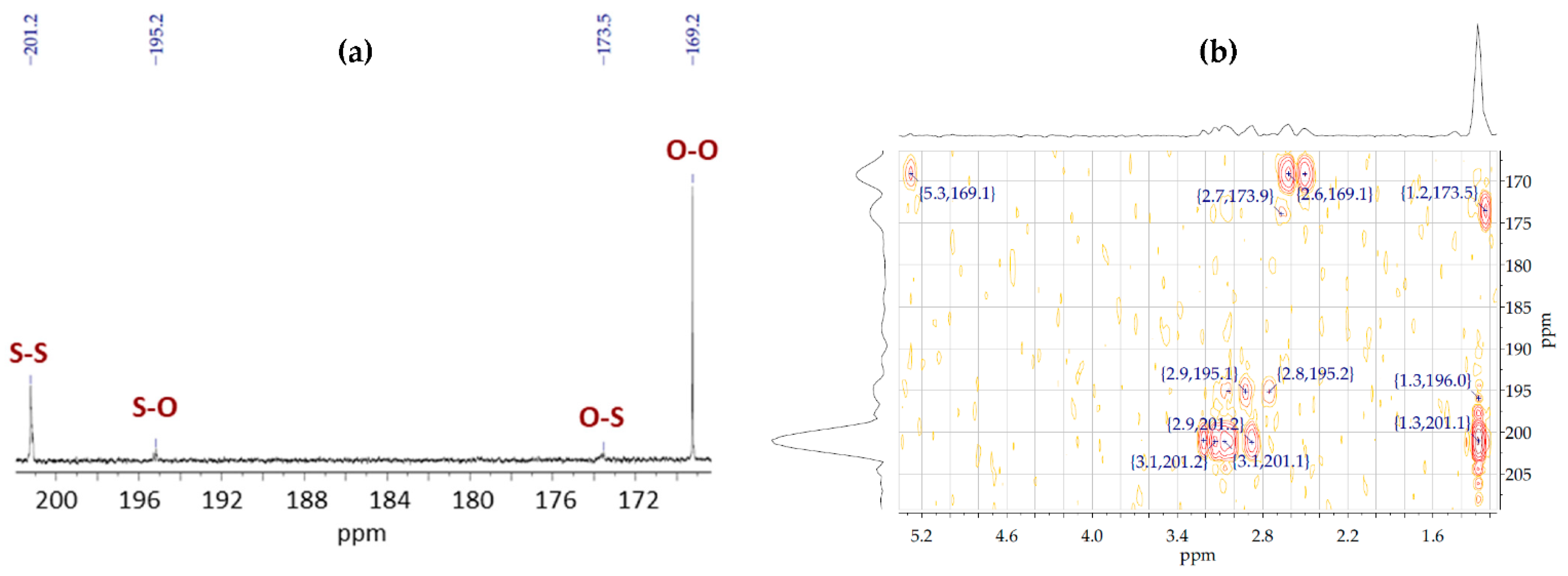
The 13C NMR spectra of the copolymer samples showed two extra peaks 195.2 and 173.5 ppm that were not present in the spectra of their homopolymers. Thereafter, based on the 1H-13C HMBC for the copolymer samples, these peaks were found to interact with the protons of the 3M2MP and 3HB units. Accordingly, these are the carbonyl peaks of 3M2MP-3HB and 3HB-3M2MP diad, respectively. Figure 5a shows the 13C NMR spectrum and Figure 5b the 1H-13C HMBC spectrum of Sample 5. The D parameter was calculated for the copolymer samples using the carbonyl peaks detected in the 13C NMR spectra. Table 2 presents the results for the FXY and D parameters for each sample and shows that all samples had D values significantly greater than 1, indicating that they have blocky sequence distributions. When the amount of the 3M2MP precursor added to the medium was small, the degree of the block sequence tended to be high.

3.3. Molecular Weight and Thermal Properties

The weight-average molecular weight (Mw) of the samples was considered high, varying from 7.7 × 10⁵ to 17.5 × 10⁵ g/mol. The values are in the upper limit of the 1 to 20 × 105 g/mol range commonly found for biosynthesized P(3HB) [34]. In contrast, other PTE and PTE copolymers with 3HB have markedly smaller Mw and Mw/Mn, as reported previously [22,23,24,25,26]. Moreover, except for in Sample 4, decreasing Mw and increasing Mw/Mn trends were observed, possibly because of the increasing amounts of unreacted 3-mercapto-2-methylpropionic acid; at increasing dosage concentrations, this reagent acted as a chain-transfer agent, ultimately decreasing the average molecular weight of the polymer and increasing the number of synthesized chains because of its chain termination function [34].
Based on the thermal properties, the homopolymer (Sample 7) displayed clear amorphous behavior, with a negative Tg and no melting or cold crystallization peaks. Consequently, in Samples 1–3, with increasing amounts of 3M2MP incorporated into the polymer structure, Tg and ΔHm decreased until only Tg remained, resulting in amorphous characteristics for Sample 4. Samples 5 and 6 exhibited different behaviors. The 3M2MP unit content of Sample 5 was similar to that of Sample 4 but was not amorphous. Sample 5 had two Tg peaks with lower ΔHm and Tcc values but a higher Tm compared with those of Sample 3. This finding may be related to differences in its microstructure compared with that of Sample 4. The Tg and Tcc of Sample 6 were similar to those of Sample 1; however, this sample exhibited a higher Tm and ΔHm, possibly because of its higher crystallinity, higher Mw and Mw/Mn, and lower 3M2MP fraction, despite the higher precursor addition. Moreover, as there are no published reports of biosynthesis and material property of its equivalent oxoester polymer [35,36], we could not confirm the high thermal stability of P(3M2MP). However, compared with other bio-PTE homopolymers, P(3M2MP) is the first amorphous PTE [25].
Table 3 shows the results of the molecular weight and thermal property analyses of the samples, whereas Figure 6a,b show the 1st and 2nd heating scans, respectively.

3.4. Physical and Mechanical Properties

Figure 7 shows the physical aspects of the polymer films as the 3M2MP fraction increased from 5.5 mol% to 100 mol%. Accordingly, the polymers became more transparent and softer as the 3M2MP content increased. Notably, the homopolymer tends to be very sticky, leading to handling difficulties.
Table 4 shows the mechanical properties based on the physical observations. As the 3M2MP fraction increased from Samples 1 to 7, the yield strength, tensile strength, and Young’s modulus decreased, whereas elongation at break increased. Sample 6 differed from the other samples, as explained above. The 3M2MP unit conferred the copolymers with great flexibility, allowing them to achieve over 1500% elongation at break with only 53.9 mol% of 3M2MP. Although Sample 5 showed similar 3M2MP contents to Sample 4, it exhibited a significantly lower elongation of break. This difference may be related to its lower molecular weight and differences in the degree of the block sequence (Table 2 and Table 3). Therefore, the sequence distribution may greatly affect the mechanical properties of the copolymers [37]. The stress–strain curves of all samples are shown in Figure 8.
The homopolymer also showed exceptional elasticity, with 2605% of elongation at break. Notably, most deformation was instantly recoverable. Figure 9 shows the elongation and recovery after manual deformation.

4. Discussion

NMR spectroscopy confirmed that P(3M2MP) homopolymer and copolymers with 3HB were biosynthesized by recombinant E. coli LSBJ using a similar method to previous studies to produce P(3H2MB) and its copolymers [19,38]. The biosynthesis data indicated that the maximum uptake of 3-mercapto-2-methylpropionic acid by the cells occurred under the culture conditions used in this study, unlike those observed for other sulfuric fatty acids [22,23,24]. Notably, the polymer content achieved for copolymer production was markedly higher than that observed for other P(3HB-co-3-mercaptoalkanoate)s [22,23,24], demonstrating that the point mutation D4N in PhaPAc and double-point mutations NSDG in PhaCAc lead to high polymer accumulation and high incorporation of 3M2MP unit into the 3HB copolymer. These findings indicate the relatively high polymerization activity of PhaCAc NSDG for 3M2MP unit. The lower production of the homopolymer relative to its 3HB copolymers suggests that the presence of 3HB unit facilitates the polymerization of 3M2MP unit by PhaCAc NSDG.
In contrast to P(3MP) and other bio-PTEs [25,39], P(3M2MP) can be easily dissolved in chloroform for extraction; however, the range of solvent solubility, which may impact its recovery from cells, remains unclear. Thakor et al. [40] efficiently extracted a PTE homopolymer from cells containing up to 45 wt% polymer by adding SDS to the cultivation broth, performing intensive stirring for 20 min at 90 °C, and conducting centrifugation and water rinsing cycles. This extraction method may also be applied to P(3M2MP).
Based on the sequence distribution, the D values of the copolymers were markedly higher than those typically found for most PHA copolymers [32,41,42] and other P(3HB-co-3-mercaptoalkanoate)s [26,43], indicating a high block sequence distribution. When the amount of 3M2MP precursor added to the medium was small, the degree of the block sequence tended to be high (Table 2) because polymerization of the 3HB monomer was prioritized over that of the 3M2MP monomer. Therefore, increasing the amount of 3M2MP precursor increased the intracellular 3M2MP concentration, which facilitated the polymerization of 3M2MP, thereby reducing the degree of block sequence in the biosynthesized copolymers. However, as the copolymers were not fractionated with a solvent depending on the solubility difference between the 3M2MP and 3HB units, some polymer samples may be blends of copolymers with different comonomer compositions.
The molecular weight of the copolymers was exceptionally high compared with that of normally biosynthesized P(3HB) but showed a decreasing trend with higher addition of 3M2MP precursor, which may induce a chain-transfer reaction during polymerization [34]. Moreover, regarding its thermal properties, in contrast to other bio-PTEs [25], P(3M2MP) exhibited amorphous behavior, possibly because a racemic 3M2MP precursor was used. Therefore, as the 3M2MP fraction in the copolymer increased, Tg and ΔHm decreased. On the other hand, Huang et al. [44] demonstrated that the higher thioester linkage contents in 3HB-based copolymers may lead to higher thermal stability by reducing thermal degradation by hindering random scission of the polymer chain. Thus, thermal degradation study of P(3M2MP) and its copolymers are of interest as a potential target for thermostable material.
P(3M2MP) showed extraordinary elongation at break and instant recovery, indicating that its elasticity is higher than those of most commercial elastomeric polymers, such as natural or synthetic rubber (typically ranging from 100% to 800%) [45,46,47], or well-known elastic PHAs, such as poly(4-hydroxybutirate), which have copolymer and/or homopolymer elongations of approximately 1000% [48]. Because P(3HB-co-3MP) also has significant elastic properties [26], the superior elasticity of PTE may be related to the sulfur atom in the polymer backbone. Because the Pauling electronegativity of the sulfur atom (2.58) is markedly lower than that of the oxygen atom (3.44) and closer to the carbon atom (2.55) [25], the smaller difference in electronegativity may enable easier mobilization of the polymer structure during stretching [26]. α-Carbon methylation of 3M2MP may also affect its mechanical properties by hindering packing of the polymer structure.
Despite the lack of oxoester-equivalent polymers for analogy, a unique property of α-methylated monomers in their materials can be identified relative to those in other PTEs. It is interesting to study the biodegradability of P(3M2MP) because P(3MP), a non-α-methylated PTE, does not show biodegradability [49]. Bacterial PTEs are now considered non-biodegradable biopolymers [50]. Although the biodegradability of P(3MB) and P(3MV) has not been evaluated, other enzymatically polymerized PTEs were reported to be degradable with lipase [51].

Author Contributions

Conceptualization, L.V.S.C., M.M. and T.T.; methodology, L.V.S.C., M.M., Y.M. and T.T.; software, L.V.S.C. and M.M.; validation, M.M., H.A. and T.T.; formal analysis, L.V.S.C. and M.M.; investigation, L.V.S.C. and M.M.; resources, C.T.N., S.T. and T.T.; data curation, L.V.S.C.; writing—original draft preparation, L.V.S.C.; writing—review and editing, T.T.; visualization, L.V.S.C. and T.T.; supervision, T.T.; project administration, T.T.; funding acquisition, T.T. All authors have read and agreed to the published version of the manuscript.

Funding

This paper is based on the results obtained from a project, JPNP18016, commissioned by the New Energy and Industrial Technology Development Organization (NEDO) and partially supported by a Grant-in-Aid for Scientific Research (KAKENHI, 21H03640), Japan.

Institutional Review Board Statement

Not applicable.

Informed Consent Statement

Not applicable.

Data Availability Statement

Not applicable.

Acknowledgments

The authors acknowledge the Biomaterials Analysis Division and Materials Analysis Division, Open Facility Center, Tokyo Institute of Technology, for their support with DNA sequencing analysis and NMR measurements, respectively.

Conflicts of Interest

The authors declare no conflict of interest.

References

  1. PlasticsEurope. Plastics the Facts-2021. Available online: https://plasticseurope.org/wp-content/uploads/2021/12/Plastics-the-Facts-2021-web-final.pdf (accessed on 16 February 2022).
  2. Geyer, R.; Jambeck, J.R.; Law, K.L. Production, use, and fate of all plastics ever made. Sci. Adv. 2017, 3, e1700782. [Google Scholar] [CrossRef] [PubMed] [Green Version]
  3. Kaza, S.; Yao, L.; Bhada-Tata, P.; van Woerden, P. What a Waste 2.0: A Global Snapshot of Solid Waste Management to 2050. Urban Development Series; World Bank: Washington, DC, USA, 2018; ISBN 978-1-4648-1347-4. [Google Scholar]
  4. World Economic Forum; Ellen MacArthur Foundation; McKinsey & Company. The New Plastics Economy–Rethinking the Future of Plastics. Available online: https://emf.thirdlight.com/link/faarmdpz93ds-5vmvdf/@/preview/1?o (accessed on 16 February 2022).
  5. Ross, G.; Ross, S.; Tighe, B.J. Bioplastics: New routes, new products. In Brydson’s Plastics Materials, 8th ed.; Gilbert, M., Ed.; Butterworth-Heinemann: Cambridge, MA, USA, 2017; pp. 631–652. ISBN 978-0-323-35824-8. [Google Scholar]
  6. Chen, G.-Q.; Hajnal, I.; Wu, H.; Lv, L.; Ye, J. Engineering biosynthesis mechanisms for diversifying polyhydroxyalkanoates. Trends Biotechnol. 2015, 33, 565–574. [Google Scholar] [CrossRef] [PubMed]
  7. Anjum, A.; Zuber, M.; Zia, K.M.; Noreen, A.; Anjum, M.N.; Tabasum, S. Microbial production of polyhydroxyalkanoates (PHAs) and its copolymers: A review of recent advancements. Int. J. Biol. Macromol. 2016, 89, 161–174. [Google Scholar] [CrossRef] [PubMed]
  8. Choi, S.Y.; Cho, I.J.; Lee, Y.; Kim, Y.-J.; Kim, K.-J.; Lee, S.Y. Microbial polyhydroxyalkanoates and nonnatural polyesters. Adv. Mater. 2020, 32, 1907138. [Google Scholar] [CrossRef] [PubMed]
  9. Suzuki, M.; Tachibana, Y.; Kasuya, K.I. Biodegradability of poly (3-hydroxyalkanoate) and poly (ε-caprolactone) via biological carbon cycles in marine environments. Polym. J. 2021, 53, 47–66. [Google Scholar] [CrossRef]
  10. Narancic, T.; Verstichel, V.; Chaganti, S.R.; Morales-Gamez, L.; Kenny, S.T.; De Wilde, B.; Padamati, R.B.; O’Connor, K.E. Biodegradable plastic blends create new possibilities for end-of-life management of plastics but they are not a panacea for plastic pollution. Environ. Sci. Technol. 2018, 52, 10441–10452. [Google Scholar] [CrossRef]
  11. Choi, S.Y.; Rhie, M.N.; Kim, H.T.; Joo, J.C.; Cho, I.J.; Son, J.; Lee, Y. Metabolic engineering for the synthesis of polyesters: A 100-year journey from polyhydroxyalkanoates to non-natural microbial polyesters. Metab. Eng. 2020, 58, 47–81. [Google Scholar] [CrossRef]
  12. Taguchi, S.; Iwata, T.; Abe, H.; Doi, Y. Poly(hydroxyalkanoate)s. In Polymer Science: A Comprehensive Reference, 1st ed.; Matyjaszewski, K., Möller, M., Eds.; Elsevier: Amsterdam, The Netherlands, 2012; pp. 157–182. ISBN 978-0-08-087862-1. [Google Scholar]
  13. McAdam, B.; Fournet, M.B.; McDonald, P.; Mojicevic, M. Production of Polyhydroxybutyrate (PHB) and Factors Impacting Its Chemical and Mechanical Characteristics. Polymers 2020, 12, 2908. [Google Scholar] [CrossRef]
  14. Yeo, J.C.C.; Muiruri, J.K.; Thitsartarn, W.; Li, Z.; He, C. Recent advances in the development of biodegradable PHB-based toughening materials: Approaches, advantages and applications. Mater. Sci. Eng. C Mater. Biol. Appl. 2018, 92, 1092–1116. [Google Scholar] [CrossRef]
  15. Sudesh, K.; Abe, H.; Doi, Y. Synthesis, structure and properties of polyhydroxyalkanoates: Biological polyesters. Prog. Polym. Sci. 2000, 25, 1503–1555. [Google Scholar] [CrossRef]
  16. Taguchi, S.; Matsumoto, K. Evolution of polyhydroxyalkanoate synthesizing systems toward a sustainable plastic industry. Polym. J. 2021, 53, 67–79. [Google Scholar] [CrossRef]
  17. Ishii-Hyakutake, M.; Mizuno, S.; Tsuge, T. Biosynthesis and characteristics of aromatic polyhydroxyalkanoates. Polymers 2018, 10, 1267. [Google Scholar] [CrossRef] [PubMed] [Green Version]
  18. Brydson, J.A. Silicone and other heat-resisting polymers. In Plastics Materials, 7th ed.; Brydson, J.A., Ed.; Butterworth-Heinemann: Oxford, UK, 1999; pp. 814–852. ISBN 0-7506-4132-0. [Google Scholar]
  19. Furutate, S.; Kamoi, J.; Nomura, C.T.; Taguchi, S.; Abe, H.; Tsuge, T. Superior thermal stability and fast crystallization behavior of a novel, biodegradable α-methylated bacterial polyester. NPG Asia Mater. 2021, 13, 31. [Google Scholar] [CrossRef]
  20. Marvel, C.S.; Kotch, A. Polythiolesters. J. Am. Chem. Soc. 1951, 73, 1100–1102. [Google Scholar] [CrossRef]
  21. Xia, Y.; Wübbeler, J.H.; Qi, Q.; Steinbüchel, A. Employing a recombinant strain of Advenella mimigardefordensis for biotechnical production of homopolythioesters from 3,3′-dithiodipropionic acid. Appl. Environ. Microbiol. 2012, 78, 3286–3297. [Google Scholar] [CrossRef] [Green Version]
  22. Lütke-Eversloh, T.; Bergander, K.; Luftmann, H.; Steinbüchel, A. Identification of a new class of biopolymer: Bacterial synthesis of a sulfur-containing polymer with thioester linkages. Microbiol. Read. 2001, 147, 11–19. [Google Scholar] [CrossRef] [Green Version]
  23. Lütke-Eversloh, T.; Bergander, K.; Luftmann, H.; Steinbüchel, A. Biosynthesis of poly(3-hydroxybutyrate-co-3-mercaptobutyrate) as a sulfur analogue to poly(3-hydroxybutyrate) (PHB). Biomacromolecules 2001, 2, 1061–1065. [Google Scholar] [CrossRef]
  24. Lütke-Eversloh, T.; Steinbüchel, A. Novel precursor substrates for polythioesters (PTE) and limits of PTE biosynthesis in Ralstonia eutropha. FEMS Microbiol. Lett. 2003, 221, 191–196. [Google Scholar] [CrossRef] [Green Version]
  25. Lütke-Eversloh, T.; Fischer, A.; Remminghorst, U.; Kawada, J.; Marchessault, R.H.; Bögershausen, A.; Kalwei, M.; Eckert, H.; Reichelt, R.; Liu, S.J.; et al. Biosynthesis of novel thermoplastic polythioesters by engineered Escherichia coli. Nat. Mater. 2002, 1, 236–240. [Google Scholar] [CrossRef]
  26. Yu, F.; Dong, T.; Zhu, B.; Tajima, K.; Yazawa, K.; Inoue, Y. Mechanical properties of comonomer-compositionally fractionated poly[(3-hydroxybutyrate)-co-(3-mercaptopropionate)] with low 3-mercaptopropionate unit content. Macromol. Biosci. 2007, 7, 810–819. [Google Scholar] [CrossRef]
  27. Watanabe, Y.; Ishizuka, K.; Furutate, S.; Abe, H.; Tsuge, T. Biosynthesis and characterization of novel poly(3-hydroxybutyrate-co-3-hydroxy-2-methylbutyrate): Thermal behavior associated with α-carbon methylation. RSC Adv. 2015, 5, 58679. [Google Scholar] [CrossRef] [Green Version]
  28. Tappel, R.C.; Wang, Q.; Nomura, C.T. Precise control of repeating unit composition in biodegradable poly(3-hydroxyalkanoate) polymers synthesized by Escherichia coli. J. Biosci. Bioeng. 2012, 113, 480–486. [Google Scholar] [CrossRef] [PubMed]
  29. Ushimaru, K.; Watanabe, Y.; Hiroe, A.; Tsuge, T. A single-nucleotide substitution in phasin gene leads to enhanced accumulation of polyhydroxyalkanoate (PHA) in Escherichia coli harboring Aeromonas caviae PHA biosynthetic operon. J. Gen. Appl. Microbiol. 2015, 61, 63–66. [Google Scholar] [CrossRef] [PubMed]
  30. Tsuge, T.; Watanabe, S.; Shimada, D.; Abe, H.; Doi, Y.; Taguchi, S. Combination of N149S and D171G mutations in Aeromonas caviae polyhydroxyalkanoate synthase and impact on polyhydroxyalkanoate biosynthesis. FEMS Microbiol. Lett. 2007, 277, 217–222. [Google Scholar] [CrossRef] [Green Version]
  31. Arikawa, H.; Sato, S.; Fujiki, T.; Matsumoto, K. Simple and rapid method for isolation and quantitation of polyhydroxyalkanoate by SDS-sonication treatment. J. Biosci. Bioeng. 2017, 124, 250–254. [Google Scholar] [CrossRef] [PubMed]
  32. Kamiya, N.; Yamamoto, Y.; Inoue, Y.; Chujo, R.; Doi, Y. Microstructure of bacterially synthesized poly(3-hydroxybutyrate-co-3-hydroxyvalerate). Macromolecules 1989, 22, 1676–1682. [Google Scholar] [CrossRef]
  33. Socrates, G. The Carbonyl Group: C==O. In Infrared and Raman Characteristic Group Frequencies: Tables and Charts, 3rd ed.; Socrates, G., Ed.; John Wiley & Sons, Ltd.: Chichester, UK, 2001; pp. 115–156. ISBN 978-0-470-09307-8. [Google Scholar]
  34. Tsuge, T. Fundamental factors determining the molecular weight of polyhydroxyalkanoate during biosynthesis. Polym. J. 2016, 48, 1051–1057. [Google Scholar] [CrossRef]
  35. Marx, A.; Poetter, M.; Buchholz, S.; May, A.; Siegert, H.; Alber, B.; Fuchs, G.; Eggeling, L. Microbiological Production Of 3-Hydroxyisobutyric Acid. Available online: https://patentimages.storage.googleapis.com/1a/8f/54/e04f10eb8e2206/CA2654133A1.pdf (accessed on 27 February 2022).
  36. Vermeer, C.M.; Bons, L.J.; Kleerebezem, R. Production of a newly discovered PHA family member with an isobutyrate-fed enrichment culture. Appl. Microbiol. Biotechnol. 2022, 106, 605–618. [Google Scholar] [CrossRef]
  37. Kajita, T.; Noro, A.; Matsushita, Y. Design and properties of supramolecular elastomers. Polymer 2017, 128, 297–310. [Google Scholar] [CrossRef]
  38. Furutate, S.; Abe, H.; Tsuge, T. Thermal properties of poly(3-hydroxy-2-methylbutyrate-co-3-hydroxybutyrate) copolymers with narrow comonomer-unit compositional distributions. Polym. J. 2021, 53, 1451–1457. [Google Scholar] [CrossRef]
  39. Kawada, J.; Lütke-Eversloh, T.; Steinbüchel, A.; Marchessault, R.H. Physical properties of microbial polythioesters: Characterization of poly(3-mercaptoalkanoates) synthesized by engineered Escherichia coli. Biomacromolecules 2003, 4, 1698–1702. [Google Scholar] [CrossRef]
  40. Thakor, N.; Lütke-Eversloh, T.; Steinbüchel, A. Application of the BPEC pathway for large-scale biotechnological production of poly(3-mercaptopropionate) by recombinant Escherichia coli, including a novel in situ isolation method. Appl. Environ. Microbiol. 2005, 71, 835–841. [Google Scholar] [CrossRef] [Green Version]
  41. Hu, D.; Chung, A.L.; Wu, L.P.; Zhang, X.; Wu, Q.; Chen, J.C.; Chen, G.Q. Biosynthesis and characterization of polyhydroxyalkanoate block copolymer P3HB-b-P4HB. Biomacromolecules 2011, 12, 3166–3173. [Google Scholar] [CrossRef]
  42. Tripathi, L.; Wu, L.P.; Chen, J.; Chen, G.Q. Synthesis of Diblock copolymer poly-3-hydroxybutyrate -block-poly-3-hydroxyhexanoate [PHB-b-PHHx] by a β-oxidation weakened Pseudomonas putida KT2442. Microb. Cell Fact. 2012, 11, 11. [Google Scholar] [CrossRef] [Green Version]
  43. Impallomeni, G.; Steinbüchel, A.; Lütke-Eversloh, T.; Barbuzzi, T.; Ballistreri, A. Sequencing microbial copolymers of 3-hydroxybutyric and 3-mercaptoalkanoic acids by NMR, electrospray ionization mass spectrometry, and size exclusion chromatography NMR. Biomacromolecules 2007, 8, 985–991. [Google Scholar] [CrossRef]
  44. Huang, P.; Furutate, S.; Mizuno, S.; Tsuge, T. Thermal degradation behavior of bacterial poly (3-hydroxybutyrate-co-3-mercaptopropionate). Polym. Degrad. Stab. 2019, 165, 35–42. [Google Scholar] [CrossRef]
  45. Goff, J.; Sulaiman, S.; Arkles, B.; Lewicki, J.P. Soft Materials with Recoverable Shape Factors from Extreme Distortion States. Adv. Mater. 2016, 28, 2393–2398. [Google Scholar] [CrossRef]
  46. Arkles, B.; Goff, J.; Sulaiman, S.; Sikorsky, A. Ultra-High Elongation Silicone Elastomers. Available online: https://www.gelest.com/wp-content/uploads/Ultra-high-Elongation-Silicone-Elastomers.pdf (accessed on 27 February 2022).
  47. Hiroe, A.; Ishii, N.; Ishii, D.; Kabe, T.; Abe, H.; Iwata, T.; Tsuge, T. Uniformity of monomer composition and material properties of medium-chain-length polyhydroxyalkanoates biosynthesized from pure and crude fatty acids. ACS Sustain. Chem. Eng. 2016, 4, 6905–6911. [Google Scholar] [CrossRef]
  48. Saito, Y.; Doi, Y. Microbial synthesis and properties of poly(3-hydroxybutyrate-co-4-hydroxybutyrate) in Comamonas acidovorans. Int. J. Biol. Macromol. 1994, 16, 99–104. [Google Scholar] [CrossRef]
  49. Kim, D.Y.; Lütke-Eversloh, T.; Elbanna, K.; Thakor, N.; Steinbüchel, A. Poly(3-mercaptopropionate): A nonbiodegradable biopolymer? Biomacromolecules 2005, 6, 897–901. [Google Scholar] [CrossRef]
  50. Steinbüchel, A. Non-biodegradable biopolymers from renewable resources: Perspectives and impacts. Curr. Opin. Biotechnol. 2005, 16, 607–613. [Google Scholar] [CrossRef]
  51. Kato, M.; Toshima, K.; Matsumura, S. Preparation of aliphatic poly(thioester) by the lipase-catalyzed direct polycondensation of 11-mercaptoundecanoic acid. Biomacromolecules 2005, 6, 2275–2280. [Google Scholar] [CrossRef]
Figure 1. P(3HB-co-3M2MP) and P(3M2MP) biosynthetic pathway.
Bioengineering 09 00228 g001
Figure 2. NMR spectra of P(3M2MP). (a) 1H NMR; (b) 1H-1H COSY NMR; (c) 13C NMR; and (d) 1H-13C HSQC NMR.
Bioengineering 09 00228 g002
Figure 3. FTIR spectra of P(3M2MP).
Bioengineering 09 00228 g003
Figure 4. 1H NMR spectra of Sample 1 (3M2MP 5.5 mol%), Sample 2 (10.1 mol%), Sample 3 (34.2 mol%), Sample 4 (53.9 mol%), Sample 5 (54.8 mol%), Sample 6 (10.7 mol%), and Sample 7 (100 mol%).
Bioengineering 09 00228 g004
Figure 5. (a) 13C NMR of Sample 5 (3M2MP 54.8 mol%) showing combination peaks; (b) 1H-13C HMBC of Sample 5.
Bioengineering 09 00228 g005
Figure 6. DSC thermogram of Samples 1 (3M2MP 5.5 mol%), 2 (10.1 mol%), 3 (34.2 mol%), 4 (53.9 mol%), 5 (54.8 mol%), 6 (10.7 mol%), and 7 (100 mol%). (a) 1st heating scan; (b) 2nd heating scan.
Bioengineering 09 00228 g006
Figure 7. Polymer solvent cast films with increasing 3M2MP units. (a) Sample 1 (5.5 mol% 3M2MP); (b) Sample 2 (10.1 mol%); (c) Sample 3 (34.2 mol%); (d) Sample 4 (53.9 mol%); (e) Sample 5 (54.8 mol%); (f) Sample 7 (100 mol%).
Bioengineering 09 00228 g007
Figure 8. Stress–strain curve of Samples 1 (3M2MP 5.5 mol%), 2 (10.1 mol%), 3 (34.2 mol%), 4 (53.9 mol%), 5 (54.8 mol%), 6 (10.7 mol%), and 7 (100 mol%).
Bioengineering 09 00228 g008
Figure 9. Elongation of P(3M2MP) (Sample 7) by manual deformation. (a) Before deformation; (b) during deformation; (c) after deformation.
Bioengineering 09 00228 g009
Table 1. P(3HB-co-3M2MP) and P(3M2MP) biosynthesis by recombinant E. coli LSBJ.
PhaAB and PhaJ ExpressionPrecursor
(g/L) 1
Dry Cell wt. (g/L)Polymer Content (wt%)Monomer Composition (mol%) 2Sample ID
3HB3M2MP
+ 30.253.50 ± 0.0369.1 ± 1.994.55.51
+ 30.503.69 ± 0.2568.7 ± 1.089.910.12
+ 31.003.48 ± 0.0267.3 ± 0.165.834.23
+ 31.503.63 ± 0.0269.1 ± 0.246.153.94
+ 32.004.29 ± 0.0772.1 ± 0.845.254.85
+ 32.505.06 ± 0.0477.2 ± 0.389.310.76
41.201.28 ± 0.018.4 ± 1.301007
3HB, 3-hydroxyburyrate; 3M2MP, 3-mercapto-2-methylpropionate. Results are expressed as the mean ± standard deviation (n = 3). 1 (R,S)-3-Mercapto-2-methylpropionic acid. 2 Determined by comparison of the methine (>CH-) peaks in the 1H NMR spectra. 3 Strain harboring pTTQ19PCT and pBBR1phaP(D4N)JCAcABRe NSDG. Cells were cultured in 100 mL M9-modified medium containing 0.25–2.5 g/L precursor (3-mercapto-2-methylpropionic acid), 20 g/L glucose, and 1 mM IPTG at 30 °C for 72 h. 4 Strain harboring pTTQ19PCT and pBBR1phaP(D4N)CAc NSDG. Cells were cultured in 100 mL M9-modified medium at 30 °C for 4 h, and 1.2 g/L precursor (3-mercapto-2-methylpropionic acid), 3.75 g/L glucose, and 1 mM IPTG were added before further culture for 72 h.
Table 2. D value and sequence distribution of P(3HB-co-3M2MP) samples.
Sample ID3M2MP (mol%)Diad Sequence DistributionDDegree of Block Sequence 1
FOOFOSFSOFSS
15.50.9600.0160.0090.015100High
210.10.9340.0120.0060.048623High
334.20.7410.0060.0370.216720High
453.90.3290.1820.1520.3374Low
554.80.4490.0670.0450.43965Medium
610.70.8680.0570.0210.05439Medium
1 1 < D < 10: low, 10 ≤ D < 100: medium, 100 ≤ D: high.
Table 3. Molecular weight and thermal properties of the samples.
Sample ID3M2MP
(mol%)
Mw
(×105)
Mw/MnTg
(°C)
Tcc
(°C)
Tm
(°C)
ΔHm
(J/g)
P(3HB) 105.22.34.0NA17679
15.514.63.38.157150, 16428
210.112.03.26.662149, 16426
334.210.03.65.470159, 17313
453.913.74.4−1.2---
554.87.74.2−3.1, 7.065167, 1799
610.717.55.68.450166, 18151
710015.02.6−3.1---
NA: not available, -: not detectable. 1 Data from Ref. [19].
Table 4. Mechanical properties of the samples (n = 3).
Sample ID3M2MP (mol%)Yield Strength (MPa)Tensile Strength (MPa)Elongation at Break (%)Young’s Modulus (MPa)
P(3HB) 10NA 258 ± 712 ± 01420 ± 80
15.513.6 ± 2.023.0 ± 2.324 ± 13880 ± 75
210.114.6 ± 2.219.3 ± 3.0470 ± 131644 ± 41
334.26.5 ± 0.317.1 ± 0.8825 ± 2187 ± 8
453.92.2 ± 0.112.7 ± 0.2 1549 ± 10313 ± 1
554.81.3 ± 0.25.8 ± 0.4436 ± 4918 ± 1
610.78.2 ± 3.515.5 ± 1.4158 ± 58709 ± 303
71001.0 ± 0.14.2 ± 0.52605 ± 40.8 ± 0.1
1 Ref. [19], 2 Not available.
Publisher’s Note: MDPI stays neutral with regard to jurisdictional claims in published maps and institutional affiliations.