Article

A Molecular Tool for Rapid Detection and Traceability of Cyclospora cayetanensis in Fresh Berries and Berry Farm Soils

by
Carolina N. Resendiz-Nava
1,
Guadalupe E. Orozco-Mosqueda
2,
Edmundo M. Mercado-Silva
1,
Susana Flores-Robles
1,
Hilda V. Silva-Rojas
3 and
Gerardo M. Nava
1,*
1
Facultad de Química, Universidad Autónoma de Querétaro, Cerro de las Campanas S/N, Querétaro 76010, Mexico
2
Hospital Infantil de Morelia Eva Samano de López Mateos, Servicio de Salud de Michoacán, Interior Bosque Cuauhtémoc S/N, Michoacán 58020, Mexico
3
Posgrado en Recursos Genéticos y Productividad, Producción de Semillas, Colegio de Postgraduados, Km. 36.5 Carretera Mexico-Texcoco, Estado de Mexico 56230, Mexico
*
Author to whom correspondence should be addressed.
Foods 2020, 9(3), 261; https://doi.org/10.3390/foods9030261
Submission received: 29 January 2020 / Revised: 16 February 2020 / Accepted: 23 February 2020 / Published: 2 March 2020
(This article belongs to the Special Issue Advances in Foodborne Pathogen Analysis)

Abstract

:
Due to recent outbreaks of cyclosporiasis associated with consumption of fresh berries, producers are demanding modern microbiological tools for the rapid and accurate identification of the human pathogen Cyclospora cayetanensis in berries and environmental samples. The aim of the present work was to develop a molecular tool based on a PCR approach for the rapid and accurate detection of C. cayetanensis. A nested PCR assay was validated for the amplification of a 294 bp size region of the 18S rRNA gene from C. cayetanensis. The limit of detection for the nested PCR assay was validated using 48 berry samples spiked with ~0, 10, 100, and 1000 oocyst per gram of sample. With this assay, it was possible to detect as few as 1 oocyst per gram of berry, in a 50 g sample. Sanger DNA sequencing and phylogenetic analysis were carried out to confirm the presence of C. cayetanensis in berry (n = 17) and soil (n = 5) samples. The phylogenetic analysis revealed that the C. cayetanensis sequences obtained from Mexico clustered within a group recovered from China, Peru, Guatemala-Haiti, and Japan. The PCR protocol designed in the present study could be an important tool for the rapid and accurate detection of this human pathogen in environmental and food samples.

1. Introduction

The consumption of raspberries (Rubus idaeus), blackberries (Rubus sp.), and blueberries (Vaccinium corymbosum) has increased worldwide in recent years because these fruits are considered an important source of antioxidant compounds [1,2]. Unfortunately, consumption of berries is associated with a risk of acquiring foodborne parasites, such as Cyclospora cayetanensis [1,3]. For example, in the USA and Canada, 4250 cyclosporiasis cases have been linked to consumption of raspberries, blackberries, and strawberries [1]. In the UK, 43 cases of cyclosporiasis were linked to the consumption of fresh strawberries and raspberries [4]. At the farm level, the presence of C. cayetanensis in berries is directly associated with the presence of the parasite in soil [5,6,7]. Thus, it is fundamental that producers monitor the presence of this pathogen on farms and packing facilities.
Conventionally, detection of C. cayetanensis in clinical and environmental samples is based on identification of oocysts by microscopy, following modified acid fast staining or by autofluorescence under ultraviolet (UV) light [8]. However, this technique is time-consuming, non-specific, and lacks sensitivity [9]. To overcome these issues, molecular methods have been developed to detect C. cayetanensis in clinical and environmental samples [10,11,12]; however, the food producing industry requires a molecular method able to detect a low oocyst concentration (40–1500 oocyst per gram) as found in food and environmental samples [13]. Also, it is important to have a tool available to perform molecular traceability and identification of contamination sources. Thus, the objective of the present study was to develop and validate a highly sensitive and specific PCR assay for the rapid and accurate detection of C. cayetanensis, as well as its molecular traceability in fresh berries and farm soils.

2. Materials and Methods

2.1. DNA Extractions

Oocysts of C. cayetanensis (preserved in 2.5% potassium dichromate solution) obtained from a laboratory strain collection and environmental (fruit and soil) samples were subjected to DNA extraction using the ZymoBIOMICS DNA Kit (Zymo Research, Irvine, CA, USA) following manufacturer’s instructions. Purified DNA was diluted to reach a concentration of 1 ng/µL and stored at −20 °C.

2.2. Nested PCR Assay

To identify the most effective conditions for nested PCR, gradient PCR amplifications were performed using different annealing temperatures (ranging from 48 °C to 75 °C) for each primer set. Various rounds of nested PCR reactions were also carried out using C. cayetenensis purified DNA and DNA from PCR amplification. Overall, the first round of PCR amplification using primer pair CYCF1E (5′-TACCCAATGAAAACAGTTT-3′) and CYCR2B (5′-CAGGAGAAGCCAAGGTAGG-3′), generated a ~630 bp amplicon [10]; this primer pair amplifies a segment of the 18S rRNA gene found in different members of the Eimeriidae family [14]. Each PCR reaction (17 µL) contained: 3.4 µL of 5X Phire Hot Start II DNA Polymerase (Thermo Scientific, Waltham, MA, USA) reaction buffer, 0.34 µL of dNTP solution mix (10 mM) (Thermo Scientific.), 0.34 µL of Phire Hot Start II DNA Polymerase, 0.68 µL of each primer (1.0 µM), 2 µL of bovine serum albumin (Bioline, London, UK), and 3.0 ng of DNA extracted from oocysts. The amplification program consisted of 1 min at 95 °C followed by 35 cycles of denaturing at 95 °C for 30 s, annealing at 53.6 °C for 30 s, an extension at 72 °C for 30 s. and a final extension for 2 min at 72 °C. Genomic DNA (3.0 ng) obtained from C. cayetanensis oocysts was used for positive control reactions. The second round of PCR amplification was carried out using primer pair CC719 (5′-GTAGCCTTCCGCGCTTCG-3′) and CRP999 (5′-CGTCTTCAAACCCCCTACTGTCG-3′), which generates a ~298 bp amplicon [15]. The specificity of this primer pair has been previously validated against other Cyclospora species and genera of the Eimeriidae family [15,16]. The PCR reaction was performed as described above, using an annealing temperature of 66.5 °C and 2.0 µL of 1:100 diluted PCR products from the first reaction. All PCR products were subjected to electrophoresis using 1.5% (wt/vol) agarose-TBE (89 mM Tris-borate, 2 mM EDTA) gels, stained with an ethidium bromide solution(10 µg/35 mL gel) (Bio-Rad, Hercules, CA, USA). Specificity of the nested PCR assay was corroborated by Sanger sequencing and phylogenetic analysis.

2.3. Sensitivity of the Nested PCR Assay

To establish the limit of detection of the PCR assay, 50 g of fresh blueberries were spiked (applied as droplets) with approximately 1, 10, 100, and 1000 C. cayetanensis oocysts per gram of sample, using an inoculation solution (10 oocysts/µL). Briefly, approximately 3 mL of oocysts in 2.5% potassium dichromate solution were washed three times with 10 mL sterile distilled water by centrifugation (4500 rpm) at room temperature during 10 min to remove potassium dichromate. Washed cells were resuspended in 1 mL of sterile saline solution. Concentration of oocysts was estimated by counting three replicates using a hemocytometer. To evaluate reproducibility, experiments were carried out in triplicate as follows. Spiked blueberries were kept overnight at 4 °C to promote oocyst adherence to the fruit [17]. Then, samples were rinsed manually (~5 min) with 50 mL of sterile distilled water using a sterile plastic bag; washes were transferred to a conical tube and centrifuged for 10 min at 10,000 rpm [17] to recover a sample pellet. Approximately, 250 mg (between 80–100%) of the pellet was used for DNA extraction, and the genomic DNA was subjected to the nested PCR assay as described above.

2.4. Detection of C. cayetanensis in Fresh Berries and Farm Soils

To validate the usefulness of the nested PCR assay under field conditions, samples of blueberries (n = 6), raspberries (n = 6), blackberries (n = 11), and farm soils (n = 5) were obtained from commercial farms located in central Mexico. Each fruit sample consisted of 500 g of berries, which were rinsed (as described above) with 100 mL of sterile distilled water and subjected to DNA extraction (as described above). Soil samples consisted of ~50 g of soil collected at the base of the plants; ~250 mg of each soil sample was used for the DNA extraction. Purified DNA was subjected to the nested PCR protocol as described above. PCR products were subjected to Sanger sequencing and phylogenetic analysis. Each fragment was sequenced at least twice to eliminate sequencing errors.

2.5. Molecular Traceability of C. cayetanensis

The potential use of the 18S rRNA fragments, obtained with the nested PCR, was evaluated for studies of molecular traceability of pathogen origin. To accomplish this goal, a genomic survey at the GenBank was carried out using BLAST search tool [18]. Fifty-five sequences from C. cayetanensis 18S rRNA genes were found and retrieved from the GenBank. This set contained sequences from ten different countries (Table 1). Sequences were aligned, manually trimmed, and edited using MEGA 6 software [19]. Nucleotide alignments were subjected to phylogenetic analysis using the Maximum Likelihood [20], Maximum Parsimony [21], and Neighbor Joining [22] methods, Jukes-Cantor evolutionary model [23], and bootstrap analysis after 1000 replicates. The 18S rRNA gene sequences obtained in the present study were deposited in the GenBank under accession numbers: MK332310 to MK332315.

3. Results and Discussion

3.1. Nested PCR Standardization

To improve sensitivity in PCR-based detection of C. cayetanensis, two strategies were implemented; first, a nested PCR approach was designed because this pathogen is found at low concentrations in fruits and environmental samples [13]. Second, PCR assays were designed to amplify the 18S rRNA locus because multi-copy genes increase PCR sensitivity [30]. The first round of amplification using primer pair CYCF1E and CYCR2B generated specific and strong PCR signals when 3.0 ng of DNA template per reaction and an annealing temperature of 53.6 °C were used. For the second round of amplification, primer pair CC719 and CRP999 generated specific and strong PCR signals when the first-round PCR products were diluted 1:100 and an annealing temperature of 66.5 °C was used (Figure 1). Specificity of the nested PCR assay was corroborated by Sanger sequencing and phylogenetic analysis. These analyses revealed that nested PCR fragments obtained with the present protocol share high genetic similarity (~98.0% identity) with 18S rRNA genes from C. cayetanensis archived in the GenBank (accession numbers: EU861001.1, KY770759.1 and GU557063.1), confirming the specificity of the nested PCR assay. Importantly, these primer sets have been included in a new U.S. Food and Drug Administration method developed for detection of C. cayetanensis on cilantro and raspberries [31].

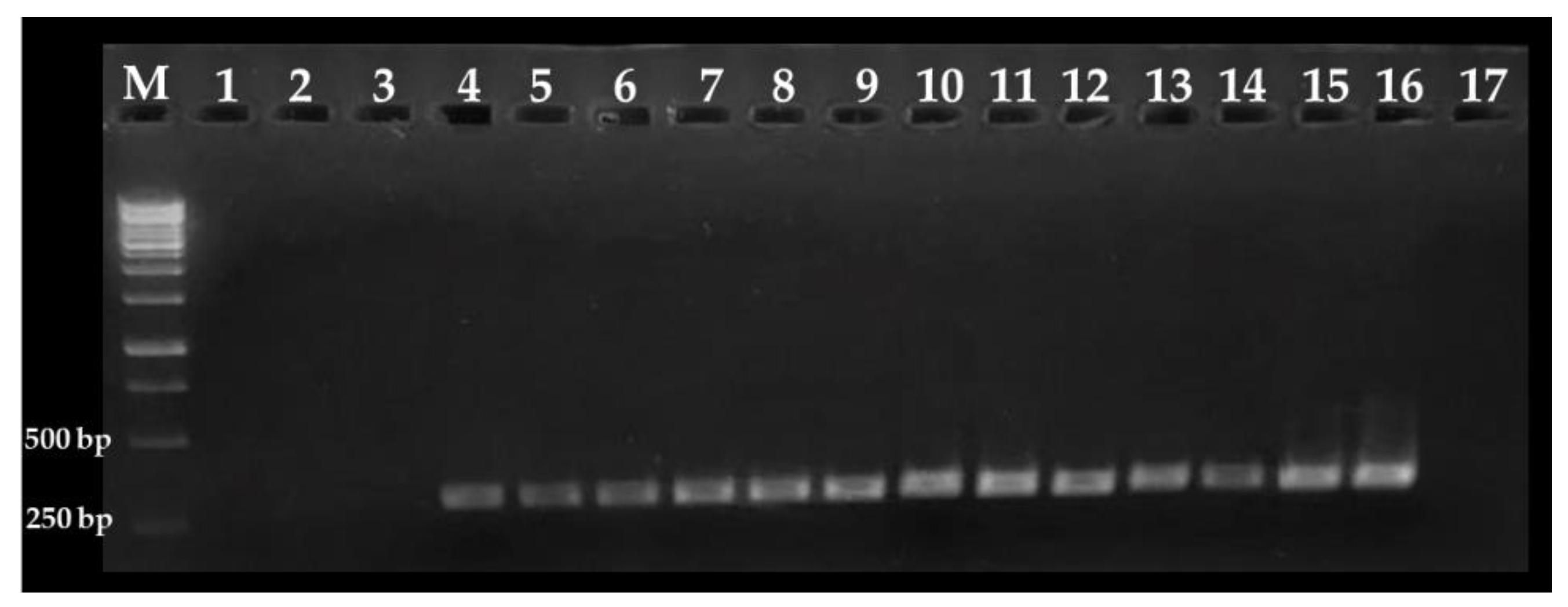
3.2. Sensitivity of the Nested PCR Assay

To determine the sensitivity of the nested PCR protocol, blueberry samples were spiked with approximately 1, 10, 100, and 1000 C. cayetanensis oocysts per gram of sample. It was observed that this PCR assay can detect as few as 1 oocyst per gram of sample; i.e., 50 oocysts in 50 g of sample (Figure 2). This level of sensitivity could be attributed to the inclusion of bovine serum albumin in each PCR reaction to overcome the effects of PCR-inhibitory substances found in environmental samples [32]. Thus, this nested PCR protocol could be a useful tool for the rapid and accurate detection of C. cayetanensis in environmental samples where oocyst concentrations are considered low; for example, 102 to 103 per gram of produce [13], 103 per liter of river water [6], 101 per liter of tap water [11], and 102 to 105 per gram of stool [33]. Because an infective dose of C. cayetanensis has been estimated between 101 and 102 oocysts [34,35], this PCR assay is sensitive enough to detect these levels of contamination in berries and environmental samples. Other studies have also accomplished low levels of detection using nested PCR assays. For example, in fresh basil, lettuce, and raspberries it was possible to detect from 1 to 10 C. cayetanensis oocysts per gram of product [36,37]. Taken together, these results reveal that nested PCR assays are a sensitive and effective tool for the detection of C. cayetanensis in fresh produce.

3.3. Detection of C. cayetanensis in Berries and Farm Soils

To validate the effectiveness of the nested PCR assay in detecting C. cayetanensis in environmental samples, fresh berries and farm soil were collected, processed, and subjected to the PCR protocol. The presence of C. cayetanensis was detected in 16.6% (1/6), 36.4% (4/11) and 20.0% (1/5) of blueberry, blackberry, and farm soil samples, respectively (Figure 3). These results were confirmed by Sanger sequencing and phylogenetic analysis. All seven PCR products obtained from fresh berries and soil samples share high genetic similarity (~ 99.9%) with 18S rRNA genes from C. cayetanensis archived in the GenBank (accession numbers: EU861001.1, KY770759.1 and GU557063.1). To the best of our knowledge, these sequences represent the first evidence of C. cayetanensis 18S rRNA PCR products obtained from environmental samples. Altogether, these results confirmed the high specificity of the nested PCR assay for the detection of C. cayetanensis in environmental samples, such as fresh berries and farm soil.
Importantly, the present study revealed the presence of C. cayetanensis in samples (berries and soil) collected from commercial farms, suggesting that these products could represent a potential threat to human health. Comparable results have been reported in South Korea where 2.3% of blueberries were positive for C. cayetanensis [38]. These results are of relevance because consumption of fresh berries [1,3,4] and contact with contaminated soil [5,6,7] have been linked to numerous C. cayetanensis outbreaks in humans. Together, these data highlight the importance of performing molecular surveillance for the rapid and opportune detection of this human pathogen.

3.4. Molecular Traceability of C. cayetanensis

An important and current challenge in food microbiology is to rapidly identify sources or origin of contamination. To this end, genomic analyses of pathogens are an important tool to accomplish this goal [39]; unfortunately, genomic information from C. cayetanensis is very limited, thus it is essential to generate this molecular evidence. In the present study, the use of the 18S rRNA locus is proposed not only for rapid and accurate detection of C. cayetanensis, but also, as a potential tool for molecular traceability. To perform this analysis, fifty-five C. cayetanensis 18S rRNA gene sequences were found and retrieved from the GenBank database. These nucleotide sequences were obtained from countries such as China, Guatemala, Iran, Japan, Korea, Mexico, Nepal, Peru, Poland, and Singapore [24,25,26,27,28,40]. Six additional 18S rRNA gene sequences (accession numbers MK332310 to MK332315) from Mexico, obtained in the present study, were included in the analysis. Phylogenetic analysis of these fifty-five gene fragments (position 760 to 986, C. cayetanensis AF111183.1 numbering) revealed the presence of seven different genotypes (Figure 4).
Genotype I combined 18S rRNA gene sequences from Japan and Mexico. Interestingly, information available at the GenBank indicates that sequences AB368541.1 and AB368542.1 were obtained from an infected Japanese patient who traveled to Mexico, and sequence MK332314 was recovered from an environmental sample from Mexico, indicating that genotype I found in Japan is also prevalent in Mexico. This result underlines the potential use of this molecular tool for identification of pathogen origin source. Genotype II and III are integrated by C. cayetanensis genetic variants found in Iran and China, respectively. Genotype IV is integrated by sequences from China. Genotype V is broadly distributed and integrates a variant found in China, Guatemala, Japan, Korea, Mexico, Nepal, Peru, Poland, and Singapore. Genotype VI and VII are integrated by variants found in China. Overall, these results indicate that in China and Mexico at least five and two different genotypes, respectively; are prevalent (Figure 4). Moreover, phylogenetic analyses of 18S rRNA genes revealed the presence of seven new polymorphic sites at positions 816 (T/G), 823 (T/C), 824 (T/C), 852 (G/A), 873 (G/A), 940 (G/C), and 958 (T/A) (C. cayetanensis AF111183.1 numbering) (Figure 5). These results provide preliminary information regarding molecular diversity of C. cayetanensis causing disease in humans. Very recently, other studies have proposed alternative loci for C. cayetanensis source tracking; for example; the use of whole-genome sequence analysis allowed the identification of multiple genomic regions enriched in single-nucleotide-polymorphisms, that have been used as markers to differentiate, to some extent, source of origin [41,42,43]. However, implementation of these molecular markers requires the use of multilocus sequence typing protocols (i.e., DNA Sanger sequencing of multiple genes), which remain relatively expensive and laborious in many world regions. Moreover, these protocols have been implemented only in clinical samples [41,42,43] where concentration of oocysts is elevated [41].

4. Conclusions

Due to the current cyclosporiasis outbreaks associated with the consumption of fresh raspberries, blueberries, and blackberries, producers require a rapid and effective microbiological method for effective detection of this human pathogen. The PCR assays described in the present study showed high sensitivity and specificity for the rapid and accurate detection of low number of cells (1 oocyst per g of blueberry sample). The use of this molecular approach could provide additional nucleotide sequences from C. cayetanensis 18S rRNA gene, from fresh produce and environmental samples, to increase our knowledge about the diversity and distribution of this pathogen.

Author Contributions

C.N.R.-N., G.E.O.-M., E.M.M.-S., S.F.-R., H.V.S.-R., and G.M.N. performed conceptualization of the study and designed methodology; C.N.R.-N., E.M.M.-S., and G.M.N. performed analyses and validation of the assays; C.N.R.-N., E.M.M.-S., S.F.-R., H.V.S.-R., and G.M.N. performed data analysis and interpretation results; C.N.R.-N., G.E.O.-M., E.M.M.-S., S.F.-R., H.V.S.-R., and G.M.N. performed writing—original draft preparation, writing—review and editing. All authors have read and agreed to the published version of the manuscript.

Funding

C.N.R.-N. was supported by a CONACYT graduate scholarship during the development of this work.

Acknowledgments

The authors thank berries producers that provided samples for the present study.

Conflicts of Interest

The authors declare no conflict of interest.

References

  1. Tefera, T.; Tysnes, K.R.; Utaaker, K.S.; Robertson, L.J. Parasite contamination of berries: Risk, occurrence, and approaches for mitigation. Food Waterborne Parasitol. 2018, 10, 23–38. [Google Scholar] [CrossRef]
  2. Skrovankova, S.; Sumczynski, D.; Mlcek, J.; Jurikova, T.; Sochor, J. Bioactive compounds and antioxidant activity in different types of berries. Int. J. Mol. Sci. 2015, 16, 24673–24706. [Google Scholar] [CrossRef] [Green Version]
  3. Herwaldt, B.L.; Ackers, M.L. An outbreak in 1996 of cyclosporiasis associated with imported raspberries. The Cyclospora Working Group. N. Engl. J. Med. 1997, 336, 1548–1556. [Google Scholar] [CrossRef] [PubMed]
  4. Nichols, G.L.; Freedman, J.; Pollock, K.G.; Rumble, C.; Chalmers, R.M.; Chiodini, P.; Hawkins, G.; Alexander, C.L.; Godbole, G.; Williams, C.; et al. Cyclospora infection linked to travel to Mexico, June to September 2015. Euro. Surveill. 2015, 20, 30048. [Google Scholar] [CrossRef] [PubMed]
  5. Chacín-Bonilla, L. Transmission of Cyclospora cayetanensis infection: A review focusing on soil-borne cyclosporiasis. Trans. R. Soc. Trop. Med. Hyg. 2008, 102, 215–216. [Google Scholar] [CrossRef]
  6. Bern, C.; Hernandez, B.; Lopez, M.B.; Arrowood, M.J.; Alvarez de Mejia, M.; Maria de Merida, A.; Hightower, A.W.; Venczel, L.; Herwaldt, B.L.; Klein, R.E. Epidemiologic studies of Cyclospora cayetanensis in Guatemala. Emerg. Infect. Dis. 1999, 5, 766–774. [Google Scholar] [CrossRef]
  7. Koumans, E.H.; Katz, D.J.; Malecki, J.M.; Kumar, S.; Wahlquist, S.P.; Arrowood, M.J.; Hightower, A.W.; Herwaldt, B.L. An outbreak of cyclosporiasis in Florida in 1995: A harbinger of multistate outbreaks in 1996 and 1997. Am. J. Trop. Med. Hyg. 1998, 59, 235–242. [Google Scholar] [CrossRef]
  8. Ortega, Y.R.; Sanchez, R. Update on Cyclospora cayetanensis, a food-borne and waterborne parasite. Clin. Microbiol. Rev. 2010, 23, 218–234. [Google Scholar] [CrossRef] [Green Version]
  9. Mundaca, C.C.; Torres-Slimming, P.A.; Araujo-Castillo, R.V.; Morán, M.; Bacon, D.J.; Ortega, Y.; Gilman, R.H.; Blazes, D.L. Use of PCR to improve diagnostic yield in an outbreak of cyclosporiasis in Lima, Peru. Trans. R. Soc. Trop. Med. Hyg. 2008, 102, 712–717. [Google Scholar] [CrossRef]
  10. Relman, D.A.; Schmidt, T.M.; Gajadhar, A.; Sogin, M.; Cross, J.; Yoder, K.; Sethabutr, O.; Echeverria, P. Molecular phylogenetic analysis of Cyclospora, the human intestinal pathogen, suggests that it is closely related to Eimeria species. J. Infect. Dis. 1996, 173, 440–445. [Google Scholar]
  11. Giangaspero, A.; Marangi, M.; Arace, E. Cyclospora cayetanensis travels in tap water on Italian trains. J. Water Health 2015, 13, 210–216. [Google Scholar] [CrossRef]
  12. Shapiro, K.; Kim, M.; Rajal, V.B.; Arrowood, M.J.; Packham, A.; Aguilar, B.; Wuertz, S. Simultaneous detection of four protozoan parasites on leafy greens using a novel multiplex PCR assay. Food Microbiol. 2019, 84, 103252. [Google Scholar] [CrossRef]
  13. Caradonna, T.; Marangi, M.; Del Chierico, F.; Ferrari, N.; Reddel, S.; Bracaglia, G.; Normanno, G.; Putignani, L.; Giangaspero, A. Detection and prevalence of protozoan parasites in ready-to-eat packaged salads on sale in Italy. Food Microbiol. 2017, 67, 67–75. [Google Scholar] [CrossRef]
  14. Shields, J.M.; Olson, B.H. PCR-restriction fragment length polymorphism method for detection of Cyclospora cayetanensis in environmental waters without microscopic confirmation. Appl. Environ. Microbiol. 2003, 69, 4662–4669. [Google Scholar] [CrossRef] [Green Version]
  15. Orlandi, P.A.; Carter, L.; Brinker, A.M.; da Silva, A.J.; Chu, D.-M.; Lampel, K.A.; Monday, S.R. Targeting single-nucleotide polymorphisms in the 18S rRNA gene to differentiate Cyclospora species from Eimeria species by multiplex PCR. Appl. Environ. Microbiol. 2003, 69, 4806–4813. [Google Scholar] [CrossRef] [Green Version]
  16. Chu, D.-M.T.; Sherchand, J.B.; Cross, J.H.; Orlandi, P.A. Detection of Cyclospora cayetanensis in animal fecal isolates from Nepal using an FTA filter-base polymerase chain reaction method. Am. J. Trop. Med. Hyg. 2004, 71, 373–379. [Google Scholar] [CrossRef] [Green Version]
  17. Lalonde, L.F.; Gajadhar, A.A. Optimization and validation of methods for isolation and real-time PCR identification of protozoan oocysts on leafy green vegetables and berry fruits. Food Waterborne Parasitol. 2016, 2, 1–7. [Google Scholar] [CrossRef] [Green Version]
  18. Altschul, S.F.; Gish, W.; Miller, W.; Myers, E.W.; Lipman, D.J. Basic local alignment search tool. J. Mol. Biol. 1990, 215, 403–410. [Google Scholar] [CrossRef]
  19. Tamura, K.; Stecher, G.; Peterson, D.; Filipski, A.; Kumar, S. MEGA6: Molecular Evolutionary Genetics Analysis version 6.0. Mol. Biol. Evol. 2013, 30, 2725–2729. [Google Scholar] [CrossRef] [Green Version]
  20. Felsenstein, J. Evolutionary trees from DNA sequences: A maximum likelihood approach. J. Mol. Evol. 1981, 17, 368–376. [Google Scholar] [CrossRef]
  21. Cavalli-Sforza, L.L.; Edwards, A.W.F. Phylogenetic analysis. Models and estimation procedures. Am. J. Hum. Genet. 1967, 19, 233–257. [Google Scholar] [PubMed]
  22. Saitou, N.; Nei, M. The neighbor-joining method: A new method for reconstructing phylogenetic trees. Mol. Biol. Evol. 1987, 4, 406–425. [Google Scholar] [PubMed]
  23. Jukes, T.H.; Cantor, C.R. Evolution of protein molecules. Mamm. Protein Metab. 1969, 3, 132. [Google Scholar]
  24. Eberhard, M.L.; da Silva, A.J.; Lilley, B.G.; Pieniazek, N.J. Morphologic and molecular characterization of new Cyclospora species from Ethiopian monkeys: C. cercopitheci sp.n., C. colobi sp.n., and C. papionis sp.n. Emerg. Infect. Dis. 1999, 5, 651–658. [Google Scholar] [CrossRef] [PubMed]
  25. Zhou, Y.; Lv, B.; Wang, Q.; Wang, R.; Jian, F.; Zhang, L.; Ning, C.; Fu, K.; Wang, Y.; Qi, M.; et al. Prevalence and molecular characterization of Cyclospora cayetanensis, Henan, China. Emerg. Infect. Dis. 2011, 17, 1887–1890. [Google Scholar] [CrossRef] [PubMed]
  26. Yu, J.-R.; Sohn, W.-M. A case of human cyclosporiasis causing traveler’s diarrhea after visiting Indonesia. J. Korean Med. Sci. 2003, 18, 738–741. [Google Scholar] [CrossRef]
  27. Sulaiman, I.M.; Ortega, Y.; Simpson, S.; Kerdahi, K. Genetic characterization of human-pathogenic Cyclospora cayetanensis parasites from three endemic regions at the 18S ribosomal RNA locus. Infect. Genet. Evol. 2014, 22, 229–234. [Google Scholar] [CrossRef]
  28. Bednarska, M.; Bajer, A.; Welc-Falęciak, R.; Pawełas, A. Cyclospora cayetanensis infection in transplant traveller: A case report of outbreak. Parasit. Vectors. 2015, 8, 411. [Google Scholar] [CrossRef] [PubMed] [Green Version]
  29. Li, J.; Chang, Y.; Shi, K.E.; Wang, R.; Fu, K.; Li, S.; Xu, J.; Jia, L.; Guo, Z.; Zhang, L. Multilocus sequence typing and clonal population genetic structure of Cyclospora cayetanensis in humans. Parasitology 2017, 144, 1890–1897. [Google Scholar] [CrossRef]
  30. Habets, M.N.; Cremers, A.J.H.; Bos, M.P.; Savelkoul, P.; Eleveld, M.J.; Meis, J.F.; Hermans, P.W.M.; Melchers, W.J.; de Jonge, M.I.; Diavatopoulos, D.A. A novel quantitative PCR assay for the detection of Streptococcus pneumoniae using the competence regulator gene target comX. J. Med. Microbiol. 2016, 65, 129–136. [Google Scholar] [CrossRef]
  31. Murphy, H.R.; Cinar, H.N.; Gopinath, G.; Noe, K.E.; Chatman, L.D.; Miranda, N.E.; Wetherington, J.H.; Neal-McKinney, J.; Pires, G.S.; Sachs, E.; et al. Interlaboratory validation of an improved method for detection of Cyclospora cayetanensis in produce using a real-time PCR assay. Food Microbiol. 2018, 69, 170–178. [Google Scholar] [CrossRef]
  32. Schrader, C.; Schielke, A.; Ellerbroek, L.; Johne, R. PCR inhibitors-occurrence, properties and removal. J. Appl. Microbiol. 2012, 113, 1014–1026. [Google Scholar] [CrossRef]
  33. Fryauff, D.J.; Krippner, R.; Prodjodipuro, P.; Ewald, C.; Kawengian, S.; Pegelow, K.; Yun, T.; von Heydwolff-Wehnert, C.; Oyofo, B.; Gross, R. Cyclospora cayetanensis among expatriate and indigenous populations of West Java, Indonesia. Emerg. Infect. Dis. 1999, 5, 585–588. [Google Scholar] [CrossRef]
  34. Karanja, R.M.; Gatei, W.; Wamae, N. Cyclosporiasis: An emerging public health concern around the world and in Africa. Afr. Health. Sci. 2007, 7, 62–67. [Google Scholar]
  35. Dixon, B.R.; Bussey, J.M.; Parrington, L.J.; Parenteau, M. Detection of Cyclospora cayetanensis oocysts in human fecal specimens by flow cytometry. J. Clin. Microbiol. 2005, 43, 2375–2379. [Google Scholar] [CrossRef] [Green Version]
  36. Jinneman, K.C.; Wetherington, J.H.; Hill, W.E.; Adams, A.M.; Johnson, J.M.; Tenge, B.J.; Dang, N.L.; Manger, R.L.; Wekell, M.M. Template preparation for PCR and RFLP of amplification products for the detection and identification of Cyclospora sp. and Eimeria spp. oocysts directly from raspberries. J. Food Prot. 1998, 61, 1497–1503. [Google Scholar] [CrossRef]
  37. Steele, M.; Unger, S.; Odumeru, J. Sensitivity of PCR detection of Cyclospora cayetanensis in raspberries, basil, and mesclun lettuce. J. Microbiol. Methods 2003, 54, 277–280. [Google Scholar] [CrossRef]
  38. Sim, S.; Won, J.; Kim, J.-W.; Kim, K.; Park, W.-Y.; Yu, J.-R. Simultaneous molecular detection of Cryptosporidium and Cyclospora from raw vegetables in Korea. Korean J. Parasitol. 2017, 55, 137–142. [Google Scholar] [CrossRef] [PubMed] [Green Version]
  39. Allard, M.W.; Strain, E.; Melka, D.; Bunning, K.; Musser, S.M.; Brown, E.W.; Timme, R. Practical value of food pathogen traceability through building a whole-genome sequencing network and database. J. Clin. Microbiol. 2016, 54, 1975–1983. [Google Scholar] [CrossRef] [Green Version]
  40. Chavatte, J.-M.; Roland, J. Incidental detection of Cyclospora cayetanensis during general health screening: A case study from Singapore. J. Trop. Dis. Public Health 2016, 4, 1–5. [Google Scholar] [CrossRef]
  41. Barratt, J.L.N.; Park, S.; Nascimento, F.S.; Hofstetter, J.; Plucinski, M.; Casillas, S.; Bradbury, R.S.; Arrowood, M.J.; Qvarnstrom, Y.; Talundzic, E. Genotyping genetically heterogeneous Cyclospora cayetanensis infections to complement epidemiological case linkage. Parasitology 2019, 146, 1275–1283. [Google Scholar] [CrossRef] [PubMed] [Green Version]
  42. Guo, Y.; Wang, Y.; Wang, X.; Zhang, L.; Ortega, Y.; Feng, Y. Mitochondrial genome sequence variation as a useful marker for assessing genetic heterogeneity among Cyclospora cayetanensis isolates and source-tracking. Parasit. Vectors 2019, 12, 1–6. [Google Scholar] [CrossRef] [PubMed] [Green Version]
  43. Hofstetter, J.N.; Nascimento, F.S.; Park, S.; Casillas, S.; Herwaldt, B.L.; Arrowood, M.J.; Qvarnstrom, Y. Evaluation of Multilocus Sequence Typing of Cyclospora cayetanensis based on microsatellite markers. Parasite 2019, 26, 3. [Google Scholar] [CrossRef] [PubMed] [Green Version]
Figure 1. Cyclospora cayetanensis 18S rRNA DNA fragments obtained with the nested PCR assays. Lanes 1 to 14: DNA (3 ng) extracted from C. cayetanensis oocyst. M: molecular weight marker.
Foods 09 00261 g001
Figure 2. Detection limit of the Cyclospora cayetanensis nested PCR assay. Lanes 1 to 3: non-spiked blueberries; lanes 4 to 6: samples spiked with ~1 oocyst per gram of sample; lanes 7 to 9: samples spiked with ~10 oocysts per gram of sample; lanes 10 to 12: samples spiked with ~100 oocysts per gram of sample, and lanes 13 to 15: samples spiked with ~1000 oocysts per gram of sample. Lane 16: C. cayetanensis DNA as positive control and lane 17: water PCR grade as negative control. Fifty grams of blueberries were used for each sample. M: molecular weight marker.
Foods 09 00261 g002
Figure 3. Detection of Cyclospora cayetanensis in berry and soil samples from Mexican farms. Lanes 1 to 3: blueberry; lanes 4 to 6: raspberry; lanes 7 to 14: blackberry samples; and lanes 15 and 16: soil samples. Lanes 17 to 19: C. cayetanensis DNA as positive control and lanes 20 to 23: water PCR grade as negative control. M: molecular weight marker.
Foods 09 00261 g003
Figure 4. Phylogenetic analysis of Cyclospora cayetanensis 18S rRNA genes. Red circles: sequences from Mexico (present study); orange circles: sequences from Japan; brown circle: sequence from Iran; black circles: sequences from Singapore, Korea, Nepal, Guatemala/Haiti, Poland, and Peru; green circles: sequences from China. Roman numbers (I–VII) depict different genotypes identified in the present study. Countries of origin and accession numbers are provided for each sequence.
Foods 09 00261 g004
Figure 5. Analysis of polymorphic sites in the Cyclospora cayetanensis 18S rRNA gene. Red boxes enclosed new polymorphic sites identified in the present analyses. Nucleotide positions are based on C. cayetanensis AF111183.1 numbering.
Foods 09 00261 g005
Table 1. Accession numbers of the downloaded sequences from the GenBank database used for the phylogenetic analysis.
Accession NumbersOriginReference
AB368541–AB368543JapanUnpublished
AF111183Guatemala-Haiti[24]
EU860998–EU861002China[25]
FJ009120–FJ009129China[25]
GQ292774–GQ292776China[25]
GQ292778–GQ292782China[25]
GU557063Korea[26]
KC662279–KC662280Mexico[27]
KC662292Peru[27]
KJ569531–KJ569535ChinaUnpublished
KP642664Poland[28]
KY769936IranUnpublished
KX618190Singapore[26]
KY770755–KY770763China[29]
XR_003297358NepalUnpublished

