Article

Matrix-Assisted Laser Desorption Ionization Time-of-Flight Mass Spectrometry Combined with Chemometrics for Protein Profiling and Classification of Boiled and Extruded Quinoa from Conventional and Organic Crops

1
Department of Chemical Engineering and Analytical Chemistry, Institute for Research on Nutrition and Food Safety (INSA·UB), University of Barcelona, 08028 Barcelona, Spain
2
Serra Húnter Program, Generalitat de Catalunya, 08007 Barcelona, Spain
3
National Institute of Agricultural Innovation (INIA), Lima 15024, Peru
*
Author to whom correspondence should be addressed.
Foods 2024, 13(12), 1906; https://doi.org/10.3390/foods13121906
Submission received: 14 May 2024 / Revised: 3 June 2024 / Accepted: 11 June 2024 / Published: 17 June 2024
(This article belongs to the Special Issue Application of Mass Spectrometry-Based Omics and Chemometrics in Food)

Abstract

:
Quinoa is an Andean crop that stands out as a high-quality protein-rich and gluten-free food. However, its increasing popularity exposes quinoa products to the potential risk of adulteration with cheaper cereals. Consequently, there is a need for novel methodologies to accurately characterize the composition of quinoa, which is influenced not only by the variety type but also by the farming and processing conditions. In this study, we present a rapid and straightforward method based on matrix-assisted laser desorption ionization time-of-flight mass spectrometry (MALDI-TOF-MS) to generate global fingerprints of quinoa proteins from white quinoa varieties, which were cultivated under conventional and organic farming and processed through boiling and extrusion. The mass spectra of the different protein extracts were processed using the MALDIquant software (version 1.19.3), detecting 49 proteins (with 31 tentatively identified). Intensity values from these proteins were then considered protein fingerprints for multivariate data analysis. Our results revealed reliable partial least squares-discriminant analysis (PLS-DA) classification models for distinguishing between farming and processing conditions, and the detected proteins that were critical for differentiation. They confirm the effectiveness of tracing the agricultural origins and technological treatments of quinoa grains through protein fingerprinting by MALDI-TOF-MS and chemometrics. This untargeted approach offers promising applications in food control and the food-processing industry.

1. Introduction

Quinoa (Chenopodium quinoa Willd.) is an important crop originally from the Andes Mountains in Peru, Bolivia, and Chile. This “Golden Grain” is in global demand for its exceptional nutritional and immuno-nutritional properties [1,2,3,4]. Quinoa is a rich source of gluten-free proteins containing all essential amino acids, important minerals, omega-3 fatty acids, polyphenols, and vitamins, along with other interesting bioactive compounds [3,5]. Among these compounds, saponins exhibit haemolytic activity and induce bitterness. However, they are effectively removed from the seeds using various methods, such as washing and abrasion [6]. Quinoa is resilient to environmental stress and poor soil, making cultivation a viable option worldwide [3,7,8]. In recent years, the cultivation of organic quinoa has experienced a dramatic increase, because it is perceived as safer, healthier, and more environmentally friendly than quinoa from conventional farming [9,10,11]. White quinoa, known for its high productivity, is the most widely cultivated commercial variety [12].
The extensive exploration of technological approaches has been undertaken to improve the nutritional and functional potential of quinoa-based products, aiming to enhance the potential benefits of incorporating quinoa into the diet. The typical technological methods described in the literature can be classified based on whether they involve heat energy input during processing. Thermal treatment methods typically include extrusion, drying, and boiling, under or without pressure. Conversely, nonthermal treatment methods involve high hydrostatic pressure, atmospheric pressure, cold plasma, and sonication [2]. Among thermal treatment methods, extrusion is considered a versatile and efficient technique for processing instant foods with diverse textures and shapes. This process involves the application of heat, mechanical energy, and pressure. During extrusion, the starch in quinoa seeds undergoes gelatinization, and the proteins denature, improving digestibility. However, it is noteworthy that the protein and lipid content in extruded quinoa typically diminishes due to the formation of protein–lipid or starch–lipids complexes, resulting in a remarkable decrease in solubility [2,13]. On the other hand, boiling can also enhance digestibility and bioavailability while promoting sensory properties like palatability, taste, flavour, and the development of soft and mushy textures. However, cooking also affects the composition of numerous chemical constituents, including proteins, amino acids, vitamins, and minerals [14].
Research on the impact of agricultural production methods and technological treatments on quinoa proteins remains very limited [15,16,17,18,19]. These studies are a necessary part of the quality control of quinoa grains and their derived products, which face the threat of adulteration with cheaper cereals [20,21,22,23]. Food adulteration is a widespread malpractice aimed at maximizing economic benefits, posing potential risks to human health by either depriving consumers of vital nutrients or exposing them to allergenic or toxic compounds [20,24,25,26]. Consequently, there is an urgent need to develop analytical methods for quinoa characterization aimed at enhancing quality control, food-safety, and fraud-prevention programs.
Different analytical techniques assisted by chemometrics for data deconvolution, multivariate data analysis, and classification have been described for the characterization of quinoa [20,21,22,23,27,28,29,30,31,32]. Several authors have demonstrated the potential of using infrared or fluorescence spectroscopic techniques to obtain global profiles of quinoa flour components for tracing adulteration [20,21,22,23]. Other authors have targeted the volatile fraction of compounds in quinoa flour for the same purpose, using headspace–gas chromatography–ion mobility spectrometry (HS-GC-IMS) [27]. Alternatively, we have been focused on the global profiling of quinoa proteins, which has proven to be an efficient way to characterize commercial varieties of quinoa grains [28,29,30,31]. We have developed different methods based on capillary electrophoresis and liquid chromatography with ultraviolet absorption spectrophotometric detection (CE-UV and LC-UV, respectively) [28,29], shotgun proteomics using label-free liquid chromatography coupled to tandem mass spectrometry (LC-MS/MS) [30], and matrix-assisted laser desorption ionization time-of-flight mass spectrometry (MALDI-TOF-MS) [31]. In particular, the MALDI-TOF-MS method proved to be highly convenient, enabling the rapid, straightforward, and reliable differentiation of commercial quinoa grains based on the proteins detected in their characteristic mass spectra [31]. Additionally, the most relevant proteins for discriminating between different quinoa grains were tentatively identified based on their molecular masses (Mr), comparing them with the experimental proteome map obtained by LC-MS/MS shotgun proteomics [30]. Protein identification not only enhances the reliability of differentiation but also provides valuable information, such as the potential bioactivity of the present proteins [32].
In this study, we extend the previously developed MALDI-TOF-MS global profiling approach to discriminate among commercial quinoa grain varieties, aiming to investigate the impact of agricultural production methods and technological treatments on quinoa proteins. We employ MALDI-TOF-MS to obtain global profiles of quinoa proteins from white quinoa varieties cultivated under two distinct farming practices (organic and conventional) and subjected to different processing methods (boiling and extrusion). Subsequently, MALDIquant and chemometrics are applied for efficient data processing and multivariate analysis. Additionally, the method tentatively identifies the most critical proteins for discrimination within the analysed samples, providing insights that may have important nutritional, functional, and technological implications. Ultimately, this information can contribute to the improvement of agricultural and food production practices.

2. Materials and Methods

2.1. Chemicals

All the chemicals were of at least analytical reagent grade. Hydrochloric acid (37% (v/v)), sodium hydroxide (≥99.0%, pellets), boric acid (≥99.5%), water (LC-MS grade), acetonitrile (ACN, LC-MS grade), acetone (99.8%), sinapinic acid (SA, ≥99.0%), and trifluoroacetic acid (TFA, 99.0%) were provided by Merck (Darmstadt, Germany). Milli-Q ultra-pure water system (Millipore, Molsheim, France) was employed for water purification.

2.2. Samples

The investigation involved triplicate analysis of four distinct white quinoa variety samples, including raw quinoa (seeds and grains), two crop conditions (conventional and organic), and two processing conditions (boiling and extrusion). The quinoa varieties, namely Quillahuaman INIA (Quillahuaman, V1), INIA 433-Santa Ana/AIQ/FAO (Santa Ana, V2), INIA 431-Altiplano (Altiplano, V3), and Salcedo INIA (Salcedo, V4), were provided by the National Institute of Agrarian Innovation (INIA) from Lima, Peru. These four quinoa varieties were cultivated in both conventional and organic conditions in La Molina (Lima, Peru) (latitude 12°04′36″ S, longitude 76° 56′43″ W, altitude 241 m above sea level (masl)) and Omas (Lima, Peru) (latitude 12°33′25.6″ S, longitude 76°19′9″ W, altitude 1227 masl), respectively. They were grown in the same year (2018) to minimize environmental effects.
Conventional soil fertilization was performed using a mixture containing urea, potassium chloride, and diammonium phosphate, while organic soil fertilization employed ‘Bokashi’, a fermented food-based fertilizer comprising organic materials such as animal dung, yeast, and molasses. Quinoa seeds were processed using a scarifier machine (Vulcano, Lima, Peru) to separate the grain from the pericarp. To eliminate saponins responsible for the bitter flavour, the obtained quinoa grains were washed three times for 5 min in a quinoa-to-water 1:10 (m/v) bath at room temperature (rt). Finally, the washed quinoa grains were dried at 40 °C in an oven (Memmert, Schwabach, Germany) and stored in a dry environment at rt.

2.3. Extrusion Process

White quinoa grains from the four varieties, cultivated under both conventional and organic farming methods, were preconditioned with water (12–14% moisture) to achieve optimal heat transfer during the extrusion process and ensure starch gelatinization. Extrusion took place in a co-rotating twin-screw extruder (Inbramaq, São Paulo, Brazil) with a total barrel length of 960 mm, a screw diameter of 30 mm, and a cylindrical die diameter of 10 mm. The extruder featured three independent zones: a feeding zone, a heating zone, and a die zone. Temperature settings were as follows: the feeding zone was maintained at 30 °C, gradually increasing to 40 °C and then 50 °C. The heating zone had variable temperatures of 70 °C, 85 °C, and 100 °C, while the die zone was set at temperatures of 100 °C, 110 °C, and 125 °C. The grain feed rate was established at 14 kg/h, with a screw speed of 800 rpm. The cut-off frequency was configured at 17 Hz, keeping the retention time between 10 and 15 s. After the extrusion process, the extruded grains were cooled for 15 min and subsequently stored in polyethylene (PE) bags at rt until further analysis.

2.4. Boiling Process

Another batch of white quinoa grain samples was milled utilizing a laboratory ultra-centrifugal mill (Restch, Schwabach, Germany) at 18,000 rpm for 30 s. The milling process involved sieving through a mesh with a 0.5 mm opening. The resulting sieved flour was dispersed in water before boiling to prevent lump formation, ensuring a homogeneous mixture. This mixture was then boiled in a cooking pot at 100 °C for 20 min, maintaining a flour-to-water mixture ratio of 1:20 (m/v) with continuous stirring. Finally, the boiled quinoa was cooled for 20 min, dried at 40 °C for 72 h, and subsequently stored in PE bags at rt until further analysis.

2.5. Sample Preparation

Protein extraction from raw (i.e., seeds and grains), boiled, and extruded quinoa from conventional and organic farming was carried out in triplicate for each variety (V1, V2, V3, and V4), resulting in a total of 96 quinoa protein extracts. The extraction protocol was as described in our previous work [30], with some modifications. Briefly, 250 mg of each sample was mixed with 2 mL of water and 39 µL of 1 M NaOH (final pH of 10.0) using a vortex Genius 3 (Ika®, Staufen, Germany) for 3 h at rt. The resulting suspension was centrifuged at 23,000× g for 60 min at 4 °C in a cooled Rotanta 460 centrifuge (Hettich Zentrifugen, Tuttlingen, Germany). The supernatant was collected, and the pH value was adjusted to 5.0 with 22 µL of 1 M HCl. After centrifugation at 30,000× g for 30 min at 4 °C, precipitated proteins were resuspended in 1 mL of a solution of 60 mM H3BO3 (pH adjusted to 9.0 with NaOH). The resulting solution was filtered through 0.22 µm nylon filters (MSI, Westboro, MA, USA) before analysis. All pH measurements were made using a Crison 2002 potentiometer and a Crison electrode 52-03 (Crison Instruments, Barcelona, Spain).
The estimation of protein content in the quinoa extracts was determined spectrophotometrically utilizing a capillary electrophoresis (CE) instrument equipped with a diode array detector (7100 CE, Agilent Technologies, Waldbronn, Germany). Three independent replicates of samples, obtained from seed, grain, boiled, and extruded quinoa from conventional and organic farming, were injected at 50 mbar for 10 s in a fused silica capillary of 58 cm total length (LT), 50 μm internal diameter (i.d.), and 365 μm outer diameter (o.d.) (Polymicro Technologies, Phoenix, AZ, USA). A calibration curve was established using BSA standard solutions at 100 to 1000 mg·L−1. Flow injection experiments were performed without voltage, with the sample plug mobilized through applications of 50 mbar pressure after the injection. Absorbance measurements were taken at 214 nm within the region of the detected protein peaks.

2.6. MALDI-TOF-MS

For the preparation of the protein extracts for MALDI-TOF-MS analyses, MF-Millipore® membrane filters (Merck) and Milli-Q water were employed for desalting [31]. Briefly, 10 µL of protein extracts were deposited onto the membrane filter, and desalting was achieved by dialyzing with water for 45 min at rt. The dialyzed extracts were then collected and stored at −20 °C until the analyses.
A 4800 MALDI TOF/TOF mass spectrometer (Applied Biosystems, Waltham, MA, USA) was employed to acquire mass spectra in mid-mass positive mode within a 3000–25,000 m/z range. Data acquisition and processing were conducted using the 4000 Series ExplorerTM (Applied Biosystems, version 3.5) and Data Explorer® (Applied Biosystems, version 4.5) software. Sample-MALDI matrix mixtures were freshly prepared as described in our previous work [31]. Briefly, the procedure involved manually spotting, droplet-by-droplet, onto a steel MALDI plate 1 μL of a 27 mg·mL−1 SA solution in 99:1 (v/v) acetone:water, 1 µL of dialyzed sample solution, an additional 1 µL of dialyzed sample solution (for enhanced homogeneity), and finally 1 µL of a 10 mg·mL−1 SA solution in 50:50 (v/v) ACN:water with 0.1% (v/v) of TFA. Between each droplet addition, spots were allowed to dry at rt. The resulting layer-by-layer spots ensured maximal homogeneity and reproducibility in the MALDI-TOF-MS analyses. Each of the 96 quinoa protein extracts was spotted and analysed in triplicate.

2.7. Data Analysis

The MALDI-TOF mass spectra were processed and analysed employing MALDIquant and multivariate data analysis [31].

2.7.1. MALDIquant Data Processing

The raw mass spectra were initially converted to text format (.txt) using Data Explorer® software (version 4.5, accessed on 1 December 2023). Afterward, the raw mass spectra were imported into the R platform (version 4.0.4, http://www.R-project.org/, accessed on 1 January 2024) [33] with the MALDIquantForeign package (version 0.12) [34]. MALDIquant (version 1.19.3) [35] was then employed to detect protein peaks in the mass spectra based on their characteristic m/z and intensity values. Imported data from the 96 protein extracts (3 spots c/u) were first transformed for variance stabilization through a square root transformation [36]. Smoothing was applied to enhance the signal-to-noise ratio (SNR) and reduce noise in the mass spectra using the Savitsky–Golay algorithm filter in profile mode [37]. Subsequently, the baseline was subtracted using the sensitive nonlinear iterative peak (SNIP) algorithm [38]. The denoised data were then normalized, setting the total ion current to one [39]. After that, alignment was achieved using the warping algorithm facilitated by locally weighted scatterplot smoothing (LOWESS) [40]. Following alignment, the mass spectra from replicates were averaged to derive a mean mass spectrum for each of the 96 protein extracts. Then, a peak detection algorithm based on the median absolute deviation (MAD) was applied to detect features of potential proteins [41]. Finally, a peak binning procedure, using the binpeaks function, was implemented to compensate for small variations in the m/z values.

2.7.2. Multivariate Data Analysis

Multivariate data analysis was conducted using the PLS Toolbox (Version 9.0, Eigenvector Research Incorporated, Wenatchee, WA, USA) in Matlab R2016a (The MathWorks Incorporated, Natick, MA, USA). Principal component analysis (PCA) and partial least squares discriminant analysis (PLS-DA) were performed using the scaled intensities of the proteins detected with MALDIquant. PCA served for the unsupervised assessment of general clustering trends among different farming and treatment conditions, as well as for detecting potential outliers. Subsequently, PLS-DA was used to maximize the separation between observed sample classes, constructing a classification model [42,43]. For model optimization, a leave-one-out cross-validation model was performed [44]. Membership within each class was examined within a 95% confidence ellipse in the PLS-DA score plot [45]. Variable importance in the projection (VIP) scores [44,46] was also calculated to investigate the degree of influence of each individual protein on discrimination. Finally, the most relevant proteins for discriminating between the sample classes were tentatively identified based on their Mr, comparing them with the experimental proteome map of the Salcedo white quinoa grains obtained in a separate study by LC-MS/MS shotgun proteomics [47].

3. Results and Discussion

3.1. MALDI-TOF-MS Analysis

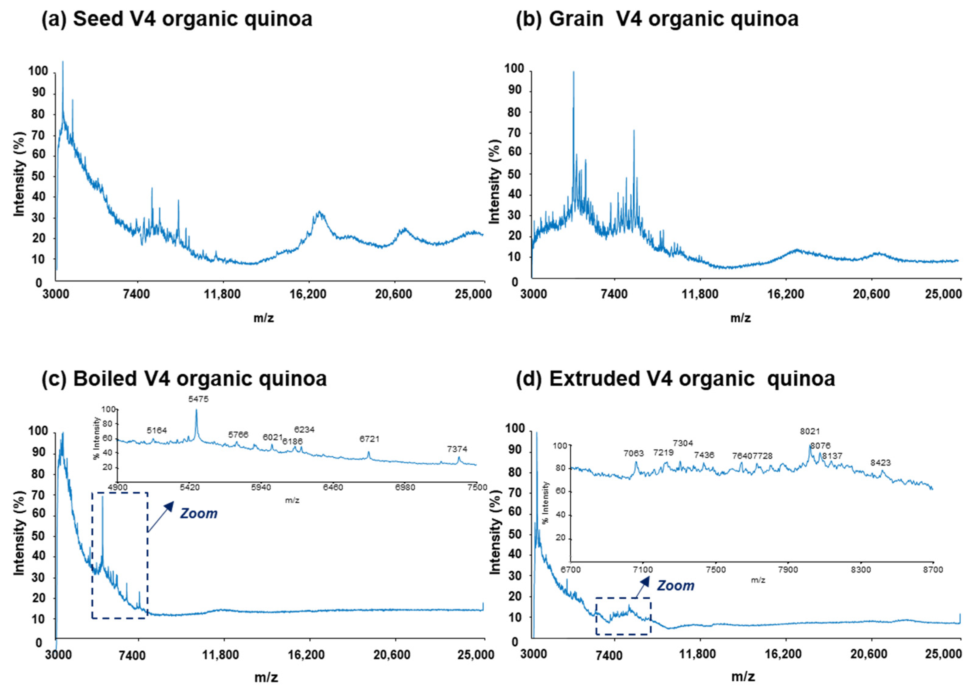
To obtain characteristic mass spectra profiles of protein extracts from quinoa, we employed a reliable and reproducible sample preparation method described in our previous study [31]. This sandwich method, previously used on raw commercial quinoa grains, was applied to the preparation of sample-MALDI matrix mixtures and spot deposition. Figure 1 and Figure 2 present representative mass spectra for the protein extracts of seed, grain, boiled, and extruded V4 quinoa varieties from conventional and organic farming, respectively.
In particular, Figure 1 and Figure 2a,b display the mass spectra of the protein extracts from seeds and grains under both farming conditions. As can be observed, characteristic mass spectra rich in proteins were obtained within the scanned range of 3000 to 25,000 m/z in all cases. Moreover, the differences in mass spectra were more pronounced when comparing seeds and grains for a specific farming type. This could be attributed to the technological treatments applied to quinoa seeds to prepare grains, including scarification and, particularly, washing and drying, aimed at reducing the high levels of saponins.
On the other hand, Figure 1 and Figure 2c,d illustrate the mass spectra of the protein extracts from boiled and extruded quinoa under both farming conditions. As can be observed, both boiling and extrusion treatments led to a reduction in the detected proteins. Indeed, the total amount of protein in these processed quinoa extracts was lower compared to the raw quinoa (i.e., seeds and grains), with, for example, 1.1% and 5.5% (m/m) for extruded and seed V4 quinoa varieties from conventional farming, respectively. Such behaviour can be attributed to the effect of heat and pressure treatments on the reduction in protein solubility, resulting in protein denaturation, oxidation, and aggregation [14,48,49,50].
To generate a reliable large data set for multivariate data analysis, mass spectra were collected in triplicate (n = 288 spots (96 × 3)) for all the protein extracts of seed, grain, boiled, and extruded quinoa samples from the four varieties grown under both conventional and organic farming methods. However, direct peak detection for protein fingerprinting was challenging due to the complexity of the mass spectra, which exhibited numerous overlapped protein peaks with varying intensities. Consequently, we employed MALDIquant software (version 1.19.3) for the efficient quantitative processing of the mass spectra, as described in our previous work [31]. This approach facilitated improved peak detection, reliably providing distinctive protein features with characteristic m/z and intensities. Such accuracy was essential for subsequent multivariate data analysis by PCA and PLS-DA to discriminate between quinoa samples.
Following this data processing strategy, a total of 49 proteins were detected across the different quinoa samples, including the four varieties, the two raw materials (seeds and grains), the two farming conditions (conventional and organic), and the two processing methods (boiling and extrusion). To tentatively identify these proteins based on their Mr, we compared them with the experimental proteome map of the Salcedo samples obtained in a separate study by LC-MS/MS shotgun proteomics [47]. Table 1 lists the experimental Mr calculated for the detected proteins, their theoretical Mr, the accession number (ID), and the names of the 31 tentatively identified proteins out of the 49 detected proteins. Note that in many cases, several possible identifications were provided because the mass accuracy and resolution of the full-scan MALDI-TOF mass spectrometer was not enough for an unequivocal identification.

3.2. Multivariate Data Analysis

3.2.1. Discrimination of Conventional and Organic Quinoa

Multivariate data analysis was carried out, considering the intensities of the 49 detected proteins in the different protein extracts. To simplify data interpretation for differentiating conventional and organic quinoa samples, only the protein fingerprints from the 48 raw samples corresponding to seeds and grains grown under both farming conditions were considered. Initially, unsupervised PCA was employed to visualize trends and identify outliers from the scores plot (Supplementary Figure S1) [28,31]. Two principal components (PCs) explained a total variance of 52.9% (Supplementary Figure S1). Given the absence of distinct trends in the scores plot across samples from the four different white quinoa varieties, even when increasing the number of components, the representation of the samples was solely based on farming conditions. As can be observed, PC1 (33.9% of the explained variance) revealed differential clustering between conventional and organic quinoa samples, while PC2 (19.0% of the explained variance) separated samples within these two groups. Additionally, only two samples corresponding to the V2 quinoa variety from conventional farming appeared outside the 95% confidence ellipse of the scores plot and were identified as outliers, thus excluding them from the supervised PLS-DA analysis.
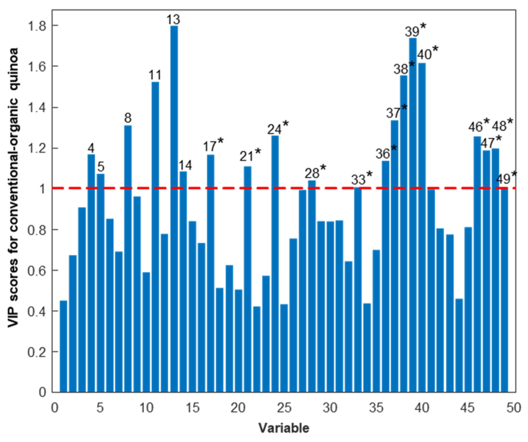
A PLS-DA model considering two classes was established to enhance discrimination and identify the protein variables significantly contributing to the differentiation between quinoa farming conditions. The scores plot of the PLS-DA model, with two latent variables (LVs), accounting for 45.5% of the explained variance and illustrated in Figure 3a, effectively demonstrated discrimination between conventional and organic quinoa samples, suggesting that farming conditions induced differences at the protein level [51]. The loadings plot depicted the contribution of the different protein variables to the LVs (Figure 3b), while the VIP scores provided additional information to reveal the relevant contribution of these variables for discrimination (Figure 4). As shown in Figure 4, 20 of the 49 detected proteins were found to be the most important for discriminating between conventional and organic quinoa (VIP > 1) [44]. The Mr values of this subset of 20 proteins ranged between 5000 and 25,000. Additionally, 14 of these relevant proteins were tentatively identified, as summarized in Table 1. Notably, several of the tentatively identified proteins ranked at the top of VIP values (VIP > 1.5), including protein 38 (VIP value of 1.56), protein 39 (VIP value of 1.74), and protein 40 (VIP value of 1.62), which emerged as primary discriminants between conventional and organic quinoa (see Table 1 for the identities). Overall, these 20 proteins, selected based on their discriminatory potential, could be considered critical markers for discriminating quinoa grown under varying agroecological conditions.

3.2.2. Discrimination of Raw and Processed Quinoa

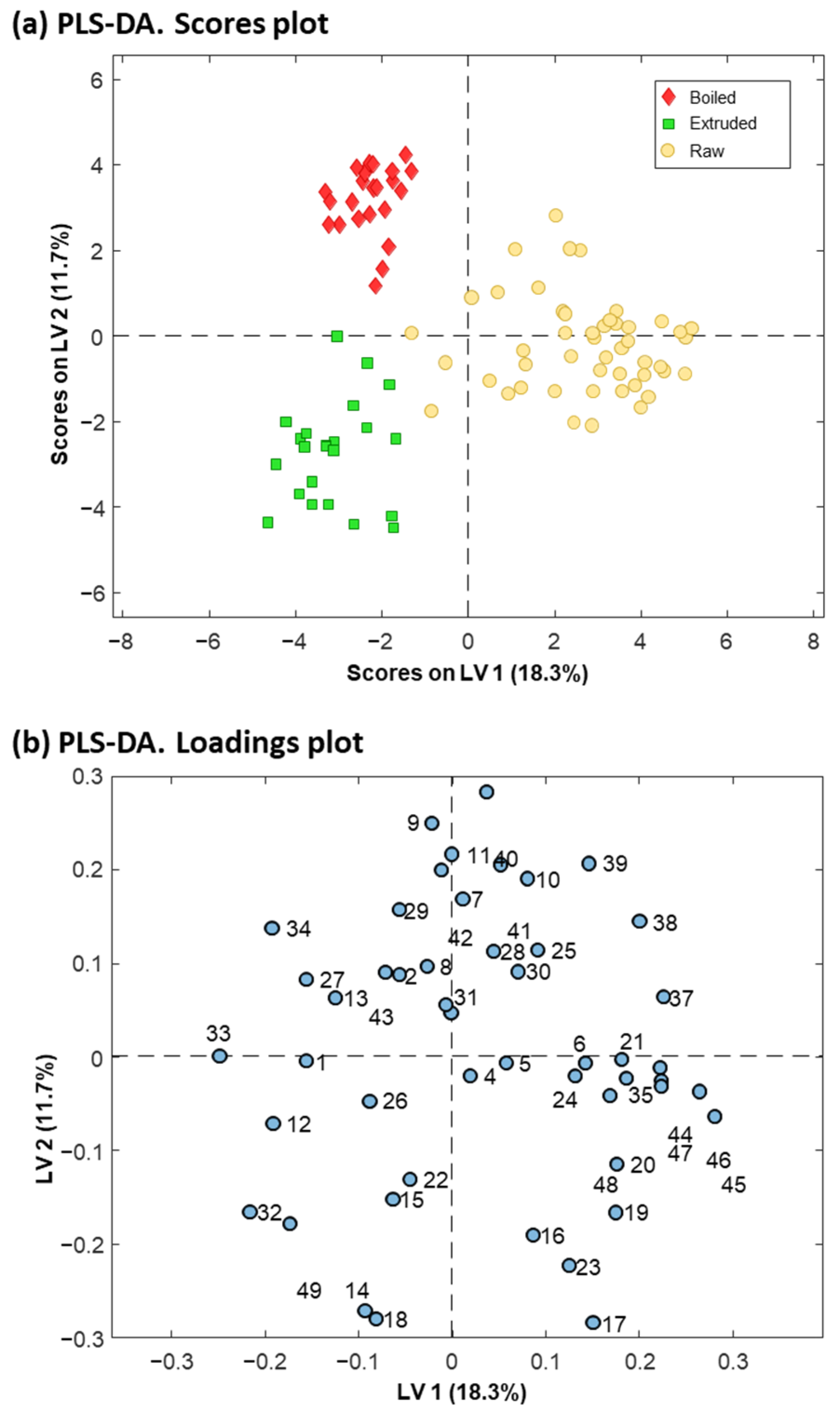
In order to differentiate between raw and processed quinoa, a PCA was conducted considering the intensities of the 49 detected proteins in the different protein extracts of seed, grain, boiled, and extruded quinoa samples from conventional and organic farming. As can be observed in the scores plot of Supplementary Figure S2, two PCs explained a total variance of 38.3%. To focus on the impact of boiling and extrusion on raw quinoa proteins and clarify the evaluation of sample trends and clustering, samples were represented in the score plot without considering the different varieties and farming conditions. PC2 (16.4% of the explained variance) facilitated the separation of boiled and extruded quinoa from grain and seed samples, predominantly located along the positive axis of PC2. Furthermore, PC1 (21.9% of the explained variance) led to a very slight separation between boiled and extruded quinoa samples, while grain and seed samples were distributed and overlapped along the axis of this component. Since no clear clustering was observed for seed and grain samples, we decided to consider raw quinoa samples as a single class for subsequent PLS-DA analysis (Figure 5).
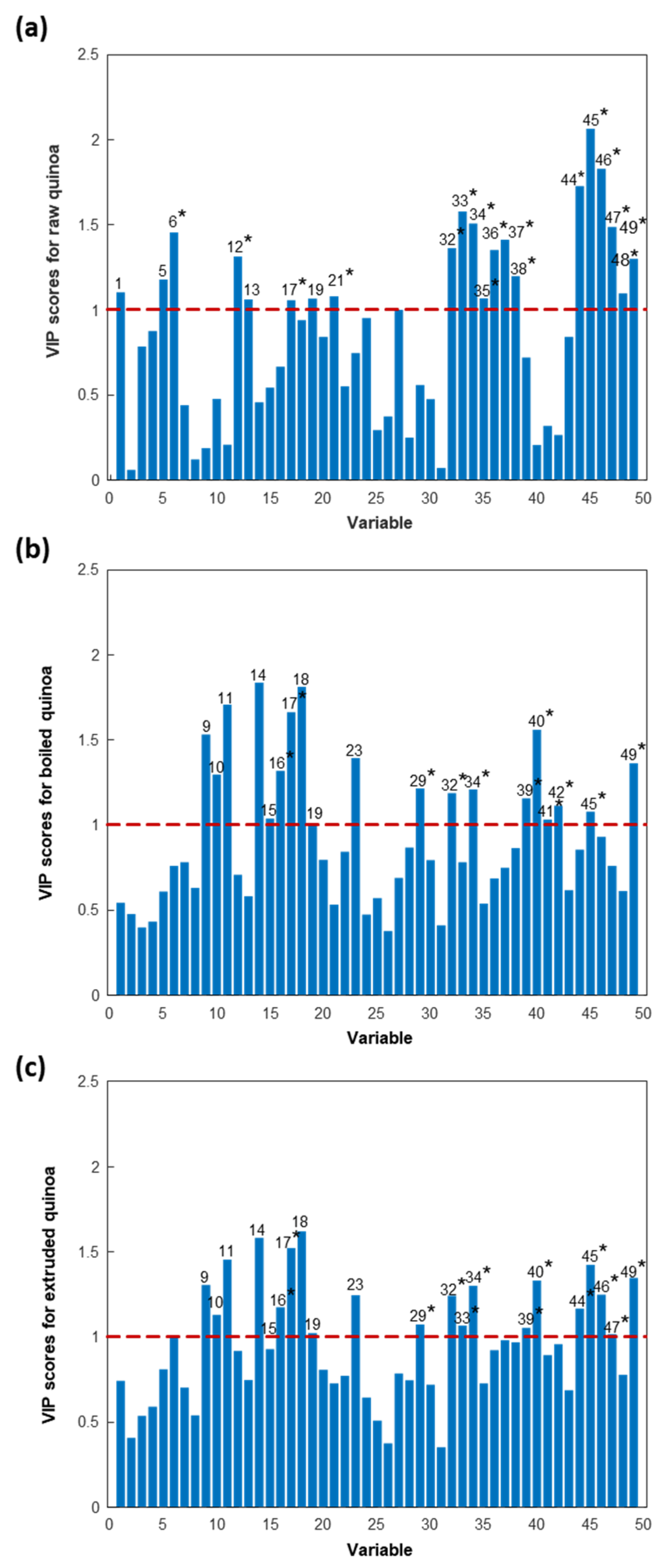
A PLS-DA model considering three classes (i.e., raw (seed and grain), boiled, and extruded quinoa samples) was established for improved discrimination between raw and processed quinoa samples, as well as to identify the most relevant variables for the discrimination. Figure 5a displays the scores plot of the PLS-DA model with two LVs (accounting for 30% of the explained variance), revealing a complete separation with a clear division between boiled, extruded, and raw quinoa samples. This suggested that quinoa processing affected raw quinoa proteins, as well as demonstrating a differential effect of boiling and extrusion. VIP values (Figure 6) were calculated to assess the level of contribution of the different protein variables represented in the loadings plot of Figure 5b for the discrimination of the three classes of quinoa samples.
Figure 6 shows, in each case, the VIP plots for the discrimination of raw, boiled, and extruded quinoa samples from the other two sample classes. Analysing Table 1 and Figure 6, it can be concluded that 38 of the 49 detected proteins were significant for the discrimination of quinoa sample classes (VIP > 1), constituting critical markers of quinoa processing. It is important to note that this protein set included the 20 proteins necessary to distinguish between farming practices. Additionally, 25 of the 38 relevant proteins were tentatively identified (5000 < Mr < 25,000), as summarized in Table 1 and marked with an asterisk in Figure 6. Notably, several of the tentatively identified proteins exhibited high VIP values (VIP > 1.5), underscoring their significance in distinguishing between quinoa processing methods. Specifically, proteins 33 (VIP value of 1.58), 44 (VIP value of 1.72), 45 (VIP value of 2.06), and 46 (VIP value of 1.83) emerged as primary discriminants between raw and boiled/extruded quinoa. Similarly, proteins 17 (VIP value of 1.66) and 40 (VIP value of 1.56) were pivotal in discriminating between boiled and raw/extruded quinoa, while protein 17 (VIP value of 1.52) also played a crucial role in differentiating extruded and raw/boiled quinoa.

4. Conclusions

In this study, we presented a rapid and simple chemometrics-assisted MALDI-TOF-MS method to assess the influence of conventional and organic farming, boiling, and extrusion on protein profiles across various white quinoa grain varieties. Once the raw mass spectra had been acquired appropriately, we employed MALDIquant for data processing, enabling the resolution of complexities within the mass spectra and the reliable detection of proteins. A total of 49 proteins were detected, with 31 tentatively identified. The global fingerprints, comprising the intensity values of these proteins, were subsequently subjected to multivariate data analysis. Our results revealed a PLS-DA model for distinguishing between conventional and organic farming samples, with 20 out of the 49 detected proteins proving critical for differentiation (14 of which were identified). These 20 proteins were also relevant for discriminating between raw and processed samples, which required a total of 38 proteins for an effective differentiation by PLS-DA (25 of which were identified). This global profiling approach allows protein fingerprinting and chemometrics analysis to evaluate differences at the protein level in quinoa grains, facilitating the assessment of farming practices and quality changes during food processing. Further research will be needed to assess the impact of these differences at the nutritional and immunonutritional levels. Additionally, the potential application of the presented approach extends to other areas of food analysis, especially when dealing with complex mass spectra with highly overlapped peaks.

Supplementary Materials

The following supporting information can be downloaded at: https://www.mdpi.com/article/10.3390/foods13121906/s1, Supplementary Figure S1. PCA scores plot derived from the analysis of 48 protein extracts from conventional and organic raw quinoa varieties (seed and grain) using the intensities of the 49 protein peaks detected by MALDIquant.; Supplementary Figure S2. PCA scores plot derived from the analysis of 96 protein extracts from seed, grain, boiled, and extruded quinoa varieties from conventional and organic farming using the intensities of the 49 protein peaks detected by MALDIquant.

Author Contributions

R.G.-L.: Methodology, Investigation, Writing original draft. L.P.: Conceptualization, Supervision, Investigation, Writing—review and editing, F.Q.: Conceptualization, Writing—review and editing. V.S.-N.: Conceptualization, Supervision, Writing—review and editing. F.B.: Conceptualization, Supervision, Writing—review and editing, Funding acquisition. All authors have read and agreed to the published version of the manuscript.

Funding

This study was supported by grant PID2021-127137OB-I00, funded by MCIN/AEI/10.13039/501100011033, and by “ERDF A way of making Europe”. The Bioanalysis group of the University of Barcelona is part of the INSA-UB Maria de Maeztu Unit of Excellence (Grant CEX2021-001234-M) funded by MCIN/AEI/FEDER, UE.

Institutional Review Board Statement

Not applicable.

Informed Consent Statement

Not applicable.

Data Availability Statement

The original contributions presented in the study are included in the article/Supplementary Materials, further inquiries can be directed to the corresponding author.

Acknowledgments

R.G. thanks the Ministry of Education of Peru for a PhD fellowship.

Conflicts of Interest

The authors declare no conflicts of interest.

References

  1. Aloisi, I.; Parrotta, L.; Ruiz, K.B.; Landi, C.; Bini, L.; Cai, G.; Biondi, S.; Del Duca, S. New Insight into Quinoa Seed Quality under Salinity: Changes in Proteomic and Amino Acid Profiles, Phenolic Content, and Antioxidant Activity of Protein Extracts. Front. Plant Sci. 2016, 7, 183977. [Google Scholar] [CrossRef] [PubMed]
  2. Mu, H.; Xue, S.; Sun, Q.; Shi, J.; Zhang, D.; Wang, D.; Wei, J. Research Progress of Quinoa Seeds (Chenopodium quinoa Wild.): Nutritional Components, Technological Treatment, and Application. Foods 2023, 12, 2087. [Google Scholar] [CrossRef] [PubMed]
  3. Chaudhary, N.; Walia, S.; Kumar, R. Functional Composition, Physiological Effect and Agronomy of Future Food Quinoa (Chenopodium Quinoa Willd.): A Review. J. Food Compos. Anal. 2023, 118, 105192. [Google Scholar] [CrossRef]
  4. Angeli, V.; Silva, P.M.; Massuela, D.C.; Khan, M.W.; Hamar, A.; Khajehei, F.; Graeff-Hönninger, S.; Piatti, C. Quinoa (Chenopodium quinoa Willd.): An Overview of the Potentials of the “Golden Grain” and Socio-Economic and Environmental Aspects of Its Cultivation and Marketization. Foods 2020, 9, 216. [Google Scholar] [CrossRef] [PubMed]
  5. Niro, S.; D’Agostino, A.; Fratianni, A.; Cinquanta, L.; Panfili, G. Gluten-Free Alternative Grains: Nutritional Evaluation and Bioactive Compounds. Foods 2019, 8, 208. [Google Scholar] [CrossRef] [PubMed]
  6. Mhada, M.; Metougui, M.L.; El Hazzam, K.; El Kacimi, K.; Yasri, A. Variations of Saponins, Minerals and Total Phenolic Compounds Due to Processing and Cooking of Quinoa (Chenopodium quinoa Willd.) Seeds. Foods 2020, 9, 660. [Google Scholar] [CrossRef] [PubMed]
  7. Hussain, M.I.; Farooq, M.; Syed, Q.A.; Ishaq, A.; Al-Ghamdi, A.A.; Hatamleh, A.A. Botany, Nutritional Value, Phytochemical Composition and Biological Activities of Quinoa. Plants 2021, 10, 2258. [Google Scholar] [CrossRef] [PubMed]
  8. Ceyhun Sezgin, A.; Sanlier, N. A New Generation Plant for the Conventional Cuisine: Quinoa (Chenopodium quinoa Willd.). Trends Food Sci. Technol. 2019, 86, 51–58. [Google Scholar] [CrossRef]
  9. El-Serafy, R.S.; El-Sheshtawy, A.-N.A.; Abd El-Razek, U.A.; Abd El-Hakim, A.F.; Hasham, M.M.A.; Sami, R.; Khojah, E.; Al-Mushhin, A.A.M. Growth, Yield, Quality, and Phytochemical Behavior of Three Cultivars of Quinoa in Response to Moringa and Azolla Extracts under Organic Farming Conditions. Agronomy 2021, 11, 2186. [Google Scholar] [CrossRef]
  10. Gomiero, T. Food Quality Assessment in Organic vs. Conventional Agricultural Produce: Findings and Issues. Appl. Soil Ecol. 2018, 123, 714–728. [Google Scholar] [CrossRef]
  11. Cancino-Espinoza, E.; Vázquez-Rowe, I.; Quispe, I. Organic Quinoa (Chenopodium quinoa L.) Production in Peru: Environmental Hotspots and Food Security Considerations Using Life Cycle Assessment. Sci. Total Environ. 2018, 637–638, 221–232. [Google Scholar] [CrossRef] [PubMed]
  12. FAO; CIRAD. State of the Art Report on Quinoa around the World in 2013; Bazile, D., Bertero, D., Nieto, C., Eds.; FAO: Rome, Italy, 2015; ISBN 978-92-5-108558-5. [Google Scholar]
  13. Huang, R.; Huang, K.; Guan, X.; Li, S.; Cao, H.; Zhang, Y.; Lao, X.; Bao, Y.; Wang, J. Effect of Defatting and Extruding Treatment on the Physicochemical and Storage Properties of Quinoa (Chenopodium quinoa Wild) Flour. LWT 2021, 147, 111612. [Google Scholar] [CrossRef]
  14. Naozuka, J.; Oliveira, P.V. Cooking Effects on Iron and Proteins Content of Beans (Phaseolus vulgaris L.) by GF AAS and MALDI-TOF MS. J. Braz. Chem. Soc. 2012, 23, 156–162. [Google Scholar]
  15. Scanlin, L.; Lewis, K.A. Quinoa as a Sustainable Protein Source: Production, Nutrition, and Processing. In Sustainable Protein Sources; Nadathur, S.R., Wanasundara, J.P.D., Scanlin, L., Eds.; Elsevier Inc.: Amsterdam, The Netherlands, 2017; pp. 223–238. ISBN 9780128027769. [Google Scholar]
  16. Poza-Viejo, L.; Redondo-Nieto, M.; Matías, J.; Granado-Rodríguez, S.; Maestro-Gaitán, I.; Cruz, V.; Olmos, E.; Bolaños, L.; Reguera, M. Shotgun Proteomics of Quinoa Seeds Reveals Chitinases Enrichment under Rainfed Conditions. Sci. Rep. 2023, 13, 4951. [Google Scholar] [CrossRef] [PubMed]
  17. Di Silvestre, D.; Passignani, G.; Rossi, R.; Ciuffo, M.; Turina, M.; Vigani, G.; Mauri, P.L. Presence of a Mitovirus Is Associated with Alteration of the Mitochondrial Proteome, as Revealed by Protein–Protein Interaction (PPI) and Co-Expression Network Models in Chenopodium Quinoa Plants. Biology 2022, 11, 95. [Google Scholar] [CrossRef] [PubMed]
  18. Derbali, W.; Manaa, A.; Spengler, B.; Goussi, R.; Abideen, Z.; Ghezellou, P.; Abdelly, C.; Forreiter, C.; Koyro, H.W. Comparative Proteomic Approach to Study the Salinity Effect on the Growth of Two Contrasting Quinoa Genotypes. Plant Physiol. Biochem. 2021, 163, 215–229. [Google Scholar] [CrossRef] [PubMed]
  19. Rasouli, F.; Kiani-Pouya, A.; Shabala, L.; Li, L.; Tahir, A.; Yu, M.; Hedrich, R.; Chen, Z.; Wilson, R.; Zhang, H.; et al. Salinity Effects on Guard Cell Proteome in Chenopodium Quinoa. Int. J. Mol. Sci. 2021, 22, 428. [Google Scholar] [CrossRef] [PubMed]
  20. Wang, Z.; Wu, Q.; Kamruzzaman, M. Portable NIR Spectroscopy and PLS Based Variable Selection for Adulteration Detection in Quinoa Flour. Food Control 2022, 138, 108970. [Google Scholar] [CrossRef]
  21. Shotts, M.L.; Plans Pujolras, M.; Rossell, C.; Rodriguez-Saona, L. Authentication of Indigenous Flours (Quinoa, Amaranth and Kañiwa) from the Andean Region Using a Portable ATR-Infrared Device in Combination with Pattern Recognition Analysis. J. Cereal Sci. 2018, 82, 65–72. [Google Scholar] [CrossRef]
  22. Rodríguez, S.D.; Rolandelli, G.; Buera, M.P. Detection of Quinoa Flour Adulteration by Means of FT-MIR Spectroscopy Combined with Chemometric Methods. Food Chem. 2019, 274, 392–401. [Google Scholar] [CrossRef]
  23. Xue, S.S.; Tan, J.; Xie, J.Y.; Li, M.F. Rapid, Simultaneous and Non-Destructive Determination of Maize Flour and Soybean Flour Adulterated in Quinoa Flour by Front-Face Synchronous Fluorescence Spectroscopy. Food Control 2021, 130, 108329. [Google Scholar] [CrossRef]
  24. Kelis Cardoso, V.G.; Poppi, R.J. Cleaner and Faster Method to Detect Adulteration in Cassava Starch Using Raman Spectroscopy and One-Class Support Vector Machine. Food Control 2021, 125, 107917. [Google Scholar] [CrossRef]
  25. Ellis, D.I.; Brewster, V.L.; Dunn, W.B.; Allwood, J.W.; Golovanov, A.P.; Goodacre, R. Fingerprinting Food: Current Technologies for the Detection of Food Adulteration and Contamination. Chem. Soc. Rev. 2012, 41, 5706–5727. [Google Scholar] [CrossRef] [PubMed]
  26. Bansal, S.; Singh, A.; Mangal, M.; Mangal, A.K.; Kumar, S. Food Adulteration: Sources, Health Risks, and Detection Methods. Crit. Rev. Food Sci. Nutr. 2017, 57, 1174–1189. [Google Scholar] [CrossRef] [PubMed]
  27. Yang, X.; Xing, B.; Guo, Y.; Wang, S.; Guo, H.; Qin, P.; Hou, C.; Ren, G. Rapid, Accurate and Simply-Operated Determination of Laboratory-Made Adulteration of Quinoa Flour with Rice Flour and Wheat Flour by Headspace Gas Chromatography-Ion Mobility Spectrometry. LWT 2022, 167, 113814. [Google Scholar] [CrossRef]
  28. Galindo-Luján, R.; Pont, L.; Sanz-Nebot, V.; Benavente, F. Classification of Quinoa Varieties Based on Protein Fingerprinting by Capillary Electrophoresis with Ultraviolet Absorption Diode Array Detection and Advanced Chemometrics. Food Chem. 2021, 341, 128207. [Google Scholar] [CrossRef] [PubMed]
  29. Galindo-Luján, R.; Caballero-Alcazar, N.; Pont, L.; Sanz-Nebot, V.; Benavente, F. Fingerprinting of Quinoa Grain Protein Extracts by Liquid Chromatography with Spectrophotometric Detection for Chemometrics Discrimination. LWT 2023, 187, 115289. [Google Scholar] [CrossRef]
  30. Galindo-Luján, R.; Pont, L.; Minic, Z.; Berezovski, M.V.; Sanz-Nebot, V.; Benavente, F. Characterization and Differentiation of Quinoa Seed Proteomes by Label-Free Mass Spectrometry-Based Shotgun Proteomics. Food Chem. 2021, 363, 130250. [Google Scholar] [CrossRef] [PubMed]
  31. Galindo-Luján, R.; Pont, L.; Sanz-Nebot, V.; Benavente, F. Protein Profiling and Classification of Commercial Quinoa Grains by MALDI-TOF-MS and Chemometrics. Food Chem. 2023, 398, 133895. [Google Scholar] [CrossRef] [PubMed]
  32. Galindo-Luján, R.; Pont, L.; Sanz-Nebot, V.; Benavente, F. A Proteomics Data Mining Strategy for the Identification of Quinoa Grain Proteins with Potential Immunonutritional Bioactivities. Foods 2023, 12, 390. [Google Scholar] [CrossRef]
  33. R Development Core Team: R: A Language and Environment for Statistical Computing R Foundation for Statistical Computing. Available online: http://www.r-project.org/ (accessed on 1 January 2024).
  34. Gibb, S. MALDIquantForeign: Import/Export Routines for MALDIquant. 2014; pp. 1–7. Available online: https://cran.r-project.org/package=MALDIquantForeign (accessed on 1 January 2024).
  35. Gibb, S.; Strimmer, K. Mass Spectrometry Analysis Using MALDIquant. In Statistical Analysis of Proteomics, Metabolomics, and Lipidomics Data Using Mass Spectrometry; Springer: Cham, Switzerland, 2017; pp. 101–124. [Google Scholar]
  36. Purohit, P.V.; Rocke, D.M. Discriminant Models for High-Throughput Proteomics Mass Spectrometer Data. Proteomics 2003, 3, 1699–1703. [Google Scholar] [CrossRef]
  37. Savitzky, A.; Golay, M.J.E. Smoothing and Differentiation of Data by Simplified Least Squares Procedures. Anal. Chem. 1964, 36, 1639–1643. [Google Scholar] [CrossRef]
  38. Ryan, C.G.; Clayton, E.; Griffin, W.L.; Sie, S.H.; Cousens, D.R. SNIP, a Statistics-Sensitive Background Treatment for the Quantitative Analysis of PIXE Spectra in Geoscience Applications. Nucl. Instrum. Methods Phys. Res. B 1988, 34, 396–402. [Google Scholar] [CrossRef]
  39. Borgaonkar, S.P.; Hocker, H.; Shin, H.; Markey, M.K. Comparison of Normalization Methods for the Identification of Biomarkers Using MALDI-TOF and SELDI-TOF Mass Spectra. OMICS 2010, 14, 115–126. [Google Scholar] [CrossRef]
  40. Cleveland, W.S. Robust Locally Weighted Regression and Smoothing Scatterplots. J. Am. Stat. Assoc. 1979, 74, 829–836. [Google Scholar] [CrossRef]
  41. Friedman, J.H. A Variable Span Smoother. Laboratory for Computational Statistics, Stanford University Technical Report No. 5. J. Am. Stat. Assoc. 1984, 5, 1–32. [Google Scholar] [CrossRef]
  42. Barker, M.; Rayens, W. Partial Least Squares for Discrimination. J. Chemom. 2003, 17, 166–173. [Google Scholar] [CrossRef]
  43. Ballabio, D.; Consonni, V. Classification Tools in Chemistry. Part 1: Linear Models. PLS-DA. Anal. Methods 2013, 5, 3790–3798. [Google Scholar] [CrossRef]
  44. Wold, S.; Sjöström, M.; Eriksson, L. PLS-Regression: A Basic Tool of Chemometrics. Chemom. Intell. Lab. Syst. 2001, 58, 109–130. [Google Scholar] [CrossRef]
  45. Worley, B.; Halouska, S.; Powers, R. Utilities for Quantifying Separation in PCA/PLS-DA Scores Plots. Anal. Biochem. 2013, 433, 102–104. [Google Scholar] [CrossRef]
  46. Mehmood, T.; Liland, K.H.; Snipen, L.; Sæbø, S. A Review of Variable Selection Methods in Partial Least Squares Regression. Chemom. Intell. Lab. Syst. 2012, 118, 62–69. [Google Scholar] [CrossRef]
  47. Galindo-Luján, R.; Pont, L.; Minic, Z.; Berezovski, M.V.; Quispe, F.; Sanz-Nebot, V.; Benavente, F. Comprehensive Characterization of Raw and Processed Quinoa from Conventional and Organic Farming by Label-Free Shotgun Proteomics. 2024. Manuscript submitted for publication. Available online: https://ssrn.com/abstract=4774018 (accessed on 27 March 2024).
  48. Soladoye, O.P.; Juárez, M.L.; Aalhus, J.L.; Shand, P.; Estévez, M. Protein Oxidation in Processed Meat: Mechanisms and Potential Implications on Human Health. Compr. Rev. Food Sci. Food Saf. 2015, 14, 106–122. [Google Scholar] [CrossRef] [PubMed]
  49. Santé-Lhoutellier, V.; Astruc, T.; Marinova, P.; Greve, E.; Gatellier, P. Effect of Meat Cooking on Physicochemical State and in Vitro Digestibility of Myofibrillar Proteins. J. Agric. Food Chem. 2008, 56, 1488–1494. [Google Scholar] [CrossRef] [PubMed]
  50. Tang, H.; Fu, T.; Feng, Y.; Zhang, S.; Wang, C.; Zhang, D. Effect of Heat Treatment on Solubility, Surface Hydrophobicity and Structure of Rice Bran Albumin and Globulin. Qual. Assur. Saf. Crops Foods 2019, 11, 499–509. [Google Scholar] [CrossRef]
  51. Xiao, R.; Li, L.; Ma, Y. A Label-Free Proteomic Approach Differentiates between Conventional and Organic Rice. J. Food Compos. Anal. 2019, 80, 51–61. [Google Scholar] [CrossRef]
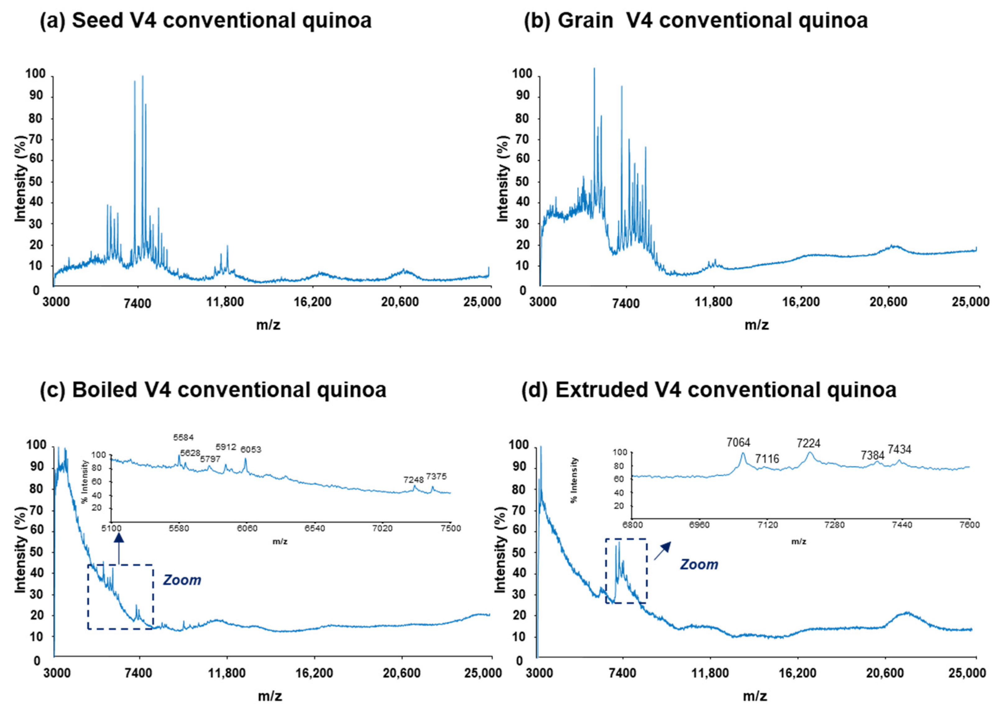
Figure 1. Raw MALDI-TOF mass spectra for the protein extracts of (a) seed, (b) grain, (c) boiled, and (d) extruded V4 quinoa varieties from conventional farming (V4 = Salcedo). Different regions of the mass spectra are zoomed in for boiled and extruded quinoa.
Foods 13 01906 g001
Figure 2. Raw MALDI-TOF mass spectra for the protein extracts of (a) seed, (b) grain, (c) boiled, and (d) extruded V4 quinoa varieties from organic farming (V4 = Salcedo). Different regions of the mass spectra are zoomed in for boiled and extruded quinoa.
Foods 13 01906 g002
Figure 3. PLS−DA scores plot (a) and loadings plot (b) derived from the analysis of 46 protein extracts from conventional and organic raw quinoa varieties (seed and grain) using the intensities of the 49 protein peaks detected by MALDIquant.
Foods 13 01906 g003
Figure 4. VIP scores of the different protein variables when considering the separation between conventional and organic raw quinoa sample classes. Protein variables with a VIP score greater than one are numbered, and those tentatively identified are marked with an asterisk (*) (as in Table 1).
Foods 13 01906 g004
Figure 5. PLS−DA scores plot (a) and loadings plot (b) derived from the analysis of 96 protein extracts from seed, grain, boiled, and extruded quinoa varieties from conventional and organic farming using the intensities of the 49 protein peaks detected by MALDIquant.
Foods 13 01906 g005
Figure 6. VIP scores of the different protein variables when considering the separation of (a) raw (seed and grain), (b) boiled, and (c) extruded quinoa sample classes from the other two sample classes. Protein variables with a VIP score greater than one are numbered, and those tentatively identified are marked with an asterisk (*) (as in Table 1).
Foods 13 01906 g006
Table 1. List of proteins detected by MALDI-TOF-MS used as variables for multivariate data analysis with their corresponding experimental Mr, PLS-DA VIP score values for discrimination between farming and processing conditions, and tentative identifications. Theoretical Mr, accession number (ID), and protein name were based on an experimental proteome map of the Salcedo samples obtained in a separate study by LC-MS/MS shotgun proteomics [47].
Multivariate Data Analysis Protein Variables aTentative Identifications b
ProteinExperimental Mr cPLS-DA VIP Scores dTheoretical MrAccession Number (ID) e and Protein Name
FarmingProcessing
Craw fOraw fRawBoiledExtruded
142310.450.451.100.540.74 -
244450.670.670.060.480.41 -
346500.910.910.780.400.53 -
449511.171.170.870.430.59 -
551801.071.071.180.610.81 -
653040.850.851.45 *0.761.005307XP_021736943.1 wound-induced basic protein-like
754600.690.690.440.780.70 -
855871.311.310.120.630.54 -
957670.960.960.191.531.30 -
1059340.590.590.471.291.13 -
1161841.521.520.201.701.45 -
1263910.780.781.31 *0.700.916413XP_021764391.1 40S ribosomal protein S29
1368181.801.801.060.580.75 -
1470631.081.080.461.841.58 -
1574350.840.840.541.040.93
1677300.730.730.671.32 *1.17 *7747XP_021714409.1 uncharacterized protein LOC110682385
1779831.17 *1.17 *1.06 *1.66 *1.52 *7974XP_021773921.1 metallothionein-like protein 4B
1882210.510.510.941.811.62 -
1984650.620.621.061.011.02 -
2086350.500.500.840.790.81 -
2188401.11 *1.11 *1.08 *0.530.738806XP_021718956.1 protein DELETION OF SUV3 SUPPRESSOR 1(I)-like
8814XP_021729007.1 uncharacterized protein LOC110696048
8823XP_021762602.1 defensin-like protein
2290540.420.420.550.840.779076XP_021768105.1 late seed maturation protein P8B6-like
2393140.570.570.741.391.24 -
2496951.26 *1.26 *0.950.470.649692XP_021747091.1 sm-like protein LSM5
2510,1060.430.430.290.570.51 -
2610,7030.750.750.370.370.3710,689XP_021714810.1 uncharacterized protein LOC110682782
10,724XP_021756490.1 sm-like protein LSM8
10,736XP_021756753.1 60S ribosomal protein L37-3
10,750XP_021768220.1 sm-like protein LSM7
2710,9470.990.991.000.690.7910,920XP_021761138.1 mitochondrial import inner membrane translocase subunit Tim9
10,928XP_021746329.1 probable steroid-binding protein 3
10,989XP_021761862.1 peamaclein-like
2811,2741.04 *1.04 *0.250.860.7511,270XP_021771595.1 sm-like protein LSM3A
11,308XP_021716413.1 60S acidic ribosomal protein P2-4-like
2911,4890.840.840.561.21 *1.07 *11,449XP_021727941.1 NADH dehydrogenase
11,458XP_021766637.1 non-specific lipid-transfer protein-like
11,517XP_021754863.1 thioredoxin M-type, chloroplastic-like isoform X2
3011,7790.840.840.470.790.7211,723XP_021772578.1 RNA polymerase II transcriptional coactivator KIWI-like isoform X1
11,772XP_021755694.1 uncharacterized protein LOC110720913
11,797XP_021763483.1 small ubiquitin-related modifier 1-like
3112,0430.840.840.070.410.3511,992YP_009380236.1 ribosomal protein S18 (chloroplast)
12,050XP_021776279.1 peptidyl-prolyl cis-trans isomerase FKBP12-like
12,055XP_021765385.1 NADH dehydrogenase
12,064XP_021774292.1 huntingtin-interacting protein K-like
3212,3620.640.641.36 *1.19 *1.24 *12,301XP_021760438.1 gibberellin-regulated protein 9-like
12,315XP_021716755.1 uncharacterized protein At2g27730, mitochondrial-like
12,332XP_021765334.1 V-type proton ATPase subunit G 1-like
12,375XP_021720641.1 60S ribosomal protein L30
12,407XP_021761775.1 uncharacterized protein LOC110726608
12,413XP_021773050.1 60S ribosomal protein L36-2-like
12,420XP_021738644.1 40S ribosomal protein S25-like
3312,8011.01 *1.01 *1.58 *0.781.06 *12,827XP_021769120.1 60S ribosomal protein L35a-3
12,849XP_021757241.1 nodulin-related protein 1-like
12,855XP_021718430.1 nodulin-related protein 1-like
3413,2200.430.431.50 *1.21 *1.30 *13,205XP_021758336.1 thioredoxin H-type 1-like
13,231XP_021759897.1 thioredoxin H-type 1-like
3516,2150.700.701.06 *0.540.7216,134XP_021716351.1 ferredoxin, root R-B2-like
16,149XP_021747601.1 uncharacterized protein LOC110713466
16,165XP_021730244.1 outer envelope pore protein 16-2, chloroplastic-like isoform X2
16,200XP_021717733.1 high mobility group B protein 3-like
16,215XP_021716749.1 ferredoxin, root R-B2-like
16,216XP_021754488.1 high mobility group B protein 3-like
16,239XP_021762815.1 uncharacterized protein At5g48480-like
16,250XP_021766528.1 40S ribosomal protein S14-2
16,289XP_021721762.1 oleosin 1-like
3616,5141.13 *1.13 *1.35 *0.680.9216,458XP_021733518.1 uncharacterized protein At5g48480-like
16,464XP_021761922.1 uncharacterized protein LOC110726743
16,469XP_021746531.1 60S ribosomal protein L27a-3-like
16,474XP_021769235.1 glycine cleavage system H protein 2, mitochondrial-like
16,524XP_021768671.1 60S ribosomal protein L27a-3-like
16,568XP_021762909.1 uncharacterized protein LOC110727639
16,570XP_021732568.1 uncharacterized protein LOC110699354
3716,6931.34 *1.34 *1.41 *0.750.9816,616XP_021755504.1 2S albumin-like
16,624XP_021751394.1 60S ribosomal protein L26-1
16,625XP_021730224.1 probable calcium-binding protein CML13
16,636XP_021760375.1 eukaryotic translation initiation factor 1A
16,651XP_021731588.1 glycine-rich RNA-binding, abscisic acid-inducible protein-like
16,685XP_021735190.1 ubiquitin-conjugating enzyme E2 variant 1D-like
16,693XP_021774210.1 60S ribosomal protein L28-1-like
16,702XP_021717270.1 blue copper protein-like isoform X2
16,742XP_021720407.1 17.4 kDa class III heat shock protein-like
16,758XP_021766054.1 uncharacterized protein LOC110730552
3816,8971.56 *1.56 *1.20 *0.860.9716,833XP_021733717.1 40S ribosomal protein S16-like
16,834XP_021776507.1 calmodulin-7-like
16,860XP_021754554.1 calmodulin
16,877XP_021749775.1 peptidyl-prolyl cis-trans isomerase FKBP15-1-like
16,884XP_021716580.1 17.4 kDa class III heat shock protein-like
16,933XP_021735458.1 probable prefoldin subunit 5
16,942XP_021731073.1 thiosulfate sulfurtransferase 16, chloroplastic-like isoform X2
16,946XP_021743153.1 uncharacterized protein LOC110709246
16,962XP_021758167.1 transcription initiation factor TFIID subunit 15b-like
3917,1011.74 *1.74 *0.721.16 *1.05 *17,026XP_021751891.1 NADH dehydrogenase
17,040XP_021771944.1 DNA-directed RNA polymerases II, IV and V subunit 8B-like
17,048XP_021739940.1 uncharacterized protein LOC110706342
17,111XP_021765383.1 40S ribosomal protein S13-like
17,129XP_021766190.1 uncharacterized protein LOC110730679
17,131XP_021740721.1 MLP-like protein 423
17,143XP_021769150.1 17.8 kDa class I heat shock protein-like
4017,3261.62 *1.62 *0.201.56 *1.33 *17,290XP_021770408.1 outer envelope pore protein 16-3, chloroplastic/mitochondrial-like
17,301XP_021729636.1 NADH dehydrogenase
17,330XP_021717756.1 uncharacterized protein LOC110685525
17,340XP_021747441.1 eukaryotic translation initiation factor 5A-4-like
17,350XP_021764293.1 40S ribosomal protein S15-4-like
17,355YP_009380273.1 ribosomal protein S7 (chloroplast)
17,366XP_021747435.1 eukaryotic translation initiation factor 5A-like
17,376XP_021720177.1 ubiquitin-NEDD8-like protein RUB2
17,385XP_021748235.1 60S ribosomal protein L23A
4117,6171.00*1.00*0.321.03 *0.8917,532XP_021765685.1 glycine cleavage system H protein, mitochondrial
17,543XP_021768154.1 glycine cleavage system H protein, mitochondrial-like
17,560XP_021731505.1 oleosin 1-like
17,562XP_021736891.1 peroxiredoxin-2B-like
17,572XP_021732018.1 peroxiredoxin-2B-like
17,592XP_021756471.1 putative 4-hydroxy-4-methyl-2-oxoglutarate aldolase 3
17,604XP_021735589.1 nascent polypeptide-associated complex subunit beta-like
17,622XP_021733122.1 protein mago nashi homolog 2
17,652XP_021743932.1 histidine-containing phosphotransfer protein 1-like
17,665XP_021753630.1 uncharacterized protein LOC110719020
17,675XP_021759953.1 nascent polypeptide-associated complex subunit beta-like
17,700XP_021745442.1 40S ribosomal protein S11-3
4217,8790.800.800.261.11 *0.9617,803XP_021769395.1 40S ribosomal protein S11-like
17,855XP_021773311.1 60S ribosomal protein L12-1
17,916XP_021748317.1 desiccation protectant protein Lea14 homolog
17,939XP_021749487.1 MLP-like protein 43
17,969XP_021715429.1 universal stress protein PHOS34-like
4318,3110.770.770.840.620.6918,221XP_021738830.1 oleosin 16 kDa
18,224XP_021737967.1 MFP1 attachment factor 1-like
18,238XP_021765145.1 60S ribosomal protein L24-like
18,240XP_021753128.1 peptidyl-prolyl cis-trans isomerase 1-like
18,252XP_021763237.1 pathogenesis-related protein STH-21-like
18,254XP_021775867.1 peptidyl-prolyl cis-trans isomerase 1
18,258XP_021769094.1 18.3 kDa class I heat shock protein-like
18,271XP_021730326.1 universal stress protein PHOS32
18,276XP_021744114.1 17.3 kDa class II heat shock protein-like
18,317XP_021752091.1 probable NADH dehydrogenase
18,348XP_021738936.1 17.3 kDa class II heat shock protein-like
18,348XP_021732306.1 pathogenesis-related protein STH-21-like
18,348XP_021725562.1 deoxyuridine 5-triphosphate nucleotidohydrolase
18,366XP_021774711.1 50S ribosomal protein L18, chloroplastic
4420,3490.460.461.72 *0.851.16 *20,301XP_021763546.1 30S ribosomal protein 3, chloroplastic
20,406XP_021734303.1 HMG-Y-related protein A-like
4520,5560.810.812.06 *1.08 *1.42 *20,466XP_021727144.1 21 kDa seed protein-like
20,499XP_021763320.1 photosystem II reaction center Psb28 protein-like
20,522XP_021744010.1 succinate dehydrogenase assembly factor 2, mitochondrial-like
20,523XP_021729294.1 uncharacterized protein LOC110696308
20,557XP_021766022.1 PLAT domain-containing protein 3-like
20,565XP_021741243.1 putative H/ACA ribonucleoprotein complex subunit 1-like protein 1
20,592XP_021769990.1 ADP-ribosylation factor 1-like
20,619XP_021752903.1 thioredoxin-like protein CITRX, chloroplastic
4620,7801.25 *1.25 *1.83 *0.931.25 *20,736XP_021773813.1 adenylate kinase isoenzyme 6 homolog
20,739XP_021740322.1 protein CutA, chloroplastic-like
20,778XP_021761077.1 peroxiredoxin-2F, mitochondrial-like isoform X1
20,799XP_021753718.1 60S ribosomal protein L11-1
20,801XP_021772257.1 HMG-Y-related protein A-like
20,844XP_021763208.1 60S ribosomal protein L18-3-like
20,848XP_021738998.1 protein OPI10 homolog
20,852XP_021763370.1 monothiol glutaredoxin-S10-like
4721,0751.19 *1.19 *1.48 *0.761.01 *21,027XP_021750037.1 uncharacterized protein LOC110715738
21,031XP_021730777.1 thioredoxin O2, mitochondrial-like isoform X2
21,055XP_021766443.1 lactoylglutathione lyase isoform X2
21,077XP_021756715.1 uncharacterized protein LOC110721825
21,107XP_021727997.1 50S ribosomal protein L27, chloroplastic
21,121XP_021733985.1 glycine-rich RNA-binding protein 3, mitochondrial-like
21,149XP_021736893.1 probable inactive nicotinamidase At3g16190
21,170XP_021763161.1 uncharacterized protein Os08g0359500-like
21,172XP_021732021.1 probable inactive nicotinamidase At3g16190
4821,3431.20 *1.20 *1.09 *0.610.7821,241XP_021720070.1 ankyrin repeat and SAM domain-containing protein 6-like isoform X2
21,268XP_021771518.1 uncharacterized protein LOC110735639
21,294XP_021764214.1 cyclic phosphodiesterase-like
21,326XP_021763910.1 60S ribosomal protein L18a-2
21,364XP_021754795.1 50S ribosomal protein L24, chloroplastic-like
21,376XP_021718085.1 60S ribosomal protein L18a
21,433XP_021730369.1 probable prefoldin subunit 3
4922,0791.01 *1.01 *1.30 *1.36*1.34 *21,984XP_021772119.1 RNA-binding protein Y14-like
22,018XP_021763572.1 40S ribosomal protein S7-like
22,034XP_021761714.1 histone H1-like
22,040YP_009380239.1 ClpP (chloroplast)
22,088XP_021766393.1 50S ribosomal protein L9, chloroplastic-like
a PLS−DA variables correspond to the protein peaks detected by MALDIquant. b The experimental proteome map of the same samples obtained in a separate study by LC-MS/MS shotgun proteomics [47] was used as a reference for the tentative identification. A mass error ±0.5% between the theoretical and experimental Mr was considered acceptable for proposing an identity. This threshold value was established considering the mass error observed for the analysis of a ribonuclease A standard (from a bovine pancreas) under the same instrumental conditions, Mr = 13,690). c Experimental Mr were calculated from the m/z values considering the formation of single-charged molecular ions by MALDI-TOF-MS. d VIP scores > 1 were considered important for discrimination and are marked in bold, and tentatively identified scores were marked with an asterisk (*). e Accession numbers (IDs) of the identified proteins correspond to the IDs of the indicated LC-MS/MS shotgun proteomics work [47]. The tentatively identified proteins fulfilling the acceptance criterium are ordered by the Andromeda score values obtained by LC-MS/MS, which are a measure of the reliability of their identification. f Craw and Oraw quinoa correspond to raw (seeds and grains) quinoa from conventional and organic farming, respectively.
Disclaimer/Publisher’s Note: The statements, opinions and data contained in all publications are solely those of the individual author(s) and contributor(s) and not of MDPI and/or the editor(s). MDPI and/or the editor(s) disclaim responsibility for any injury to people or property resulting from any ideas, methods, instructions or products referred to in the content.