Article

Evaluation of Nano-Wall Material for Production of Novel Lyophilized-Probiotic Product

1
Bioproduct Science Program, Department of Science, Faculty of Liberal Arts and Science, Kasetsart University, Kamphaeng Saen Campus, Nakhon Pathom 73140, Thailand
2
College of Medicine and Public Health, Ubon Ratchathani University, Ubon Ratchathani 34190, Thailand
3
Division of Microbiology, Department of Science, Faculty of Liberal Arts and Science, Kasetsart University, Kamphaeng Saen Campus, Nakhon Pathom 73140, Thailand
*
Author to whom correspondence should be addressed.
Foods 2022, 11(19), 3113; https://doi.org/10.3390/foods11193113
Submission received: 12 August 2022 / Revised: 27 September 2022 / Accepted: 2 October 2022 / Published: 6 October 2022
(This article belongs to the Section Food Biotechnology)

Abstract

:
Lyophilization is one of the most used methods for bacterial preservation. In this process, the cryoprotectant not only largely decreases cellular damage but also plays an important part in the conservation of viability during freeze-drying. This study investigated using cryoprotectant and a mixture of the cryoprotectant to maintain probiotic activity. Seven probiotic strains were considered: (Limosilactobacillus reuteri KUKPS6103; Lacticaseibacillus rhamnosus KUKPS6007; Lacticaseibacillus paracasei KUKPS6201; Lactobacillus acidophilus KUKPS6107; Ligilactobacillus salivarius KUKPS6202; Bacillus coagulans KPSTF02; Saccharomyces cerevisiae subsp. boulardii KUKPS6005) for the production of a multi-strain probiotic and the complex medium for the lyophilized synbiotic production. Cholesterol removal, antioxidant activity, biofilm formation and gamma aminobutyric acid (GABA) production of the probiotic strains were analyzed. The most biofilm formation occurred in L. reuteri KUKPS6103 and the least in B. coagulans KPSTF02. The multi-strain probiotic had the highest cholesterol removal. All the probiotic strains had GABA production that matched the standard of γ-aminobutyric acid. The lyophilized synbiotic product containing complex medium as a cryoprotectant and wall material retained a high viability of 7.53 × 10⁸ CFU/g (8.89 log CFU/g) after 8 weeks of storage. We found that the survival rate of the multi-strain probiotic after freeze-drying was 15.37% in the presence of complex medium that was used as high performing wall material. Our findings provided a new type of wall material that is safer and more effective and, can be extensively applied in relevant food applications.

Graphical Abstract

1. Introduction

Gums are the natural dripples from the various parts of plants, especially the main trunk. When the stem is damaged (accidentally or purposely) by tapping, the injuries and beetle penetration result in the gum exuding from the bark. Gum arabic (GA) was noted “as a dried excretion got from the parts of the tree Acacia senegal (L. Willdenow) or related species of Acacia”, as defined by Joint FAO/WHO Expert Committee of Food Additives [1]. GA is non-sticky soluble fibers and is edible when sourced from, for example, dried gum, its exudates from the trunks and branches of A. senegal and A. seyal [2,3,4]. GA is widely used as it has excellent emulsifying properties, is non-toxic, and has low viscosity when compared to other polysaccharides. An emulsifier applied from GA is used in the pharmaceutical industry and in food plants. In addition, it is used as a stabilizer and suspending agent for insoluble drugs [5]. GA is the best emulsifier and stabilizer due to the binding of high water-solubility and low viscosity. Acacia gums are film-forming agents, stabilizers, thickeners, flocculants, emulsifiers and suspending agents. GA is an excellent thickener and suspender. These criteria underpin the standard and quality of GA utilized in the food, drug and cosmetic sectors [6]. Some scientists have studied the ability of GA as a nutrient for probiotic strains as it is fermentable fiber [7].
Probiotics are normal indigenous flora in the gastrointestinal tract, which can be prepared into various kinds of products. A probiotic is defined as “live microorganisms when it is administered in adequate amount gives a health benefit on the host” [8]. Saccharomyces cerevisiae and Escherichia coli microorganisms have also been used as probiotics, and Lactobacillus and Bifidobacterium species are commonly used as probiotics [9,10]. However, the strains used as probiotics are mainly in the group of lactic acid microorganisms. The benefits of a probiotic strain are explored in different kinds of foods. Before utilization, probiotic strains must survive and maintain their function during storage. Probiotic strains can survive storage a long time in a lyophilized product [11]. In the freeze-drying method, the solvent is freeze-dried and separated using sublimation. During the process, both the microbial cell wall and membrane can be injured by the transition of the membrane phase [11]. To avoid such issues, a protective wall agent is used to ensure the survivability of probiotic strains during the lyophilization process and storage [12].
There is pressure to increase the shelf life of drug products and foods. Thus, it is important to take care of their storage characteristics. The key element causing deterioration in the quality of merchandise is the water that is included in foods and pharmaceutical products. Thus, to eliminate the water from food and medicinal goods, a suitable drying technique should be used. Lyophilization, spray drying, and reduced-pressure drying are the most well-known drying techniques [13,14,15,16]. Lyophilization or freeze-drying has been investigated and used for over one thousand years to preserve biological cells [17]. Accordingly, the process of freeze-drying is frequently employed to produce dried viable probiotic products [18,19]. The aims of the present study were to assess the potential of probiotic strains and to produce lyophilized multi-strain probiotic product prior to investigation of the changes in the presence of protective wall materials during lyophilization and storage. GA, mung bean extract, stingless-bee honey and Riceberry extract were used as protective wall materials to assess their impact on the stability and viability of probiotic cells after freeze-drying and storage.

2. Materials and Methods

2.1. Materials

Gum arabic, mung bean, stingless-bee honey and a rice cultivar “Riceberry” were purchased from a local market in Thailand, which was certified according to Good Manufacturing Practice (GMP), Hazard Analysis and Critical Control Point (HACCP) and Good Agricultural Practice (GAP).

2.2. Growth of Probiotic Strains and Culture Conditions

Probiotic strains (L. reuteri KUKPS6103, L. rhamnosus KUKPS6007, L. paracasei KUKPS6201, L acidophilus KUKPS6107, L. salivarius KUKPS6202, B. coagulans KPSTF02, and S. boulardii KUKPS6005) were obtained from the culture collection of the Division of Microbiology, Faculty of Liberal Arts and Science, Kasetsart University, Kamphaeng Saen campus, Thailand. All the probiotic strains were grown on de Man Rogosa and Sharpe (MRS; Merck, Darmstadt, Germany) agar, nutrient agar (NA; Himedia, Mumbai, India) and yeast extract-malt extract agar (YMA; Himedia, Mumbai, India). All the probiotic strains were incubated at 37 °C for 24–48 h, stored in glycerol and kept for further investigation at −20 °C.

2.3. Determination of Cholesterol Removal Activity

The probiotic strains were investigated for their cholesterol removal activity. Thirty milligrams of cholesterol (polyoxyethanylcholesterol sebacate) (Sigma—Aldrich, Saint Louis, MO, USA) were dissolved in 10 mL of Milli-Q water prior to filter-sterilization using 0.45 µm filter (Millipore, Bedford, MA, USA) to obtain a stock solution of cholesterol. MRS, YM, nutrient broth (NB) and brain heart infusion (BHI; Himedia, Mumbai, India) broth containing 0.30% bile salt ox gall (Sigma—Aldrich, Saint Louis, MO, USA) were sterilized, and 1.5 µL/mL cholesterol stock solution was inoculated with 108 colony forming units per milliliter (CFU/mL) of the activated probiotic strains (L. reuteri KUKPS6103, L. rhamnosus KUKPS6007, L. paracasei KUKPS6201, L. acidophilus KUKPS6107, L. salivarius KUKPS6202, B. coagulans KPSTF02, S. boulardii KUKPS6005 and mixed-culture of all probiotic strains) and incubated at 37 ℃ for 6, 12 and 24 h. Then, the solutions were centrifuged at 4000× g and 4 °C for 20 min. The cholesterol content in the supernatant was investigated using the method of Miremadi et al. [20]. A sample (1 mL) of the supernatant was mixed with 1 mL KOH (33%, w/v) (Univar, Ingleburn, NSW, Australia) and 2 mL 96% ethanol (Qrec, Auckland, New Zealand). The solution was vigorously shaken for 1 min and then incubated at 37 °C for 15 min. After that, the solution was cooled to 25 °C. Following cooling, 2 mL of Milli-Q water and 3 mL of hexane were added to the mixture, which was then vortexed for 1 min. The solution was left to settle until two distinct layers had formed. The upper hexane layer was collected and evaporated under nitrogen gas. Then, 2 mL O-phthalaldehyde reagent (Sigma–Aldrich, Saint Louis, MO, USA) were added and vigorously shaken for 1 min to dissolve the residues. After that, 0.5 mL of sulfuric acid (Qrec, Auckland, New Zealand) was added and vigorously shaken for 1 min and then rested at 25 °C for 10 min before the optical density was measured at 550 nm using a spectrophotometer (G10S UV-VIS; Thermo Scientific®, Carlsbad, CA, USA). The capacity of probiotic microorganisms to reduce cholesterol was expressed as the percentage of cholesterol removal at each incubation interval using following equation:
Cholesterol removal percentage (%) = [1.5 − Residual cholesterol at each incubation interval/1.5] × 100

2.4. DPPH Radical Scavenging Activity

The free radical scavenging activity of the probiotic supernatant was examined using the 1,1 diphenyl-2-picryl-hydrazyl (DPPH; Sigma–Aldrich, Saint Louis, MO, USA) method as described by Elansary et al. [21] with slight modifications. Briefly, an aliquot of 1.5 mL of stock solution of 0.1 mM DPPH in methanol was added to 0.5 mL of the sample solution (supernatant obtained from probiotic culture broth), with 1 mg/mL solution of gallic acid (Merck, Darmstadt, Germany) in methanol being used as the standard, followed by resting at room temperature in the dark for 30 min. Similarly, the same amount of methanol and DPPH was used as the control. The optical density at 517 nm was measured using a spectrophotometer (U-5100UV/VIS, Hitachi, Japan). An increase in DPPH radical scavenging activity is indicated by a decrease in the absorbance of DPPH solution. The experiment was carried out in triplicate. Free radical scavenging activity was calculated using the following formula:
% DPPH radical-scavenging = [(AcontrolAsample)]/Acontrol × 100
where, Acontrol = the absorbance of DPPH solution in the methanol
Asample = the absorbance of DPPH solution with a sample or gallic acid solution.
The control contained 1.5 mL of DPPH solution and 0.5 mL of methanol.
Antioxidant activity = IC50 of gallic acid/IC50 of sample
where, IC50 is the inhibitory concentration at 50%.

2.5. Biofilm Formation of Probiotic Strain

Sterile test tubes were filled with 1 mL of MRS, NB or YM broth per tube. Twenty microliters of probiotic cells suspension were added to each tube and incubated at 37 °C for 3 days without agitation. Unbound probiotic cells were removed by washing the test tubes twice with sterile phosphate buffer saline solution (PBS; Himedia, Mumbai, India). Any probiotic cells that are surface-attached were stained with 0.1% (w/v) crystal violet in isopropanol-methanol-PBS (1:1:18 v/v) for 30 min, as described by Watnick and Kolter [22]. Excess dye was removed by washing the tubes three times with PBS. The surface-attached cells’ remaining dye was extracted using 1 mL of dimethyl sulphoxide. The extracted solution was measured using spectrophotometer (U-5100UV/VIS, Hitachi, Japan) at an optical density of 570 nm. The amount of crystal violet (in microgram) that adhered to the surface of the tube was calculated using the standard curve of crystal violet.

2.6. Screening of Gamma Aminobutyric Acid (GABA)-Producing Probiotic Strains Using Thin Layer Chromatography (TLC)

Probiotic microorganisms were inoculated in MRS, NB or YM broth containing 2% (w/v) monosodium glutamate (MSG) at 37 °C for 48 h. The cell-free supernatant was obtained by centrifuging at 9700× g and 4 °C for 10 min, which was screened for GABA using a thin layer chromatography (TLC) method. The GABA content in the cell-free supernatant was separated and identified using TLC on activated silica gel plates (Silica gel 60 F254, Merck, Darmstadt, Germany) according to the method [23] with a slight modification. The supernatant (1 µL) was spotted onto the TLC plates. Analytical standard γ-aminobutyric acid (Sigma–Aldrich, Saint Louis, MO, USA) was used as a positive control. The silica plates were placed in the mobile phase, including a solvent mixture of n-butanol: acetic acid: distilled water (5:3:2) and sprayed with 0.2% (w/v) of ninhydrin solution (Merck, Darmstadt, Germany). After that, the plates were heated at 60 °C for 30 min. Bands appeared on the TLC plates.

2.7. Production of Freeze-Dried Probiotic Product

2.7.1. Preparation of Cell Pellets

Probiotic strains were inoculated in MRS, NB or YM broth for 24–48 h. All the probiotic strains were centrifuged at 3000× g at 4 °C for 10 min. The cell pellets were washed two times with 0.85% (w/v) sodium chloride solution (NaCl; Himedia, Mumbai, India) to remove the residue of the medium components.

2.7.2. Preparation of Gum Arabic

30% (w/v) of GA was dissolved in distilled water and autoclaved at 121 °C for 15 min. The GA solution was kept cool at room temperature (25 °C).

2.7.3. Preparation of Complex Medium

A sample of 10% (w/v) mung bean and 10% (w/v) Riceberry was heated using an autoclave (SS-245, Autoclave, Tomy, San Diego, CA, USA) at 100 °C for 20 min. After autoclaving, the extracted liquid medium was filtered using a 125 mm Whatman™ filter paper to clarify the medium. Then, 5% (w/v) stingless-bee honey was added to the medium and autoclaved at 121 °C for 15 min. Then, the medium was kept cool at room temperature (25 °C).

2.7.4. Production of Lyophilized Product

The cell pellets of 7 probiotic strains were mixed together with the complex medium obtained from Section 2.7.3, which was also used as wall materials, at a ratio of 1:9 (cells to medium). After mixing the cell pellets and the wall materials, 2 mL of the mixture were put in tubes and frozen at −20 °C for 6 h. The mixtures were then freeze-dried using an Alpha 1-4 LSC basic freeze drier (Martin Christ, Osterode am Harz, Germany) at a condenser temperature of −55 °C and 0.128 mbar of chamber pressure. After 6 h, the lyophilized samples were kept within a sealed, airtight, plastic bag containing silica gel and stored at room temperature for 60 days. The lyophilized products were taken out of the packing at various storage periods to determine viability plate counts.

2.8. Probiotic Strains Adherence on Medium Using Scanning Electron Microscopy (SEM)

The lyophilized-samples were fixed to a sample slide and subjected to SEM. The medium adherence of probiotic strains was observed based on the method of Chumphon et al. [24]. SEM (MX-2000, MIRA3 scanning electron microscope, TESCAN®, Brno-Kohoutovice, Czech Republic) was used to determine the ability of the multi-strain probiotic to fasten to the medium (wall materials). Each sample was fixed with 2.5% glutaraldehyde and allowed to dry in a K850 Critical point drier (Emitech, Chelmsford, UK). Then, the sample was mounted on SEM micrograph stubs, coated with gold and observed using SEM. Scanning electron micrographs were observed at magnifications of 15,000×, 20,000×, 30,000× and 50,000×.

2.9. Determination of Cell Viability, Storage and Microbial Safety

All the lyophilized samples were stored at 25 °C and the viability of the probiotic strains was assessed at 7-day intervals over a period of 60 days. One gram of lyophilized samples was serially diluted with 0.85% (w/v) NaCl and poured on MRS, NA or YM agar. The agar plates were then incubated at 37 °C for 24–48 h and the number of cell colonies were counted. The probiotic cell counts were expressed as log number of CFU per gram of the lyophilized products. The viable cell counts were performed immediately after the process of lyophilization (day 0) and also at the end of the storage time (day 60). For microbial safety, the lyophilized samples were serially diluted from 10−1 to 10−3 with 0.85% (w/v) NaCl using the spread plate method. To check microbial contamination, eosin methylene agar was used for E. coli, BHI agar for total coliform count and YM agar for yeast and molds. The mean value of the viability of each of the probiotic strains was reported based on three plates. The survival rate of the probiotic strains in the treatment was expressed using the formula described by Savedboworn and Wanchaitanawong [25]:
Survival rate (%) = N1/N0 × 100
where, N1 = the number of viable cells after the treatment (CFU/g) and N0 = the number of viable cells before the treatment (CFU/g).

2.10. Statistical Analysis

All experiments were recorded three times, with means and standard deviations calculated from these values. Data analysis was carried out using statistical analysis software (SSPS Inc., Chicago IL, USA, IBM®, Armonk, NY, USA). Analysis of variance was used to test significant differences between trials (p < 0.05).

3. Results and Discussion

3.1. Determination of Cholesterol Removal

A high blood cholesterol level is thought to be a threat for the cardiovascular disease [26]. Thus, decreasing the cholesterol level in serum is a main factor in preventing this disease. The cholesterol assimilation percentage was determined in vitro in the presence of 0.3% ox gall during anaerobic conditions at 37 °C. All seven probiotic strains and the multi-strain probiotic had different capacities to reduce cholesterol from the medium in the ranges 3.52–18.64% for 6 h, 17.53–38.58% for 12 h and 83.52–91.67% for 24 h (Figure 1). The strains L. reuteri KUKPS6103, L. rhamnosus KUKPS6007 and the multi-strain probiotic had significantly higher cholesterol removal activity than the other strains. Klaver et. al. [27] revealed that the removal of cholesterol by some lactobacillus strains was due to the disruption of cholesterol micelles that was caused by cholesterol deconjugation and precipitation with free bile salts as the pH media decreased due to acid production during the growth of lactobacilli. In addition, lactobacillus microorganisms are known for cholesterol assimilation, which is associated and incorporated in the cells during growth [28].

3.2. Antioxidant Activity: DPPH Radical Scavenging Assay

The antioxidant activities of the probiotic microorganisms are shown in Table 1. The DPPH assay is a free radical scavenging activity test that is stable at room temperature and is frequently employed to determine the antioxidant activities of hydrophilic molecules [29]. The strains B. coagulans KPSTF02, L. rhamnosus KUKPS6007 and L. salivarius KUKPS6202 had significantly higher antioxidant activity, compared with L. reuteri KUKPS6103 and S. boulardii KUKPS6005 based on the DPPH method. While L. acidophilus KUKPS6107 had the lowest antioxidant activity, B. coagulans KPSTF02 had the highest antioxidant activity of any of the other probiotic strains. During electron mitochondrial transport in aerobic respiration, the living microorganisms obtained an increasing level of oxygen radical by–products [30]. Numerous Lactobacillus microorganisms have hydrogen peroxide activity. The antioxidant enzymes of lactic acid bacteria, such as NADH oxidase, glutathione S-transferase, catalase, glutathione reductase and feruloyl esterase are neutralized by oxidative stress [31,32].

3.3. Biofilm Formation of Probiotic Strains

The biofilm formation capacity was strengthened in the probiotic strains, which were lyophilized with the medium (wall materials). The study measured the ability of biofilm formation based on the crystal violet fastened to biofilms produced by the probiotic strains after storage for 60 days (Table 1). The crystal violet binding was higher in L. reuteri KUKPS6103, L. paracasei KUKPS6201, L. salivarius KUKPS6202 and L. acidophilus KUKPS6107 4.61, 4.45, 3.82, and 3.74 µg, respectively. In contrast, the multi-strain probiotic, L. rhamnosus KUKPS6007, S. boulardii KUKPS6005 and B. coagulans KPSTF02 had lower crystal violet binding 3.36, 2.45, 1.25 and 0.98 µg, respectively, after storage for 60 days.

3.4. GABA-Producing Probiotic Strains Using TLC

Different probiotic strains were capable of producing GABA, showing the red spot that matched the GABA standard (Figure 2), after they had been screened using the TLC method. Seven probiotic strains and the multi-strain probiotic were screened for GABA production using 2% MSG. Other studies have revealed the highest GABA production after analysis of its culture supernatants using TLC [23,33,34]. Figure 2 represented the TLC profile of the cell supernatants obtained in the current experiment. Their refractive index (Rf) values ranged from 0.60 to 0.66, which was similar to that of the GABA standard (Rf = 0.70). The prominent production of GABA was shown with L3 and L4 compared with the other sample strains after spot quantification. Thus, these probiotic strains were selected for future experiment.

3.5. Production of Lyophilized Probiotic Product

The present research showed that the viable cell count of probiotic strains was above 108 CFU/g after a shelf life of 60 days. After storing for 30 days, the cell count of the samples steadily reduced a little. Similarly, the samples had lower cell numbers from 45 days until the end of the study (60 days). The survival rate of the multi-strain probiotic was 15.37% (7.53 ± 0.65 × 10⁸ CFU/g; Table 2) after the eighth week of storage. In addition, when compared with the standard lyophilized-products (control) that use skimmed milk as cryoprotective agent [35], the number of viable cells in control (standard lyophilized-products) was 6.53 ± 0.77 × 10⁶ CFU/g (0.19% survival rate). Thus, this reflects the fact that the potential of our wall material (complex medium) was high. In this research, it was found that the probiotic strains were suitable for the lyophilization product in combination with the complex medium (wall materials). All the probiotic strains survived well at room temperature until the end of storage. Therefore, the lyophilized samples combined with the complex medium are prone to being prominent nutraceutical products as a result of their good characteristics and the ability to maintain a constant viability.

3.6. Adhesion of Probiotic Cell as Determined Using SEM

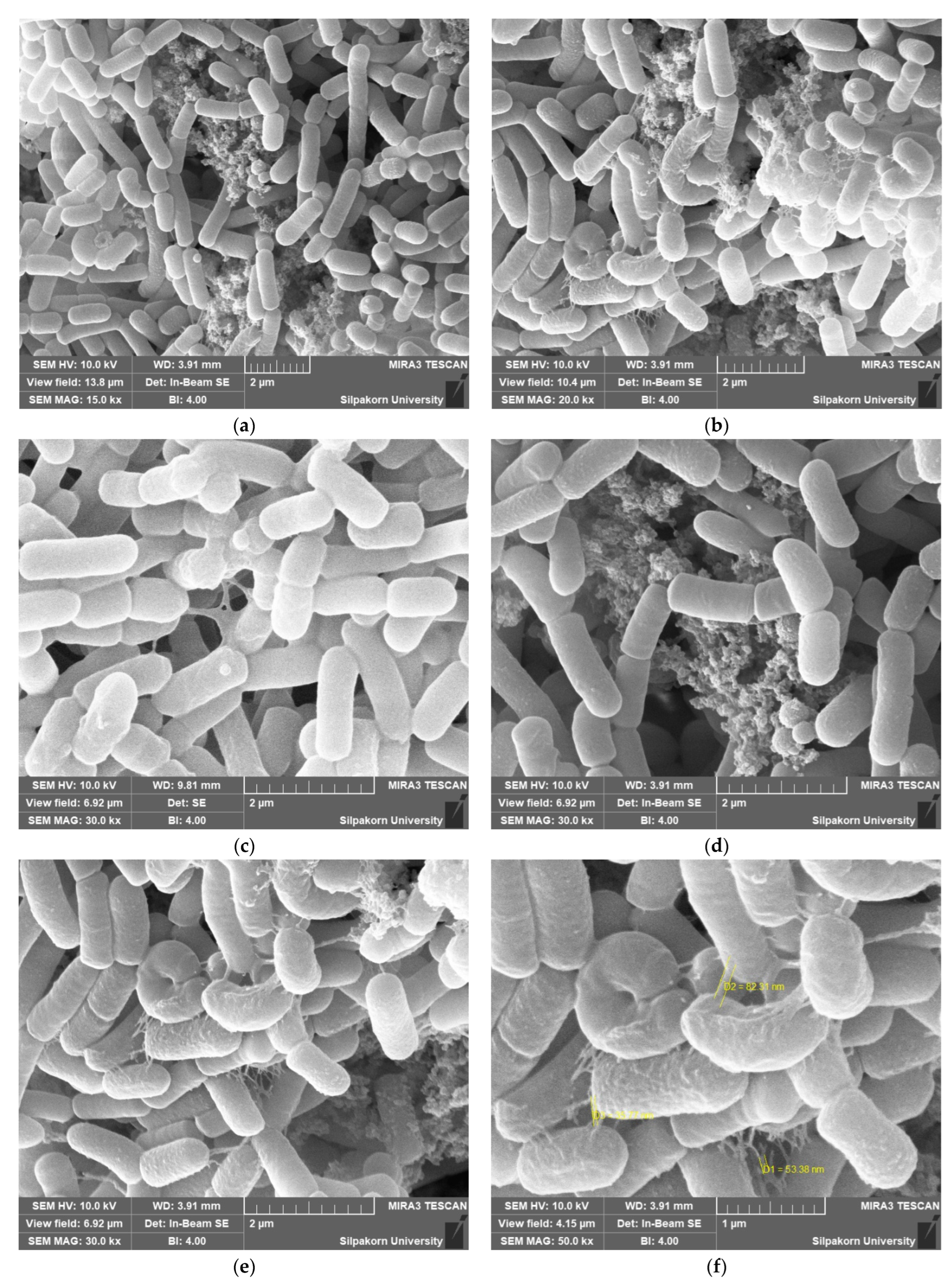
SEM was applied to investigate the adhesion of the multi-strain probiotic to the wall materials. Strains survival was essential for encapsulation with the suitable medium (wall materials). The SEM observations of samples revealed the presence of probiotic strains with several morphologies that affected their ability to adhere to the medium (wall materials), as shown in Figure 3. The adhesion of probiotic strains was considered essential to achieve a beneficial probiotic cell effect in the gastrointestinal tract [36]. From the SEM studies, the evident adhesion showed that multi-strain probiotic was capable of adhering to the medium (wall materials). The probiotic adhesion was probably assisted by secreted lectin-like bacteriocins as a result of cell surface adhesion factors, such as lectin or the adhesion proteins of S-layers and lectin-like complexes on the probiotic cell walls [37]. The colonization of the pathogenic strains was restricted by the adhesion of the probiotic strains that were able to modulate the host immune system systematically [38,39]. Some mucous in the nanometer scale could be observed around the probiotic strains in the SEM micrographs (Figure 3f) due to the numerous wall materials. It was clear that the protectant wall materials were firmly adhered around the probiotic strains as a nano wall cryoprotectant based on their magnified images. From this experiment, the complex medium (wall materials) was essential for the survival and adhesion to the wall materials of the multi-strain probiotic.

3.7. Microbial Safety

The lyophilized samples were not contaminated by E. coli, yeast and molds after 60 days of storage, with the contamination being acceptable at lower than 1 × 101–1 × 10² CFU/g. Therefore, the lyophilized samples could be safely used for consumption in a functional product.

4. Conclusions

The multi-strain probiotic microorganisms showed high resistance to the lyophilization process in the presence of the cryoprotectants used. The best results were obtained when a complex medium (wall materials) was used. Synbiotic samples from lyophilized probiotic strains on the complex medium were formulated to achieve maximum probiotic viability. This study showed that the tested cryoprotectant materials influenced the viability of probiotic cells after the lyophilization processing. Furthermore, the multi-strain probiotic maintained a high degree of viability whilst in storage. The lyophilized multi-strain probiotics maintained their viability in the synbiotic product. Additionally, all probiotic strains were promising strains that produced many beneficial substances, and would be used as health supplements. The lyophilized synbiotic formulation provided a health benefit and had the additional effect of probiotic viability and stability. The mucilage around the probiotic strains was assumed as a potential nano-wall cryoprotectant based on their magnified SEM images. According to our key findings, this current study presented the distinguished development of lyophilized-synbiotic product in combination with multi-strain probiotic microorganisms and the new complex medium as nano-wall material.

Author Contributions

Z.M.S. did the conceptualization, investigation, formal analysis, methodology, writing-original draft; T.C. did the investigation, formal analysis and methodology; M.P. did the methodology, data curation, writing-review and editing; K.P. did the methodology, formal analysis, data curation, writing-review and editing; S.P. did the supervision, conceptualization, project administration, writing-review and editing, formal analysis, funding acquisition. All authors have read and agreed to the published version of the manuscript.

Funding

This research was funded by the National Research Council of Thailand (NRCT) under the Royal Golden Jubilee (RGJ-ASEAN: PhD scholarship program, Grant No. NRCT5-RGJ63002 (Sub-Code NRCT-RGJ63012-143).

Data Availability Statement

Data is contained within the article.

Acknowledgments

The authors would like to acknowledge the Royal Golden Jubilee (RGJ-ASEAN: PhD program), National Research Council of Thailand (NRCT). We also thank Kasetsart University Research and Development Institute for editing English in manuscript.

Conflicts of Interest

The authors declare no conflict of interest.

References

  1. FAO/WHO. Specifications for identity and purity of certain food additives. FAO Food Nutr. Pap. 1992, 52, 345–355. [Google Scholar]
  2. Abd-Razig, N.M.; Sabahelkhier, M.K.; Idris, O.F. Effect of gum arabic (Acacia senegal, L. Willd) on lipid profile and performance of laying hens. J. Appl. Biosci. 2010, 32, 2002–2007. [Google Scholar]
  3. Azeez, O. Decolourization of gum Arabic using activated charcoal. Leonardo J. Sci. 2005, 4, 23–32. [Google Scholar]
  4. Ali, B.H.; Ziada, A.; Blunden, G. Biological effects of gum arabic: A review of some recent research. Food Chem. Toxicol. 2009, 47, 1–8. [Google Scholar] [CrossRef] [PubMed]
  5. Hadi, A.; Elderbi, M.A.; Mohamed, A. Effect of gum arabic on coagulation system of albino rats. Int. J. PharmTech Res. 2010, 2, 1762–1766. [Google Scholar]
  6. Ali, K.; Daffalla, H. Physicochemical and functional properties of the gum arabic from Acacia senegal. Ann. Food Sci. Technol. 2018, 19, 27–34. [Google Scholar]
  7. Bisar, G.H.; El-Saadany, K.; Khattab, A.; El Kholy, W.M. The possibility of using fibers as a prebiotic in making of probiotic based on-some dairy products. Br. Microbiol. Res. J. 2014, 4, 678. [Google Scholar] [CrossRef]
  8. Hill, C.; Guarner, F.; Reid, G.; Gibson, G.R.; Merenstein, D.J.; Pot, B.; Morelli, L.; Canani, R.B.; Flint, H.J.; Salminen, S.; et al. Expert consensus document: The international scientific association for probiotics and prebiotics consensus statement on the scope and appropriate use of the term probiotic. Nat. Rev. Gastroenterol. Hepatol. 2014, 11, 506–514. [Google Scholar] [CrossRef] [Green Version]
  9. Harish, K.; Varghese, T. Probiotics in humans–evidence-based review. Calicut Med. J. 2006, 4, e3. [Google Scholar]
  10. Macfarlane, G.T.; Cummings, J.H. Probiotics and prebiotics: Can regulating the activities of intestinal bacteria benefit health? BMJ 1999, 318, 999–1003. [Google Scholar] [CrossRef] [Green Version]
  11. Kurtmann, L.; Carlsen, C.U.; Skibsted, L.H.; Risbo, J. Water activity-temperature state diagrams of freeze-dried Lactobacillus acidophilus (La-5): Influence of physical state on bacterial survival during storage. Biotechnol. Prog. 2009, 25, 265–270. [Google Scholar] [CrossRef] [PubMed]
  12. Schwab, C.; Vogel, R.; Gänzle, M.G. Influence of oligosaccharides on the viability and membrane properties of Lactobacillus reuteri TMW1. 106 during freeze-drying. Cryobiology 2007, 55, 108–114. [Google Scholar] [CrossRef]
  13. Mahdavi, S.A.; Jafari, S.M.; Ghorbani, M.; Assadpoor, E. Spray-drying microencapsulation of anthocyanins by natural biopolymers: A review. Dry. Technol. 2014, 32, 509–518. [Google Scholar] [CrossRef]
  14. Nireesha, G.; Divya, L.; Sowmya, C.; Venkateshan, N.; Lavakumar, V. Lyophilization/freeze drying: A review. Int. J. Novel Trends Pharm. Sci. 2013, 3, 87–98. [Google Scholar]
  15. Peighambardoust, S.; Tafti, A.G.; Hesari, J. Application of spray drying for preservation of lactic acid starter cultures: A review. Trends Food Sci. Technol. 2011, 22, 215–224. [Google Scholar] [CrossRef]
  16. Freitas, S.; Merkle, H.P.; Gander, B. Ultrasonic atomisation into reduced pressure atmosphere—Envisaging aseptic spray-drying for microencapsulation. J. Control. Release 2004, 95, 185–195. [Google Scholar] [CrossRef]
  17. Costantino, H.R.; Pikal, M.J. Lyophilization of Biopharmaceuticals; Springer Science & Business Media: Berlin/Heidelberg, Germany; AAPS Press: Arlington, VA, USA, 2004; pp. 1–74. ISBN 0-9711767-6-0. [Google Scholar]
  18. Meng, X.; Stanton, C.; Fitzgerald, G.; Daly, C.; Ross, R. Anhydrobiotics: The challenges of drying probiotic cultures. Food Chem. 2008, 106, 1406–1416. [Google Scholar] [CrossRef]
  19. Fonseca, F.; Cenard, S.; Passot, S. Freeze-Drying of Lactic Acid Bacteria. In Cryopreservation and Freeze-Drying Protocols; Wolkers, W.F., Oldenhof, H., Eds.; Springer: New York, NY, USA, 2015; Volume 1257, pp. 477–488. ISBN 978-1-4939-2193-5. [Google Scholar]
  20. Miremadi, F.; Ayyash, M.; Sherkat, F.; Stojanovska, L. Cholesterol reduction mechanisms and fatty acid composition of cellular membranes of probiotic Lactobacilli and Bifidobacteria. J. Funct. Foods 2014, 9, 295–305. [Google Scholar] [CrossRef]
  21. Elansary, H.O.; Salem, M.Z.; Ashmawy, N.A.; Yacout, M.M. Chemical composition, antibacterial and antioxidant activities of leaves essential oils from Syzygium cumini L., Cupressus sempervirens L. and Lantana camara L. from Egypt. J. Agric. Sci. 2012, 4, 144. [Google Scholar] [CrossRef] [Green Version]
  22. Watnick, P.I.; Kolter, R. Steps in the development of a Vibrio cholerae El Tor biofilm. Mol. Microbiol. 1999, 34, 586–595. [Google Scholar] [CrossRef] [Green Version]
  23. Lee, B.-J.; Kim, J.-S.; Kang, Y.M.; Lim, J.-H.; Kim, Y.-M.; Lee, M.-S.; Jeong, M.-H.; Ahn, C.-B.; Je, J.-Y. Antioxidant activity and γ-aminobutyric acid (GABA) content in sea tangle fermented by Lactobacillus brevis BJ20 isolated from traditional fermented foods. Food Chem. 2010, 122, 271–276. [Google Scholar] [CrossRef]
  24. Chumphon, T.; Sriprasertsak, P.; Promsai, S. Development of rice as potential carriers for probiotic Lactobacillus amylovorus. Int. J. Food Sci. Technol. 2016, 51, 1260–1267. [Google Scholar] [CrossRef]
  25. Savedboworn, W.; Wanchaitanawong, P. Viability and probiotic properties of Lactobacillus plantarum TISTR 2075 in spray-dried fermented cereal extracts. Maejo Int. J. Sci. Technol. 2015, 9, 382. [Google Scholar] [CrossRef]
  26. Shehata, M.; El Sohaimy, S.; El-Sahn, M.A.; Youssef, M. Screening of isolated potential probiotic lactic acid bacteria for cholesterol lowering property and bile salt hydrolase activity. Ann. Agric. Sci. 2016, 61, 65–75. [Google Scholar] [CrossRef] [Green Version]
  27. Klaver, F.; Van der Meer, R. The assumed assimilation of cholesterol by lactobacilli and Bifidobacterium bifidum is due to their bile salt-deconjugating activity. Appl. Environ. Microbiol. 1993, 59, 1120–1124. [Google Scholar] [CrossRef] [Green Version]
  28. Gilliland, S.; Nelson, C.; Maxwell, C. Assimilation of cholesterol by Lactobacillus acidophilus. Appl. Environ. Microbiol. 1985, 49, 377–381. [Google Scholar] [CrossRef] [PubMed] [Green Version]
  29. Noipa, T.; Srijaranai, S.; Tuntulani, T.; Ngeontae, W. New approach for evaluation of the antioxidant capacity based on scavenging DPPH free radical in micelle systems. Food Res. Int. 2011, 44, 798–806. [Google Scholar] [CrossRef]
  30. Thibessard, A.; Borges, F.; Fernandez, A.; Gintz, B.; Decaris, B.; Leblond-Bourget, N. Identification of Streptococcus thermophilus CNRZ368 genes involved in defense against superoxide stress. Appl. Environ. Microbiol. 2004, 70, 2220–2229. [Google Scholar] [CrossRef] [Green Version]
  31. Arasu, M.V.; Kim, D.H.; Kim, P.I.; Jung, M.W.; Ilavenil, S.; Jane, M.; Lee, K.D.; Al-Dhabi, N.A.; Choi, K.C. In vitro antifungal, probiotic and antioxidant properties of novel Lactobacillus plantarum K46 isolated from fermented sesame leaf. Ann. Microbiol. 2014, 64, 1333–1346. [Google Scholar] [CrossRef]
  32. Wojcik, M.; Burzynska-Pedziwiatr, I.; Wozniak, L. A review of natural and synthetic antioxidants important for health and longevity. Curr. Med. Chem. 2010, 17, 3262–3288. [Google Scholar] [CrossRef]
  33. Kim, S.-H.; Shin, B.-H.; Kim, Y.-H.; Nam, S.-W.; Jeon, S.-J. Cloning and expression of a full-length glutamate decarboxylase gene from Lactobacillus brevis BH2. Biotechnol. Bioprocess Eng. 2007, 12, 707–712. [Google Scholar] [CrossRef]
  34. Villegas, J.M.; Brown, L.; de Giori, G.S.; Hebert, E.M.J.L.-F.S. Optimization of batch culture conditions for GABA production by Lactobacillus brevis CRL 1942, isolated from quinoa sourdough. LWT-Food Sci. Technol. 2016, 67, 22–26. [Google Scholar] [CrossRef]
  35. Savini, M.; Cecchini, C.; Verdenelli, M.C.; Silvi, S.; Orpianesi, C.; Cresci, A. Pilot-scale production and viability analysis of freeze-dried probiotic bacteria using different protective agents. Nutrients 2010, 2, 330–339. [Google Scholar] [CrossRef] [PubMed] [Green Version]
  36. Maragkoudakis, P.A.; Zoumpopoulou, G.; Miaris, C.; Kalantzopoulos, G.; Pot, B.; Tsakalidou, E. Probiotic potential of Lactobacillus strains isolated from dairy products. Int. Dairy J. 2006, 16, 189–199. [Google Scholar] [CrossRef]
  37. Lakhtin, V.; Aleshkin, V.; Lakhtin, M.; Afanas’ev, S.; Pospelova, V.; Shenderov, B. Lectins, adhesins, and lectin-like substances of lactobacilli and bifidobacteria. Vestn. Ross. Akad. Med. Nauk. 2006, 1, 28–34. [Google Scholar]
  38. Wagner, R.D.; Johnson, S.J. Probiotic bacteria prevent Salmonella–induced suppression of lymphoproliferation in mice by an immunomodulatory mechanism. BMC Microbiol. 2017, 17, 77. [Google Scholar] [CrossRef] [Green Version]
  39. Gopal, P.K.; Prasad, J.; Smart, J.; Gill, H.S. In vitro adherence properties of Lactobacillus rhamnosus DR20 and Bifidobacterium lactis DR10 strains and their antagonistic activity against an enterotoxigenic Escherichia coli. Int. J. Food Microbiol. 2001, 67, 207–216. [Google Scholar] [CrossRef]
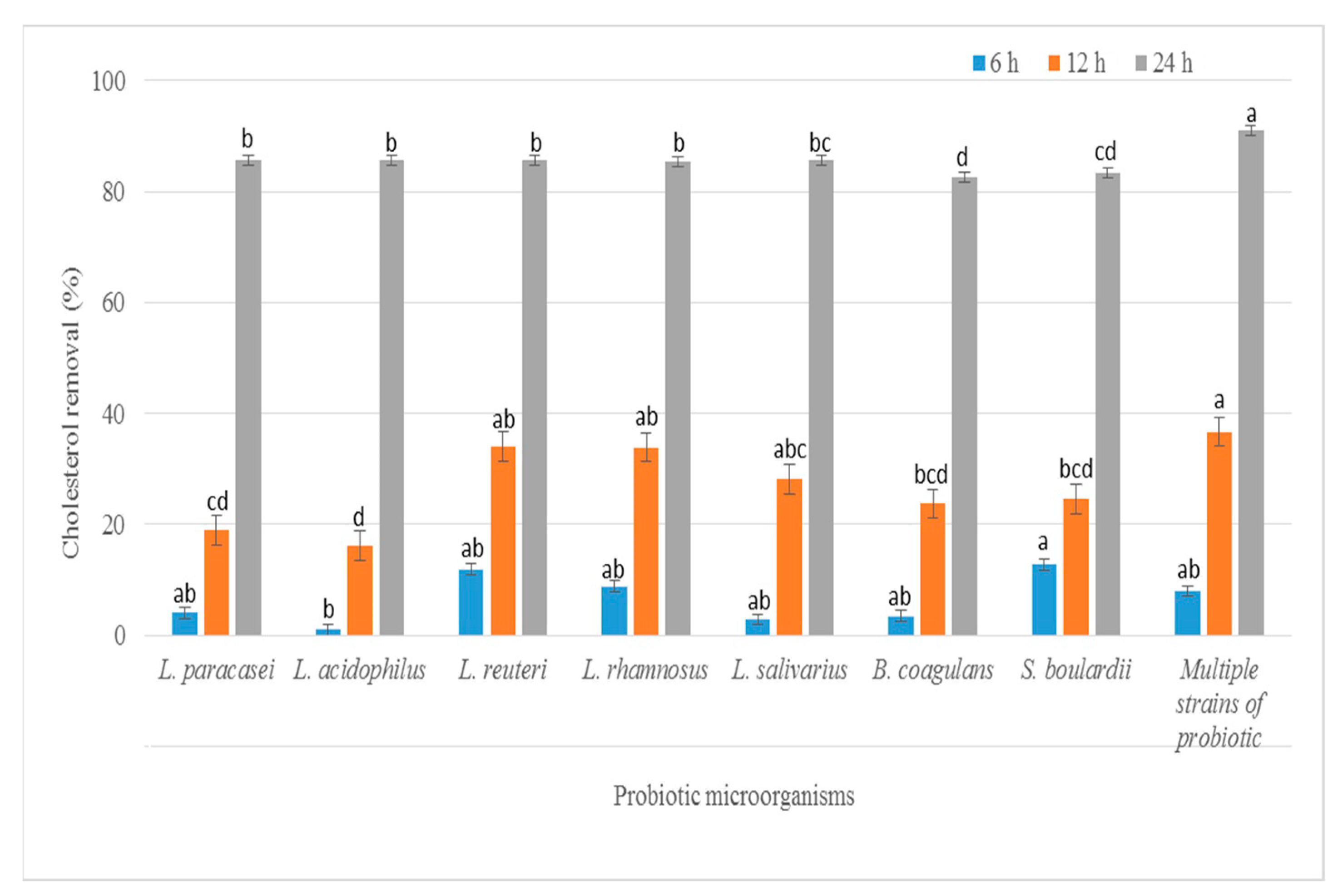
Figure 1. Cholesterol removal (%) of the seven probiotic strains and multi-strain probiotic incubated in media broth supplemented with 1.5 µg/mL water soluble cholesterol for 6, 12 and 24 h at 37 °C. Different lowercase between each time of incubation represents a significant difference using Duncan’s test with a confidence level of 95%.
Foods 11 03113 g001
Figure 2. Chromatogram of screening GABA production by TLC method. The development solvent consists of n-butanol: acetic acid: water (5:3:2, v/v). The chromatogram was observed after spraying the plates with a 2% ninhydrin solution and developing at 105 °C for 5 min. (γ std: standard gamma aminobutyric acid, L1: L. reuteri KUKPS6103, L2: L. rhamnosus KUKPS6007, L3: L. paracasei KUKPS6201, L4: L. acidophilus KUKPS6107, L5: L. salivarius KUKPS6202, B: B. coagulans KPSTF02, S: S. boulardii KUKPS6005 and Mix: multi-strain).
Foods 11 03113 g002
Figure 3. SEM of multi-strain probiotic product after freeze-drying in the presence of complex medium as protectant wall materials. (Magnification: (a) 15,000×, (b) 20,000×, (ce) 30,000×, (f) 50,000×).
Foods 11 03113 g003
Table 1. Biofilm formation and antioxidant activity of probiotic strains.
StrainAmount of Crystal Violet (µg)Antioxidant Activity (mg Gallic Acid/ mL Extract)
L. reuteri KUKPS61034.61 ± 1.17 c,*0.096 ± 0.041 a
L. rhamnosus KUKPS6007
L. paracasei KUKPS6201
L. acidophilus KUKPS6107
L. salivarius KUKPS6202
B. coagulans KPSTF02
S. boulardii KUKPS6005
Multi-strain
2.45 ± 0.47 a,b,c
4.45 ± 0.02 c
3.74 ± 1.35 c
3.82 ± 1.48 c
0.98 ± 0.52 a
1.25 ± 0.20 a,b
3.36 ± 1.45 b,c
2.246 ± 0.190 b
2.038 ± 0.219 b
1.893 ± 0.292 b
2.403 ± 0.298 b
0.220 ± 0.140 a
0.311 ± 0.090 a
2.310 ± 0.138 b
* Values represent the mean ± standard deviation in three independent experiments, n = 3. A different lower case superscript letter in a column represents a significantly different value in statistics using Tukey’s test with a confidential level of 95%.
Table 2. Survival rate of mixed-strains of probiotics in the lyophilized product after an eight-week period (at 25 °C).
Time (Week)Viability of Cell Count
(CFU/g)
Survival Rate (%)
0
1
2
3
4
5
6
7
8
4.90 ± 1.90 × 109 *
8.26 ± 0.71 × 10⁸
6.56 ± 0.78 × 10⁹
1.75 ± 0.41 × 10⁹
4.66 ± 0.86 × 10⁹
3.76 ± 0.73 × 10⁹
1.18 ± 0.08 × 10⁹
9.56 ± 0.65 × 10⁸
7.53 ± 0.65 × 10⁸
100.00
16.86
133.88
35.71
95.10
76.73
24.08
19.51
15.37
* Values show the mean ± SD of the three independent experiments.
Publisher’s Note: MDPI stays neutral with regard to jurisdictional claims in published maps and institutional affiliations.