Article

Morphology-Controlled CuO Photocatalysts for Visible-Light Degradation of Organic Pollutants

Langfang Center for Comprehensive Survey of Natural Resources, China Geological Survey, Langfang 065000, China
*
Authors to whom correspondence should be addressed.
Inorganics 2026, 14(7), 172; https://doi.org/10.3390/inorganics14070172
Submission received: 26 May 2026 / Revised: 22 June 2026 / Accepted: 22 June 2026 / Published: 24 June 2026

Abstract

Copper oxide (CuO) is a narrow-bandgap p-type semiconductor promising for visible-light photocatalysis, yet it suffers from rapid charge recombination and low carrier transfer efficiency. In this study, two distinct CuO photocatalysts were fabricated via different routes: two-dimensional CuO nanosheets derived from annealing a CuBDC metal–organic framework (MOF) precursor, and oriented one-dimensional CuO nanoflower arrays prepared by electrochemical deposition, followed by annealing. The crystal structure, morphology, optical absorption, and photoelectrochemical properties were systematically characterized by XRD, SEM, XPS, UV-Vis spectroscopy, transient photocurrent response, EIS, and PL spectroscopy. The CuO nanoflower thin film exhibits a broad visible-light absorption, a markedly higher photocurrent density (42.25 μA cm−2), and lower charge-transfer resistance compared to CuO nanosheets. When evaluated for visible-light photocatalytic degradation of methylene blue (MB), rhodamine B (RhB), and malachite green (MG), the CuO thin film completely degraded MB within 15 min, with an apparent rate constant of 20.15 h−1—approximately three times that of CuO nanosheets. It also showed 1.2- and 1.28-fold higher activity for RhB and MG, respectively. The enhanced performance is attributed to the oriented nanoflower architecture that provides continuous charge transport pathways, suppresses carrier recombination, and extends light propagation via multiple reflections. This work demonstrates that microstructural engineering is an effective strategy to overcome the intrinsic limitations of CuO photocatalysts for wastewater treatment.

1. Introduction

With the fast industrial development, a large number of organic pollutants are released into aquatic systems with industrial wastewater, which brings great difficulties to its treatment [1,2]. Thus, removing such pollutants from industrial wastewater is of great importance. Photocatalysis represents a sustainable and high-performance method for pollutant degradation and air purification [3]. As a light-driven technology based on semiconductors, photocatalysis is distinguished by its superior stability, non-toxic nature, and corrosion resistance [4,5]. The core photocatalytic mechanism involves the generation of hydroxyl radicals and superoxide anions, which act as strong oxidants to degrade organic compounds and ultimately convert them into less toxic CO2 and H2O [6,7,8]. An excellent photocatalyst is required to exhibit a narrow gap, a facile and scalable preparation process, superior stability, high-charge separation efficiency, and abundant high-active surface sites [9,10].
Traditionally studied TiO2-based photocatalysts have a wide bandgap (approximately 3.2 eV) and can only respond to ultraviolet light, which accounts for less than 5% of solar energy; their visible-light utilization is extremely low, severely limiting their practical applications [11,12,13]. Copper oxide (CuO) is a typical p-type narrow-bandgap semiconductor with an intrinsic bandgap of only 1.2–1.9 eV, which fully covers the visible-light spectrum [14,15,16]. It also has advantages in abundant global reserves, and low production costs, good chemical stability, which meets the core requirements of an ideal photocatalyst [1,17,18]. However, pure-phase CuO photocatalysts still face critical performance limitations: the rapid recombination rate of photo-generated carriers and low carrier transfer efficiency result in photocatalytic activity under visible light that is far below theoretical expectations, severely limiting their large-scale application in the treatment of wastewater.
Existing research has confirmed that controlling the morphological structures and array design of nanostructures is key to overcome the performance limitations of CuO photocatalysts [19,20,21]. The microstructure of the material directly determines its specific surface area, light-harvesting capacity, the continuity of charge transport pathways, and the density of active surface sites, thereby regulating photocatalytic performance across three dimensions: light absorption, carrier utilization, and surface reactions [22,23,24].
Based on this, this study prepared two distinct CuO photocatalytic materials with significantly different microstructures and structural properties using two different processes: first, the metal–organic framework (MOF)-derived annealing method can be used to prepare two-dimensional CuO nanosheets with a high specific surface area [25]; second, the electrochemical deposition-annealing process enables the controlled preparation of oriented one-dimensional CuO nanoflower arrays [26]. The study systematically investigated the effects of two preparation methods on the crystal structure, microstructure, light absorption capacity, and photoelectrochemical properties of CuO materials. In addition, to systematically compare the influence of different morphologies on photocatalysis, simple-structured CuO nanoparticles were added for comparison (synthesized by wet chemical method) [27]. Using MB, malachite green (MG), and RhB as the target organic pollutants, the visible-light photocatalytic degradation performance of the two CuO materials was compared and evaluated. This provides insights into enhancing photocatalytic efficiency by regulating microstructural properties.

2. Results and Discussions

2.1. Morphological and Structural Studies

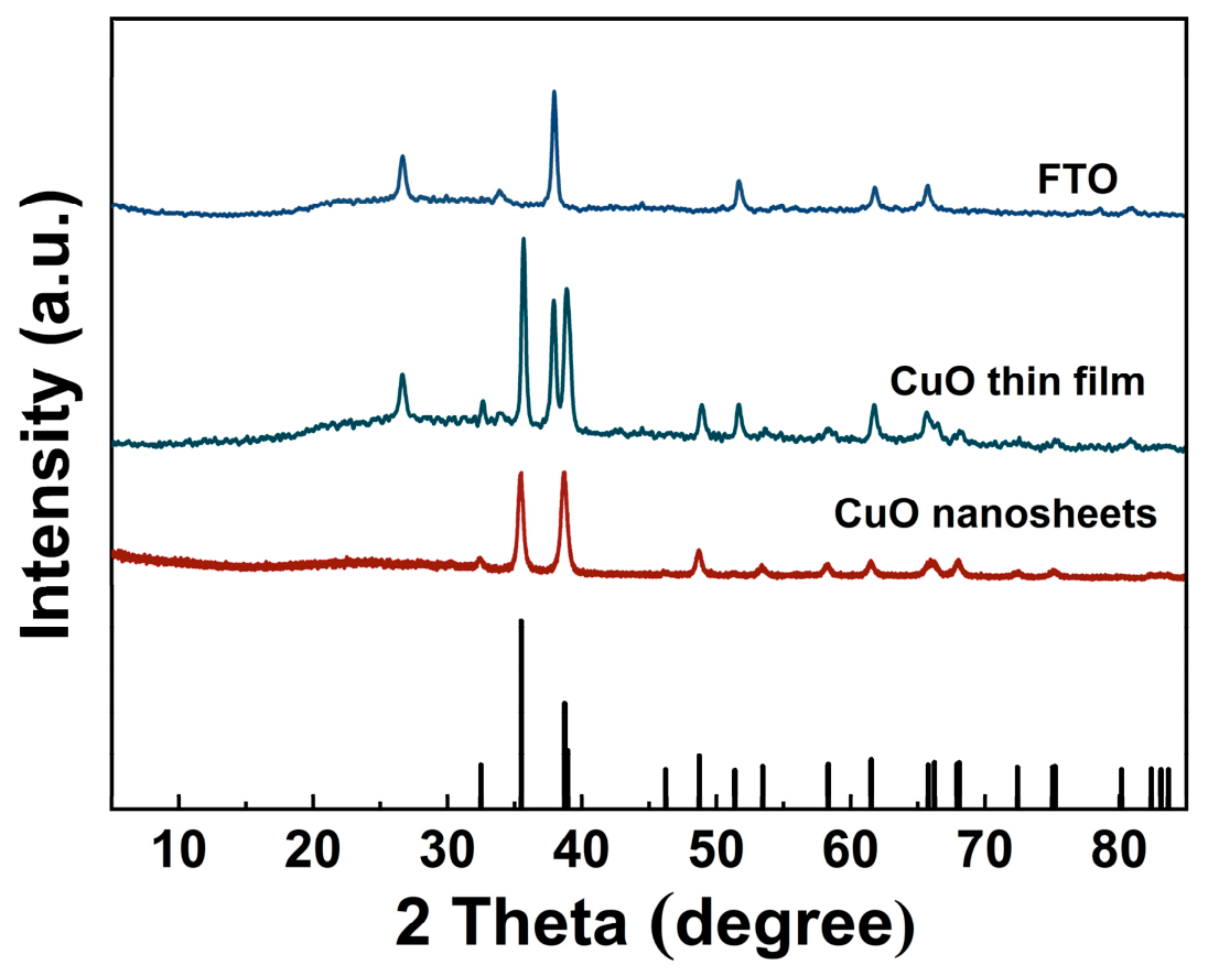
The crystal structure of the synthesized CuO nanosheets and CuO nanoflower thin films was analyzed using X-ray diffraction (XRD). As shown in Figure 1, the XRD pattern reveals distinct peaks at 2θ = 32.68°, 35.66°, 38.94°, 48.94° and 58.54°, which are indexed to the (−110), (002), (111), (−202) and (202) planes of monoclinic CuO, respectively. The strong intensity of these peaks suggests that the synthesized CuO nano-oxides are highly crystalline, and the pattern is in excellent agreement with the JCPDS # 45-0937 [28].
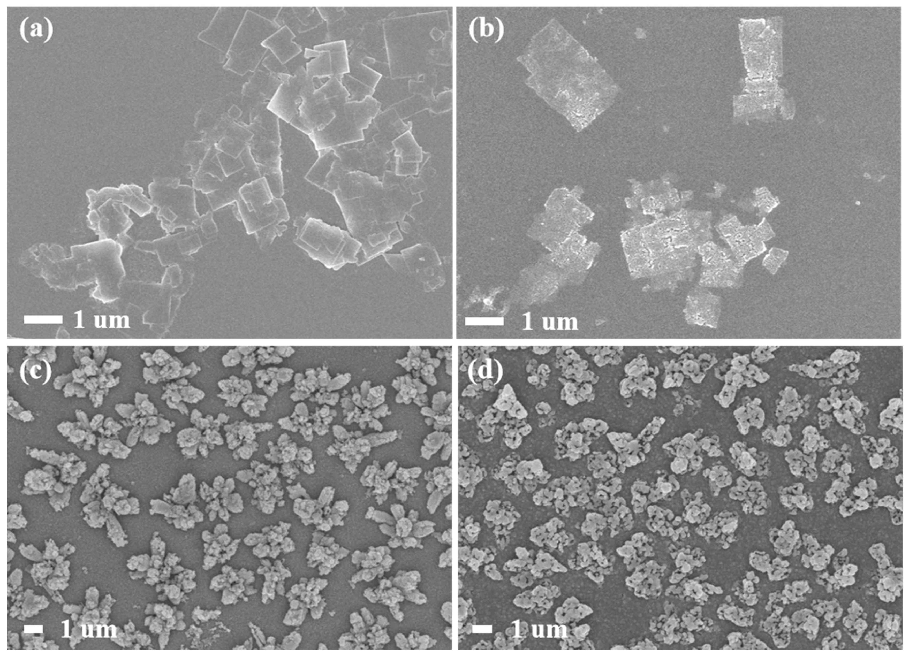
SEM images were used to observe the different nanostructures. CuBDC MOF nanosheets synthesized by bottom-up synthesis strategy were shown in Figure 2a, with lateral dimensions of 0.6–2.7 um. The annealed CuBDC MOF was followed by CuO nanosheets with lateral size of 0.5–3.4 um (Figure 2b). Figure 2c shows the SEM images of electrochemically deposited nanostructured Cu(OH)2, which exhibits an array of nanoflower-like structure, with lengths ranging from 2 to 4 um. After annealing at 600°C in air, the CuO were obtained (Figure 2d), which mainly maintained the nanoflower structure with a size of 2–3 um. The SEM images clearly show significant difference between CuO nanosheets and CuO nanoflowers.
To obtain quantitative information about the specific surface area, pore-volume and pore-size distribution of the synthesized materials, the BET experiment was conducted. Figure S1a shows N2 adsorption–desorption isotherms at 77 K and b shows the BJH pore-size distributions for CuO thin films and CuO NSs. All isotherms are type IV with H4 hysteresis loops, characteristic of mesoporous materials. Textural properties are summarized in Table S3. The CuO films show a BET surface area of 670 m2·g−1, total pore volume of 0.28 cm3·g−1, micropore volume of 0.11 cm3·g−1, mesopore volume of 0.16 cm3·g−1, and a pore diameter of 4.17 nm. The CuO NSs exhibit similar values: 648 m2·g−1, 0.26 cm3·g−1, 0.10 cm3·g−1, 0.14 cm3·g−1, and 4.29 nm, respectively.
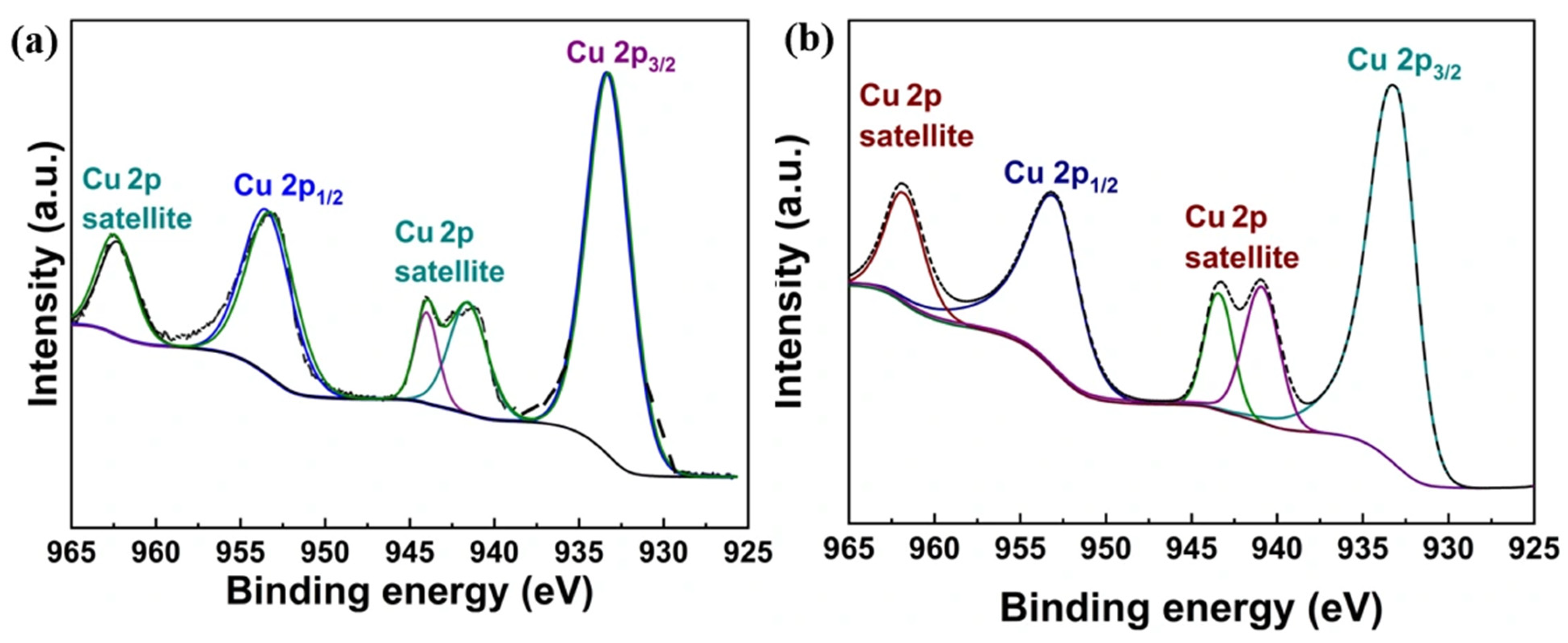
The X-ray photoelectron spectroscopy (XPS) spectra of CuO nanosheets and CuO thin films are shown in Figure 3a,b. The Cu 2p spectrum exhibits two sharp peaks at binding energies of 933 eV and 953 eV, which are ascribed to Cu 2p3/2 and Cu 2p1/2 electrons, respectively, confirming the presence of copper. Furthermore, the shake-up satellite peaks at 941 eV, 943 eV and 962 eV unequivocally indicate the existence of CuO [29,30,31].
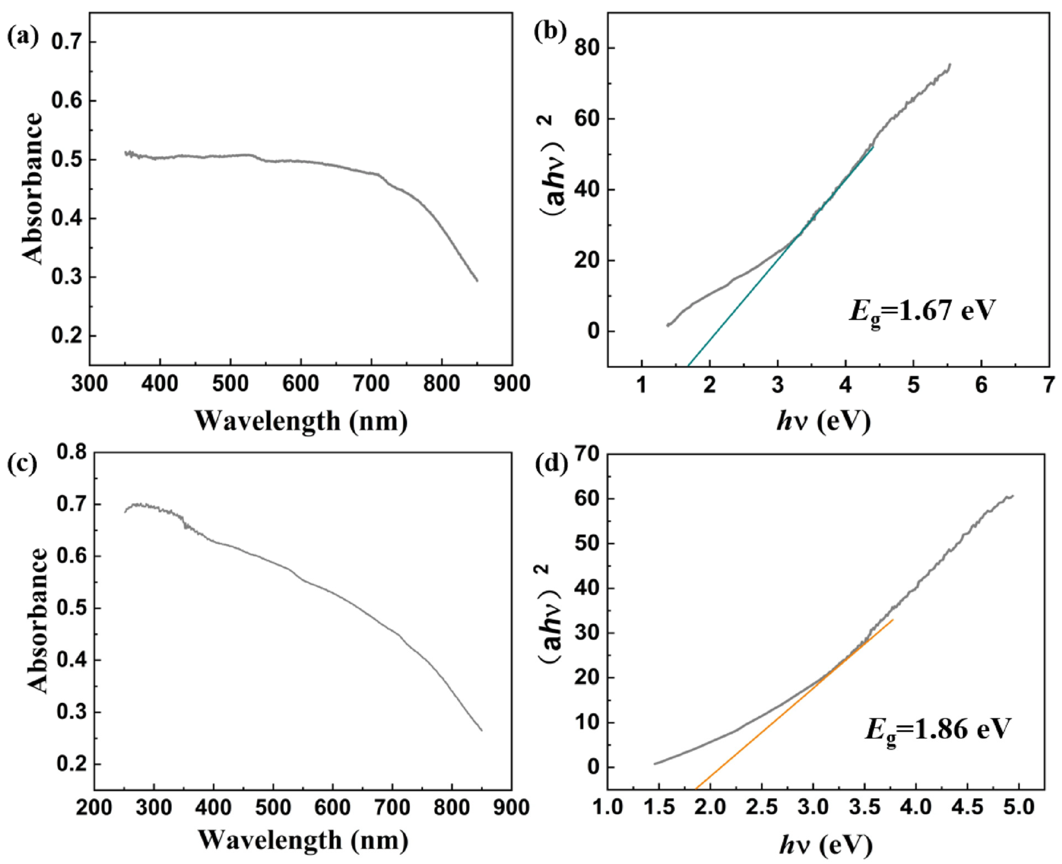
To monitor the optical response of the synthesized CuO sample, UV-Vis absorption spectroscopy was performed. Figure 4a,c exhibits the broadband light absorption ranging from 300 to 800 nm of CuO nanosheets and CuO thin film. The Tauc relation was employed to calculate the optical band gap (e.g., the CuO of nanosheets and CuO thin film are 1.67 eV and 1.86 eV, respectively). The flower-like structure of the CuO thin film contains a large number of pores and interfaces. The incident light undergoes multiple reflections, scattering and diffuse reflection within the structure, greatly extending the light propagation path, resulting in the higher light absorption intensity.

2.2. Photogenerated Carrier Behavior

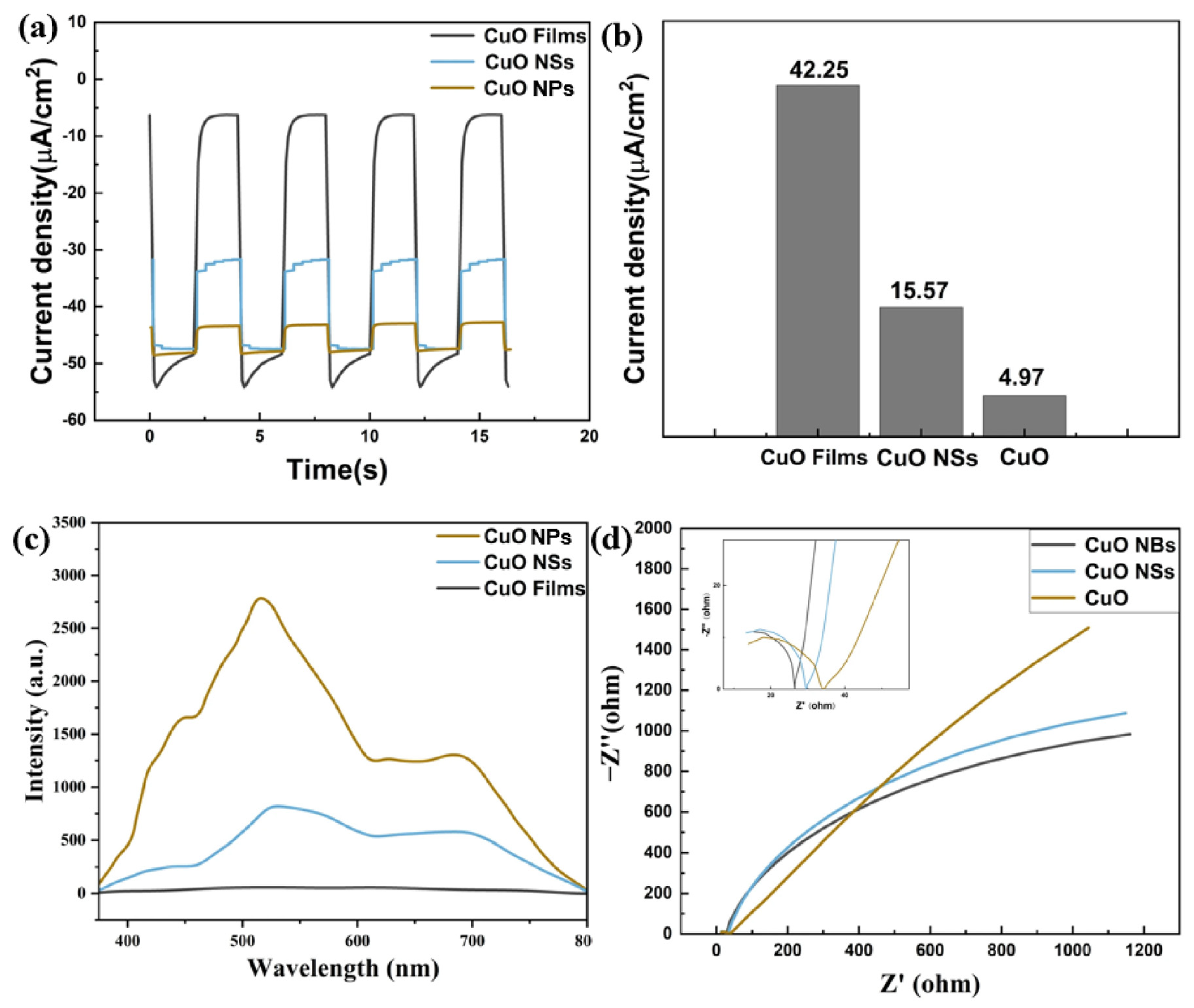
To examine the behavior of photogenerated charge carriers regarding separation and transport, the CuO nanosheets (NSs), CuO thin film and CuO nanoparticles were prepared, and transient photocurrent response (TPR) and electrochemical impedance spectroscopy (EIS) were measured. As shown in Figure 5a,b, the CuO thin film exhibit a markedly higher photocurrent density (42.25 μA·cm−2) than CuO NPs (4.97 μA·cm−2) and CuO NSs (15.57 μA·cm−2) under intermittent illumination, indicating more efficient separation of photogenerated carriers. The recombination of electron–hole pairs was further probed using steady-state photoluminescence (PL) spectroscopy (Figure 5c). Among the three samples, CuO thin film shows the lowest emission intensity, suggesting that the incorporation of nanoflower suppresses carrier recombination and boosts interfacial charge transfer, thereby promoting the photocatalytic reaction. The EIS Nyquist plots (Figure 5d) reveal a smaller arc radius for the CuO thin film than for CuO NSs, implying lower charge transfer resistance. This is attributed to the oriented structure of CuO thin film, which creates continuous charge transfer pathways and accelerates the migration of photogenerated carriers to the electrode surface. In addition, the large specific surface area of the nanoflower arrays enhances electrolyte contraction and improves the interfacial electrochemical reaction kinetics [32,33].

2.3. Evaluation of Photocatalytic Studies

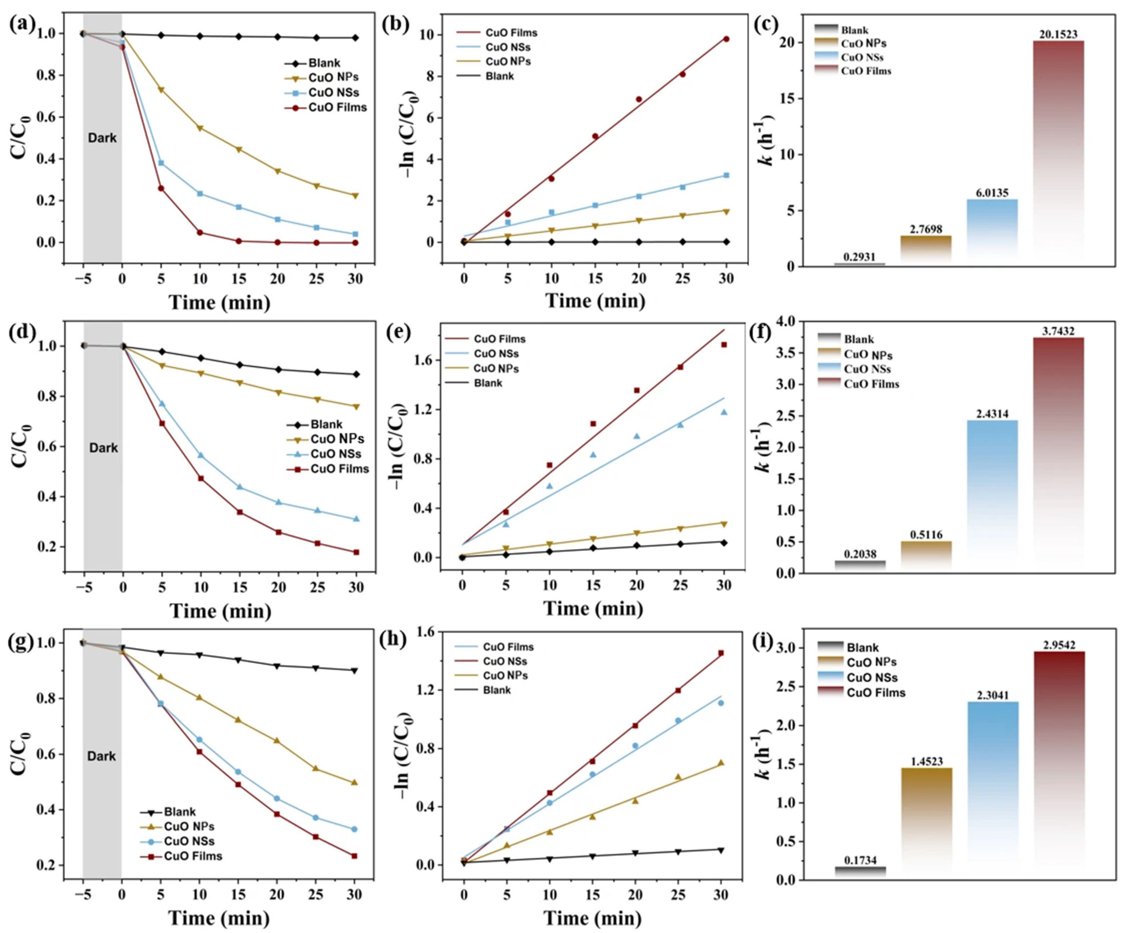
Under visible-light irradiation, the photocatalytic activity of the samples was assessed by measuring the degradation of MB, malachite green (MG) and RhB. The initial evaluation of photocatalytic performance utilized MB as a representative pollutant. As shown in Figure 6a,b, the residual concentration is plotted against irradiation time for different samples. In the blank experiment, the MB exhibited negligible degradation under visible light, indicating its good photostability. For CuO nanoparticle, only about 75% of MB was degraded within 30 min, and the CuO NSs degraded to 92% of MB within 30 min. However, after adding the CuO thin film catalyst, it only took 15 min for the MB to be completely degraded.
A pseudo-first-order kinetic model, −ln(C/C0) = kt (with C0 and C being the initial and time-dependent MB concentrations), was applied to determine the apparent reaction rate constants (k) for quantitative comparison [34,35]. The results in Figure 6c show that CuO NSs and CuO thin films exhibited k values of 6.0135 h−1 and 20.1523 h−1, respectively—approximately a 3-fold increase in degradation rate. This enhancement stems from the nanoflower thin film increases the contact area with the electrolyte, promoting the interfacial redox reaction [36,37]. It does not only generate photogenerated carriers more efficiently, but also allow the carriers to fully participate in the degradation reaction, thereby increasing the degradation rate.
Further evaluation of degradation performance was conducted using a dye—RhB. As shown in Figure 6d–f, roughly 80% of RhB was removed over the CuO thin film within 30 min, the photocatalytic efficiency was 1.2 times higher than that of CuO NSs. In addition, the photodegradation of MG was studied (Figure 6g–i). Relative to CuO NSs, the CuO thin film significantly enhanced photocatalytic performance with a degradation rate about 1.28-fold increased. Among the three contaminants tested, MB was degraded the fastest, indicating that CuO thin film photocatalysts are particularly well suited for the degradation of MB. In addition, in order to further verify that CuO exhibits great photocatalytic performance through morphology control, a comparison was made with the photocatalytic materials used in previous studies (Table S4). The results confirmed that the CuO nanoflower thin films exhibit a good apparent reaction rate constants and degradation rate, which provides a viable approach for the elimination of organic contaminants in wastewater [38,39,40,41,42,43].
In order to further verify whether the photocatalytic effect is complete, the degradation of RhB over CuO films was monitored for 120 min. Figure S2a shows the time-dependent spectral changes, shifts in the maximum absorption wavelength (λmax), and degradation efficiencies of RhB during photodegradation over CuO film samples. As the irradiation time increased, the maximum absorbance gradually dropped, and λmax displayed a blue shift (Figure S2a). This decrease in absorbance and the limited shift in λmax are attributed to the structural breakdown of RhB and its gradual deethylation. The CuO films exhibited strong photocatalytic activity, achieving 97.1% RhB removal within 120 min of illumination (inset of Figure S2a). However, the destruction and decolorization of RhB do not guarantee complete mineralization. Smaller organic molecules, formed during the process, may still pose environmental risks. Therefore, TOC analysis and conventional volumetric methods were employed to evaluate the removal of total organic carbon (TOC) and chemical oxygen demand (COD) during the mineralization of RhB over the synthesized micron-sized CuO films. After 120 min of irradiation, the removal efficiencies were 66.8% for COD and 59.2% for TOC (Figure S2b). Both efficiencies and the corresponding rate constants were lower than those for RhB degradation, which is due to the continuous formation of organic intermediates during the photocatalytic process. TOC analysis further confirmed that more than 59.2% of the carbon originally present in the RhB solution was mineralized into CO2.
To identify the primary active species responsible for RhB photodegradation over the CuO thin film, trapping experiments were carried out using disodium ethylenediaminetetraacetic acid (EDTA–2Na), isopropanol (IPA), and benzoquinone (BQ) as scavengers for holes (h+), hydroxyl radicals (•OH), and superoxide radicals (•O2), respectively. As shown in Figure S3a, the addition of EDTA–2Na nearly completely suppressed the degradation of RhB, indicating that h+ played a dominant role. Meanwhile, a marked inhibition was also observed upon BQ addition, suggesting that •O2 was another key reactive species. In contrast, the introduction of IPA led to no significant change in the degradation rate, implying that •OH was not among the major active species involved.
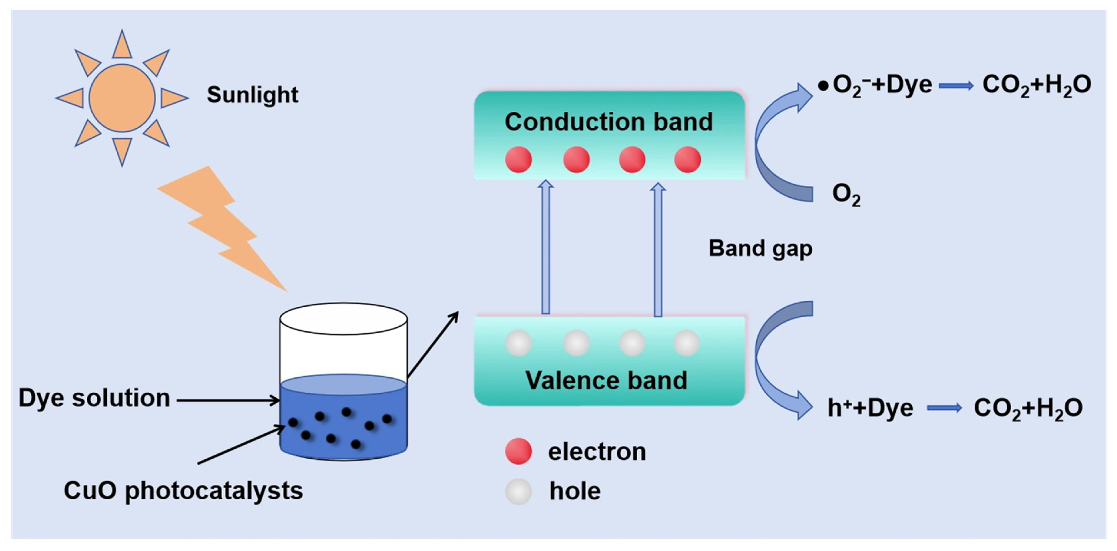
ESR spectroscopy was further employed to identify the active species (h+, •OH, and •O2) generated during RhB photodegradation over the CuO thin film. As illustrated in Figure S3b, TEMPO (2,2,6,6-tetramethylpiperidine-1-oxyl), a spin-label molecule, was used to detect holes (h+). TEMPO can react with charge carriers to form spin adducts that exhibit no ESR signal. In the dark, a characteristic triplet ESR signal of TEMPO with an intensity ratio of 1:1:1 was observed. Upon visible-light irradiation, this signal intensity decreased markedly, indicating the generation of effective holes by the CuO thin film under illumination. This result suggests that h+ radicals play a significant role in the photocatalytic degradation process. In contrast to TEMPO, DMPO (5,5-dimethyl-1-pyrroline N-oxide) was used as a spin-trapping agent to detect •OH in aqueous solution and •O2 in methanol solution. DMPO can react with oxygen-containing radicals to form spin adducts that yield characteristic ESR signals. As shown in Figure S3c, a weak •OH signal was detected upon irradiation, suggesting that only a small amount of •OH radicals participated in the degradation process. In contrast, under the same conditions, pronounced characteristic peaks of •O2 radicals were observed (Figure S3d), demonstrating that •O2 radicals are crucial contributors to the photocatalytic degradation of RhB over the CuO film. The mechanism of dye degradation for CuO photocatalyst is shown in Figure 7. Under visible-light irradiation, the electrons (e−) are excited from the violence band (VB) to the conduction band (CB), leaving holes (h+) behind in the VB. The generated electrons with oxygen molecules produce superoxide (•O2) radicals. The superoxide (•O2) radicals are responsible for the dye degradation [44].
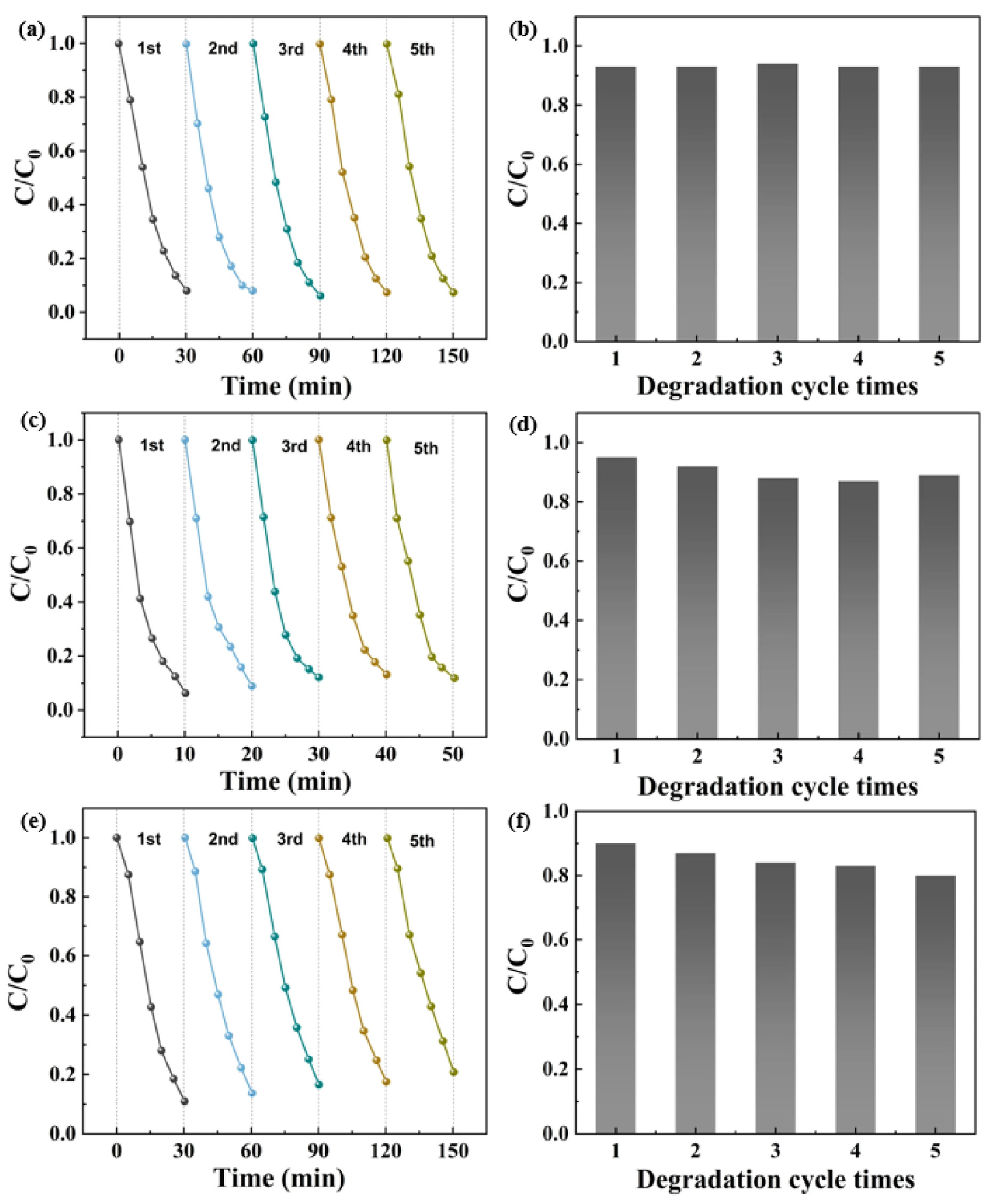
We tested the recyclability and stability of the CuO thin films by running repeated photodegradation experiments on three organic dyes—RhB, MB, and MG—as shown in Figure 8. In each cycle, the film-coated structures were exposed to visible light for a set time, rinsed with deionized water to wash off residual pollutants, and then dried with the air flow. After that, the same films were placed back into fresh dye solutions of identical concentration for another round of degradation. Among the three dyes, MB showed consistently high photocatalytic performance. Over five cycles, the degradation efficiency for MB reached 92.8%, 93.1%, 92.5%, 92.6%, and 92.8%, respectively. These stable results confirm that the CuO thin films are both efficient and durable, with a long working life. Futhermore, because thin films are grown directly on FTO, they are easy to recover and reuse, making them a practical and cost-effective choice for photocatalytic applications [45,46]. After the recyclability and stability test, the comparison of XRD and XPS data for CuO thin films before and after catalytic tests has been presented in Figure S4. The results demonstrated that the nanoflower structure exhibited excellent stability, and no significant changes occurred before and after the tests.

3. Materials and Methods

3.1. Preparation of CuO Nanosheets

CuBDC MOF were synthesized in a glass test tube. First, 30 mg of 1,4-dicarboxybenzene (H2BDC) was dissolved in a mixed solvent containing 2 mL of N,N-dimethylformamide (DMF) and 1 mL of CH3CN. This linker solution was then placed at the bottom of the tube. Next, to avoid early contact between the two precursor solutions, a gentle addition of 1 mL DMF and 1 mL CH3CN mixture was introduced as an intermediate layer. Finally, another solution was prepared by dissolving 30 mg of Cu(NO3)2·3H2O in 1 mL DMF and 2 mL CH3CN, and this metal precursor solution was carefully layered on top as the upper layer. Allow the reaction to stand at 40 °C for 24 h. Centrifuge and collect the blue pellets, clean it, and dry the reactants at 50 °C for 24 h to obtain CuBDC MOF nanosheets. Anneale the CuBDC MOF nanosheets at 600 °C for 1 h to obtain CuO nanosheets.

3.2. Preparation of CuO Nanoflower Thin Films

First, dissolve CH3COONa·3H2O and Cu(CH3COO)2·H2O in deionized water to make a 0.1 M sodium acetate solution and a 0.02 M copper acetate solution. Then, use acetic acid to adjust the pH of the mixed solution to 5.7. For electrochemical deposition, use an ITO-coated glass plate as the working electrode, a platinum plate as the counter electrode, and a saturated calomel electrode (SCE) as the reference electrode. Apply a voltage of 3 V for 10 min to deposit the thin film. Finally, anneal the deposited film at 600 °C for 1 h to obtain the CuO nanoflower thin film.

3.3. Photocatalytic Experiment

To evaluate the photocatalytic performance, three types of CuO samples (nanosheets, nanoflower thin film, and nanoparticles) were tested for their ability to degrade three common dyes: RhB, MB, and MG. For each test, 0.1 g of the photocatalyst was mixed with 100 mL of dye solution and stirred magnetically in the dark for 30 min to reach absorption–desorption equilibrium. The mixture was then exposed to visible light (420–800 nm) from a 300 W xenon lamp. At regular time points, 5 mL of the suspension was taken, filtered through a 0.22 μm filter, and analyzed using UV–Vis spectroscopy to measure the remaining dye concentration. In addition, the CuO thin film composite was reused over multiple cycles to assess its stability and recyclability.

3.4. Characterization

Scanning electron microscopy (SEM, JSM-7500F) was used to observe the morphology of CuO nanosheets and CuO thin films; X-ray photoelectron spectroscopy (XPS, ESCALAB 250XI, light source: Al kα, 15 kV, 100 W) was employed to determine their composition; X-ray diffraction (XRD, Shimadzu, Full-width scanning) was applied to identify their crystallographic phase; the light absorption capacity was evaluated using a UV-Vis absorption spectrometer (UV-Vis, UV-2600) in the wavelength range of 200 to 900 nm. For photoluminescence measurements (PL, FS5, excitation wavelength: 450 nm), a fluorescence spectrophotometer was used to characterize optical properties of the samples. The reagents and equipment used in the experiment are all listed in Tables S1 and S2.

4. Conclusions

This study successfully prepared CuO nanosheets via MOF annealing and oriented CuO nanoflower array thin films through electrochemical deposition followed by annealing, and systematically compared their microstructures, photoelectrochemical properties and photocatalytic performance. Optical and photoelectrochemical tests show that CuO nanoflower thin films exhibit higher photocurrent density (42.25 μA·cm−2), lower charge-transfer resistance and weaker photoluminescence intensity, indicating more efficient carrier separation and suppressed recombination.
In photocatalytic degradation of MB, RhB and MG, the CuO nanoflower thin film shows remarkably higher activity. It completely degrades MB within 15 min with a rate constant three times that of nanosheets, and presents 1.2- and 1.28-fold higher activity for RhB and MG, respectively. Cycling tests confirm its excellent stability and reusability. The oriented nanoflower structure provides continuous charge transport pathways and extends light propagation via multiple reflections, effectively overcoming the intrinsic limitations of CuO. This study emphasizes that tailoring the microstructure constitutes a powerful approach to boosting the photocatalytic efficacy of CuO, presenting a promising and feasible solution for the degradation of organic pollutants in wastewater.

Supplementary Materials

The following supporting information can be downloaded at: https://www.mdpi.com/article/10.3390/inorganics14070172/s1, Figure S1: N2 adsorption-desorption isotherms (a) and BJH pore size distributions (b) for CuO Films, CuO NSs nanomaterials; Figure S2: (a) Temporal evolution of the spectral changes (inset of degradation efficiencies of RhB) and (b) the COD and TOC removal rate of RhB over CuO film; Figure S3: (a) Active species trapping experiments for Rh B degradation over CuO thin film. ESR spectra of (b) TEMPO-h+, (c) DMPO-•OH, and (d) DMPO-•O2 adducts under visible light irradiation (blank system); Figure S4: (a) XRD and (b) XPS data for catalysts before and after catalytic tests; Table S1: Chemical reagents used in the experiments; Table S2: Instruments used in the experiment; Table S3: Textural properties of CuO Films, CuO NSs nanomaterials; Table S4: Photocatalysis Comparison of Recently Reported Photocatalytic under visible light.

Author Contributions

Conceptualization, methodology, formal analysis, writing—original draft preparation Q.G.; data curation, validation software, formal analysis, writing—original draft preparation, H.Y.; validation, supervision, writing—review and editing, funding acquisition, X.L.; resources, software, L.F.; resources, software, H.D.; methodology, X.S.; visualization, Y.J.; software L.W. All authors have read and agreed to the published version of the manuscript.

Funding

This research was funded by National Technical Support and Services for Gold and Other Strategic Mineral Analysis, grant number DD20250209112.

Data Availability Statement

The data supporting the reported results are available on reasonable request to the authors.

Conflicts of Interest

The authors declare no conflicts of interest.

References

  1. Imran, M.; Abdullah, A.Z.; Khan, M.E.; Kim, Y.-M.; Khan, F. Excellent successive photo-induced degradation of tetracycline using CuO/g-C3N4 nanocomposites: Synergistic effects of CuO integration and H2O2 in a photo-Fenton system. J. Taiwan Inst. Chem. Eng. 2025, 171, 106068. [Google Scholar]
  2. Rahimi-Nasrabadi, M.; Pourmortazavi, S.M.; Aghazadeh, M.; Ganjali, M.R.; Karimi, M.S.; Novrouzi, P. Optimizing the procedure for the synthesis of nanoscale gadolinium(III) tungstate as efficient photocatalyst. J. Mater. Sci. Mater. Electron. 2017, 28, 3780–3788. [Google Scholar]
  3. Duta, A.; Enesca, A.; Bogatu, C.; Gyorgy, E. Solar-active photocatalytic tandems. A compromise in the photocatalytic processes design. Mater. Sci. Semicond. Process. 2016, 42, 94–97. [Google Scholar] [CrossRef]
  4. Chen, C.; Wang, B.Y.; Xu, J.J.; Fei, L.Y.; Raza, S.; Li, B.S.; Zeng, Q.Q.; Shen, L.G.; Lin, H.J. Recent Advancement in Emerging MXene-Based Photocatalytic Membrane for Revolutionizing Wastewater Treatment. Small 2024, 20, e2311427. [Google Scholar] [CrossRef] [PubMed]
  5. Yin, C.; Qian, J.; Guo, T.; Wang, L.Q. Antibacterial photocatalytic films for effective degradation of organic compounds. Res. Chem. Intermed. 2025, 51, 195–215. [Google Scholar]
  6. Xiang, W.H.; Yuan, J.L.; Wu, Y.W.; Luo, H.Y.; Xiao, C.B.; Zhong, N.B.; Zhao, M.F.; Zhong, D.J.; He, Y.Y. Working principle and application of photocatalytic optical fibers for the degradation and conversion of gaseous pollutants. Chin. Chem. Lett. 2022, 33, 3632–3640. [Google Scholar] [CrossRef]
  7. Fu, J.L.; Wang, X.J.; Ma, Z.; Hao, W.M.; Li, J.Y.; Wang, Z.P.; Wang, L.G. Photocatalytic ultrafiltration membranes based on visible light responsive photocatalyst: A review. Desalin. Water Treat. 2019, 168, 42–55. [Google Scholar] [CrossRef]
  8. Chen, C.; Fei, L.Y.; Wang, B.Y.; Xu, J.J.; Li, B.S.; Shen, L.G.; Lin, H.J. MOF-Based Photocatalytic Membrane for Water Purification: A Review. Small 2024, 20, e2305066. [Google Scholar] [PubMed]
  9. Pedanekar, R.S.; Shaikh, S.K.; Rajpure, K.Y. Thin film photocatalysis for environmental remediation: A status review. Curr. Appl. Phys. 2020, 20, 931–952. [Google Scholar] [CrossRef]
  10. Chang, Y.-C.; Syu, S.-Y.; Lu, M.-Y. Fabrication of In(OH)3–In2S3–Cu2O nanofiber for highly efficient photocatalytic hydrogen evolution under blue light LED excitation. Int. J. Hydrogen Energy 2023, 48, 9318–9332. [Google Scholar] [CrossRef]
  11. Guo, Q.; Zhou, C.Y.; Ma, Z.B.; Yang, X.M. Fundamentals of TiO2 Photocatalysis: Concepts, Mechanisms, and Challenges. Adv. Mater. 2019, 31, 1901997. [Google Scholar] [CrossRef]
  12. Nakata, K.; Ochiai, T.; Murakami, T.; Fujishima, A. Photoenergy conversion with TiO2 photocatalysis: New materials and recent applications. Electrochim. Acta 2012, 84, 103–111. [Google Scholar] [CrossRef]
  13. Milosevic, M.; Radoicic, M.; Ohara, S.; Abe, H.; Spasojevic, J.; Mancic, L.; Saponjic, Z. Advanced photocatalysis mediated by TiO2/Ag/TiO2 nanoparticles modified cotton fabric. Cellulose 2023, 30, 4749–4771. [Google Scholar]
  14. Bruno, E.; Haris, M.; Mohan, A.; Senthilkumar, M. Formation of self-assembled hierarchical structure on Zn doped in CuO nanoparticle using a microwave-assisted chemical precipitation approach. J. Mater. Sci. Mater. Electron. 2021, 32, 19339–19351. [Google Scholar] [CrossRef]
  15. Sahu, K.; Bisht, A.; Khan, S.A.; Pandey, A.; Mohapatra, S. Engineering of morphological, optical, structural, photocatalytic and catalytic properties of nanostructured CuO thin films fabricated by reactive DC magnetron sputtering. Ceram. Int. 2020, 46, 7499–7509. [Google Scholar] [CrossRef]
  16. Harish, S.; Archana, J.; Sabarinathan, M.; Navaneethan, M.; Nisha, K.D.; Ponnusamy, S.; Muthamizhchelvan, C.; Ikeda, H.; Aswal, D.K.; Hayakawa, Y. Controlled structural and compositional characteristic of visible light active ZnO/CuO photocatalyst for the degradation of organic pollutant. Appl. Surf. Sci. 2017, 418, 103–112. [Google Scholar] [CrossRef]
  17. Ghosh, M.K.; Sahu, S.; Gupta, I.; Ghorai, T.K. Green synthesis of copper nanoparticles from an extract of Jatropha curcas leaves: Characterization, optical properties, CT-DNA binding and photocatalytic activity. RSC Adv. 2020, 10, 22027–22035. [Google Scholar] [PubMed]
  18. Yao, B.B.; Kang, J.; Li, S.J.; Mao, Z.X.; Tu, T.X.; Wu, Z.L.; Bi, M.F.; Chen, J.Y.; Chen, S.; Yin, H.J. CuO Nanoparticle-Loaded TiO2 Catalyst for High-Performance Photocatalytic Hydrogen Peroxide Generation. Small 2025, 21, e10644. [Google Scholar] [CrossRef] [PubMed]
  19. Chu, W.; Guo, Q.; Zou, H.; Liu, Z.; Ren, X.; Wang, X.; Chen, R.; Zhang, H.; Ni, H. Hydrothermal Fabrication of Low-Dimensional CuO Nanosheets for Enhancing Carbon Reduction Product Selectivity. ACS Omega 2025, 10, 46079–46086. [Google Scholar] [CrossRef] [PubMed]
  20. Fan, Q.; Lan, Q.; Zhang, M.; Fan, X.; Zhou, Z.; Zhang, C. Preparation and photocatalytic activities of 3D flower-like CuO nanostructures. J. Semicond. 2016, 37, 083002. [Google Scholar] [CrossRef][Green Version]
  21. Madona, J.; Sridevi, C.; Velraj, G.; Dhayal Raj, A.; George, A. Surfactant assisted morphology controlled CuO nanostructures for enhanced photocatalytic performance and bacterial growth inhibition. Mater. Sci. Eng. B 2023, 294, 116562. [Google Scholar] [CrossRef]
  22. Yang, T.; Wang, B.; Chu, P.K.; Xia, J.; Li, H. Self-sacrificing MOF-derived hierarchical porous In2S3 nanostructures with enhanced photocatalytic performance. Chin. J. Catal. 2024, 59, 204–213. [Google Scholar] [CrossRef]
  23. Xu, N.; Zheng, Y.; Chen, J.; Dai, J.; Zhao, X.; Ma, J.; Liu, R. Ti-doped synergistic hollow thin-walled Bi2O3 nano-microspheres for efficient tetracycline hydrochloride photodegradation. Colloids Surf. A 2024, 701, 134887. [Google Scholar] [CrossRef]
  24. Huang, Q.-Q.; Li, N.; Han, M.-S.; Liu, J.; Lan, Y.-Q. Conductive Knitting of Covalent Organic Framework Manipulates Spin Density, Orbital Reorganization, and Charge Mobility for Outstanding Photoreactivity. Angew. Chem. Int. Ed. 2025, 64, e202513848. [Google Scholar] [CrossRef]
  25. Rodenas, T.; Luz, I.; Prieto, G.; Seoane, B.; Miro, H.; Corma, A.; Kapteijn, F.; Llabrés i Xamena, F.X.; Gascon, J. Metal–organic framework nanosheets in polymer composite materials for gas separation. Nat. Mater. 2015, 14, 48–55. [Google Scholar] [PubMed]
  26. Wang, Y.; Jiang, T.; Meng, D.; Yang, J.; Li, Y.; Ma, Q.; Han, J. Fabrication of nanostructured CuO films by electrodeposition and their photocatalytic properties. Appl. Surf. Sci. 2014, 317, 414–421. [Google Scholar] [CrossRef]
  27. Parekh, Z.R.; Chaki, S.H.; Hirpara, A.B.; Patel, G.H.; Kannaujiya, R.M.; Khimani, A.J.; Deshpande, M.P. CuO nanoparticles—Synthesis by wet precipitation technique and its characterization. Phys. B 2021, 610, 412950. [Google Scholar] [CrossRef]
  28. Yang, C.P.; Wu, Q.; Jiang, Z.W.; Wang, X.; Huang, C.Z.; Li, Y.F. Cu vacancies enhanced photoelectrochemical activity of metal-organic gel-derived CuO for the detection of l-cysteine. Talanta 2021, 228, 122261. [Google Scholar] [CrossRef] [PubMed]
  29. Gangaja, B.; Chandrasekharan, S.; Vadukumpully, S.; Nair, S.V.; Santhanagopalan, D. Surface chemical analysis of CuO nanofiber composite electrodes at different stages of lithiation/delithiation. J. Power Sources 2017, 340, 356–364. [Google Scholar] [CrossRef]
  30. Bhattacharjee, A.; Morya, V.; Ghoroi, C. Enzyme-mimetic activity of sugar cane juice stabilized CuO nanospheres and CuO/GO nanocomposite: Green synthesis and applications. Colloid Interface Sci. Commun. 2020, 35, 100239. [Google Scholar] [CrossRef]
  31. Chen, X.; Li, M.; Ouyang, Y.; Liu, T.; Chen, Y.; Song, Y.; Wang, Y.; Qiu, R.; Shen, Y. Fabrication of CuO nanorod loaded with a single gold nanoparticle for low-temperature NO2 detection. Mater. Lett. 2022, 328, 133115. [Google Scholar] [CrossRef]
  32. Sahu, K.; Choudhary, S.; Khan, S.A.; Pandey, A.; Mohapatra, S. Thermal evolution of morphological, structural, optical and photocatalytic properties of CuO thin films. Nano-Struct. Nano-Objects 2019, 17, 92–102. [Google Scholar] [CrossRef]
  33. Zerouali, M.; Bouras, D.; Daïra, R.; Fellah, M.; Boudjema, B.; Barille, R.; Sakher, E.-F.; Bellucci, S.; El-Hiti, G.A. Effect of Zn-doped CuO thin films on structural, morphological, optical, and electrical properties for photocatalysis application. Opt. Mater. 2024, 152, 115495. [Google Scholar] [CrossRef]
  34. Sayoud, N.; Bouchair, A.; Khen, O.; Laib, S.; Boudellioua, H.; Hadji, F.; Zoukel, A.; Touati, H. Sustainable NiO nanoparticles photocatalysts for efficient methylene blue removal: Synthesis, characterization, and kinetic studies. React. Kinet. Mech. Catal. 2025, 138, 3455–3480. [Google Scholar] [CrossRef]
  35. Abbas, S.M.; Abas, K.M. In situ decoration of Ag@exfoliated graphite composite catalyst for Fenton-like oxidation of methylene blue dye: Kinetic and thermodynamic studies. BMC Chem. 2025, 19, 221. [Google Scholar] [CrossRef] [PubMed]
  36. Zhu, F.; Zhan, Y.; Chen, X.; Chen, Y.; Lei, Y.; Jia, H.; Li, Y.; Duan, X. Photocatalytic PAN Nanofibrous Membrane through Anchoring a Nanoflower-Branched CoAl-LDH@PANI Heterojunction for Organic Hazards Degradation and Oil-Containing Emulsified Wastewater Separation. Langmuir 2024, 40, 14368–14383. [Google Scholar] [CrossRef] [PubMed]
  37. Tian, H.; Zhang, R.; Song, Y.; Ning, Y.; Wang, Q.; Liu, B. Construction of 3D nanoflower Bi2WO6/Bi4O5Br2 Z-scheme heterojunction with an internal electric field for enhanced photocatalytic activity. Appl. Surf. Sci. 2025, 684, 161863. [Google Scholar] [CrossRef]
  38. Navya, B.S.; Chen, L.; Nguyen, T.-B.; Arshad, M.; Chen, C.-W.; Dong, C.-D. Graphene-assisted CuO nanoparticles enhanced the photocatalytic degradation of methylene blue under visible light: Performance and electron transfer mechanisms. J. Taiwan Inst. Chem. Eng. 2025, 184, 105436. [Google Scholar]
  39. Ahsan, A.; Bibi, S.; Jabeen, S.; Ali, M.A.; Wattoo, M.A.; Fallatah, A.M.; Elsharkawy, E.R.; Qureshi, M.Z.; Rehman, A.U. ZnO-derived M/MOF-2 hybrid catalysts via solvothermal route for enhanced photocatalytic degradation of methylene blue under visible light irradiation. Solid State Sci. 2026, 177, 108191. [Google Scholar] [CrossRef]
  40. Al-Mamun, M.R.; Kader, S.; Islam, M.S.; Khan, M.Z.H. TiO2/g-C3N4 visible-light-driven photocatalyst for methylene blue decomposition. J. Nanomater. 2023, 2023, 9967890. [Google Scholar]
  41. Sarkar, T. Green synthesized ZnO nanocatalysts for rapid and effective visible-light degradation of industrial dyes. RSC Adv. 2026, 16, 2671–2684. [Google Scholar] [CrossRef] [PubMed]
  42. Kouser, H.A.; Vinay Kumar, E.; Kamat, V.; Bhojya Naik, H.S. Photocatalytic degradation phenomena of methylene blue dye by ZnFe2O4 decorated with rGO nanocomposites under visible light irradiation. Next Nanotechnol. 2026, 9, 100342. [Google Scholar] [CrossRef]
  43. Krishnan, S.G.; Nand, D. A comparative study of the individual and mixed oxide nanostructures on sunlight driven degradation of methylene blue dye and antimicrobial efficacy. Braz. J. Phys. 2024, 54, 230. [Google Scholar] [CrossRef]
  44. Bekele, T.; Alamnie, G. The photocatalytic degradation of organic pollutants-a comprehensive overview. Results Chem. 2025, 18, 102758. [Google Scholar] [CrossRef]
  45. Ibrahim, A.M.; Galaly, A.R.; Abdel-wahab, M.S.; Shaban, M.; Tawfik, W.Z.; Tammam, M.T. Hierarchical CuO photocathodes with cobalt doping for efficient photoelectrochemical water-splitting. RSC Adv. 2025, 15, 24612–24623. [Google Scholar] [CrossRef]
  46. Althamthami, M.; Temam, H.B.; Temam, E.G.; Rahmane, S.; Gasmi, B.; Hasan, G.G. Impact of surface topography and hydrophobicity in varied precursor concentrations of tenorite (CuO) films: A study of film properties and photocatalytic efficiency. Sci. Rep. 2024, 14, 7928. [Google Scholar] [CrossRef] [PubMed]
Figure 1. The XRD of CuO nanosheets and CuO thin films.
Inorganics 14 00172 g001
Figure 2. The SEM images of (a) CuBDC nanosheets; (b) CuO nanosheets; (c) Cu(OH)2 thin film and (d) CuO thin film.
Inorganics 14 00172 g002
Figure 3. The XPS of (a) CuO nanosheets and (b) CuO thin films.
Inorganics 14 00172 g003
Figure 4. (a) UV-Vis absorption spectra of the prepared CuO nanosheets and (b) the corresponding Tauc curve; (c) UV-Vis absorption spectra of the prepared CuO thin film and (d) the corresponding Tauc curve.
Inorganics 14 00172 g004
Figure 5. (a) Transient photocurrent response (TPR) curves. (b) Corresponding photocurrent density data. (c) Steady-state photoluminescence (PL) spectra. (d) Nyquist plots of electrochemical impedance spectroscopy (EIS).
Inorganics 14 00172 g005
Figure 6. (a,d,g) Photocatalytic degradation of MB, RhB, and MG under visible-light irradiation, respectively; (b,e,h) corresponding kinetic curves for the degradation of MB, RhB, and MG; (c,f,i) comparison of the rate constants (k) for MB, RhB, and MG.
Inorganics 14 00172 g006
Figure 7. The mechanism of dye degradation for CuO photocatalyst.
Inorganics 14 00172 g007
Figure 8. Recycle experiments of CuO thin films for (a) MB, (b) RhB, (c) MG solution under UVC light exposure and its corresponding bar chart data for (d) MB, (e) RhB, and (f) MG.
Inorganics 14 00172 g008
Disclaimer/Publisher’s Note: The statements, opinions and data contained in all publications are solely those of the individual author(s) and contributor(s) and not of MDPI and/or the editor(s). MDPI and/or the editor(s) disclaim responsibility for any injury to people or property resulting from any ideas, methods, instructions or products referred to in the content.