Article

Chitosan/Polyacrylic Acid Functionalized Side-Polish Polymer Optical Fiber-Based SPR Sensor for Cu2+ Ion Detection

1
College of Optoelectronic Engineering, Guilin University of Electronic Technology, Guilin 541004, China
2
School of Physics and Optoelectronic Engineering, Hangzhou Institute for Advanced Study, Hangzhou 310000, China
3
Jiangxi Daishing POF Co., Ltd., Jinggangshan 343600, China
4
Geological Survey of Finland, Vuorimiehentie 5, 02151 Espoo, Finland
*
Authors to whom correspondence should be addressed.
Photonics 2025, 12(5), 461; https://doi.org/10.3390/photonics12050461
Submission received: 9 April 2025 / Revised: 3 May 2025 / Accepted: 8 May 2025 / Published: 9 May 2025
(This article belongs to the Special Issue Emerging Technologies and Applications in Fiber Optic Sensing)

Abstract

:
A polymer optical fiber SPR sensor for detecting Cu2+ ion concentration in water is proposed. The sensor employs a simple side-polish structure and realizes the detection of Cu2+ ion concentration by employing the chitosan (CS)/polyacrylic acid (PAA) bilayer film on the gold film of the optical fiber surface. The structure of the fiber probe is optimized, and the sensing performances for the Cu2+ ion detection are analyzed experimentally. The experimental results demonstrate that the sensor exhibits a high sensitivity of 465.539 nm/ppm for the Cu2+ ion detection in the concentration range of 0–0.04 ppm. And it has a fast response speed and good selectivity for Cu2+ ions. The sensor has the advantages of simple structure and low cost, and has potential applications in the field of heavy metal detection.

1. Introduction

In recent years, with the rapid development of technology and industry, pollution of soil, rivers, and even drinking water has become increasingly severe. Among these, large amounts of industrial wastewater containing heavy metal ions and other harmful impurities may be directly discharged into the environment without treatment, leading to severe environmental pollution. Heavy metals exhibit extremely low solubility in water and are highly resistant to natural degradation. The majority of these metals accumulate in the environment, eventually entering the human body through biomagnification in the food chain. Consequently, they can induce various fatal diseases, including cancer, cardiovascular disorders, neurological damage, and renal failure [1], posing severe threats to human health and life. Therefore, the monitoring and treatment of heavy metal pollution have attracted increasing research attention.
Cu2+ ions, as essential trace elements for human physiology, are also among the most prevalent pollutants in industrial effluents [2]. Excessive intake of copper in humans can easily lead to skin, bone, kidney, or liver diseases. Therefore, monitoring of Cu2+ ions is of great significance for environmental improvement and human health protection. Common detection methods for Cu2+ ions mainly include electrochemical analysis [3], atomic absorption spectroscopy [4], colorimetry [5], and fluorescence spectroscopy [6]. Although these methods enable highly sensitive detection of Cu2+ ions, they suffer from drawbacks such as complex procedures, high costs, lengthy testing cycles, and bulky analytical instruments.
Optical fibers, as versatile photonic components, have been widely employed in sensing applications due to their unique advantages, including immunity to electro-magnetic interference, compact size, and real-time monitoring capability [7]. The operational principle of optical fiber sensors relies on the interaction between guided light and external environment, which can modulate optical properties such as intensity, phase, or wavelength transmitted in the optical fiber. In recent years, optical fiber-based heavy metal sensors have gained increasing interest due to their compact size, low cost, rapid response, high sensitivity, and miniaturization potential. Currently, reported optical fiber heavy metal sensors employ various techniques, such as optical fiber Bragg gratings (FBGs) [8], optical fiber interferometers [9], optical fiber evanescent waves [10], and optical fiber surface plasmon resonance (SPR) [11,12,13,14]. Among them, SPR-based optical fiber sensors have become a powerful tool for ultrasensitive detection of refractive index, owing to their remarkable ability to confine electro-magnetic fields at metal–dielectric interfaces [15], which makes it highly suitable for heavy metal ion detection [16]. The SPR phenomenon occurs when the wave vector of evanescent waves of the light in optical fibers matches that of surface plasmon polaritons (SPPs), which will generate a sharp peak at the specific wavelength in the transmission spectrum.
Currently, various types of optical fibers, including single-mode fibers, multimode fibers, multicore fibers, hollow-core fibers, and glass capillaries, are employed in SPR sensor fabrication [17]. To excite the SPR effect, structural modifications of optical fibers are required to generate evanescent waves that interact with surface plasmon waves. Common modification approaches include chemical etching [18], microstructure fabrication [19], mechanical polishing [20], and tapering techniques [21]. Among these structures, the side-polish fiber is fabricated by mechanically or chemically removing the cladding and a portion of the core, allowing the surrounding medium to directly act as the cladding in the polishing region. This side-polish fiber can provide a stable platform for evanescent wave excitation, which is suitable to design the SPR sensor. For example, Ahmed A. Saleh Falah et al. have achieved maximum refractive index sensitivity of 21,200 nm/RIU and resolution of 4.72 × 10⁻⁶ RIU using this structure [22]. E. Gonzalez-Valencia et al. utilized this structure to enhance sensor sensitivity and accuracy, opening new avenues for the application of optical surface waves in biosensing with high figure of merit (FOM) [23]. Nevertheless, these sensors still face challenges in selective recognition of heavy metals and long-term stability in aqueous media.
However, most reported SPR sensors are based on silica optical fibers, after structural modifications, they are prone to damage and fracture. In comparison, polymer optical fibers offer superior flexibility, low melting points, and low-loss window in the visible spectrum, and they can maintain good mechanical strength after processing, making them highly suitable for SPR sensor development and design [24]. Nevertheless, only a few studies have reported polymer optical fiber SPR sensors for heavy metal ion detection. For instance, Bijoy Sankar Boruah et al. proposed a U-shaped polymer optical fiber sensor for lead ion detection in water [25], but the fabrication process was relatively complex, and the U-shaped structure exhibited poor stability. K Bhavsar et al. proposed a dithizone-based polymer optical fiber sensor with a simple procedure to detect heavy metal ions in the aqueous environment using an evanescent wave sensing approach [26]. However, the intensity demodulation method employed by these sensors is susceptible to environmental variations.
To address these limitations, this study presents a simple and easily fabricated side-polish polymer optical fiber SPR sensor functionalized with a chitosan (CS)/polyacrylic acid (PAA) self-assembled film for Cu2+ ion detection in water. The proposed side-polish polymer optical fiber can provide a stable structure with a large sensing area. And the CS/PAA bilayer can provide a selective Cu2+ binding through amine and carboxyl groups, enabling refractive index modulation proportional to ion concentration. The Cu2+ ion concentration can be detected by monitoring the SPR peak wavelength shifts. The proposed sensor demonstrates advantages of simple fabrication and strong mechanical stability, showing promising potential for applications in heavy metal detection.

2. Preparation and Working Principle

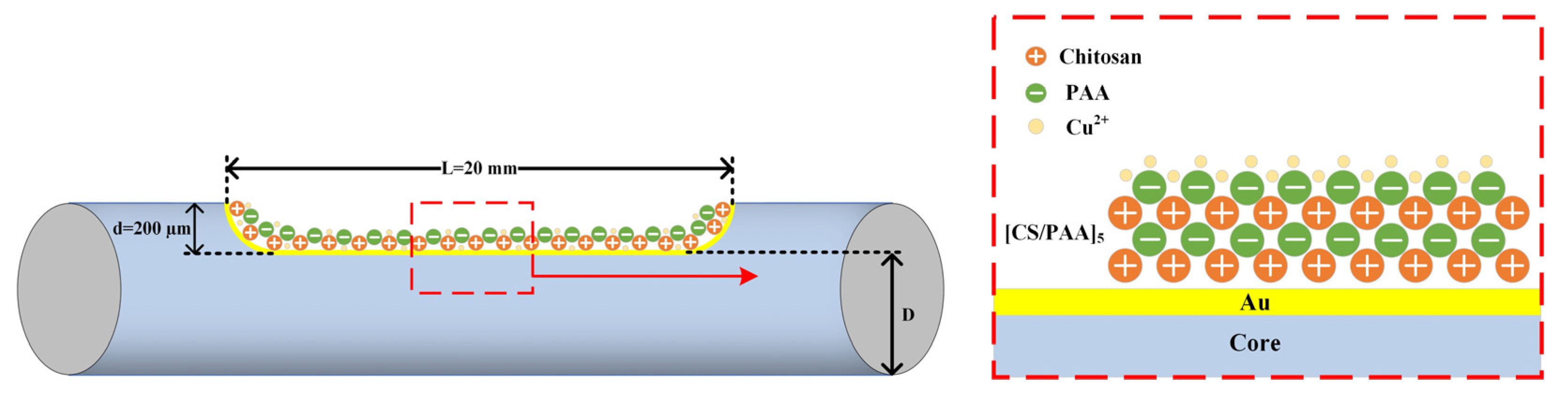
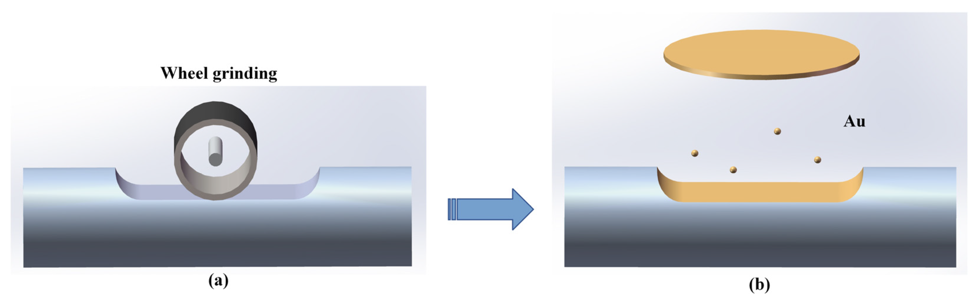
2.1. Structure and Preparation

As shown in Figure 1, the sensing probe structure consists of a gold-coated side-polish polymer optical fiber with a CS/PAA functional sensing layer. The probe employs a step-index polymer optical fiber from Jiangxi Daishing POF Co., Ltd. (Jinggangshan, China), with a fiber diameter of 500 µm, and numerical aperture of 0.5. The refractive indices of the core and cladding are 1.49 and 1.40, respectively. Here, L represents the polishing length, d denotes the polishing depth, and D indicates the residual diameter after side polishing.
The fabrication process of the gold-coated side-polish polymer optical fiber is shown in Figure 2, which primarily consists of two stages: side polishing and gold film coating. As shown in Figure 2a, the side-polish structure is fabricated using a wheel-type fiber polishing machine [27], where a 2000-mesh sandpaper is attached to the rotating wheel for grinding the optical fiber along its axis. During this process, both fiber ends are secured with fiber clamps, and the polishing parameters can be computer controlled. Different polishing depths can be achieved by controlling the wheel’s rotation time and speed, and the residual fiber thickness can be monitored and measured in real-time using a microscopic imaging system. The uncertainty polishing depth of this preparation method is about ±10 μm. After this process, the wheel polished fiber surface requires further polishing; in this step, a small amount of alumina-based polishing paste is applied to the polished surface; after 4–5 min of manual polishing, the polished surface is sufficiently smooth. Then, the side-polish polymer optical fiber is ultrasonically cleaned to remove residual paste and dried for gold film deposition. After the side polishing procedure, a gold film with a thickness of about 50 nm is deposited on the polished region of the fiber by ion sputtering [28], as shown in Figure 2b.
The CS/PAA functional film layer can be prepared onto the surface of the gold-coated optical fiber by using the layer-by-layer (LBL) electrostatic self-assembly technique. The fabrication process proceeded as follows: First, 1 g CS powder was dissolved in 4% acetic acid solution under continuous stirring for 12 h at room temperature to prepare a 1% CS solution, and then its pH was adjusted to 2 by adding hydrochloric acid solution. Subsequently, 35% PAA solution was diluted with distilled water and stirred continuously for 15 min at room temperature to obtain a 4% PAA solution; then, its pH value was adjusted to 5 by using sodium hydroxide solution. For self-assembly, the gold-coated fiber area was initially immersed in chitosan solution for 5 min, withdrawn at constant speed, and air dried naturally; then, the PAA layer was similarly assembled onto the CS layer to form the CS/PAA functional film. Multiple CS/PAA bilayers can be fabricated on the gold surface through repeated cycles of this process. Finally, the sensor was air dried for 12 h to ensure complete drying. Figure 3 shows the SEM images of the probes after polishing the surface, after gold layer deposition, and after CS/PAA coating, respectively.
The CS powder used in the above fabrication process (deacetylation degree ≥ 95%, viscosity 100–200 mPa·s) was purchased from Shanghai Macklin Biochemical Technology Co., Ltd. (Shanghai, China), the PAA solution (35 wt%) was obtained from Sigma-Aldrich (Shanghai) Trading Co., Ltd. (Shanghai, China), and hydrochloric acid and sodium hydroxide solutions were acquired from Guangzhou YuJiaGaihua Biotechnology Co., Ltd. (Guangzhou, China).

2.2. Working Principle

When light is incident on the polished area of the fiber, the transmitted light undergoes total internal reflection at the interface between the polished area and the metal, generating evanescent waves. When the horizontal wave vector of the evanescent wave matches that of the metal surface plasmon wave (SPW), the SPR effect can be excited. This wave vector matching condition can be expressed as follows [29]:
k x = ω c ε 0 ( λ ) · sin θ s p r = k s p = ω c · ε 1 ( λ ) ε 2 ( λ ) ε 1 ( λ ) + ε 2 ( λ )
where ε1 and ε2 represent the dielectric constants of the metal and surrounding medium, respectively. When the SPR effect occurs, the incident light intensity at a specific wavelength position will produce a significant decrease, forming the SPR characteristic peaks. When the ambient refractive index changes, the coupling conditions change and will result in SPR peak shifts. For Cu2+ ion concentration measurements, CS enables Cu2+ adsorption on the self-assembled film through chelation, while PAA blending enhances CS’s Cu2+ adsorption capacity [30]. The adsorption of Cu2+ ions causes a change in the refractive index of the CS/PAA sensitive membrane, consequently inducing the shift in SPR peak. Therefore, the concentration of Cu2+ ions can be obtained by monitoring the peak position of the SPR.
The normalized transmission spectra for refractive index sensing can be analyzed with a three-layer model (fiber core, gold film, and measured liquid), which can be expressed as follows [31]:
T = θ c r π 2 R G N r e f θ P θ d θ θ c r π 2 P θ d θ
where θcr is the incident light critical angle, RG is the reflectivity at the interface between fiber core and gold film, and P(θ) is the modal power for light incidence angle θ. N r e f θ = L / 2 D tan θ is the total number of reflections of the incident light on the polishing surface, and L and D are the polishing length and the remaining diameter of the fiber, respectively.
The simulation results of refractive index sensing for the side-polish polymer optical fiber SPR sensor are shown in Figure 4. In the simulation, the gold film thickness was set to 50 nm, the incident angle θ range was configured between 0.485π and 0.5π, the parameter Nref(θ) was set to 12, and the refractive indices of the fiber core and cladding were defined as 1.49 and 1.40, respectively. The simulation results demonstrate that when the external environmental refractive index increases, the SPR characteristic peak exhibits a red shift.

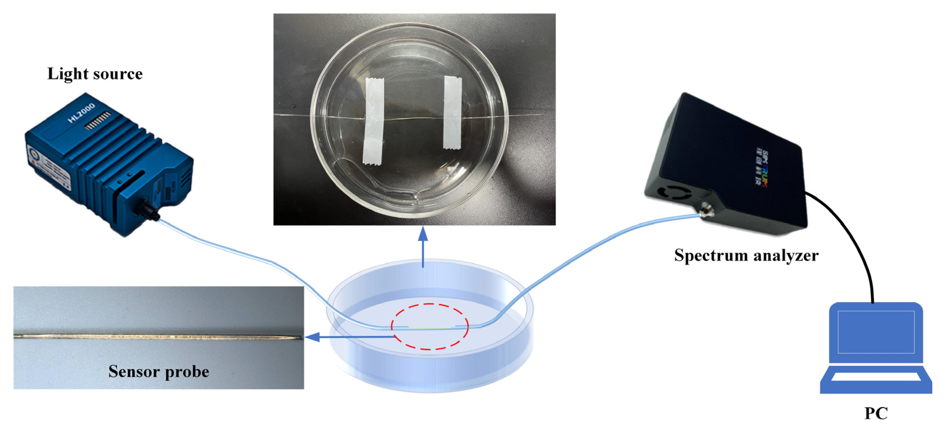
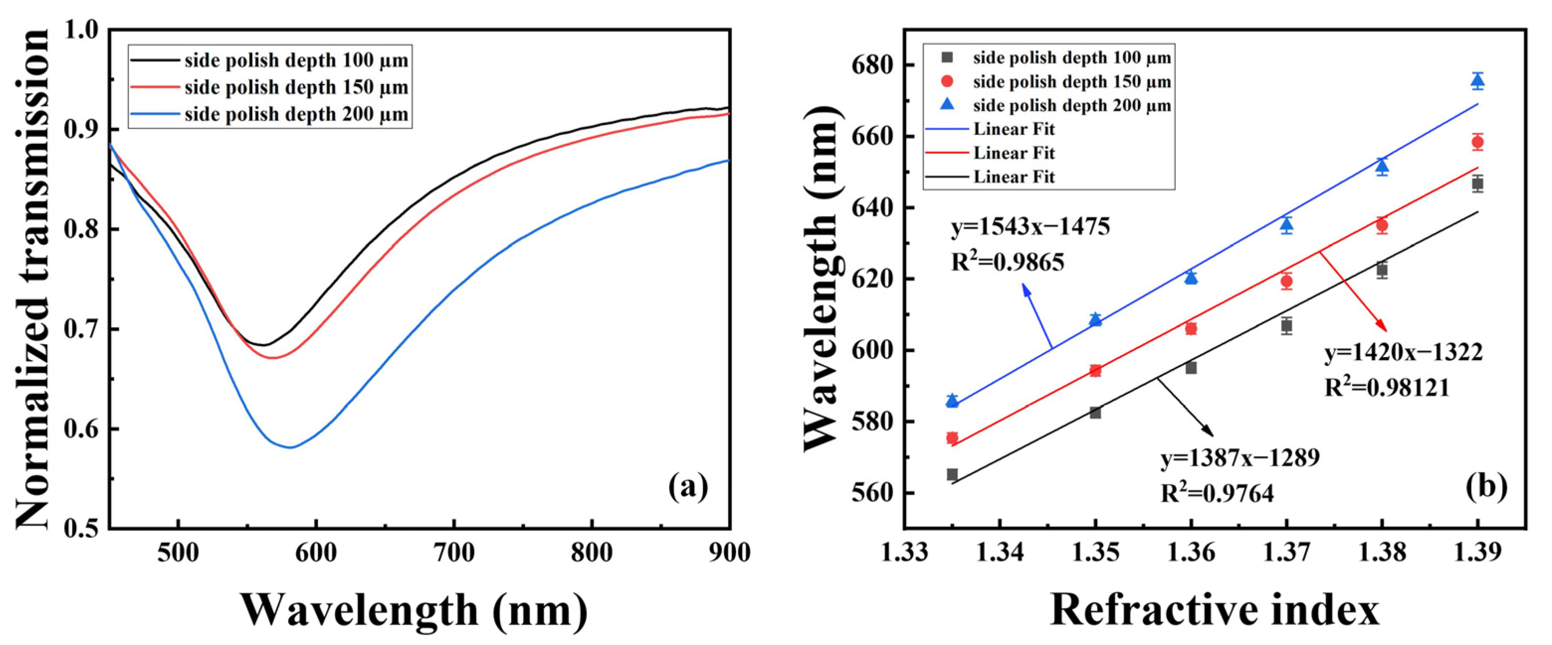
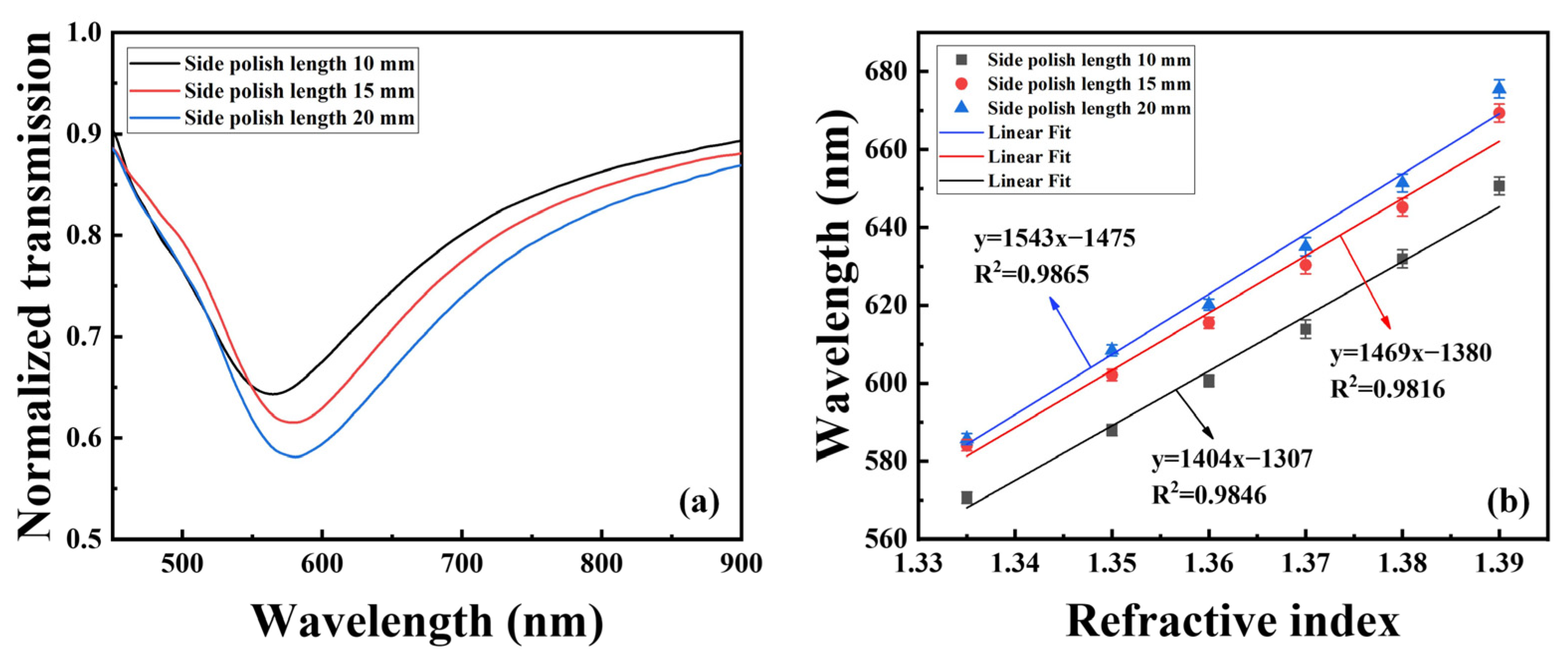
3. Experiment Setup and Structure Optimization

The experiment setup is shown in Figure 5, which consists of a halogen light source (ideaoptics, HL2000, Shanghai, China) with a wavelength range of 360–2500 nm, a spectrometer (ideaoptics, Shanghai, China) with a wavelength range of 325–1100 nm, an optical fiber sensing probe, and a computer. The two ends of the polymer optical fiber are connected to the light source and spectrometer, respectively, by using the optical fiber adapters.
To achieve an optimal sensing performance, the fiber structure needs to be optimized before preparing the CS/PAA film layer. Figure 6 presents the refractive index sensing performance of probes with polishing depths of 100, 150, and 200 μm with a polishing length of 20 mm. As shown in Figure 6a, it can be seen that the SPR peak is deepest for the probe with a polishing depth of 200 μm. Figure 6b displays the sensitivity linear curves of the probes with different depths, and the results show that the linear relationship between refractive index and wavelength position within is 1.335–1.39 RIU, and the probe with a polishing depth of 200 μm has the highest refractive index sensitivity of 1543 nm/RIU. Therefore, optical fiber with a polishing depth of 200 μm was employed for the following study.
Figure 7 presents the refractive index sensing performance of probes with polishing lengths of 10, 15, and 20 mm, respectively; all the probes had the same polishing depth of 200 μm. Figure 7a demonstrates that the sensing probe with a polishing length of 20 mm has a deeper SPR peak. From the sensitivity fitting curves in Figure 7b, it can be seen that the refractive index sensitivity of the probe with a polishing length of 20 mm is higher than that of the other two sensing probes. Consequently, the CS/PAA sensing film was fabricated on the optimized fiber probe with 200 μm polishing depth and 20 mm polishing length.

4. Results and Discussions

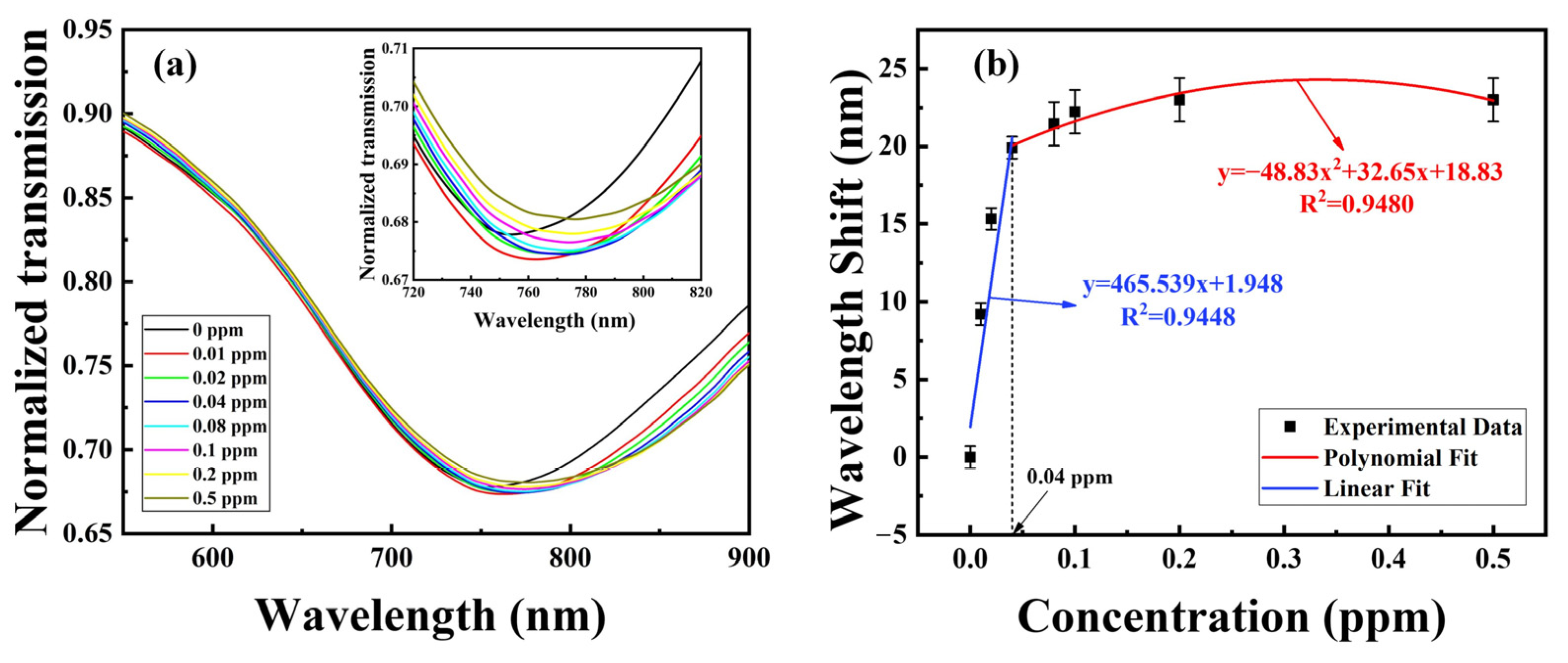
4.1. Analysis of the Spectral Response to Cu2+ Ion

Figure 8 displays the refractive indices of solutions with varying concentrations of Cu2+ ions. It can be observed that the refractive index of Cu2+ ion solutions remains constant at 1.333 within the concentration range of 0–0.5 ppm.
Figure 9a shows the spectral response of the sensor with a sensitive membrane layer of five CS/PAA bilayer structures in different concentrations of Cu2+ ion solutions. The Cu2+ ion solutions with concentrations in the range of 0.01–0.5 ppm were obtained by preparing copper chloride solutions with different concentrations. As shown in Figure 9a, the resonance peak exhibits a rightward shift with increasing Cu2+ ion concentration. This is because Cu2+ ions bind to the amino groups of chitosan through chelation and to the carboxyl groups on PAA through electrostatic interaction [30], leading to an increase in the level of cross-linking in the CS/PAA network and, consequently, an increase in the refractive index of the CS/PAA membrane layer [32]. Figure 9b presents the piecewise fitting curve between wavelength shift and Cu2+ ion concentration, revealing a linear relationship in the concentration range of 0–0.04 ppm with an average sensitivity up to 465.539 nm/ppm and a limit of detection (LOD) of 0.01 ppm. However, a nonlinear relationship was observed in the concentration range of 0–0.5 ppm, and the sensitivity decreased significantly for the high concentration of Cu2+ ion. This is because as the concentration of Cu2+ ions increases, the number of sites in the CS/PAA membrane layer used for binding Cu2+ ions gradually saturates, resulting in a small wavelength shift.

4.2. Effect of the Number of Sensitive Film Layers
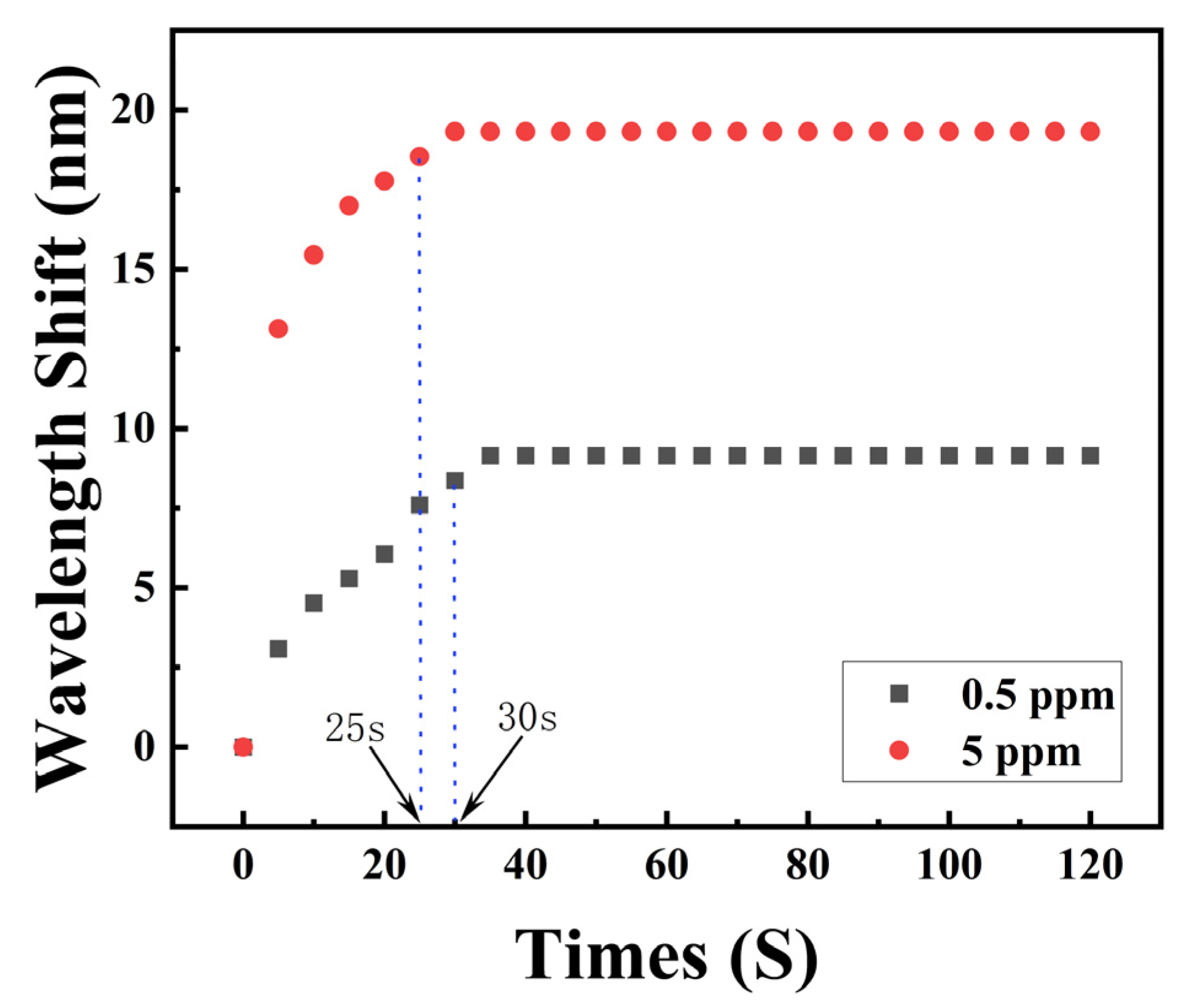
To investigate the effect of sensing bilayer number on Cu2+ ion detection, the response characteristics of sensors coated with two, five, and seven CS/PAA bilayers were compared, as shown in Figure 10. It can be seen that the five-bilayer sensor exhibited the most pronounced wavelength shift with increasing Cu2+ ion concentration, demonstrating 22.9 nm total shift from 0 ppm to 0.5 ppm concentration, which is 8.48 nm for the two-bilayer and only 5.43 nm for the seven-bilayer sensor. Furthermore, sensors with more bilayers are prone to falling off from the optical fiber along with the gold film during the preparation process, leading to device damage. Therefore, the five-bilayer structure was selected for the following experiments.

4.3. Response Time

To analyze the sensor’s response time under different concentrations, temporal responses were measured at both 0.5 ppm and 5 ppm Cu2+ ion concentrations, as shown in Figure 11. It reveals that at 0.5 ppm concentration, the sensor reached 90% of maximum wavelength shift in approximately 30 s, whereas only 25 s was required to achieve 90% of the maximum wavelength shift for 5 ppm concentration. This demonstrates that the speed of the SPR characteristic peak wavelength moving to the maximum wavelength position is related to the Cu2+ ion concentration. Higher concentrations of Cu2+ ions have a faster response time, which is mainly because the diffusion rate is higher for the solution with the higher concentrations of Cu2+ ions, and their binding rate to CS and PAA will also be faster.

4.4. Ion Selectivity

Furthermore, we investigated the ion selectivity of the sensor and compared the wavelength shifts of Na+, Ca2+, Cu2+, and Cr6+ ions with concentrations ranging from 0.02 to 0.5 ppm. As shown in Figure 12, the resonance wavelength shifts caused by Na+, Ca2+, and Cr6+ were significantly less pronounced than those induced by Cu2+. This occurs because CS cannot chelate alkali metals, resulting in a smaller response to Na+. Although the sensor responds to Ca2+, its sensitivity is low. This is because CS does not chelate Ca2+, whereas PAA has an affinity for Ca2+. The line graph on the right side demonstrates the sensing sensitivity of the sensor to these ions, and it can be seen that the sensor’s sensitivity to Na+, Ca2+, and Cr6+ at low concentrations was substantially inferior to its Cu2+ sensitivity. Therefore, the sensor exhibits good specificity for Cu2+ ion detection.

4.5. Stability

To test the stability of the proposed probe, the probe was immersed in a 0.5 ppm Cu2+ solution for 5 h, and the transmission spectra were recorded at 0.5 h intervals. The time-dependent resonance wavelength shift is shown in Figure 13. The results demonstrate that the CS/PAA sensing film exhibits excellent long-term stability.
To investigate the effect of temperature on the sensor, the probe was immersed in water at different temperatures from 25 °C to 65 °C. Figure 14 depicts the relationship between the SPR peak wavelength shift and temperature. It can be observed that as temperature increases, the resonance peak shifts toward shorter wavelengths. This may be attributed to the thermal expansion of the sensitive film, which leads to a decrease in the refractive index due to swelling. The test results demonstrate that the sensor exhibits temperature instability. Therefore, a stable temperature environment is required for the practical applications of this sensor.
Table 1 presents a comparison of Cu2+ ion sensing performance between the proposed sensor and other types of optical fiber SPR sensors. The results demonstrate that the side-polish polymer optical fiber SPR sensor proposed in this paper achieves a higher sensitivity for the probe with the CS/PAA sensing film, and it has a better suitability for practical applications because of its simple structure, easy preparation, and better mechanical strength.

5. Conclusions

This study presents a side-polish polymer optical fiber-based SPR sensor for Cu2+ ion detection in water. The functional sensitive layer of CS/PAA was fabricated on the gold-coated polymer optical fiber surface. The structure of the optical fiber probe was optimized. The sensing performance of the sensor to Cu2+ ion, the effect of the number of the CS/PAA functional membrane layer, and the response time and ion selectivity of the sensor were experimentally investigated. Experimental results demonstrate that the five-bilayer CS/PAA self-assembled film exhibits an optimal Cu2+ sensing performance, which achieves an average sensitivity of 465.539 nm/ppm within the 0–0.04 ppm concentration range, and the sensor has a rapid response speed, as well as good selectivity for Cu2+ ions. In addition, the sensor has the advantages of simple fabrication, low cost, and stable mechanical structure, showing promising potential for water quality monitoring applications.

Author Contributions

Conceptualization, C.T.; methodology, C.T. and S.Y.; software, R.Y. and H.Q.; validation, R.Y. and S.Y.; formal analysis, R.Y. and S.Y.; investigation, C.T., R.Y. and S.Y.; resources, C.T. and Z.C.; data curation, R.Y. and S.Y.; writing—original draft preparation, R.Y. and S.Y.; writing—review and editing, C.T., H.X. and H.Q.; visualization, L.S.; supervision, L.Y.; project administration, C.T.; funding acquisition, C.T., S.D. and Y.Z. All authors have read and agreed to the published version of the manuscript.

Funding

This research was funded by Natural Science Foundation of Guangxi Province, grant number 2023GXNSFDA026040; Central Guiding Local Science and Technology Development Project, grant number ZY23055018; Guangxi Key Research and Development Project, grant number AB25069261; National Natural Science Foundation of China, grant number 62365003, W2421029, 61965009, 62364006, U23A20282; and Innovation Project of GUET Graduate Education, grant number 2023YCXS217.

Institutional Review Board Statement

Not applicable.

Informed Consent Statement

Not applicable.

Data Availability Statement

The original data presented in this study are included in the article. Further inquiries can be directed to the corresponding authors.

Conflicts of Interest

Author Zining Chen was employed by the company Jiangxi Daishing POF Co., Ltd. The remaining authors declare that the research was conducted in the absence of any commercial or financial relationships that could be construed as a potential conflict of interest.

References

  1. Roli, V.; Banshi, D.G. Detection of heavy metal ions in contaminated water by surface plasmon resonance based optical fiber sensor using conducting polymer and chitosan. Food Chem. 2015, 166, 568–575. [Google Scholar]
  2. Wan Ngah, W.S.; Fatinathan, S. Adsorption of Cu (II) ions in aqueous solution using chitosan beads, chitosan–GLA beads and chitosan–alginate beads. Chem. Eng. J. 2008, 143, 62–72. [Google Scholar] [CrossRef]
  3. Mohammadi, A.; Ranjith, K.S.; Vilian, A.E.; gil Lee, S.; Won, J.; Huh, Y.S.; Han, Y.-K. Synergistic on engineering layered N-doped carbon/MXene heterostructure: A potential scaffold for simultaneous electrochemical detection of Cu2+ and Hg2+ ions. Sens. Actuators B Chem. 2025, 422, 136661. [Google Scholar] [CrossRef]
  4. Li, Z.; Che, T.; Yang, M.; Hu, X. Flame atomic absorption spectrometry combined with surface-modified magnetic mesoporous silica microspheres by polyethyleneimine for enrichment, isolation and determination of Cu2+ in preserved eggs after high-temperature digestion. Food Addit. Contam. Part A 2022, 39, 1828–1842. [Google Scholar] [CrossRef]
  5. You, Y.; Li, D.; Chen, Z.; Zhang, X.; Hu, Y.; Ouyang, S.; Li, N. Fluorescent and colorimetric dual-mode detection of Cu2+ based on carbon dots. Microchim. Acta 2024, 191, 563. [Google Scholar] [CrossRef]
  6. Liu, S.; Zheng, L.-N.; Sun, Y.-Z.; Dong, S.-W.; Xue, Q.-W.; Xue, N.; Ding, T. Highly stable Zn (II)-based metal-organic framework as a bifunctional fluorescence sensor for efficient detection of Cu2+ and CrO42− ions. J. Mol. Struct. 2025, 1326, 141094. [Google Scholar] [CrossRef]
  7. Grattan, K.T.V.; Sun, T. Fiber optic sensor technology: An overview. Sens. Actuators A Phys. 2000, 82, 40–61. [Google Scholar] [CrossRef]
  8. Yang, J.; Chen, L.H.; Zheng, Y.; Dong, X.; Raghunandhan, R.; So, P.L.; Chan, C.C. Heavy metal ions probe with relative measurement of fiber Bragg grating. Sens. Actuators B Chem. 2016, 230, 353–358. [Google Scholar] [CrossRef]
  9. Ravikumar, R.; Chen, L.H.; Chan, C.C.; So, P.L.; Tou, Z.Q.; Peng, Z. Chitosan-hydrogel-based fiber optic sensor for heavy metal ion detection. In Proceedings of the 24th International Conference on Optical Fibre Sensors, Curitiba, Brazil, 28 September–2 October 2015. [Google Scholar]
  10. Raikar, U.S.; Kulkarni, V.K.; Lalasangi, A.S.; Pattanashetti, I.I.; Akki, J.F. Evanescent field absorption sensor for detection of copper (II) in water using multimode optical fiber. Optoelectron. Lett. 2009, 5, 224–226. [Google Scholar] [CrossRef]
  11. Ding, Z.W.; Ravikumar, R.; Zhao, C.L.; Chen, L.H.; Chan, C.C. Chitosan/Poly (Acrylic Acid) Based Fiber-Optic Surface Plasmon Resonance Sensor for Cu2+ Ions Detection. J. Light. Technol. 2019, 37, 2246–2252. [Google Scholar] [CrossRef]
  12. Zhang, M.; Zhu, G.; Li, T.; Lou, X.; Zhu, L. A dual-channel optical fiber sensor based on surface plasmon resonance for heavy metal ions detection in contaminated water. Opt. Commun. 2020, 462, 124750. [Google Scholar] [CrossRef]
  13. Huang, Q.; Zhu, W.; Wang, Y.; Deng, Z.; Li, Z.; Peng, J.; Lyu, D.; Lewis, E.; Yang, M. Optical fiber plasmonic sensor for the ultrasensitive detection of copper (II) ion based on trimetallic Au@ AgPt core-shell nanospheres. Sens. Actuators B Chem. 2020, 321, 128480. [Google Scholar] [CrossRef]
  14. Shrivastav, A.M.; Gupta, B.D. Ion-imprinted nanoparticles for the concurrent estimation of Pb (II) and Cu (II) ions over a two channel surface plasmon resonance-based fiber optic platform. J. Biomed. Opt. 2018, 23, 017001. [Google Scholar]
  15. Maleki, M.J.; Soroosh, M.; Akbarizadeh, G. A compact low-loss 2-to-4 plasmonic decoder based on suspended graphene for surface plasmon polariton transmission. Diam. Relat. Mater. 2024, 144, 110983. [Google Scholar] [CrossRef]
  16. Brolo, A. Plasmonics for future biosensors. Nat. Photon 2012, 6, 709–713. [Google Scholar] [CrossRef]
  17. Homola, J.; Slavik, R. Fiber-optic sensor based on surface plasmon resonance. Electron. Lett. 1996, 32, 480. [Google Scholar] [CrossRef]
  18. Coelho, L.C.C.; de Almeida, J.M.M.M.; Moayyed, H.; Santos, J.L.; Viegas, D. Multiplexing of Surface Plasmon Resonance Sensing Devices on Etched Single-Mode Fiber. J. Light. Technol. 2015, 33, 432–438. [Google Scholar] [CrossRef]
  19. Wang, X.M.; Zhao, C.L.; Wang, Y.R.; Shen, C.Y.; Dong, X.Y. A Highly Sensitive Fibre-Optic Nano-Displacement Sensor Based on Surface Plasmon Resonance. J. Light. Technol. 2016, 34, 2324–2330. [Google Scholar] [CrossRef]
  20. Patnaik, A.; Senthilnathan, K.; Jha, R. Graphene-Based Conducting Metal Oxide Coated D-Shaped Optical Fiber SPR Sensor. IEEE Photonics Technol. Lett. 2015, 27, 2437–2440. [Google Scholar] [CrossRef]
  21. Zhao, Y.; Cai, L.; Hu, H.F. Fiber-Optic Refractive Index Sensor Based on Multi-Tapered SMS Fiber Structure. IEEE Sens. J. 2015, 15, 6348–6353. [Google Scholar] [CrossRef]
  22. Falah, A.A.S.; Wong, W.R.; Adikan, F.R.M. Single-mode eccentric-core D-shaped photonic crystal fiber surface plasmon resonance sensor. Opt. Laser Technol. 2022, 145, 107474. [Google Scholar] [CrossRef]
  23. Gonzalez-Valencia, E.; Reyes-Vera, E.; Del Villar, I.; Torres, P. Side-polished photonic crystal fiber sensor with ultra-high figure of merit based on Bloch-like surface wave resonance. Opt. Laser Technol. 2024, 169, 110129. [Google Scholar] [CrossRef]
  24. Teng, C.; Min, R.; Zheng, J.; Deng, S.; Li, M.; Hou, L.; Yuan, L. Intensity-modulated Polymer Optical Fiber-based Refractive Index Sensor: A Review. Sensors 2021, 22, 81. [Google Scholar] [CrossRef]
  25. Boruah, B.S.; Biswas, R. Localized surface plasmon resonance based U-shaped optical fiber probe for the detection of Pb2+ in aqueous medium. Sens. Actuators B Chem. 2018, 276, 89–94. [Google Scholar] [CrossRef]
  26. Bhavsar, K.; Prabhu, R.; Pollard, P. Development of dithizone based fibre optic evanescent wave sensor for heavy metal ion detection in aqueous environments. J. Phys. Conf. Series. IOP Publ. 2013, 450, 012011. [Google Scholar] [CrossRef]
  27. Zhang, Y.; Wang, L.; Liu, Z. The polishing detection method of side-polished fiber. In Proceedings of the 2011 International Conference on Optical Instruments and Technology, Beijing, China, 23 November 2011. [Google Scholar]
  28. Liu, L.; Zheng, J.; Deng, S.; Yuan, L.; Teng, C. Teng. Parallel Polished Plastic Optical Fiber-Based SPR Sensor for Simultaneous Measurement of RI and Temperature. IEEE Trans. Instrum. Meas. 2021, 70, 1–8. [Google Scholar]
  29. Raether, H. Surface plasmons on smooth and rough surfaces and on gratings. Springer 2006, 111, 4–39. [Google Scholar]
  30. Yan, H.; Yang, L.; Yang, Z.; Yang, H.; Li, A.; Cheng, R. Preparation of chitosan/poly (acrylic acid) magnetic composite microspheres and applications in the removal of copper (II) ions from aqueous solutions. J. Hazard. Mater 2012, 229, 371–380. [Google Scholar] [CrossRef]
  31. Velazquez-Gonzalez, J.S.; Monzon-Hernandez, D.; Martinez-Pinon, F.; May-Arrioja, D.A.; Hernandez-Romano, I. Surface plasmon resonance-based optical fiber embedded in PDMS for temperature sensing. IEEE J. Sel. Top. Quantum Electron. 2016, 23, 126–131. [Google Scholar] [CrossRef]
  32. Raghunandhan, R.; Chen, L.H.; Long, H.Y.; Leam, L.L.; So, P.L.; Ning, X.; Chan, C.C. Chitosan/PAA based fiber-optic interferometric sensor for heavy metal ions detection. Sens. Actuators B Chem. 2016, 233, 31–38. [Google Scholar] [CrossRef]
Figure 1. A diagram of the sensor probe structure.
Photonics 12 00461 g001
Figure 2. Schematic of the fabrication process of gold-coated side-polish fiber: (a) side polishing process; (b) gold film coating.
Photonics 12 00461 g002
Figure 3. The SEM images of the probes after polishing surface (a), after gold layer deposition (b), and after CS/PAA coating (c).
Photonics 12 00461 g003
Figure 4. Simulation of SPR peaks with different refractive indices.
Photonics 12 00461 g004
Figure 5. A diagram of the experimental setup.
Photonics 12 00461 g005
Figure 6. (a) Transmission spectra of probes with different polishing depths in water; (b) the relationships between the SPR peaks and the refractive index for probes with different polishing depths.
Photonics 12 00461 g006
Figure 7. (a) Transmission spectra of probes with different polishing lengths in water; (b) the relationships between the SPR peaks and the refractive index for probes with different polishing lengths.
Photonics 12 00461 g007
Figure 8. The refractive indices of Cu2+ ion solutions with varying concentrations.
Photonics 12 00461 g008
Figure 9. (a) Transmission spectra for the probe with different concentrations of Cu2+ ions; (b) piecewise fitting curve between wavelength shift and Cu2+ ion concentration.
Photonics 12 00461 g009
Figure 10. Wavelength shift in sensor probes with different CS/PAA bilayer numbers.
Photonics 12 00461 g010
Figure 11. Response time of the sensor in Cu2+ ion solutions with 0.5 ppm and 5 ppm concentrations.
Photonics 12 00461 g011
Figure 12. Wavelength shifts caused by Na+, Ca2+, Cu2+, and Cr6+ ions (histogram); the sensitivity of the sensor for Na+, Ca2+, Cu2+, and Cr6+ ions (line chart).
Photonics 12 00461 g012
Figure 13. The wavelength shift versus time for the proposed probe in 0.5 ppm Cu2+ solution.
Photonics 12 00461 g013
Figure 14. The relationship between the wavelength shift and temperature variation.
Photonics 12 00461 g014
Table 1. Performance comparison of different optical fiber SPR Cu2+ ion sensors.
StructureSensitive MaterialsSensitivity (nm/ppm)Measurement Range (ppm)Ref.
Side-polish polymer optical fiberCS/PAA465.5390.01–0.5This work
MMF-NCF-MMF pull taperCS/PAA1.86312.71–635.5[11]
PCS optical fiber strippingCS/PAA3.9191.27–635.5[12]
MMF-SMF-MMFPEI-Au@AgPt NS-6.355 × 10⁻¹²–6.355 × 10⁻⁷[13]
PCS optical fiber strippingIon-imprinted nanoparticles40,7000–1000[14]
Disclaimer/Publisher’s Note: The statements, opinions and data contained in all publications are solely those of the individual author(s) and contributor(s) and not of MDPI and/or the editor(s). MDPI and/or the editor(s) disclaim responsibility for any injury to people or property resulting from any ideas, methods, instructions or products referred to in the content.

