Article

Extraction, Chemical Characterization, In Vitro Antioxidant, and Antidiabetic Activity of Canola (Brassica napus L.) Meal

by
Saira Hussain
1,2,3,*,
Ata Ur Rehman
1,2,
Hassan K. Obied
3,
David J. Luckett
2 and
Christopher L. Blanchard
1,2,3
1
ARC Industrial Transformation Training Centre for Functional Grains & Graham Centre for Agricultural Innovation, Boorooma Street, Wagga Wagga, NSW 2650, Australia
2
Graham Centre (An Alliance between Charles Sturt University and the NSW Department of Primary Industries), Boorooma Street, Wagga Wagga, NSW 2678, Australia
3
School of Biomedical Sciences, Charles Sturt University, Boorooma Street, Wagga Wagga, NSW 2678, Australia
*
Author to whom correspondence should be addressed.
Separations 2022, 9(2), 38; https://doi.org/10.3390/separations9020038
Submission received: 21 November 2021 / Revised: 13 January 2022 / Accepted: 16 January 2022 / Published: 30 January 2022
(This article belongs to the Section Analysis of Natural Products and Pharmaceuticals)

Abstract

:
Canola (Brassica napus L.) meal is a by-product after oil extraction from canola seed and is of relatively low value. This meal may have additional value in the biotechnology, food, and pharmaceutical industries if health-promoting useful bioactive compounds can be identified. Hence, seven canola meal extracts (CMEs) were generated using different organic solvents for two genotypes. HPLC and LCMS analyses were employed for the determination of the phenolic and antioxidant activity of meal extracts, including recovery of major biological compounds. When comparing genotype-1 with genotype-2, the latter had higher antioxidant activity in acetone extract (AE). This study also indicated seven major glucosinolates in CMEs in which water (WE) appeared to be the best solvent for the recovery of glucosinolates. Higher quantities of phenolic, glucosinolate, and antioxidant were present in genotype-2 compared with genotype-1. Using HPLC-DAD and LC-MS analysis 47 compounds were detected. We could identify 32 compounds in canola meal extracts: nine glucosinolates and twenty-three phenolic derivatives. Phenolic compounds in canola meal were conjugates and derivatives of hydroxycinnamic acid (sinapic, ferulic, and caffeic acids). Among phenolics, kaempherol as conjugate with sinapic acid was found; sinapine and trans-sinapic acid were the most abundant, as well as major contributors to the antioxidant and free radical scavenging activities of canola meal extracts. Some samples exhibited mild to moderate in-vitro antidiabetic activity in a Dipeptidyl Peptidase-IV inhibition assay.

1. Introduction

Canola was first used in Canada in the 1970s through breeding to reduce toxicity and produce highly valuable consumable rapeseed varieties [1]. Canola meal is a waste product of the oil extraction of canola, which is usually produced by solvent extraction procedure along with the oil. Hence, the extraction process is essential to make high-quality meals having low levels of anti-nutritional factors. An ideal extraction procedure has been optimized to deliver a high yield of chemical(s) in a relatively pure form using a simple, cheap, safe, reliable, and repeatable method [2]. Phenolic compounds are key secondary metabolites in all plants and are reported for their potential health effects [3]. Therefore, optimal extraction is a prerequisite in laboratory-based research for the subsequent identification and classification of individual chemicals; it is also required in an industry where the combined extraction and purification process must be financially viable [4]. For extraction of plant phenols, many solvents have been tried, including neat solvents or solvent mixtures [5]. The organic solvent choice and its percentage are primarily dependent on the plant matrix’s phenolic composition and nature [6]. Selecting a suitable solvent for the extraction of phytochemicals from canola meals using different genotypes is a crucial step in subsequent chemical analysis. It determines the type and the number of compounds that will be recovered. No doubt, the prime characteristic of plant phenols is to have their antioxidant properties. The need for natural antioxidants is at an all-time high to fight oxidative stress [7] and many chronic diseases, including diabetes [8]. Therefore, antioxidant activity is considered a multidimensional property that should be assessed using multiple assays for a more accurate evaluation.
Diabetes mellitus (DM) is a metabolic syndrome caused by a high level of blood glucose following deficiencies in insulin secretion, insulin action, or both [9], where the enzyme dipeptidyl peptidase-IV (DPP-IV) is involved. DM is widely spread worldwide [10], with an increasing rate of occurrence in Australia [11]. A range of health problems is recognized to be linked with DM [12]. DPP-IV is a serine protease that removes dipeptides from the N-terminus of the glucose-dependent insulinotropic polypeptide (GIP) and glucose-like peptide (GLP-1) [13]. This cleavage makes them inactive, rendering their half-life of 1–2 min [14]. Hence, there is a need to identify inhibitors for DPP- IV with minimal side effects, which may be able to enhance the amount of endogenous active GLP-1, GIP, and insulin secretion.
Canola oil as a cardioprotective substance [15], has lots of applications, with potential oxidative activity in neutrophils in vivo [16], including people with central adiposity [17]. However, there is limited research for its meal produced after the extraction of oil. Canola meal is used mainly for animal feed. However, this meal may have additional value in the pharmaceutical industry if bioactive compounds with potential health benefits in reducing the risk of disease or optimal nutrition are identified. The present work was aimed to examine the power of hydro-organic solvents in extracting canola meal phenols. In addition to aqueous-acetone, aqueous-methanol, and aqueous-ethanol mixtures, water and aqueous mixtures with butanol, chloroform, and hexane were also investigated for the first time to determine phytochemicals and antioxidant activity. A recovery of major compounds in seven extracts of canola meal was also performed. Additionally, antioxidant and antidiabetic activity of all the extracts were assessed.

2. Materials and Methods

Canola seed samples from two different cultivars, Bln-3347 (genotype-1) and Rivette (genotype-2) were obtained from the New South Wales Department of Primary Industries (NSW DPI) Wagga Wagga, Australia. Genotype-1 is a local, open-pollinated breeding line with high-yielding, well-adapted, early-flowering with high blackleg resistance. Genotype-2 is an open-pollinated commercial cultivar with a typical, local cultivar with mid-flowering, high blackleg resistance, and good adaptation (high yield and canola quality).
HPLC-grade methanol, n-hexane, n-butanol, and acetone were purchased from Fisher Scientific (Pittsburgh, PA, USA). Glacial acetic acid and hydrochloric acid were acquired from Merck (Melbourne, Australia). Dipeptidyl peptidase-IV (DPP-IV, CD26) from porcine kidney and Diprotin-A (Ile-pro-ile) were purchased from Merck (Darmstadt, Germany).
Anhydrous acetonitrile, chloroform and ethanol were obtained from UNICHROME (Sydney, Australia). Sodium carbonate, ferric chloride, Folin–Ciocalteu’s reagent, (±)-6-hydroxy-2,5,7,8-tetramethylchromane-2-carboxylic acid (Trolox), 2,2′-azinobis (3-ethylbenzothiazoline-6-sulfonic acid) diammonium salt (ABTS), 2-4-6-tripyridyl-s-triazine (TPTZ), potassium persulphate, naringenin, gallic acid, catechin hydrate, caffeic acid, chlorogenic acid, ferulic acid, rutin, luteolin, sinapic acid, trans-cinnamic acid, 4-hydroxybenzoic acid, phenylisothiocyanate, quercetin, ellagic acid, tyramine, pyrogallol, and vanillic acid were purchased from Sigma Aldrich (Sydney, Australia).
Epicatechin and kaempferol were purchased from Extrasynthese (Genay, France). Membrane nylon filters (0.22 μm) were obtained from Sigma Aldrich (Sydney, Australia). Gly-pro-p-nitroanilide, and tris hydrochloride (Tris-HCl) were obtained from Sigma Aldrich (Sydney, Australia). Syringe filters (0.22 μm) were purchased from Millex GP (Sydney, Australia). Water was prepared by GenPure water purification system (0.22 μm filter) ThermoFisher Scientific (Melbourne, Australia).

2.1. Preparation of Canola Meal

Approximately 200 g of seeds were used for canola meal (CM) preparation by grinding seed four times using a mechanical grinder (Foss Knifetec™ 1095, Slangerupgade, Denmark). The oil was removed from the ground seeds using soxhlet (soxtec™ 2050, Tector™ Technology, Slangerupgade, Denmark) with Buchi B-811 Extraction System (BUCHI Labortechnik, Flawil, Switzerland) using absolute n-hexane for 16 h. The remaining meal after oil extraction was air-dried, mixed to remove lumps, and left overnight in a fume hood at room temperature to remove all residual hexane. The resultant material was the base meal used for extraction purposes in all subsequent analyses.

2.2. Preparation of Canola Meal Extracts (CMEs)

Exactly 5-g samples of CM were dissolved in either 100 mL in water or 80% organic solvent in water (v/v). Organic solvents used were methanol, ethanol, acetone, butanol, chloroform, and hexane. These solvents were mixed with CM using a vortex (Super-Mixer Lab-Line Instruments, Melrose Park, IL, USA) and then continuously mixed in a suspension mixer (Ratex, Rowe Scientific Pty Ltd., Sydney, NSW, Australia) for 12 h at room temperature, then centrifuged (Eppendorf centrifuge 5810R) at 3000 rpm for 30 min to produce a supernatant extract. Samples of supernatant were filtered using a syringe filter for maximum recovery of the soluble extract.
These extracts were transferred to a round bottom flask and passed through a rotary evaporator (BUCHI Labortechnik, Flawil, Switzerland) for solvent removal. The samples were then dissolved in a minimum amount of water, after which the whole extract was stored at −80 °C for approximately 12 h, before being freeze-dried using a Christ-Alpha 2-4 LD Plus freeze dryer (Biotech International, Osterode, Germany). The freeze-drying was done for 72 h, after which the samples were stored at −20 °C until required. This freeze-dried material is “extract” and is used frequently in all assays and experiments. All canola meal extracts (CMEs) were named according to the solvent used for extraction with a meal, such as water extract (WE), methanol extract (ME), ethanol extract (EE), acetone extract (AE), butanol extract (BE), chloroform extract (CE) and hexane extract (HE).

2.3. Total Phenolic Contents (TPC)

All freeze-dried extracts were mixed with 50% methanol and filtered for measuring total phenolic content using Folin-Ciocalteu reagent as reported earlier [17]. All results were repeated three times.

2.4. High-Performance Liquid Chromatography-Diode Array Detection with Online ABTS (2,2′-Azinobis(3-ethylbenzothiazoline-6-sulfonic Acid) Diammonium Salt) Scavenging (HPLC-DAD-Online ABTS)

Phenolic compounds were characterized using HPLC-DAD and LC-MS, where all CMEs were prepared as 15 mg/10 mL in 50% of methanol-water mixture and filtered. All samples were processed in the vortex and/or underwent sonication where required for complete solubilization. For the HPLC, the same conditions were maintained as described by Obied et al. [1], with modification of time used for each run.
All samples were analyzed with three replicates by a reversed-phase column (150 mm × 4.6 mm (i.d.), 3 μm) Gemini C-18 (Phenomenex, Sydney, Australia). The HPLC-DAD online ABTS scavenging system consists of a Varian Prostar 240 solvent delivery process, a Varian Prostar 335 diode array detector, and a Varian Prostar 410 autosampler. The blank (50% aqueous methanol) and standards were also analyzed for quality control and identification. The system was initiated and equilibrated with 5% solvent B, and then solvent B increased to 80% over 70 min. All solvent gradients were filtered (Nylon filter, 0.22 μm). The HPLC-DAD flow rate was maintained at 0.7 mL/min, while the injection volume was 10 μL. Online ABTS with HPLC-DAD was performed as described [1] with three replicates, while data analysis was performed using Star Chromatography workstation version 6.41 2004 (Varian, Inc., Walnut Creek, CA, USA).

2.5. Liquid Chromatography-Mass Spectrometry (LC-MS) for Identification of Canola Meal Extracts (CMEs)

For the HPLC-DAD-MS/MS, all conditions were maintained as described by Obied et al. [1] with the following modifications. All samples were analyzed by Agilent 1200 series liquid chromatograph (Agilent Technologies, Waldbronn, Germany) using gradient elution and a Gemini C-18 column [50 mm × 4.6 mm (i.d.), 3 μm], (Phenomenex, Sydney, Australia) using three replicates. The total run time was 70 min. A gradient consisting of five steps was as follows: initial condition, 5% solvent B; followed by solvent B increased to 80% over 65 min; then solvent B increased to 100% over 2 min; back to initial conditions in 3 min and finally, the system was isocratic for 15 min. The effluent from the DAD was connected to a 6410 triple-quadrupole LC-MS (Agilent Technologies, Santa Clara, CA, USA) equipped with an electrospray ionization (ESI) interface.
The MS procedure was performed in both the negative and positive ion mode (m/z 100–1200) under the following conditions: nitrogen gas; at a temperature of 350 °C; with a gas flow rate of 9 L/min and a nebulizer pressure, 40 psi. The capillary voltage was 4 kV, and the cone voltage, 100 V. Results were analyzed using an Agilent Mass Hunter workstation version B.01.04 2008 (Agilent Technologies, Waldbronn, Germany).

2.6. ABTS Assay Radical Scavenging Activity

The scavenging activity of seven different extracts against the ABTS was measured in 96-well microplates by Greiner, VWR, USA as described by Obied et al. [1]. Trolox was used as a control, and all samples were used with three replicates.

2.7. Ferric Reducing Antioxidant Power (FRAP) Assay

The FRAP assay was carried out as described by Benzie and Strain [18,19] and modified to a 96-well microplate [20] with the following additional steps. Three reagents were prepared for this assay named reagent A, B, and C. Reagent A consisted of 300 mM acetate buffer at pH 3.6 (16 mL of glacial acetic acid in 3.1 g of sodium acetate trihydrate, made to a total volume of 1 L with water). Reagent B was prepared by using 5 mM of TPTZ in 10 mL of 40 mM of HCl. Reagent C was prepared using 5 mM of ferric chloride in 10 mL of water. Reagents B and C were always prepared fresh. Finally, FRAP reaction mixture was prepared by mixing 2.5 mL of each reagents B and C, and 25 mL of reagent A, making a total volume of 30 mL for FRAP reagent. This reagent mixture was heated at 37 °C for 10 min.
Samples and standard solutions were prepared in 50% methanol. For the FRAP assay, 10 µL of the sample (1 mg/mL) was mixed with 40 µL of water, to which 200 µL of FRAP reagent assay mixture was added. Change in absorbance was measured at 593 nm using FLUOstar omega UV-VIS spectrophotometer (BMG Labtech, Offenburg, Germany). Trolox (1000 µM/mL) was used as a standard. All samples were used in three replicates, and results for FRAP assays were expressed as Trolox equivalents per 100 g of dry weight (mM TE/100 g DW).

2.8. Antidiabetic Assay

The antidiabetic activity was conducted as described by Lacroix and Li-Chan [21] with slight modifications. All samples were prepared in 100 mM Tris-HCl buffer. The assay was carried out in 96 micro-well plates using Diprotin-A (Ile-Pro-Ile) as a standard inhibitor. A total reaction volume of 250 μL was prepared and consisted of 100 mM Tris-HCl buffer (pH 8.4), 7.5 μL of DPP-IV enzyme (0.2 U/mL) and 232.5 μL of either test samples, standard inhibitor or the buffer as blank. This mixture was incubated at 37 °C for 30 min, followed by the addition of 10 μL of 1.59 mM Gly-Pro-p-nitroanilide (substrate). The reaction mixture was mixed and further incubated at 37 °C for 30 min. All samples were used in three replicates. The absorbance at 410 nm was measured in a microplate (FLUOstar Omega microplate reader; BMG Labtech, Offenburg, Germany).
The percentage inhibition of DPP-IV was calculated as:
Inhibition ( % ) = Absorbance control Absorbance inhibitor Absorbance control × 100
Here, the control corresponds to the enzyme while the inhibitor corresponds to sample absorbance. The results were expressed as the IC50 value or concentration (the sample concentration needed to inhibit DPP-IV by 50%. The IC50 of each sample was calculated from the least-squares regression line of the plot of the logarithm of the sample concentration (log) versus the DPP-IV inhibition activity (%).

2.9. Quantitative Analysis of Major Compounds from Canola Meal Extracts (CMEs)

All seven extracts were analyzed for the quantitative determination of sinapine, feruloyl choline, kaempherol-sinapoyl-trihexoside, sinapic acid, and feruloyl choline (4-O-8) guaiacyl di sinapoyl. Quantitative HPLC-DAD measured the amounts of these compounds. Peak areas from duplicate extracts were measured at 280 nm, and the mean was taken. Sinapic acid was used as a standard sample for quantification purposes, and all recoveries of samples were measured as milligram of sinapic acid equivalent per gram of dry weight (mg SAE/g DW).

2.10. Statistical Analysis

All experiments were done in triplicates. Data are presented as the mean ± standard deviation (SD), and results were explored using Graph pad prism 5, Microsoft Excel 2017, and one-way analysis of variance (ANOVA) using statistical analysis system SAS® system for Window V8 (SAS Institute, Carey, NC, USA). Differences between sample means were evaluated using the Duncan multiple range test at a 5% probability level (p < 0.05).

3. Results and Discussion

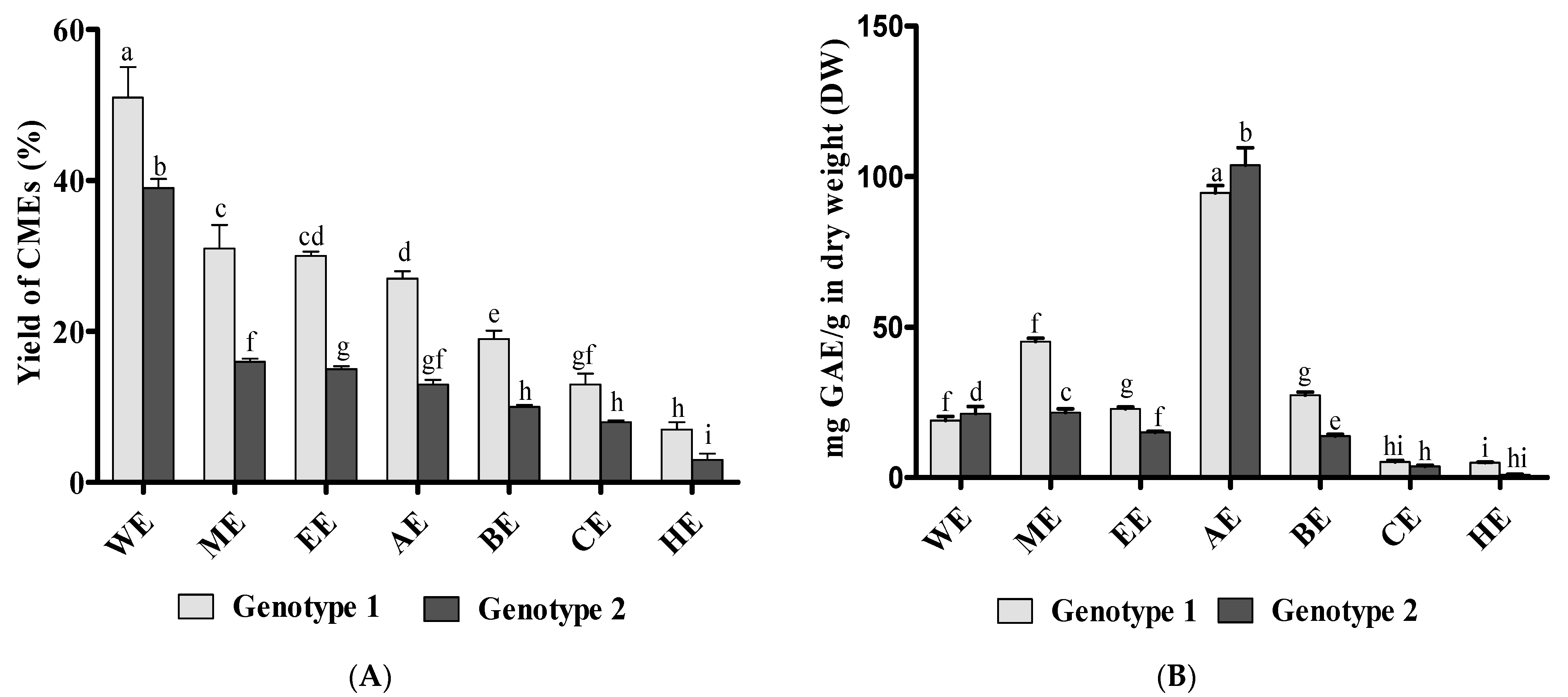
3.1. Preparation of Canola Extracts (CMEs) and Total Phenolic Content (TPC)

In the current study, seven solvents with different polarities were used for the generation of extracts from canola meal. Results reported in the literature displayed that different solvents possess different extraction properties [22]. Hence, chemical composition and biological activity can vary based on the extraction solvent used. In addition, the chemical composition of different plant genotypes or cultivars can represent quantitative and sometimes qualitative differences. This is the first time these features have been studied (using different solvents and genotypes) in canola meal, and no other reports in the literature could be found. Figure 1A compares the extraction yield for genotype-1 and genotype-2 using different solvents, where the extraction yield varied widely (5–55%) among different solvents and genotypes. WE appeared to extract more material (40–50% yield) from canola meal than any other solvent. This suggests the polar nature of the matrix components [23]. Irrespective of the solvent used, genotype-1 had a higher yield than genotype-2.
TPC is measured using Folin-Ciocalteu reagent, which measures the reducing power of antioxidants. This assay has been repeatedly scrutinized, and many researchers have questioned its validity and highlighted its limitations [2]. We totally understand and acknowledge these limitations. Folin–Ciocalteu results should not be used to compare samples from different botanical origins due to the potential interference from nonphenolic phytochemicals and matrix components. However, Folin–Ciocalteu remains an unbeatable assay for estimating the phenolic content of samples with similar botanical origin due to its robustness, ease of application, cost, high throughput, and high correlation with other antioxidant assays, including chromatographic quantitative data.
After Soxhlet complete hexane extraction of lipids from crushed canola seeds, nil or minimal recovery with a room-temperature short-time extraction with hexane was expected. However, adding water to hexane apparently had pulled out some hydrophilic components. It is evident that chloroform/water and hexane/water are immiscible, but acetone and water are miscible and used extensively in polyphenol extraction. Both chloroform/water and hexane/water (80:20 v/v) have been used to extract polyphenol compounds from natural products [24]. The solvent polarity and the solubility of polyphenols compounds in the extraction solvent influences the TPC and the composition of the extract [25]. Water is more polar and hence will extract more polar compounds. Conversely, both chloroform and hexane are highly non-polar solvents and will extract non-polar polyphenol compounds. Therefore, these two solvents were used to determine any such non-polar compounds in the canola meal.
Genotype-1 demonstrated higher TPC than genotype-2 in all extracts apart from WE and AE (Figure 1B). With regard to total phenol recovery, the current results for TPC were higher than the literature values reported by Obied et al. [1]. This can be explained by genetic variability as both genotype-1 and 2 have not been studied before for their phenol content, or can be due to experimental factors such as freeze-drying. Freeze drying revealed superior properties in preserving plant phenols and antioxidant activity [26]. Different genotypes demonstrated different TPC. This has been frequently reported in canola and other plants [27]. Genotype-1 has higher TPC than genotype-2 in all extracts apart from AE and WE, indicating that the TPC data depends on the genotype and the solvent used.
ME had a higher recovery than EE. EE is less polar than ME, yet their extracting abilities for phenolic compounds differ based on the plant extracted [28]. However, EE and WE have been previously reported to have major contributions to antioxidant activities [29]. Though WE had the highest yield, their TPC was not as high. It can also be concluded that WE contain mostly non-phenolic constituents on a weight basis. Regardless of the genotype, acetone stands out amongst other solvents with more than a four-fold higher phenol recovery. The exceptionally high extraction abilities of acetone compared to methanol (the most popular polyphenol extraction solvent) cannot be explained solely by being less polar than methanol. Both ethanol and n-butanol are less polar than methanol, yet they exhibited much lower total phenol recoveries. The superior recovery of total phenols by acetone can be attributed to its unique physicochemical properties: polar aprotic nature, miscibility with water and viscosity. Another interesting feature of the acetone and water extracts in this experiment is that they could recover more phenols from genotype 2 than genotype 1, contrary to all other used solvents. This finding is very interesting, stressing the importance of solvent selection in the recovery of plant phenols and interpreting/comparing data from different samples.

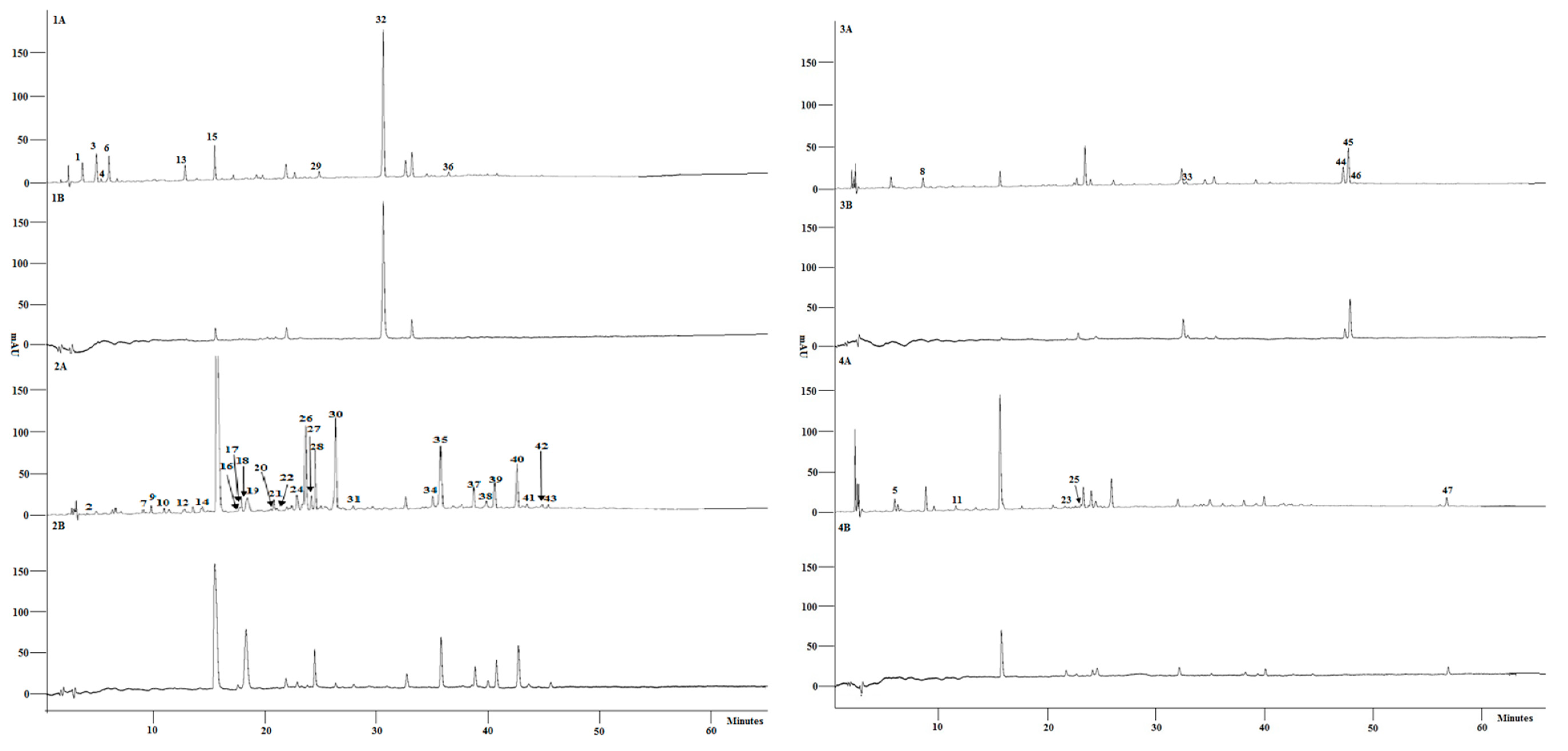
3.2. Characterisation of Phenolic Composition and Free Radical Scavenging Activity of Individual Compounds

The phenolic composition in different canola extracts was analyzed by reverse phase-HPLC-DAD with online detection of ABTS scavenging activity (Figure 2). In this technique, we combine HPLC-DAD (the most popular plant phenol profiling technique) with an online assessment of the free radical scavenging activity for simultaneous identification and antioxidant assessment of the individual components of the canola meal extracts. This technique has shown great potential in studying polyphenols from canola and other plants [1]. The m/z data and molecular masses were detected by LC-MS (Table 1). The collected data from UV-Vis spectra, ABTS scavenging activity, relative retention times, and mass spectra analysis was used in combination with reference standards and literature data to characterize the chemical composition of CMEs.
A large number of peaks, close to 100, were detected In DAD chromatogram at 280 nm and total ion chromatogram (TIC). Only prominent peaks appearing at 280 nm or in the TIC were selected for further identification. UV-Vis and mass spectra (Table 1) provides spectral data of 47 peaks (Figure 2) from all the CMEs. Thirty-two compounds were fully identified using standards or tentatively identified based on their spectral data. Fifteen compounds could not be identified. Early eluting peaks (highly polar compounds) were prominent in WE (Figure 2 (2A)). Compounds detected in CMEs were mostly glucosinolates or phenols. Glucosinolates identified were progitrin, sinigrin, glucalyssin, glucoraphanin, gluconapoleiferin, gluconapin, 4-hydroxyglucobrassicin, and gluconasturtiin. All glucosinolates were eluted within the first 18 min, excluding gluconasturtiin, which came out at 29.4 min. WE appeared as the best solvent for the recovery of glucosinolates.
Overall, ME had the most significant qualitative recovery (number of peaks) from CM, though AE revealed a much higher quantitative phenol recovery (peak area). This phenomenon has been observed before in literature where methanolic solvents seem to have a qualitative advantage over acetone solvents. Similar findings were reported earlier in other plants e.g., olives [28]. Sinapic acid derivatives (glycoside and choline conjugates) comprise the chief constituents of canola meal, a characteristic feature of Brassica plants, including canola [35]. Sinapine (peak-15) appeared as the major phenol in all CMEs (Figure 2), with the highest recovery in AE. Also, ME, EE, AA, BE, and HE appeared to have trans-sinapic acid as a dominant peak, with its highest recovery in AE again. Surprisingly, water extracts had a higher recovery of trans-sinapic acid (peak-32) compared with sinapine. Sinapine is more polar than trans-sinapic acid as it is a positively charged molecule. Hence, it dissolves more in water and its recovery is supposed to be higher from water extracts. However, the lower recovery of sinapine from WE can be explained by extensive hydrolysis in water to produce trans-sinapine. It is evident that water extraction increased the recovery of non-phenolic compounds and promoted the chemical and enzymatic hydrolysis of labile compounds such as sinapine.
Comparing both genotypes, WE of genotype-2 displayed more trans-sinapic acid compared to sinapine, suggesting more hydrolysis. The cis-isomer of sinapic acid (peak-34) was detected in all extracts. Peak-40, tentatively identified as tetra-sinapoyl dihexoside was a prominent peak in ME, while other extracts have only traces of this compound. ABTS scavenging activities of extracts are exclusively due to their sinapic acid derivatives. Though sinapine was the main contributor to ABTS scavenging activity as reported in the literature [39], some minor constituents demonstrated higher antioxidant activity that is not proportional to their concentration. This comes in accord with previous findings [1]. A feruloyl choline (4-O-8’) guiacyl (peak-18), and its isomers (peak-19), including feruloyl choline (5-8’) guaiacyl (peak-28), were observed in ME in both genotypes, while traces were observed only in other extracts. Meanwhile, feruloyl choline (4-O-8’) guaiacyl-di-sinapoyl (peak-35) was detected in genotype-1 only with the highest recovery in ME.
Although there was no free kaempferol identified in canola meal extracts, there were two glycoside derivatives of kaempferol—namely, kaempferol-sinapoyl-trihexoside (peak-30), and kaempferol 3-dihexoside-7-sinapoyl-hexoside (peak-33), which were also reported in Brassica napus seed meal [38]. Flavonoid compounds with C6C3C6 chains were commonly observed all extracts such as sinapic acid, feruloyl and kaempferol derivatives including compounds at peak 27 and 44 etc. Glucosinolates were always co-extracted with phenols due to the water component in all solvents. WE demonstrated the highest content and number of glucosinolates. A cyclic spermidine alkaloid and its isomers (peak-22 and 23) were detected in CMEs.

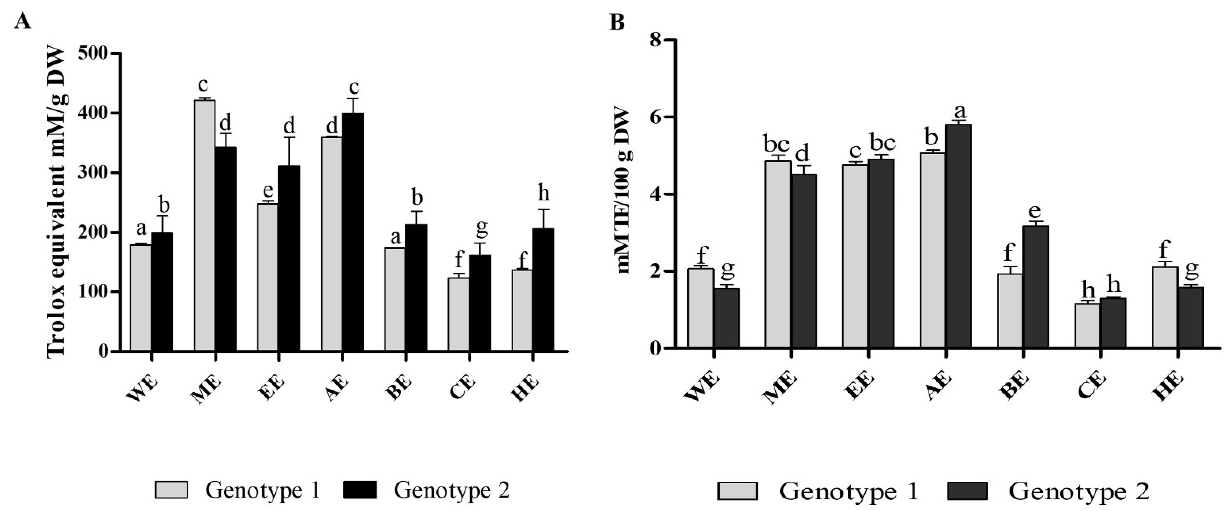
3.3. Antioxidant Properties

Antioxidant activities of CMEs were assessed by ABTS and FRAP assays, in addition to online ABTS assay. The trend for the free radical scavenging activity was different for the two genotypes (Figure 3A). For genotype-1, higher antioxidant activity was observed in ME, while for genotype-2, it was observed in AE. The FRAP activity of CMEs is presented in Figure 3B. The reducing power activity for the extract in both genotypes was higher in AE. Parallel to their radical scavenging activities, AE exhibited the highest, while CE had the least potential to reduce Fe (III). On the other hand, ME and EE demonstrated similar reducing power abilities to AE.
ME and AE proved superior solvents with the highest ABTS scavenging (Figure 3A) and reduction capacity (Figure 3B) from CMEs. This is because AE appeared to recover more quantity of antioxidant compounds than other extracts. AE is recommended for the highest antioxidant recovery [40]. Similarly, the extracts obtained by chloroform and hexane from both genotypes indicated the lowest scavenging activity. Both are non-polar organic solvents compared to acetone, butanol, ethanol, and water (polar solvents), making them unsuitable for extracting antioxidant phenolic compounds from canola meal. Overall, ABTS scavenging activity (Figure 3A) and the FRAP activity (Figure 3B) could be correlated with total phenolic content (TPC) for each extract as in Figure 1B as suggested for other plants extracts [29]. Online-ABTS HPLC data (Figure 2) confirm that phenolic compounds are the essential constituents with antioxidant activity in CMEs, mainly sinapine and sinapic acid. These results are supported by lipid models where sinapine and its derivates revealed higher antioxidant activity [41].
Although consumption of plant phenols has been seen to be linked to a wide range of physiological activities and health benefits, their exact in-vivo mechanism of action is still largely an area of debate and controversy. Currently, plant phenolic compounds are identified as excellent antioxidants [42] sources in the process of metabolism in the body. It has been confirmed that after the intake of plant polyphenols by humans, these compounds undergo extensive metabolic modification in the gastrointestinal tract [43]. It is repeatedly proven that compounds with high antioxidant activity in vitro tests have demonstrated promising in vivo activities. Although the exact pharmacological role of dietary antioxidants has not been well established yet, simple chemical assays of antioxidant activity can be correlated with potential in-vivo bioactivities. They will continue to be a useful screening tool of compounds with potential pharmacological activities [44]. On the other hand, antioxidants from canola meals can be utilized as preservatives or to fortify other food products [18,45].

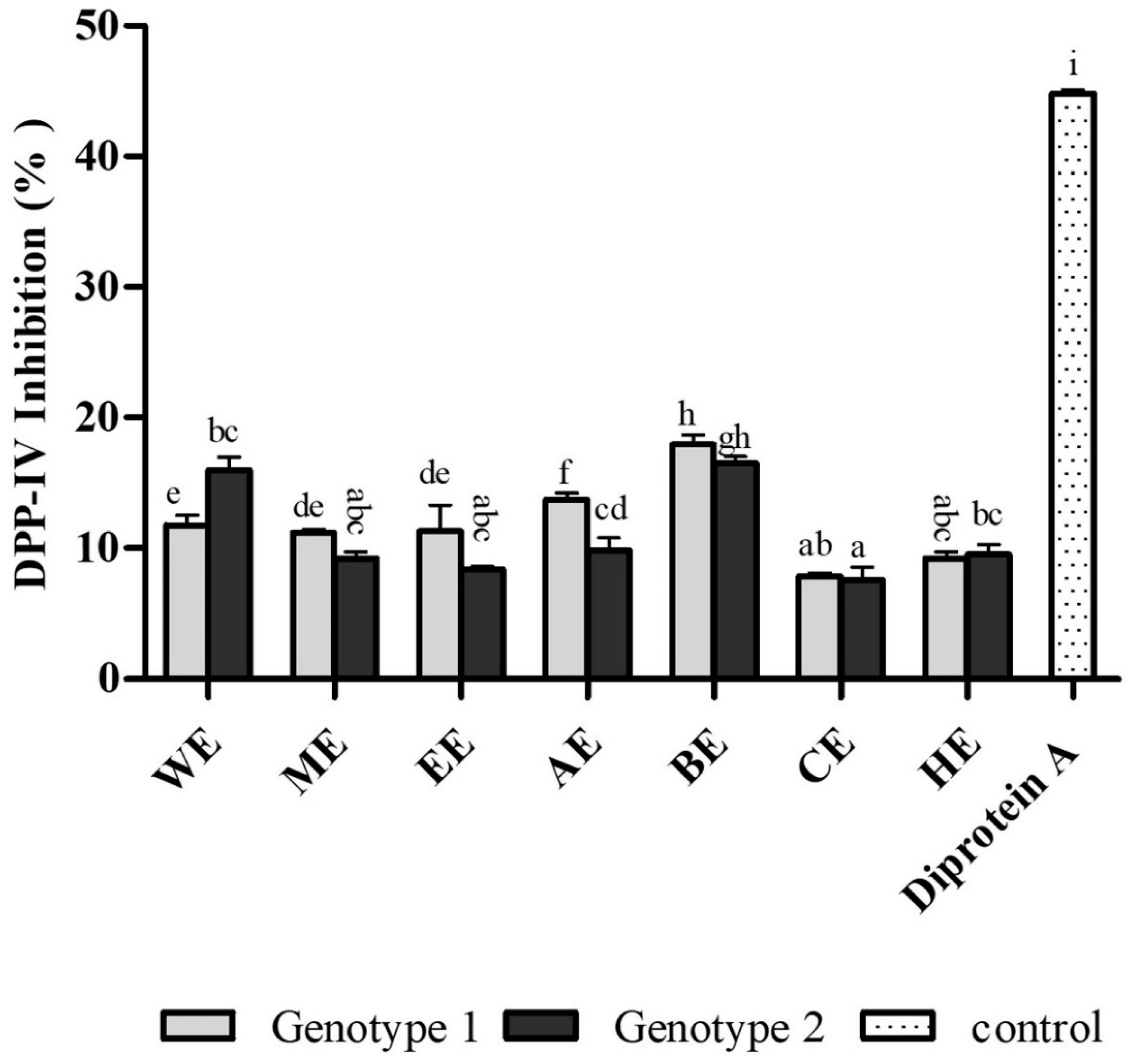
3.4. Antidiabetic Activity

The antidiabetic activity of various canola meal extracts has been assessed using DPP-IV enzyme inhibition assay (Figure 4). DPP-IV inactivates incretins responsible for postprandial insulin secretion and plays an important role in glucose homeostasis. DPP-IV inhibitors are clinically useful in the management of type 2 diabetes. Many plant extracts and purified phytochemicals have displayed DPP-IV inhibitory properties [46]. Some of the canola meal phenolic constituents (sinapic acid, caffeic acid, and kaempferol) presented the potential to inhibit DPP-IV [47].
Canola meal extracts indicated statistically significant DPP-IV inhibitory activities (7–18%) at the concentration used (2 mg/mL). The order of inhibition was found to be: BE > AE > WE > EE > ME > HE > CE for genotype-1 and BE > WE > AE > ME> HE > EE> CE for genotype-2. Unlike antioxidant activity assays (Figure 3), Figure 4 illustrates that the DPP-IV inhibitory activity was highest in BE (18%) for both genotypes, followed by WE (11–16%) and AE (9–14%). Although acetone extracts continue to demonstrate potential for the recovery of DPP-IV inhibitors, butanol and water extracts are more promising for the recovery of antidiabetic components. These findings also suggest that the antidiabetic activity of canola meal extracts cannot solely be attributed to its antioxidant polyphenols, and there are other phytochemicals that probably are more potent DPP-IV inhibitors. Future research should employ bioactivity-guided fractionation to isolate bioactive compounds from canola meal butanol, water, and acetone extracts.

3.5. Recovery of Sinapine, Feruloyl Choline, Kaempherol-Sinapoyl-Trihexoside, Sinapic Acid, and Feruloyl Choline (4-O-8) Guaiacyl Disinapoyl

Recoveries of the five major phenolic compounds in canola meal extracts were analyzed to provide a better insight into the chemical composition of this valuable agricultural waste and assist any future endeavors to recover any of these compounds on a commercial scale. Sinapine, feruloyl choline, kaempherol-sinapoyl-trihexoside, sinapic acid, and feruloyl choline (4-O-8) guaiacyl disinapoyl in both genotypes were quantified using peak areas from chromatograms at 280 nm and a six-point sinapic acid calibration curve (Table 2). Sinapine, feruloyl choline, and kaempherol sinapoyl-trihexoside were recovered in AE in both genotypes. Sinapic acid was recovered in more significant amounts from water extracts (Figure 2(1A)); however, its recovery varied between genotypes and in accord with earlier literature [35]. Sinapic acid was present in higher concentrations in genotype-1 than genotype-2. Feruloyl choline (4-O-8) guaiacyl disinapoyl was recovered in EE from genotype-1 and in ME from genotype-2 (Table 2).
Sinapine is the most abundant phenolic compound in canola meal. The highest recoveries were achieved with acetone, followed by methanol and ethanol. Higher recoveries obtained from genotype 1 (13 ± 0.3 mg SAE/g DW) while the difference was not statistically significant. Sinapine was the primary compound with the highest yields, as described in earlier literature [48]. Only traces of feruloyl choline were recovered by water, chloroform, and hexane. Highest recoveries were obtained from AE of genotype 1 (1.6 mg SAE/g DW). Kaempferol sinapoyl-trihexoside was present in all extracts, but the highest recoveries were obtained from AE (2.4 mgSAE/g DW). Meanwhile, higher recoveries of sinapic acid were obtained from both genotypes from WE. High recovery of sinapic acid from water extracts were reported in the literature [49]. Feruloyl choline (4-O-8) guaiacyl disinapoyl had significantly higher recoveries from AE and ME (1.4 mgSAE/g DW).

4. Conclusions

Different solvent extracts of canola meal exhibited qualitative and quantitative differences, and consequently varying potencies in antioxidant and antidiabetic assays used in the current study. While water extracts presented the highest yield of extractable materials, aqueous acetone exhibited superior selectivity for the recovery of antioxidant phenolic compounds. On the other hand, aqueous butanol and water extracts revealed higher DPP-IV inhibitory activities. This study emphasizes the occasionally underestimated role of solvent selection in the study of the chemical composition and biological activity of plant samples. In the case of canola meal, aqueous acetone could achieve the highest quantitative recovery of antioxidant phenols.
Meanwhile, aqueous methanol extracts indicated higher qualitative recovery (more compounds detected). Furthermore, extraction of canola meal using water only without organic solvents increased the hydrolysis of sinapic acid conjugates (such as sinapine) and recovered higher quantities of the free sinapic acid. It has also been observed that all employed solvents apart from aqueous acetone revealed that genotype 1 has a higher content of total phenols than genotype 2. Yet, when aqueous acetone was used, the genotype 2 exhibited a higher recovery of total phenols than genotype 1.
HPLC-DAD-ABTS and LC-MS analyses detected 47 compounds, identifying 32 compounds, while 15 remained unknown. Water extracts appeared superior for the recovery of potentially health-beneficial glucosinolates. BE presented the highest potential for the inhibition of DPP-IV. These findings highlight the potential of canola meal as a source for the recovery of valuable phytochemicals for dietary and pharmaceutical applications. The current study presents a preliminary investigation of the polyphenol content of canola meal and highlights its potential utilization for the recovery of compounds with antioxidant and antidiabetic properties. Further investigations are necessary to elucidate the mechanism of these compounds, their bioavailability, and in-vivo biological activities. Unknown compounds need to be characterized, as some of these possess high antioxidant activity and may have potentially useful bioactivities.

Author Contributions

Conceptualization, S.H., C.L.B. and A.U.R.; methodology, S.H.; software, S.H. and H.K.O.; validation, C.L.B. and H.K.O.; formal analysis, S.H. and C.L.B.; investigation, H.K.O., S.H., C.L.B., A.U.R. and D.J.L.; data curation, S.H. and H.K.O.; writing—original draft preparation, S.H., D.J.L. and C.L.B.; writing—review and editing, S.H., C.L.B. and H.K.O.; visualization, C.L.B. and A.U.R.; supervision, C.L.B., A.U.R., H.K.O. and D.J.L.; project administration, S.H. and C.L.B.; funding acquisition, C.L.B. All authors have read and agreed to the published version of the manuscript.

Funding

This research was funded by Charles Sturt University and the Australian Research Council (ARC) Industrial Transformation Training Centre for Functional Grains.

Data Availability Statement

The data submitted in this research are available on request from the corresponding author. The data are not publicly presented owing to confidentiality.

Acknowledgments

The authors are thankful to the NSW Department of Primary Industries (DPI), Wagga Wagga, Australia, for providing canola seeds; the Australian Oilseed Laboratory, Department of Primary Industries, NSW, Wagga Wagga, Australia for using their laboratory facilities; Charles Sturt University laboratory staff for technical assistance; and to Peter Thompson for proofreading the manuscript.

Conflicts of Interest

The authors declare no conflict of interest.

References

  1. Obied, H.K.; Song, Y.; Foley, S.; Loughlin, M.; Rehman, A.U.; Mailer, R.; Masud, T.; Agboola, S. Biophenols and antioxidant properties of Australian canola meal. J. Agric. Food Chem. 2013, 61, 9176–9184. [Google Scholar] [CrossRef] [PubMed]
  2. Taşpınar, H.; Elik, A.; Kaya, S.; Altunay, N. Optimization of green and rapid analytical procedure for the extraction of patulin in fruit juice and dried fruit samples by air-assisted natural deep eutectic solvent-based solidified homogeneous liquid phase microextraction using experimental design and computational chemistry approach. Food Chem. 2021, 358, 129817. [Google Scholar] [PubMed]
  3. Veiga, M.; Costa, E.M.; Silva, S.; Pintado, M. Impact of plant extracts upon human health: A review. Crit. Rev. Food Sci. Nutr. 2020, 60, 873–886. [Google Scholar] [CrossRef] [PubMed]
  4. Belwal, T.; Chemat, F.; Venskutonis, P.R.; Cravotto, G.; Jaiswal, D.K.; Bhatt, I.D.; Devkota, H.P.; Luo, Z. Recent advances in scaling-up of non-conventional extraction techniques: Learning from successes and failures. TrAC Trends Anal. Chem. 2020, 127, 115895. [Google Scholar] [CrossRef]
  5. Belwal, T.; Ezzat, S.M.; Rastrelli, L.; Bhatt, I.D.; Daglia, M.; Baldi, A.; Devkota, H.P.; Orhan, I.E.; Patra, J.K.; Das, G. A critical analysis of extraction techniques used for botanicals: Trends, priorities, industrial uses and optimization strategies. TrAC Trends Anal. Chem. 2018, 100, 82–102. [Google Scholar] [CrossRef]
  6. Alara, O.R.; Abdurahman, N.H.; Ukaegbu, C.I. Extraction of phenolic compounds: A review. Curr. Res. Food Sci. 2021, 4, 200–214. [Google Scholar] [CrossRef] [PubMed]
  7. Zeng, Z.; Centner, C.; Gollhofer, A.; König, D. Effects of Dietary Strategies on Exercise-Induced Oxidative Stress: A Narrative Review of Human Studies. Antioxidants 2021, 10, 542. [Google Scholar] [CrossRef]
  8. Domingueti, C.P.; Dusse, L.M.S.A.; das Graças Carvalho, M.; de Sousa, L.P.; Gomes, K.B.; Fernandes, A.P. Diabetes mellitus: The linkage between oxidative stress, inflammation, hypercoagulability and vascular complications. J. Diabetes Its Complicat. 2016, 30, 738–745. [Google Scholar] [CrossRef]
  9. Wondafrash, D.Z.; Nire’a, A.T.; Tafere, G.G.; Desta, D.M.; Berhe, D.A.; Zewdie, K.A. Thioredoxin-interacting protein as a novel potential therapeutic target in diabetes mellitus and its underlying complications. Diabetes Metab. Syndr. Obes. Targets Ther. 2020, 13, 43. [Google Scholar] [CrossRef] [PubMed] [Green Version]
  10. Ginter, E.; Simko, V. Global prevalence and future of diabetes mellitus. In Diabetes; Springer: Berlin, Germany, 2013; pp. 35–41. [Google Scholar]
  11. Weber, C.; Hung, J.; Hickling, S.; Li, I.; Murray, K.; Briffa, T. Changing age-specific trends in incidence, comorbidities and mortality of hospitalised heart failure in Western Australia between 2001 and 2016. Int. J. Cardiol. 2021, 343, 56–62. [Google Scholar] [CrossRef]
  12. Aynalem, S.B.; Zeleke, A.J. Prevalence of diabetes mellitus and its risk factors among individuals aged 15 years and above in Mizan-Aman town, Southwest Ethiopia, 2016: A cross sectional study. Int. J. Endocrinol. 2018, 2018, 9317987. [Google Scholar] [CrossRef] [PubMed]
  13. Clapham, J.C. Sixty years of drug discovery for type 2 diabetes: Where are we now? Type 2 Diabetes 2020, 2076, 1–30. [Google Scholar]
  14. Liu, R.; Cheng, J.; Wu, H. Discovery of Food-Derived Dipeptidyl Peptidase IV Inhibitory Peptides: A Review. Int. J. Mol. Sci. 2019, 20, 463. [Google Scholar] [CrossRef] [PubMed] [Green Version]
  15. Bowen, K.J.; Kris-Etherton, P.M.; West, S.G.; Fleming, J.A.; Connelly, P.W.; Lamarche, B.; Couture, P.; Jenkins, D.J.A.; Taylor, C.G.; Zahradka, P.; et al. Diets Enriched with Conventional or High-Oleic Acid Canola Oils Lower Atherogenic Lipids and Lipoproteins Compared to a Diet with a Western Fatty Acid Profile in Adults with Central Adiposity. J. Nutr. 2019, 149, 471–478. [Google Scholar] [CrossRef]
  16. Kanikowska, D.; Kanikowska, A.; Rutkowski, R.; Wlochal, M.; Orzechowska, Z.; Juchacz, A.; Zawada, A.; Grzymislawski, M.; Roszak, M.; Sato, M.; et al. Amaranth (Amaranthus cruentus L.) and canola (Brassica napus L.) oil impact on the oxidative metabolism of neutrophils in the obese patients. Pharm. Biol. 2019, 57, 140–144. [Google Scholar] [CrossRef] [Green Version]
  17. Hussain, S.; Rehman, A.U.; Luckett, D.J.; Blanchard, C.L.; Obied, H.K.; Strappe, P. Phenolic Compounds with Antioxidant Properties from Canola Meal Extracts Inhibit Adipogenesis. Int. J. Mol. Sci. 2019, 21, 1. [Google Scholar] [CrossRef] [Green Version]
  18. Benzie, I.F.; Strain, J. The ferric reducing ability of plasma (FRAP) as a measure of “antioxidant power”: The FRAP assay. Anal. Biochem. 1996, 239, 70–76. [Google Scholar] [CrossRef] [Green Version]
  19. Benzie, I.F.; Devaki, M. The ferric reducing/antioxidant power (FRAP) assay for non-enzymatic antioxidant capacity: Concepts, procedures, limitations and applications. In Measurement of Antioxidant Activity & Capacity: Recent Trends and Applications; Wiley: New York, NY, USA, 2018; Volume 11, pp. 77–106. [Google Scholar]
  20. Merola, N.; Alonso, F.J.G.; Ros, G.; Castón, M.J.P. Antioxidant activity comparison between [6S]-5-methyltetrahydrofolic acid calcium salt and the related racemate form. Food Chem. 2013, 136, 984–988. [Google Scholar] [CrossRef]
  21. Lacroix, I.M.E.; Li-Chan, E.C.Y. Dipeptidyl peptidase (DPP)-IV inhibitory activity of dairy protein hydrolysates. Int. Dairy J. 2012, 25, 97–102. [Google Scholar] [CrossRef]
  22. Ngo, T.V.; Scarlett, C.J.; Bowyer, M.C.; Ngo, P.D.; Vuong, Q.V. Impact of Different Extraction Solvents on Bioactive Compounds and Antioxidant Capacity from the Root of Salacia chinensis L. J. Food Qual. 2017, 2017, 9305047. [Google Scholar] [CrossRef] [Green Version]
  23. Xu, M.; Ran, L.; Chen, N.; Fan, X.; Ren, D.; Yi, L. Polarity-dependent extraction of flavonoids from citrus peel waste using a tailor-made deep eutectic solvent. Food Chem. 2019, 297, 124970. [Google Scholar] [CrossRef] [PubMed]
  24. Ignat, I.; Volf, I.; Popa, V.I. A critical review of methods for characterisation of polyphenolic compounds in fruits and vegetables. Food Chem. 2011, 126, 1821–1835. [Google Scholar] [CrossRef] [PubMed]
  25. Babbar, N.; Oberoi, H.S.; Sandhu, S.K.; Bhargav, V.K. Influence of different solvents in extraction of phenolic compounds from vegetable residues and their evaluation as natural sources of antioxidants. J. Food Sci. Technol. 2014, 51, 2568–2575. [Google Scholar] [CrossRef] [PubMed] [Green Version]
  26. Saifullah, M.; McCullum, R.; McCluskey, A.; Vuong, Q. Effects of different drying methods on extractable phenolic compounds and antioxidant properties from lemon myrtle dried leaves. Heliyon 2019, 5, e03044. [Google Scholar] [CrossRef] [Green Version]
  27. Shah, U.N.; Mir, J.I.; Ahmed, N.; Jan, S.; Fazili, K.M. Bioefficacy potential of different genotypes of walnut Juglans regia L. J. Food Sci. Technol. 2018, 55, 605–618. [Google Scholar] [CrossRef]
  28. Sepahpour, S.; Selamat, J.; Abdul Manap, M.Y.; Khatib, A.; Abdull Razis, A.F. Comparative analysis of chemical composition, antioxidant activity and quantitative characterization of some phenolic compounds in selected herbs and spices in different solvent extraction systems. Molecules 2018, 23, 402. [Google Scholar] [CrossRef] [Green Version]
  29. Hassas-Roudsari, M.; Chang, P.R.; Pegg, R.B.; Tyler, R.T. Antioxidant capacity of bioactives extracted from canola meal by subcritical water, ethanolic and hot water extraction. Food Chem. 2009, 114, 717–726. [Google Scholar] [CrossRef]
  30. Kusznierewicz, B.; Iori, R.; Piekarska, A.; Namiesnik, J.; Bartoszek, A. Convenient identification of desulfoglucosinolates on the basis of mass spectra obtained during liquid chromatography–diode array–electrospray ionisation mass spectrometry analysis: Method verification for sprouts of different Brassicaceae species extracts. J. Chromatogr. A 2013, 1278, 108–115. [Google Scholar] [CrossRef]
  31. Lelario, F.; Bianco, G.; Bufo, S.A.; Cataldi, T.R. Establishing the occurrence of major and minor glucosinolates in Brassicaceae by LC-ESI-hybrid linear ion-trap and Fourier-transform ion cyclotron resonance mass spectrometry. Phytochemistry 2012, 73, 74–83. [Google Scholar] [CrossRef]
  32. Lee, I.; Boyce, M.C. Extraction and Purification of Glucoraphanin by Preparative High-Performance Liquid Chromatography (HPLC). J. Chem. Educ. 2011, 88, 832–834. [Google Scholar] [CrossRef]
  33. Velasco, P.; Francisco, M.; Moreno, D.A.; Ferreres, F.; Garcia-Viguera, C.; Cartea, M.E. Phytochemical fingerprinting of vegetable Brassica oleracea and Brassica napus by simultaneous identification of glucosinolates and phenolics. Phytochem. Anal. 2010, 22, 144–152. [Google Scholar] [CrossRef] [PubMed] [Green Version]
  34. Thiyam-Hollander, U.; Aladedunye, F.; Logan, A.; Yang, H.; Diehl, B.W. Identification and quantification of canolol and related sinapate precursors in Indian mustard oils and Canadian mustard products. Eur. J. Lipid Sci. Technol. 2014, 116, 1664–1674. [Google Scholar] [CrossRef]
  35. Yang, S.-C.; Arasu, M.V.; Chun, J.-H.; Jang, Y.-S.; Lee, Y.-H.; Kim, I.H.; Lee, K.-T.; Hong, S.-T.; Kim, S.-J. Identification and Determination of Phenolic Compounds in Rapeseed Meals (Brassica napus L.). J. Agric. Chem. Environ. 2015, 4, 14–23. [Google Scholar] [CrossRef] [Green Version]
  36. Fang, J.; Reichelt, M.; Hidalgo, W.; Agnolet, S.; Schneider, B. Tissue-specific distribution of secondary metabolites in rapeseed (Brassica napus L.). PLoS ONE 2012, 7, e48006. [Google Scholar] [CrossRef] [PubMed]
  37. Obied, H.K. Biography of biophenols: Past, present and future. Funct. Foods Health Dis. 2013, 3, 230–241. [Google Scholar] [CrossRef]
  38. Siger, A.; Czubinski, J.; Dwiecki, K.; Kachlicki, P.; Nogala-Kalucka, M. Identification and antioxidant activity of sinapic acid derivatives in Brassica napus L. seed meal extracts. Eur. J. Lipid Sci. Technol. 2013, 115, 1130–1138. [Google Scholar] [CrossRef]
  39. Yates, K.; Pohl, F.; Busch, M.; Mozer, A.; Watters, L.; Shiryaev, A.; Kong Thoo Lin, P. Determination of sinapine in rapeseed pomace extract: Its antioxidant and acetylcholinesterase inhibition properties. Food Chem. 2019, 276, 768–775. [Google Scholar] [CrossRef] [PubMed]
  40. Thavamoney, N.; Sivanadian, L.; Tee, L.H.; Khoo, H.E.; Prasad, K.N.; Kong, K.W. Extraction and recovery of phytochemical components and antioxidative properties in fruit parts of Dacryodes rostrata influenced by different solvents. J. Food Sci. Technol. 2018, 55, 2523–2532. [Google Scholar] [CrossRef] [PubMed]
  41. Martinović, N.; Poklar Ulrih, N.; Abramovič, H. Sinapic acid and its derivatives increase oxidative stability in different model lipid systems. Eur. J. Lipid Sci. Technol. 2019, 121, 1800326. [Google Scholar] [CrossRef]
  42. Chaves, N.; Santiago, A.; Alías, J.C. Quantification of the antioxidant activity of plant extracts: Analysis of sensitivity and hierarchization based on the method used. Antioxidants 2020, 9, 76. [Google Scholar] [CrossRef] [Green Version]
  43. Gonçalves, R.F.; Martins, J.T.; Duarte, C.M.; Vicente, A.A.; Pinheiro, A.C. Advances in nutraceutical delivery systems: From formulation design for bioavailability enhancement to efficacy and safety evaluation. Trends Food Sci. Technol. 2018, 78, 270–291. [Google Scholar] [CrossRef] [Green Version]
  44. Granato, D.; Shahidi, F.; Wrolstad, R.; Kilmartin, P.; Melton, L.D.; Hidalgo, F.J.; Miyashita, K.; van Camp, J.; Alasalvar, C.; Ismail, A.B. Antioxidant activity, total phenolics and flavonoids contents: Should we ban in vitro screening methods? Food Chem. 2018, 264, 471–475. [Google Scholar] [CrossRef] [PubMed]
  45. Hussain, S.; Ata-ur-rehman, D.L.; Blanchard, C.L. Can canola meal be used to treat cancer? New South Wales 2014, 89–209. Available online: https://www.researchgate.net/publication/282664401_Can_canola_meal_be_used_to_treat_cancer (accessed on 15 January 2022).
  46. Turdu, G.; Gao, H.; Jiang, Y.; Kabas, M. Plant dipeptidyl peptidase-IV inhibitors as antidiabetic agents: A brief review. Future Med. Chem. 2018, 10, 1229–1239. [Google Scholar] [CrossRef] [PubMed]
  47. Fan, J.; Johnson, M.H.; Lila, M.A.; Yousef, G.; de Mejia, E.G. Berry and Citrus Phenolic Compounds Inhibit Dipeptidyl Peptidase IV: Implications in Diabetes Management. Evid. Based Complement. Altern. Med. 2013, 2013, 479505. [Google Scholar] [CrossRef] [Green Version]
  48. Zardo, I.; Rodrigues, N.P.; Sarkis, J.R.; Marczak, L.D. Extraction and identification by mass spectrometry of phenolic compounds from canola seed cake. J. Sci. Food Agric. 2020, 100, 578–586. [Google Scholar] [CrossRef] [PubMed]
  49. Moreno-González, M.; Girish, V.; Keulen, D.; Wijngaard, H.; Lauteslager, X.; Ferreira, G.; Ottens, M. Recovery of sinapic acid from canola/rapeseed meal extracts by adsorption. Food Bioprod. Processing 2020, 120, 69–79. [Google Scholar] [CrossRef]
Figure 1. Comparison of extraction yields and total phenolic content of seven canola meal extracts (CMEs) namely WE (water extract); ME (methanol extract); EE (ethanol extract); AE (acetone extract); BE (butanol extract); CE (chloroform extract); HE (hexane extract). (A) Comparison of extraction yields (in percentage) of CMEs from two canola genotypes [Bln-3347 (genotype-1) and Rivette (genotype-2)], (B) Comparison of total phenolic contents represented as milligrams of gallic acid equivalent per gram dry weight (mg GAE/g DW) in CMEs from two canola genotypes. The values are means ± SD (n = 3). Bars with the different letters are significantly different using the Duncan multiple range test at p < 0.05.
Separations 09 00038 g001
Figure 2. Online High-Performance Liquid Chromatography with Diode-Array Detection (HPLC-DAD) antioxidant chromatograms of CMEs. Eluting time in minutes (x-axis) and absorbance in mAU (y-axis). Chromatogram 1, 2, 3 and 4 A detected at 280 nm implies presence of compounds while chromatogram 1, 2, 3 and 4 B detected at 414 nm implies their ABTS scavenging activity (antioxidant activity). (1A) WE (water extract) at 280 nm; (1B) WE (water extract) at 414 nm; (2A) ME (methanol extract) at 280 nm; (2B) ME (methanol extract) at 414 nm; (3A) CE (chloroform extract) at 280 nm; (3B) CE (chloroform extract) at 414 nm; (4A) BE (butanol extract) at 280 nm; (4B) BE (butanol extract) at 414 nm. All identified peaks have been allocated a number and these are given in Table 1.
Separations 09 00038 g002
Figure 3. (A) Antioxidant properties by ABTS scavenging activity representing the effectiveness of extracts that scavenge radicals, where results are expressed as Trolox equivalent mM/g DW. (B) Comparison of antioxidant properties as measured by FRAP assay. The values are means ± SD (n = 3). Bars with the different letters are significantly different using the Duncan multiple range test at p < 0.05.
Separations 09 00038 g003
Figure 4. Percentage (%) of DPP-IV inhibition activity of canola extracts from two different genotypes. Diprotin A was used as standard control. The values are means ± SD (n = 3). Bars with the different letters are significantly different using the Duncan multiple range test at p < 0.05.
Separations 09 00038 g004
Table 1. HPLC and LCMS peaks in canola meal extracts (CMEs).
PIdentityGNHECEBEAEEEMEWERTλmaxABTSESI+ESIMWReference
1UnknownGN2✓✓✓4.1258124122123
2UnknownGN1✓✓✓✓✓✓5.0275613611612
3UnknownGN2✓✓✓5.5249137135136
4UnknownBoth✓✓6.0262268243244
5UnknownBoth✓✓✓✓✓✓✓✓6.2256233231232
6ProgoitrinBoth✓✓✓✓✓✓✓✓✓6.7220NI388389[30]
7SinigrinBoth7.8285NI358359[30]
8UnknownBoth✓✓✓✓✓✓✓✓✓8.8252, 270s277NI276
9GlucoalyssinBoth✓✓✓✓✓✓✓9.5225452450451[31]
10GlucoraphaninBoth✓✓10.5230NI435436[32]
11GluconapoleiferinBoth✓✓✓✓✓✓11.1275NI402403[33]
12GluconapinBoth✓✓✓✓✓✓✓✓11.3220NI372373[1]
13UnknownBoth✓✓✓✓✓✓✓✓✓13.7278451449450
14UnknownBoth✓✓✓✓✓✓✓✓✓13.7288NI315316
15SinapineBoth✓✓✓✓✓✓✓✓✓✓✓15.7237, 328++310294, 663310[34]
16Caffeoyl dihexosideBoth✓✓✓✓✓✓✓17.2297s, 327++NI503504[1]
174-hydroxyglucobrassicinBoth✓✓✓✓✓✓✓17.6292NI463464[31]
18Feruloyl choline (4-0-8’) guiacylBoth✓✓✓✓✓✓✓17.7270b, 325b+++476NI476[35]
19Feruloyl choline guiacyl isomerBoth✓✓18.3270b, 325b++476NI476[35]
204’-glucosylsinapic acidBoth✓✓✓✓✓✓✓✓✓20.6317bNI385386[1]
21Sinapoyl dihexosideBoth✓✓✓✓✓19.0328.54+NI547548[1]
22Cyclic spermidin derivativeBoth✓✓✓✓✓✓✓✓20.8316496494495[36]
23Cyclic spermidin derivativeBoth✓✓✓✓✓✓✓✓✓✓✓21.7320496494495[36]
24Sinapoyl hexosideBoth✓✓✓✓✓✓✓22.7330++NI385386[37]
25Sinapoyl hexoside isomerGN1✓✓✓✓✓✓✓23.1330++NI385386[37]
26UnknownGN1✓✓✓✓✓✓✓✓✓23.5305b533531532
27UnknownBoth✓✓✓✓✓✓✓23.6310b449447448
28Feruloyl choline (5-8’)
guaiacyl
Both✓✓✓✓✓✓✓24.4328, 280s++457458459[35]
29UnknownBoth✓✓✓24.6260s, 280s, 290s++429427428
30Kaempherol-sinapoyl-trihexosideBoth✓✓✓✓✓✓✓✓✓26.3268, 333+979978977[38]
31GluconasturtiinBoth✓✓29.4230NI422423[31]
32Trans-Sinapic acidBoth✓✓✓✓✓✓✓✓✓✓✓✓✓✓✓32.6324++NI223224[1]
33Kaempherol 3-dihexoside-7-sinapoyl-hexosideBoth✓✓33.0266, 323++979978977[37]
34Cis-sinapic acidBoth✓✓✓✓✓✓✓✓✓✓✓35.3266, 323++NI223224[37]
35Feruloyl choline (4-O-8’) guaiacyl-di-sinapoylGN1✓✓✓✓✓✓✓36.1323++682NI682[35]
36Unknown (methyl sinapate dihexoside)Both✓✓36.4329++NI561562
37Disinapoyl dihexosideBoth✓✓✓✓✓✓✓✓✓38.6230, 330++NI753754[1]
38Trisinapoyl dihexoside 1Both✓✓✓✓✓✓✓39.7327++NI959960[1]
39Disinapoyl hexosideBoth✓✓✓✓✓✓✓✓✓40.7330++NI591592[1]
40Tetrasinapoyl dihexoside ✓✓✓41.9326++NI11831184[37]
41Methyl sinapateBoth✓✓✓42.5325++NI237238[1]
42Unknown ✓✓✓✓43.1325+NI545546[36]
43Disinapoyl hexoside isomerBoth✓✓45326+NI591592[1]
44UnknownBoth✓✓✓47.2320++275NI274
45UnknownGN1✓✓✓47.6325++454NI453
46UnknownBoth✓✓48.7325+245NI244
47UnknownBoth✓✓✓✓✓58.2327+303279280
P, peak number from Figure 2; GN, genotypes; GN-1, only in genotype-1; GN-2, only in genotype-2; Both, genotype-1 and 2; HE, hexane extract; CE, chloroform extract; BE, butanol extract; AE, acetone extract; EE, ethanol extract; ME, methanol extract; WE, water extract; ✓, traces; ✓✓, present; ✓✓✓, major peak; ✗, could not be detected; RT, retention time; λmax, UV-Vis spectra; ESI, electrospray ionization peaks in negative mode; ESI+, electrospray ionization peaks in positive mode; MW, molecular weight; NI, did not ionize under ESI modes; b, broad peak; s, peak shoulder; ABTS, ABTSscavenging activity in online assay (antioxidant); +, low ABTS scavenging activity; ++, good ABTS scavenging activity; +++, high ABTS scavenging activity.
Table 2. Recovery of major compounds in seven extracts of canola meal.
Recovery mg SAE/g DW
Extract (Sample)SinapineFeruloyl CholineKaempherol-Sinapoyl-TrihexosideSinapic Acid *Feruloyl Choline (4-O-8) Guaiacyl di Sinapoyl
WE (GN-1)2.2 ± 0.0 atraces0.6 ± 0.0 a1.5 ± 0.0 a0.6 ± 0.0 a
WE (GN-2)2.2 ± 0.1 atraces0.6 ± 0.0 a1.7 ± 0.0 a,d0.7 ± 0.0 a
ME (GN-1)7.0 ± 0.1 b0.8 ± 0.0 a1.4 ± 0.0 b0.2 ± 0.0 b1.0 ± 0.0 a
ME (GN-2)12.1 ± 0.2 c1.1 ± 0.0 a2.2 ± 0.0 c0.9 ± 0.0 c1.4 ± 0.0 b
EE (GN-1)7.7 ± 0.0 b0.8 ± 0.0 a1.5 ± 0.0 b0.2 ± 0.0 b1.3 ± 0.0 b
EE (GN-2)10.1 ± 0.0 d0.7 ± 0.0 a1.7 ± 0.1 b1.0 ± 0.0 c0.9 ± 0.0 a
AE (GN-1)13.0 ± 0.3 c1.6 ± 0.0 b2.4 ± 0.0 c0.3 ± 0.0 b0.6 ± 0.1 a
AE (GN-2)12.6 ± 0.3 c1.2 ± 0.0 a2.2 ± 0.0 c1.0 ± 0.0 c1.4 ± 0.0 b
BE(GN-1)5.7 ± 0.0 e0.7 ± 0.0 a1.3 ± 0.1 b0.4 ± 0.0 b0.4 ± 0.0 c
BE (GN-2)5.9 ± 0.1 e0.7 ± 0.0 a1.6 ± 0.0 b0.8 ± 0.0 c0.6 ± 0.0 a
CE (GN-1)0.3 ± 0.0 ftraces0.2 ± 0.0 a0.7 ± 0.0 c0.2 ± 0.0 c
CE (GN-2)2.0 ± 0.0 atraces0.6 ± 0.0 a1.8 ± 0.0 a0.6 ± 0.0 a
HE (GN-1)0.2 ± 0.0 ftraces0.2 ± 0.1 b0.7 ± 0.0 c0.3 ± 0.0 c
HE (GN-2)0.5 ± 0.0 ftraces0.4 ± 0.0 a2.0 ± 0.0 d0.6 ± 0.0 a
* Recovery is in milligram sinapic acid equivalent/gram dry weight (mg SAE/g DW); GN-1 (Genotype-1); GN-2 (Genotype-2); WE (water extract); ME (methanol extract); EE (ethanol extract); AE (acetone extract); BE (butanol extract); CE (chloroform extract); HE (hexane extract). Different letters are significantly different using the Duncan multiple range test at p < 0.05.
Publisher’s Note: MDPI stays neutral with regard to jurisdictional claims in published maps and institutional affiliations.

