Article

GC-MS and SPME-GC/MS Analysis and Bioactive Potential Evaluation of Essential Oils from Two Viola Species Belonging to the V. calcarata Complex

by
Sara Vitalini
1,2,3,
Marcello Iriti
1,2,3,4 and
Stefania Garzoli
5,*
1
Department of Agricultural and Environmental Sciences, Università degli Studi di Milano, 20133 Milan, Italy
2
Phytochem Lab, Department of Agricultural and Environmental Sciences, Università degli Studi di Milano, 20133 Milan, Italy
3
National Interuniversity Consortium of Materials Science and Technology (INSTM), Università degli Studi di Milano, 20133 Milan, Italy
4
Center for Studies on Bioispired Agro-Environmental Technology (BAT Center), Università degli Studi di Milano, 20133 Milan, Italy
5
Department of Drug Chemistry and Technology, Sapienza University, 00185 Rome, Italy
*
Author to whom correspondence should be addressed.
Separations 2022, 9(2), 39; https://doi.org/10.3390/separations9020039
Submission received: 31 December 2021 / Revised: 26 January 2022 / Accepted: 28 January 2022 / Published: 31 January 2022

Abstract

:
Viola calcarata L. and Viola dubyana Burnat ex Gremli belong to the V. calcarata complex in Viola section Melanium (Violaceae family). For the first time, the essential oils (EOS) obtained by hydrodistillation from dried flowers, were analyzed by GC/MS and SPME-GC/MS to describe their volatile chemical profile. Differences in the qualitative and quantitative composition between the two violets have been found. A total of 43 compounds were identified among which methyl salicylate was the most abundant (from 45.5 to 68.0%) both in the vapor and liquid phase. The performed bioactivity tests pointed out the greater effect of V. dubyana EO compared to that obtained from V. calcarata. Nevertheless, both EOs proved to be good scavengers, especially toward the ABTS⋅+ radical. They also showed a dose-dependent phytotoxic action against Sinapis alba and Lolium multiflorum. Their seed germination was inhibited up to 100% and 25%, respectively, in response to the highest used dose (100 μL) of each EO. Furthermore, a significant decrease in root and shoot length was observed. The resulting seedling vigor index was reduced by 15–100% and 8–82% for S. alba and by 11–91% and 4–91% for L. multiflorum by V. dubyana and V. calcarata EOS, respectively.

1. Introduction

Essential oils (EOs) are a more or less complex mixture of natural organic substances of the secondary metabolism of plants where they play different roles as elements of interaction with the environment. For example, EOs are able to defend plants from pests and predators or to attract pollinators and disseminators [1]. Furthermore, men exploit the EO properties for food and therapeutic purposes, also applying them in perfumery, cosmetics, pharmaceutical, and agriculture field [2]. The bioactivity of EOs was widely demonstrated. Among others, they have shown remarkable antimicrobial, anti-inflammatory, antioxidant, cytotoxic, and anticancer effects both in in vitro and in vivo models [3,4,5,6]. Some EOs with allelopathic potential have also been identified [7,8,9].
EOs obtained from Viola species have previously been the subject of study for their chemical composition and biological activity. Many compounds including aliphatics, shikimic acid derivatives, sesquiterpenes, and monoterpenes have been identified in different relative quantities based on species and its origin. Antibacterial, antifungal, and antiplasmodial properties have been documented with mixed results [10,11,12,13,14,15,16,17].
Only a recent work has covered these aspects of Viola calcarata L. [16], however, no reports have dealt with documenting the non-volatile chemical composition of V. dubyana Burnat ex Gremli and Viola calcarata L. These are two rare Viola species belonging to the V. calcarata complex in Viola section Melanium (Violaceae) and growing on the Italian Alps and Prealps, linked together by a common aspect and similar ecology, but with constant botanical characteristics well correlated to the geographical distribution [18]. V. calcarata is typical of meadows, pastures, and screes, mostly above 1500 m, while V. dubyana grows on calcareous dry pastures and rocky places between 900 and 2100 m in altitude [19].
The aim of this work was to investigate, for the first time, the volatile chemical fraction of the EOs of the two violets by solid-phase micro-extraction (SPME) and gas chromatography-mass spectrometry (GC-MS) techniques to identify, quantify, and compare their content. In addition to this, the antiradical and phytotoxic activities were also evaluated to make initial assessments on their bioactive potential.

2. Materials and Methods

2.1. Materials

Ethanol, methanol, 6-hydroxy-2,5,7,8-tetramethylchroman-2-carboxylic acid (Trolox), 2,2-diphenyl-1-picrylhydrazyl (DPPH), 2,2′-azinobis (3-ethylbenzothiazoline-6-sulfonic acid) diammonium salt (ABTS), potassium persulfate (K2S2O8) were from Merck (Darmstadt, Germany).

Plant Material

Undamaged flowers of V. calcarata and V. dubyana were collected in full bloom during summer 2018 in the Graian Alps (45°70′84.06″ N 6°92′21.14″ E at the Little St. Bernard Pass, Aosta, Italy) and in the Garda Prealps (45°79′62.92″ N 10°63′61.91″ E at Monte Caplone, Brescia, Italy), at 2100 and 1500 m above sea level, respectively. They were air dried away from light and heat sources. Herbarium samples (No. VC-PPSB-VO18 and No. VD-VS-BS18) were deposited at the Department of Agricultural and Environmental Sciences of the Milan State University (Milan, Italy), after the plant identification according to Flora d’Italia [18].
Seeds of Sinapis alba L. were purchased from the company “Padana Sementi” (Padua, Italy) while those of Lolium multiflorum Lam. were provided by the organic farm “Terre di Lomellina” (Pavia, Italy). Both types of seeds were sterilized for 10 min in a 1% sodium hypochlorite solution, then repeatedly rinsed with distilled water before use.

2.2. Distillation of Essential Oils

Dried flowers of V. calcarata and V. dubyana (25 g and 27 g, respectively) were subjected to hydrodistillation for 3 h using a clevenger-type apparatus. Both EOs were dried over anhydrous sodium sulfate and kept in the fridge (4 °C) until use.

2.3. SPME Sampling

To describe the chemical profile of the headspace from two EOs, a SPME fiber was used for the sampling. About 1.0 mL of each EO was individually placed into a 20 mL glass vial with PTFE-coated silicone septum. In order to reach thermal equilibrium, a thermostatic bath for 15 min with constant magnetic stirring was utilized. For the extraction of volatile compounds, a SPME device from Supelco (Bellefonte, PA, USA) with 1 cm fiber coated with 50/30 μm DVB/CAR/PDMS (divinylbenzene/carboxen/polydimethylsiloxane) was chosen. Before use, the fiber was conditioned at 270 °C for 30 min. After conditioning, the fiber was inserted into the vials and exposed to the headspace for 30 min at 60 °C to adsorb the volatiles. Lastly, the SPME fiber was taken to the GC injector port maintained at 250 °C in split mode for the desorption phase.

2.4. GC-MS Analisys of EOs

To better describe the volatile chemical profile of the two oils, headspace analysis by SPME and the liquid phase analysis for both the oils were performed by direct injection. In both cases the analyses were performed by the use of a Clarus 500 model Perkin Elmer (Waltham, MA, USA) gas chromatograph coupled with a mass spectrometer and equipped with a FID (flame detector ionization). For the separation of compounds, a Varian Factor Four VF-1 capillary column was housed in the GC oven. The chromatographic conditions was followed as in [20] with some modifications. Briefly, the oven GC temperature program was: isothermal at 60 °C for 5 min, then ramped to 220 °C at a rate of 6 °C min1, and finally isothermal at 220 °C for 20 min. Carrier gas was He at flow rate of 1.0 mL min1 in constant mode. The mass spectra were obtained in the electron impact mode (EI), at 70 eV in scan mode in the range 35–400 m/z. The identification of volatile compounds was performed by the matching their mass spectra with those stored in the Wiley 2.2 and Nist 02 mass spectra libraries database and by comparison of their linear retention indices (LRIs), relative to C8–C25 n-alkanes analyzed under the same conditions, with those available in the literature. Relative amounts of compounds, expressed as percentage, were calculated in relation to the total area of the chromatogram by normalizing the peak area without the use of an internal standard and any factor correction. All analyses were carried out in triplicate.

2.5. DPPH Test

The radical-scavenging capacity of EOs against DPPH⋅ was assessed following Iriti et al. [21] with some modifications. Briefly, the DPPH⋅ solution was diluted with methanol to obtain 1.00 ± 0.03 absorbance units at 517 nm. Then, 10 μL of each EO was added to 1990 μL of this solution. After vortexing and a reaction time of 30 min in the dark, at room temperature, the decrease in absorbance was spectrophotometrically measured and the obtained results are expressed as µM eq Trolox mL−1 EO. A DPPH⋅ solution without EO was used as control. Test was performed in triplicate.

2.6. ABTS Test

The ABTS⋅+ radical cation-scavenging activity was determined following Iriti et al. [21]. The ABTS⋅+ radical cation was produced by reacting ABTS 7 mM with potassium persulfate 2.45 mM (final concentration) and keeping the mixture in the dark at room temperature for at least 6 h before use. The ABTS⋅+ solution was diluted with ethanol to an absorbance of 0.7 (±0.02) at 734 nm and equilibrated at 30 °C. Then, 1 mL of this solution was mixed for 30 s with 10 μL of each EO. Ethanol and a standard solution of the synthetic antioxidant 6-hydroxy-2,5,7,8-tetramethychroman-2-carboxylic acid (Trolox) were used as negative and positive controls, respectively. Their absorbance was read at 734 nm, at room temperature, 20 s after the end of the mixing. The inhibition percentage was calculated and the results reported as μM eq Trolox mL−1 EO. Test was performed in triplicate.

2.7. Phytotoxicity Test

A dose-response assay was performed following Vitalini et al. [22], with some modifications. EOs were tested at 2, 20, 50, and 100 μL by placing them in a small handmade aluminum container inside each Petri dish (9 cm in diameter). In this way, it is possible to avoid direct contact with the target seeds distributed evenly on a layer of filter paper moistened with 4 mL of deionized water. Petri dishes were set up in a vertical laminar flow hood using sterile materials and sealed with a double layer of parafilm to prevent leakage of the volatile compounds. Afterwards, they were transferred for 7 days in a growth chamber settled with a 16/8 h light/dark photoperiod and a corresponding temperature of 25/18 °C. For each combination of “target species × amount of EO”, the experimental design was as follows: 15 seeds of L. multiflorum or S. alba × 4 levels of EO concentration plus distilled water as control × 3 replicates × 2 runs.
The germination of the seeds (when their radicle exceeded 1 mm) was recorded daily, while root and shoot lengths of all seedlings developed in each Petri dish were measured on the seventh day, at the end of each run. The collected data were used to calculate some indices such as (i) Germination percentage [G% = (germinated seed number)/(seed total number) × 100]; (ii) Coefficient of Velocity of Germination (CVG = N1 + N2 + ... + Ni/100 × N1T1 + ... + NiTi, where N is the number of seeds germinated every day while T is the number of days from seeding corresponding to N) [23]; (iii) Mean Germination Time (MGT = ∑D × Germinated seed number/∑Germinated seed number, where D is the number of days from the beginning of germination) [24]; (iv) Seedling Vigor Index [SVI = (mean root length + mean shoot lenght) × germination percentage] [25].

2.8. Statistical Analysis

All data were expressed as means ± standard deviation (SD). Statistical analysis was preformed using one-way ANOVA test with a Stat-Plus software (AnalystSoft VC 2009) with the threshold of significance set at p < 0.05.

3. Results

3.1. EO Chemical Composition

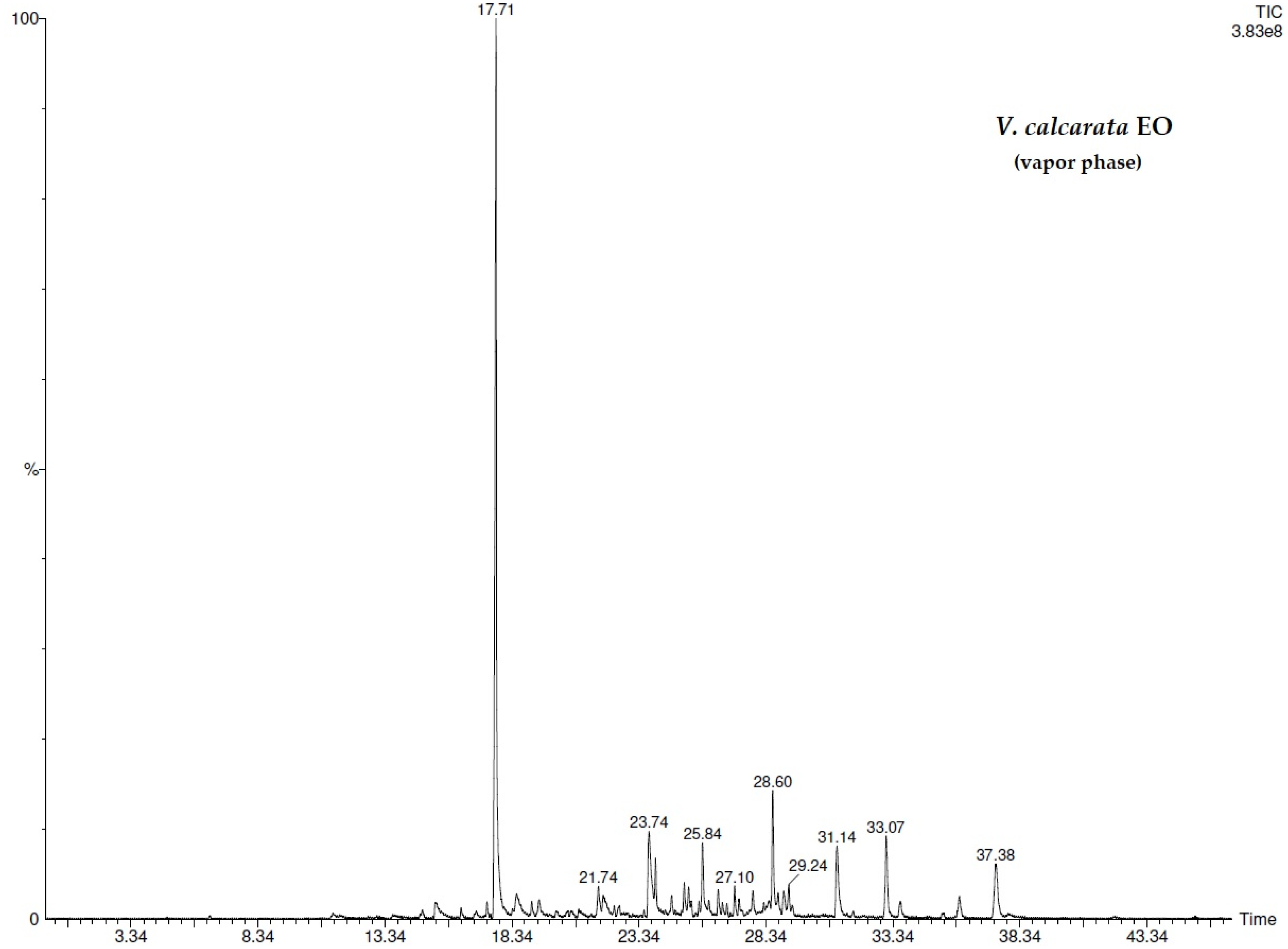
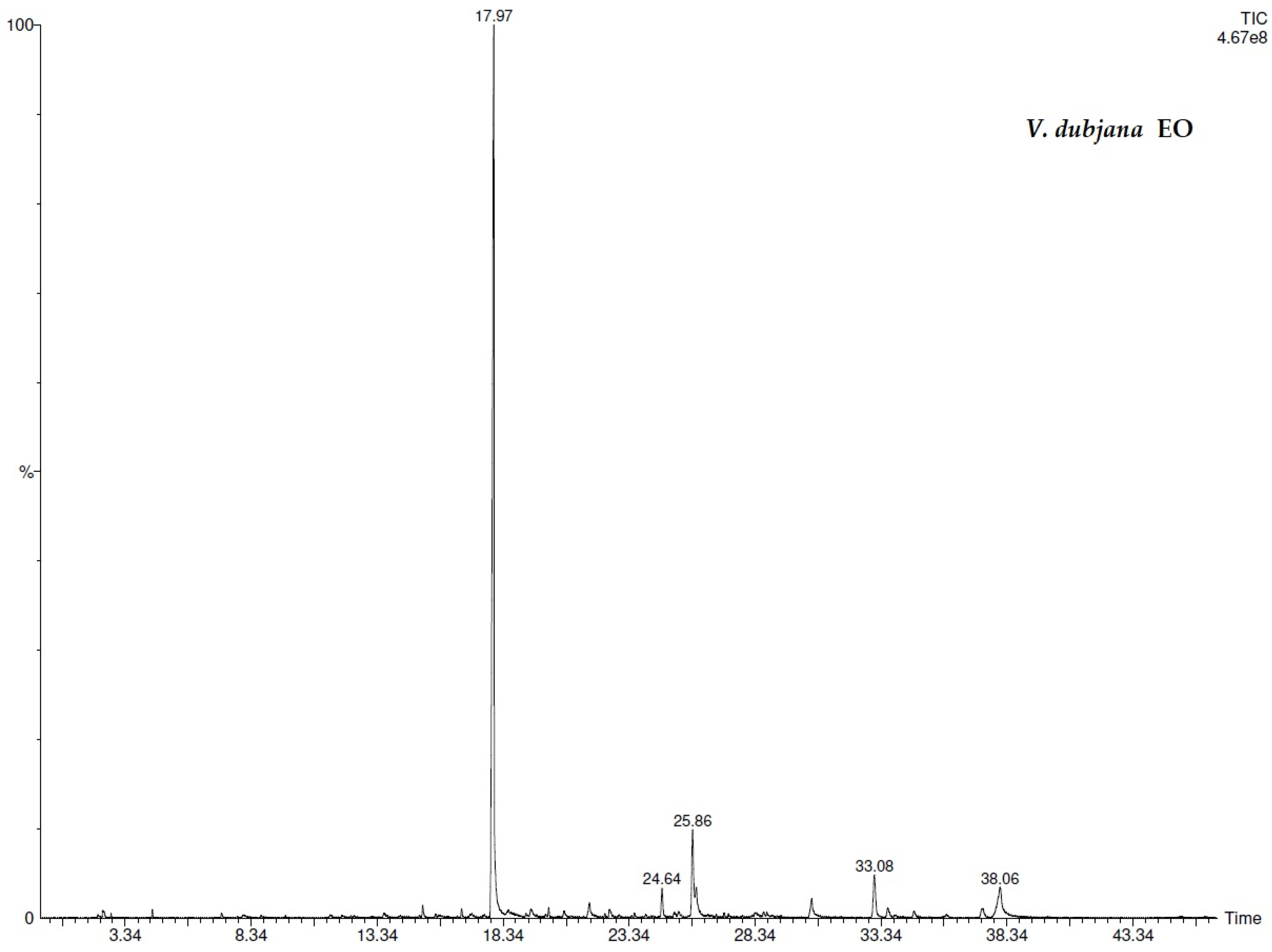
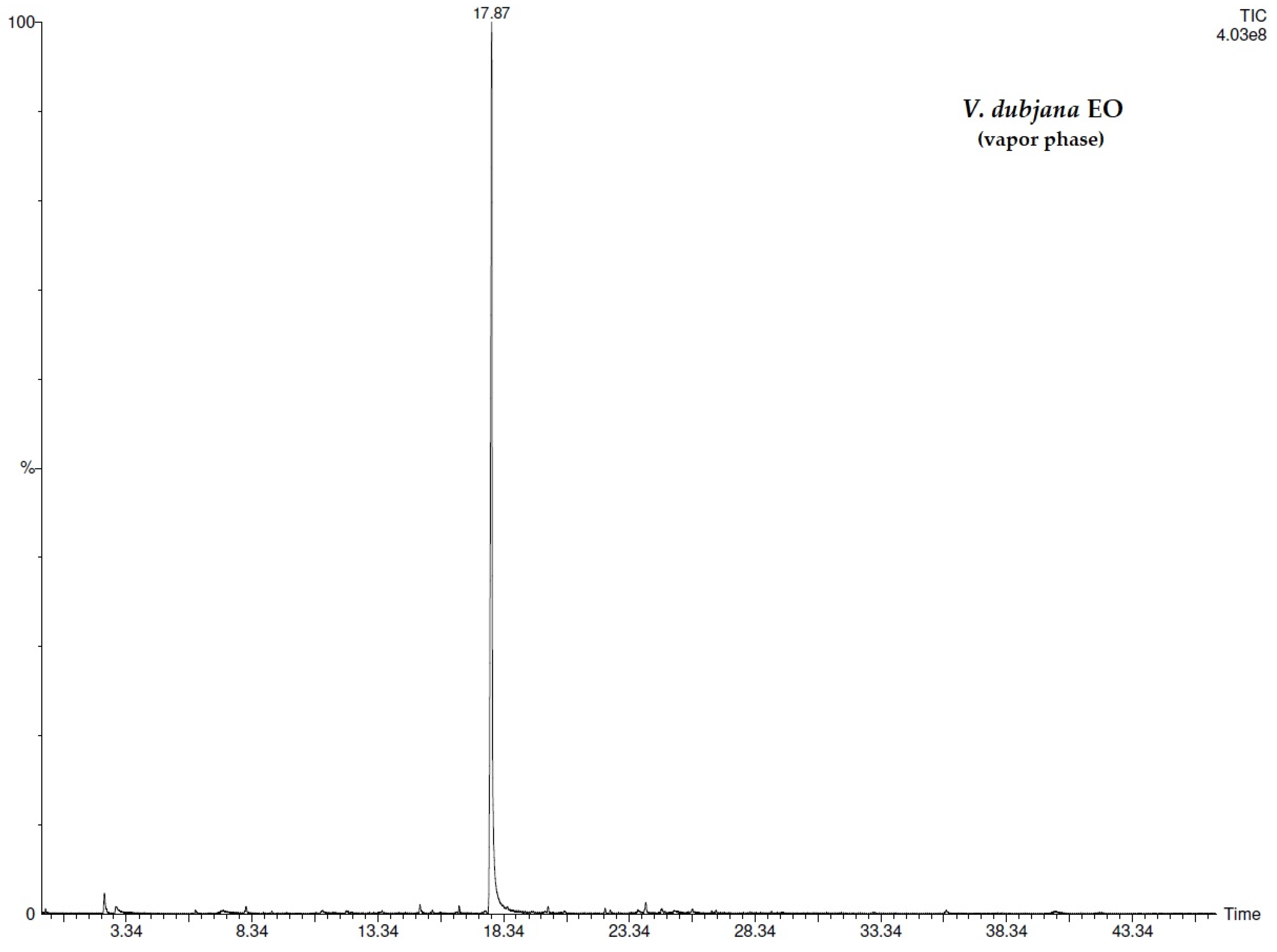
For both Viola species, the analysis of the vapor phase and that of the liquid phase was carried out to obtain a more accurate description of the volatile chemical composition. The respective GC chromatograms were reported (Figure 1, Figure 2, Figure 3 and Figure 4). A total of 43 compounds were identified and listed in the Table 1, among which methyl salicylate (MeSA), was the most abundant in both EOs but with higher percentage values in liquid and vapor phase of V. dubjana EO (67.3% and 68.0%) with respect to V. calcarata EO (45.5% and 46.3%). Differences in the qualitative composition between the two violets were found. For example, coumaran (2.9% and 0.2%), coumarin (6.2% and 5.8%), and epimanool (2.2% and 4.0%) were present only in the V. calcarata, as were the two monoterpenes terpinen-4-ol (0.7%; 1.1%) and linalol (0.5%; 0.7%). On the contrary, 2(4H)-benzofuranone, 5,6,7,7a-tetrahydro-4,4,7a-trimethyl-, a terpenic compound, reached major percentage values in V. dubjana (6.6%; 12.8%) compared to V. calcarata (4.2%; 3.0%) EO. The sesquiterpene content was higher in V. calcarata than in V. dubyana. Indeed, several sesquiterpenes such as sesquicineole (0.4%; 0.4%), nerolidol (0.7%; 0.8%), caryophyllene oxide (0.7%, 0.6%), β-eudesmol (1.3%; 1.3%), and α-bisabolol (1.2%; 1.6%) were characteristic only of V. calcarata sample. On the contrary, δ-cadinol (0.3%; 1.0%) and α-cadinol (0.3%; 0.4%) were characteristic only of V. dubjana sample. The only two sesquiterpene compounds common to both violets were spathulenol (from 0.2 to 1.1%) and hexahydrofarnesyl acetone (from 2.9 to 5.4%). Fatty acids were also detected in both samples except for dodecanoic (1.4% vapor phase; 3.2% liquid phase) and tetradecanoic (1.2%; 1.5%) acids which were present only in V. dubjana while hexadecenoic acid was only in V. calcarata vapor phase (0.3%).

3.2. EO Antiradical Activity

In terms of antiradical activity, both EOs showed different reactions with the two used radicals and the V. dubyana EO proved to be the most effective as a scavenger. EOs were able to appreciably inhibit ABTS⋅+ cation radical while they were weakly active in the removal of the stable radical DPPH⋅ (Table 2).

3.3. EO Phytotoxic Activity

As for the phytotoxic potential, the EOs of the two violets were found active, however revealing a greater efficacy of V. dubyana compared to V. calcarata, especially against the dicotyledon S. alba. EOs decreased, in a dose dependent manner, both the seed germination and seedling growth parameters (Table 3 and Table 4). In detail, the germination percentage of the monocot L. multiflorum, the most resistant target species, was similarly reduced by 5–25% and by 10–20%, respectively, by all used doses (2–100 μL) of V. calcarata and V. dubyana EOs. Alike, its CVG values dropped 4–60% and 3–57% while MGT was only affected by the two highest doses of 50 and 100 μL (+4% to +15% and +8% to +23%) of both EOs. The same doses (50 and 100 μL) also significantly inhibited the development of L. multiflorum roots (−44% to −92% and 50% to −90%) and shoots (−13% to −82% and −25% to −87%) lowering the corresponding SVI values (−42% to −91% and 52% to −91%).
More marked differences between the effects of the two EOs were identified on S. alba. In detail, V. dubyana EO, in the highest dose (100 μL), prevented its germination (0%) (Table 4), while V. calcarata EO was able to almost halve it (−46%) (Table 3). The lower doses of V. dubyana EO decreased the germination of S. alba by 5–77%, those of V. calcarata EO by 5–39% (Table 3 and Table 4). Accordingly, CVG and MGT indices were also influenced with values reduced up to −100% and −60%. When it occurred, the growth of roots and shoots of S. alba exposed to V. dubyana EO (2, 20 and 50 μL) stopped from −14% to −85% and from −5% to −63%, respectively, with reductions in SVI values up to 95% (Table 4). On the other hand, the length of S. alba roots and shoots under the effects of V. calcarata EO (2–100 μL) was found to be shorter than the controls by 6–74% and 2–58%. Hence, the SVI values decreased 1 to 6 fold (Table 3).

4. Discussion

In this work, the volatile profile of two Viola species was described owing to the use of SPME-GC/MS techniques. The chemical composition highlighted the presence of MeSA as the principal compound in both investigated samples with higher percentages in V. dubyana rather than in V. calcarata. Our results agree with previous studies on several populations of another rare Italian Viola species, namely V. etrusca Erben, reporting MeSA as the main component [10,26]. MeSA is a volatile compound and a constituent of floral fragrance of various plant species capable of acting as a defense agent against pathogens and some species of herbivores including aphids [27]. Furthermore, MeSA presents a good phytotoxic and allelopathic activity toward invasive plants [27,28], and plays a role as a pollinator attractor and in insect defense [29,30]. It has been referred to as a means of plant-to-plant aerial communication [31,32] and as the major constituent (96%) of Gaultheria procumbens L., whose EO also showed good antioxidant and antiradical activity [33]. Through brine shrimp lethality test, the toxicity of Laportea aestuans (Gaud) EOs was also demonstrated, the most abundant compound of which was MeSa (54.50%) [34]. Jayasekara et al. [35] also reported MeSA repellent and toxic properties against invertebrate pest adult Sitophilus zeamais and a dose-dependent fumigant effect against S. zeamais, Rhyzopertha dominica, and Prostephanus truncates. MeSA compound has even been described as an inducer of programmed cell death (PCD) [36]. Lastly, MeSA compound is used topically as a counter-irritant. Once absorbed it is rapidly hydrolyzed to salicylic acid, which has analgesic, antipyretic, and anti-inflammatory effects. In general, salicylates are used precisely for these beneficial effects, but cases of salicylate intoxication may occur following overdoses due to ingestion or skin absorption [37,38,39].
In the two violet EOs, we have found two other bioactive compounds such as 2(4H)-benzofuranone,5,6,7,7a-tetrahydro-4,4,7a-trimethyl and hexahydrofarnesyl acetone with relative high percentages. Both compounds were reported in the chemical composition of the EOs obtained from the leaves of V. odorata L. [40] and dried aerial parts of V. tricolor L. [41]. 2(4H)-Benzofuranone,5,6,7,7a-tetrahydro-4,4,7a-trimethyl- was also identified in the dichloromethane extract obtained from Alstonia scholaris (L.) R. Br. [42] and in the methanolic fractions of Azadirachta indica A.Juss. and Aegle marmelos (L.) Corrêa [43,44] showing antioxidant and antibacterial activity. Hexahydrofarnesyl acetone is a sesquiterpenoid associated with allelopathic activity [45]. It has been reported several times as one of the agents responsible for the phytotoxicity of various EOs [46,47,48,49]. Although present in lower percentages in the EO of V. calcarata and V. dubyana than the previous compounds, decanoic and hexadecanoic acids have already been detected as constituents of samples (EOs, exudates or extracts) capable of selectively reducing the growth of some plant species including weeds [50,51,52].
V. calcarata EO was characterized by the presence of bisabolone oxide A, bisabolol oxide A and B, and α-bisabolol. The first three compounds were also reported by Anca et al. [41] in their study on V. tricolor. Bisabolone oxide A and bisabolol oxide B were identified among the most abundant components of the Chamomilla recutita (L.) Rauschert EO, which, tested together with other EOs against the germination of some weeds, showed one of the lowest activities [53]. Otherwise, the phytotoxic potential of the EO of an Egyptian ecospecies of Pulicaria undulata (L.) C.A. Mey as well the allelopathic properties of the EO of Prangos pabularia Lindl. was ascribed to their major compounds including α-bisabolol [54,55]. V. calcarata also differed from the composition of V. dubyana for the presence of coumarin and coumaran. Coumarin is a natural compound known for its negative effects on weed germination [56]. Dodecanoic acid in V. dubyana EO showed phytotoxic effect toward some plant species, able to reduce their germination and root development [52]. The inhibitory effects of EOs can be due to the single component but also be related to the mixture of compounds [57]. For example, it is known that sesquiterpene allelochemicals including spathulenol and β-caryophyllene detected in our investigation presented phytotoxicity against various invasive plants [58]. In our EOs, the activity of these two compounds could be added to that of other components present in greater quantities. Furthermore, it was proved that the phytotoxic potential of an EO also depends on the used dose and the tested plant species [53].
Lastly, the presence of fatty acids recognized as effective antioxidants, with hexadecanoic acid reaching 6.1% in V. dubyana, may have contributed to the antiradical activity showed by both Viola species [59].

5. Conclusions

This is the first paper dealing with the volatile chemical composition of V. dubjana and V. calcarata. SPME-GC/MS techniques allowed the separation and identification of several volatile compounds highlighting a different profile, both from a qualitative and quantitative point of view, between the two species, however, both having MeSA as the dominant component. Despite this, the detected effectiveness of their EOs could also be due to the synergistic and/or additive action of the minor components, which, as has been widely reported, are able to play a significant role in various biological activities.
The findings, although to be further investigated, revealed an interesting bioactive potential of these violets, which could have various implications including ecological ones supporting the development of new products respectful of the environment and its resources.

Author Contributions

Conceptualization, S.G. and S.V.; methodology, S.G., M.I. and S.V.; investigation, S.G. and S.V.; resources, S.G. and M.I.; writing—original draft preparation, S.G. and S.V.; writing—review and editing, S.G. and S.V.; supervision, S.G., M.I. and S.V.; funding acquisition, S.G. and M.I. All authors have read and agreed to the published version of the manuscript.

Funding

This research received no external funding.

Acknowledgments

The authors thank Cristina Uva for the collection of Viola calcarata.

Conflicts of Interest

The authors declare no conflict of interest.

References

  1. Butnariu, M.; Sarac, I. Essential oils from plants. Int. J. Biotechnol. Biomed. Sci. 2018, 1, 35–43. [Google Scholar] [CrossRef] [Green Version]
  2. Raut, J.S.; Karuppayil, S.M. A status review on the medicinal properties of essential oils. Ind. Crops Prod. 2014, 62, 250–264. [Google Scholar] [CrossRef]
  3. Bakkali, F.; Averbeck, S.; Averbeck, D.; Idaomar, M. Biological effects of essential oils—A review. Food Chem. Ecol. 2008, 46, 446–475. [Google Scholar] [CrossRef] [PubMed]
  4. Shaaban, H.A.E.; El-Ghorab, A.H.; Shibamoto, T. Bioactivity of essential oils and their volatile aroma components: Review. J. Essent. Oil Res. 2012, 24, 203–212. [Google Scholar] [CrossRef]
  5. Sharif-Rad, J.; Sureda, A.; Tenore, G.C.; Daglia, M.; Sharif-Rad, M.; Valussi, M.; Tindis, R.; Sharif-Rad, M.; Loizzo, M.R.; Ademiluyi, A.O.; et al. Biological activities of essential oils: From plant chemoecology to traditional healing systems. Molecules 2017, 22, 70. [Google Scholar] [CrossRef]
  6. Mancianti, F.; Ebani, V.V. Biological activity of essential oils. Molecules 2020, 25, 678. [Google Scholar] [CrossRef] [Green Version]
  7. Azirak, S.; Karaman, S. Allelopathic effect of some essential oils and components on germination of weed species. Acta Agric. Scand. B Soil Plant Sci. 2008, 58, 88–92. [Google Scholar] [CrossRef]
  8. El Sawi, S.A.; Ibrahim, M.E.; El-Rokiek, K.G.; El-Din, S.A.S. Allelopathic potential of essential oils isolated from peels of three citrus species. Ann. Agric. Sci. 2019, 64, 89–94. [Google Scholar] [CrossRef]
  9. Grul’ová, D.; Caputo, L.; Elshafie, H.S.; Baranová, B.; De Martino, L.; Sedlák, V.; Gogal’ová, Z.; Poráčová, J.; Camele, I.; De Feo, V. Thymol chemotype Origanum vulgare L. essential oil as a potential selective bio-based herbicide on monocot plant species. Molecules 2020, 25, 595. [Google Scholar] [CrossRef] [Green Version]
  10. Flamini, G.; Cioni, P.L.; Morelli, I. Variability of the essential oil of Viola etrusca. Ann. Bot. 2003, 91, 493–497. [Google Scholar] [CrossRef] [Green Version]
  11. Hammami, I.; Kamoun, N.; Rebai, A. Biocontrol of Botrytis cinerea with essential oil and methanol extract of Viola odorata L. flowers. Arch. Appl. Sci. Res. 2011, 3, 44–51. [Google Scholar]
  12. Muhammad, N.; Saeed, M.; Awan, A.A.; Khan, H. Ethnomedicinal, phytochemical and pharmacological profile of genus Viola. Phytopharmacology 2012, 3, 214–226. [Google Scholar]
  13. Chandra, D.; Kohli, G.; Prasad, K.; Bisht, G.; Punetha, V.D.; Pandey, H.K. Chemical composition of the essential oil of Viola serpens from Bageshwar (Shama), Uttarakhad, India. J. Med. Plant Res. 2017, 11, 513–517. [Google Scholar]
  14. Kirillov, V.; Stikhareva, T.; Atazhanova, G.; Vasiliv, A.M. Composition of essential oil of the aerial parts of Viola canina L. growing wild in Northern Kazakhistan. Nat. Prod. Res. 2019, 35, 2285–2288. [Google Scholar] [CrossRef]
  15. Yousuf, S.; Yousuf, T.; Bachheti, R.K.; Deshmukh, C. Reducing potential of essential oil extracted from Viola patrinii for some powerful oxidants like DPPH, FeCl3 and nitric oxide. Ann. Rom. Soc. Cell. Biol. 2021, 25, 20562–20571. [Google Scholar]
  16. Falla, N.M.; Demasi, S.; Caser, M.; Scariot, V. Preliminary observations on bioactive compounds: Antioxidant activity and phytochemical profile of two alpine subspecies. Agronomy 2021, 11, 2241. [Google Scholar] [CrossRef]
  17. Chandra, D.; Kohli, G.; Prasad, K.; Bisht, G.; Punetha, V.D.; Khetwal, K.S.; Devrani, M.K.; Pandey, H.K. phytochemical and ethnomedicinal uses of family Violaceae. Curr. Res. Chem. 2015, 7, 44–52. [Google Scholar] [CrossRef] [Green Version]
  18. Pignatti, S. Flora d’Italia, 2nd ed.; Edagricole: Bologna, Italy, 2017. [Google Scholar]
  19. Tutin, T.G.; Heywood, V.H.; Burges, N.A.; Moore, D.M.; Valntine, D.H.; Walters, S.M.; Webb, D.A. Flora Europaea, 3rd ed.; Cambridge University Press: Cambridge, UK, 1981. [Google Scholar]
  20. Garzoli, S.; Laghezza Masci, V.; Caradonna, V.; Tiezzi, A.; Giacomello, P.; Ovidi, E. Liquid and Vapor Phase of Four Conifer-Derived Essential Oils: Comparison of Chemical Compositions and Antimicrobial and Antioxidant Properties. Pharmaceuticals 2021, 14, 134. [Google Scholar] [CrossRef] [PubMed]
  21. Iriti, M.; Vitalini, S.; Apostolides, N.A.; El Beyrouthy, M. Chemical composition and antiradical capacity of essential oils from Lebanese medicinal plants. J. Ess. Oil Res. 2014, 26, 466–472. [Google Scholar] [CrossRef]
  22. Vitalini, S.; Orlando, F.; Iriti, M. Selective phytotoxic activity of eugenol towards monocot and dicot target species. Nat. Prod. Res. 2021, 1–4. [Google Scholar] [CrossRef] [PubMed]
  23. Al-Mudaris, M. Notes on various parameters recording the speed of seed germination. Der Trop. 1998, 99, 147–154. [Google Scholar]
  24. Ellis, R.A.; Roberts, E.H. The quantification of ageing and survival in orthodox seeds. Seed Sci. Technol. 1981, 9, 373–409. [Google Scholar]
  25. Abdul-Baki, A.A.; Anderson, J.D. Vigour determination in soybean seed by multiple criteria. Crop Sci. 1973, 1, 630–633. [Google Scholar] [CrossRef]
  26. Flamini, G.; Cioni, P.L.; Morelli, I. Analysis of the essential oil of the aerial parts of Viola etrusca from Monte Labbro (South Tuscany, Italy) and in vivo analysis of flower volatiles using SPME. Flavour Fragr. J. 2002, 17, 147–149. [Google Scholar] [CrossRef]
  27. Batish, D.R.; Singh, H.P.; Kaur, M.; Kohli, R.K.; Singh, S. Chemical characterization and phytotoxicity of volatile essential oil from leaves of Anisomeles indica (Lamiaceae). Biochem. Syst. Ecol. 2012, 41, 104–109. [Google Scholar] [CrossRef]
  28. Bi, H.H.; Zeng, R.S.; Su, L.M.; An, M.; Luo, S.M. Rice allelopathy induced by methyl jasmonate and methyl salicylate. J. Chem. Ecol. 2007, 33, 1089–1103. [Google Scholar] [CrossRef] [PubMed]
  29. Van Poecke, R.M.P.; Posthumus, M.A.; Dicke, M. Herbivore-induced volatile production by Arabidopsis thaliana leads to attraction of the parasitoid Cotesia rubecula: Chemical, behavioral, and gene expression analysis. J. Chem. Ecol. 2001, 27, 1911–1928. [Google Scholar] [CrossRef]
  30. Zhu, J.W.; Park, K.C. Methyl salicylate, a soybean aphid-induced plant volatile attractive to the predator Coccinella septempunctata. J. Chem. Ecol. 2005, 31, 1733–1746. [Google Scholar] [CrossRef]
  31. Shulaev, V.; Silverman, P.; Raskin, I. Airborne signaling by methyl salicylate in plant pathogen resistance. Nature 1997, 385, 718–721. [Google Scholar] [CrossRef]
  32. Koo, Y.J.; Kim, M.A.; Kim, E.H.; Song, J.T.; Jung, C.; Moon, J.K.; Kim, J.H.; Seo, H.S.; Song, J.K.; Lee, J.S.; et al. Overexpression of salicylic acid carboxyl methyltransferase reduces salicylic acid-mediated pathogen resistance in Arabidopsis thaliana. Plant Mol. Biol. 2007, 64, 1–15. [Google Scholar] [CrossRef]
  33. Nikolić, M.; Marković, T.; Mojović, M.; Pejin, B.; Savić, A.; Perić, T.; Marković, D.; Stević, T.; Soković, M. Chemical composition and biological activity of Gaultheria procumbens L. essential oil. Ind. Crops Prod. 2013, 49, 561–567. [Google Scholar] [CrossRef]
  34. Oloyede, G.K. Toxicity, antimicrobial and antioxidant activities of methyl salicylate dominated essential oils of Laportea aestuans (Gaud). Arab. J. Chem. 2016, 9, 840–845. [Google Scholar] [CrossRef] [Green Version]
  35. Jayasekara, T.K.; Stevenson, P.C.; Hall, D.R.; Belmain, S.R. Effect of volatile constituents from Securidaca Longepedunculata on insect pests of stored grain. J. Chem. Ecol. 2005, 31, 303–313. [Google Scholar] [CrossRef]
  36. Yun, L.J.; Chen, W.L. SA and ROS are involved in methyl salicylate-induced programmed cell death in Arabidopsis thaliana. Plant Cell Rep. 2011, 30, 1231–1239. [Google Scholar] [CrossRef] [PubMed]
  37. Brubacher, J.R.; Hoffman, R.S. Salicylism from topical salicylates review of the literature. J. Toxicol. Clin. Toxicol. 1996, 34, 431–436. [Google Scholar] [CrossRef] [PubMed]
  38. Heng, M.C. Local necrosis and interstitial nephritis due to topical methyl salicylate and menthol. Cutis 1987, 39, 442–444. [Google Scholar] [PubMed]
  39. Baron, J.A.; Raia, J.J.; Huang, Y.C. Evaluation of the effects of multiple-dose activated charcoal on the absorption of orally administered salicylate in a simulated toxic ingestion model. Ann. Emerg. Med. 1988, 17, 34–37. [Google Scholar] [CrossRef]
  40. Akhbari, M.; Batooli, H.; Kashi, F.J. Composition of essential oil and biological activity of extracts of Viola odorata L. from central Iran. Nat. Prod. Res. Form. Nat. Prod. Lett. 2012, 26, 802–809. [Google Scholar]
  41. Anca, T.; Philippe, V.; Ilioara, O.; Mircea, T. Composition of essential oils of Viola tricolor and V. arvensis from Romania. Chem. Nat. Comp. 2009, 45, 91–92. [Google Scholar] [CrossRef]
  42. Carranza, M.S.S.; Oyong, G.G.; Linis, V.C.; Ajero, M.D.M.; Tan, M.C.S. The antioxidant and antiproliferative agents from the bark of Philippine Alstonia scholaris (L.) R. Br. (Apocynaceae). Jordan J. Pharm. Sci. 2020, 13, 207–224. [Google Scholar]
  43. Moorthy, V.; Boominathan, M. The antimicrobial activities of crude extracts and fraction of Psidium guajava and Azadirachta indica against Staphylococcus aureus in chronic disease affected patients. Int. J. Univers. Pharm. Life Sci. 2011, 1, 2249–6793. [Google Scholar]
  44. Mujeeb, F.; Bajpai, P.; Pathak, N. Phytochemical evaluation, antimicrobial activity, and determination of bioactive components from leaves of Aegle marmelos. Biomed Res. Int. 2014, 2014, 497606. [Google Scholar] [CrossRef] [PubMed] [Green Version]
  45. Abd-ElGawad, A.M.; El Gendy, A.E.-N.G.; Assaeed, A.M.; Al-Rowaily, S.L.; Alharthi, A.S.; Mohamed, T.A.; Nassar, M.I.; Dewir, Y.H.; Elshamy, A.I. Phytotoxic effects of plant essential oils: A systematic review and structure-activity relationship based on chemometric analyses. Plants 2021, 10, 36. [Google Scholar] [CrossRef] [PubMed]
  46. El Ayeb-Zakhama, A.; Ben Salem, S.; Sakka-Rouis, L.; Flamini, G.; Ben Jannet, H.; Harzallah-Skhiri, F. Chemical composition and phytotoxic effects of essential oils obtained from Ailanthus altissima (Mill.) swingle cultivated in Tunisia. Chem. Biodivers. 2014, 11, 1216–1227. [Google Scholar] [CrossRef] [PubMed]
  47. Elshamy, A.; Abd El-Gawad, A.M.; El-Amier, Y.A.; El Gendy, A.; Al-Rowaily, S. Interspecific variation, antioxidant and allelopathic activity of the essential oil from three Launaea species growing naturally in heterogeneous habitats in Egypt. Flavour Fragr. J. 2019, 34, 316–328. [Google Scholar] [CrossRef]
  48. Abd-ElGawad, A.M.; Elshamy, A.I.; El Gendy, A.E.-N.G.; Al-Rowaily, S.L.; Assaeed, A.M. Preponderance of oxygenated sesquiterpenes and diterpenes in the volatile oil constituents of Lactuca serriola L. revealed antioxidant and allelopathic activity. Chem. Biodivers. 2019, 16, 1900278. [Google Scholar] [CrossRef] [PubMed]
  49. Abd-ElGawad, A.; El Gendy, A.; El-Amier, Y.; Gaara, A.; Omer, S.; Al-Rowaily, S.; Assaeed, A.; Al-Rashed, S.; Elshamy, A. Essential oil of Bassia muricata: Chemical characterization, antioxidant activity, and allelopathic effect on the weed Chenopodium murale. Saudi J. Biol. Sci. 2020, 27, 1900–1906. [Google Scholar] [CrossRef]
  50. Xuan, T.D.; Chung, I.M.; Khanh, T.D.; Tawata, S. Identification of phytotoxic substances from early growth of barnyard grass (Echinochloa crus-galli) root exudates. J. Chem. Ecol. 2006, 32, 895. [Google Scholar] [CrossRef]
  51. Mancini, E.; Arnold, N.A.; De Feo, V.; Formisano, C.; Rigano, D.; Piozzi, F.; Senatore, F. Phytotoxic effects of essential oils of Nepeta curviflora Boiss. and Nepeta nuda L. subsp. albiflora growing wild in Lebanon. J. Plant Interact. 2009, 4, 253–259. [Google Scholar] [CrossRef]
  52. Cruz-Estrada, A.; Ruiz-Sánchez, E.; Cristóbal-Alejo, J.; González-Coloma, A.; Fe Andrés, M.; Gamboa-Angulo, M. Medium-chain fatty acids from Eugenia winzerlingii leaves causing insect settling deterrent, nematicidal, and phytotoxic effects. Molecules 2019, 24, 1724. [Google Scholar] [CrossRef] [Green Version]
  53. Synowiec, A.; Kaemba, D.; Drozdek, E.; Bocianowski, J. Phytotoxic potential of essential oils from temperate climate plants against the germination of selected weeds and crops. J. Pest Sci. 2017, 90, 407–419. [Google Scholar] [CrossRef]
  54. Razavi, S.M. Chemical and allelopathic analyses of essential oils of Prangos pabularia Lindl. from Iran. Nat. Prod. Res. 2011, 26, 2148–2151. [Google Scholar] [PubMed]
  55. Abd-ELGawad, A.M.; Al-Rowaily, S.L.; Assaeed, A.M.; EI-Amier, Y.A.; El Gendy, A.E.-N.G.; Omer, E.; Al-Dosari, D.H.; Bonanomi, G.; Kassem, H.S.; Elshamy, A.I. Comparative chemical profiles and phytotoxic activity of essential oils of two ecospecies of Pulicaria undulata (L.) C.A.Mey. Plants 2021, 10, 2366. [Google Scholar] [CrossRef] [PubMed]
  56. Araniti, F.; Mancuso, R.; Lupini, A.; Giofrè, S.V.; Sunseri, F.; Gabriele, B.; Abenavoli, M.R. Phytotoxic potential and biological activity of three synthetic coumarin derivatives as new natural-like herbicides. Molecules 2015, 20, 17883–17902. [Google Scholar] [CrossRef] [PubMed] [Green Version]
  57. Kaur, S.; Singh, H.; Mittal, S.; Batish, D.R.; Kohli, R.K. Phytotoxic effects of volatile oil from Artemisia scoparia against weeds and its possible use as a bioherbicide. Ind. Crops Prod. 2010, 32, 54–61. [Google Scholar] [CrossRef]
  58. Kobaisy, M.; Tellez, M.R.; Dayan, F.E.; Duke, S.O. Phytotoxicity and volatile constituents from leaves of Callicarpa japonica Thunb. Phytochemistry 2002, 61, 37–40. [Google Scholar] [CrossRef]
  59. Çakmak, Y.S.; Aktumsek, A.; Duran, A. Studies on antioxidant activity, volatile compound and fatty acid composition of different parts of Glycyrrhiza echinata L. EXCLI J. 2012, 11, 178–187. [Google Scholar]
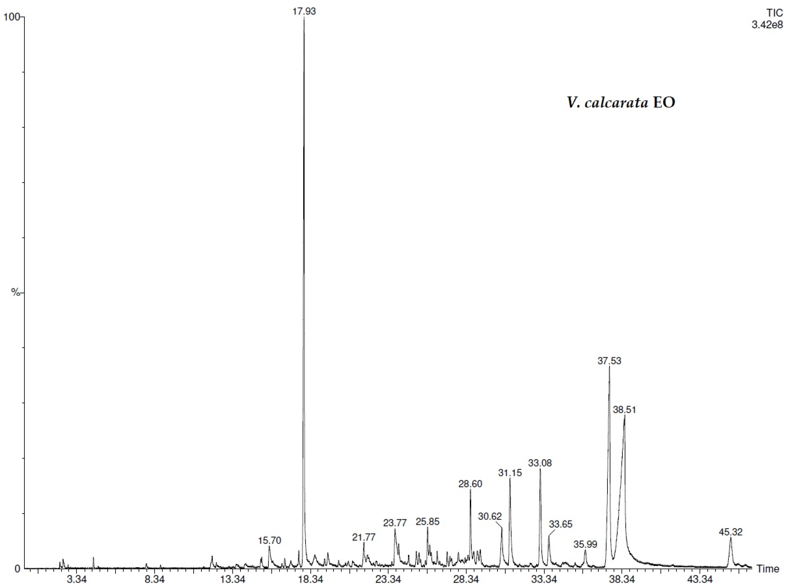
Figure 1. GC/GC-FID chromatograms of V. calcarata EO.
Separations 09 00039 g001
Figure 2. SPME/GC-FID chromatograms of V. calcarata EO.
Separations 09 00039 g002
Figure 3. GC/GC-FID chromatograms of V. dubjana EO.
Separations 09 00039 g003
Figure 4. SPME/GC-FID chromatograms of V. dubjana EO.
Separations 09 00039 g004
Table 1. Chemical composition (peak area percentage) of Viola dubyana and Viola calcarata EOs.
COMPONENT 1LRI 2V.d.3 (%)V.d.4 (%)V.c.5 (%)V.c.6 (%)
1hexanal7700.3 ± 0.02---
2furfural8320.3 ± 0.02---
3heptanal8740.1 ± 0.01---
4hexanoic acid976--tr0.3 ± 0.02
5octanal10050.1 ± 0.00---
6linalol1078--0.5 ± 0.020.7 ± 0.02
7nonanal10910.6 ± 0.020.1 ± 0.02-0.2 ± 0.02
8camphor11570.4 ± 0.010.2 ± 0.010.5 ± 0.030.4 ± 0.01
9trans-sabinene hydrate11620.2 ± 0.01---
10octanoic acid11720.2 ± 0.010.5 ± 0.020.3 ± 0.010.6 ± 0.01
11terpinen-4-ol1182--0.7 ± 0.021.1 ± 0.02
12β-cyclocitral12000.8 ± 0.02---
13methyl salicylate121867.3 ± 0.0268.0 ± 0.0245.5 ± 0.0246.3 ± 0.02
14coumaran1225--2.9 ± 0.020.2 ± 0.02
15nonanoic acid1263-0.9 ± 0.011.2 ± 0.011.4 ± 0.02
16p-vinylguaiacol12790.6 ± 0.020.5 ± 0.02tr0.4 ± 0.02
17bornyl acetate12800.4 ± 0.010.3 ± 0.02--
18decanoic acid13491.4 ± 0.022.6 ± 0.021.6 ± 0.022.8 ± 0.01
192-(4-methoxyphenyl)-ethanol1360--1.6 ± 0.022.0 ± 0.02
20β-copaen-4α-ol14000.7 ± 0.02tr--
21coumarin1430- 6.2 ± 0.025.8 ± 0.02
22trans-geranyl acetone14370.2 ± 0.020.3 ± 0.02--
23trans-β-ionone14631.8 ± 0.002.2 ± 0.02--
24sesquicineole1500--0.4 ± 0.020.4 ± 0.02
252(4H)-benzofuranone, 5,6,7,7a-tetrahydro-4,4,7a-trimethyl-15306.6 ± 0.0112.8 ± 0.014.2 ± 0.033.0 ± 0.02
26nerolidol1537--0.7 ± 0.010.8 ± 0.02
27dodecanoic acid15553.2 ± 0.021.4 ± 0.02--
28spathulenol15720.2 ± 0.020.5 ± 0.021.1 ± 0.021.1 ± 0.02
29caryophyllene oxide1578--0.7 ± 0.010.6 ± 0.04
30humulene epoxide II1598--1.3 ± 0.02-
31δ-cadinol16210.3 ± 0.021.0 ± 0.02--
32bisabolol oxide B1660--6.0 ± 0.035.9 ± 0.02
33β-eudesmol1668--1.3 ± 0.021.3 ± 0.02
34α-cadinol16720.3 ± 0.020.4 ± 0.02--
35α-bisabolol1681--1.2 ± 0.021.6 ± 0.02
36bisabolone oxide A1700--1.2 ± 0.011.3 ± 0.02
37bisabolol oxide A1710--5.8 ± 0.025.7 ± 0.03
38tetradecanoic acid17551.5 ± 0.011.1 ± 0.01--
39hexahydrofarnesyl acetone18444.2 ± 0.022.9 ± 0.025.4 ± 0.044.7 ± 0.02
40diisobutyl phthalate18680.9 ± 0.020.7 ± 0.021.1 ± 0.032.2 ± 0.04
41dibutyl phthalate19111.3 ± 0.020.2 ± 0.022.5 ± 0.013.3 ± 0.02
42hexadecanoic acid19756.1 ± 0.013.0 ± 0.011.6 ± 0.031.9 ± 0.04
43epimanool2061--2.2 ± 0.024.0 ± 0.02
SUM 100.099.697.7100.0
Terpenoids 8.213.35.95.2
Sesquiterpenoids 5.74.810.810.5
Fatty acids 12.49.54.77.0
Other 73.772.076.377.3
1 The components are reported according to their elution order on apolar column; 2 linear retention indices measured on apolar column; V.d. 3: peak area percentage of Viola dubyana liquid phase components (%); V.d. 4: peak area percentage of Viola dubyana vapor phase components; V.c. 5: peak area percentage of Viola calcarata liquid phase components (%); V.c. 6: peak area percentage of Viola calcarata vapor phase components; -: not detected; tr: traces (mean value < 0.1%).
Table 2. Antiradical power of EOs from Viola dubyana and Viola calcarata.
EOABTS
(μM Trolox eq/mL)
DPPH
(μM Trolox eq/mL)
Viola calcarata1.03 ± 0.010.29 ± 0.01
Viola dubyana1.32 ± 0.030.37 ± 0.01
Table 3. Germination and growth parameters of two target species (Lolium multiflorum and Sinapis alba) under the phytotoxic effects of different doses of Viola calcarata EO.
Viola calcarata
Target
Species
EO Doses
(μL)
G
(%)
CVGMGTSVIRoot
(mm)
Shoot
(mm)
Lolium multiflorum292.0 ± 9.0101.0 ± 8.04.8 ± 0.310,332 ± 95462.3 ± 8.950.0 ± 5.3
2090.0 ± 7.099.0 ± 5.04.8 ± 0.210,368 ± 68664.1 ± 22.051.1 ± 7.23
5080.0 ± 8.079.0 ± 4.05.0 ± 0.26240 ± 55534.7 ± 7.443.3 ± 12.4
10073.0 ± 5.042.0 ± 6.05.5 ± 0.31022 ± 2865.1 ± 1.48.9 ± 1.9
CTRL97.0 ± 5.0105.0 ± 9.04.8 ± 0.110,796 ± 73161.4 ± 7.949.9 ± 4.0
Sinapis alba283.0 ± 5.0115.0 ± 6.04.2 ± 0.24001 ± 30126.1 ± 7.722.1 ± 5.5
2080.0 ± 8.0108.0 ± 7.64.2 ± 0.33200 ± 14321.7 ± 9.218.3 ± 4.0
5053.0 ± 5.060.0 ± 7.34.3 ± 0.3912 ± 697.3 ± 3.99.9 ± 2.2
10047.0 ± 3.048.0 ± 3.34.5 ± 0.0785 ± 987.3 ± 2.59.4 ± 1.4
CTRL87.0 ± 5.0119.0 ± 9.84.1 ± 0.14359 ± 35327.8 ± 3.422.3 ± 6.8
Values are mean ± standard deviation. G%, germination percentage; CVG, coefficient of velocity of germination; MGT, mean germination time, SVI, seedling vigor index.
Table 4. Germination and growth parameters of two target species (Lolium multiflorum and Sinapis alba) under the phytotoxic effects of different doses of Viola dubyana EO.
Viola dubyana
Target
Species
EO Doses
(μL)
G
(%)
CVGMGTSVIRoot
(mm)
Shoot
(mm)
Lolium multiflorum290.0 ± 14.0104.5 ± 9.34.9 ± 0.211,601 ± 87774.9 ± 11.254.0 ± 9.4
2083.0 ± 9.0101.0 ± 15.04.9 ± 0.110,234 ± 94169.9 ± 12.853.4 ± 8.3
5080.0 ± 5.074.0 ± 2.85.2 ± 0.16320 ± 45838.0 ± 6.641.0 ± 9.8
10080.0 ± 0.047.0 ± 4.55.9 ± 0.01192 ± 2377.8 ± 2.67.1 ± 1.7
CTRL100.0 ± 5.0108.0 ± 4.04.8 ± 0.113,100 ± 68776.3 ± 10.654.7 ± 11.2
Sinapis alba283.0 ± 8.0102.0 ± 9.04.2 ± 0.14116 ± 30129.4 ± 6.720.2 ± 4.5
2057.0 ± 5.063.0 ± 2.04.2 ± 0.01533 ± 14312.8 ± 4.514.1 ± 2.6
5020.0 ± 5.020.0 ± 6.04.1 ± 0.1258 ± 695.1 ± 1.17.8 ± 1.5
1000.0 ± 0.0n.d.n.d.n.d.n.d.n.d.
CTRL87.0 ± 5.0112.0 ± 11.04.2 ± 0.14829 ± 35334.3 ± 8.521.2 ± 5.9
Values are mean ± standard deviation. G%, germination percentage; CVG, coefficient of velocity of germination; MGT, mean germination time; SVI, seedling vigor index; n.d., not determined.
Publisher’s Note: MDPI stays neutral with regard to jurisdictional claims in published maps and institutional affiliations.