
Article

Isolation of Neuroprotective Constituents from Dryopteris crassirhizoma Rhizomes Inhibiting Beta-Amyloid Production and BACE1 Activity
1
Laboratory of Pharmacognosy, College of Pharmacy, Dankook University, Cheonan 31116, Republic of Korea
2
College of Pharmacy, Chung-Ang University, 84 Heukseok-ro, Dongjak-gu, Seoul 06974, Republic of Korea
*
Author to whom correspondence should be addressed.
These authors contributed equally to this work.
Separations 2026, 13(1), 35; https://doi.org/10.3390/separations13010035
Submission received: 19 December 2025 / Revised: 13 January 2026 / Accepted: 15 January 2026 / Published: 16 January 2026
(This article belongs to the Special Issue Isolation and Identification of Biologically Active Natural Compounds)

Abstract

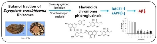
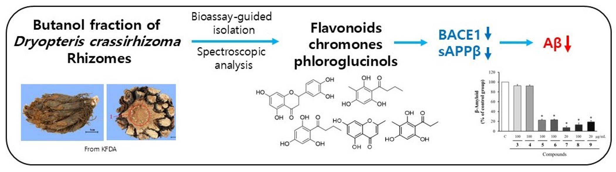
Alzheimer’s disease (AD) is a prevalent neurodegenerative condition that progressively impairs cognitive processes, particularly learning and memory. A key pathological feature of AD involves senile plaques mainly composed of β-amyloid (Aβ) peptides, generated via the amyloidogenic pathway from amyloid precursor protein (APP) through sequential β-secretase (BACE1) and γ-secretase cleavage, positioning BACE1 inhibition as a prime therapeutic target. In this study, we applied bioassay-guided fractionation of the butanol-soluble fraction from Dryopteris crassirhizoma rhizomes, previously reported to inhibit Aβ production, to isolate and characterize Aβ-lowering constituents. Through successive chromatographic steps, nine compounds were isolated and structurally classified into flavonoids, chromones, and phloroglucinols, including epicatechin (1), β-carboxymethyl-(-)-epicatechin (2), 7-methoxy-isobiflorin (3), biflorin (4), eriodictyol (5), noreugenin (6), phloroglucinols (butyrylphloroglucinol (7), 2-propionyl-4-methylphloroglucinol (8), and 2-butyryl-4-methylphloroglucinol (9) by comprehensive spectroscopic analysis (NMR, MS, UV, IR). These compounds were assessed for effects on sAPPβ and BACE1 (β-secretase) levels by Western blot, with Aβ production quantified via ELISA in a cellular AD model (APP-CHO cells). Compounds 59 significantly reduced sAPPβ and BACE1 expression while potently suppressing Aβ generation. These results demonstrate that diverse constituents from D. crassirhizoma rhizomes inhibited Aβ production through BACE1 suppression, highlighting their potential as natural lead compounds for AD prevention or therapy.

Graphical Abstract

1. Introduction

Alzheimer’s disease (AD) is the leading cause of neurodegeneration worldwide and accounts for about 71% of irreversible dementia cases in the elderly population [1]. The disease is incurable and is characterized clinically by gradual memory loss, cognitive impairment, and neuropsychiatric symptoms such as depression and anxiety [2,3]. As AD progresses, patients eventually lose the ability to perform daily activities [1,2].
Neuropathologically, AD is characterized by the accumulation of extracellular β-amyloid (Aβ) plaques and intracellular neurofibrillary tangles formed by hyperphosphorylated tau, accompanied by extensive neuronal degeneration. Accumulations of Aβ and tau are considered the principle factors that contribute to neuronal degeneration and clinical deficits [4,5].
Aβ arises from aberrant proteolytic cleavage of amyloid precursor protein (APP), a transmembrane protein. BACE1 (β-secretase)–mediated processing of APP produces two distinct fragments: an extracellularly released N-terminal sAPPβ and a membrane-associated C-terminal C99 fragment. The C99 fragment is subsequently cleaved by the γ-secretase complex, which consists of presenilin-1 or -2, presenilin enhancer-2, anterior pharynx-defective protein, and nicastrin, thereby generating Aβ peptides. These Aβ monomers undergo progressive aggregation into oligomeric species and insoluble fibrils that exhibit cytotoxicity and contribute to neuronal cell death. Therefore, inhibiting Aβ production represents a promising strategy for the prevention and treatment of AD [6,7].
Our previous study identified the butanol-soluble fraction of the rhizomes of Dryopteris crassirhizoma Nakai (commonly known as Gwanjung) significantly reduced Aβ production to 26% of the DMSO-treated control, accompanied by suppression of BACE1 to 20% [8]. Traditionally, this medicinal plant has been used for its anti-parasitic, detoxifying, antipyretic, hemostatic, and uterotonic properties, as well as in the treatment of parasitic infections, colds, cancer, and meningitis. However, its effects against dementia have not been previously reported, and the active compounds responsible for its anti-amyloidogenic activity have not been identified [9].
Based on these observations, this study employed bioassay-guided fractionation of the D. crassirhizoma butanol fraction using successive chromatographic techniques to isolate and structurally characterize Aβ-lowering compounds. Nine constituents were purified and evaluated for effects on sAPPβ secretion, BACE1 expression (Western blot), and Aβ production (ELISA) in APP-CHO cells, aiming to identify novel natural BACE1 inhibitors through systematic separation science.

2. Materials and Methods

2.1. Column Chromatography

For HPLC analyses, ACE-121-2546 columns (5 μm, 250 × 4.6 mm, Advanced Chromatography Technologies, Aberdeen, UK) and YMC-Pack ODS-A columns (5 μm, 250 × 10 mm, Yamamura Chemical, Kyoto, Japan) were used. Open column chromatography employed silica gel (200–400 mesh, Fisher Scientific, Pittsburgh, PA, USA), Lichroprep RP-18 (40–63 μm, Merck, Darmstadt, Germany), and MCI gel CHP 20P (75–150 μm, Mitsubishi Chemical, Tokyo, Japan) as packing materials. Thin-layer chromatography (TLC) was performed on Kieselgel 60 F254 plates (0.2 mm, Merck). Vanillin-sulfuric acid spray reagent was prepared using 10% vanillin and 10% sulfuric acid (Sigma, St. Louis, MO, USA).

2.2. Preparation of Extract and Partitioned Fractions

Six kilograms of D. crassirhizoma rhizomes were purchased from a commercial market (Incha Herb Market, Seoul, Republic of Korea). Botanical authentication was carried out by one of the authors (S.-Y. Park), and a voucher specimen (D2015007) has been deposited in the Pharmacognosy Laboratory, College of Pharmacy, Dankook University (Cheonan, Republic of Korea). The dried and pulverized D. crassirhizoma rhizomes were extracted with 90% methanol at room temperature. The extracts were filtered using filter paper (Nalgene, NY, USA) and solvents were removed under vacuum using a rotary evaporator. The methanol extract was suspended in water and sequentially fractionated using solvents with increasing polarity: n-hexane, methylene chloride, ethyl acetate, butanol, and water. After evaporation, butanol fraction yielded approximately 80 g (24.6% of total methanol extract).

2.3. Isolation of Compounds

The butanol fraction was first separated by MCI gel column chromatography (CC) eluted with a water:methanol gradient (7:3 to 100% methanol) to yield six fractions (CRB 1–CRB 6) [10]. Of the six fractions, CRB 2 and CRB 5 showed notable inhibitory effects on sAPPβ production, reducing its level to 41% and 55% of the control at 100 μg/mL without any cytotoxicity. Based on their superior activity compared with the other fractions and their suitability for compound isolation, these two fractions were selected for further investigation. Fraction CRB 2 (22 g) was separated by silica gel CC using a methylene chloride:methanol:water solvent system (7:2:1 to 7:5:1) into five subfractions (CRB 2-1 to CRB 2-5). CRB 2-1 and CRB 2-3 exhibited greater inhibition of sAPPβ production (61% and 56% of control, respectively) at 100 μg/mL compared with the other subfractions (>80%). Subfraction CRB 2-1 (198 mg) was purified using semi-preparative C18 HPLC (flow rate 3 mL/min, 12% → 20% acetonitrile gradient, 45 min), yielding compounds 1 (1.8 mg) and 2 (1.6 mg). Subfraction CRB 2-3 (206 mg) was similarly purified under the same conditions for 40 min, resulting in compounds 3 (6.7 mg) and 4 (4.5 mg) [11]. Fraction CRB 5 (3.5 g) was separated by C18 MPLC using a methylene chloride:methanol:water gradient (10:1:1 to 10:5:1) into 10 subfractions (CRB 5-1 to CRB 5-10). Among the subfractions, CRB 5-1 to CRB 5-4 exhibited strong inhibitory effects, reducing sAPPβ levels to below 50% of the control at 100 μg/mL, while the other subfractions showed levels above 80%. Due to their low yields (<20 mg) despite chemical complexity, CRB 5-3 and CRB 5-4 were excluded, and only CRB 5-1 and CRB 5-2 were selected for compound isolation. Subfraction CRB 5-1 (255 mg) was purified by semi-preparative HPLC (3 mL/min, 40% → 50% acetonitrile gradient, 20 min) to yield compounds 5 (12 mg), 7 (10.0 mg), 8 (5.5 mg), and 9 (15.0 mg). Subfraction CRB 5-2 (26 mg) was also purified under the same HPLC conditions to isolate compound 6 (11.1 mg). The isolated compounds were structurally identified as epicatechin (1) [12], β-carboxymethyl-(-)-epicatechin (2) [13], 7-methoxy-isobiflorin (3) [14], biflorin (4) [15], eriodictyol (5) [16], noreugenin (6) [17], butyrylphloroglucinol (7) [18], 2-propionyl-4-methylphloroglucinol (8) [18] and 2-butyryl-4-methylphloroglucinol (9) [18] by comparing their 1H and 13C NMR spectroscopic data with those reported in the literature.
Epicatechin (1)—White powder; 1H-NMR (400 MHz, acetone-d6) δ: 7.05 (1H, d, J = 2.0 Hz, H-2′), 6.84 (1H, dd, J = 8.1, 2.0 Hz, H-6′), 6.79 (1H, d, J = 8.1 Hz, H-5′), 6.02 (1H, d, J = 2.3 Hz, H-6), 5.92 (1H, d, J = 2.3 Hz, H-8), 4.88 (1H, s, H-2), 4.21 (1H, m, H-3), 2.87 (1H, dd, J = 16.5, 4.6 Hz, H-4β), 2.74 (1H, dd, J = 16.5, 3.2 Hz, H-4α); 13C-NMR (100 MHz, acetone-d6) δ: 157.6 (C-5), 157.6 (C-7), 157.2 (C-8a), 145.5 (C-5′), 145.4 (C-3′), 145.3 (C-4′), 132.3 (C-1′), 119.4 (C-6′), 115.5 (C-2′), 115.3 (C-2′), 99.9 (C-4a), 96.2 (C-6), 95.8 (C-8), 79.5 (C-2), 67.0 (C-3), 29.0 (C-4).
β-Carboxymethyl-(-)-epicatechin (2)—Light-brown solid; 1H-NMR (600 MHz, acetone-d6) δ: 7.08 (1H, d, J = 1.7 Hz, H-2′), 6.87 (1H, dd, J = 8.2, 1.8 Hz, H-6′), 6.81 (1H, d, J = 2.2 Hz, H-5′), 6.04 (1H, d, J = 2.2 Hz, H-8), 5.94 (1H, d, J = 2.2 Hz, H-6), 4.93 (1H, s, H-2), 4.01 (1H, s, H-3), 3.46 (1H, d, J = 6.2 Hz, H-4), 3.05 (1H, dd, J = 16.4, 3.5 Hz, H-1″b), 2.45 (1H, dd, J = 16.4, 11.2 Hz, H-1″a); 13C-NMR (150 MHz, acetone-d6) δ: 173.9 (C-2″), 157.9 (C-7, 9), 156.3 (C-5), 145.5 (C-4′), 145.3 (C-3′), 132.2 (C-1′), 119.3 (C-6′), 115.6 (C-5′), 115.3 (C-2′), 102.7 (C-10), 96.6 (C-8), 95.8 (C-6), 75.4 (C-2), 70.2 (C-3), 39.1 (C-1″), 36.0 (C-4).
7-Methoxy-isobiflorin (3)—White powder; 1H-NMR (400 MHz, CD3OD) δ: 6.24 (1H, s, H-6), 6.09 (1H, s, H-3), 4.39 (1H, d, J = 7.6 Hz, H-1″), 4.10 (1H, m, H-2″), 3.97 (3H, s, 7-OCH3), 3.94 (1H, dd, J = 11.6, 4.8 Hz, H-6″a), 3.70 (1H, br d, J = 11.6 Hz, H-6″b), 3.48 (1H, m, H-3″), 3.45 (2H, m, H-4″, H-5″), 2.40 (3H, s, 2-CH3); 13C-NMR (100 MHz, CD3OD) δ: 184.3 (C-4), 169.3 (C-2), 164.6 (C-9), 162.8 (C-7), 149.4 (C-5), 108.7 (C-3), 106.3 (C-10), 105.2 (C-8), 94.5 (C-6), 82.4 (C-5″), 80.0 (C-3″), 74.9 (C-1″), 72.8 (C-2″), 71.7 (C-4″), 62.9 (C-6″), 56.8 (7-OCH3), 20.3 (2-CH3).
Biflorin (4)—Purple solid; 1H-NMR (400 MHz, CD3OD) δ: 7.55 (1H, d, J = 8.3 Hz, H-4), 7.43 (1H, d, J = 8.3 Hz, H-5), 7.10 (1H, br s, H-2), 5.18 (1H, t, H-12), 2.74 (3H, br s, H-16), 2.56 (2H, t, J = 7.4 Hz, H-10), 2.31 (2H, dt, H-11), 2.01 (3H, s, H-17), 1.74 (3H, br s, H-15), 1.62 (3H, br s, H-14); 13C-NMR (100 MHz, CD3OD) δ: 182.4 (C-7), 178.4 (C-8), 162.1 (C-9a), 146.9 (C-6), 141.1 (C-2), 136.7 (C-5), 133.9 (C-13), 129.3 (C-3a), 128.6 (C-4), 126.9 (C-6a), 124.5 (C-9b), 122.8 (C-12), 116.3 (C-3), 113.7 (C-9), 27.6 (C-10), 27.5 (C-11), 26.1 (C-15), 23.5 (C-16), 18.2 (C-14), 8.1 (C-17).
Eriodictyol (5)—Yellow powder; 1H-NMR (400 MHz, CD3OD) δ: 6.91 (1H, H-2′), 6.78 (2H, H-5′, H-6′), 5.90 (1H, H-6), 5.88 (1H, H-8), 5.28 (1H, dd, J = 13, 3 Hz, H-2), 3.06 (1H, dd, J = 17, 13 Hz, H-3a), 2.70 (1H, dd, J = 17, 3 Hz, H-3b); 13C-NMR (100 MHz, CD3OD) δ: 197.7 (C-4), 168.3 (C-7), 165.4 (C-5), 164.8 (C-9), 146.9 (C-4′), 146.5 (C-3′), 131.8 (C-1′), 119.2 (C-6′), 116.2 (C-5′), 114.7 (C-2′), 103.3 (C-10), 97.0 (C-6), 96.1 (C-8), 80.5 (C-2), 44.1 (C-3).
Noreugenin (6)—brown powder; 1H-NMR (400 MHz, acetone-d6) δ: 12.89 (1H, br s, OH), 6.36 (1H, d, J = 2.0 Hz, H-8), 6.23 (1H, d, J = 2.0 Hz, H-6), 6.07 (1H, br s, H-3), 2.37 (3H, br s, H-11); 13C-NMR (100 MHz, acetone-d6) δ: 168.3 (C-2), 108.9 (C-3), 182.9 (C-4), 164.1 (C-5), 99.5 (C-6), 164.8 (C-7), 94.5 (C-8), 159.3 (C-9), 14.9 (C-10), 120.3 (C-11).
Butyrylphloroglucinol (7)—Red powder; 1H-NMR (400 MHz, CD3OD) δ: 5.79 (2H, s, H-3, H-5), 3.01 (2H, t, J = 7.4 Hz, H-8), 1.68 (2H, m, H-9), 0.96 (3H, s, H-10); 13C-NMR (100 MHz, CD3OD) δ: 207.4 (C=O), 166.1 (C-4), 165.9 (C-2), 165.9 (C-6), 105.6 (C-1), 95.8 (C-3), 95.8 (C-5), 46.8 (C-8), 19.6 (C-9), 14.5 (C-10).
2-Propionyl-4-methylphloroglucinol (8)—Red powder; 1H-NMR (400 MHz, CD3OD) δ: 5.88 (1H, s, H-5), 3.07 (2H, t, J = 7.5 Hz, H-9), 1.90 (3H, S, H-7), 1.13 (3H, t, J = 7.5 Hz, H-10); 13C-NMR (100 MHz, CD3OD) δ: 208.0 (C=O), 164.9 (C-6), 163.7 (C-4), 161.4 (C-2), 103.7 (C-1), 94.8 (C-5), 38.1 (C-9), 9.4 (C-10), 7.4 (C-7).
2-Butyryl-4-methylphloroglucinol (9)—Red powder; 1H-NMR (400 MHz, CD3OD) δ: 5.86 (1H, s, H-5), 3.01 (2H, t, J = 7.6 Hz, H-9), 1.91 (3H, s, H-7), 1.68 (2H, m, H-10), 0.96 (3H, s, H-11); 13C-NMR (100 MHz, CD3OD) δ: 207.4 (C=O), 164.9 (C-6), 163.7 (C-4), 161.4 (C-2), 103.6 (C-1), 94.8 (C-5), 46.9 (C-9), 19.5 (C-10), 14.4 (C-11), 7.4 (C-7).

2.4. Cell Culture

APP-CHO cells generated by transfecting Chinese hamster ovary (CHO) cells with an APP-pcDNA 3.1 vector, were cultured in RPMI medium containing 10% fetal bovine serum (FBS) and 2.5 mg/mL geneticin to ensure stable selection [8,19]. Cells were maintained in T75 flasks at 37 °C in a humidified 5% CO2 incubator, with medium changes performed every three days.

2.5. Cytotoxicity Assay

The cytotoxic effects of the compounds on APP-CHO cells were evaluated using an MTT assay. Cells were incubated with the test samples for 24 h, after which MTT solution (5 mg/mL) was added and incubation was continued for 3 h. After removing the medium, DMSO was added to dissolve the formazan crystals for 30 min. Optical density at 570 nm was measured with an Emax precision microplate reader (Molecular Devices, San Jose, CA, USA).

2.6. Western Blot Analysis

APP-CHO cells seeded in 6-well plates were treated with the compounds for 24 h. Conditioned media were harvested and clarified by centrifugation at 3000× g to eliminate cell debris. Cells were rinsed with PBS, lysed in 100 μL of lysis buffer per well, incubated on ice for 30 min, and stored at −20 °C. Protein levels was measured using a SMART™ BCA kit. Both media and cell lysates were mixed with loading buffer, heated at 100 °C for 10 min, and kept at −20 °C until analysis. Proteins were separated on 7.5% polyacrylamide gels by electrophoresis and transferred to PVDF membranes. After blocking with 5% skim milk in PBS for 1 h, membranes were washed with PBST and incubated overnight at 4 °C with primary antibodies: sAPPβ (1:1000, IBL, Fujioka, Japan), BACE1 (1:1000, Abcam, Cambridge, UK), and α-Tubulin (1:10,000, Abcam). Membranes were washed three times with PBST and then incubated with secondary antibodies at room temperature for 1 h: goat anti-rabbit IgG (1:5000, Bio-Rad, Hercules, CA, USA) or goat anti-mouse IgG (1:5000, Bio-Rad). Detection was performed using an ECL spray (Advansta, San Jose, CA, USA) and visualized using a Bio-rad ChemiDoc imaging system [20].

2.7. Measurement of Aβ

APP-CHO cells (6 × 10⁴ cells/well) were seeded in 96-well plates, incubated for 4 h, and treated with test compounds for 24 h. Supernatants were then collected and clarified by centrifugation at 3000× g for 5 min. Aβ concentrations were quantified using a Human Aβ40 Quantikine® ELISA kit (R&D Systems, Minneapolis, MN, USA). Briefly, standard and samples (20 μL supernatant plus 180 μL diluent RD2-7) were dispensed into the wells and incubated at 2–8 °C for 2 h. After aspiration of the reaction mixture, wells were washed three times with 400 μL washing buffer. Human Amyloid β (aa1–40) Conjugate (200 μL) was added and incubated for 2 h at 2–8 °C, followed by four additional washes. Substrate solution (200 μL) was applied to the wells and incubated for 30 min in the dark. The enzymatic reaction was terminated by adding 50 μL of stop solution, and absorbance was measured at 450 nm using a microtiter plate reader.

2.8. Statistical Analysis

All results are expressed as the mean ± SD. Statistical analysis was initiated by assessing variance homogeneity using Levene’s test. When the p-value exceeded 0.05, equal variances were assumed, and comparisons among two or more groups were performed using one-way analysis of variance (ANOVA) followed by Fisher’s least significant difference (LSD) post hoc test (SPSS version 27.0; Armonk, NY, USA). In cases where Levene’s test indicated unequal variances (p < 0.05), alternative statistical approaches were applied, including Tamhane’s T2 and Dunnett’s tests for multiple group comparisons. Differences were considered statistically significant at p < 0.05.

3. Results

3.1. Structural Characterization of the Isolated Compounds

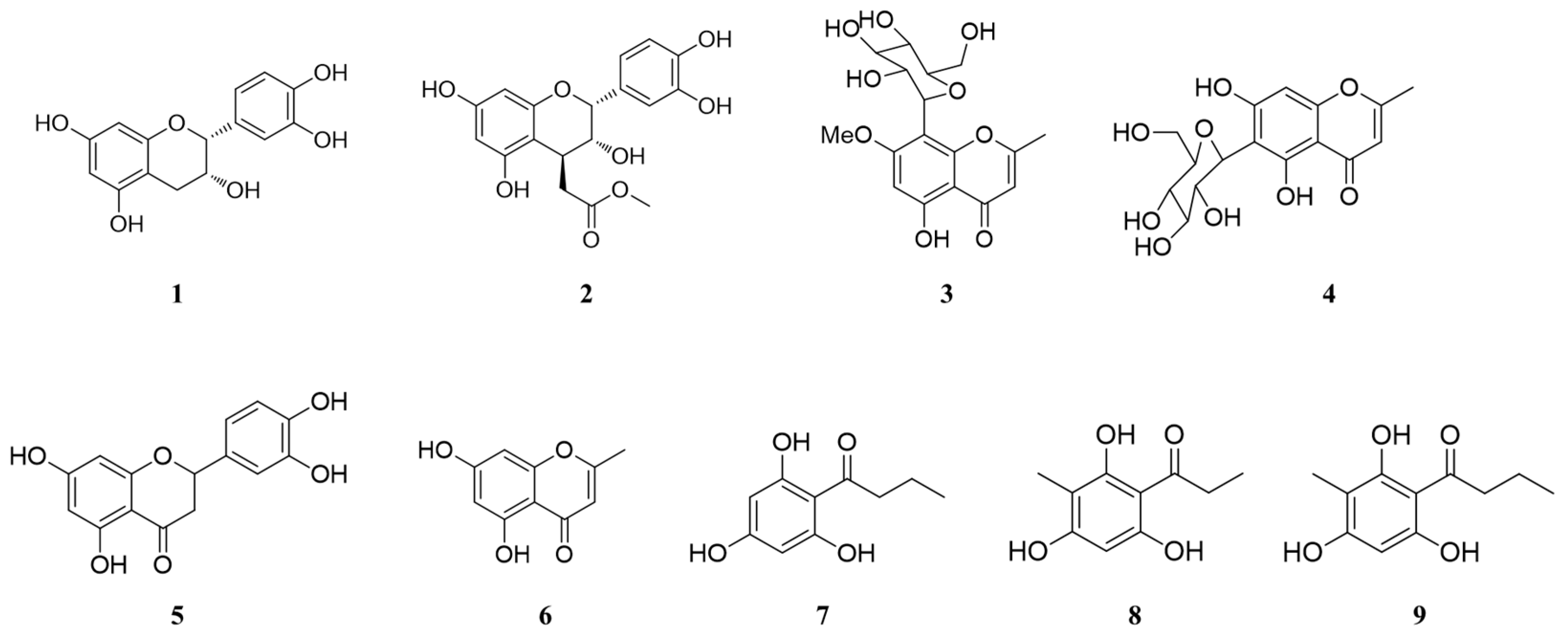
Nine compounds were isolated from the butanol fraction of D. crassirhizoma rhizomes via bioassay-guided chromatography and identified by comprehensive spectroscopic analysis (1D/2D NMR, HRMS, UV, IR) with literature comparisons (Figure 1). These comprised flavonoids, chromones, and phloroglucinols including epicatechin (1), 4β-carboxymethyl-(-)-epicatechin (2), 7-methoxy-isobiflorin (3), biflorin (4), eriodictyol (5), noreugenin (6), butyrylphloroglucinol (7), 2-propionyl-4-methylphloroglucinol (8), and 2-butyryl-4-methylphloroglucinol (9). All assignments were confirmed by characteristic spectral features and supported by literature comparisons.

3.2. Cytotoxicity Assesments of Isolated Compounds in APP-CHO Cells

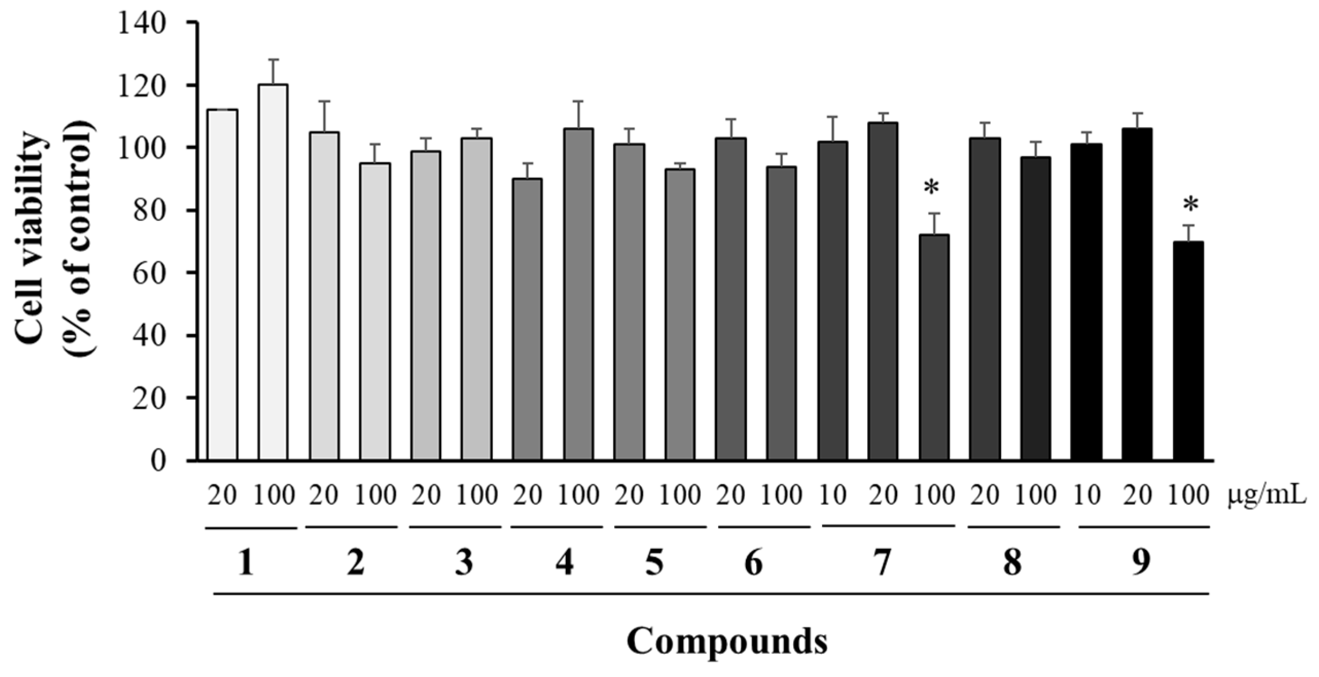
MTT assay evaluated cytotoxicity of compounds 19 in APP-CHO cells (Figure 2). Compounds 16 and 8 were tested at concentrations of 20 and 100 μg/mL, while compounds 7 and 9 were tested at 10, 20, and 100 μg/mL The results showed that compounds 16 and 8 showed no toxicity at both concentrations. Compounds 7 and 9 were non-toxic at 10 and 20 μg/mL but cytotoxic at 100 μg/mL. Thus, Subsequent bioassays used 10–20 μg/mL for compounds 7 and 9 and 20–100 μg/mL for others.

3.3. Modulation of sAPPβ and BACE1 Levels by the Isolated Compounds

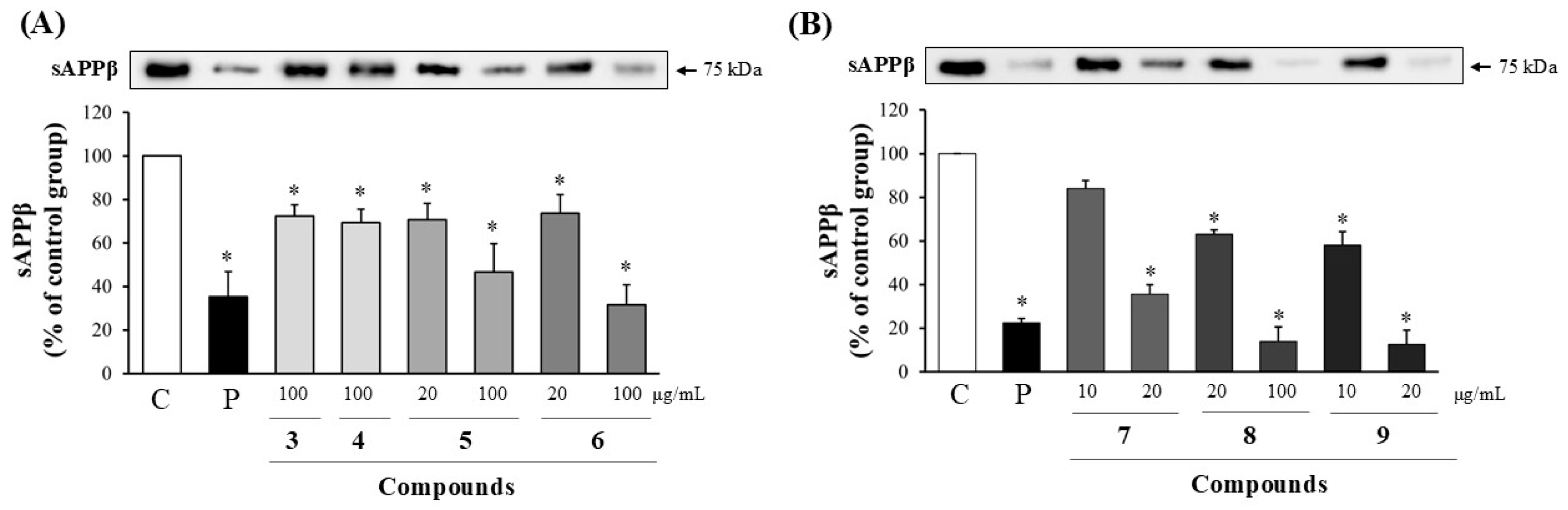
To assess the Aβ inhibitory potential of compounds 19, we measured BACE1 protein expression and the amount of sAPPβ secreted extracellularly in APP-CHO cells by Western blot analysis at non-cytotoxic concentrations. The amount of sAPPβ produced was compared to that of control cells treated with DMSO alone. Compounds 1 and 2 showed no significant inhibition at 100 μg/mL. However, compounds 3 and 4 reduced sAPPβ secretion by approximately 25% at 100 μg/mL. Compounds 5 and 6 decreased sAPPβ levels by about 30% at 20 μg/mL and over 50% at 100 μg/mL. Compounds 7, 8, and 9 exhibited dose-dependent suppression, with compound 7 by 18% at 10 μg/mL and over 60% at 20 μg/mL, compound 8 by 40% at 20 μg/mL and over 80% at 100 μg/mL, and compound 9 by 40% at 10 μg/mL and over 80% at 20 μg/mL (Figure 3).
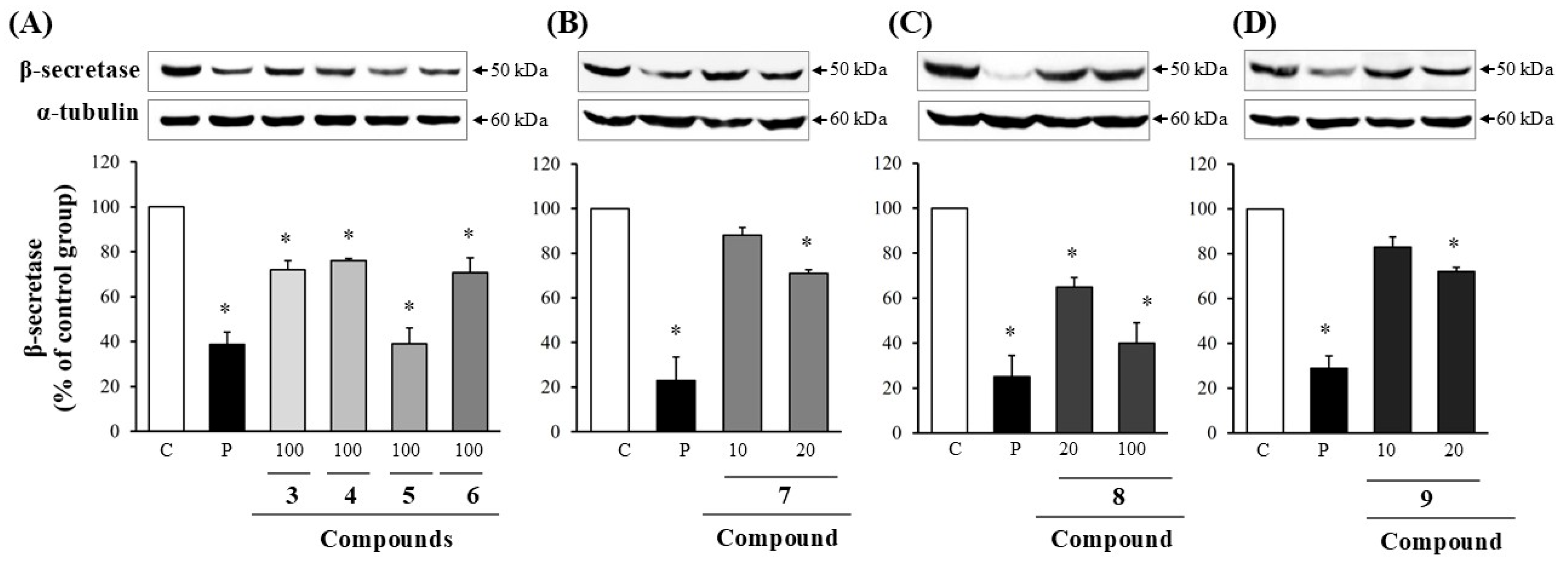
BACE1 protein levels were also quantified by Western blot in APP-CHO cells at non-cytotoxic concentrations. Compounds 1 and 2 showed no significant inhibition. Compounds 3 and 4 showed approximately 25% reduction of BACE1 at 100 μg/mL, while compounds 5 and 6 reduced BACE1 by 60% and 30%, respectively, at the same concentration. Compound 7 decreased BACE1 levels by 10% at 10 μg/mL and 30% at 20 μg/mL. Compound 8 reduced by 40% at 20 μg/mL and 60% at 100 μg/mL. Compound 9 caused 20% and 30% reductions at 10 μg/mL and 20 μg/mL, respectively. Overall, compounds 39 demonstrated significant inhibitory effects on BACE1 expressions (Figure 4). Taken together, compounds 39 significantly decreased both sAPPβ secretion and BACE1 expression at higher concentrations, suggesting their possible role in modulating Aβ generation. No significant effects were observed on α-secretase activity.

3.4. Modulation of Aβ Production by Compounds 3 to 9

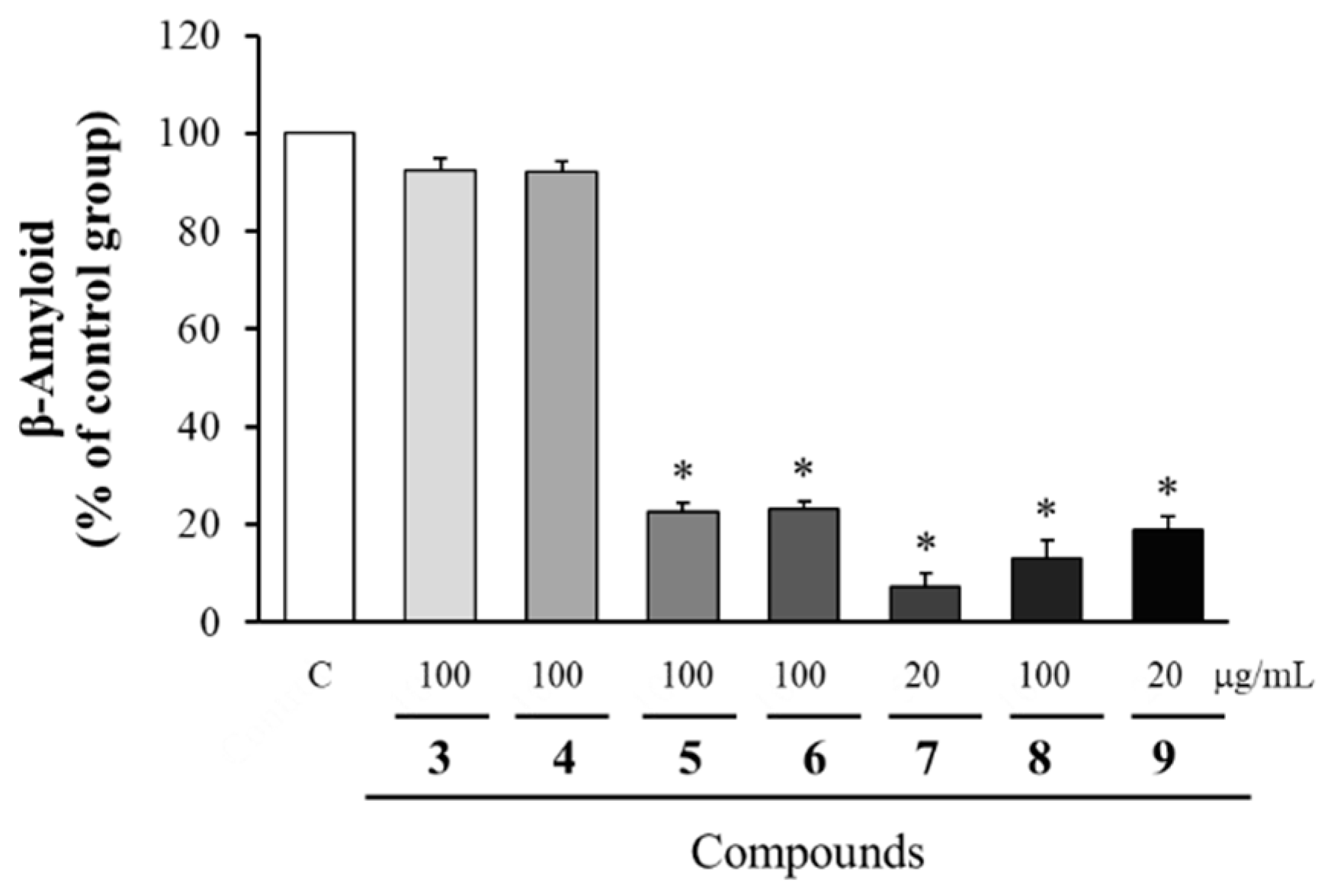
Among the compounds isolated from the butanol fraction, compounds 39 which showed the inhibitory effects on the levels of sAPPβ and BACE1 were tested for their effects on Aβ production. Compounds 3 and 4 slightly reduced Aβ levels, approximately 90% of the control at 100 μg/mL, while compounds 5, 6 and 8 decreased Aβ production by about 22–23% of the control at the same concentration. Compounds 7 and 9 exhibited the strongest efficacy at 20 μg/mL, decreasing Aβ levels by approximately 85-90% compared to the control. These findings suggest that compounds 59 possess notable anti-amyloidogenic activity, with compounds 7 and 9 being particularly effective (Figure 5).

4. Discussion

Understanding neuroprotection mechanisms is fundamental for advancing AD research. AD pathogenesis is explained by several interrelated hypotheses. According to the amyloid cascade hypothesis, aberrant cleavage of APP by BACE1 and γ-secretase results in excessive Aβ accumulation, which is considered a central pathogenic factor in AD [21]. Familial AD models with APP or presenilin mutations further support the link between elevated Aβ levels and neurotoxicity, as these mutations directly increase Aβ aggregation [21,22]. In addition to Aβ, the tau hypothesis describes how hyperphosphorylated tau protein forms neurofibrillary tangles, while the neuroinflammation hypothesis implicates chronic glial activation in progressive neuronal loss [23].
Aβ aggregates are particularly neurotoxic due to their extracellular deposition, which induces oxidative stress, promotes inflammation, disrupts calcium homeostasis, and impairs synaptic function [24,25]. Recent research also highlights the role of microglial activation and chronic inflammatory responses in exacerbating AD pathology [26].
D. crassirhizoma, a medicinal fern, contains bioactive phloroglucinol derivatives, triterpenes, and flavonoids that have demonstrated antioxidant, anti-cancer, anti-obesity, antibacterial, and antiviral activities [9,27]. While pharmacological research on AD and neuroprotection has been limited, recent studies indicate that the butanol fraction of D. crassirhizoma can inhibit Aβ generation in vitro [8]. Building on these findings, we isolated and purified nine compounds, including three flavonoids (1, 2, 5), three chromones (3, 4, 6), and three phloroglucinol derivatives (7, 8, 9), to evaluate their potential neuroprotective effects in AD-related pathways. To clarify their relevance, an overview of the known pharmacological activities and properties of each compound is presented.
The compounds epicatechin (1) and β-carboxymethyl-(-)-epicatechin (2) have been isolated from multiple plant species, such as Dalbergia monetaria and Pterocarpus marsupium. Epicatechin has been widely reported to exhibit strong antioxidant and anti-inflammatory activities, which contribute to neuroprotection, including in models of cognitive impairment and neurodegeneration [28,29]. In addition, epicatechin (1) is known as a potent β-secretase inhibitor [30], but it showed no activity in our study. This discrepancy may stem from variations in experimental design, as previous studies demonstrated that epicatechin (1) does not directly inhibit recombinant BACE1 enzymatic activity but rather modulates endogenous APP processing in specific neuronal cell systems. β-Carboxymethyl-(-)-epicatechin (2) similarly exhibits antioxidant and immunomodulatory effects, but specific studies on its CNS activity remain limited. 7-Methoxy-isobiflorin (3) and biflorin (4) are chromone derivatives identified in D. crassirhizoma. Biflorin (4) displays anti-inflammatory effects that may ameliorate neurodegeneration. Similarly, 7-methoxy-isobiflorin (3) has been reported with antioxidant activity, supporting its potential as a neuroprotective agent [31]. Noreugenin (6), a chromone derivative also isolated from D. crassirhizoma, has demonstrated inhibitory effects on Aβ aggregation. This suggests a direct role in mitigating amyloid plaque formation [32]. Phloroglucinol derivatives such as butyrylphloroglucinol (7), 2-propionyl-4-methylphloroglucinol (8), and 2-butyryl-4-methylphloroglucinol (9) have been isolated from D. crassirhizoma and related species, showing significant antioxidant, antiplatelet [18]. Among nine compounds, the inhibitory effects of isobiflorin (3), biflorin (4), noreugenin (6), butyrylphloroglucinol (7), 2-propionyl-4-methylphloroglucinol (8), and 2-butyryl-4-methylphloroglucinol (9) on Aβ production through the reduced levels of BACE1 have not been reported previously.
Investigation of compound permeability across the blood–brain barrier (BBB) requires consideration of key physicochemical properties, including molecular weight, lipophilicity (logP/logD), topological polar surface area (TPSA), hydrogen bond donor and acceptor counts, and molecular flexibility [33]. Additionally, potential interactions with efflux and influx transporters, as well as plasma protein binding, should be evaluated, as these factors collectively affect a compound’s penetration of the BBB and achieve therapeutic concentrations within the central nervous system [34,35]. The isolated compounds exhibit varied potentials for crossing the BBB. 7-Methoxy-isobiflorin (3) (predicted log P ~2.1) and biflorin (4) (predicted log P ~1.8) demonstrate moderate lipophilicity and relatively low polar surface areas, supporting their higher likelihood of passive BBB diffusion. This aligns with their reported anti-inflammatory and enzyme-inhibitory activities relevant to neurodegeneration [26]. Eriodictyol (5) (predicted log P ~1.6), a flavanone, shows higher polarity and hydrogen bonding capacity, which likely restricts its BBB permeability, implying its effects may be mediated mainly outside the CNS or through indirect pathways [36]. Noreugenin (6) (predicted log P ~1.5) exhibits physicochemical properties conducive to BBB penetration, correlating with its capacity to inhibit Aβ aggregation directly in the brain [37]. Acylated phloroglucinols such as butyrylphloroglucinol (7) (predicted log P ~2.4), 2-propionyl-4-methylphloroglucinol (8) (predicted log P ~2.1), and 2-butyryl-4-methylphloroglucinol (9) (predicted log P ~2.7) contain structural features that afford moderate lipophilicity, suggesting partial BBB permeability and regional CNS effects, potentially contributing to their overall neuroprotective profiles [18,28]. These differential BBB permeability profiles provide insight into the mechanisms through which each compound may contribute to anti-AD effects and guide prioritization for further pharmacokinetic and efficacy studies. However, these predictions need to be confirmed by experimental results [38,39].
Impaired insulin signaling and chronic inflammation have been shown to upregulate BACE1 expression and promote amyloidogenic APP processing, while activation of kinases such as GSK-3β further contributes to tau pathology and synaptic dysfunction [40,41]. In this context, the inhibitory effects of compounds 39 from D. crassirhizoma on BACE1 expression, sAPPβ generation, and Aβ production may extend beyond direct modulation of amyloidogenic pathways. Given the well-documented antioxidant and anti-inflammatory properties of flavonoids, chromones, and phloroglucinol derivatives, these compounds may also alleviate inflammation- and metabolism-associated neuronal stress, which is central to the AD–T2D pathological axis [42]. Notably, compounds 7 and 9 exhibited the strongest effects, underscoring their potential as multi-target therapeutic agents. Such a multi-mechanistic profile is particularly relevant considering the limitations and safety issues of current passive anti-amyloid immunotherapies, including amyloid-related imaging abnormalities (ARIA) [43]. In this study, compounds 39 significantly reduced sAPPβ and BACE1 levels in a dose-dependent manner, accompanied by decreased Aβ production, as confirmed by Western blot and ELISA. Since sAPPβ is generated by BACE1 cleavage of APP, reduced sAPPβ indicates BACE1 inhibition leading to decreased Aβ production [6]. Among these, compounds 7 and 9 showed the strongest inhibitory effects. The relatively low yields of the isolated compounds suggest that additional, more potent bioactive constituents may be present in the active fractions. It is also possible that minor components or synergistic interactions contribute to the observed activity. Further bioactivity-guided isolation combined with advanced analytical techniques will be required to fully elucidate these active principles.
These findings suggest that compounds 39 from the butanol fraction of Dryopteris crassirhizoma have promising potential as natural agents for the prevention and treatment of AD. However, in vivo studies using animal models are required to further confirm their efficacy in inhibiting Aβ.

Author Contributions

Conceptualization, S.-Y.P.; methodology, H.B.J. and T.E.P.; validation, C.H.L. and M.S.K.; formal analysis, H.B.J.; investigation, H.B.J. and T.E.P.; resources, S.-Y.P.; data curation, T.E.P.; writing—original draft preparation, S.-Y.P.; writing—review and editing, T.E.P. and K.W.H.; visualization, T.E.P.; supervision, S.-Y.P.; project administration, S.-Y.P.; funding acquisition, S.-Y.P. All authors have read and agreed to the published version of the manuscript.

Funding

This research was funded by the National Research Foundation of Korea (NRF) grant funded by the Korean government (MSIT) (No. RS-2024-00344498).

Data Availability Statement

The original contributions presented in the study are included in the article, further inquiries can be directed to the corresponding authors.

Conflicts of Interest

The authors declare no conflicts of interest.

Abbreviations

The following abbreviations are used in this manuscript:
ADAlzheimer’s disease
β-amyloid peptides
APPamyloid precursor protein
CHOChinese hamster ovary
NMRNuclear magnetic resonance
MSMass spectrometry
ELISAEnzyme-linked immunosorbent assay
HPLCHigh-performance liquid chromatography
FBSFetal bovine serum
DMSODimethyl sulfoxide
MTT3-(4,5-Dimethylthiazol-2-yl)-2,5-Diphenyltetrazolium Bromide
PVDFPolyvinylidene fluoride
CNSCentral nervous system
BBBBlood–brain barrier
TPSATopological polar surface area

References

  1. Sheppard, O.; Coleman, M. Alzheimer’s Disease: Etiology, Neuropathology and Pathogenesis. In Alzheimer’s Disease: Drug Discovery; Huang, X., Ed.; Exon Publications: Brisbane, Australia, 2020; pp. 1–28. [Google Scholar]
  2. Botto, R.; Callai, N.; Cermelli, A.; Causarano, L.; Rainero, I. Anxiety and depression in Alzheimer’s disease: A systematic review of pathogenetic mechanisms and relation to cognitive decline. Neurol. Sci. 2022, 43, 2601–2611. [Google Scholar] [CrossRef]
  3. Ma, C.J. Cognitive Enhancing Activity of Cynaroside Isolated from Lysimachia christinae on Memory Impairment Insulted by Scopolamine. Nat. Prod. Sci. 2024, 30, 175–181. [Google Scholar] [CrossRef]
  4. Hampel, H.; Hardy, J.; Blennow, K.; Chen, C.; Perry, G.; Kim, S.H.; Villemagne, V.L.; Aisen, P.; Vendruscolo, M.; Iwatsubo, T.; et al. The Amyloid-β Pathway in Alzheimer’s Disease. Mol. Psychiatry 2021, 26, 5481–5503. [Google Scholar] [CrossRef]
  5. Park, C.-W.; Lee, J.; Pel, P.; Lee, S.-E.; Chin, Y.-W. Benzoquinone Derivatives with Neuroprotective Activities from Lysimachia vulgaris L. Nat. Prod. Sci. 2024, 30, 1–7. [Google Scholar] [CrossRef]
  6. Zhang, H.; Wei, W.; Zhao, M.; Ma, L.; Jiang, X.; Pei, H.; Cao, Y.; Li, H. Amyloid β-based therapy for Alzheimer’s disease. Signal Transduct. Target. Ther. 2023, 8, 261. [Google Scholar] [CrossRef]
  7. Zhang, J.; Zhang, Z.; Dong, Y.; Wang, Y.; Li, Y.; Wang, X.; Li, X.; Zhang, H. Recent advances in Alzheimer’s disease: Mechanisms, clinical trials and new drug development strategies. Signal Transduct. Target. Ther. 2024, 9, 211. [Google Scholar] [CrossRef]
  8. Kim, J.-M.; Hwang, K.W.; Joo, H.B.; Park, S.Y. Anti-amyloidogenic Properties of Dryopteris crassirhizoma Roots in Alzheimer’s Disease Cellular Model. J. Food Biochem. 2015, 39, 478–484. [Google Scholar] [CrossRef]
  9. Bai, Q.X.; Zhang, Y.; Yang, Y.J.; Zhang, H.; Zhang, T.T.; He, J. Dryopteris crassirhizoma Nakai: A review of its botany, traditional use, phytochemistry, pharmacological activity, toxicology and pharmacokinetics. J. Ethnopharmacol. 2024, 319, 117144. [Google Scholar] [CrossRef] [PubMed]
  10. Pham, T.L.; Ha, M.T.; Min, B.S.; Kim, J.A. PTP1B Inhibitory Activity of Flavonoids from the Roots of Astragalus membranaceus Bunge. Nat. Prod. Sci. 2025, 31, 62–73. [Google Scholar] [CrossRef]
  11. Lee, D.; Lee, J.W. Cytotoxicity against MDA-MB-231 Breast Cancer Cells of Fungal Metabolites of Trichoderma sp. Collected from Medicinal Herbal Garden. Nat. Prod. Sci. 2025, 31, 6–12. [Google Scholar] [CrossRef]
  12. Davis, A.L.; Cai, Y.; Davies, A.P.; Lewis, J.R. 1H and 13C NMR Assignments of Some Green Tea Polyphenols. Magn. Reson. Chem. 1996, 34, 887–890. [Google Scholar] [CrossRef]
  13. Gyeltshen, T.; Smith, J.A.; Bissember, A.C. Natural products isolation studies of native Australian fern species. Aust. J. Chem. 2022, 75, 422–437. [Google Scholar] [CrossRef]
  14. Gao, H.-Y.; Wang, H.-Y.; Li, G.-Y.; Du, X.-W.; Zhang, X.-T.; Han, Y.; Huang, J.; Li, X.-X.; Wang, J.-H. Constituents from Zhuyeqing Liquor and their inhibitory effects on nitric oxide production. Phytochem. Lett. 2014, 7, 150–155. [Google Scholar] [CrossRef]
  15. Fonseca, A.M.; Pessoa, O.D.L.; Silveira, E.R.; Monte, F.J.Q.; Braz-Filho, R.; Lemos, T.L.G. Total assignments of 1H and 13C NMR spectra of biflorin and bis-biflorin from Capraria biflora. Magn. Reson. Chem. 2003, 41, 1038–1040. [Google Scholar] [CrossRef]
  16. Miyake, Y.; Yamamoto, K.; Osawa, T. Isolation of Eriocitrin (Eriodictyol 7-rutinoside) from Lemon Fruit (Citrus limon Burm. f.) and Its Antioxidative Activity. Food Sci. Technol. Int. Tokyo 1997, 3, 84–89. [Google Scholar] [CrossRef]
  17. Lima, T.C.; Souza, R.J.; Santos, A.D.C.; Moraes, M.H.; Biondo, N.E.; Barison, A.; Steindel, M.; Biavatti, M.W. Evaluation of leishmanicidal and trypanocidal activities of phenolic compounds from Calea uniflora Less. Nat. Prod. Res. 2016, 30, 551–557. [Google Scholar] [CrossRef] [PubMed]
  18. Yim, N.-H.; Lee, J.-J.; Lee, B.; Li, W.; Ma, J.Y. Antiplatelet Activity of Acylphloroglucinol Derivatives Isolated from Dryopteris crassirhizoma. Molecules 2019, 24, 2212. [Google Scholar] [CrossRef] [PubMed]
  19. Citron, M.; Oltersdorf, T.; Haass, C.; McConlogue, L.; Hung, A.Y.; Seubert, P.; Vigo-Pelfrey, C.; Lieberburg, I.; Selkoe, D.J. Mutation of the β-amyloid precursor protein in familial Alzheimer’s disease increases β-protein production. Nature 1992, 360, 672–674. [Google Scholar] [CrossRef]
  20. Sudeep, H.V.; Raj, A.; Kumara, T.P.P.; Lingaraju, H.B.; Shyamprasad, K. A Standardized Cichorium intybus L. Leaf Extract Abrogates P53/caspase-Dependent Apoptosis and Oxidative Stress via Activation of PI3K/Akt/Nrf2/HO-1 Signaling Pathway in Human Embryonic Kidney (HEK293) Cell Line. Nat. Prod. Sci. 2025, 31, 92–101. [Google Scholar] [CrossRef]
  21. Zhang, X.; Li, Y.; Xu, H.; Zhang, Y.W. Secretases Related to Amyloid Precursor Protein Processing. Biomolecules 2021, 11, 1856. [Google Scholar]
  22. Uddin, M.S.; Kabir, M.T.; Rahman, M.S.; Behl, T.; Jeandet, P.; Ashraf, G.M.; Aleya, L.; Barreto, G.E.; Uddin, M.S. Revisiting the Amyloid Cascade Hypothesis: From Anti-Aβ Therapeutics to Auspicious New Ways for Alzheimer’s Disease. Int. J. Mol. Sci. 2020, 21, 5858. [Google Scholar] [CrossRef]
  23. Wang, C.; Fan, L.; Khawaja, R.R.; Liu, B.; Zhan, X.; Kodama, L.; Chin, K.R.; Li, Y.; Le, D.E.; Zhou, Y. The effects of microglia-associated neuroinflammation on Alzheimer’s disease therapeutics. Front. Pharmacol. 2023, 14, 1040838. [Google Scholar]
  24. Zhang, Y.; Wang, Z.; Li, Y.; Zhang, X.; Wang, J. Neurotoxic β-amyloid oligomers cause mitochondrial dysfunction—The trigger for PANoptosis in neurons. Front. Aging Neurosci. 2024, 16, 1400544. [Google Scholar]
  25. Braidy, N.; Essa, M.M.; Poljak, A.; Selvaraju, S.; Al-Adawi, S.; Manivasagm, T.; Thenmozhi, A.J.; Ooi, L.; Sachdev, P.; Guillemin, G.J. The Dual Role of Amyloid Beta-Peptide in Oxidative Stress and Inflammation: Unveiling Their Connections in Alzheimer’s Disease Etiopathology. Int. J. Mol. Sci. 2024, 25, 10839. [Google Scholar]
  26. Leng, F.; Edison, P. Neuroinflammation and microglial activation in alzheimer disease: Where do we go from here? Nat. Rev. Neurol. 2021, 17, 157–172. [Google Scholar] [CrossRef]
  27. Phong, N.V.; Zhao, Y.; Min, B.S.; Yang, S.Y.; Kim, J.A. Inhibitory Activity of Bioactive Phloroglucinols from the Rhizomes of Dryopteris crassirhizoma on Escherichia coli β-Glucuronidase: Kinetic Analysis and Molecular Docking Studies. Metabolites 2022, 12, 938. [Google Scholar] [CrossRef] [PubMed]
  28. Cox, K.H.; Pipingas, A.; Scholey, A.B. Evaluation of Cognitive and Mood Effects of an Epicatechin-Rich Cocoa Supplement in Healthy Adults. Front. Nutr. 2019, 6, 74. [Google Scholar]
  29. Cox, C.J.; Choudhry, F.; Peacey, E.; Perkinton, M.S.; Richardson, J.C.; Howlett, D.R.; Lichtenthaler, S.F.; Francis, P.T.; Williams, R.J. Dietary (−)-Epicatechin as a Potent Inhibitor of βγ-Secretase Amyloid Precursor Protein Processing. Neurobiol. Aging 2015, 36, 178–187. [Google Scholar] [CrossRef]
  30. Diaz, A.; Muñoz-Arenas, G.; Guevara-Guzmán, R.; López-Gómez, A.B.; Brambila, E.; Treviño, S. Epicatechin Reduces Spatial Memory Deficit Caused by Amyloid-Beta 25-35 Toxicity in the Hippocampus of Rats. Nutrients 2019, 11, 983. [Google Scholar]
  31. Li, S.; Wang, H.; Hu, B.; Li, F.; Liang, Y.; Wang, Y. Biflorin from Oldenlandia diffusa Suppresses Lipopolysaccharide-Induced Inflammatory Responses in RAW 264.7 Macrophages and Zebrafish. J. Ethnopharmacol. 2017, 202, 62–68. [Google Scholar]
  32. Nain Walia, V.; Kumar, D.; Kumar, H.; Kumar, V. In Vitro and In Vivo Investigations of Chromone Derivatives as Potential Multitarget-Directed Ligands for the Effective Treatment of Alzheimer’s Disease. ACS Chem. Neurosci. 2024, 15, 2392–2412. [Google Scholar]
  33. Lin, Y.C.; Tsai, P.C.; Chen, Y.C.; Chen, C.T.; Huang, H.T.; Yang, J.M. Predicting blood–brain barrier permeability of molecules with a large language model and in vitro assay. Sci. Rep. 2024, 14, 15694. [Google Scholar]
  34. Wu, D.; Chen, Q.; Chen, X.; Han, F.; Chen, Z.; Wang, Y. The blood–brain barrier: Structure, regulation and drug delivery. Signal Transduct. Target. Ther. 2023, 8, 217. [Google Scholar] [CrossRef] [PubMed]
  35. Cornelissen, F.M.G.; Woodfield, G.K.; Lammertink, B.H.A.; Capacchione, C.; de Vries, H.E.; de Lange, E.C.M. Explaining Blood–Brain Barrier Permeability of Small Molecules by Molecular Descriptors. J. Med. Chem. 2023, 66, 7193–7207. [Google Scholar] [CrossRef]
  36. Li, D.; Liu, Y.; Sun, W.; Zhou, Y.; Li, Y.; Chen, Y.; Li, S. Eriodictyol ameliorates cognitive dysfunction in APP/PS1 mice and inhibits ferroptosis via Nrf2/HO-1 pathway. Cell Death Discov. 2022, 8, 33. [Google Scholar] [CrossRef]
  37. Li, F.; Zhu, M.; Zhang, C.; Zhao, Y. Synthesis and Pharmacological Evaluation of Novel Chromone Derivatives as Potential Multitarget-Directed Ligands for the Treatment of Alzheimer’s Disease. Bioorg. Med. Chem. 2017, 25, 4405–4419. [Google Scholar] [CrossRef]
  38. Kato, R.; Liu, X.; Xu, X.; McKew, J.C.; Zheng, W.; Huang, R. Development and Validation of PAMPA-BBB QSAR Model for Virtual Screening of Blood-Brain Barrier Permeability. Front. Pharmacol. 2023, 14, 1291246. [Google Scholar]
  39. Shaker, B.; Yu, M.S.; Song, J.S.; Ahn, S.; Oh, K.J.; Na, D. LightBBB: Computational Prediction Model of Blood–Brain Barrier Penetration Based on LightGBM. Bioinformatics 2021, 37, 1135–1143. [Google Scholar] [CrossRef]
  40. Sharma, K.; Rai, P.; Tapadia, M.G. Impaired insulin signaling and diet-induced type 3 diabetes pathophysiology increase amyloid β expression in the Drosophila model of Alzheimer’s disease. Biochim. Biophys. Acta Mol. Cell Res. 2025, 1872, 119875. [Google Scholar] [CrossRef]
  41. Salian, V.S.; Tang, X.; Thompson, K.J.; Curan, G.L.; Lowe, V.J.; Li, L.; Kandimalla, K.K. Molecular mechanisms underlying amyloid Beta peptide mediated upregulation of vascular cell adhesion Molecule-1 in Alzheimer disease. J. Pharmacol. Exp. Ther. 2024, 391, 430–440. [Google Scholar] [CrossRef]
  42. Yoo, J.; Lee, J.; Ahn, B.; Han, J.; Lim, M.H. Multi-target-directed therapeutic strategies for Alzheimer’s disease: Controlling amyloid-β aggregation, metal ion homeostasis, and enzyme inhibition. Chem. Sci. 2025, 16, 2105–2135. [Google Scholar] [CrossRef] [PubMed]
  43. Heneka, M.T.; Morgan, D.; Jessen, F. Passive anti-amyloid β immunotherapy in Alzheimer’s disease—Opportunities and challenges. Lancet 2024, 404, 2198–2208. [Google Scholar] [CrossRef] [PubMed]
Figure 1. Structures of compounds isolated from D. crassirhizoma. 1: epicatechin, 2: β-carboxymethyl-(-)-epicatechin, 3: 7-methoxy-isobiflorin, 4: biflorin, 5: eriodictyol, 6: noreugenin, 7: butyrylphloroglucinol, 8: 2-propionyl-4-methylphloroglucinol and 9: 2-butyryl-4-methylphloroglucinol.
Separations 13 00035 g001
Figure 2. Viability of APP-CHO cells following the treatment with compounds 19. The cell viability after the treatment with the compounds were determined by MTT assay. The average viability lower than 80% compared to DMSO-treated group was considered cytotoxic. Data represent the mean ± SD of three independent experiments and are expressed as a percentage of the DMSO-treated group. * p < 0.05, significantly different from the DMSO-treated control group (0.1% DMSO).
Separations 13 00035 g002
Figure 3. Inhibition of sAPPβ production by compounds 39. Western blot analysis was performed on supernatants from APP-CHO cells treated with the compounds 36 (A) and 79 (B) were to determine sAPPβ levels. Changes in sAPPβ levels are shown in the accompanying graphs. Data presented the means ± SD of three independent experiments and are presented as a percentage of the DMSO-treated control. Equal amount of total protein was loaded in each lane. Statistical significance was defined as p < 0.05. * p < 0.05, significantly different from the control group. C: control (0.1% DMSO), P: positive control, D. crassirhizoma rhizome extract (50 μg/mL).
Separations 13 00035 g003
Figure 4. Inhibitory effects of compounds 39 on BACE1. Western blot analysis of BACE1 was performed on cell lysates from APP-CHO cells treated with the compounds 36 (A), 7 (B), 8 (C), 9 (D). Data (mean ± SD, n = 3) are expressed as a percentage of the DMSO control. * p < 0.05 was considered significant compared to the control group. C: control (0.1% DMSO), P: positive control, D. crassirhizoma rhizome extract (50 μg/mL).
Separations 13 00035 g004
Figure 5. Inhibition of Aβ production by compounds 39. Supernatants from APP-CHO cells treated with compounds 39 were analyzed by ELISA to quantify Aβ levels. Data represent the means ± SD of three independent experiments and are presented as a percentage of the DMSO-treated control. Statistical significance was defined as * p < 0.05 vs. the control group. C: control (0.1% DMSO).
Separations 13 00035 g005
Disclaimer/Publisher’s Note: The statements, opinions and data contained in all publications are solely those of the individual author(s) and contributor(s) and not of MDPI and/or the editor(s). MDPI and/or the editor(s) disclaim responsibility for any injury to people or property resulting from any ideas, methods, instructions or products referred to in the content.