Article

Extraction of Phycocyanin and Chlorophyll from Spirulina by “Green Methods”

1
Department of Physics and Biophysics, Medical University of Varna, 9000 Varna, Bulgaria
2
Department of Organic Chemistry and Inorganic Chemistry, University of Food Technologies, 4002 Plovdiv, Bulgaria
3
Department of Biotechnology, University of Food Technologies, 4002 Plovdiv, Bulgaria
4
Department of Chemistry and Biochemistry, Medical University-Pleven, 5800 Pleven, Bulgaria
5
Department of Pharmaceutical Technologies, Medical University of Varna, 9000 Varna, Bulgaria
*
Authors to whom correspondence should be addressed.
Separations 2024, 11(2), 57; https://doi.org/10.3390/separations11020057
Submission received: 19 January 2024 / Revised: 7 February 2024 / Accepted: 8 February 2024 / Published: 12 February 2024

Abstract

:
Phycocyanin is a pigment–protein complex from the group of phycobiliproteins obtained from Spirulina (Arthrospira platensis), with possibilities for various applications in food and pharmaceutical technologies. It is a natural colorant for food and cosmetic products. This study aimed to investigate the effect of ultrasonic and microwave extraction conditions on antioxidant activity (AOA), chlorophyll content, and the content and purity index of phycocyanin in aqueous and alcoholic extracts of Spirulina (Arthrospira platensis). For this purpose, ultrasonic extraction with water or ethanol was performed at 20 °C, 30 °C, and 40 °C for 1, 2, and 3 h at an ultrasonic frequency of 36 kHz, 40 kHz, and 45 kHz. Microwave water extraction was performed for 60 s, 120 s, and 180 s. For each of the obtained samples, three parallel measurements of antioxidant activity were made by DPPH and FRAP methods, and chlorophyll content and phycocyanin yield and purity index were determined spectrophotometrically. Ultrasonic extraction resulted in a higher yield and purity index of phycocyanin compared to microwave extraction. The highest yield of 14.88 mg g−1 with a purity index of 1.60 was achieved at a temperature of 40 °C for one hour and an ultrasonic wave frequency of 40 kHz. A relatively low yield of 4.21 mg g−1, but with a purity index of 2.67, was obtained at a temperature of 30 °C, a time of two hours, and an ultrasonic frequency of 40 kHz. Chlorophyll b content at 20 °C, for two hours and ultrasonic frequency 40 kHz was 1.400 mg g−1. The study proposes ultrasonic extraction as a green method to obtain phycocyanin of varying purity index that may be used for food, cosmetic, or biomedical purposes.

1. Introduction

Spirulina (Arthrospira platensis) is increasingly used in pharmacy, cosmetics, medicine, and food technology [1] Due to its rich chemical composition and high nutritional value, it has been recognized by the World Health Organization as a superfood [2]. It is an essential component in “space food” for astronauts [3]. Cyanobacteria are rich in proteins, lipids, carbohydrates, carotenoids, phycobiliproteins [4], unsaturated fatty acids, vitamins, polysaccharides, minerals such as zinc, selenium, manganese, iron, calcium, magnesium [5], chlorophylls, and flavonoids [3]. Spirulina (Arthrospira platensis) enhances endogenous enzymatic antioxidants, captures free radicals, and inhibits lipid peroxidation in vivo [6]. It is increasingly used as a food supplement due to its established anti-inflammatory and immunomodulatory properties [7,8], as well as hepatoprotective [9] and antioxidant action [10]. Spirulina contains up to 1 g kg−1 carotenoids [11], between 6 and 11 g kg−1 chlorophyll [12], and phycobiliproteins contained in phycobilisomes.
Phycobiliproteins are divided into phycoerythrin (C-PE, red color) and phycocyanin (PC, blue color) [13]. Phycocyanins include C-phycocyanin (C-PC), R-phycocyanin (R-PC), and allophycocyanin (APC), which differ in their spectral properties, structural composition, and color [14]. Depending on the growing conditions and environment, Spirulina (Arthrospira platensis) can contain up to 20% phycocyanin. This pigment is used as a natural colorant for food and cosmetic products. Due to the multi-layered cell walls of Spirulina, their destruction is crucial for the extraction of phycocyanin [15]. This is achieved through various methods, some of which are presented below:
  • Physical destruction: through successive cycles of freezing and thawing of fresh Spirulina as the cell membrane is ruptured due to an increase in the volume of the ice crystals and subsequent purification. Another way to release intracellular substances is through the use of micro- or nano-sized spheres [16,17];
  • Supercritical fluid extraction, using supercritical carbon dioxide as a solvent [18];
  • Methods using alternating electric fields in an aqueous environment [19];
  • Green methods using ultrasonic waves or microwave extraction [20,21,22].
According to the method of the supercritical fluid extraction of phycocyanin, the purity is about 75%, and the yield is about 90% [18]. When using the freeze–thaw technique [16], a purity ratio (R) of 3.31 was achieved. Both techniques, however, require additional purification of the resulting phycocyanin. For example, after breaking the cell walls by the freeze–thaw method, fractionation follows; by using ammonium sulfate, further purification by chromatographic methods or by using absorbents occurs [16]. The main problems in the conventional extraction process are the large amount of solvents used, moderate selectivity, and extraction efficiency [23,24]. A large part of the conventional extraction methods is carried out with fresh raw material, which is associated with some inconveniences. Some of these are the rapid deterioration of the raw material during transportation and the development of microorganisms in it.
The use of green extraction methods reduces the extraction time and increases the yield of the protein in question and does not require the use of additional purification methods, resulting in the preservation of a large part of the biologically active substances. Therefore, extraction techniques without the use of solvents, called “green methods”, working with cyanobacteria dried under suitable conditions are increasingly used. These are pressurized liquid extraction (PLE), supercritical fluid extraction (SFE), microwave extraction (MW), ultrasonic extraction (US), and high-pressure homogenization (HPH) [14]. Ultrasonic extraction has several advantages over other methods. Some of them are as follows:
  • Speed and efficiency: Due to the presence of cavitation in the sample, mechanical waves arise that quickly destroy the cell walls;
  • Ensuring high concentrations of biologically active substances that are heat sensitive: This process avoids a significant increase in temperature and reduces the temperature degradation of the specified components;
  • No contamination of the extracts: This specified method uses a minimal amount of chemicals and solvents;
  • Ability to easily optimize the process to improve the quality of the extracts in real time;
  • Reduction of adverse influences such as oxidation, for example, etc.;
  • Multi-functionality: This method can be successfully combined with enzymatic and hydrothermal methods to achieve sustainability and efficiency in the extraction of a larger spectrum of biologically active substances;
  • It is suitable for the extraction of substances on a large scale.
This study aimed to investigate the effect of ultrasonic and microwave extraction conditions on antioxidant activity (AOA), chlorophyll content, and the content and purity index of phycocyanin in aqueous and alcoholic extracts of Spirulina (Arthrospira platensis).

2. Materials and Methods

2.1. Investigated Sample

A sample of Spirulina (Arthrospira platensis) grown in a bioreactor in Varvara, Bulgaria was studied. A laboratory biomass accumulation facility was used to create the crop. It is made of glass tubs with a volume of 50 L, in which the water temperature is maintained between 33 °C and 35 °C, and illumination is maintained from 8000 to 10,000 Lx. For 1 h, 100 L of a gas mixture of air and carbon dioxide is passed through 1 L of Spirulina suspension. In order to increase the amount of biomass, its habitat is changed from the laboratory to production conditions. It takes 25–30 days to accumulate a suspension with a sufficiently large mass [25]. Samples of the suspension taken for analysis are centrifuged and dried to constant weight at 42 °C, then tempered, homogenized, and packed in paper bags stored in the dark at 25 °C.

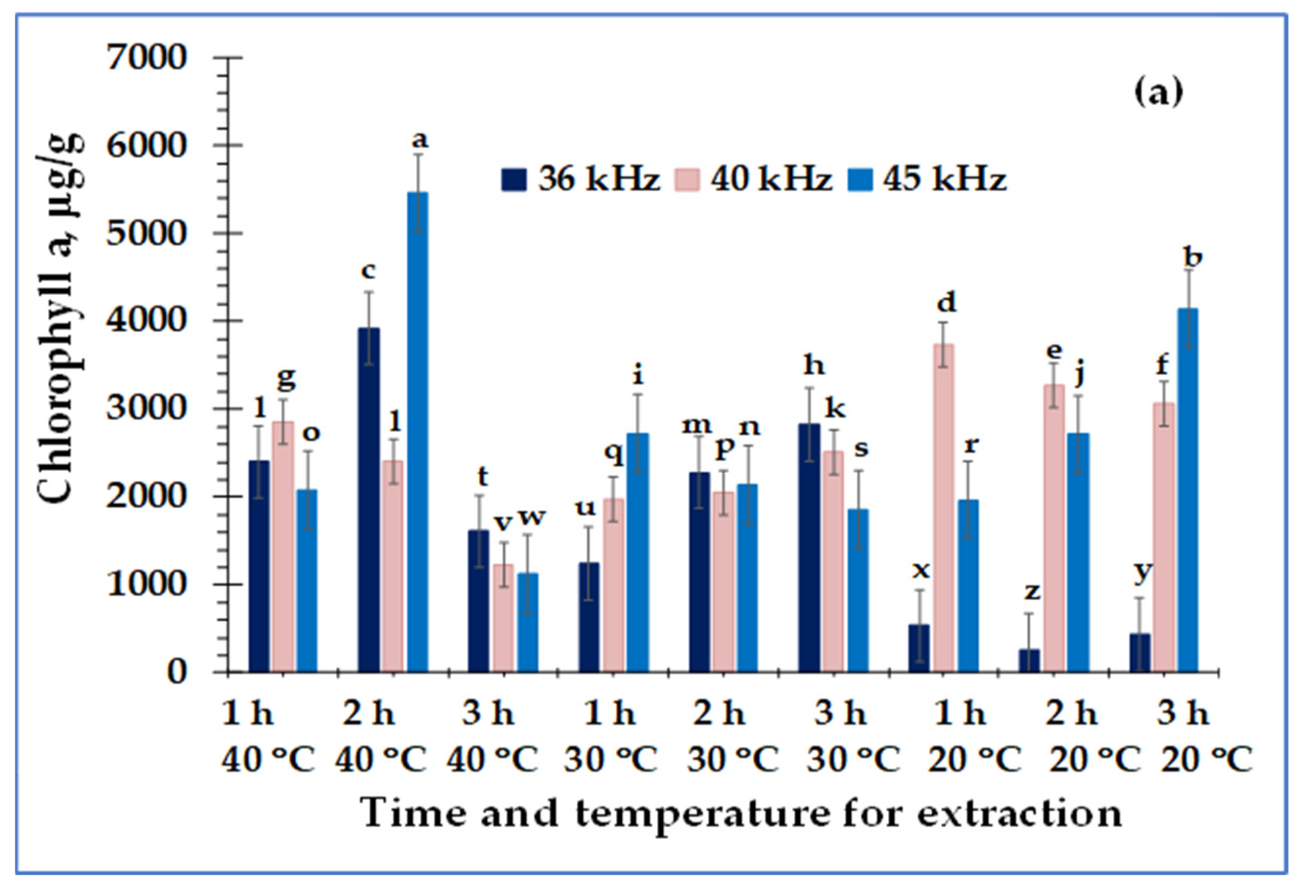
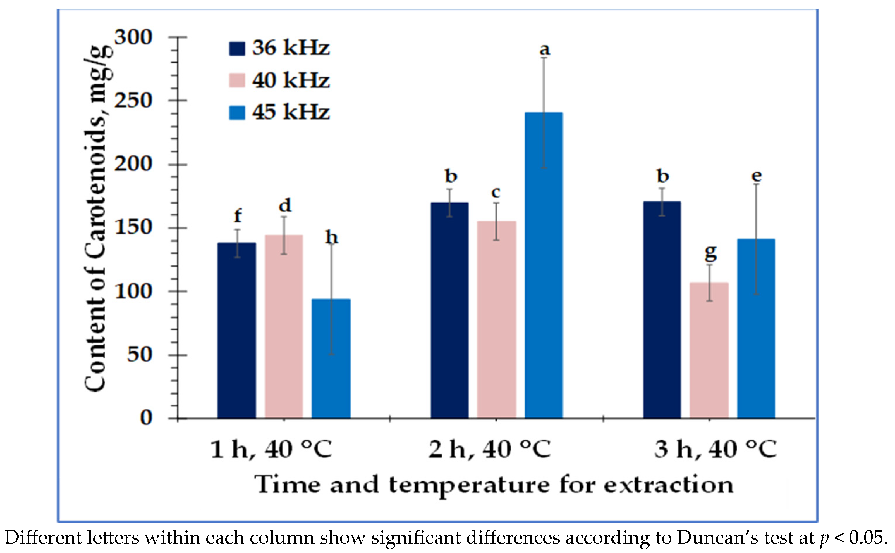
2.2. Determination of Chlorophyll and Carotenoids

Ethanol extracts were prepared in a ratio of 1:50 (sample: solvent) for the determination of the content of chlorophyll a (Ca), chlorophyll b (Cb), and total carotenoids (Cx+b). For this purpose, 96% pure ethanol (Merck, Germany) was used. Extracts were obtained in ultrasonic baths SIEL (Gabrovo, Bulgaria), Isolab (Eschau, Germany) and VWR (Seri Kembangan, Selangor, Malaysia) at 36 kHz, 40 kHz, and 45 kHz. We performed the extraction at 20 °C, 30 °C, and 40 °C for 1, 2, and 3 h. The researchers conducted the extraction procedure for each sample in triplicate. We filtered the extracts through filter paper and examined them spectrophotometrically. To determine pigment content, absorption at wavelengths 662 nm, 645 nm, and 470 nm were measured, and Equations (1)–(3) reported by Lichtenthaler and Wellburn [26] were used.
C a ,   μ g   m L 1 = 13.95 × A 665 6.88 × A 649
  C b , μ g   m L 1 = 24.9 × A 649 7.32 × A 665
C x + b ,   g   m L 1 = 1000 × A 470 2.05 × C a 114.8   × C b / 245

2.3. Determination of the Antioxidant Activity of Water and Ethanol Extracts of Spirulina (Arthrosphira platensis)
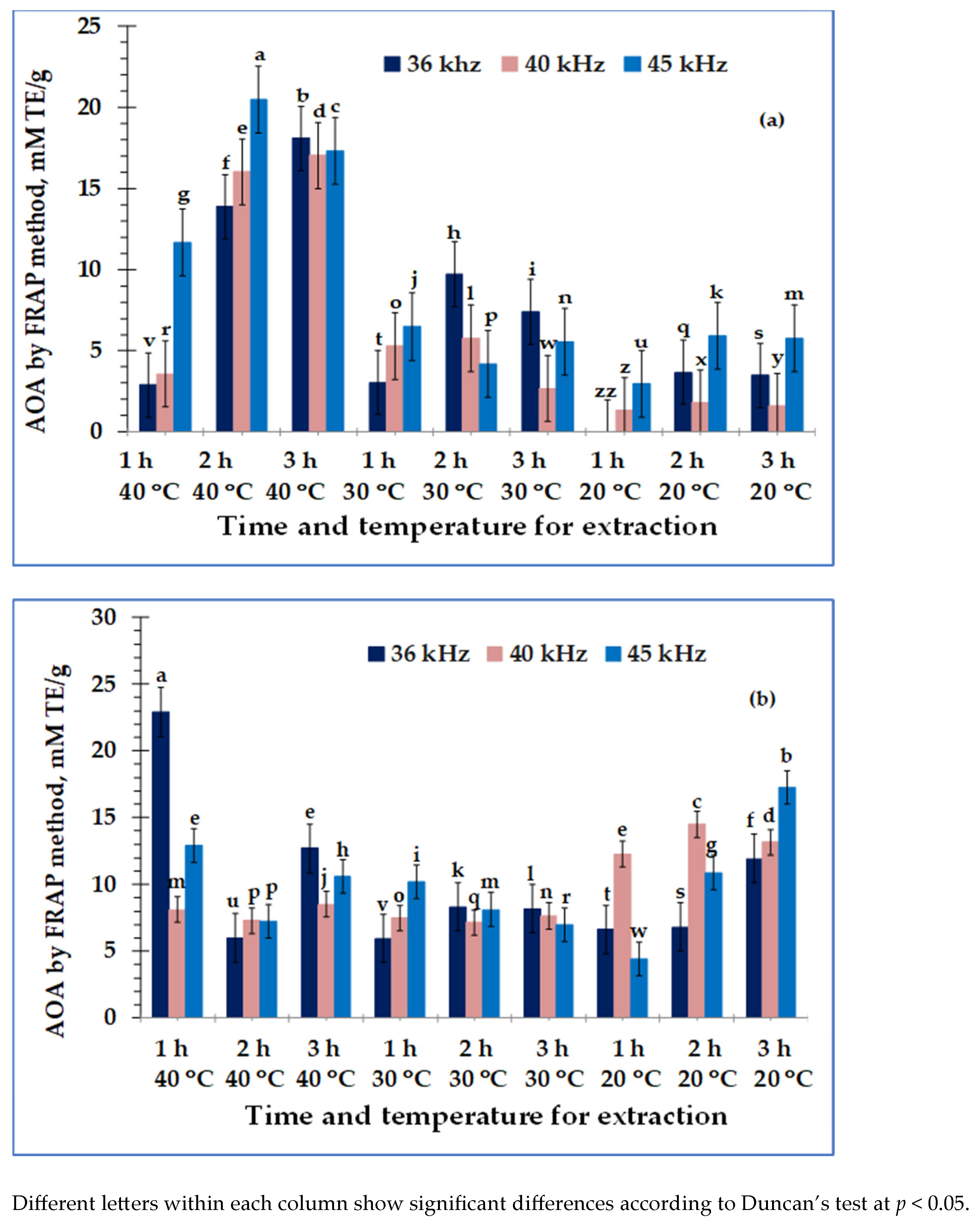
Spirulina (Arthrosphira platensis) is rich in biologically active substances; some are watersoluble, while others are better extracted in ethanol.
The antioxidant activity was determined by using two methods based on different mechanisms—DPPH and FRAP assays.

2.3.1. FRAP Method

The FRAP reagent was prepared by mixing 0.3 M acetate buffer with a pH value of 3.6 (Sigma-Aldrich Chemie GmbH, Darmstadt, Germany), 10 mM 2,4,6-tripyridyl-s-triazine (TPTZ), and 20 mM FeCl3·6H2O (Merck KGaA, Darmstadt, Germany) in a ratio of 10:1:1. A total of 0.1 mL of the test extract was added to 3 mL of FRAP reagent. The reaction mixture was incubated in a water bath for 4 min at 37 °C. The absorbance of the colored substance formed was measured at a wavelength of 593 nm against a blank sample [27]. We calculated the results of antioxidant activity using Equation (4) and expressed them as mM TE per g of dry plant material and dry extracts.
m M   T E m L   e x t r a c t = A 593 + 0.0113 1.4138

2.3.2. DPPH Method

From the examined extract, 0.15 mL was mixed with 2.85 mL of a freshly prepared 0.1 mM DPPH (2,2’-diphenyl-1-picrylhydrazyl) solution (Sigma-Aldrich Chemie GmbH, Darmstadt, Germany) in methanol (puriss. p.a., ACS reagent, reag. ISO, reag. Ph. Eur., ≥99.8% (GC); Merck KGaA, Darmstadt, Germany). The reaction mixture underwent incubation in a water bath at 37 °C for 15 min. The absorbance of the colored substance formed was measured at a wavelength of 517 nm against a blank sample [28]. Percentage inhibition was calculated (I %), using the following equation:
I   % = A 0 A 1 A 0 100
where A0 was the absorbance of the control, and A1 was the absorbance of the sample. We substituted the results in the linear regression equation, where A0 represents the absorbance of the control, and A1 represents the absorbance of the sample. Then, the antioxidant activity was calculated according to Equation (6)
m M   T E m L = 102.06 I   % + 0.7954
and then expressed as a millimole Trolox equivalent per g of dry weight.

2.4. Green Methods of Extraction

Ultrasonic and microwave extraction were performed under our chosen conditions to determine phycocyanin’s yield and purity index.

2.4.1. Microwave Extraction

Samples of 0.5 g of Spirulina in 35 mL of distilled water were used to extract phycocyanin. The extraction process involved using samples of 0.5 g of Spirulina in 35 mL of distilled water, which we performed in duplicate in a microwave oven (Daewoo KOR) with microwave output power at 700 W and 2450 MHz frequency for 60, 120, and 180 s, respectively.

2.4.2. Ultrasonic Extraction

The Spirulina samples (0.5 g) were weighed into 35 mL screw-capped centrifuge tubes, and the samples were extracted with distilled water in three ultrasonic baths operated under the following conditions:
(1)
Ultrasonic bath SIEL (Bulgaria) with frequency 36 kHz, power 300 W-(UAE 36 kHz);
(2)
Ultrasonic bath ISOLAB (Germany) with frequency 40 kHz, power 60 W-(UAE 40 kHz);
(3)
Ultrasonic bath VWR (Malaysia) with frequency 45 kHz, power 30 W-(UAE 45 kHz).
Ultrasonic extraction was performed at these three frequencies for 1, 2, and 3 h at a temperature of 20 °C, 30 °C, and 40 °C. The extracts were filtered through filter paper and used for further studies on the yield and purity index of phycocyanin, as well as the evaluation of antioxidant activity.

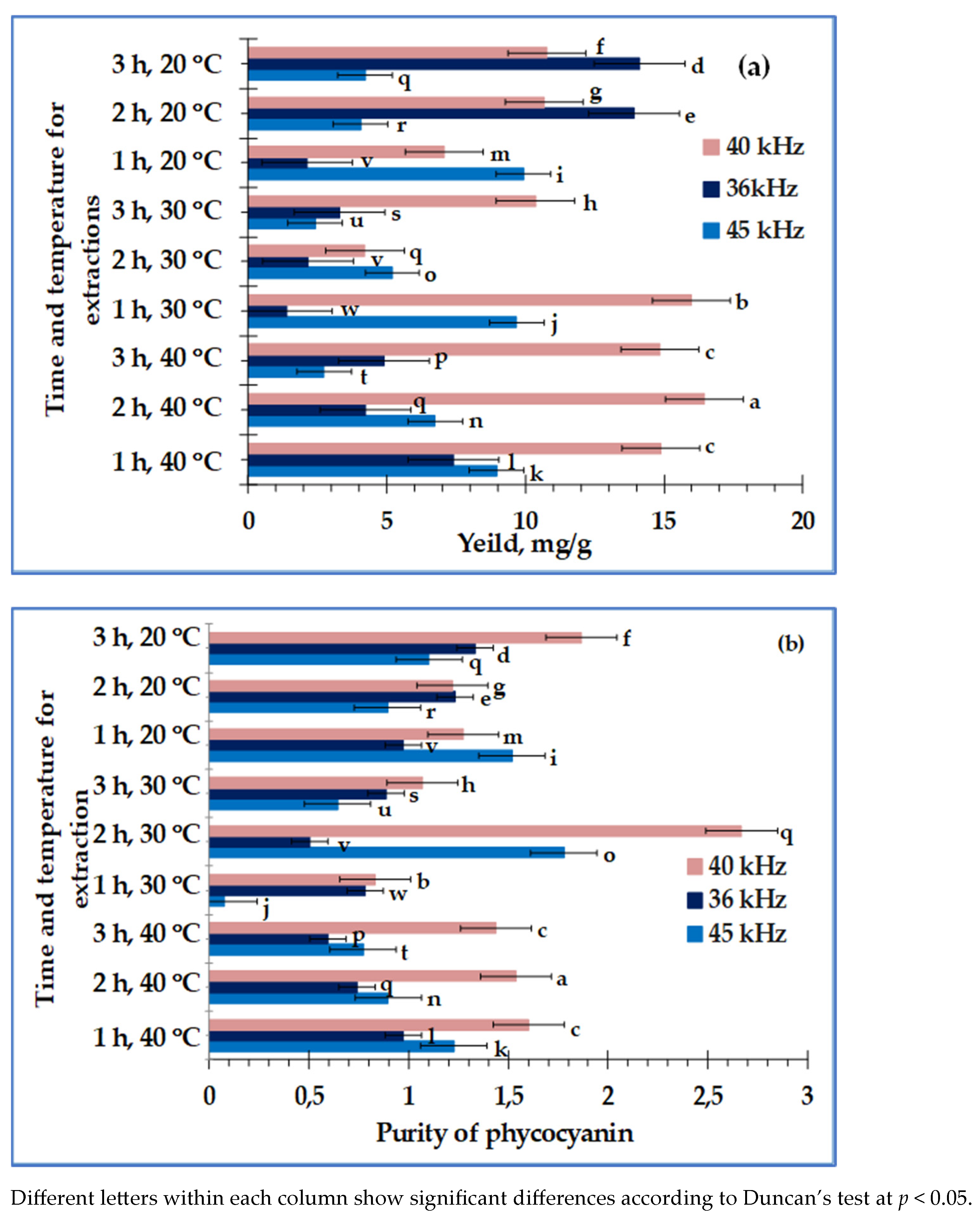
2.5. Spectrophotometric Method for Phycocyanin Analysis

After filtration, the phycocyanin extracts were examined UV-Vis spectrophotometrically using LLG-uniSPEC 2 spectrophotometer (LLG Labware, Meckenheim, Germany) at wavelengths of 280 nm, 615 nm, and 682 nm. The concentration of the specified pigment, its yield, and purity index were determined according to Equations (7)–(9) indicated, respectively in [29,30].
P h y c o c y a n i n   c o n c e n t r a t i o n , m g   m L 1 = A 615 0.474 × A 652 5.34
P u r i t y   i n d e x = A 615 A 280
Y i e l d = P C × V D B
where
  • PC—concentration of phycocyanin in mg mL−1;
  • V—volume of extract in mL;
  • DB—dry biomass of Spirulina in grams.

2.6. Data Analysis

All the measurements for the extracts were performed three times. The average result ± standard deviation (SD) is given in the section results. Duncan’s test for multiple comparisons was performed to determine statistically significant differences between samples. Statistical analysis was performed with SPSS v. 24 (IBM, New York, USA). The results have a significance level of p < 0.05.

3. Results

The results for the content of chlorophyll a and b, as well as the total carotenoid contents for ethanolic extracts, are presented in Figure 1 and Figure 2.
Regarding the content of chlorophyll a and chlorophyll b obtained under the different extraction conditions, all the investigated variants fall into different groups. Therefore, the obtained results are statistically significant.
When grouping the variants in terms of carotenoid content, only those extracted at 40 °C, 35 kHz, and for 1 or 2 h fall into one group. The remaining variants fall into different groups, and we have statistically different results in terms of total carotenoid content.
FRAP and DPPH methods were used to determine the antioxidant activity of water and ethanolic extracts of Spirulina, obtained under the same conditions. The results were expressed in mM TE g−1 and presented in Figure 3 and Figure 4.
The results for phycocyanin content in water extracts, yield, and purity index during ultrasonic extractions of biomass were summarized in Figure 5.
In the extraction of phycocyanin, a large part of the variants falls into different groups, and for the specified parameter, there are significant statistical differences among them. The variants extracted at 35 kHz, for a time of 1 h, and a temperature of 20 °C and those extracted for a time of 2 h, at a temperature of 30 °C were statistically indistinguishable. One group included variants extracted at 40 kHz for 1 or 3 h at t = 40 °C. There was no difference in pigment yield using frequencies of 35 kHz, 40 kHz, and 45 kHz at 2 h, 40 °C; 2 h, 30 °C; and 3 h, 20 °C, respectively. Farthest from each other were the variants with the highest yield (extracted at 40 kHz, 40 °C, 2 h) and with the smallest (35 kHz, 30 °C, 1 h).
The results for AOA, purity index, and yield of phycocyanin, obtained by microwave-assisted extraction are given in Table 1.

4. Discussion

Ultrasound for extracting intracellular compounds is increasingly used as a green method and is based on acoustic cavitation. In the extraction process, the passing of ultrasonic waves through the prepared mixture of distilled water and Spirulina results in tiny bubbles in the liquid. When tiny bubbles rupture at the micro level, the temperature can reach 5000 K, and the pressure can rise to 50 MPa [31]. This leads to the thinning of the cell membrane, destruction of the cell, and increased yield of desired intracellular substances [31].
The temperature and time of extraction indicate a strong influence on the purity index and yield of phycocyanin. An increase in temperature facilitates the extraction of substances through the cell membrane. The yield increases at the expense of the phycocyanin purity index at higher temperatures [30]. Su et al. noted an increase in yield from 30 °C to 50 °C [32]. The extracted phycocyanin content decreases at higher temperatures due to protein denaturation. The yield of phycocyanin from dry Spirulina, in our study, was the highest during an extraction time of 2 h, temperature of 40 °C, and 40 kHz frequency of the ultrasonic waves (16.45 mg g−1), followed by that obtained during an extraction time of 1 h, temperature of 30 °C, and 40 kHz frequency of the ultrasonic waves (16 mg g −1) (Figure 5a). About two times lower results under similar extraction conditions were reported by Silveira et al. (8.89 mg g−1) [33]. Ultrasonic extraction is more efficient than classical extraction. Irawati et al. reported a phycocyanin yield of 3.09 mg g−1 with a purity index of 0.6 extracted from a 6% aqueous extract of Spirulina for 12 h at room temperature [34].
Kamble et al. obtained a yield of 0.26 mg mL−1 of phycocyanin by using ultrasound at a frequency of 40 kHz for 40 min [35]. Other reports indicate that phycocyanin extraction is most effective at 50 kHz. Phycocyanin yields ranged between 0.57 mg g−1 (sonication only) and 43.75 mg g−1 with a C-phycocyanin concentration of 0.21 mg mL−1 (glass bead sonication) [17].
The purity index of the blue pigment depends on various factors: the biomass/solvent ratio [30], extraction time and temperature [22,36], type of solvent [9], isoelectric point [30], etc. The purity index of phycocyanin was determined by the ratio between the absorbance of the pigment at 620 nm and the absorbance of the aromatic amino acids in all proteins in Spirulina. The purity index of the commercial phycocyanin, spectrophotometrically estimated as A615/A280 and confirmed by HPLC, was higher than that of the fractionated phycocyanin after phosphate buffer (2.0 vs. 1.5) [37]. According to Rito-Palomares et al., a purity index value greater than 0.7 allows the pigment to be used in food technology as a colorant; with a purity index above 3.9, it is a reactive class pigment and above four analytical classes [38]. Companies extracting biologically active substances and proteins from plant sources offer phycocyanin with a purity index between 0.5 and 1.5 as a food colorant, between 1.5 and 2.5 as a cosmetic colorant, between 2.5 and 3.5 as a biomarker, and with a purity index over 4 for biomedical applications [39,40].
The purity index of phycocyanin in our study ranged from 0.08 to 2.7. The highest purity index was observed for an extraction time of 2 h, a temperature of 30 °C, and a frequency of 40 kHz, and the lowest was observed at an extraction time of 1 h, temperature of 30 °C, and frequency of 45 kHz. Therefore, the purity index is mainly dependent on the frequency of the ultrasound wave. The maximum purity index in our study is higher than the number of results reported in the literature using salts, ultrafiltration, and dialysis [41,42]. Therefore, the purity index of phycocyanin for most extracts in this study is suitable for food industry purposes. The resulting pigments obtained under conditions 1 h, 40 °C, 40 kHz; 2 h, 30 °C, 45 kHz; and 3 h, 20 °C, 40 kHz are suitable for colorants in the cosmetic industry. One of the samples extracted for 2 h, at a temperature of 30 °C, and a frequency of 40 kHz has a purity index close to those used for a biomarker (Figure 5b).
The ultrasonic extraction of phycocyanin is also preferable to methods using acid treatment of fresh Spirulina with acetic and hydrochloric acids [17,43] since it does not use chemical reagents and can also be used for dry biomass; the destructuring of cell membranes takes place at a low temperature, limiting the thermal denaturation of sensitive compounds. It is preferable to classical extraction, as it reduces extraction time. The yield of phycocyanin of 2.13 mg g−1 in the first hour at 20 °C at the frequency of 35 kHz is comparable to a classical extraction carried out for 24 h at room temperature [44], and, at 30 °C, 3 h time, and a frequency of 35 kHz, it is 3.29 mg g−1, which is 1.34 times higher than that of classical extraction for 48 h [44].
Another green method used in this study is the microwave-assisted extraction of phycocyanin. Data in the literature on this method are mixed. Some authors indicate a power of 133 W and an extraction time of 166 s as optimized extraction conditions for a lower purity index and concentration of the blue pigment, compared to our results [9]. Others have reported higher yields with microwave-assisted extraction (1400 W, 2.5 GHz, 120 s) than with a porcelain bead-milling method [45].
This study yielded between 3.30 mg g−1 extract and 4.60 mg g−1 extract, with a purity index ratio between 0.9 and 1.2 (Table 1). The highest yield and purity index of phycocyanin was obtained at 60 s. One reason for the lower yield in comparison with ultrasonic extraction is the high temperature during the microwave-assisted extraction and the degradation of some of the phycocyanin [9]. Phycocyanin is very sensitive to heat and undergoes rapid changes when exposed to high temperatures. It was reported previously that phycocyanin was stable at temperatures of up to 45 °C [8,46].
In addition to the blue pigment phycocyanin, Spirulina is rich in chlorophyll a and chlorophyll b. Chlorophyll a, similar to phycocyanin, has antioxidant activity, antitumor effects, anti-obesity effects, and anti-ageing effects on cells [47,48,49,50]. Chlorophyll content in this study is presented in Figure 1. Total carotenoid content was found only in the ethanolic extracts at a temperature of 40 °C for each indicated temperature and ultrasonic wave frequency (Figure 1).
Chlorophyll a dominates in all ethanol extracts of Spirulina except those obtained at a duration of 2 and 3 h, at a temperature of 20 °C, and at an ultrasonic wave frequency of 35 kHz. The maximum content of chlorophyll a at 5.46 mg g−1 was obtained during an extraction time of 2 h, temperature of 40 °C, and a frequency of 45 kHz.
Marzorati et al. reported a slightly higher result of 5.7 mg g−1 for the content of chlorophyll a obtained by supercritical carbon dioxide extraction of dry Spirulina [51]. The chlorophyll b content in the investigated samples varied from 0.262 mg g−1 up to 1.404 mg g−1. In the supercritical extraction in [51], values around 3400 mg g−1 were reported. A chlorophyll a content of 9.85 mg g−1 was obtained in the study of Choi et al., where high pressure, 650 bar, and shear stress of about 20 s−1 were applied [3]. However, these values are about twice the maximum yield obtained in our study. The maximum yield of chlorophyll a under the extraction conditions in our investigation was about four times lower than that reported by Choi and Lee obtained under optimal ultrasonic extraction conditions (20.52 kHz, 32.59 °C, in 4.91 h) from ethanol extracts of dry Spirulina [3].
Chlorophyll a content increased with an increase in the frequency of the ultrasonic waves from 35 kHz to 45 kHz, as well as with an increase in the extraction time between 2 and 3 h. The influence of temperature on the extraction of chlorophyll a is lower, while chlorophyll b has a maximum content in the extracts at temperatures around 20 °C. Similar data for an increase in the content of chlorophyll a with an increase in temperature from 30 °C to 40 °C are reported in [52].
Most studies deal with the evaluation of antioxidant activity as a percentage inhibition [53,54,55]. However, this data could not be compared in most of the studies and did not give exact information about extract or dry Spirulina. In our case, two methods based on different mechanisms were used. The FRAP method is based on electron transfer, while the DPPH method is based on radical scavenging ability. DPPH selectively reacts with radicals and hydrogen atom donors at different reaction sites [56].
The AOA of aqueous extracts by the DPPH method was the highest at an extraction temperature 30 °C, a frequency of 35 kHz, and time of 3 h (14.67 mM TE g−1), and by FRAP methods, it was the highest at 40 °C, 45 kHz, and 2 h (20.48 mM TE g−1) (Figure 5a). Ethanol extracts by the DPPH and FRAP methods have maximum antioxidant activity at a temperature of 40 °C, extraction time of 1 h, and frequency of 36 kHz–4.74 mM TE g−1, and 22.92 mM TE g−1, respectively (Figure 3b and Figure 4b). The possible explanation for antioxidant activity is related to phycocyanin and carotenoid content. The results obtained for antioxidant activity in our study were higher than the values reported by some authors for phycocyanin extracted with lysozyme and ammonium sulfate precipitation [57]. The information obtained from this current research will be valuable for further studies on the isolation and structural elucidation of phycocyanin obtained by ultrasonic irradiation.

5. Conclusions

Green extraction methods were successfully performed to obtain phycocyanin, chlorophylls, and antioxidants in water and ethanol extracts from Spirulina. From the results obtained, we can conclude that the purity index and yield of phycocyanin depended on the frequency of applied waves, as well as the temperature of extraction. Ultrasonic extraction was evaluated as a better approach to obtain phycocyanin with acceptable yield and purity index for future applications in pharmaceutical and nutritional products. The best potential for extracting phycocyanin and chlorophylls is an ultrasonic frequency of 40 kHz. In this sense, in-depth studies on the use of this frequency can be the basis of future research works.

Author Contributions

Conceptualization, N.P., D.M. and V.A.; Methodology, G.G. (Galia Gentscheva), K.N., N.P. and V.A.; Formal Analysis, G.G. (Galia Gentscheva) and K.N.; Investigation, I.P. and G.G. (Georgi Gavrailov); Resources, K.N.; Writing—Original Draft Preparation, K.N.; Writing—Review and Editing, D.M. and V.A.; Visualization, I.P.; Supervision, V.A.; Project Administration, K.N.; Funding Acquisition, K.N. All authors have read and agreed to the published version of the manuscript.

Funding

This research was funded by the MU-Varna with the project code № 21001, “Development of a green method for the production of phycocyanin from Spirulina with potential applicability in pharmacy and food technology”.

Data Availability Statement

Datasets from the time of this study are available from the respective authors upon reasonable request.

Conflicts of Interest

The authors declare no conflicts of interest.

References

  1. Costa, J.A.V.; Freitas, B.C.B.; Moraes, L.; Zaparoli, M.; Morais, M.G. Progress in the physicochemical treatment of microalgae biomass for value-added product recovery. Bioresour. Technol. 2020, 301, 122727. [Google Scholar] [CrossRef] [PubMed]
  2. Shao, W.; Ebaid, R.; El-Sheekh, M.; Abomohra, A.; Eladel, H. Pharmaceutical applications and consequent environmental impacts of Spirulina (Arthrospira): An overview. Grasas Aceites 2019, 70, e292. [Google Scholar] [CrossRef]
  3. Choi, W.Y.; Lee, H.Y. Enhancement of neuroprotective effects of Spirulina platensis extract from a high-pressure homogenization process. Appl. Sci. 2018, 8, 634. [Google Scholar] [CrossRef]
  4. Hsieh-Lo, M.; Castillo, G.; Ochoa-Becerra, M.A.; Mojica, L. Phycocyanin and phycoerythrin: Strategies to improve production yield and chemical stability. Algal Res. 2019, 42, 101600. [Google Scholar] [CrossRef]
  5. Gabr, G.A.; El-Sayed, S.M.; Hikal, M.S. Antioxidant Activities of Phycocyanin: A bioactive compound from Spirulina platensis. J. Pharm. Res. Int. 2020, 32, 73–85. [Google Scholar] [CrossRef]
  6. Abdelkhalek, N.K.M.; Ghazy, E.W.; Abdel-Daim, M.M. Pharmacodynamic interaction of Spirulina platensis and deltamethrin in freshwater fish Nile tilapia, Oreochromis niloticus: Impact on lipid peroxidation and oxidative stress. Environ. Sci. Pollut. Res. 2015, 22, 3023–3031. [Google Scholar] [CrossRef] [PubMed]
  7. Wu, Q.; Liu, L.; Miron, A.; Klímov’a, B.; Wan, D.; Kŭca, K. The antioxidant, immunomodulatory, and anti-inflammatory activities of spirulina: An overview. Arch. Toxicol. 2016, 90, 1817–1840. [Google Scholar] [CrossRef] [PubMed]
  8. Wu, H.L.L.; Wang, G.H.H.; Xiang, W.Z.Z.; Li, T.; He, H. Stability and antioxidant activity of food-grade phycocyanin isolated from Spirulina platensis. Int. J. Food Prop. 2016, 19, 2349–2362. [Google Scholar] [CrossRef]
  9. İlter, I.; Akyıl, S.; Demirel, Z.; Koç, M.; Conk-Dalay, M.; Kaymak-Ertekin, F. Optimization of phycocyanin extraction from Spirulina platensis using different techniques. J. Food Compos. Anal. 2018, 70, 78–88. [Google Scholar] [CrossRef]
  10. Rame, R.; Nilawati, D.S.; Novarina, I.H.; Agus, P.; Harjanto, G.R.D. Cell-wall disruption and characterization of phycocyanin from microalgae: Spirulina platensis using Catalytic ozonation. In Proceedings of the 3rd International Conference on Energy, Environmental and Information System (ICENIS 2018), Semarang, Indonesia, 14–15 August 2018; 2018; Volume 73, pp. 1–4. [Google Scholar] [CrossRef]
  11. Danesi, E.D.G.; Rangel-Yagui, C.O.; Carvalho, J.C.M.; Sato, S. Effect of reducing the light intensity on the growth and production of chlorophyll by Spirulina platensis. Biomass Bioenergy 2004, 26, 329–335. [Google Scholar] [CrossRef]
  12. Pawar, S.T.; Puranik, P.R. C-phycocyanin production by halotolerant cyanobacteria. PHYKOS-Off. J. Phycol. Soc. India 2014, 44, 25–32. [Google Scholar]
  13. Puzorjov, A.; McCormick, A.J. Phycobiliproteins from extreme environments and their potential applications. J. Exp. Bot. 2020, 71, 3827–3842. [Google Scholar] [CrossRef] [PubMed]
  14. Ruiz-Domínguez, M.C.; Jáuregui, M.; Medina, E.; Jaime, C.; Cerezal, P. Rapid green extractions of C-phycocyanin from Arthrospira maxima for functional applications. Appl. Sci. 2019, 9, 1987. [Google Scholar] [CrossRef]
  15. Jaeschke, D.P.; Mercali, G.D.; Marczak, L.D.F.; Müller, G.; Frey, W.; Gusbeth, C. Extraction of valuable compounds from Arthrospira platensis using pulsed electric field treatment. Bioresour. Technol. 2019, 283, 207–212. [Google Scholar] [CrossRef] [PubMed]
  16. Soni, B.; Kalavadia, B.; Trivedi, U.; Madamwar, D. Extraction, purification and characterization of phycocyanin from Oscillatoria quadripunctulata-Isolated from the rocky shores of Bet-Dwarka, Gujarat, India. Process Biochem. 2006, 41, 2017–2023. [Google Scholar] [CrossRef]
  17. Moraes, C.C.; Sala, L.; Cerveira, G.P.; Kalil, S.J. C-phycocyanin extraction from Spirulina platensis wet biomass. Braz. J. Chem. Eng. 2011, 28, 45–49. [Google Scholar] [CrossRef]
  18. Deniz, I.; Ozen, M.O.; Yesil-Celiktas, O. Supercritical fluid extraction of phycocyanin and investigation of cytotoxicity on human lung cancer cells. J. Supercrit. Fluids 2016, 108, 13–18. [Google Scholar] [CrossRef]
  19. Martínez, J.M.; Luengo, E.; Saldaña, G.; Álvarez, I.; Raso, J. C-phycocyanin extraction assisted by pulsed electric field from Artrosphira platensis. Food Res. Int. 2017, 99, 1042–1047. [Google Scholar] [CrossRef]
  20. Aftari, R.V.; Rezaei, K.; Bandani, A.R.; Mortazavi, A. Antioxidant activity optimisation of Spirulina platensis C-phycocyanin obtained by freeze-thaw, microwave-assisted and ultrasound-assisted extraction methods. Qual. Assur. Saf. Crops Foods 2017, 9, 1–9. [Google Scholar] [CrossRef]
  21. Jain, T.; Jain, V.; Pandey, R.; Shukila, S.S. Microwave assisted extraction for phytoconstituents-an overview. Asian J. Res. Chem. 2009, 2, 19–25. [Google Scholar]
  22. Aftari, R.V.; Rezaei, K.; Mortazavi, A.; Bandani, A.R. The optimized concentration and purity index of Spirulina platensis C-phycocyanin: A comparative study on microwave-assisted and ultrasound-assisted extraction methods. J. Food Process. Preserv. 2015, 39, 3080–3091. [Google Scholar] [CrossRef]
  23. Viskari, P.J.; Colyer, C.L. Rapid extraction of phycobiliproteins from cultured cyanobacteria samples. Anal. Biochem. 2003, 319, 263–271. [Google Scholar] [CrossRef]
  24. Poojary, M.M.; Barba, F.J.; Aliakbarian, B.; Donsì, F.; Pataro, G.; Dias, D.A.; Juliano, P. Innovative alternative technologies to extract carotenoids from microalgae and seaweeds. Mar. Drugs 2016, 14, 214. [Google Scholar] [CrossRef]
  25. Gentscheva, G.; Milkova-Tomova, I.; Pehlivanov, I.; Gugleva, V.; Nikolova, K.; Petkova, N.; Pisanova, E. Chemical characterization of selected algae and cyanobacteria from Bulgaria as sources of compounds with antioxidant activity. Appl. Sci. 2022, 12, 9935. [Google Scholar] [CrossRef]
  26. Lichtenthaler, H.K.; Wellburn, A.R. Determination of total carotenoids and chlorophylls a and b of leaf in different solvents. Biochem. Soc. Trans. 1983, 11, 591–592. [Google Scholar] [CrossRef]
  27. Benzie, I.F.F.; Strain, J.J. The ferric reducing ability of plasma (FRAP) as a measure of “antioxidant power”: The FRAP assay. Anal. Biochem. 1996, 239, 70–76. [Google Scholar] [CrossRef] [PubMed]
  28. Kivrak, I.; Duru, M.E.; Öztürk, M.; Mercan, N.; Harmandar, M.; Topçu, G. Antioxidant, anticholinesterase and antimicrobial constituents from the essential oil and ethanol extract of Salvia potentillifolia. Food Chem. 2009, 116, 470–479. [Google Scholar] [CrossRef]
  29. Abalde, J. Purification and characterization of phycocyanin from the marine cyanobacterium Synechococcus sp. IO9201. Plant Sci. 1998, 136, 109–120. [Google Scholar] [CrossRef]
  30. Silveira, S.T.; Quines, L.K.D.M.; Burkert, C.A.V.; Kalil, S.J. Separation of phycocyanin from Spirulina platensis using ion exchange chromatography. Bioprocess Biosyst. Eng. 2008, 31, 477–482. [Google Scholar] [CrossRef] [PubMed]
  31. Tiwari, B.K. Ultrasound: A clean, green extraction technology. TrAC Trends Anal. Chem. 2015, 71, 100–109. [Google Scholar] [CrossRef]
  32. Su, C.H.; Liu, C.S.; Yang, P.C.; Syu, K.S.; Chiuh, C.C. Solid-liquid extraction of phycocyanin from Spirulina platensis: Kinetic modeling of influential factors. Sep. Purif. Technol. 2014, 123, 64–68. [Google Scholar] [CrossRef]
  33. Silveira, S.T.; Burkert, J.D.M.; Costa, J.A.V.; Burkert, C.A.V.; Kalil, S.J. Optimization of phycocyanin extraction from Spirulina platensis using factorial design. Bioresour. Technol. 2007, 98, 1629–1634. [Google Scholar] [CrossRef] [PubMed]
  34. Irawati, D.; Abdillah, A.A.; Pramono, H.; Sulmartiwi, L. The effect of different polar solvents on the stability of thermal extraction phycocyanin from Spirulina platensis. IOP Conf. Ser. Earth Environ. Sci. 2020, 441, 012050. [Google Scholar] [CrossRef]
  35. Kamble, S.P.; Gaikar, R.B.; Padilla, R.B.; Shinde, K.D. Extraction and purification of C-phycocyanin from dry Spirulina powder and evaluation of its antioxidant, anticoagulation, and prevention of DNA damage activity. J. Appl. Pharm. Sci. 2013, 3, 149–153. [Google Scholar] [CrossRef]
  36. Dejsungkranont, M.; Chisti, Y.; Sirisansaneeyakul, S. Simultaneous production of C-phycocyanin and extracellular polymeric substances by photoautotrophic cultures of Arthrospira platensis. J. Chem. Technol. Biotechnol. 2017, 92, 2709–2718. [Google Scholar] [CrossRef]
  37. Puglisi, R.; Biazzi, E.; Gesmundo, D.; Vanni, R.; Tava, A.; Cenadelli, S. The Antioxidant Activity of a Commercial and a Fractionated Phycocyanin on Human Skin Cells in Vitro. Molecules 2022, 27, 5276. [Google Scholar] [CrossRef] [PubMed]
  38. Rito-Palomares, M.; Nũnez, L.; Amador, D. Practical application of aqueous two-phase systems for developing a prototype process for c-phycocyanin recovery from Spirulina maxima. J. Chem. Technol. Biotechnol. 2001, 76, 1273–1280. [Google Scholar] [CrossRef]
  39. Figueira, F.S.; Moraes, C.C.; Kalil, S.J. C-phycocyanin purification: Multiple processes for different applications. Technol. Biotechnol. 2001, 76, 1273–1280. [Google Scholar] [CrossRef]
  40. Fernandez-Rojas, B.; Hernandez-Juarez, J.; Pedraza-Chaverri, J. Nutraceutical properties of phycocyanin. J. Funct. Foods 2014, 11, 375–392. [Google Scholar] [CrossRef]
  41. Purohit, A.; Kumar, V.; Chownk, M.; Yadav, S.K. Processing-independent extracellular production of high purity index C-Phycocyanin from Spirulina platensis. ACS Biomater. Sci. Eng. 2019, 5, 3237–3245. [Google Scholar] [CrossRef]
  42. Park, W.S.; Kim, H.J.; Li, M. Two Classes of pigments, carotenoids and c-phycocyanin, in spirulina powder and their antioxidant activities. Molecules 2018, 23, 2065. [Google Scholar] [CrossRef] [PubMed]
  43. Sivasankari, S.; Naganandhini, N.; Ravindran, D. Comparison of Different Extraction methods for Phycocyanin Extraction and Yield, from Spirulina platensis. Int. J. Curr. Microbiol. Appl. Sci. 2014, 3, 904–909. [Google Scholar]
  44. Minchev, I.; Petkova, N.; Milkova-Tomova, I. Ultrasound-assisted extraction of chlorophylls and phycocyanin from Spirulina platensis. Biointerface Res. Appl. Chem. 2020, 11, 9296–9304. [Google Scholar]
  45. Larrosa, A.P.Q.; Camara, A.S.; Moura, J.M.; Pinto, L.A.A. Spirulina sp. biomass dried/disrupted by different methods and their application in biofilms production. Food Sci. Biotechnol. 2018, 27, 1659–1665. [Google Scholar] [CrossRef] [PubMed]
  46. Jespersen, L.; Stromdahl, L.D.; Olsen, K.; Skibsted, L.H. Heat and Light Stability of Three Natural Blue Colorants for Use in Confectionery and Beverages. Eur. Food Res. Technol. 2005, 220, 261–266. [Google Scholar] [CrossRef]
  47. Wells, M.L.; Potin, P.; Craigie, J.S.; Raven, J.A.; Merchant, S.S.; Helliwell, K.E.; Smith, A.G.; Camire, M.E.; Brawley, S.H. Algae as nutritional and functional food sources: Revisiting our understanding. J. Appl. Phycol. 2017, 29, 949–982. [Google Scholar] [CrossRef] [PubMed]
  48. Hernadez, A.C.; Nieves, I.; Meckes, M.; Chamorro, G.; Barron, B.L. Antiviral activity of Spirulina maxima against herpes simplex virus type 2. Antivir. Res. 2002, 56, 279–285. [Google Scholar]
  49. García, J.L.; Vicente, M.; Galán, B. Microalgae, old sustainable food and fashion nutraceuticals. Microb. Biotechnol. 2017, 10, 1017–1024. [Google Scholar] [CrossRef]
  50. Clark, J.G.; Kostal, K.M.; Marino, B.A. Modulation of collagen production following bleomycin-induced pulmonary fibrosis in hamsters. The presence of a factor in the lung that increases fibroblast prostaglandin E2 and cAMP and suppresses fibroblast proliferation and collagen production. J. Biol. Chem. 1982, 257, 8098–8105. [Google Scholar] [CrossRef]
  51. Marzorati, S.; Schievano, A.; Idà, A.; Verotta, L. Carotenoids, chlorophylls and phycocyanin from Spirulina: Supercritical CO2 and water extraction methods for added value products cascade. Green Chem. J. 2020, 22, 187–196. [Google Scholar] [CrossRef]
  52. Erge, H.S.; Karadeniz, F.; Koca, N.; Soyer, Y. Effect of heat treatment on chlorophyll degradation and color loss in green peas. Gida 2008, 33, 225–233. [Google Scholar]
  53. Wu, L.C.; Ho, J.A.A.; Shieh, M.C.; Lu, I.W. Antioxidant and antiproliferative activities of Spirulina and Chlorella water extracts. J. Agric. Food Chem. 2005, 53, 4207–4212. [Google Scholar] [CrossRef] [PubMed]
  54. de Souza, T.D.; Prietto, L.; de Souza, M.M.; Furlong, E.B. Profile, antioxidant potential, and applicability of phenolic compounds extracted from Spirulina platensis. Afr. J. Biotechnol. 2015, 14, 2903–2909. [Google Scholar]
  55. Ali, H.E.A.; Shanab, S.M.M.; Abo-State, M.A.M.; Shalaby, E.A.A.; El Demerdash, U.M.N.; Abdullah, M.A. Evaluation of antioxidants, pigments and secondary metabolites contents in Spirulina platensis. Appl. Mech. Mater. 2014, 625, 160–163. [Google Scholar] [CrossRef]
  56. Gulcin, İ.; Alwasel, S.H. DPPH Radical Scavenging Assay. Processes 2023, 11, 2248. [Google Scholar] [CrossRef]
  57. Safari, R.; Amiri, Z.R.; Kenari, R.E. Antioxidant and antibacterial activities of C-phycocyanin from common name Spirulina platensis. Iran. J. Fish. Sci. 2020, 19, 1911–1927. [Google Scholar] [CrossRef]
Figure 1. Chlorophyll content of ethanolic extracts of Spirulina; (a) content of chlorophyll a; (b) content of chlorophyll b.
Separations 11 00057 g001aSeparations 11 00057 g001b
Figure 2. Carotenoid content of ethanolic extracts of Spirulina obtained under different conditions.
Separations 11 00057 g002
Figure 3. Antioxidant activity (AOA) by the DPPH method for extracts of Spirulina; (a) for water extracts; (b) for ethanolic extracts.
Separations 11 00057 g003
Figure 4. Antioxidant activity (AOA) by the FRAP method for extracts of Spirulina; (a) for water extracts; (b) for ethanolic extracts.
Separations 11 00057 g004
Figure 5. Yield and purity index of phycocyanin, obtained via ultrasonic extraction of water extracts; (a) Yield of phycocyanin; (b) Purity index of phycocyanin.
Separations 11 00057 g005
Table 1. Main characteristics of phycocyanin and AOA of water extracts, obtained via microwave extraction.
Extraction Time, sPhycocyanin Yield ± SD,
mg g−1 Extract
Phycocyanin Purity Index ± SDAOA by DPPH,
mM TE g−1 Extract
AOA by FRAP,
mM TE g−1 Extract
604.58 a ± 0.231.17 a ± 0.0713.31 b ± 0.561869.6 b ± 5.34
1203.34 b ± 0.350.92 b ± 0.0413.35 b ± 1.341595.6 c± 7.23
1803.40 b ± 0.300.88 b ± 0.0816.13 a ± 1.671925.4 a ± 11.23
Different letters within each column show significant differences according to Duncan’s test at p < 0.05.
Disclaimer/Publisher’s Note: The statements, opinions and data contained in all publications are solely those of the individual author(s) and contributor(s) and not of MDPI and/or the editor(s). MDPI and/or the editor(s) disclaim responsibility for any injury to people or property resulting from any ideas, methods, instructions or products referred to in the content.

