Article

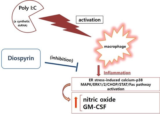
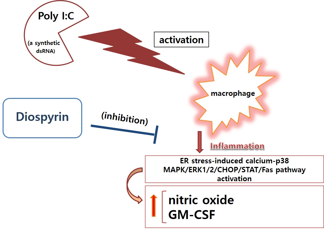
Diospyrin Modulates Inflammation in Poly I:C-Induced Macrophages via ER Stress-Induced Calcium-CHOP Pathway

1
Department of Pathology, College of Korean Medicine, Gachon University, Seong-nam 13120, Korea
2
Department of Pharmacy, University of Peshawar, Peshawar 25120, Pakistan
3
Department of Chemistry, University of Swabi, Anbar 23561, KPK, Pakistan
*
Author to whom correspondence should be addressed.
Processes 2020, 8(9), 1050; https://doi.org/10.3390/pr8091050
Submission received: 29 July 2020 / Revised: 19 August 2020 / Accepted: 25 August 2020 / Published: 27 August 2020
(This article belongs to the Special Issue Pharmacodynamics Modeling of Anti-inflammatory Drugs)

Abstract

:
Diospyrin, plant-derived bisnaphthoquinonoid, is known to have anticancer activity. However, pharmacological activity of diospyrin on viral infection is not well known. We investigated effects of diospyrin on macrophages induced by polyinosinic-polycytidylic acid (poly I:C), a mimic of double-stranded viral RNA. Various cytokines, intracellular calcium, nitric oxide (NO), phosphorylated p38 MAPK, and phosphorylated ERK1/2 as well as mRNA expressions of transcription factors were evaluated. Diospyrin significantly reduced NO production, granulocyte-macrophage colony-stimulating factor production, and intracellular calcium release in poly I:C-induced RAW 264.7. The phosphorylation of p38 MAPK and ERK1/2 was also significantly suppressed. Additionally, diospyrin inhibited mRNA levels of nitric oxide synthase 2, C/EBP homologous protein (CHOP), calcium/calmodulin dependent protein kinase II alpha, signal transducers and activators of transcription 1 (STAT1), STAT3, STAT4, Janus kinase 2, first apoptosis signal receptor, c-Jun, and c-Fos in poly I:C-induced RAW 264.7. Taken together, this study represents that diospyrin might have the inhibitory activity against viral inflammation such as excessive production of inflammatory mediators in poly I:C-induced RAW 264.7 via ER stress-induced calcium-CHOP pathway.

Graphical Abstract

1. Introduction

Innate immune activity is thought to be essential for overcoming infectious diseases [1]. Gilroy reported that immune reaction and inflammatory activity are important against infection [2]. Recently, Nahrendorf and Swirski reported that when stimuli persist and inflammation is not resolved, monocytes might cause a chronic inflammation [3]. Then, it is reasonable that the regulation of inflammatory responses is becoming more meaningful [4].
Macrophages are important in immunity. Zong et al. reported that various inflammatory diseases are related with a complex reaction generated by macrophages [5]. In inflammatory processes, macrophages and monocytes not only produce cytokines and nitric oxide (NO) but also release intracellular calcium.
Cho et al. reported that viral infection can activate immune responses and trigger inflammatory diseases [6]. Alexopoulou et al. reported that double-stranded RNA (dsRNA) induces macrophages [7].
Diospyrin (Figure 1), the plant-derived bisnaphthoquinonoid, is known to have anti-leishmanial effect [8]. In the previous report, we already reported the anti-inflammatory activity of diospyrin on RAW 264.7 stimulated by lipopolysaccharide (LPS) [9]. However, the activity of diospyrin on viral inflammation is not yet reported.
In this experiment, we investigated inhibitory effects of diospyrin on RAW 264.7 induced by polyinosinic-polycytidylic acid (poly I:C), a mimic of double-stranded viral RNA. Diospyrin significantly reduced productions of GM-CSF and NO as well as calcium release in RAW 264.7 induced with poly I:C. The phosphorylation of p38 MAPK and ERK1/2 was also significantly suppressed. The mRNA expressions of nitric oxide synthase 2 (NOS2), C/EBP homologous protein (CHOP), calcium/calmodulin dependent protein kinase II alpha (Camk2a), signal transducers and activators of transcription 1 (STAT1), STAT3, STAT4, Janus kinase 2 (Jak2), first apoptosis signal receptor (Fas), c-Jun, and c-Fos were reduced. Data means diospyrin might have the inhibitory activity against viral inflammation such as excessive production of inflammatory mediators in dsRNA-stimulated macrophages via ER stress-induced calcium-CHOP pathway.

2. Materials and Methods

2.1. Materials

Dulbecco’s modified Eagle’s medium (DMEM), other cell culture reagents, and MILLIPLEX MAP Mouse Cytokine/Chemokine assay kits were obtained from Millipore (Billerica, MA, USA). Indomethacin, baicalein, chrysin, and poly I:C were obtained from Sigma-Aldrich (St. Louis, MO, USA). Diospyrin was isolated from Diospyros lotus by Dr. Inamullah Khan. Phospho-ERK1/2 Antibody (Thr202, Tyr204) (eBioscience 12-9109-42), Phospho-p38 MAPK Antibody (T180/Y182) (eBioscience 17-9078-42), Mouse IgG1 kappa Isotype Control (eBioscience 12-4714-81), and Mouse IgG2b kappa Isotype Control (eBioscience 12-4732-81) were from Life Technologies Corporation (Carlsbad, CA, USA).

2.2. Cell Culture

RAW 264.7 was purchased from Korea Cell Line Bank (Seoul, Korea). In the previous study [9], we reported that diospyrin did not show any cytotoxicity up to a concentration of 10 μM, which was chosen for subsequent experiments.

2.3. NO Production

RAW 264.7 was seeded in a 96-well plate (1 × 10⁴ cells/well) and treated with diospyrin and poly I:C for 24 h. After treatment, supernatants were collected, and NO content was measured using the modified Griess reagent assay kit (Millipore) [9,10].

2.4. Calcium Release

RAW 264.7 was seeded in a 96-well plate (1 × 10⁵ cells/well) and treated with diospyrin and poly I:C for 18 h. Then, calcium release was measured using Fluo-4 NW Calcium Assay Kits (Thermo Fisher Scientific, Waltham, MA, USA) by a spectrofluorometer (Dynex, West Sussex, UK) at 485 nm (excitation) and 535 nm (emission) [11,12,13]

2.5. Cytokines Production

RAW 264.7 was seeded in a 96-well plate (1 × 10⁴ cells/well) and treated with diospyrin and poly I:C. After 24 h treatment, the production of cytokines was measured using MILLIPLEX MAP Mouse Cytokine/Chemokine assay kits and Bio-Plex 200 suspension array system (Bio-Rad) [9,10,11,12,13].

2.6. Quantitative Polymerase Chain Reaction

RAW 264.7 was treated with diospyrin and poly I:C for 18 h in a six-well plate (1 × 10⁶ cells/well). After 18 h treatment, RNA quantity of each well was measured using NucleoSpin RNA kit (Macherey-Nagel, Duren, Germany) and Experion RNA StdSens Analysis kit (Bio-Rad) with Experion Automatic Electrophoresis System (Bio-Rad). Each cDNA was produced using iScript cDNA Synthesis kit (Bio-Rad). Then, gene expressions were measured using quantitative polymerase chain reaction with iQ SYBR Green Supermix (Bio-Rad) [14,15]. The target genes are listed in Table 1. The β-actin was used as a housekeeping gene.

2.7. Flow Cytometry Assay

RAW 264.7 was seeded in a six-well plate (1 × 10⁶ cells/well) and treated with diospyrin and poly I:C for 15 min. After incubation, cells were harvested and washed with Flow Cytometry Staining Buffer. Prior to antibody staining, cells were fixed and permeabilized using Fix Buffer I and Perm Buffer III, respectively. RAW 264.7 stained with Phospho-ERK1/2 (Thr202, Tyr204) Antibody (eBioscience 12-9109-42) and phospho-p38 MAPK (T180/Y182) Antibody (eBioscience 17-9078-42) were analyzed on the Attune NxT flow cytometer (Thermo Fisher Scientific) using Attune NxT software.

2.8. Statistics

Data are presented as mean ± SD. All data were analyzed by one-way analysis of variance test followed by Tukey’s multiple comparison test using GraphPad Prism (ver. 4; GraphPad Software, San Diego, CA, USA).

3. Results

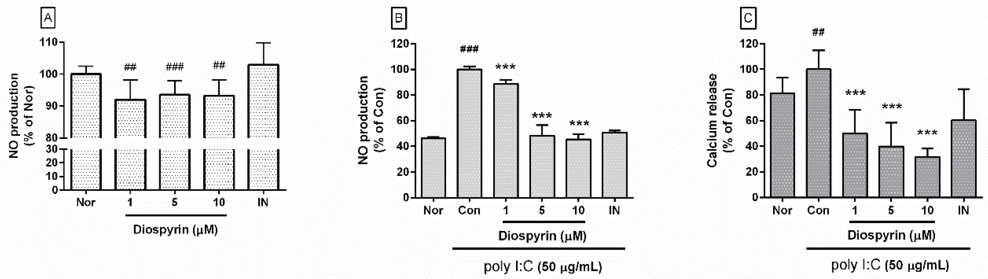
3.1. NO Production from RAW 264.7

NO production from RAW 264.7 after 24 h treatment of diospyrin at concentrations of 1, 5, and 10 µM were 92.02 ± 6.15%, 93.52 ± 4.44%, and 93.27 ± 4.99% of media group (Figure 2A). In the case of poly I:C stimulation, diospyrin at concentrations of 1, 5, and 10 µM showed 88.58 ± 3.24%, 48.4 ± 8.08%, and 45.48 ± 3.95% of the poly I:C alone (p < 0.001) (Figure 2B).

3.2. Calcium Release in RAW 264.7

Calcium release with diospyrin at concentrations of 1, 5, and 10 μM were 50.07 ± 18.52%, 39.82 ± 18.39%, and 31.61 ± 6.69% of the poly I:C alone (p < 0.001) (Figure 2C).

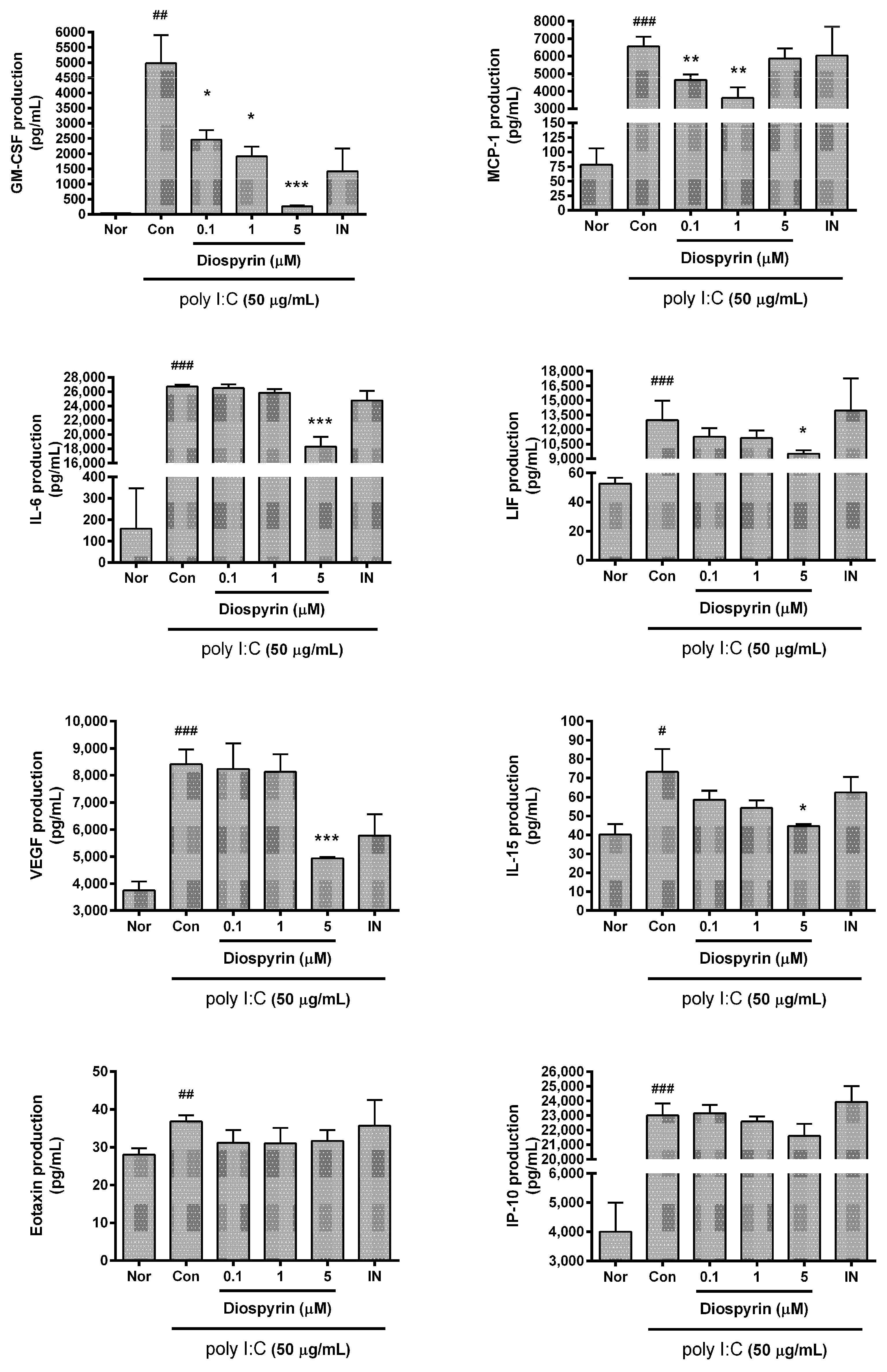
3.3. Cytokine Production from RAW 264.7

Diospyrin significantly reduced GM-CSF production in RAW 264.7 stimulated by poly I:C. Concretely, GM-CSF production from RAW 264.7 with diospyrin at concentrations of 0.1, 1, and 5 µM were 49.46 ± 6.26%, 38.45 ± 6.45%, and 5.37 ± 0.58% of the poly I:C alone; MCP-1 were 70.66 ± 4.79%, 55.11 ± 9.17%, and 89.39 ± 8.72%; IL-6 were 99.15 ± 1.98%, 96.72 ± 1.98%, and 68.5 ± 5.13%; LIF were 86.75 ± 6.77%, 85.79 ± 6.03%, and 73.29 ± 2.74%; VEGF were 97.94 ± 11.18%, 96.66 ± 7.7%, and 58.73 ± 0.48%; IL-15 were 80 ± 6.44%, 74.09 ± 5.51%, and 60.91 ± 1.56%; Eotaxin were 84.62 ± 9.24%, 84.16 ± 11.11%, and 85.97 ± 7.84%; IP-10 were 84.62 ± 9.24%, 84.16 ± 11.11%, and 85.97 ± 7.84% (Figure 3).

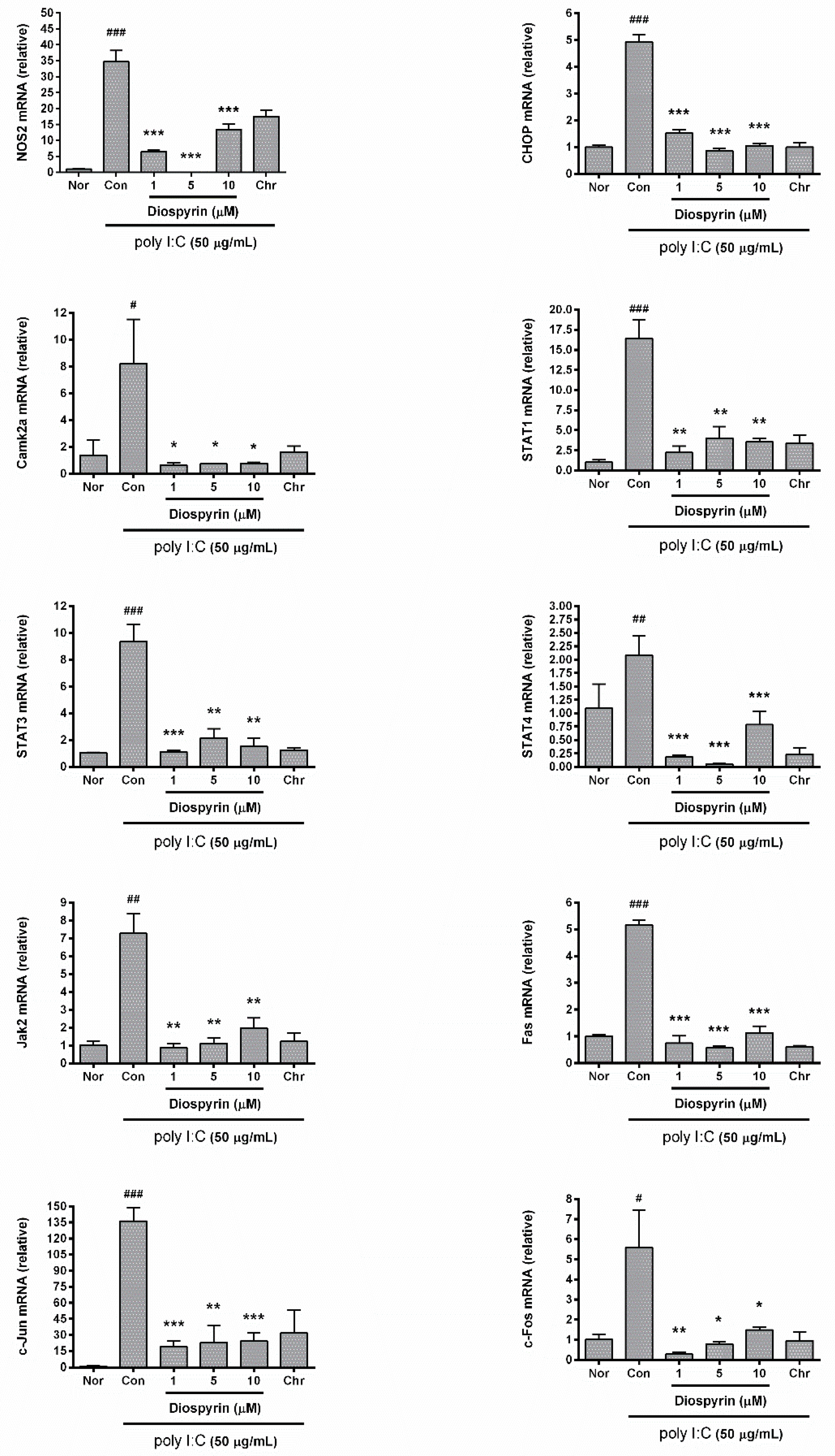
3.4. mRNA Expression of NOS2, CHOP, Camk2a, STAT1, STAT3, STAT4, Jak2, Fas, c-Jun, and c-Fos

Diospyrin inhibited expressions of NOS2, CHOP, Camk2a, STAT1, STAT3, STAT4, Jak2, Fas, c-Jun, and c-Fos in RAW 264.7 stimulated by poly I:C (p < 0.05) (Figure 4). Concretely, NOS2 expression in RAW 264.7 treated with diospyrin at concentrations of 1, 5, and 10 µM were 18.9 ± 1.33%, 0.05 ± 0.01%, and 38.72 ± 4.72% of the poly I:C alone; CHOP were 31.05 ± 2.64%, 17.47 ± 1.87%, and 21.43 ± 1.69%; Camk2a were 7.64 ± 2.28%, 8.93 ± 0.43%, and 9.44 ± 0.69%; STAT1 were 13.58 ± 4.69%, 24.54 ± 8.59%, and 21.68 ± 2.62%; STAT3 were 12.01 ± 1.36%, 22.9 ± 7.41%, and 16.37 ± 6.56%; STAT4 were 8.89 ± 1.35%, 2.31 ± 0.82%, and 37.79 ± 11.83%; Jak2 were 11.97 ± 3.03%, 15.41 ± 3.99%, and 27.1 ± 7.84%; Fas were 14.39 ± 5.29%, 10.98 ± 1.26%, and 21.98 ± 4.75%; c-Jun were 13.99 ± 4.17%, 16.82 ± 11.55%, and 17.71 ± 5.93%; c-Fos were 5.04 ± 1.94%, 14.15 ± 2%, and 26.54 ± 2.44%.

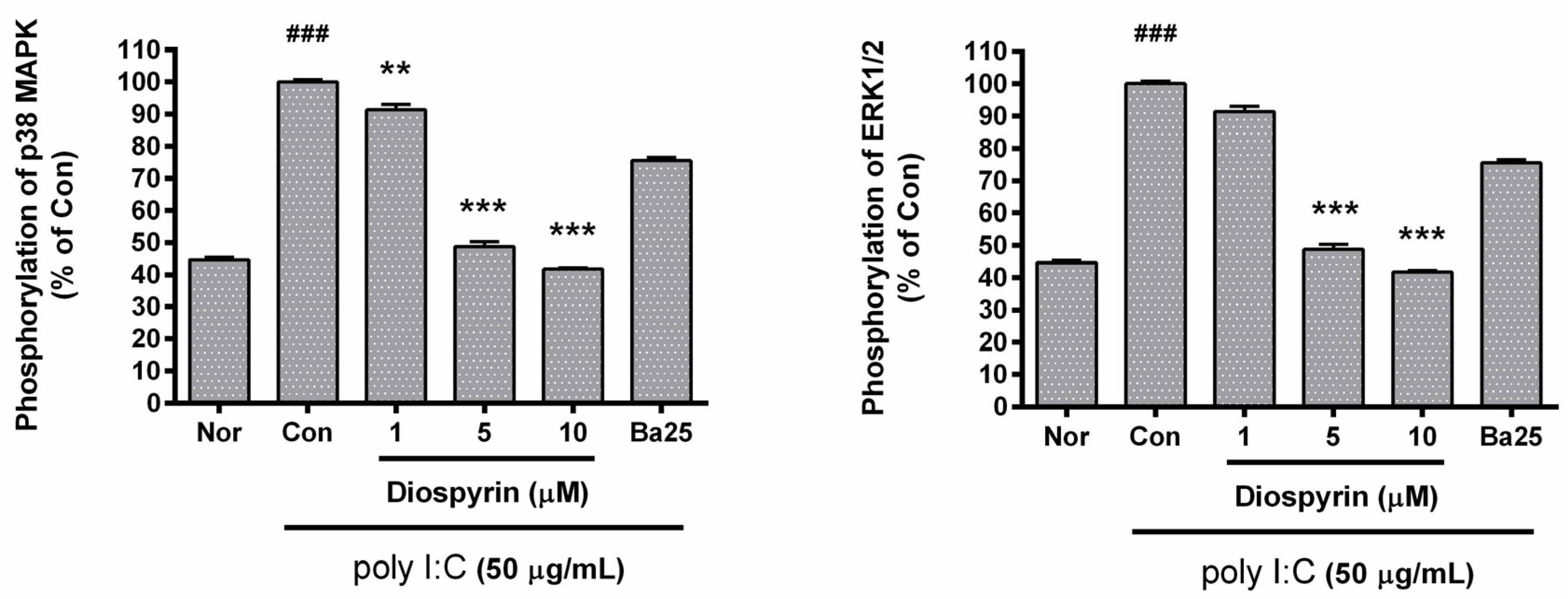
3.5. Phosphorylation of p38 MAPK and ERK1/2 in RAW 264.7

The phosphorylation of p38 MAPK and ERK 1/2 was inhibited by diospyrin significantly (Figure 5). To be concrete, p38 MAPK phosphorylation in RAW 264.7 with diospyrin at concentrations of 1, 5, and 10 µM were 91.38 ± 1.64%, 48.69 ± 1.58%, and 41.73 ± 0.46% of the poly I:C alone; ERK 1/2 were 91.23 ± 3.59%, 53.79 ± 1.74%, and 31.64 ± 0.34%.

4. Discussion

Diospyrin, the medicinal plant (i.e., species of Diospyros such as Diospyros lotus and Diospyros montana)-derived bisnaphthoquinonoid, is known to suppress cancer [16], tuberculosis [17], and leishmaniasis [18,19]. In detail, Bailly reported in 2000 that diospyrin exerts the activity of a topoisomerase I suppressor, resulting in anti-tumor effect [16]. Lall et al. reported in 2005 that diospyrin exerted the inhibitory activity against Mycobacterium tuberculosis [20]. In 2013, Hazra et al. reported that diospyrin represented the anti-leishmanial activity [19]. But effects of diospyrin on viral inflammation have been rarely reported so far.
Although the innate immunity is essential for overcoming hazardous infections, the uncontrolled immuno-inflammatory reaction such as cytokine storm also might be fatal for human life. Thus, the resolution of excessive inflammation is being required continuously because there is no pertinent therapeutics against the newly emerged pandemic viral infection (such as Coronavirus Disease-19), which sometimes provokes cytokine storm.
Karpuzoglu and Ahmed reported that excessive NO production might be concerned with inflammatory diseases such as inflammatory autoimmune diseases [21]. Moreover, it is well reported that excessive production of NO contributes to septic shock [22]. Cooper et al. reported that viral infections might cause airway inflammation and increase the production of various cytokines excessively [23]. Interestingly, respiratory tract viral infections such as Influenza A and Parainfluenza virus were reported to show higher levels of GM-CSF, IL-17A, and IL-22 [24]. In this year, Crisci et al. reported that cytokines such as GM-CSF, VEGF, MCP-1, IL-6, and LIF were increased in severe patients with COVID-19 [25]. It is reasonable that excessive production of cytokines becomes a major target for relieving inflammatory illnesses [26]. In this study, we tried to find a material able to inhibit hyper-production of cytokines in macrophages induced with virus-like particles. During viral infection, dsRNA is produced in host cells and brings about viral inflammatory reaction in macrophages producing NO, ILs, TNF-α, and other cytokines [27]. Then, multiplex cytokine assay is regarded to be effective for checking anti-inflammatory activity of drug candidates [28,29,30,31,32]. For example, Kim et al. reported that baicalein showed inhibitory effects on productions of various cytokines in poly I:C–induced RAW 264.7 [14]. Meanwhile, Alexopoulou et al. reported in 2001 that dsRNA recognized by mammalian Toll-like receptor 3 might provoke inflammatory response via activation of p38 MAPK and ERK1/2 [7]. However, it is not yet fully reported for effects of diospyrin on viral inflammation.
In this experiment, bio-activity of diospyrin in poly I:C–induced RAW 264.7 was evaluated using multiplex cytokine assay, flow cytometry assay, etc. Experimental data means that diospyrin reduces the production of NO and GM-CSF as well as phosphorylation of p38 MAPK and ERK1/2 in poly I:C–induced RAW 264.7. As NO is related with septic shock, diospyrin could be tested for relieving cytokine storm with viral sepsis.
Recently, pandemic viral infection COVID-19 has produced a global threat. Interestingly, TNF-α-converting enzyme (TACE) was reported to be involved in the cell entry of SARS-CoV [33,34]. Xiao et al. reported that SARS-CoV-2 binds angiotensin-converting enzyme 2 (ACE2) for cell entry [35]. ACE2 is known to be shed by TACE and Palau et al. suggested that TACE inhibition may be important for protecting COVID-19 [36]. Additionally, Scott et al. reported in 2011 that TACE activity is upregulated in LPS-induced human monocytes through p38 activation [37]. In this study, the authors presented TACE inhibitory potential of diospyrin predicted by structure-based virtual screening in Figure 6. However, more studies are needed to ascertain whether diospyrin exerts a meaningful effect on TACE activity.
With an infection of microorganisms, macrophages show a distinct reaction, called pyroptosis, which is different from both apoptosis and necrosis. In pyroptosis, various inflammatory mediators are produced from infected macrophages [38]. In 2016, Broz and Dixit also reported about pyroptotic cells to produce many kinds of cytokines [39]. In 2019, Goddard et al. reported that microbial infections can stimulate macrophages [40]. However, it is not yet fully reported for virus-induced pyroptosis accompanying massive production of inflammatory mediators with the calcium signaling pathway.
With ER stress, NO is well known to increase CHOP expression with releasing calcium from ER calcium store and leads to macrophage apoptosis [41,42,43,44]. In 2009, Timmins et al. suggested in their impressive report that ER stress-induced Camk2a activation might enable macrophage apoptosis through Fas induction and/or activation of STAT1 in macrophages [43]. In 2006, Endo et al. reported that LPS causes the overexpression of CHOP, which mediates apoptosis in macrophages and ER stress [45]. Interestingly, Stout et al. reported in 2007 that a brief activation of STAT1 induces ER stress and calcium release from ER calcium store in IFN-gamma-induced airway epithelial cell death with increase of STAT1 protein level [46]. In 2008, Lim et al. reported atheromata-related macrophage apoptosis might be provoked by ER stress through a rise in cytosolic calcium [47].
In the current study, poly I:C-induced RAW 264.7 represents propyrototic ER stress with enhancing mRNA expression of NOS2, CHOP, Camk2a, STAT1, STAT3, STAT4, Jak2, Fas, c-Jun, and c-Fos. Additionally, diospyrin modulates calcium release and mRNA expression of NOS2, CHOP, Camk2a, STAT1, STAT3, STAT4, Jak2, Fas, c-Jun, and c-Fos in poly I:C-induced RAW 264.7. This data means diospyrin could regulate macrophage activation in viral infection via ER stress-induced calcium-CHOP pathway.
However, this study could not elucidate whether intracellular calcium level is increased through influx of extracellular calcium or ER calcium store depletion in poly I:C-induced RAW 264.7. Further study is needed for the exact mechanism for anti-viral activity of diospyrin and medicinal usefulness of diospyrin on viral inflammatory diseases.

5. Conclusions

Diospyrin inhibits excessive production of pro-inflammatory mediators such as GM-CSF and NO via ER stress-induced calcium-CHOP pathway in poly I:C-induced RAW 264.7. Further study is required to clarify pharmacological usefulness for virus-triggered hyper-inflammation such as cytokine storm syndromes from SARS-CoV-2 infection.

Author Contributions

Conceptualization, H.-J.K., I.K., and S.M.A.H.; methodology, A.R. and W.P.; formal analysis, H.-J.K. and J.-Y.L.; investigation, H.-J.K., J.-Y.L., Y.-J.K., B.-Y.K., and W.P.; resources, A.S. and A.R.; data curation, W.P.; writing—original draft preparation, W.P.; visualization W.P.; supervision, W.P.; funding acquisition, W.P. All authors have read and agreed to the published version of the manuscript.

Funding

This research was supported by the Basic Science Research Program through the National Research Foundation of Korea, funded by the Ministry of Education, Science and Technology (2017R1A2B4004933).

Conflicts of Interest

The authors declare no conflict of interest. The funders had no role in the design of the study; in the collection, analyses, or interpretation of data; in the writing of the manuscript, or in the decision to publish the results.

References

  1. Goff, W.L.; Bastos, R.G.; Brown, W.C.; Johnson, W.C.; Schneider, D.A. The Bovine Spleen: Interactions among Splenic Cell Populations in the Innate Immunologic Control of Hemoparasitic Infections. Vet. Immunol. Immunopathol. 2010, 138, 1–14. [Google Scholar] [CrossRef] [PubMed]
  2. Gilroy, D.W. Eicosanoids and the Endogenous Control of Acute Inflammatory Resolution. Int. J. Biochem. Cell Biol. 2010, 42, 524–528. [Google Scholar] [CrossRef] [PubMed]
  3. Nahrendorf, M.; Swirski, F.K. Immunology. Neutrophil-Macrophage Communication in Inflammation and Atherosclerosis. Science 2015, 349, 237–238. [Google Scholar] [CrossRef] [PubMed]
  4. Chen, H.L.; Liao, F.; Lin, T.N.; Liu, F.T. Galectins and Neuroinflammation. Adv. Neurobiol. 2014, 9, 517–542. [Google Scholar] [PubMed]
  5. Zong, Y.; Sun, L.; Liu, B.; Deng, Y.S.; Zhan, D.; Chen, Y.L.; He, Y.; Liu, J.; Zhang, Z.J.; Sun, J.; et al. Resveratrol Inhibits LPS-Induced MAPKs Activation Via Activation of the Phosphatidylinositol 3-Kinase Pathway in Murine RAW 264.7 Macrophage Cells. PLoS ONE 2012, 7, e44107. [Google Scholar] [CrossRef] [PubMed]
  6. Cho, Y.S.; Challa, S.; Moquin, D.; Genga, R.; Ray, T.D.; Guildford, M.; Chan, F.K. Phosphorylation-Driven Assembly of the RIP1-RIP3 Complex Regulates Programmed Necrosis and Virus-Induced Inflammation. Cell 2009, 137, 1112–1123. [Google Scholar] [CrossRef] [Green Version]
  7. Alexopoulou, L.; Holt, A.C.; Medzhitov, R.; Flavell, R.A. Recognition of Double-Stranded RNA and Activation of NF-kappaB by Toll-Like Receptor 3. Nature 2001, 413, 732–738. [Google Scholar] [CrossRef]
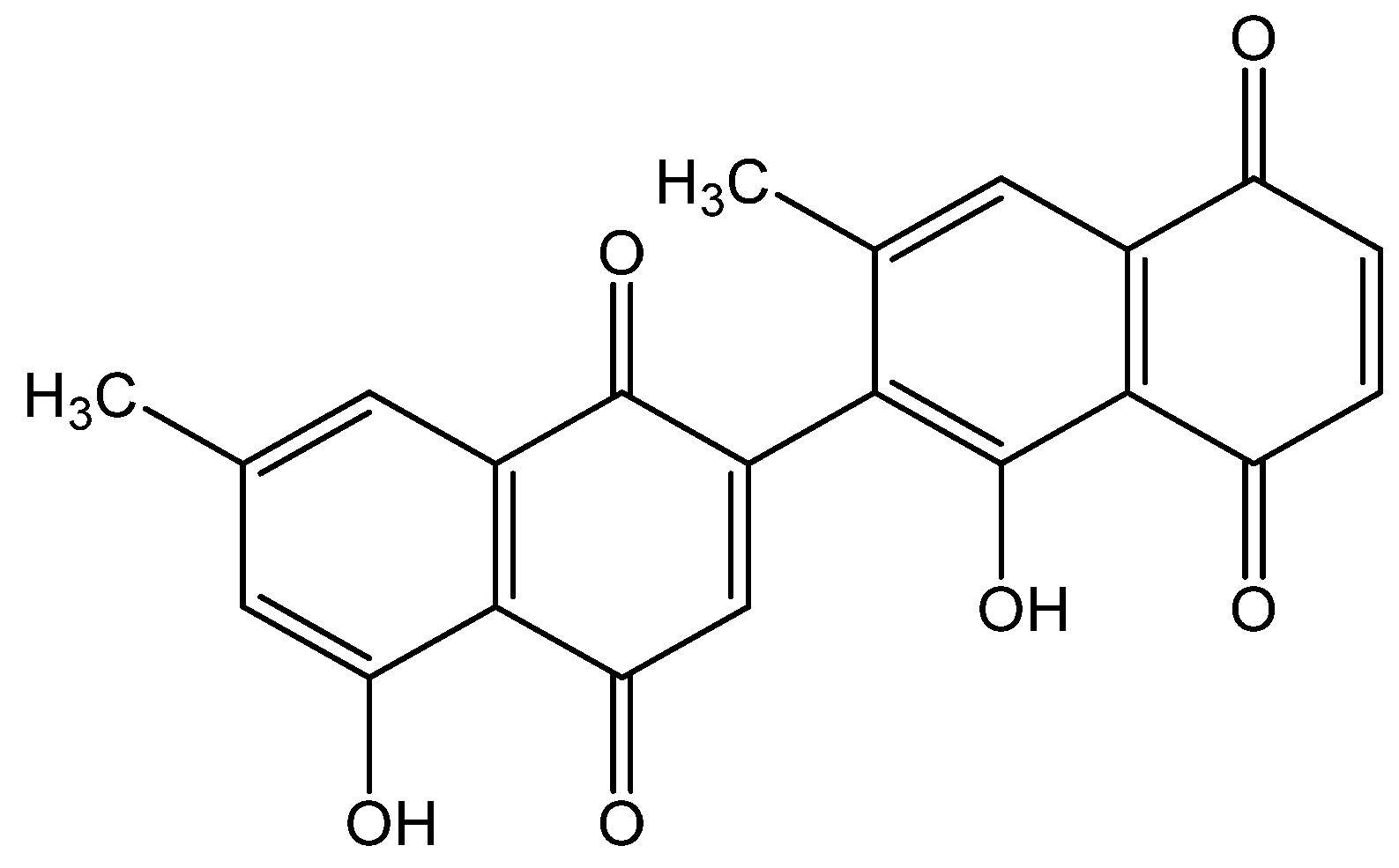
  8. Ray, S.; Hazra, B.; Mittra, B.; Das, A.; Majumder, H.K. Diospyrin, a Bisnaphthoquinone: A Novel Inhibitor of Type I DNA Topoisomerase of Leishmania Donovani. Mol. Pharmacol. 1998, 54, 994–999. [Google Scholar] [CrossRef] [Green Version]
  9. Shahidullah, A.; Lee, J.-Y.; Kim, Y.-J.; Halimi, S.M.A.; Rauf, A.; Kim, H.-J.; Kim, B.-Y.; Park, W. Anti-Inflammatory Effects of Diospyrin on Lipopolysaccharide-Induced Inflammation Using RAW 264.7 Mouse Macrophages. Biomedicines 2020, 8, 11. [Google Scholar] [CrossRef] [Green Version]
  10. Lee, J.Y.; Park, W. Anti-Inflammatory Effect of Myristicin on RAW 264.7 Macrophages Stimulated with Polyinosinic-Polycytidylic Acid. Molecules 2011, 16, 7132–7142. [Google Scholar] [CrossRef] [Green Version]
  11. Lee, J.Y.; Park, W.; Yi, D.K. Immunostimulatory Effects of Gold Nanorod and Silica-Coated Gold Nanorod on RAW 264.7 Mouse Macrophages. Toxicol. Lett. 2012, 209, 51–57. [Google Scholar] [CrossRef] [PubMed]
  12. Lee, J.Y.; Park, W. Anti-Inflammatory Effect of Wogonin on RAW 264.7 Mouse Macrophages Induced with Polyinosinic-Polycytidylic Acid. Molecules 2015, 20, 6888–6900. [Google Scholar] [CrossRef] [PubMed]
  13. Kim, Y.J.; Park, W. Anti-Inflammatory Effect of Quercetin on RAW 264.7 Mouse Macrophages Induced with Polyinosinic-Polycytidylic Acid. Molecules 2016, 21, 450. [Google Scholar] [CrossRef] [PubMed]
  14. Kim, Y.J.; Kim, H.J.; Lee, J.Y.; Kim, D.H.; Kang, M.S.; Park, W. Anti-Inflammatory Effect of Baicalein on Polyinosinic(-)Polycytidylic Acid-Induced RAW 264.7 Mouse Macrophages. Viruses 2018, 10, 224. [Google Scholar] [CrossRef] [PubMed] [Green Version]
  15. Kim, Y.J.; Lee, J.Y.; Kim, H.J.; Kim, D.H.; Lee, T.H.; Kang, M.S.; Park, W. Anti-Inflammatory Effects of Angelica Sinensis (Oliv.) Diels Water Extract on RAW 264.7 Induced with Lipopolysaccharide. Nutrients 2018, 10, 647. [Google Scholar] [CrossRef] [PubMed] [Green Version]
  16. Bailly, C. Topoisomerase I Poisons and Suppressors as Anticancer Drugs. Curr. Med. Chem. 2000, 7, 39–58. [Google Scholar] [CrossRef] [PubMed] [Green Version]
  17. Sundarrajan, S.; Lulu, S.; Arumugam, M. Computational Evaluation of Phytocompounds for Combating Drug Resistant Tuberculosis by Multi-Targeted Therapy. J. Mol. Model. 2015, 21, 247. [Google Scholar] [CrossRef] [PubMed]
  18. Mukherjee, P.; Majee, S.B.; Ghosh, S.; Hazra, B. Apoptosis-Like Death in Leishmania Donovani Promastigotes Induced by Diospyrin and its Ethanolamine Derivative. Int. J. Antimicrob. Agents 2009, 34, 596–601. [Google Scholar] [CrossRef] [PubMed]
  19. Hazra, S.; Ghosh, S.; Das Sarma, M.; Sharma, S.; Das, M.; Saudagar, P.; Prajapati, V.K.; Dubey, V.K.; Sundar, S.; Hazra, B. Evaluation of a Diospyrin Derivative as Antileishmanial Agent and Potential Modulator of Ornithine Decarboxylase of Leishmania Donovani. Exp. Parasitol. 2013, 135, 407–413. [Google Scholar] [CrossRef]
  20. Lall, N.; Meyer, J.J.M.; Wang, Y.; Bapela, N.B.; van Rensburg, C.E.J.; Fourie, B.; Franzblau, S.G. Characterization of Intracellular Activity of Antitubercular Constituents the Roots of Euclea Natalensis. Pharm. Biol. 2005, 43, 353–357. [Google Scholar] [CrossRef]
  21. Karpuzoglu, E.; Ahmed, S.A. Estrogen Regulation of Nitric Oxide and Inducible Nitric Oxide Synthase (iNOS) in Immune Cells: Implications for Immunity, Autoimmune Diseases, and Apoptosis. Nitric Oxide 2006, 15, 177–186. [Google Scholar] [CrossRef] [PubMed]
  22. Thiemermann, C.; Vane, J. Inhibition of Nitric Oxide Synthesis Reduces the Hypotension Induced by Bacterial Lipopolysaccharides in the Rat in Vivo. Eur. J. Pharmacol. 1990, 182, 591–595. [Google Scholar] [CrossRef]
  23. Cooper, P.R.; Lamb, R.; Day, N.D.; Branigan, P.J.; Kajekar, R.; San Mateo, L.; Hornby, P.J.; Panettieri, R.A., Jr. TLR3 Activation Stimulates Cytokine Secretion without Altering Agonist-Induced Human Small Airway Contraction Or Relaxation. Am. J. Physiol. Lung Cell. Mol. Physiol. 2009, 297, L530–L537. [Google Scholar] [CrossRef] [Green Version]
  24. Antalis, E.; Spathis, A.; Kottaridi, C.; Kossyvakis, A.; Pastellas, K.; Tsakalos, K.; Mentis, A.; Kroupis, C.; Tsiodras, S. Th17 Serum Cytokines in Relation to Laboratory-Confirmed Respiratory Viral Infection: A Pilot Study. J. Med. Virol. 2019, 91, 963–971. [Google Scholar] [CrossRef] [PubMed]
  25. Crisci, C.D.; Ardusso, L.R.F.; Mossuz, A.; Muller, L. A Precision Medicine Approach to SARS-CoV-2 Pandemic Management. Curr. Treat. Options Allergy 2020. [Google Scholar] [CrossRef]
  26. Teijaro, J.R. Cytokine storms in infectious diseases. Semin. Immunopathol. 2017, 39, 501–503. [Google Scholar] [CrossRef] [PubMed] [Green Version]
  27. Shaheen, Z.R.; Corbett, J.A. Macrophage Expression of Inflammatory Genes in Response to EMCV Infection. Biomolecules 2015, 5, 1938–1954. [Google Scholar] [CrossRef] [PubMed] [Green Version]
  28. Lee, J.Y.; Kim, Y.J.; Kim, H.J.; Kim, Y.S.; Park, W. Immunostimulatory Effect of Laminarin on RAW 264.7 Mouse Macrophages. Molecules 2012, 17, 5404–5411. [Google Scholar] [CrossRef] [Green Version]
  29. Kim, H.K.; Lee, J.Y.; Han, H.S.; Kim, Y.J.; Kim, H.J.; Kim, Y.S.; Kim, H.M.; Ko, S.G.; An, H.J.; Lee, Y.J.; et al. Immunomodulatory Effects of Liriope Platyphylla Water Extract on Lipopolysaccharide-Activated Mouse Macrophage. Nutrients 2012, 4, 1887–1897. [Google Scholar] [CrossRef] [Green Version]
  30. Lee, J.Y.; Park, W. Anti-Inflammatory Effects of Oroxylin A on RAW 264.7 Mouse Macrophages Induced with Polyinosinic-Polycytidylic Acid. Exp. Ther. Med. 2016, 12, 151–156. [Google Scholar] [CrossRef] [Green Version]
  31. Lee, J.Y.; Park, W. Anti-Inflammatory Effect of Chrysin on RAW 264.7 Mouse Macrophages Induced with Polyinosinic-Polycytidylic Acid. Biotechnol. Bioprocess Eng. 2015, 20, 1026–1034. [Google Scholar] [CrossRef]
  32. Kim, Y.J.; Lee, J.Y.; Kim, H.J.; Kim, D.H.; Lee, T.H.; Kang, M.S.; Choi, Y.K.; Lee, H.L.; Kim, J.; An, H.J.; et al. Inhibitory Effect of Emodin on Raw 264.7 Activated with Double Stranded Rna Analogue Poly I:C. Afr. J. Tradit. Complement. Altern. Med. 2017, 14, 157–166. [Google Scholar] [CrossRef] [PubMed] [Green Version]
  33. Haga, S.; Yamamoto, N.; Nakai-Murakami, C.; Osawa, Y.; Tokunaga, K.; Sata, T.; Yamamoto, N.; Sasazuki, T.; Ishizaka, Y. Modulation of TNF-Alpha-Converting Enzyme by the Spike Protein of SARS-CoV and ACE2 Induces TNF-Alpha Production and Facilitates Viral Entry. Proc. Natl. Acad. Sci. USA 2008, 105, 7809–7814. [Google Scholar] [CrossRef] [PubMed] [Green Version]
  34. Heurich, A.; Hofmann-Winkler, H.; Gierer, S.; Liepold, T.; Jahn, O.; Pohlmann, S. TMPRSS2 and ADAM17 Cleave ACE2 Differentially and Only Proteolysis by TMPRSS2 Augments Entry Driven by the Severe Acute Respiratory Syndrome Coronavirus Spike Protein. J. Virol. 2014, 88, 1293–1307. [Google Scholar] [CrossRef] [Green Version]
  35. Xiao, L.; Sakagami, H.; Miwa, N. ACE2: The Key Molecule for Understanding the Pathophysiology of Severe and Critical Conditions of COVID-19: Demon or Angel? Viruses 2020, 12, 491. [Google Scholar] [CrossRef] [PubMed]
  36. Palau, V.; Riera, M.; Soler, M.J. ADAM17 Inhibition may Exert a Protective Effect on COVID-19. Nephrol. Dial. Transplant. 2020. [Google Scholar] [CrossRef]
  37. Scott, A.J.; O’Dea, K.P.; O’Callaghan, D.; Williams, L.; Dokpesi, J.O.; Tatton, L.; Handy, J.M.; Hogg, P.J.; Takata, M. Reactive Oxygen Species and p38 Mitogen-Activated Protein Kinase Mediate Tumor Necrosis Factor Alpha-Converting Enzyme (TACE/ADAM-17) Activation in Primary Human Monocytes. J. Biol. Chem. 2011, 286, 35466–35476. [Google Scholar] [CrossRef] [Green Version]
  38. Sarhan, J.; Liu, B.C.; Muendlein, H.I.; Li, P.; Nilson, R.; Tang, A.Y.; Rongvaux, A.; Bunnell, S.C.; Shao, F.; Green, D.R.; et al. Caspase-8 Induces Cleavage of Gasdermin D to Elicit Pyroptosis during Yersinia Infection. Proc. Natl. Acad. Sci. USA 2018, 115, E10888–E10897. [Google Scholar] [CrossRef] [Green Version]
  39. Broz, P.; Dixit, V.M. Inflammasomes: Mechanism of Assembly, Regulation and Signalling. Nat. Rev. Immunol. 2016, 16, 407–420. [Google Scholar] [CrossRef]
  40. Goddard, P.J.; Sanchez-Garrido, J.; Slater, S.L.; Kalyan, M.; Ruano-Gallego, D.; Marches, O.; Fernandez, L.A.; Frankel, G.; Shenoy, A.R. Enteropathogenic Escherichia Coli Stimulates Effector-Driven Rapid Caspase-4 Activation in Human Macrophages. Cell Rep. 2019, 27, 1008–1017.e6. [Google Scholar] [CrossRef] [Green Version]
  41. Gotoh, T.; Terada, K.; Oyadomari, S.; Mori, M. Hsp70-DnaJ Chaperone Pair Prevents Nitric Oxide- and CHOP-Induced Apoptosis by Inhibiting Translocation of Bax to Mitochondria. Cell Death Differ. 2004, 11, 390–402. [Google Scholar] [CrossRef] [PubMed]
  42. Tabas, I.; Seimon, T.; Timmins, J.; Li, G.; Lim, W. Macrophage Apoptosis in Advanced Atherosclerosis. Ann. N. Y. Acad. Sci. 2009, 1173 (Suppl. 1), E40–E45. [Google Scholar] [CrossRef] [Green Version]
  43. Timmins, J.M.; Ozcan, L.; Seimon, T.A.; Li, G.; Malagelada, C.; Backs, J.; Backs, T.; Bassel-Duby, R.; Olson, E.N.; Anderson, M.E.; et al. Calcium/calmodulin-Dependent Protein Kinase II Links ER Stress with Fas and Mitochondrial Apoptosis Pathways. J. Clin. Invest. 2009, 119, 2925–2941. [Google Scholar] [CrossRef] [PubMed] [Green Version]
  44. Mori, M. Regulation of Nitric Oxide Synthesis and Apoptosis by Arginase and Arginine Recycling. J. Nutr. 2007, 137, 1616S–1620S. [Google Scholar] [CrossRef] [PubMed] [Green Version]
  45. Endo, M.; Mori, M.; Akira, S.; Gotoh, T. C/EBP Homologous Protein (CHOP) is Crucial for the Induction of Caspase-11 and the Pathogenesis of Lipopolysaccharide-Induced Inflammation. J. Immunol. 2006, 176, 6245–6253. [Google Scholar] [CrossRef] [PubMed] [Green Version]
  46. Stout, B.A.; Melendez, K.; Seagrave, J.; Holtzman, M.J.; Wilson, B.; Xiang, J.; Tesfaigzi, Y. STAT1 Activation Causes Translocation of Bax to the Endoplasmic Reticulum during the Resolution of Airway Mucous Cell Hyperplasia by IFN-Gamma. J. Immunol. 2007, 178, 8107–8116. [Google Scholar] [CrossRef] [PubMed] [Green Version]
  47. Lim, W.S.; Timmins, J.M.; Seimon, T.A.; Sadler, A.; Kolodgie, F.D.; Virmani, R.; Tabas, I. Signal Transducer and Activator of Transcription-1 is Critical for Apoptosis in Macrophages Subjected to Endoplasmic Reticulum Stress in Vitro and in Advanced Atherosclerotic Lesions in Vivo. Circulation 2008, 117, 940–951. [Google Scholar] [CrossRef] [PubMed] [Green Version]
Figure 1. Chemical structure of diospyrin.
Processes 08 01050 g001
Figure 2. Effect of diospyrin on production of nitric oxide (A,B) and calcium release (C). Values are mean ± SD of the three independent experiments. Nor, normal group (media only); Con, control group (poly I:C alone); IN, indomethacin (0.5 µM). ## p < 0.01 vs. Nor; ### p < 0.001 vs. Nor; *** p < 0.001 vs. Con.
Processes 08 01050 g002
Figure 3. Effect of diospyrin on cytokine production from RAW 264.7. Values are the mean ± SD of the three independent experiments. Nor, media alone; IN, indomethacin (0.5 µM). # p < 0.05 vs. Nor; ## p < 0.01 vs. Nor; ### p < 0.001 vs. Nor; * p < 0.05 vs. Con; ** p < 0.01 vs. Con; *** p < 0.001 vs. Con.
Processes 08 01050 g003
Figure 4. Expressions of NOS2, CHOP, Camk2a, STAT1, STAT3, STAT4, Jak2, Fas, c-Jun, and c-Fos mRNA in RAW 264.7. mRNA expressions were normalized to β-actin. Values are the mean ± SD of the three independent experiments. Nor, media alone; Chr, Chrysin (10 µM). # p < 0.05 vs. Nor; ## p < 0.01 vs. Nor; ### p < 0.001 vs. Nor; * p < 0.05 vs. Con; ** p < 0.01 vs. Con; *** p < 0.001 vs. Con.
Processes 08 01050 g004
Figure 5. The phosphorylation of p38 MAPK and ERK 1/2 in RAW 264.7. Values are the mean ± SD of the three independent experiments. Nor, media alone; Ba25, Baicalein (25 µM). ### p < 0.001 vs. Nor. ** p < 0.01 vs. Con; *** p < 0.001 vs. Con.
Processes 08 01050 g005
Figure 6. Predicted binding mode of lowest energy conformation of diospyrin inside the active site of TNF-α-converting enzyme (TACE).
Processes 08 01050 g006
Table 1. Primers used for quantitative PCR.
Name 1Forward Primer (5′–3′)Reverse Primer (5′–3′)
NOS2TGGAGGTTCTGGATGAGAGCAATGTCCAGGAAGTAGGTGAGG
CHOPCCACCACACCTGAAAGCAGTCCTCATACCAGGCTTCCA
Camk2aAGCCATCCTCACCACTATATTCCTTCACGCCATCATT
STAT1TGAGATGTCCCGGATAGTGGCGCCAGAGAGAAATTCGTGT
STAT3GTCTGCAGAGT TCAAGCACCTTCCTCAGTCACGATCAAGGAG
STAT4TTCAGAGCAGCTCAACATGCGGTGAGGTGACCATCATTGTAG-3
Jak2TTGGTTTTGAATTATGGTGTCTGTTCCAAATTTTACAAATTCTTGAACC
FasCGCTGTTTTCCCTTGCTGCCTTGAGTATGAACTCTTAACTGTGAG
c-Jun
c-Fos
ACTGGGTTGCGACCTGAC
AGAGCGGGAATGGTGAAGA
CAATAGGCCGCTGCTCTC
TCTTCCTCTTCAGGAGATAGCTG
β-ActinCTAAGGCCAACCGTGAAAAGACCAGAGGCATACAGGGACA
1. Primer names; nitric oxide synthase 2 (NOS2), C/EBP homologous protein (CHOP), calcium/calmodulin dependent protein kinase II alpha (Camk2a), signal transducers and activators of transcription 1 (STAT1), STAT3, STAT4, Janus kinase 2 (Jak2), first apoptosis signal receptor (Fas), c-Jun, c-Fos, and β-Actin.

