Article

Andrographis paniculata Extract Relieves Pain and Inflammation in Monosodium Iodoacetate-Induced Osteoarthritis and Acetic Acid-Induced Writhing in Animal Models

1
Department of Herbal Pharmacology, College of Korean Medicine, Gachon University, 1342 Seongnamdae-ro, Sujeong-gu, Seongnam 13120, Korea
2
Natural F&P Corp., 767, Sinsu-ro, Suji-gu, Yongin-si, Gyeonggi-do 16827, Korea
*
Author to whom correspondence should be addressed.
Processes 2020, 8(7), 873; https://doi.org/10.3390/pr8070873
Submission received: 29 June 2020 / Revised: 14 July 2020 / Accepted: 16 July 2020 / Published: 20 July 2020
(This article belongs to the Special Issue Pharmacodynamics Modeling of Anti-inflammatory Drugs)

Abstract

:
Osteoarthritis (OA), being the most prominent degenerative joint disease is affecting millions of elderly people worldwide. Although Andrographis paniculata is an ethnic medicine with a long history of being used as analgesic agent, no study using a monosodium iodoacetate (MIA) model has investigated its potential activities against OA. In this study, experimental OA was induced in rats with a knee injection of MIA, which represents the pathological characteristics of OA in humans. A. paniculata extract (APE) substantially reversed the loss of hind limb weight-bearing and the cartilage damage resulted from the OA induction in rats. Additionally, the levels of serum pro-inflammatory cytokines, such as IL-1β, IL-6, and TNF-α as well as the concentration of matrix metalloproteinases, including MMP-1, MMP-3, MMP-8, and MMP-13 were decreased by APE administration. Acetic acid-induced writhing responses in mice which quantitatively measure pain were significantly reduced by APE. In vitro, APE inhibited the generation of NO and downregulated the expression of IL-1β, IL-6, COX-2, and iNOS in lipopolysaccharide (LPS)-stimulated RAW264.7 cells. The above results suggest the potential use APE as a therapeutic agent against OA.

Graphical Abstract

1. Introduction

Osteoarthritis (OA) is the leading degenerative joint disease with an incidence of more than 50% among people aged 65 or older throughout the world [1]. The common characteristics of OA include the erosion of cartilage, synovial swelling, and a disruption in chondrocyte metabolism which cause pain and immobility [2,3]. Arthritic pain is the direct result of the cartilage damage in the joint caused by inflammation; and in the absence of joint cartilage, bones can rub against each other, which causes severe pain and immobility [4,5]. The involvement of inflammatory mediators in osteoarthritic inflammation and pain is now well-known. Inflammatory cytokines are secreted primarily by the synovial chondrocytes and can be found in the synovial fluids of OA patients [6]. As pain and synovial inflammation are two of the major complaints of OA patients, the available treatments for OA are designed mainly to alleviate pain and inflammation [7,8]. Currently, non-steroidal anti-inflammatory drugs (NSAIDS) and steroids are being used as a first-line therapy against OA to control pain and inflammation [9]. However, their prolonged use in elderly is associated with adverse health consequences, such as gastro-intestinal, cardiovascular, and urinary disorders [10,11]. An effective medicine with less or no side effects is immensely needed for the treatment of OA. As a consequence, natural products are receiving tremendous attention for OA treatment.
Natural products for health are gaining popularity thanks to their availability and safety [12]. A large number of investigations have been conducted with numerous medicinal herbs that are widely being used as ethnic medicines in Korea for treating inflammatory diseases including arthritis [13]. We selected Andrographis paniculata (Burm.f.) Nees for its anti-inflammatory and analgesic effects observed through previous screening. A. paniculata has been traditionally used as analgesic agent for its properties, including heat clearing, detoxifying, fire draining, and dampness drying [14,15]. Pharmacological evidence suggests that A. paniculata possesses anti-inflammatory [16], analgesic [17], antinociceptive [18], antiviral [19], anti-cancer [20], and antidiabetic [21] properties. The diverse biological activities of A. panicaulata are attributed to its bioactive compounds such as diterpene, flavonoid, and polyphenol [22]. Diterpene, which consists of andrographolide, 14-deoxyandrographolide, and neoandrographolide, has anti-inflammatory, anti-bacterial, anti-hypertension, and pain-relieving effects [23,24]. In particular, andrographolide of diterpene has strong anti-inflammatory effects [25], which reduces the manifestation of pro-inflammatory agents, including COX-2, IL-6, IL-1β, iNOS, and NF-kB [26,27,28]. Nevertheless, no study using a monosodium iodoacetate (MIA) model has investigated the potential effects of Andrographis paniculata extract (APE) on OA. This study examined the anti-osteoarthritic effects of APE in MIA and acetic acid-induced animal models.
MIA rat model has been used extensively to study the pathophysiological characteristics of OA and the pain behavior in rodents [29]. After being injected, MIA disrupts the chondrocyte metabolism and causes inflammation and cartilage damage in the joint which mimics the characteristics of OA [30]. Matrix metalloproteinase (MMP) is a substrate proteinase that forms cartilage such as proteoglycan, collagen, gelatin, and aggrecan, which are induced by inflammatory cytokines such as IL-1β and TNF-α [31]. MMPs are known to play crucial roles in a number of inflammatory diseases including rheumatoid arthritis and OA [32]. In this experiment, we analyzed the mRNA and protein expression in knee articular cartilage tissues of MIA rats using interstitial collagenase MMP-1, MMP-8, MMP-13, and stromelysin MMP-3 [33]. Acetic acid-induced writhing is the most practiced model to evaluate the peripheral analgesic activities of treatments, which can quantitatively measure inflammatory pain [34]. Lipopolysaccharide (LPS)-stimulated RAW264.7 cells have been widely used to study inflammatory responses, which upon stimulated with LPS, release several inflammatory cytokines and mediators, such as NO, COX-2, iNOS, TNF-α, IL-1β, and IL-6 through the activation of NF-kB and MAPKs [35].
The present study evaluated the pain-relieving and anti-inflammatory effects of APE in MIA rats, acetic acid-induced writhing in mice, and LPS-stimulated RAW264.7 cells.

2. Materials and Methods

2.1. Preparation of APE

The leaf extract of Andrographis paniculata Nees. (Acanthaceae) used in the experiment was purchased from HP Ingredients (Bradenton, USA). A voucher specimen (18060301-1) was deposited by professor Donghun Lee, Department of Herbal pharmacology, College of Korean Medicine, Gachon University. The dried leaf of A. paniculata was extracted in a reflux apparatus in 75% ethanol (2 h at 85 °C) with a ratio of 10:1. The extract was filtered and concentrated under decreased pressure, then, spray dried to yield a powder. After that, the extract was lyophilized under −80 °C.

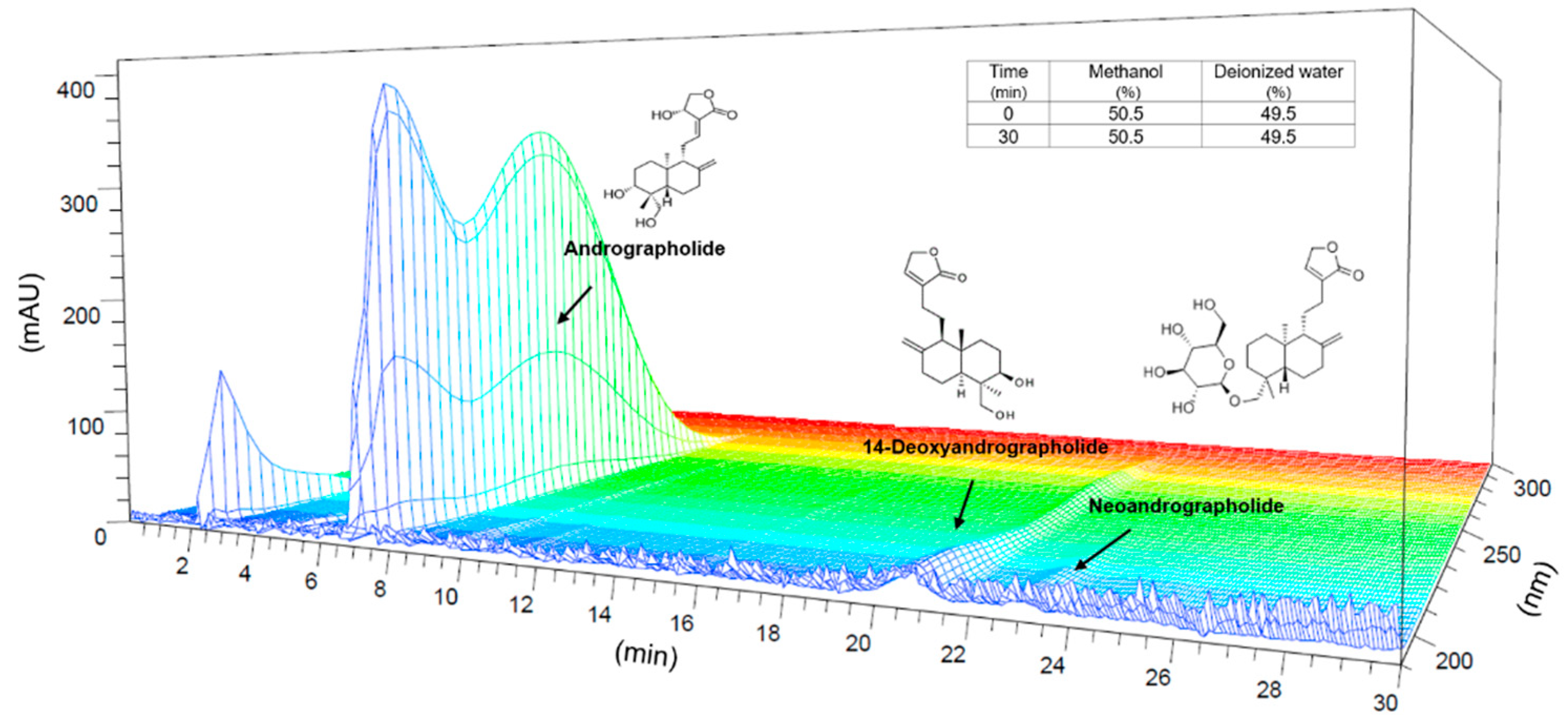
2.2. High Performance Liquid Chromatography (HPLC) Analysis of APE

The chromatographic quantitative analysis of A. paniculata was performed using a high performance liquid chromatography (HPLC) analysis system equipped with a 1200 series HPLC system (Agilent, Palo Alto, USA). The chromatic separation was accomplished on C18 column, 4.6 × 150 mm, 5 µm (Shiseido capcell pak UG120, Shiseido, Yokohama, Japan) at 35 °C. A total of 50 mg of the sample was put in 50 mL of methanol for sonication for 20 min. Additionally, each sample was filtered with a 0.45 μm syringe filter (Waters Corp., Milford, USA). The mobile phase composition was 50.5% methanol and 49.5% deionized water set in isocratic mode and flow rate of 1.2 mL/min. An aliquot of 20 μL of the sample was injected and the effluent was monitored at 220 nm and the total run time was fixed at 30 min. Data was obtained and analyzed with Agilent Chemstation Software. A total of 1 mg of standard compounds was dissolved in 1 mL of methanol, then diluted to concentrations (mg/mL) of 0.0625, 0.125, 0.25, 0.50, and 1. A standard was formed. The contents of Andrographolide (Sigma-Aldrich, St. Louis, USA), 14-deoxy-andrographolide (Rs-interpharm, Gyeonggi, Korea), neoandrographolide (Sigma-Aldrich, St. Louis, USA) contents were quantified by comparing with the standard curve.

2.3. Animal Treatment

Five-week-old (weight 180–200 g) male Sprague-Dawley (SD) rat and six-week-old (weight 25–33 g) male Institute of Cancer Research (ICR) mice were received from Samtako Inc. (Osan, Korea). Prior to the expert to experiment, the animals were acclimatized to the standard laboratory conditions (22 ± 2 °C, 55 ± 10% humidity, and 12 h light/dark cycle) for more than 7 days. Before the experiment, the animals were adapted for a week. The animals were allowed to eat and drink freely. We followed the Guidelines for Animal Experimentation allowed by Gachon University in this study (GIACUC-R2019003 at 29/01/2019).

2.4. MIA Injection and Diet Preparation

SD rats were separated into four groups (n = 9 per group) as: sham, control, indomethacin, APE. MIA solution (40 mg/mL) was prepared by dissolving MIA (Sigma, USA) in 0.9% saline. Rats were first anesthetized with 2% isofluorane and then injected with 50 µL of MIA solution into the knee joint to induce experimental OA. Sham rats were injected with same volume of saline. The animals were maintained as follows: sham and control rats were supplied with basic AIN-93G diet only with no treatment, indomethacin rats were given AIN-93G diet containing with 0.003% indomethacin (equivalent to 3 mg/kg indomethacin), and the APE rats were supplemented with AIN-93G diet containing 0.11% or 0.33% of APE (equivalent to 100 or 300 mg/kg of APE). From the day of MIA injection, the diet was continued over 24 days at 17–20 g per 180–200 g body weight each day.

2.5. Hind Limb Weight-Bearing Measurement

Starting from the initial day of the MIA induction, weight-bearing was recorded until 24 days after MIA solution injection with an Incapacitance Meter Tester 600 (IITC Life Science Inc., Woodland Hills, USA) and averaged the strength applied to each limb over 10 s. The percentage of weight distributed in the hind limb of the treated side (right) was measured using the following equation: weight bearing ratio (%) = (weight on right hind limb/weight on left hind limb + weight on right hind limb) × 100.

2.6. Serum Measurement of OA Induced Model

Blood samples were collected from the abdominal vein and left for 30 min to produce blood clots. The serum was isolated by centrifuging the samples at 4000 rpm for 10 min and kept at −70 °C analysis. The concentration of IL-1β, IL-6, and TNF-α (#LXSARM-03) was measured with Premixed Multi-Analyte Kit (R&D Systems Inc., Minneapolis, USA), and the obtained data were analyzed using a Luminex MAGPIX analyzer (Luminex Co., Austin, USA). The experimental procedure of all multiplex assays was carried out according to the manufacturers’ protocols.

2.7. Real-Time Quantitative PCR Analysis of Knee Cartilage Tissue

Extraction of total RNA in OA induced knee cartilage tissues were performed using Tissue Grinder Moter (Korea Ace Co Ltd., Seoul, Korea) and TRIeasyTM Total RNA Ultra Purification Kit (Real Biotech Corporation Ltd., Banqiao, Taiwan). cDNA was converted by cDNA Reverse AccuPower® CycleScript RT PreMix (Bioneer Corporation, Gyeonggi, KOREA) according to the manufacturer’s instructions. The Real-Time Quantitative PCR was conducted by StepOnePlus real-time PCR system (Applied Biosystems, Foster City, USA). Then, 4 µg of cDNA was amplified using 10 µg of PowerUpTM SYBRTM Green Master mix (Applied Biosystems, Foster City, USA) and forward/reverse primers (Macrogen, Seoul, Korea). The conditions of PCR progress were to be repeated, as follows: 15 s of reacting at 95 °C and 60 s of reacting at 63 °C by 40 cycles after 10 min of pre-incubation step at 95 °C. The sequences of the primers used in the experiment are shown in Table 1.

2.8. Western Blot Analysis for Knee Cartilage Tissue

The protein expression of MMP-1 (#10371-2-AP 1:500), MMP-3 (#SC-21732 1:500), MMP-8 (#SC-514803 1:500), MMP-13 (#18165-1-Ap 1:500), and β-actin (SC-47778 1:1000) was analyzed through Western blot analysis. Extraction of total protein in OA induced knee cartilage tissues was performed using RIPA buffer (Cell Signaling Technology Inc., Danvers, USA) and cOmplete™ Mini EDTA-free Protease Inhibitor Cocktail (Sigma, USA) with Tissue Grinder Moter (Korea Ace Co Ltd., Seoul, Korea). Equal amounts of protein samples were loaded onto SDS-polyacrylamide gel for electrophoresis and the isolated proteins were transferred to PVDF membranes with the Semidry Transfer Cell (Bio-Rad Laboratories, Inc., California, USA) for 1 h at 15 V. Membranes were incubated at room temperature with 5% skim milk in TBS-T for an hour to block non-specific antibody binding. After that, membrane was washed three times with TBS-T and primary antibodies (MMP-1, MMP-3, MMP-8, MMP-13, and β-actin) were applied to react for 24 h at 4°C. The antibodies were purchased from Proteintech Group, Inc., and Santa Cruz Biotechnology, Inc. The membrane was probed using secondary antibody at room temperature for 2 h and then reacted using D-Plus™ ECL Femto System (Dongin, Seoul, Korea) solution. The Western blot image was identified with Azure 280 (Azure Biosystems, Dublin, USA).

2.9. Measurement of Acetic Acid-Induced Writhing

ICR mice were divided into five groups (n = 8 per group) and administrated with APE (200, 600, and 1000 mg/kg), control (distilled water), and ibuprofen 200 mg/kg (Sigma, St. Louis, USA). Then, 30 min after oral administration, 0.7% acetic acid was injected in peritoneal cavity at 10 mL/kg, and after 10 min writhing responses were recorded. A twist reaction was composed of a contraction of the abdominal wall and a turn of the pelvis following the swelling of the hind limbs. A significant decrease in writhing response in the administered group compared with the control group was considered as pain response.

2.10. Measurement of NO and Cytotoxicity

RAW264.7 cells seeded at 5 × 10⁵/well, were incubated at 37 °C, and 5% CO2 for 24 h. Cells were treated with different concentrations of APE (10–1000 µg/mL) and LPS (1 µg/mL) and incubated for 24 h. After mixing the culture supernatant with Griess reagent (1:1), the amount of nitric oxide (NO) in the mixture was recorded at an absorbance of 540 nm. Cell toxicity was recorded using MTT assay. After 24 h of seeding, 5 mg/mL MTT solution was included to RAW264.7 cells and kept at 37 °C, 5% CO2 for 1 h. The supernatant was removed, 100 μL DMSO was included, stored for 10 min, and the absorbance was recorded at 540 nm. The RAW2647 cell line was purchased from Korean Cell Line Bank (Seoul, Korea). All cell experiments were performed three times with n = 3.

2.11. Real-Time Quantitative PCR Analysis of LPS-Stimulated RAW264.7 Cells

Cells were cultured with different concentrations of APE, 1 µg/mL of dexamethasone, and 1 µg/mL LPS for 24 h. RNA extraction was performed with QIAzol Lysis reagent (Qiagen Ltd., Manchester, UK). After cDNA synthesis with using Reverse Transcription Kit (R&D Systems, Minneapolis, USA), Real-Time Quantitative PCR was performed with StepOnePlus real-time PCR system (Applied Biosystems, USA). Power SYBR® Green PCR Master mix (Applied Biosystems, Foster City, USA) was used to amplify the 4 µg of cDNA with forward/reverse primers (Bioneer, Daejeon, Korea). The conditions of PCR progress were to be repeated, as follows: 15 s of reacting at 95 °C, and 60 s of reacting at 60 °C by 40 cycles after 10 min of pre-incubation step at 95 °C. The sequences of primers used in the experiment are shown in Table 2.

2.12. Statistics

Statistical analysis was performed using GraphPad Prism® 5.0 (GraphPad Software, San Diego, USA) with two-sided one-way ANOVA and Dunnett’s post hoc test. The significance was verified at p < 0.05, and measurements were indicated as mean ± standard error of the mean.

3. Results

3.1. HPLC Analysis

Quantification of andrographolide, 14-deoxyandrographolide, and neoandrographolide in APE was carried out through HPLC analysis. APE contained 485.42 mg/g of andrographolide, 131.31 mg/g of 14-deoxyandrographolide, and 26.26 mg/g of neoandrographolide. Three dimensional HPLC chromatogram and the structures of the constituent compounds are shown in Figure 1.

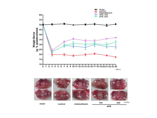
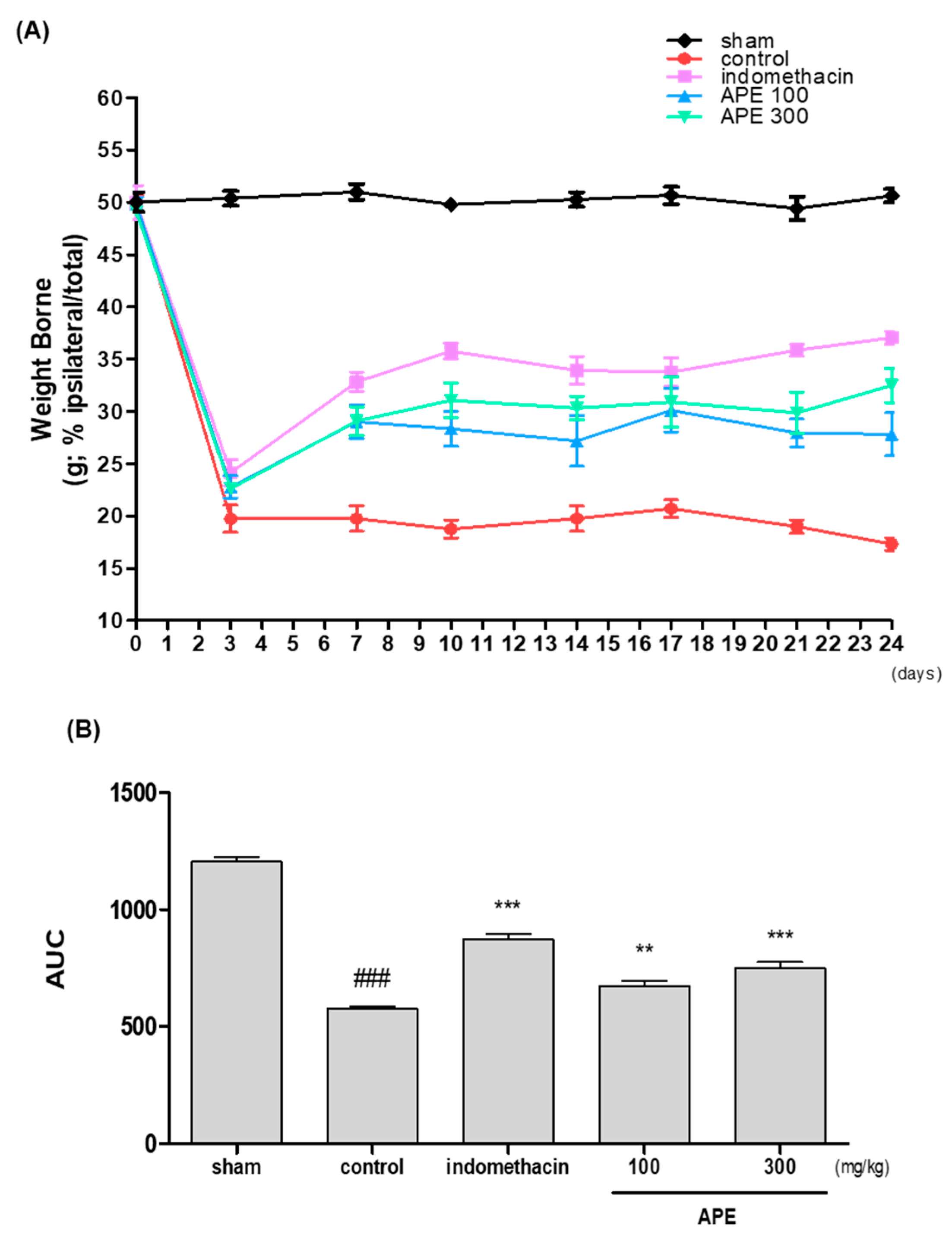
3.2. Effects on Weight-Bearing Distribution in MIA Model

The weight-bearing ratios were recorded during 24 days after OA induction. As shown in Figure 2A, the weight-bearing distribution in the control (MIA) group was significantly reduced on day 3 and continued to be lower afterwards in contrast with the sham group. On the other hand, APE administration resulted in the significant increase in the weight-bearing of MIA rats. Particularly, the increase of weight-bearing by 300 APE was comparable to that of indomethacin (Figure 2B).

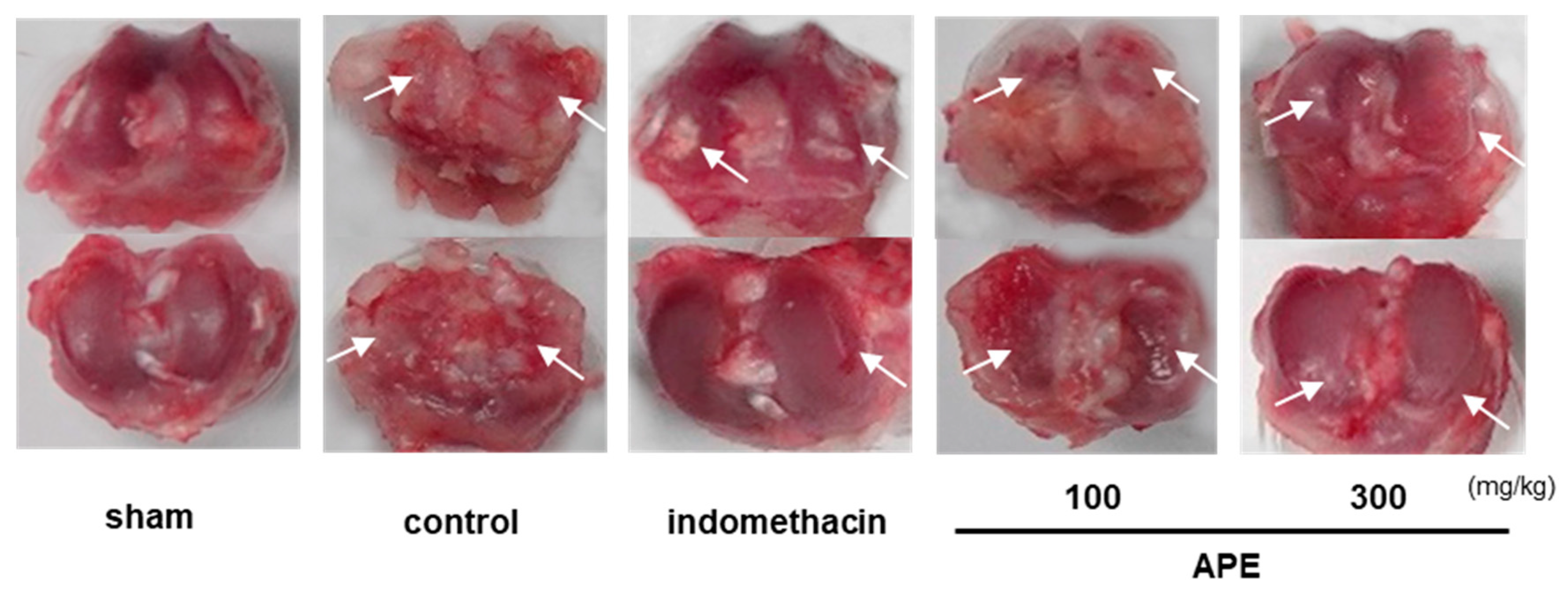
3.3. Preventing Effects on Knee Joint Damage in MIA Model

The representative images of the knee joints in each experimental group show that APE prevented the cartilage erosion triggered by MIA injection. As illustrated in Figure 3, the joint cartilage of the sham rat was in a lustrous, smooth state; while the cartilage of the control rat was precisely less polished and rougher with damages in some areas. The erosion of cartilage induced with MIA was significantly prevented and recovered in APE and indomethacin administered rats. Noticeably, the preventing and recovery of cartilage erosion by APE was similar with that by indomethacin.

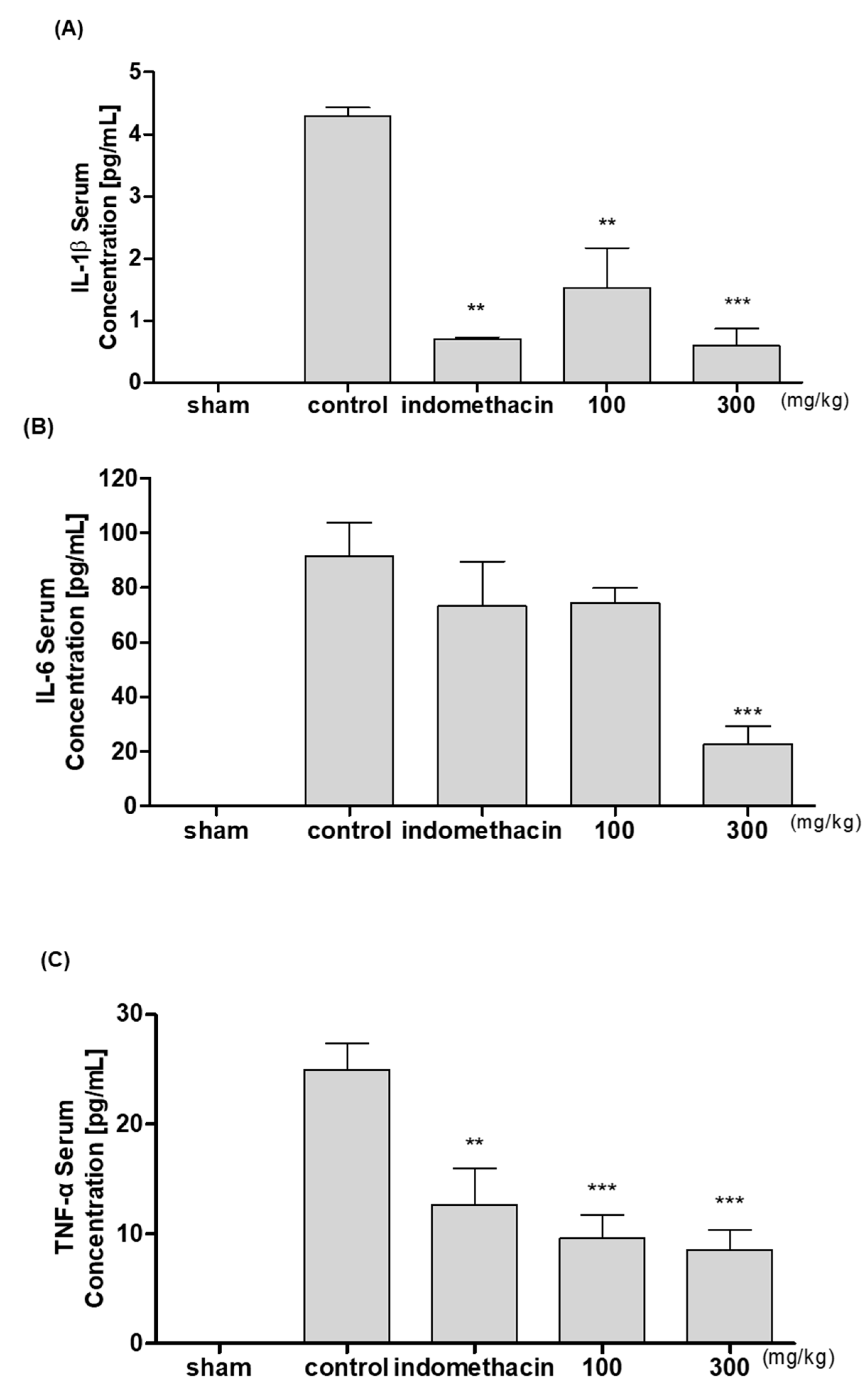
3.4. Effects on Inflammatory Cytokines in MIA-Induced Rat Model

The levels of serum inflammatory cytokines were recorded after isolating serum from blood collected from each experimental group. APE administrated rats had a significant reduction in the concentration of IL-1β, IL-6, and TNF-α in comparison with the control group in a dose-pendent manner. Interestingly, 300 APE reduced the cytokine levels similar to the indomethacin group (Figure 4).

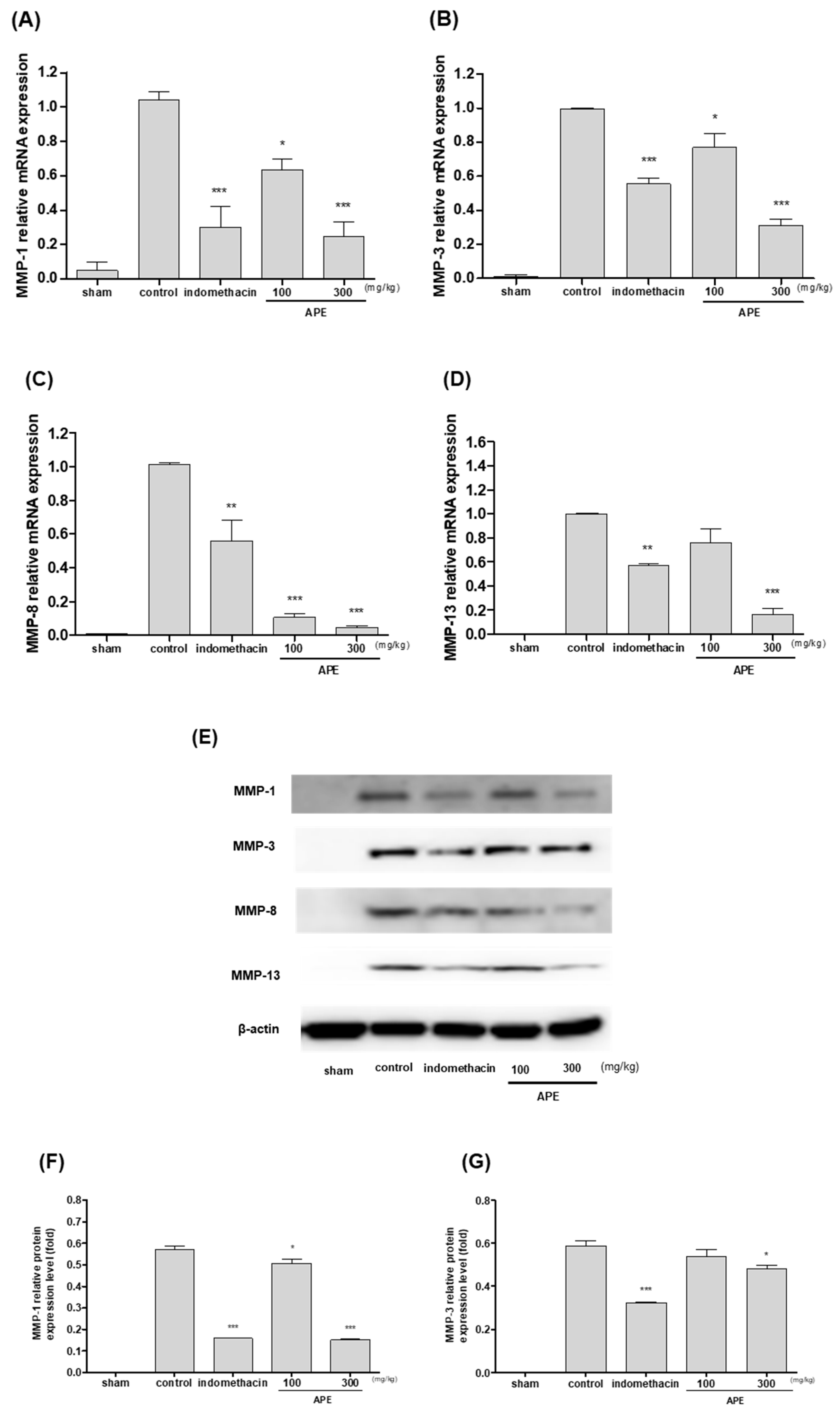
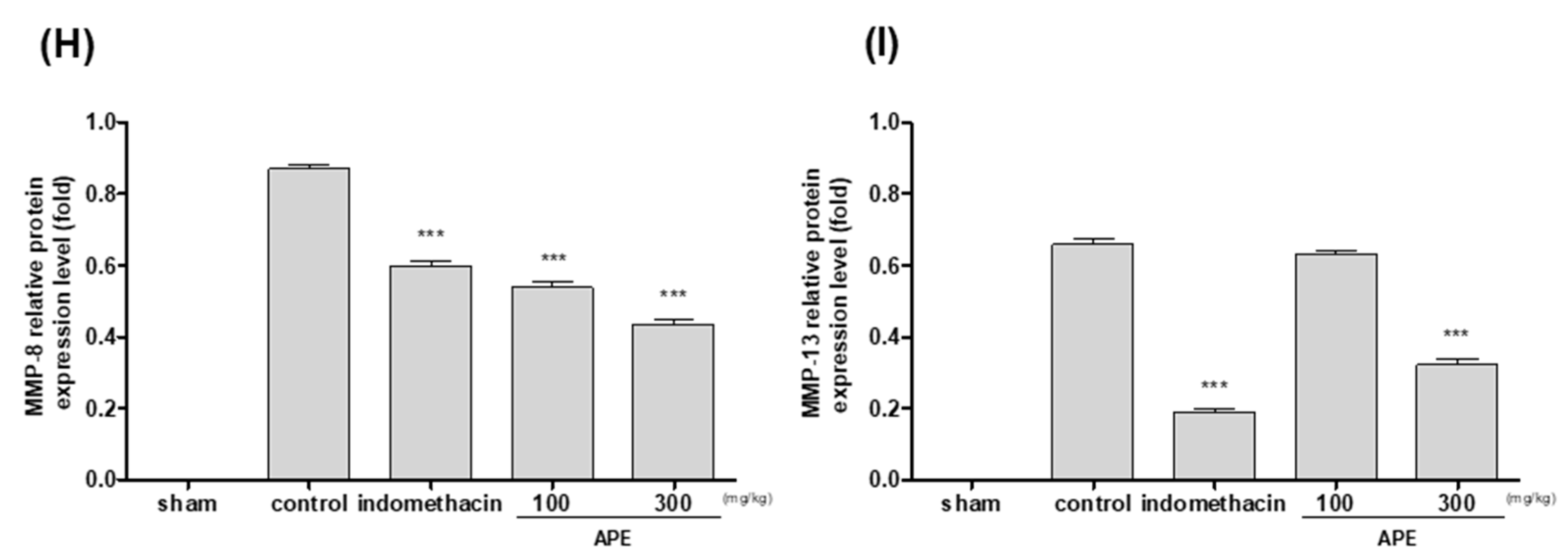
3.5. Effects on Matrix Metalloproteinases Responses in Knee Joint Cartilage Tissue

The measurement of the mRNA levels of MMP-1, MMP-3, MMP-8, and MMP-13 in the rats revealed that APE administration significantly reduced the MMP-1, MMP-3, MMP-8, and MMP-13 levels in knee joint cartilage tissue compared to the control rats (Figure 5A–D). Noticeably, 300 APE rats had lower levels of all four MMPs than the indomethacin group. Western blot analysis also demonstrated APE’s downregulating effects on MMP-1, MMP-3, MMP-8, and MMP-13 in MIA rats in a dose-dependent manner (Figure 5E–I).

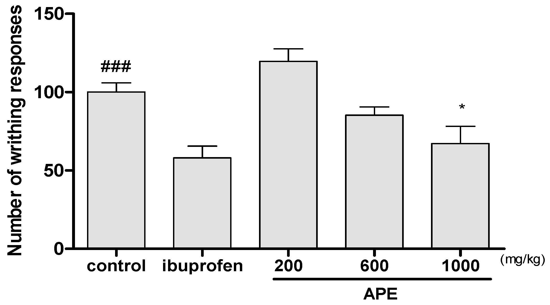
3.6. Effect on Acetic Acid-Induced Writhing Responses

The analgesic effects of APE were investigated against the writhing responses in mice induced with acetic acid. The average writhing number in the control group for 10 min was recorded as 100%. APE administration resulted in the reduction in the number of writhing compared to the control. Rats fed with 1000 mg/kg of APE had the average writhing number of 67.22%, which was close to that of the ibuprofen group (58.09%). This result shows the analgesic effects of APE against peripheral pain (Figure 6).

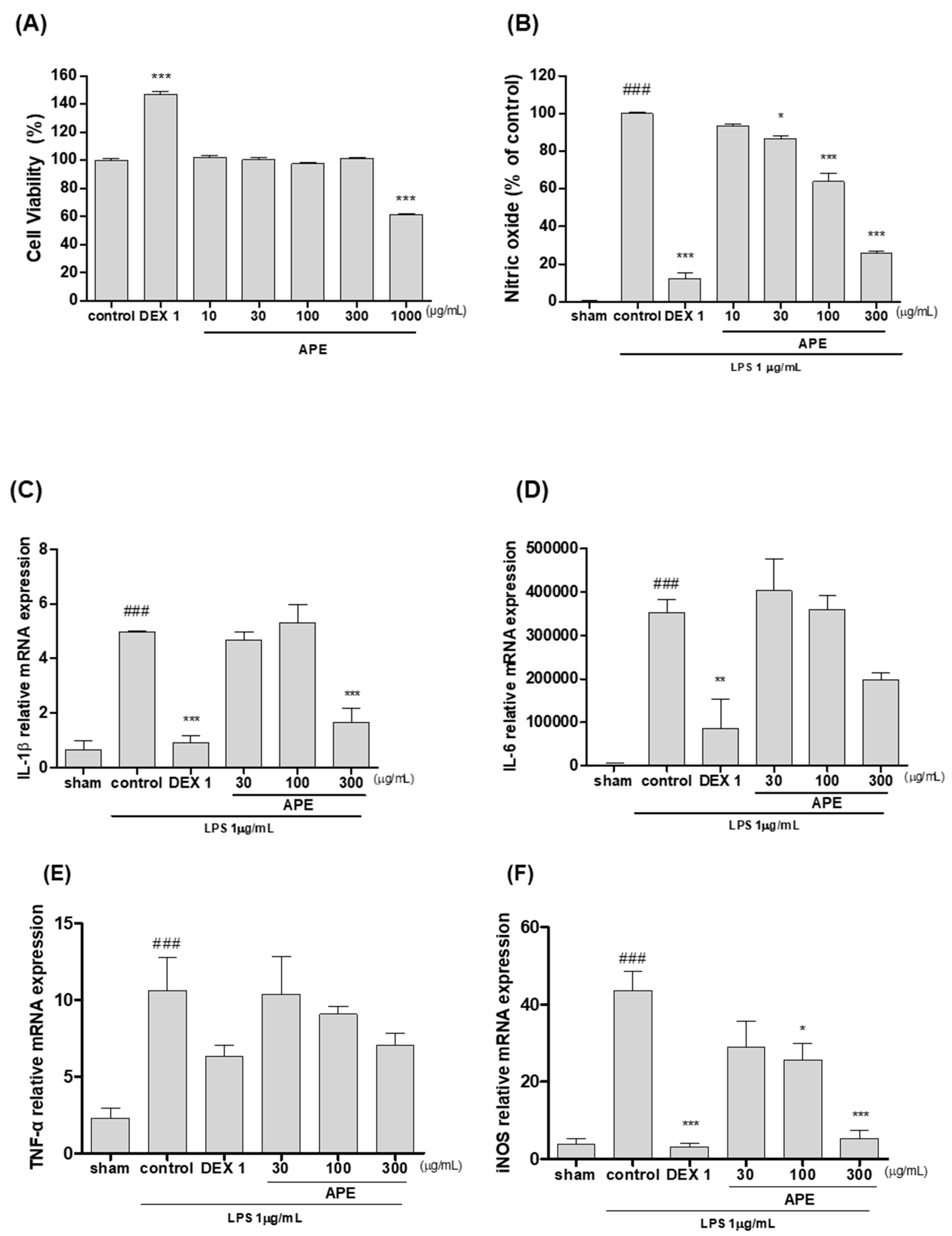
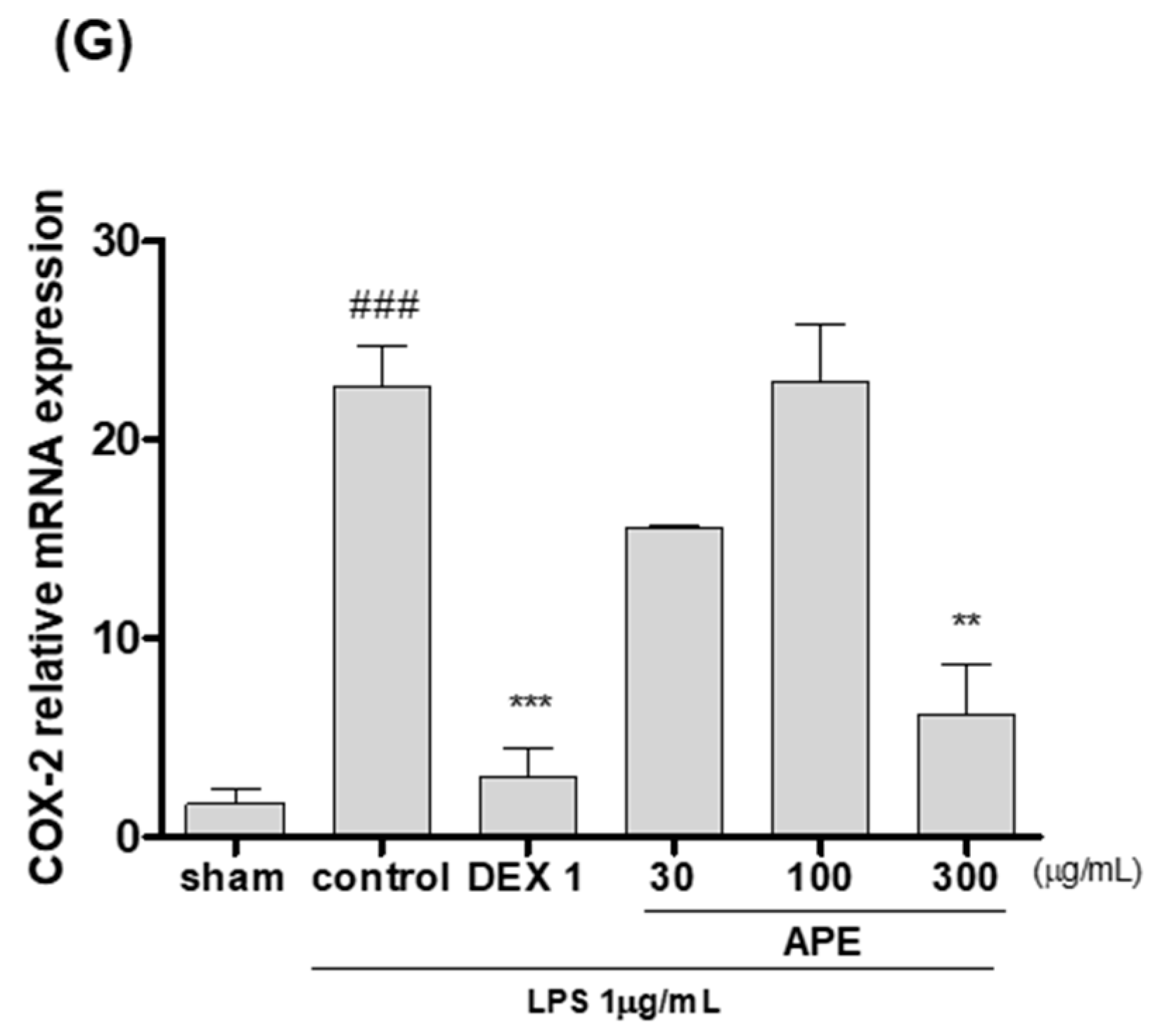
3.7. Anti-Inflammatory Effects in LPS-Stimulated RAW264.7 Cells

In LPS-stimulated RAW264.7 cells, APE suppressed the anti-inflammatory responses by reducing the level of NO and the expression of IL-1β, IL-6, iNOS, TNF-α, and COX-2. In cell viability assay, no apparent cytotoxicity by APE was found with up to 300 µg/mL (Figure 7A). The NO generation of LPS-stimulated RAW264.7 cells was dose-dependently decreased by APE at 300 µg/mL, the reduction of NO was more than 60% compared to the control (Figure 7B). APE suppressed the expression of IL-1β, iNOS, and COX-2 in a dose-dependent manner (Figure 7C,F,G). The reduction of these cytokines by 300 µg/mL APE was comparable to dexamethasone (1 µg/mL).

4. Discussion

The study demonstrated that APE improved weight-bearing capacity of MIA rats and decreased acetic acid-induced writhing responses in mice. APE also suppressed IL-1β, IL-6, and MMPs in MIA rats and IL-1β, iNOS, COX-2 in LPS-induced RAW264.7 cells. Furthermore, APE administration prevented the MIA-induced cartilage degradation in rats.
Previous studies have shown that APE can decrease paw edema and the expressions of inflammatory cytokines in Freund’s adjuvant or collagen injected rats which mimic the human pathophysiology of rheumatoid arthritis and most of them were conducted with samples of its active compounds, andrographolide [36,37,38]. Even though APE has been reported to improve osteoarthritic pain in OA patients [28], no studies were performed for APE using MIA animal model targeting OA, to our knowledge. While a number of surgical and chemically induced arthritis models are available, the MIA model is one of the most accepted systems for studying OA, as it can produce much of the pain related behaviors as well as the patho-physiological characteristics that are found in human OA, including synovial inflammation, cartilage degeneration, and the erosion of subchondral bone [39]. The incapacitance test that measures the hind limb’s weight-bearing capacity is a representative behavioral test frequently used in joint pain studies including MIA-induced OA model, based on the fact that the degree of weight-bearing on hind limbs decreases as the animal feels pain [40,41]. The weight-bearing capacity of the rats fed with APE was superior to the control rats, and was comparable with that of indomethacin treated rats. The improved weight load on the hind limbs by APE in the rats translates a pain-relieving effect in the osteoarthritic knees, which is the primary goal of current OA treatments. These results suggest that APE have analgesic effects against OA pain.
OA patients are often diagnosed with increased concentrations of inflammatory cytokines, including IL-4, IL-10, and IL-13 that are spontaneously released by synovial membrane and cartilage [42,43]. The anti-inflammatory properties of any therapeutic agent includes the decreased production of IL-1β, TNF-α, and MMPs, upregulation of IL-1Rα and TIMP-1, and the inhibition of PGE2 release [44,45]. Pro-inflammatory cytokines play active roles in the pathogenesis of OA by promoting synovial inflammation and the cartilage erosion [46,47]. Particularly, IL-1β is regarded as a key mediator in the inflammatory process in OA [48] and MMPs induced by IL-1β degrades extracellular matrix components, which plays significant roles in the pathogenesis of OA [49]. IL-1β initiates an initial inflammatory reaction, elevates the production of MMPs that impaired chondrocyte in OA, and interrupts the proteoglycan and collagen, which constitute the knee cartilage [50,51]. We conducted an experiment on MMPs that have critical function in causing OA. MMP-1, MMP-8, and MMP-13 are the main collagenases that are distributed in synovial membrane and in synovial fluid [52]. MMP-3 is an enzyme called stromelysin, which plays an important role in joint substrate degradation and has a significant impact on other collagenase activations [53]. This study found a significant increase in IL-1β in serum and MMPs in knee cartilage tissue following MIA injection in rats, while APE administration resulted in a dramatic decrease in serum IL-1β and significant decline in MMP-1, MMP-3, MMP-8, and MMP-13 in knee cartilage tissues. Since the excessive production of IL-1β and MMPs enhances OA progression and hinders weight-bearing functions by promoting synovial inflammation, cartilage damage, and the release of prostaglandins, the reduction of IL-1β and MMPs by APE implicates the improvement in weight-bearing capacities by inhibiting the inflammatory responses and cartilage loss.
The loss of cartilage is one of the prominent indicators of OA and the cartilage erosion observed in MIA rats resemble the pathologic features of human OA [54,55]. Research evidence suggests that MIA causes the breakdown of proteoglycan matrix of the cartilage and induces joint impairment in rodents with characteristics similar to those of human OA [56]. The representative photos also showed a remarkable improvement in the cartilage tissue of MIA rats by APE. The degradation of cartilage in arthritic knees is commonly caused by pro-inflammatory cytokines and mediators. Our results showed that APE prevented the articular cartilage degradation in OA.
While central inputs are clearly involved in OA pain, clinical evidence suggests that the pain in OA patients is largely driven by peripheral inputs active in the affected joints since joint replacement, which eliminates peripheral pain inputs or the application of peripheral antinociceptors can suppress OA pain in the majority of the cases [57]. APE has been previously reported to alleviate pain in acetic acid-induced writhing and tail clip models [58]. In order to evaluate the analgesic effects of APE against peripheral pain, a writhing test was conducted. The injection of acetic acid into the peritoneal cavity causes the production of prostaglandins, such as PGE2 and PGF in the peritoneal fluid of the mice [59]. In this study, APE suppressed the acetic acid-induced writhing response in mice; noticeably, at 1000 mg/kg APE lowered the frequency of writhing similar as the ibuprofen group. When injected into the peritoneal cavity, acetic acid induces pain by releasing pain mediators, such as PGE2 and PGF, which leads to writhing response by releasing inflammatory pain mediators [60]. Hence, the substantial reduction of writhing response in acetic acid-induced mice by APE indicates its high efficacy in suppressing peripheral pain. This result suggests that the peripheral analgesic effect of APE contributes to the pain-relief by APE in the MIA-induced OA.
Macrophages are immune cells, which upon activation with LPS produce inflammatory cytokines and mediators [61]. This study investigated whether APE could suppress inflammatory responses using LPS-stimulated RAW264.7 macrophages. APE decreased the production of NO and the levels of IL-1β, IL-6, iNOS, and COX-2 dose-dependently. Andrographolide, a representative diterpene of APE, has been reported to reduce the production of NO and inhibit releasing IL-1β in LPS-stimulated macrophage [62,63]. The excessive amounts of COX-2 and iNOS in joint induce the production of pro-inflammatory cytokines and cause the destruction of cartilage, swelling, and pain [64,65]. IL-1β is known as the most important pro-inflammatory cytokine in OA, which stimulates the production of COX-2, iNOS, IL-6, and MMPs [66,67]. The present study demonstrated that APE inhibited IL-1β and other pro-inflammatory cytokines and mediators, and therefore could be a useful therapeutic agent against OA inflammation.
Andrographolide is well known as an active ingredient in A. panniculata, and other minor diterpenes 14-deoxyandrographolide, neoandrographolide, isoandrographolide, and 14-deoxy-11,12-didehydroandrographide are also known to have anti-inflammatory, anti-atherosclerotic, and immunomodulatory effects [68,69,70,71]. In this study, the most contained and the representative active ingredient, andrographolide, was set as a standard compound and the effect of the extract was observed, but it is also necessary to evaluate the effect of these minor diterpenes in a future study to fully understand the effects of A. paninculata.
In summary, this study indicated that APE could relieve OA pain and reverse the cartilage degeneration in knee joints by suppressing the inflammatory responses. Notably, the pain-relieving effects of APE measured by weight-bearing in MIA rats were as close as indomethacin. The acetic acid-induced writhing responses were substantially reduced by APE. These analgesic effects were accompanied with protection of knee joints and reduced expressions of pro-inflammatory cytokines. These results demonstrate that APE could be a therapeutic candidate for relieving pain and inflammation in OA patients.

Author Contributions

Conceptualization and writing—original draft preparation D.L.; investigation and data curation C.Y.B.; investigation J.H.H.; supervision, writing—review and editing M.-Y.K. All authors have read and agreed to the published version of the manuscript.

Funding

This work was supported by the National Research Foundation of Korea (NRF) grant funded by the Korea government (MSIT) (No. 2020R1A2C1013956).

Conflicts of Interest

The authors declare no conflict of interest.

References

  1. Xing, D.; Xu, Y.; Liu, Q.; Ke, Y.; Wang, B.; Li, Z.; Lin, J. Osteoarthritis and all-cause mortality in worldwide populations: Grading the evidence from a meta-analysis. Sci. Rep. 2016, 6, 1–7. [Google Scholar] [CrossRef] [PubMed] [Green Version]
  2. Beyreuther, B.; Callizot, N.; Stöhr, T. Research article Antinociceptive efficacy of lacosamide in the monosodium iodoacetate rat model for osteoarthritis pain. Arthritis Res. 2007, 9. [Google Scholar]
  3. Pharmacy, C.; Pharmacy, F.; Mulyorejo, J.; Unair, C.C. Attenuation of IL-1ß on the use of glucosamine as an adjuvant in meloxicam treatment in rat models with osteoarthritis. Basic Clin. Physiol Pharm. 2020, 1–9. [Google Scholar]
  4. Perrot, S. Osteoarthritis pain. Best Pract. Res. Clin. Rheumatol. 2015, 29, 90–97. [Google Scholar] [CrossRef]
  5. Ding, C. Do NSAIDs Affect the Progression of Osteoarthritis? Inflamm. 2002, 26. [Google Scholar]
  6. Goldring, M.B.; Otero, M. Inflammation in osteoarthritis. Curr. Opin. Rheumatol. 2014, 23, 471–478. [Google Scholar] [CrossRef] [PubMed]
  7. Che Ahmad Tantowi, N.A.; Lau, S.F.; Mohamed, S. Ficus deltoidea Prevented Bone Loss in Preclinical Osteoporosis/Osteoarthritis Model by Suppressing Inflammation. Calcif. Tissue Int. 2018, 103, 388–399. [Google Scholar] [CrossRef]
  8. Maldonado, M.; Nam, J. The Role of Changes in Extracellular Matrix of Cartilage in the Presence of Inflammation on the Pathology of Osteoarthritis. Biomed. Res. Int. 2013, 1–10. [Google Scholar] [CrossRef] [Green Version]
  9. Sebaldt, R.J.; Petrie, A.; Goldsmith, C.H.; Marentette, M.A. Appropriateness of NSAID and Coxib Prescribing for Patients with Osteoarthritis by Primary Care Physicians in Ontario: Results From the CANOAR Study. Am. J. Manag. Care. 2004, 10, 10. [Google Scholar]
  10. Hungin, A.P.S.; Kean, W.F. Nonsteroidal anti-inflammatory drugs: Overused or underused in osteoarthritis? Am. J. Med. 2001, 110, S8–S11. [Google Scholar] [CrossRef]
  11. Al-Afify, A.S.A.; El-Akabawy, G.; El-Sherif, N.M.; El-Safty, F.E.N.A.; El-Habiby, M.M. Avocado soybean unsaponifiables ameliorates cartilage and subchondral bone degeneration in mono-iodoacetate-induced knee osteoarthritis in rats. Tissue Cell 2018, 52, 108–115. [Google Scholar] [CrossRef] [PubMed]
  12. Goudarzi, R.; Reid, A.; McDougall, J.J. Evaluation of the novel avocado/soybean unsaponifiable Arthrocen to alter joint pain and inflammation in a rat model of osteoarthritis. PLoS ONE 2018, 13, e0191906. [Google Scholar] [CrossRef] [PubMed] [Green Version]
  13. Kang, M.; Jung, I.; Hur, J.; Kim, S.H.; Lee, J.H.; Kang, J.Y.; Jung, K.C.; Kim, K.S.; Yoo, M.C.; Park, D.S.; et al. The analgesic and anti-inflammatory effect of WIN-34B, a new herbal formula for osteoarthritis composed of Lonicera japonica Thunb and Anemarrhena asphodeloides BUNGE in vivo. J. Ethnopharmacol. 2010, 131, 485–496. [Google Scholar] [CrossRef] [PubMed]
  14. Roxas, M.; Jurenka, J. Colds and influenza: A review of diagnosis and conventional, botanical, and nutritional considerations. Altern. Med. Rev. 2007, 12, 25–48. [Google Scholar] [PubMed]
  15. TAYLOR, W.C. Cytotoxic Diterpenoid Constituents from Andrographis Paniculata Ness. Leaves. ScienceAsia 1992, 18, 187. [Google Scholar]
  16. Shen, Y.C.; Chen, C.F.; Chiou, W.F. Andrographolide prevents oxygen radical production by human neutrophils: Possible mechanism(s) involved in its anti-inflammatory effect. Br. J. Pharmacol. 2002, 135, 399–406. [Google Scholar] [CrossRef] [PubMed] [Green Version]
  17. Sheeja, K.; Shihab, P.K.; Kuttan, G. Antioxidant and anti-inflammatory activities of the plant Andrographis paniculata nees. Immunopharmacol. Immunotoxicol. 2006, 28, 129–140. [Google Scholar] [CrossRef]
  18. Greco, R.; Siani, F.; Demartini, C.; Zanaboni, A.; Nappi, G.; Davinelli, S.; Scapagnini, G.; Tassorelli, C. Andrographis paniculata shows anti-nociceptive effects in an animal model of sensory hypersensitivity associated with migraine. Funct. Neurol. 2016, 31, 53–60. [Google Scholar] [CrossRef]
  19. Basak, A.; Cooper, S.; Roberge, A.G.; Banik, U.K.; Chre, M. Inhibition of proprotein convertases-1, -7 and furin by diterpines of Andrographis paniculata and their succinoyl esters. Biochem. J. 1999, 113, 107–113. [Google Scholar] [CrossRef]
  20. Attla, M.A.M.; Weiss, D.W. Immunology of Spontaneous Mammary Carcinomas in Mice V. Acquired Tumor Resistance and Enhancement in Strain A Mice Infected with Mammary Tumor Virus. Cancer Res. 1966, 26, 1787–1800. [Google Scholar]
  21. Subramanian, R.; Asmawi, M.Z.; Sadikun, A. In vitro α-glucosidase and α-amylase enzyme inhibitory effects of Andrographis paniculata extract and andrographolide. Acta Biochim. Pol. 2008, 55, 391–398. [Google Scholar] [CrossRef] [PubMed] [Green Version]
  22. Koteswara Rao, Y.; Vimalamma, G.; Venkata Rao, C.; Tzeng, Y.M. Flavonoids and andrographolides from Andrographis paniculata. Phytochemistry 2004, 65, 2317–2321. [Google Scholar] [CrossRef] [PubMed]
  23. Chao, W.W.; Kuo, Y.H.; Lin, B.I.F. Anti-inflammatory activity of new compounds from Andrographis paniculata by nf-κb transactivation inhibition. J. Agric. Food Chem. 2010, 58, 2505–2512. [Google Scholar] [CrossRef] [PubMed]
  24. Hu, X.Y.; Wu, R.H.; Logue, M.; Blondel, C.; Lai, L.Y.W.; Stuart, B.; Flower, A.; Fei, Y.T.; Moore, M.; Shepherd, J.; et al. Andrographis paniculata (Chuān Xīn Lián) for symptomatic relief of acute respiratory tract infections in adults and children: A systematic review and meta-analysis. PLoS ONE 2017, 12, 1–30. [Google Scholar] [CrossRef] [PubMed] [Green Version]
  25. Parichatikanond, W.; Suthisisang, C.; Dhepakson, P.; Herunsalee, A. Study of anti-inflammatory activities of the pure compounds from Andrographis paniculata (burm.f.) Nees and their effects on gene expression. Int. Immunopharmacol. 2010, 10, 1361–1373. [Google Scholar] [CrossRef]
  26. Chandrasekaran, C.V.; Gupta, A.; Agarwal, A. Effect of an extract of Andrographis paniculata leaves on inflammatory and allergic mediators in vitro. J. Ethnopharmacol. 2010, 129, 203–207. [Google Scholar] [CrossRef]
  27. Lin, H.C.; Li, C.C.; Yang, Y.C.; Chiu, T.H.; Liu, K.L.; Lii, C.K.; Chen, H.W. Andrographis paniculata diterpenoids and ethanolic extract inhibit TNFα-induced ICAM-1 expression in EA. hy926 cells. Phytomedicine 2019, 52, 157–167. [Google Scholar] [CrossRef]
  28. Hancke, J.L.; Cáceres, D.D.; Burgos, R.A. RESEARCH ARTICLE A double-blind, randomized, placebo-controlled study to assess the efficacy of Andrographis paniculata standardized extract (ParActin®) on pain reduction in subjects with knee osteoarthritis. Phytother. Res. 2019, 1469–1479. [Google Scholar] [CrossRef]
  29. Chien, T.; Huang, S.K.; Lee, C.; Tsai, P. Antinociceptive and Anti-Inflammatory Effects of Zerumbone against Mono-Iodoacetate-Induced Arthritis. Int. J. Mol. Sci. 2016, 17, 249. [Google Scholar] [CrossRef] [Green Version]
  30. Jeong, J.W.; Lee, H.H.; Kim, J.; Choi, E.O.; Hwang-Bo, H.; Kim, H.J.; Kim, M.Y.; Ahn, K.I.; Kim, G.Y.; Lee, K.W.; et al. Mori Folium water extract alleviates articular cartilage damages and inflammatory responses in monosodium iodoacetate-induced osteoarthritis rats. Mol. Med. Rep. 2017, 16, 3841–3848. [Google Scholar] [CrossRef]
  31. Monfort, J.; Tardif, G.; Roughley, P.; Reboul, P.; Boileau, C.; Bishop, P.N.; Ophth, F.R.C.; Barcelona, U.A.; De Mar, H.; Mar, P. Identification of opticin, a member of the small leucine-rich repeat proteoglycan family, in human articular tissues: A novel target for MMP-13 in osteoarthritis. Osteoarthr. Cartil. 2008, 16, 749–755. [Google Scholar] [CrossRef] [PubMed]
  32. Vincenti, M.P.; Brinckerhoff, C.E. Transcriptional regulation of collagenase (MMP-1, MMP-13) genes in arthritis: Integration of complex signaling pathways for the recruitment of gene-specific transcription factors. Arthritis Res. 2002, 4, 8. [Google Scholar]
  33. Malemud, C.J. Inhibition of MMPs and ADAM / ADAMTS. Biochem. Pharmacol. 2019, 165, 33–40. [Google Scholar] [CrossRef]
  34. Hughes, F.M., Jr. Development of a Peptide-Derived Orally-Active Kappa-Opioid Receptor Agonist Targeting Peripheral Pain. Open Med. Chem. J. 2013, 7, 16–22. [Google Scholar] [CrossRef] [PubMed]
  35. Nareika, A.; He, L.; Game, B.A.; Slate, E.H.; Sanders, J.J.; London, S.D.; Lopes-virella, M.F.; Huang, Y.; He, L.; Game, B.A.; et al. Sodium lactate increases LPS-stimulated MMP and cytokine expression in U937 histiocytes by enhancing AP-1 and NF- kB transcriptional activities. Am. J. Physiol. Endocrinol. Metab. 2020, 29403, 534–542. [Google Scholar]
  36. Gupta, S.; Mishra, K.P.; Singh, S.B.; Ganju, L. Inhibitory effects of andrographolide on activated macrophages and adjuvant-induced arthritis. Inflammopharmacol. 2018, 26, 447–456. [Google Scholar] [CrossRef] [PubMed]
  37. Li, Z.; Tan, J.; Wang, L.; Li, Q. Andrographolide Benefits Rheumatoid Arthritis via Inhibiting MAPK Pathways. Inflammation 2017, 40, 1599–1605. [Google Scholar] [CrossRef]
  38. Balap, A.; Lohidasan, S.; Sinnathambi, A.; Mahadik, K. Herb-drug interaction of Andrographis paniculata (Nees) extract and andrographolide on pharmacokinetic and pharmacodynamic of naproxen in rats. J. Ethnopharmacol. 2017, 195, 214–221. [Google Scholar] [CrossRef]
  39. De Sousa Valente, J. The pharmacology of pain associated with the monoiodoacetate model of osteoarthritis. Front. Pharmacol. 2019, 10, 1–8. [Google Scholar] [CrossRef]
  40. Mihara, M.; Higo, S.B.S.; Uchiyama, Y.M.S.; Tanabe, K.M.S.; Saito, K. Different effects of high molecular weight sodium hyaluronate and NSAID on the progression of the cartilage degeneration in rabbit OA model. Osteoarthr. Cartil. 2007, 15, 543–549. [Google Scholar] [CrossRef] [Green Version]
  41. Uryu, N.; Okada, K.; Kawakita, K. Analgesic effects of indirect moxibustion on an experimental rat model of osteoarthritis in the knee. Acupunct. Med. 2007, 25, 175–183. [Google Scholar] [CrossRef]
  42. Fernandes, J.C.; Martel-Pelletier, J.; Pelletier, J.P. The role of cytokines in osteoarthritis pathophysiology. Biorheology 2002, 39, 237–246. [Google Scholar] [PubMed]
  43. Alaaeddine, N.; Di Battista, J.A.; Pelletier, J.P.; Kiansa, K.; Cloutier, J.M.; Martel-Pelletier, J. Inhibition of tumor necrosis factor α-induced prostaglandin E2 production by the antiinflammatory cytokines interleukin-4, interleukin-10, and interleukin-13 in osteoarthritic synovial fibroblasts: Distinct targeting in the signaling pathways. Arthritis Rheum. 1999, 42, 710–718. [Google Scholar] [CrossRef]
  44. Hart, P.H.; Vitti, G.F.; Burgess, D.R.; Whitty, G.A.; Piccoli, D.S.; Hamilton, J.A. Potential antiinflammatory effects of interleukin 4: Suppression of human monocyte tumor necrosis factor α, interleukin 1, and prostagandin E2. Proc. Natl. Acad. Sci. USA 1989, 86, 3803–3807. [Google Scholar] [CrossRef] [Green Version]
  45. Hart, P.H.; Ahern, M.J.; Smith, M.D.; Finlay-Jones, J.J. Comparison of the suppressive effects of interleukin-10 and interleukin-4 on synovial fluid macrophages and blood monocytes from patients with inflammatory arthritis. Immunology 1995, 84, 536–542. [Google Scholar]
  46. Kim, J.; Xu, M.; Xo, R.; Mates, A.; Wilson, G.L.; Iv, A.W.P.; Grishko, V. Mitochondrial DNA damage is involved in apoptosis caused by pro-inflammatory cytokines in human OA chondrocytes z Department of Cell Biology and Neuroscience, University of South Alabama, College of Medicine. Osteoarthr. Cartil. 2010, 18, 424–432. [Google Scholar] [CrossRef] [PubMed] [Green Version]
  47. Walakovits, L.A.; Moore, V.L.; Bhardwaj, N.; Gallick, G.S.; Lark, M.W. Detection of Stromelysin and Collagenase in Synovial Fluid From Patients with Rheumatoid Arthritis and Posttraumatic Knee Injury. Arthritis Rheum. 1992, 35, 35–42. [Google Scholar] [CrossRef]
  48. Woo, Y.J.; Joo, Y.B.; Jung, Y.O.; Ju, J.H.; La Cho, M.; Oh, H.J.; Jhun, J.Y.; Park, M.K.; Park, J.S.; Kang, C.M.; et al. Grape seed proanthocyanidin extract ameliorates monosodium iodoacetate induced osteoarthritis. Exp. Mol. Med. 2011, 43, 561–570. [Google Scholar] [CrossRef]
  49. International, S.; No, A. The effects of hyaluronan on matrix metalloproteinase-3 (MMP-3), interleukin-1 (IL-1), and tissue inhibitor of metalloproteinase-1 (TIMP-1) gene expression during the development of osteoarthritis. J. Orthop. Res. 1999, 1, 182–190. [Google Scholar]
  50. Sutton, S.; Clutterbuck, A.; Harris, P.; Gent, T.; Freeman, S.; Foster, N.; Barrett-jolley, R.; Mobasheri, A. The contribution of the synovium, synovial derived inflammatory cytokines and neuropeptides to the pathogenesis of osteoarthritis. Vet. J. 2009, 179, 10–24. [Google Scholar] [CrossRef]
  51. Manicourt, D.; Fujimoto, N.; Obata, K.E.N.I.; Thonar, E.J.A. SERUM LEVELS OF COLLAGENASE, STROMELYSIN-1, AND TIMP-1 Age- and Sex-Related Differences in Normal Subjects and Relationship to the Extent of Joint Involvement and Serum Levels of Antigenic Keratan Sulfate in Patients with Osteoarthritis. Arthritis Rheum. 1994, 37, 1774–1783. [Google Scholar] [CrossRef] [PubMed]
  52. Koskinen, A.; Vuolteenaho, K.; Nieminen, R.; Moilanen, T.; Moilanen, E. Leptin enhances MMP-1, MMP-3 and MMP-13 production in human osteoarthritic cartilage and correlates with MMP-1 and MMP-3 in synovial fluid from OA patients. Clin. Exp. Rheumatol. 2011, 29, 57–64. [Google Scholar] [PubMed]
  53. Sacco, H.L. Effect of nimesulide on the serum levels of hyaluronan and stromelysin-1 in patients with osteoarthritis: A pilot study. Int. J. Clinic. Pract. 2004, 13–19. [Google Scholar]
  54. Guzman, R.E.; Evans, M.G.; Bove, S.; Morenko, B.; Kilgore, K. Mono-Iodoacetate-Induced Histologic Changes in Subchondral Bone and Articular Cartilage of Rat Femorotibial Joints: An Animal Model of Osteoarthritis. Toxicol. Pathol. 2003, 31, 619–624. [Google Scholar] [CrossRef]
  55. Hayer, S.; Bauer, G.; Willburger, M.; Sinn, K.; Alasti, F.; Plasenzotti, R.; Shvets, T.; Niederreiter, B.; Aschauer, C.; Steiner, G.; et al. Cartilage damage and bone erosion are more prominent determinants of functional impairment in longstanding experimental arthritis than synovial inflammation. Dis. Models Mech. 2016, 9. [Google Scholar] [CrossRef] [Green Version]
  56. Soon-sun An, K.M.D.; Dong-seok Heo, K.M.D. Effects of Kyejiinsam-tang in MIA-Induced Osteoarthritis Rats. J. Korean Med. 2013, 34, 69–85. [Google Scholar]
  57. Syx, D.; Tran, P.B.; Miller, R.E.; Malfait, A.M. Peripheral Mechanisms Contributing to Osteoarthritis Pain. Curr. Rheumatol. Rep. 2018, 20, 1–11. [Google Scholar] [CrossRef]
  58. Ren, K.; Dubner, R. Inflammatory Models of Pain and Hyperalgesia. ILAR J. 1999, 40, 111–118. [Google Scholar] [CrossRef]
  59. Deraedt, R.; Jouquey, S.; Delevallée, F.; Flahaut, M. Release of prostaglandins E and F in an algogenic reaction and its inhibition. Eur. J. Pharmacol. 1980, 61, 17–24. [Google Scholar] [CrossRef]
  60. Peana, A.T.; Aquila, P.S.D.; Chessa, M.L.; Moretti, M.D.L.; Serra, G.; Pippia, P. (-) -Linalool produces antinociception in two experimental models of pain. Eur. J. Pharm. 2003, 460, 37–41. [Google Scholar] [CrossRef]
  61. Ko, W.K.; Lee, S.H.; Kim, S.J.; Jo, M.J.; Kumar, H.; Han, I.B.; Sohn, S. Anti-inflammatory effects of ursodeoxycholic acid by lipopolysaccharide-stimulated inflammatory responses in RAW 264.7 macrophages. PLoS ONE 2017, 12, 1–12. [Google Scholar] [CrossRef] [PubMed] [Green Version]
  62. Carretta, M.D.; Alarcón, P.; Jara, E.; Solis, L.; Hancke, J.L.; Concha, I.I.; Hidalgo, M.A.; Burgos, R.A. Andrographolide reduces IL-2 production in T-cells by interfering with NFAT and MAPK activation. Eur. J. Pharmacol. 2009, 602, 413–421. [Google Scholar] [CrossRef]
  63. Low, M.; Khoo, C.S.; Münch, G.; Govindaraghavan, S.; Sucher, N.J. An in vitro study of anti-inflammatory activity of standardised Andrographis paniculata extracts and pure andrographolide. BMC Complement Altern. Med. 2015, 1–9. [Google Scholar] [CrossRef] [PubMed] [Green Version]
  64. Amin, A.R.; Dave, M. COX-2, NO, and Cartilage Damage and Repair. Curr. Rheumatol. Rep. 2000, 2, 447–453. [Google Scholar] [CrossRef] [PubMed]
  65. Rifas, L.; Avioli, L. V A Novel T Cell Cytokine Stimulates Interleukin-6 in Human Osteoblastic Cells. J. Bone Min. Res. 1999, 14, 1096–1103. [Google Scholar] [CrossRef]
  66. Zhou, P.; Liu, S.; Peng, H. The Effect of Hyaluronic Acid on IL-1 b -Induced Chondrocyte Apoptosis in a Rat Model of Osteoarthritis. J. Orthop. Res. 2008, 26, 1643–1648. [Google Scholar] [CrossRef]
  67. Lopez-Armada, M.J.; Carames, B.; Lires-Dean, M.; Cillero-Pastor, B.; Ruiz-Romero, C.; Galdo, F.; Blanco, F.J. Cytokines, tumor necrosis factor-α and interleukin-1β, differentially regulate apoptosis in osteoarthritis cultured human chondrocytes 1. Osteoarthr. Cartil. 2006, 14, 660–669. [Google Scholar] [CrossRef] [Green Version]
  68. Suebsasana, S.; Pongnaratorn, P.; Sattayasai, J.; Arkaravichien, T.; Tiamkao, S.; Aromdee, C. Analgesic, antipyretic, anti-inflammatory and toxic effects of andrographolide derivatives in experimental animals. Arch. Pharm. Res. 2009, 32, 1191–1200. [Google Scholar] [CrossRef]
  69. Chao, W.-W.; Lin, B.-F. Isolation and identification of bioactive compounds in Andrographis paniculata (Chuanxinlian). Chin. Med. 2010, 5, 17. [Google Scholar] [CrossRef] [Green Version]
  70. Liu, J.; Wang, Z.-T.; Ji, L.-L. In Vivo and In Vitro Anti-inflammatory Activities of Neoandrographolide. Am. J. Chin. Med. 2007, 35, 317–328. [Google Scholar] [CrossRef]
  71. Cheung, H.Y.; Cheung, C.S.; Kong, C.K. Determination of bioactive diterpenoids from Andrographis paniculata by micellar electrokinetic chromatography. J. Chromatogr. A. 2001, 930, 171–176. [Google Scholar] [CrossRef]
Figure 1. 3-D high performance liquid chromatography (HPLC) chromatogram of Andrographis paniculata extract. The X-axis is retention time, Y-axis is wavelength, and Z-axis is absorbance unit.
Processes 08 00873 g001
Figure 2. The effects of Andrographis paniculata extract on weight-bearing of hind paw in the monosodium iodoacetate (MIA)-induced osteoarthritis model. (A) Weight-bearing distribution of MIA rats on 0, 3, 7, 10, 14, 17, 21, 24 days with 100 and 300 mg/kg Andrographis paniculata extract or 3 mg/kg indomethacin treatment and (B) area under the curve (AUC) were recorded using incapacitance meter tester. ### p < 0.001 vs. sham, ** p < 0.01 vs. control *** p < 0.001 vs. control.
Processes 08 00873 g002
Figure 3. Photographic images of the knee joint cartilages of monosodium iodoacetate (MIA)-induced osteoarthritis rats. MIA rats were treated with indomethacin (3 mg/kg body weight) and Andrographis paniculata extract (100 and 300 mg/kg body weight).
Processes 08 00873 g003
Figure 4. The inhibitory effects of A. paniculata extract (APE) extract on serum inflammatory cytokine levels in monosodium iodoacetate (MIA) rats. Rats were treated with 100 and 300 mg/kg APE for 24 days, ** p < 0.01 vs. control, *** p < 0.001 vs. control by one-way ANOVA, Dunnett’s test.
Processes 08 00873 g004
Figure 5. Changes of matrix metalloproteinase (MMP) at knee joint cartilage tissue with Andrographis paniculata extract (100 and 300 mg/kg) treatment. mRNA expression of MMP-1, MMP-3, MMP-8, and MMP-13 (AD) determined by quantitative Real Time PCR. (EI) Protein expression of MMP-1, MMP-3, MMP-8, and MMP-13 measured with Western blot analysis. * p < 0.05 vs. control, ** p < 0.01 vs. control, *** p < 0.001 vs. control by one-way ANOVA, Dunnett’s test.
Processes 08 00873 g005aProcesses 08 00873 g005b
Figure 6. Effects of APE extract on writhing responses of acetic acid-induced Institute of Cancer Research (ICR) mice. After 30 min of oral administration, every mouse was intraperitoneally injected with 0.7% acetic acid before 10 min counting. The number of mice was 7–8 per group; ### p < 0.001 vs. ibuprofen, * p < 0.05 vs. control by one-way ANOVA, Dunnett’s test.
Processes 08 00873 g006
Figure 7. Effects of Andrographis paniculata extract on (A) cell viability and (B) LPS-induced NO generation and the expression of (C) IL-1β, (D) IL-6, (E) TNF-α, (F) iNOS, and (G) COX-2 in RAW264.7 cells. Cell were treated with Andrographis paniculata extract (10, 30, 100, 300, and 1000 μg/mL) and LPS (1 μg/mL) for 24 h. ### p < 0.001 vs. sham, * p < 0.05 vs. control, ** p < 0.01 vs. control, *** p < 0.001 vs. control by one-way ANOVA, Dunnett’s test.
Processes 08 00873 g007aProcesses 08 00873 g007b
Table 1. Sequences of mRNA primers used in knee cartilage tissues.
MMP-1F5′-GCTTAGCCTTCCTTTGCTGTTGC-3′
R5′-GACGTCTTCACCCAAGTTGTAGTAG-3′
MMP-3F5′-CTGGGCTATCCGAGGTCATG-3′
R5′-TGGACGGTTTCAGGGAGGC-3′
MMP-8F5′-ACGCTCAAGTCGCTGAACAACC-3′
R5′-ATCCAGTAGTCTCCGCTCTTCCAC-3′
MMP-13F5′-TGGTCCAGGAGATGAAGACC -3′
R5′-GTGCAGACGCCAGAAGAATC-3′
β-actinF5′-CACCCGCGAGTACAACCTTC-3′
R5′-CATCACACCCTGGTGCCTAGG-3′
Table 2. Sequences of mRNA primers used in lipopolysaccharide (LPS)-stimulated RAW264.7 cells.
IL-6F5′-ACCAGAGGAAATTTTCAATAGG-3′
R5′-TGATGCACTTGCAGAAAACA-3′
COX-2F5′-AACCGCATTGCCTCTGAAT-3′
R5′-CATGTTCCAGGAGGATGGAG-3′
TNF-αF5′-ATGGGCTTTCCGAATTCAC-3′
R5′-GAGGCAACCTGACCACTCTC-3′
IL-1βF5′-CCTAAAGTATGGGCTGGACTGT-3′
R5′-GACTAAGGAGTCCCCTGGAGAT-3′
iNOSF5′-CCCTTCCGAAGTTTCTGGCAGCAGC-3′
R5′-GGCTGTCAGAGCCTCGTGGCTTTGG-3′
GAPDHF5′-TGGCCTCCAAGGAGTAAGAAAC-3′
R5′-CAGCAACTGAGGGCCTCTCT-3′

