Abstract
The extraction of enzyme preparations from bovine pancreas is a key step in the production of pancreatin used for pharmaceutical and food industry applications. However, conventional methods (CMs) often fail to preserve enzymatic activity (EA) during processing, particularly under variable temperature and pH conditions. This study investigates the potential of ultrasound-assisted extraction (UAM) as an alternative to CMs for improving the recovery, stability, and performance of two essential pancreatic enzymes—α-amylase (AA) and protease (PA). EA was assessed over a broad temperature range (10–50 °C) and pH spectrum (5.5–8.0), with both methods evaluated under identical conditions. UAM consistently yielded higher EA across all tested parameters, with optimal AA and PA observed at pH 6.0 and 38 °C. Notably, UAM-extracted enzymes retained significant activity even at elevated temperatures (46–50 °C), whereas CM-derived samples showed a marked loss of function. These findings demonstrate that UAM enhances enzyme release and thermal resilience by minimizing denaturation and structural degradation during extraction. UAM showed improved apparent thermal tolerance under the tested conditions, which may indicate enhanced applicability in temperature-sensitive processing environments.
Keywords:
pH; temperature; enzymatic activity; bovine pancreatin; thermal stability; amylase; protease 1. Introduction
Pancreatin is a complex of digestive enzymes extracted primarily from the pancreas of cattle or pigs, playing a critical role in the hydrolysis of carbohydrates, proteins, and lipids within the digestive tract [1,2]. Due to its potent EA, pancreatin is extensively utilized in pharmaceutical formulations to support digestion and in the food industry for processing complex biomolecules [3,4,5,6]. Its key enzymatic constituents—amylase, protease, and lipase—specifically catalyze the breakdown of starch, proteins, and dietary fats, respectively [7,8,9].
For effective application, pancreatin enzymes must retain both catalytic activity and structural stability throughout production, storage, and use [10,11,12,13,14]. However, CM techniques often subject the material to thermal and mechanical stresses, potentially causing enzyme denaturation, aggregation, or partial inactivation. This can reduce enzymatic potency, thereby limiting the shelf life and functional efficacy of the product [15,16,17,18,19,20]. Consequently, an important challenge in pancreatin processing is developing extraction methods that preserve enzymatic functionality and stability [21,22,23,24].
UAM emerges as a promising non-thermal technique, utilizing high-frequency sound waves to disrupt cellular structures and enhance the release of intracellular compounds [25]. Compared to traditional agitation methods, UAM offers several advantages, including shorter processing times, lower energy consumption, and better preservation of sensitive biomolecules [26,27,28]. Although ultrasound has been shown to improve the extraction efficiency of various bioactives, such as plant metabolites and proteins, its effects on the functional properties and thermal stability of animal-derived enzymes—specifically those in bovine pancreatin—remain insufficiently characterized [29,30,31,32,33,34].
The UAM relies on acoustic cavitation, which creates localized high temperatures and pressures, microstreaming, and mechanical shear forces. These effects enhance mass transfer, disrupt cellular structures, and improve the release and preservation of enzymes. Recent findings by Qin et al. (2022) suggest that the predominant mechanism driving UAM is inertial cavitation, which induces localized mechanical and thermal effects, enhancing mass transfer and cellular disruption. In contrast, acoustic streaming has a secondary role, primarily contributing to fluid movement but not significantly affecting solid–liquid interactions under high-power ultrasound conditions [35].
This study aims to fill this knowledge gap by comparing conventional mixing the CM and UAM method in terms of their impact on pancreatin’s EA. We evaluate AA and PA over a broad pH range (5.5–8.0) and temperature spectrum (10–50 °C), reflecting physiological and industrial conditions. This study aims to provide preliminary insights into the effects of the CM and UAM on the EA of pancreatin. We comparatively assess AA and PA over a range of pH values (5.5–8.0) and temperatures (10–50 °C), which are relevant to physiological and food processing contexts. While the findings suggest a potential benefit of UAM in enhancing enzymatic performance, further structural and stability studies are necessary to fully substantiate its industrial applicability. This study aims to provide preliminary insights into the effects of the CM and UAM on the EA of pancreatin. We comparatively assess AA and PA over a range of pH values (5.5–8.0) and temperatures (10–50 °C), which are relevant to physiological and food processing contexts. While the findings suggest a potential benefit of UAM in enhancing enzymatic performance, further structural and stability studies are necessary to fully substantiate its industrial applicability.
2. Results and Discussion
2.1. Effect of pH and Temperature on AA
2.1.1. Temperature on AA
The AA of pancreatin extracted from bovine pancreas was measured using the standardized method described in Section 3.4, and the corresponding results are presented below in Figure 1.
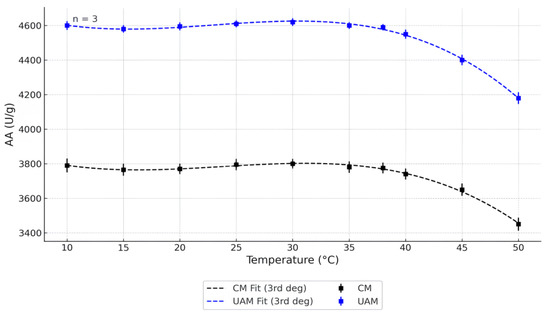
Figure 1.
Effect of temperature on AA (pH = 6.0) obtained by the CM and UAM. Mean values ± SD (n = 3). CM Fit and UAM Fit represent third-degree polynomial fits.
In previous studies, the thermal behavior of α-amylase varied depending on the source organism. For example, Dutta et al. [36] reported that α-amylase extracted from the freshwater zooplankton Heliodiaptomus viduus exhibited optimal catalytic activity at 30 °C and pH 6.0. Notably, the enzyme retained substantial activity up to 70 °C, remaining stable for 2 h at 60 °C and 1 h at 70 °C. In contrast, Fadeel et al. [37] found that α-amylase secretion was significantly inhibited at low temperatures (≤15 °C), with an optimal secretion range between 19 °C and 21 °C. Furthermore, Elmansy et al. [38] identified 45 °C as the optimal temperature for α-amylase production by thermo-halophilic bacterial strains.
Figure 1 presents the temperature-dependent AA of pancreatin extracted via the CM and UAE in the range of 10–50 °C. At nearly all tested temperatures, UAE samples exhibited significantly higher AA than those obtained by CM (p < 0.05), with the exception of 10 °C, where the difference was not statistically significant (p > 0.05). The most pronounced enhancement in AA due to UAE was observed between 30 and 35 °C, where the EA peaked at approximately 4600 U/g, compared to ~3800 U/g in the CM group. This improvement can be attributed to the mechanical and physicochemical effects of acoustic cavitation during ultrasonic treatment. The collapse of cavitation bubbles creates localized zones of high pressure and temperature that disrupt cellular structures and enhance the release of intracellular enzymes. These effects improve mass transfer and increase the exposure of catalytic sites. Additionally, ultrasound may reduce protein aggregation and promote the partial unfolding of enzyme molecules into conformations with higher catalytic efficiency, resulting in improved substrate accessibility and enhanced EA. At temperatures above 38 °C, both CM and UAE samples demonstrated a gradual decline in AA, which likely reflects thermal denaturation of the enzyme. Elevated temperatures can disrupt hydrogen bonding and the tertiary structure, leading to a loss of catalytic function. However, UAE samples retained higher activity than CM samples even at elevated temperatures, suggesting that ultrasound treatment may improve thermal stability, possibly by altering the enzyme’s structural resilience. These temperature-dependent changes were well modeled by third-degree polynomial regressions (R2 > 0.95), indicating a strong fit and supporting the reproducibility of the experimental data.
The outcomes of the two-way ANOVA (Table 1) clearly indicate that both temperature and extraction method had a statistically significant effect on AA, with p-values of 1.00 × 10⁻²⁶ and 1.21 × 10−52, respectively. In contrast, the interaction between these two factors was not significant (p = 0.099), suggesting that temperature and method influenced AA independently rather than interactively. These results confirm that the application of UAM consistently enhances enzyme activity across the tested temperature range.

Table 1.
Two-way ANOVA results for the effect of temperature on AA (pH = 6.0) obtained by the CM and UAM.
To further dissect the differences between experimental conditions, a post hoc Tukey’s Honestly Significant Difference (HSD) test was performed. This analysis revealed multiple significant pairwise differences (p < 0.05) between extraction method–temperature combinations. Notably, samples obtained via UAM displayed significantly higher AA than those obtained by the CM at nearly all tested temperatures (10–50 °C). The most pronounced differences—up to 20%—were recorded in the mid-temperature range (34–38 °C), where EA reached its peak. These differences remained statistically significant after correction for multiple comparisons (adjusted p < 0.001), reinforcing the conclusion that UAM is a more effective technique for preserving and enhancing the EA of pancreatin.
2.1.2. pH and Temperature on AA
The AA was evaluated under varying pH (5.5 to 8.0) and temperature (10–50 °C) conditions. The results for the CM and UAM are presented in Figure 2a,b.
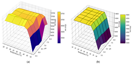
Figure 2.
Effect of pH and temperature on AA using the (a) CM and (b) UAM.
The influence of pH and temperature on AA of pancreatin obtained by the CM is shown in Figure 2a. Across the pH range tested (5.5–8.0), AA remained relatively stable between 10 °C and 38 °C, with activity values fluctuating between approximately 3727 and 3785 U/g. Beyond 38 °C, a gradual decrease in EA was observed, dropping to values between 3387 and 3709 U/g at 50 °C. The highest AA for CM samples was observed at pH 6.0 and 38 °C, registering 3785 U/g. Figure 2b presents corresponding data for pancreatin extracted via UAM. Under the same conditions, UAM samples consistently demonstrated higher AA values than the CM, maintaining stable activity between 4587 and 4606 U/g up to 38 °C. At temperatures above 38 °C, a progressive decline was recorded, with AA decreasing to approximately 4100–4500 U/g at 50 °C. The peak AA for UAM pancreatin was also observed at pH 6.0 and 38 °C, reaching 4606 U/g. Overall, these results highlight the enhanced efficacy of UAM in preserving AA across a range of physiologically relevant pH and temperature conditions, underscoring its potential advantage over the CM.
2.2. Effect of pH and Temperature on PA
2.2.1. Temperature on PA
PA serves as a critical measure of the functional potential of enzyme preparations, particularly those enriched with pancreatic proteases such as trypsin and chymotrypsin. This parameter reflects the enzymes’ capacity to hydrolyze protein substrates into peptides and free amino acids, a process essential in diverse fields including food processing, biotechnology, and pharmaceutical manufacturing. Furthermore, understanding how environmental factors like pH and temperature influence PA is crucial for optimizing extraction protocols and maintaining the quality and consistency of enzyme products [39,40,41].
The experimental results are summarized in Figure 3.
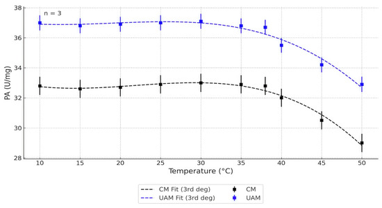
Figure 3.
Effect of temperature on the PA (pH = 6.0) obtained by the CM and UAM. Data are presented as means ± SD, n = 3. CM Fit and UAM Fit represent third-degree polynomial fits.
Majeed et al. [42] reported that protease derived from Bacillus subtilis BSP exhibited maximal catalytic activity at 50 °C and pH 8.0. The enzyme retained approximately 73% of its activity after 1 h at 70 °C, although exposure to 80 °C led to an 84% reduction. Similarly, Suleiman et al. [43] demonstrated that Geobacillus thermoglucosidasius SKF4 showed optimal growth and protease synthesis between 60 and 65 °C, with peak activity at 60 °C. In another study, Sarker et al. [44] found that protease from a halo-tolerant strain of Bacillus subtilis Rand maintained full activity after 30 min at 37–55 °C and retained 80% activity following incubation at 60 °C.
In the present study, the effect of temperature on PA of bovine pancreatin was evaluated using both the CM and UAM across a temperature range of 10–50 °C (Figure 3). The results clearly show that pancreatin obtained via UAM exhibited consistently higher PA values compared to the CM at all tested temperatures. The highest PA in UAM samples was recorded between 30 and 35 °C, after which a gradual decline was observed with increasing temperature, likely due to the partial thermal inactivation of proteases. In contrast, CM samples showed a lower peak and more pronounced loss of activity at elevated temperatures.
Third-degree polynomial regression models (R2 > 0.95) accurately described the temperature-dependent PA trends for both extraction methods. Standard deviations (n = 3) are shown as error bars. These findings align with previous results for AA and confirm that UAM enhances both the efficiency and thermal stability of proteolytic enzyme extraction from bovine pancreatic tissue.
To assess the influence of temperature and extraction method on PA, a two-way analysis of variance (ANOVA) was conducted. As shown in Table 2, both temperature (p = 2.30 × 10⁻¹⁶) and extraction method (p = 9.52 × 10⁻²⁸) had statistically significant effects on PA. However, the interaction between these two factors was not significant (p = 0.85), indicating that temperature and method influenced PA independently. The highest activity was observed in the 34–38 °C range for both extraction methods, though UAM consistently produced greater PA values across the full temperature spectrum. At elevated temperatures (≥40 °C), a marked reduction in PA was observed, likely reflecting partial thermal denaturation of the enzyme complex. These results suggest that UAM is more effective than the CM in both preserving and enhancing PA, with its advantage being remaining stable under varying thermal conditions.
Table 2.
Results of two-way ANOVA for the effect of temperature on PA (pH = 6.0) obtained by the CM and UAM.
To further explore differences among groups, Tukey’s Honestly Significant Difference (HSD) post hoc test was applied following ANOVA. The analysis revealed that UAM samples exhibited significantly higher PA values than CM samples at nearly all tested temperatures (adjusted p < 0.01). The most notable differences were recorded between 34 °C and 38 °C, where UAM-treated samples demonstrated PA levels approximately 10–15% higher than those of the CM. Although overall activity declined at temperatures above 40 °C for both methods, UAM continued to provide significantly higher PA, underscoring its consistent and robust effect on enzyme recovery. These findings validate UAM as a superior extraction approach for maintaining proteolytic enzyme functionality under thermal stress.
The observed enhancement in both EA and thermal stability of proteolytic enzymes extracted using UAM can be attributed to several structural and biochemical effects induced by ultrasonic cavitation. Ultrasonication generates localized high temperatures and pressures, which may lead to the partial unfolding of enzyme molecules, exposing more active sites and thereby increasing catalytic efficiency [45,46]. Additionally, ultrasound can facilitate the release of more enzymes from the tissue matrix due to mechanical disruption, resulting in a higher concentration of active enzymes in the extract [47]. From a biochemical standpoint, ultrasound treatment may alter the enzyme’s tertiary structure in a way that stabilizes the active conformation, thus enhancing resistance to thermal denaturation [48]. It is also possible that ultrasonication reduces the presence of endogenous protease inhibitors or aggregates that might otherwise impair EA [49]. Together, these effects help to explain the improved performance of UAM-derived pancreatin under varying temperature conditions.
2.2.2. pH and Temperature on PA
The 3D surface plots (Figure 4) illustrate the dependence of EA on temperature and pH for pancreatin samples obtained using the CM and UAM.
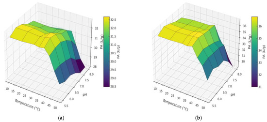
Figure 4.
Effect of pH and temperature on PA using the (a) CM and (b) UAM.
Figure 4 illustrates how PA in bovine pancreatin responds to combined variations in pH and temperature for both extraction methods. In both cases, PA remained relatively stable across the pH range tested at temperatures between 10 °C and 38 °C. However, a noticeable decline in EA was observed at higher temperatures (42–50 °C), particularly under alkaline conditions. For the CM (Figure 4a), the highest activity was recorded between pH 6.0 and 6.5, and at temperatures ranging from 30 °C to 38 °C. Beyond 42 °C, EA dropped significantly, likely due to partial thermal denaturation of the protease complex. In contrast, samples obtained using UAM (Figure 4b) consistently exhibited higher PA across all temperature and pH combinations. The highest value (~36.9 U/g) was observed at pH 6.0 and 38 °C. Importantly, even at elevated temperatures (46–50 °C), UAM-derived pancreatin maintained substantial activity levels, indicating improved thermal resistance. These results further confirm the beneficial effect of ultrasound in stabilizing proteolytic enzymes during extraction. This improvement may be attributed to more efficient enzyme release, reduced aggregation, and greater preservation of the native protein structure under processing conditions.
The observed decline in EA at elevated temperatures and alkaline pH levels can be explained by fundamental principles of enzyme structure and stability. Enzymes are globular proteins whose catalytic function depends on their three-dimensional conformation. At higher temperatures, especially beyond the optimal range (e.g., >40 °C), thermal denaturation may occur, disrupting the hydrogen bonds and hydrophobic interactions that maintain the enzyme’s active conformation, thus reducing activity. Similarly, deviation from the enzyme’s optimal pH can alter the ionization state of amino acid residues at the active site. Under increasingly alkaline conditions, essential acidic or basic residues may lose their proper charge, impairing substrate binding or catalytic turnover. These structural disruptions result in a reduction in or complete loss of EA [50,51].
The decline in PA observed at temperatures above the optimum may be attributed to partial thermal denaturation of the protease complex. Elevated temperatures can disrupt the secondary and tertiary structures of enzymes, particularly affecting hydrogen bonding and hydrophobic interactions critical for maintaining the integrity of the active site. This partial denaturation does not necessarily result in the complete loss of structure but can significantly impair the catalytic function of specific protease subunits. Such mechanistic insight aligns with the typical thermal behavior of serine proteases, which are known to be sensitive to conformational changes under thermal stress [52,53].
To better understand the observed patterns, this explanation can be extended to other experimental conditions. For example, the sharp decline in activity under alkaline pH likely reflects a similar disruption in the ionization state of catalytic residues, affecting charge–charge interactions and electrostatic stability of the enzyme complex. Together, these findings underscore the delicate balance between structural integrity and environmental conditions in determining enzymatic efficiency.
As summarized in Table 3, both extraction approaches—CM and UAM—demonstrated peak EA at pH 6.0 and 38 °C. These conditions can thus be considered optimal for maximizing the functional performance of bovine pancreatin, irrespective of the extraction strategy. However, the efficiency of enzyme recovery differed markedly between methods. Under identical conditions, the AA obtained via UAM reached 4606 U/g, compared to 3785 U/g for the CM. Similarly, PA increased from 32.8 U/g in CM samples to 36.9 U/g in UAM samples. These results underscore the superior performance of UAM in enhancing both amylolytic and proteolytic functions of pancreatin. The improvement is likely associated with more efficient disruption of tissue structures, better enzyme release, and enhanced preservation of native enzyme conformation during processing. Overall, UAM appears to be a more effective and robust extraction method, particularly in applications where high EA and thermal stability are critical for product functionality.
Table 3.
Optimal pH and temperature for bovine pancreas extraction using the CM and UAM.
3. Materials and Methods
3.1. Materials and Chemicals
Fresh bovine pancreases (n = 27; total mass of 10.0 kg) were obtained from a certified meat supplier, S-Meat LLP (Almaty Region, Kazakhstan). Upon collection, the tissues were immediately stored in a portable automotive freezer (XMSJ, Shenzhen, China) at a controlled temperature of −5 to −7 °C to preserve enzymatic integrity prior to extraction. All analytical-grade reagents and chemicals used in this study, including buffers and substrates for enzymatic assays, were supplied by Laborpharma LLP (Almaty, Kazakhstan).
3.2. Sample Preparation
Proteolytic enzyme preparations were obtained using a CM, adapted from the procedure described in patent [54]. Freshly minced bovine pancreas (10 kg) was homogenized with 15 L of distilled water containing 75 mL of glacial acetic acid. The mixture was stirred continuously at 8–12 °C for 4 h, after which the liquid phase was separated from the solid tissue residue.
The remaining solid material was subjected to a second extraction step using an additional 5 L of the same acetic acid solution. After 30 min of mixing, the extract was collected again. The combined extracts were enriched with 0.5 mol of calcium chloride (CaCl2) and supplemented with pancreatin at a concentration of 0.1 g/L. The pH of the mixture was adjusted to 8.1 using a 20% NaOH solution, and the enzymatic activation was carried out over 24 h at 0–5 °C.
Following activation, the pH was reduced to 6.0 using 5 N hydrochloric acid. Insoluble materials were removed by centrifugation using a separator. Enzymes were precipitated from the resulting supernatant with acetone, dried in a vacuum oven at 30–35 °C, and then finely ground and sieved to obtain a uniform powder.
For UAM, the above protocol was modified by incorporating an ultrasonic homogenization step. A Sonopuls HD 2200 ultrasonic processor (Bandelin electronic GmbH & Co. KG, Berlin, Germany) operating at 200 W was applied in a pulsed mode (1 s on/1 s off), enhancing cell disruption and accelerating enzyme release from the tissue matrix.
The system operated at a frequency of 20 kHz and was equipped with an MS 72 probe (tip diameter: 13 mm). The total sonication time was 15 min, with the probe immersed 1.5 cm into the sample solution. To prevent the thermal degradation of enzymes, the temperature was maintained below 25 °C using an external ice bath. The pulsed ultrasound mode (1 s on/1 s off) was employed to minimize thermal accumulation and enzyme denaturation while maintaining cavitation efficiency. The off cycles allowed the system to dissipate heat and facilitated bubble regeneration, thereby enhancing the intensity of cavitation during each subsequent pulse. This approach helped preserve EA while improving cell disruption efficiency. Although ultrasound treatment may induce local temperature increases, all extractions were conducted with the sample vessel immersed in an ice-cold water bath to limit thermal accumulation. Temperature was monitored during sonication and did not exceed 25 °C. Furthermore, enzymatic activity assays were performed independently under thermally controlled conditions using a water bath thermostat, which ensured that the temperature-dependent effects observed in the study were solely due to assay temperature and not influenced by prior ultrasonic exposure (Figure 5).
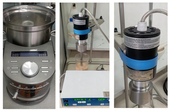
Figure 5.
Ultrasonic homogenizer (BANDELIN Sonopuls HD 2200, MS 72 probe) used for UAM.
3.3. Thawing Loss
After thawing at 2 °C for 24 h, each sample was gently blotted with filter paper to remove surface moisture. The sample weight was recorded before (m1) and after (m2) thawing. Thawing loss (%) was calculated using the following formula:
3.4. Determination of AA
All measurements, data processing, and result interpretations were carried out by qualified specialists from the Kazakh Research Institute of Processing and Food Industry (KazRIPFI, Kazakhstan), who have extensive experience in enzymatic assay methodology. The AA of the pancreatin samples was determined in accordance with GOST 34440-2018, “Enzyme preparations for the food industry. Methods for determination of amylolytic activity” [55]. This method is based on the enzymatic hydrolysis of soluble starch into dextrins of varying molecular weight by the amylolytic enzyme complex, under standardized conditions.
The hydrolysis reaction was conducted over 10 min using 6.0 units of EA under controlled pH and temperature conditions. One unit of AA was defined as the amount of enzyme required to hydrolyze 1 g of soluble starch into dextrins, corresponding to 30–50% degradation of the initial starch mass. The final EA was expressed in units per gram of dry enzyme preparation (AA/g).
The extent of starch hydrolysis was assessed by a colorimetric method, based on the reduction in iodine staining intensity, which reflects the decrease in the concentration of unhydrolyzed starch. Measurements were performed using a Cary 60 UV–Vis spectrophotometer (Agilent Technologies, Santa Clara, CA, USA) equipped with Cary WinUV software, version 5.2., operating in the spectral range of 190–1100 nm.
Reagents used in the assay included soluble starch, sodium acetate trihydrate, acetic acid, disodium phosphate, monopotassium phosphate, hydrochloric acid, and crystalline iodine. The thermostability of the AA was subsequently evaluated and expressed as enzyme units per gram, using the following equation:
where and are empirical coefficients obtained from a regression analysis of hydrolyzed starch mass versus enzyme mass per hour of enzymatic action; —degree of starch hydrolysis; —enzyme sample mass used in the analysis, g (adjusted for dilution); —density of the enzyme preparation (for liquid form), g/cm3.
Calculations were performed with one decimal precision and then rounded to the nearest whole number, as the AA values exceeded 100 U/g. Each reported result represents the average of two independent measurements carried out under repeatability conditions. The measurements met the established acceptability criteria, with the relative error maintained within ±7%, ensuring the reliability and consistency of the data.
3.5. Determination of PA
PA was assessed following the procedure outlined in GOST 34443-2018, “Enzyme Preparations for the Food Industry. Method for Determining Proteolytic Activity” [56]. This method is based on the enzymatic hydrolysis of bovine hemoglobin, a natural protein substrate, under different pH conditions (acidic pH 3.0, mildly acidic pH 5.3, neutral pH 7.0, and alkaline pH 9.0). The enzyme preparation catalyzes the breakdown of hemoglobin into smaller peptides and free amino acids. The reaction was stopped by precipitating the remaining intact protein with trichloroacetic acid (TCA), and the resulting peptides and amino acids were quantified.
One unit of PA corresponds to the amount of enzyme that releases peptides equivalent to 1 µmol of tyrosine per minute at 30 °C (where 1 µmol of tyrosine equals 0.181 mg). Enzyme activity was expressed as units of PA per gram of sample (U/g).
The concentration of hydrolyzed protein was determined by reacting the free amino groups with Folin’s reagent, producing a blue complex whose absorbance was measured at 670 nm using a colorimeter.
The reagents used in this assay included lyophilized bovine hemoglobin, tyrosine, Folin’s reagent, trichloroacetic acid, hydrochloric acid, orthophosphoric acid, glacial acetic acid, boric acid, urea, sodium carbonate, and sodium hydroxide.
PA values were calculated using the following formula:
where —optical density; 4—dilution factor after TCA addition; —tyrosine equivalent corresponding to the optical density of 1 µmol of tyrosine (determined from calibration curve); —hydrolysis time, min; —mass of the enzyme preparation used for analysis (based on a 1 cm3 working solution), g; —density of the enzyme preparation, g/cm3.
Calculations were carried out to two decimal places and rounded to one decimal place, as the PA was below 100 PA/g. The final result was reported as the arithmetic mean of two parallel measurements under repeatability conditions.
3.6. pH and Color Measurement
The pH of the samples was measured following a modified version of the method described previously [57]. Briefly, a 10 g aliquot of each sample was homogenized with 20 mL of distilled water using a digital homogenizer (S-10, Stegler, Hangzhou, China) at 1000 rpm for 15 s. The pH of the resulting homogenate was then recorded using a calibrated pH meter (F20-Std-Kit, Mettler-Toledo, Shanghai, China).
3.7. Statistical Analysis
All experiments were performed in triplicate, and the results are expressed as mean ± SD. Statistical significance of the main effects (p < 0.05) was assessed by ANOVA. Post hoc multiple comparisons were conducted using Tukey’s HSD test with adjusted p-values. To model the relationship between enzymatic activity and temperature, a second-order polynomial regression model was applied, described by the following equation:
where Y—represents the enzymatic activity, U/g; T—temperature, °C; a, b, c—regression coefficients determined by the least squares method.
For samples obtained via the CM, the regression equation
For samples obtained via UAM, the equation
4. Conclusions
This study provides new insights into the influence of extraction methods on the enzymatic functionality of bovine pancreatin, specifically α-amylase and protease, under variable pH and temperature conditions. The novelty of this work lies in the application of UAM to animal-derived enzyme systems and in the systematic comparison of its effects on EA relative to the CM, under conditions simulating those used in food and bioprocessing protocols. While promising, these findings should be considered preliminary, and further studies are needed to assess structural stability and confirm potential industrial relevance. Our results demonstrate that UAM significantly improves both the activity and thermal stability of pancreatin enzymes compared to the CM. Across a wide temperature range (10–50 °C) and pH spectrum (5.5–8.0), enzymes extracted via UAM consistently exhibited higher activity and greater resistance to denaturation. Notably, optimal EA for both α-amylase and protease was achieved at pH 6.0 and 38 °C, with UAM-extracted samples maintaining superior performance even at elevated temperatures up to 50 °C. These findings suggest that ultrasound enhances enzyme release and protects structural integrity during extraction, making it a promising strategy for the production of robust, high-activity enzyme preparations. The results contribute to the growing body of knowledge supporting ultrasound-based bioprocessing as an efficient and scalable alternative to traditional methods.
The superior EA observed in samples obtained via UAM can be mechanistically attributed to several synergistic effects induced by ultrasonic cavitation. First, ultrasound facilitates enhanced cell disruption, leading to the more efficient release of intracellular enzymes from pancreatic tissues. Second, ultrasonic energy helps reduce protein–protein aggregation, which otherwise may hinder enzyme solubility and activity. Finally, moderate ultrasonic treatment may stabilize the native conformation of enzyme molecules by preventing excessive unfolding or degradation, thereby preserving their catalytic functionality under subsequent processing conditions.
Although functional assays indicated preserved activity and thermal stability of enzymes after ultrasound-assisted extraction, the current study lacks direct structural evidence (e.g., CD spectroscopy, DSC, or molecular conformation studies) to confirm protein integrity. Future research should include such analyses to more conclusively determine the effects of ultrasound on the tertiary and quaternary structure of extracted enzymes.
Future research should further explore the molecular mechanisms underlying the stabilizing effects of ultrasound, as well as optimize acoustic parameters to maximize enzyme yield and activity.
Author Contributions
Conceptualization, G.K. and U.C.; methodology, U.C.; software, G.K. and A.T.; formal analysis, G.K. and A.T.; investigation, G.K., U.C. and A.T.; resources, G.K.; data curation, A.T.; supervision, G.K. and U.C.; project administration, G.K.; funding acquisition, G.K. and U.C. All authors have read and agreed to the published version of the manuscript.
Funding
This research was supported by the project “Development of a highly efficient technology for obtaining pancreatin for the purpose of producing export-oriented products”, scientific and technical program for 2024–2026 “Development of a technology for complex and deep processing of agricultural raw materials for food production, ensuring high quality and safety of manufactured products” BR24892775, funded by the Ministry of Agriculture of the Republic of Kazakhstan.
Informed Consent Statement
Informed consent was obtained from all subjects involved in the study.
Data Availability Statement
The data presented in this study are available on request from the corresponding author.
Conflicts of Interest
The authors declare no conflicts of interest.
Ethical Statement
The research did not involve any experiments on human subjects. Bovine pancreatic glands were sourced from animals slaughtered for food production at authorized facilities. The use of this animal-derived material adhered to all applicable ethical standards and institutional regulations. No animals were specifically killed for the purpose of this investigation.
References
- Ahmad, I.Z.; Tabassum, H.; Ahmad, A.; Kuddus, M. Food enzymes in pharmaceutical industry: Perspectives and limitations. In Enzymes in Food Technology; Kuddus, M., Ed.; Springer: Singapore, 2018; pp. 41–62. [Google Scholar] [CrossRef]
- Whitcomb, D.C.; Lowe, M.E. Human pancreatic digestive enzymes. Dig. Dis. Sci. 2007, 52, 1–17. [Google Scholar] [CrossRef]
- Gupta, R.; Beg, Q.K.; Lorenz, P. Bacterial alkaline proteases: Molecular approaches and industrial applications. Appl. Microbiol. Biotechnol. 2002, 59, 15–32. [Google Scholar] [CrossRef]
- Motta, J.F.G.; de Freitas, B.C.B.; Almeida, A.F.; Martins, G. Use of enzymes in the food industry: A review. Food Sci. Technol. 2023, 43, e106222. [Google Scholar] [CrossRef]
- Kumar, A.; Dhiman, S.; Dhewa, T. Microbial enzymes and major applications in the food industry: A concise review. Food Prod. Process. Nutr. 2024, 6, 85. [Google Scholar] [CrossRef]
- Vieira, I.R.S.; Conte-Junior, C.A. Dietary bioactive compounds and human health: The role of bioavailability. Nutrients 2025, 17, 48. [Google Scholar] [CrossRef]
- Rawlings, N.D.; Salvesen, G. Handbook of Proteolytic Enzymes, 3rd ed.; Academic Press: London, UK, 2013. [Google Scholar]
- Pandey, R.; Negi, S. Enzymatic hydrolysis of starch, protein, and fat: Structure, function and applications. In Enzymes in Food Biotechnology; Elsevier: Amsterdam, The Netherlands, 2019; pp. 175–206. [Google Scholar]
- Zakowiecki, D.; Edinger, P.; Hess, T.; Paszkowska, J.; Staniszewska, M.; Romanova, S.; Garbacz, G. Effect of compaction pressure on the enzymatic activity of pancreatin in directly compressible formulations. Pharmaceutics 2023, 15, 2224. [Google Scholar] [CrossRef] [PubMed]
- Hartmann, S.; Rydzewska, G.; Domínguez-Muñoz, J.E. Kreon® (Creon®) vs. Lipancrea®: In Vitro Comparison of Two Encapsulated Pancreatin Preparations. Pharmaceuticals 2022, 15, 1570. [Google Scholar] [CrossRef]
- Legg, E.F.; Spencer, A.M. Studies on the stability of pancreatic enzymes in duodenal fluid to storage temperature and pH. Clin. Chim. Acta 1975, 65, 175–179. [Google Scholar] [CrossRef] [PubMed]
- Fernando, I.T.; Perera, K.I.; Athauda, S.B.P.; Sivakanesan, R.; Kumar, N.S.; Jayasinghe, L. Heat stability of the in vitro inhibitory effect of spices on lipase, amylase, and glucosidase enzymes. Food Sci. Nutr. 2019, 7, 425–432. [Google Scholar] [CrossRef] [PubMed Central]
- Tessier, A.G.; Dombi, G.W.; Bouwman, D.L. Thermostability of purified human pancreatic α-amylase is increased by calcium and human serum albumin. Biochim. Biophys. Acta 1996, 1292, 71–78. [Google Scholar] [CrossRef]
- Sabadini, G.; Mellado, M.; Morales, C.; Mella, J. Arylamines QSAR-Based Design and Molecular Dynamics of New Phenylthiophene and Benzimidazole Derivatives with Affinity for the C111, Y268, and H73 Sites of SARS-CoV-2 PLpro Enzyme. Pharmaceuticals 2024, 17, 606. [Google Scholar] [CrossRef]
- de Oliveira, R.L.; de Souza Claudino, E.; Converti, A.; Porto, T.S. Use of a Sequential Fermentation Method for the Production of Aspergillus tamarii URM4634 Protease and a Kinetic/Thermodynamic Study of the Enzyme. Catalysts 2021, 11, 963. [Google Scholar] [CrossRef]
- Yu, P.; Pan, X.; Chen, M.; Ma, J.; Xu, B.; Zhao, Y. Ultrasound-assisted enzymatic extraction of soluble dietary fiber from Hericium erinaceus and its in vitro lipid-lowering effect. Food Chem. 2023, 6, 100545. [Google Scholar] [CrossRef]
- Peng, P.; Yu, H.; Xian, M.; Qu, C.; Guo, Z.; Li, S.; Zhu, Z.; Xiao, J. Preparation of acetylcholinesterase inhibitory peptides from yellowfin tuna pancreas using moderate ultrasound-assisted enzymatic hydrolysis. Mar. Drugs 2023, 21, 75. [Google Scholar] [CrossRef]
- Zhang, T.; Chen, H.; Wang, Y.; Wang, J. Effects of particle size on the extraction and functional properties of enzymes from animal by-products. Food Bioprod. Process. 2019, 117, 1352. [Google Scholar] [CrossRef]
- Li, M.; Liang, Q.; Zhang, Y.; Jiang, X.; Gu, Y.; Song, X.; Wang, X.; Shi, W. Screening of Potential Angiotensin-Converting Enzyme-Inhibitory Peptides in Squid (Todarodes pacificus) Skin Hydrolysates: Preliminary Study of Its Mechanism of Inhibition. Mar. Drugs 2025, 23, 81. [Google Scholar] [CrossRef]
- Xu, K.; Fu, H.; Chen, Q.; Sun, R.; Li, R.; Zhao, X.; Zhou, J.; Wang, X. Engineering thermostability of industrial enzymes for enhanced application performance. Int. J. Biol. Macromol. 2025, 291, 139067. [Google Scholar] [CrossRef]
- Montuori, E.; Martinez, K.A.; De Luca, D.; Ianora, A.; Lauritano, C. Transcriptome Sequencing of the Diatom Asterionellopsis thurstonii and In Silico Identification of Enzymes Potentially Involved in the Synthesis of Bioactive Molecules. Mar. Drugs 2023, 21, 126. [Google Scholar] [CrossRef]
- Razieh, A.; Abdol-Khalegh, B.; Douglas, V.L.; Ahmad, R.K.; Iraj, M.B. Thermal stability and enzymatic activity of RNase A in the presence of cationic gemini surfactants. Int. J. Biol. Macromol. 2012, 50, 1151–1157. [Google Scholar] [CrossRef]
- Abdullahi, N.; Atiku, M.K.; Umar, N.B. The roles of enzyme in food processing–an overview. Fudma J. Sci. 2021, 5, 157–164. [Google Scholar] [CrossRef]
- Song, P.; Zhang, X.; Wang, S.; Xu, W.; Wang, F.; Fu, R.; Wei, F. Microbial proteases and their applications. Front. Microbiol. 2023, 14, 1236368. [Google Scholar] [CrossRef] [PubMed]
- Chemat, F.; Zill-e-Huma; Khan, M.K. Applications of ultrasound in food technology: Processing, preservation and extraction. Ultrason. Sonochem. 2011, 18, 813–835. [Google Scholar] [CrossRef] [PubMed]
- Cornish-Bowden, A. Fundamentals of Enzyme Kinetics, 4th ed.; Wiley-Blackwell: Weinheim, Germany, 2012. [Google Scholar]
- Vera-Salgado, J.; Calderón-Chiu, C.; Calderón-Santoyo, M.; Barros-Castillo, J.C.; López-García, U.M.; Ragazzo-Sánchez, J.A. Ultrasound-Assisted Extraction of Artocarpus heterophyllus L. Leaf Protein Concentrate: Solubility, Foaming, Emulsifying, and Antioxidant Properties of Protein Hydrolysates. Colloids Interfaces 2022, 6, 50. [Google Scholar] [CrossRef]
- Kęska, P.; Wójciak, K.M.; Stasiak, D.M. Influence of Sonication and Taraxacum officinale Addition on the Antioxidant and Anti-ACE Activity of Protein Extracts from Sous vide Beef Marinated with Sour Milk and after In Vitro Digestion. Molecules 2020, 25, 4692. [Google Scholar] [CrossRef] [PubMed]
- Carreira-Casais, A.; Otero, P.; Garcia-Perez, P.; Garcia-Oliveira, P.; Pereira, A.G.; Carpena, M.; Soria-Lopez, A.; Simal-Gandara, J.; Prieto, M.A. Benefits and Drawbacks of Ultrasound-Assisted Extraction for the Recovery of Bioactive Compounds from Marine Algae. Int. J. Environ. Res. Public Health 2021, 18, 9153. [Google Scholar] [CrossRef]
- Hussain, M.; Qayum, A.; Zhang, X.; Hao, X.; Liu, L.; Wang, Y.; Hussain, K.; Li, X. Improvement in bioactive, functional, structural and digestibility of potato protein and its fraction patatin via ultra-sonication. LWT 2021, 148, 111747. [Google Scholar] [CrossRef]
- Miljanović, A.; Bielen, A.; Grbin, D.; Marijanović, Z.; Andlar, M.; Rezić, T.; Roca, S.; Jerković, I.; Vikić-Topić, D.; Dent, M. Effect of Enzymatic, Ultrasound, and Reflux Extraction Pretreatments on the Yield and Chemical Composition of Essential Oils. Molecules 2020, 25, 4818. [Google Scholar] [CrossRef]
- Iqbal, A.; Murtaza, A.; Marszałek, K.; Iqbal, M.A.; Chughtai, M.F.J.; Hu, W.; Barba, F.J.; Bi, J.; Liu, X.; Xu, X. Inactivation and structural changes of polyphenol oxidase in quince (Cydonia oblonga Miller) juice subjected to ultrasonic treatment. J. Sci. Food Agric. 2020, 100, 2065–2073. [Google Scholar] [CrossRef]
- Vinatoru, M.; Mason, T.J.; Calinescu, I. Ultrasonically assisted extraction (UAE) and microwave assisted extraction (MAE) of functional compounds from plant materials. Trends Anal. Chem. 2017, 97, 159–178. [Google Scholar] [CrossRef]
- Liao, F.-Y.; Su, Y.-L.; Weng, J.-R.; Lin, Y.-C.; Feng, C.-H. Ultrasound–Vortex-Assisted Dispersive Liquid–Liquid Microextraction Combined with High Performance Liquid Chromatography–Diode Array Detection for Determining UV Filters in Cosmetics and the Human Stratum Corneum. Molecules 2020, 25, 4642. [Google Scholar] [CrossRef]
- Qin, L.; Porfyrakis, K.; Tzanakis, I.; Grobert, N.; Eskin, D.G.; Fezzaa, K.; Mi, J. Multiscale interactions of liquid, bubbles and solid phases in ultrasonic fields revealed by multiphysics modelling and ultrafast X-ray imaging. Ultrason Sonochem. 2022, 89, 106158. [Google Scholar] [CrossRef]
- Dutta, T.K.; Jana, M.; Pahari, P.R.; Bhattacharya, T. The effect of temperature, pH, and salt on amylase in Heliodiaptomus viduus (Gurney) (Crustacea: Copepoda: Calanoida). Turk. J. Zool. 2006, 30, 167–172. Available online: https://journals.tubitak.gov.tr/zoology/vol30/iss2/11 (accessed on 4 July 2025).
- Fadeel, A.; Moll, B.A.; Jones, R.L. Effect of temperature on the synthesis and secretion of α-amylase in barley aleurone layers. Plant Physiol. 1980, 66, 466–470. Available online: https://www.ncbi.nlm.nih.gov/pmc/articles/PMC440655/ (accessed on 4 July 2025). [CrossRef] [PubMed]
- Elmansy, E.A.; Asker, M.S.; El-Kady, E.M.; Sholkamy, E.N.; Awad, M.F. Production and optimization of α-amylase from thermo-halophilic bacteria isolated from different local marine environments. Bull. Natl. Res. Cent. 2018, 42, 31. Available online: https://bnrc.springeropen.com/articles/10.1186/s42269-018-0033-2 (accessed on 4 July 2025). [CrossRef]
- Minekus, M.; Alminger, M.; Alvito, P.; Ballance, S.; Bohn, T.; Bourlieu, C.; Carrière, F.; Boutrou, R.; Corredig, M.; Dupont, D.; et al. A standardised static in vitro digestion method suitable for food—An international consensus. Food Funct. 2014, 5, 1113–1124. [Google Scholar] [CrossRef]
- Vallés, D.; Furtado, S.; Villadóniga, C.; Cantera, A.M.B. Adsorption onto alumina and stabilization of cysteine proteinases from crude extract of solanum granuloso-leprosum fruits. Process Biochem. 2011, 46, 592–598. [Google Scholar] [CrossRef]
- Sango, D.M.; Abela, D.; McElhatton, A.; Valdramidis, V.P. Assisted ultrasound applications for the production of safe foods. J. Appl. Microbiol. 2014, 116, 1067–1083. [Google Scholar] [CrossRef] [PubMed]
- Majeed, T.; Lee, C.C.; Orts, W.J.; Tabassum, R.; Shah, T.A.; Jardan, Y.A.B.; Dawoud, T.M.; Bourhia, M. Characterization of a thermostable protease from Bacillus subtilis BSP strain. BMC Biotechnol. 2024, 24, 49. [Google Scholar] [CrossRef] [PubMed]
- Suleiman, A.D.; Abdul Rahman, N.; Mohd Yusof, H.; Mohd Shariff, F.; Yasid, N.A. Effect of cultural conditions on protease production by a thermophilic Geobacillus thermoglucosidasius SKF4 isolated from Sungai Klah Hot Spring Park, Malaysia. Molecules 2020, 25, 2609. [Google Scholar] [CrossRef]
- Sarker, P.K.; Talukdar, S.A.; Deb, P.; Sayem, S.A.; Mohsina, K. Optimization and partial characterization of culture conditions for the production of alkaline protease from Bacillus licheniformis P003. SpringerPlus 2013, 2, 506. [Google Scholar] [CrossRef]
- Qian, J.; Chen, D.; Zhang, Y.; Gao, X.; Xu, L.; Guan, G.; Wang, F. Ultrasound-Assisted Enzymatic Protein Hydrolysis in Food Processing: Mechanism and Parameters. Foods 2023, 12, 4027. [Google Scholar] [CrossRef]
- Soares, A.d.S.; Duarte Augusto, P.E.; de Castro Leite Junior, B.R.; Nogueira, C.A.; Rufino Vieira, E.N.; Ribeiro de Barros, F.A.; Stringheta, P.C.; Ramos, A.M. Ultrasound assisted enzymatic hydrolysis of sucrose catalyzed by invertase: Investigation on substrate, enzyme and kinetics parameters. LWT Food Sci. Technol. 2019, 107, 164–170. [Google Scholar] [CrossRef]
- Rathnakumar, K.; Kalaivendan, R.G.T.; Eazhumalai, G.; Raja Charles, A.P.; Verma, P.; Rustagi, S.; Bharti, S.; Kothakota, A.; Siddiqui, S.A.; Manuel Lorenzo, J.; et al. Applications of ultrasonication on food enzyme inactivation—Recent review report (2017–2022). Ultrason. Sonochem. 2023, 96, 106407. [Google Scholar] [CrossRef]
- Maric, M.; Grassino, A.N.; Zhu, Z.; Barba, F.J.; Brncic, M.; Brncic, S.R. An overview of the traditional and innovative approaches for pectin extraction from plant food wastes and by-products: Ultrasound-, microwaves-, and enzyme-assisted extraction. Trends Food Sci. Technol. 2018, 76, 28–37. [Google Scholar] [CrossRef]
- Tao, Y.; Sun, D.-W. Enhancement of Food Processes by Ultrasound: A Review. Crit. Rev. Food Sci. Nutr. 2015, 55, 570–594. [Google Scholar] [CrossRef] [PubMed]
- Lehninger, A.L.; Nelson, D.L.; Cox, M.M. Principles of Biochemistry, 7th ed.; W.H. Freeman: New York, NY, USA, 2017. [Google Scholar]
- Thi Thu Tra, T.; Khanh Tien, N.; Van Viet Man, L. Effects of ultrasonication variables on the activity and properties of alpha amylase preparation. Biotechnol. Prog. 2018, 34, 702–710. [Google Scholar] [CrossRef] [PubMed]
- Rao, M.B.; Tanksale, A.M.; Ghatge, M.S.; Deshpande, V.V. Molecular and Biotechnological Aspects of Microbial Proteases. Microbiol. Mol. Biol. Rev. 1998, 62, 597–635. Available online: https://www.scirp.org/reference/referencespapers?referenceid=939650 (accessed on 4 July 2025). [CrossRef]
- Li, J.; Sun, C.; Yue, X.; Ma, W.; Wang, Y.; Zhao, J.; Zhu, G.; Bai, Y. Ultrasound-assisted immersion freezing improves the digestion properties of beef myofibrillar protein. Food Chem. X 2024, 22, 102144. [Google Scholar] [CrossRef]
- Patent No. SU651810. Method for Obtaining Pancreatin. USSR; Filed 1979 Mar 15. Russian. Available online: https://patentscope.wipo.int/search/ru/detail.jsf?docId=SU28775124 (accessed on 4 July 2025).
- GOST 34440-2018; Enzyme Preparations for Food Industry. Methods for the Determination of Amylase Activity. Standartinform: Moscow, Russia, 2018; p. 19. Available online: https://allgosts.ru/07/100/gost_34440-2018 (accessed on 4 July 2025).
- GOST 34430-2018; Enzyme Preparations for Food Industry. Methods for the Determination of Protease Activity. Standartinform: Moscow, Russia, 2018; p. 15. Available online: https://meganorm.ru/Index2/1/4293735/4293735117.htm (accessed on 4 July 2025).
- Yuan, D.; Xu, Y.; Kong, B.; Cao, C.; Zhang, F.; Xia, X.; Zhang, H.; Liu, Q.; Zhao, J. Application of seaweed dietary fiber as a potential alternative to phosphates in frankfurters with healthier profiles. Meat Sci. 2023, 196, 109044. [Google Scholar] [CrossRef]


Disclaimer/Publisher’s Note: The statements, opinions and data contained in all publications are solely those of the individual author(s) and contributor(s) and not of MDPI and/or the editor(s). MDPI and/or the editor(s) disclaim responsibility for any injury to people or property resulting from any ideas, methods, instructions or products referred to in the content. |
© 2025 by the authors. Licensee MDPI, Basel, Switzerland. This article is an open access article distributed under the terms and conditions of the Creative Commons Attribution (CC BY) license (https://creativecommons.org/licenses/by/4.0/).