Case Report

Blue Rubber Bleb Nevus Syndrome with a Novel PDGFRA Variant and Comorbid Autism Spectrum Disorder: A Case Report

by
Tae Hyeong Kim
1,
Jae Myung Cha
2 and
Sung-Hoon Chung
1,*
1
Department of Pediatrics, Kyung Hee University College of Medicine, Kyung Hee University Hospital at Gangdong, Seoul 05278, Republic of Korea
2
Department of Internal Medicine, Kyung Hee University College of Medicine, Kyung Hee University Hospital at Gangdong, Seoul 05278, Republic of Korea
*
Author to whom correspondence should be addressed.
Children 2026, 13(8), 1009; https://doi.org/10.3390/children13081009
Submission received: 30 June 2026 / Revised: 24 July 2026 / Accepted: 26 July 2026 / Published: 29 July 2026
(This article belongs to the Special Issue Advances in Pediatric Gastroenterology (2nd Edition))

Highlights

What are the main findings?
  • A pediatric patient with BRBNS and severe gastrointestinal bleeding was found to carry a heterozygous PDGFRA variant (c.2075G>T, p.Ser692Ile) of uncertain significance, with no pathogenic variant detected in TEK, PIK3CA, or GNAQ.
  • This patient also had autism spectrum disorder (ASD); whether this co-occurrence reflects a biological connection or is coincidental could not be determined from a single case.
What are the implications of the main findings?
  • A single variant of uncertain significance (VUS), identified from non-lesional tissue in one patient, raises but does not confirm the possibility of genetic contributors to BRBNS beyond TEK.
  • These findings are hypothesis-generating; lesional-tissue sequencing, segregation analysis, and functional studies in additional patients are needed before conclusions about genetic heterogeneity or shared mechanisms with ASD can be drawn.

Abstract
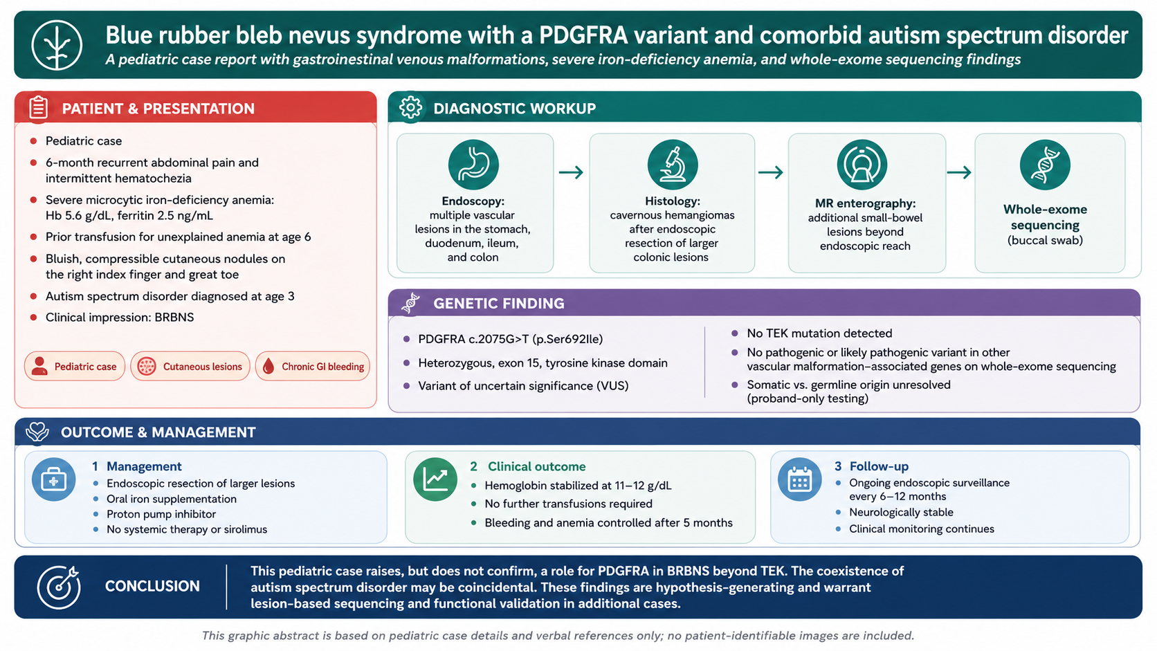
Background: Blue rubber bleb nevus syndrome (BRBNS) is a rare vascular disorder that primarily affects the skin and gastrointestinal tract, driven predominantly by somatic mosaic mutations, most commonly in TEK. Here, we report a case of a child with BRBNS and autism spectrum disorder (ASD). Case presentation: A 13-year-old boy diagnosed with ASD at 3 years presented with recurrent abdominal pain, blood in the stool, and severe anemia persisting for 6 months. At admission, his hemoglobin level was 5.6 g/dL. He had received a transfusion at age 6 for unexplained anemia. On examination, he appeared pale but stable, with bluish, compressible nodules on the right index finger and great toe, typical of BRBNS. Laboratory findings were consistent with chronic bleeding-induced iron deficiency. Endoscopic findings revealed multiple vascular lesions in the stomach, duodenum, ileum, and colon. Several colonic lesions were removed and pathologically confirmed as cavernous hemangiomas. Magnetic resonance enterography revealed additional small intestinal lesions. Whole-exome sequencing performed on buccal swab-derived DNA identified a heterozygous PDGFRA variant (c.2075G>T, p.Ser692Ile) classified as a variant of uncertain significance; no variants were identified in TEK, PIK3CA, or GNAQ. The patient underwent endoscopic resection of the larger lesions and received oral iron and a proton pump inhibitor. Hemoglobin stabilized at 11–12 g/dL, and no further transfusions were required. Conclusions: This case raises, but does not confirm, the possibility that genes other than TEK may contribute to BRBNS. The coexistence of ASD may be coincidental; a mechanistic link remains unproven. Careful endoscopic therapy and medical management controlled bleeding and anemia in this child.

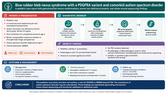
Graphical Abstract

1. Introduction

Blue rubber bleb nevus syndrome (BRBNS) is a rare vascular disorder that typically presents during childhood and most commonly affects the skin and gastrointestinal (GI) tract [1]. BRBNS arises predominantly from somatic mosaic mutations in endothelial signaling genes rather than a single germline cause [2]. Cutaneous lesions typically present as soft, compressible blue nodules, whereas GI involvement can range from asymptomatic to recurrent bleeding [3,4]. In most patients, diagnosis of BRBNS is based primarily on characteristic clinical findings. Endoscopic evaluation is often required, sometimes repeatedly, to detect intestinal lesions [5]. In a landmark study, Soblet et al. reported somatic mutations in TEK, which encodes the endothelial receptor TIE2, in 15 of 17 patients with BRBNS (approximately 88%), confirming the important role of these factors in abnormal angiogenesis [2]. In addition, activating PIK3CA mutations have been found in more than half of patients with sporadic venous malformations, further supporting the idea of genetic heterogeneity in vascular anomalies [6]. However, not all patients carry TEK mutations, suggesting the involvement of additional genetic mechanisms [7]. Platelet-derived growth factor receptor alpha (PDGFRA) encodes a receptor tyrosine kinase involved in angiogenesis and regulation of cell growth [8]. Although PDGFRA mutations are well documented in gastrointestinal stromal tumors (GISTs) and inflammatory fibroid polyps, their role in vascular anomalies such as BRBNS remains poorly understood [9]. Recent studies have observed increased PDGFRA expression in the cerebellum of individuals with autism spectrum disorder (ASD), suggesting broader developmental implications [10,11]. Here, we describe a pediatric patient with BRBNS, a heterozygous PDGFRA variant of uncertain significance, and comorbid ASD, raising the possibility of genetic heterogeneity in BRBNS and a potential overlap between angiogenic and neurodevelopmental pathways, although the variant’s uncertain significance limits any firm conclusion.

2. Detailed Case Description

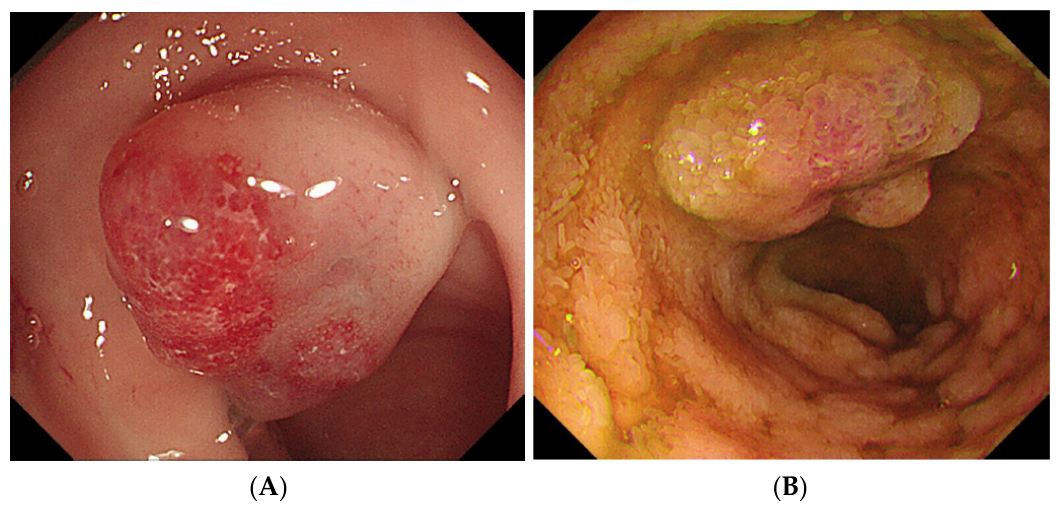
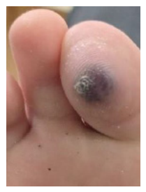
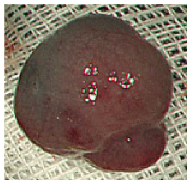
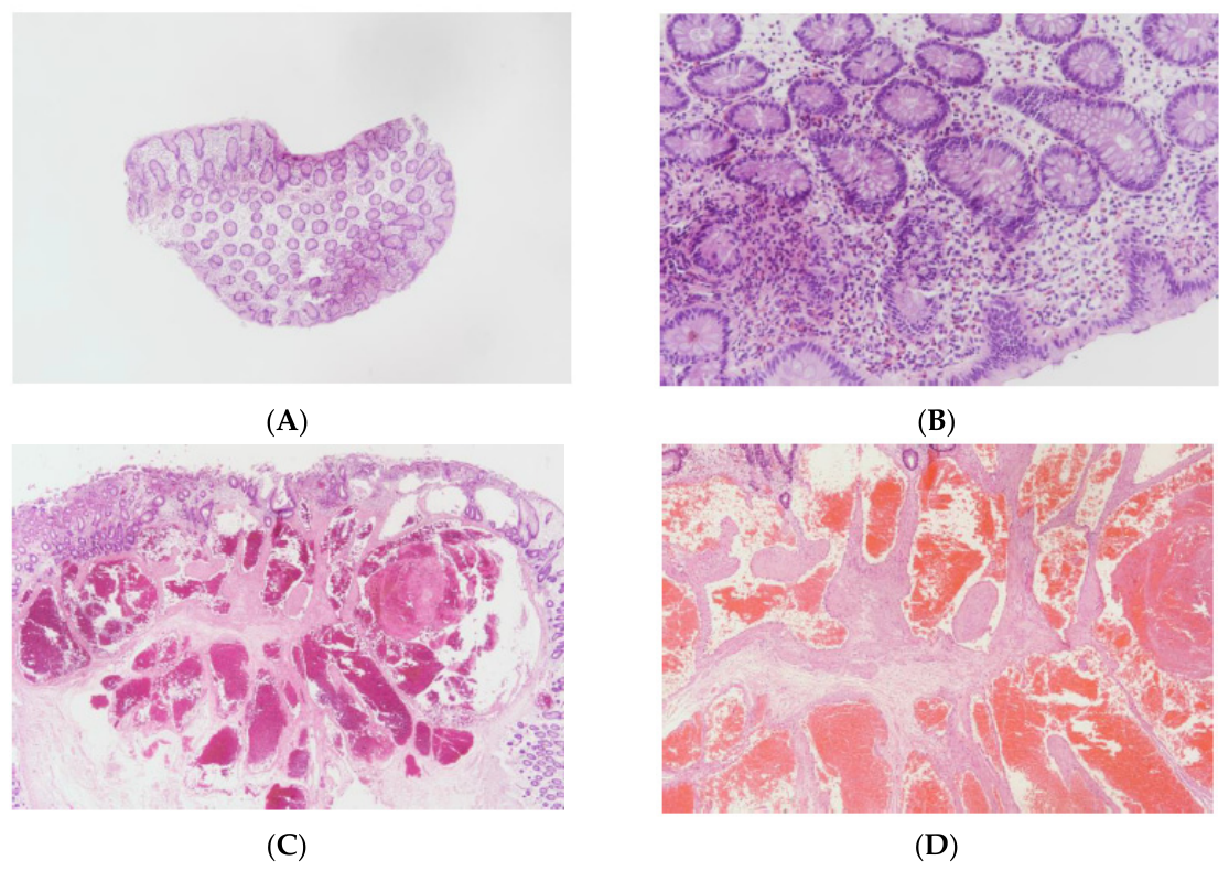
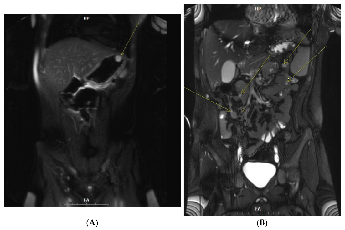
A 13-year-old boy presented to our pediatric clinic with a 6-month history of recurrent abdominal pain and intermittent hematochezia. He had been diagnosed with ASD at 3 years of age based on Diagnostic and Statistical Manual of Mental Disorders (DSM-5) criteria and had received special education and behavioral therapy. At 6 years of age, he received a red blood cell transfusion for severe anemia of unknown etiology. There was no family history of vascular malformations or bleeding disorders. On physical examination, the patient appeared pale but was hemodynamically stable. Dermatological assessment revealed pathognomonic lesions of BRBNS, including a 12-mm soft, bluish, compressible nodule on the right index finger and a similar 15-mm lesion on the plantar surface of the right great toe (Figure 1). Laboratory evaluation revealed severe microcytic anemia (hemoglobin, 5.6 g/dL) and a very low ferritin level (2.5 ng/mL), suggestive of chronic blood loss. Mild leukopenia was present, with a white blood cell count of 3.8 × 10³/μL, which was not further evaluated as it was not considered clinically significant, whereas the platelet count was within the normal range. C-reactive protein and fecal calprotectin levels were unremarkable, and stool occult blood testing yielded intermittently positive results, consistent with the patient’s known GI lesions. Endoscopy revealed multiple compressible, reddish-blue vascular lesions scattered throughout the stomach, duodenum, terminal ileum, and colon (ranging from 15 mm to 25 mm in diameter; Figure 2). Several accessible colonic lesions were removed by endoscopic mucosal resection (Figure 3). Histopathological examination of the resected specimens confirmed the presence of cavernous hemangiomas composed of dilated, thin-walled vascular channels lined with flattened endothelial cells without atypia (Figure 4). Coronal T2-weighted MR enterography demonstrated a small hyperintense polypoid lesion in the gastric fundus (Figure 5A, arrow) and several additional hyperintense small bowel lesions of varying size (Figure 5B, arrows), indicating more widespread GI involvement than was accessible by endoscopy alone. Whole-exome sequencing (NovaSeq X, Illumina; IDT xGen exome panel, Integrated DNA Technologies), performed on DNA extracted from a buccal swab, achieved a mean coverage of 111× (≥20× in 99.6% of target bases). Variant calling and classification were performed via the EVIDENCE v4.3 platform [12] (GATK v4.4.0, Manta v1.6.0, VEP v104.2) per ACMG/AMP guidelines [13], revealing a heterozygous missense variant in PDGFRA (c.2075G>T, p.Ser692Ile) in exon 15, present at a variant allele fraction of 54% (91 of 167 reads at this locus). This substitution affects a serine residue within the tyrosine kinase domain. In ClinVar (VCV001408376), the variant was listed as having uncertain significance, catalogued in association with GIST/GIST-plus syndrome (OMIM: 175510) rather than BRBNS specifically, and was not observed in the gnomAD v4.1.0 population. In silico predictions were inconclusive (REVEL 0.378; 3Cnet 0.02, a low score not supportive of pathogenicity). No pathogenic variants were detected in the TEK gene, PIK3CA, or GNAQ. As testing was proband-only, without segregation analysis or Sanger validation, whether this variant is somatic or germline could not be determined. The patient recovered without complications. The patient was started on oral iron supplementation and a proton pump inhibitor. During the 6-month follow-up period, hemoglobin levels gradually improved and stabilized between 11.0 and 12.0 g/dL, with no further transfusions required. He experienced occasional mild abdominal discomfort but no recurrent episodes of significant bleeding. Regular endoscopic surveillance was performed.

3. Discussion

To the best of our knowledge, this is among the first pediatric cases of BRBNS associated with a PDGFRA variant in the absence of a TEK mutation. This finding is consistent with the genetic heterogeneity of BRBNS beyond the TEK pathway, although this observation is based on a single variant of uncertain significance and should not be considered confirmatory [2,14]. PDGFRA encodes a receptor tyrosine kinase involved in the regulation of angiogenesis, cellular proliferation, and tissue growth [15]. Our patient harbored a p.Ser692Ile substitution in the tyrosine kinase domain, specifically within the catalytically active region [16]. In silico predictions for this variant were inconclusive: the REVEL score (0.378) fell in an intermediate range, while the 3Cnet score (0.02) did not support pathogenicity, consistent with its classification as a VUS rather than a likely pathogenic variant. In other clinical contexts, PDGFRA mutations are best known in GISTs and occur in approximately 5%–10% of cases [17]. Because DNA was obtained from a buccal swab rather than affected lesional tissue, we cannot definitively determine whether this variant is causally related to this patient’s vascular phenotype. The variant was present at a variant allele fraction of 54% in non-lesional tissue, a proportion consistent with a constitutional heterozygous variant present throughout the body rather than with low-level somatic mosaicism confined to a subset of tissues. BRBNS-associated pathogenic variants are typically somatic and lesion-restricted, arising from postzygotic mutation in endothelial precursor cells [2]; the constitutional-range allele fraction observed here is also compatible with a coincidental finding, and confirmatory sequencing of affected lesional tissue was not performed to establish a tissue-specific origin. No segregation analysis or parental testing was performed. Given these substantial uncertainties, we do not consider genetic testing of first-degree relatives to be indicated on the basis of this single proband-only VUS.
The co-occurrence of BRBNS and ASD in this patient may point to shared molecular pathways underlying these two conditions. Recent transcriptomic data have shown increased PDGFRA expression in the cerebellum of individuals with ASD [10]. Additionally, whole-genome transmission disequilibrium testing has revealed associations between PDGFRA and sensory-related behavioral phenotypes in ASD [11]. Furthermore, mouse models of maternal immune activation have demonstrated altered PDGFRA expression associated with ASD-like behaviors [18]. Although the current case does not establish a causal relationship, it suggests PDGFRA signaling could contribute to both vascular and neurodevelopmental phenotypes. However, a causal relationship in humans remains unproven, and this co-occurrence may be coincidental.
In children with BRBNS, treatment should aim to control recurrent GI bleeding while minimizing procedure-related risks. In our patient, endoscopic mucosal resection effectively controlled bleeding from accessible colonic lesions, consistent with recommendations favoring endoscopic intervention when feasible [19,20]. Given the patient’s clinical stability following endoscopic resection and iron supplementation, conservative management was chosen over systemic therapy, such as sirolimus, which is used for more complicated vascular anomalies [21]; the patient did not receive sirolimus or any other systemic agent, and this option will be reconsidered if new lesions or recurrent bleeding occur. Iron-deficiency anemia is one of the most clinically significant complications of pediatric BRBNS and requires timely and aggressive management to prevent adverse neurocognitive outcomes [22]. Our patient’s good outcome with oral iron administration and endoscopic intervention demonstrates that a staged, multimodal strategy can be effective for managing anemia in pediatric patients with BRBNS.
Our study has certain limitations. The PDGFRA variant is a VUS identified from non-lesional (buccal swab) tissue only, without segregation or orthogonal validation data. The in silico predictions and allele fraction are informative but do not establish pathogenicity or a tissue-specific somatic origin. This variant has not been functionally studied; therefore, its pathogenic role remains unknown, and any connection with ASD cannot be proven in a single case. Further patient and laboratory studies are needed to determine whether PDGFRA is involved in BRBNS and whether it is related to neurodevelopment.

4. Conclusions

In conclusion, we report a pediatric case of BRBNS associated with a PDGFRA variant and comorbid ASD. This case is consistent with genetic heterogeneity in BRBNS; however, this observation from a single VUS is hypothesis-generating and requires confirmation in additional cases. The coexistence of BRBNS and ASD may suggest shared biological pathways; however, further investigation is needed to clarify this potential association. Our patient responded favorably to endoscopic and conservative therapies, supporting a practical approach that combines early detection and multidisciplinary care.

Author Contributions

T.H.K.: Conceptualization, data collection, patient management and follow-up, investigation, literature review, data curation, interpretation of genetic findings, and writing—original draft preparation. J.M.C.: Conceptualization, methodology, resources, critical revision of the manuscript and writing—review and editing. S.-H.C.: supervision, validation, project administration, data curation, validation, critical revision of the manuscript, and writing—review and editing. All authors have read and agreed to the published version of the manuscript.

Funding

This research received no external funding.

Institutional Review Board Statement

This study was approved by the Institutional Review Board of Kyung Hee University Hospital at Gangdong (IRB No. 2025-07-035, the date of approval: September 2025).

Informed Consent Statement

Written informed consent for publication of this case and the accompanying images was obtained from the patient’s parent/legal guardian.

Data Availability Statement

The datasets used and analyzed in this study are available from the corresponding author upon reasonable request, subject to patient privacy and confidentiality requirements.

Acknowledgments

The authors thank the patient and his family for their cooperation.

Conflicts of Interest

The authors declare no conflicts of interest.

Abbreviations

ASDautism spectrum disorder
BRBNSblue rubber bleb nevus syndrome
GISTgastrointestinal stromal tumor
PDGFRAplatelet-derived growth factor receptor alpha
TEKtyrosine kinase receptor (TIE2)
VUSvariant of uncertain significance

References

  1. Ur Rashid, M.; Muhammad Khan, M.; Ullah, W.; Hussain, I.; Hurairah, A. Blue rubber bleb nevus: A rare cause of GI bleeding-review of management. BMJ Case Rep. 2019, 12, e231561. [Google Scholar] [CrossRef] [Scilit] [PubMed]
  2. Soblet, J.; Kangas, J.; Nätynki, M.; Mendola, A.; Helaers, R.; Uebelhoer, M.; Kaakinen, M.; Cordisco, M.; Dompmartin, A.; Enjolras, O.; et al. Blue rubber bleb nevus (BRBN) syndrome is caused by somatic TEK (TIE2) mutations. J. Investig. Dermatol. 2017, 137, 207–216. [Google Scholar] [CrossRef] [Scilit] [PubMed]
  3. Garg, P.K. Cutaneous and bowel vascular malformation. Indian J. Gastroenterol. 2015, 34, 91. [Google Scholar] [CrossRef] [Scilit] [PubMed]
  4. Isoldi, S.; Belsha, D.; Yeop, I.; Uc, A.; Zevit, N.; Mamula, P.; Loizides, A.M.; Tabbers, M.; Cameron, D.; Day, A.S.; et al. Diagnosis and management of children with Blue Rubber Bleb Nevus Syndrome: A multi-center case series. Dig. Liver Dis. 2019, 51, 1537–1546. [Google Scholar] [CrossRef] [Scilit] [PubMed]
  5. Li, Z.; Yuan, H.; Yu, D.; Victor, N.; Chen, Y.; Feng, J.; Teitelbaum, D.; Sun, X. The surgery for blue rubber bleb nevus syndrome. Ann. Med. Surg. 2016, 5, 93–96. [Google Scholar] [CrossRef] [Scilit]
  6. Limaye, N.; Kangas, J.; Mendola, A.; Godfraind, C.; Schlögel, M.J.; Helaers, R.; Eklund, L.; Boon, L.M.; Vikkula, M. Somatic activating PIK3CA mutations cause venous malformation. Am. J. Hum. Genet. 2015, 97, 914–921. [Google Scholar] [CrossRef] [Scilit] [PubMed]
  7. Xing, Y.; Liu, H.; Liu, H.; Ding, X.; Jing, X. Genetic mutation and blue rubber bleb nevus syndrome: Case reports and literature review. Front. Genet. 2025, 16, 1516562. [Google Scholar] [CrossRef] [Scilit] [PubMed]
  8. Heldin, C.H.; Westermark, B. Mechanism of action and in vivo role of platelet-derived growth factor. Physiol. Rev. 1999, 79, 1283–1316. [Google Scholar] [CrossRef] [Scilit] [PubMed]
  9. Wang, C.; Yantiss, R.K.; Lieberman, M.D.; Tubito-Massarano, F.; Qin, L.; Yemelyanova, A.; Solomon, J.P.; Hissong, E. A rare PDGFRA exon 15 germline mutation identified in a patient with phenotypic manifestations concerning for GIST-plus syndrome: A case report and review of literature. Int. J. Surg. Pathol. 2023, 31, 1139–1145. [Google Scholar] [CrossRef] [Scilit] [PubMed]
  10. Li, H.; Wang, X.; Hu, C.; Li, H.; Xu, Z.; Lei, P.; Luo, X.; Hao, Y. JUN and PDGFRA as crucial candidate genes for childhood autism spectrum disorder. Front. Neuroinform. 2022, 16, 800079. [Google Scholar] [CrossRef] [Scilit] [PubMed]
  11. Guo, Q.; Xia, L.; Guo, R.; Xu, W.; Zhang, Y.; Zhao, C.; Zhang, P.; Bai, T.; Ni, X.; Hao, C.; et al. Behavioural deficits of autism spectrum disorder and associations with different gene clusters: A study with the whole-genome transmission disequilibrium test. BMJ Paediatr. Open 2023, 7, e001930. [Google Scholar] [CrossRef] [Scilit] [PubMed]
  12. Seo, G.H.; Kim, T.; Choi, I.H.; Park, J.Y.; Lee, J.; Kim, S.; Won, D.G.; Oh, A.; Lee, Y.; Choi, J.; et al. Diagnostic yield and clinical utility of whole exome sequencing using an automated variant prioritization system, EVIDENCE. Clin. Genet. 2020, 98, 562–570. [Google Scholar] [CrossRef] [Scilit] [PubMed]
  13. Richards, S.; Aziz, N.; Bale, S.; Bick, D.; Das, S.; Gastier-Foster, J.; Grody, W.W.; Hegde, M.; Lyon, E.; ACMG Laboratory Quality Assurance Committee; et al. Standards and guidelines for the interpretation of sequence variants: A joint consensus recommendation of the American College of Medical Genetics and Genomics and the Association for Molecular Pathology. Genet. Med. 2015, 17, 405–424. [Google Scholar] [CrossRef] [Scilit] [PubMed]
  14. Kozai, L.; Nishimura, Y. Clinical characteristics of blue rubber bleb nevus syndrome in adults: Systematic scoping review. Scand. J. Gastroenterol. 2023, 58, 1108–1114. [Google Scholar] [CrossRef] [Scilit] [PubMed]
  15. Guérit, E.; Arts, F.; Dachy, G.; Boulouadnine, B.; Demoulin, J.B. PDGF receptor mutations in human diseases. Cell. Mol. Life Sci. 2021, 78, 3867–3881. [Google Scholar] [CrossRef] [Scilit] [PubMed]
  16. Corless, C.L.; Schroeder, A.; Griffith, D.; Town, A.; McGreevey, L.; Harrell, P.; Shiraga, S.; Bainbridge, T.; Morich, J.; Heinrich, M.C. PDGFRA mutations in gastrointestinal stromal tumors: Frequency, spectrum and in vitro sensitivity to imatinib. J. Clin. Oncol. 2005, 23, 5357–5364. [Google Scholar] [CrossRef] [Scilit] [PubMed]
  17. Huang, W.; Yuan, W.; Ren, L.; Liang, H.; Du, X.; Sun, X.; Fang, Y.; Gao, X.; Fu, M.; Sun, Y.; et al. Clinicopathological and therapeutic analysis of PDGFRA mutated gastrointestinal stromal tumor. Pathol. Res. Pract. 2022, 239, 154138. [Google Scholar] [CrossRef] [Scilit] [PubMed]
  18. Sato, A.; Kotajima-Murakami, H.; Tanaka, M.; Katoh, Y.; Ikeda, K. Influence of prenatal drug exposure, maternal inflammation, and parental aging on the development of autism spectrum disorder. Front. Psychiatry 2022, 13, 821455. [Google Scholar] [CrossRef] [Scilit] [PubMed]
  19. Samanta, A.; Poddar, U.; Sarma, M.S.; Srivastava, A.; Yachha, S.K.; Mohindra, S. Endoscopic band ligation in blue rubber bleb nevus syndrome: A report of two children. JPGN Rep. 2023, 4, e344. [Google Scholar] [CrossRef] [Scilit] [PubMed]
  20. Reddy, Y.K.; Bayoumi, M.; Barnes, M.; Gillespie, W.; Kamal, F.; Ismail, M.; Bilal, M. An unusual and challenging cause of small bowel bleeding: Isolated gastrointestinal blue rubber bleb nevus syndrome. Transl. Gastroenterol. Hepatol. 2022, 7, 12. [Google Scholar] [CrossRef] [Scilit] [PubMed]
  21. Adams, D.M.; Trenor, C.C., 3rd; Hammill, A.M.; Vinks, A.A.; Patel, M.N.; Chaudry, G.; Wentzel, M.S.; Mobberley-Schuman, P.S.; Campbell, L.M.; Brookbank, C.; et al. Efficacy and safety of sirolimus in the treatment of complicated vascular anomalies. Pediatrics 2016, 137, e20153257. [Google Scholar] [CrossRef] [Scilit] [PubMed]
  22. Aksu, T.; Ünal, S. Iron deficiency anemia in infancy, childhood, and adolescence. Turk. Arch. Pediatr. 2023, 58, 358–362. [Google Scholar] [CrossRef] [Scilit] [PubMed]
Figure 1. Bluish, compressible vascular nodule on the plantar surface of the right great toe, a pathognomonic cutaneous finding of BRBNS.
Children 13 01009 g001
Figure 2. (A) Colonoscopic view showing a reddish-blue, compressible vascular lesion in the colon. (B) Colonoscopic view showing a similar lesion in the terminal ileum.
Children 13 01009 g002
Figure 3. Gross specimen following endoscopic mucosal resection of a vascular lesion from the rectum.
Children 13 01009 g003
Figure 4. Histopathology of gastrointestinal vascular lesions. (A) Cecal lesion (H&E, ×40) and (B) cecal lesion (H&E, ×200), showing dilated, thin-walled vascular channels consistent with cavernous hemangioma. (C) Rectal lesion (H&E, ×20) and (D) rectal lesion (H&E, ×40), showing similar dilated, blood-filled vascular spaces.
Children 13 01009 g004
Figure 5. Coronal T2-weighted MR enterography. (A) A small hyperintense polypoid lesion in the gastric fundus (arrow), consistent with a venous malformation. (B) Additional hyperintense small bowel lesions of varying size (arrows), indicating involvement not fully captured by endoscopy.
Children 13 01009 g005
Disclaimer/Publisher’s Note: The statements, opinions and data contained in all publications are solely those of the individual author(s) and contributor(s) and not of MDPI and/or the editor(s). MDPI and/or the editor(s) disclaim responsibility for any injury to people or property resulting from any ideas, methods, instructions or products referred to in the content.