A Review of Fluorescent pH Probes: Ratiometric Strategies, Extreme pH Sensing, and Multifunctional Utility


Abstract
1. Introduction
- (a)
- Intramolecular Charge Transfer (ICT)
- (b)
- Photoinduced Electron Transfer (PET)
- (c)
- Fluorescent Resonance Energy Transfer (FRET)
2. Acidic Fluorescent Probes
2.1. Strongly Acidic Fluorescent Probes
2.2. Weakly Acidic Fluorescent Probes
2.3. Wide-Range Acidic Fluorescent Probes
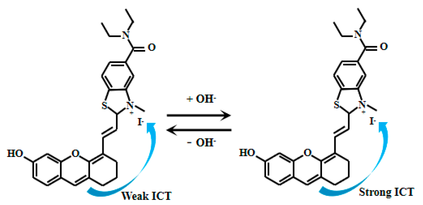
3. Alkaline Fluorescent Probes
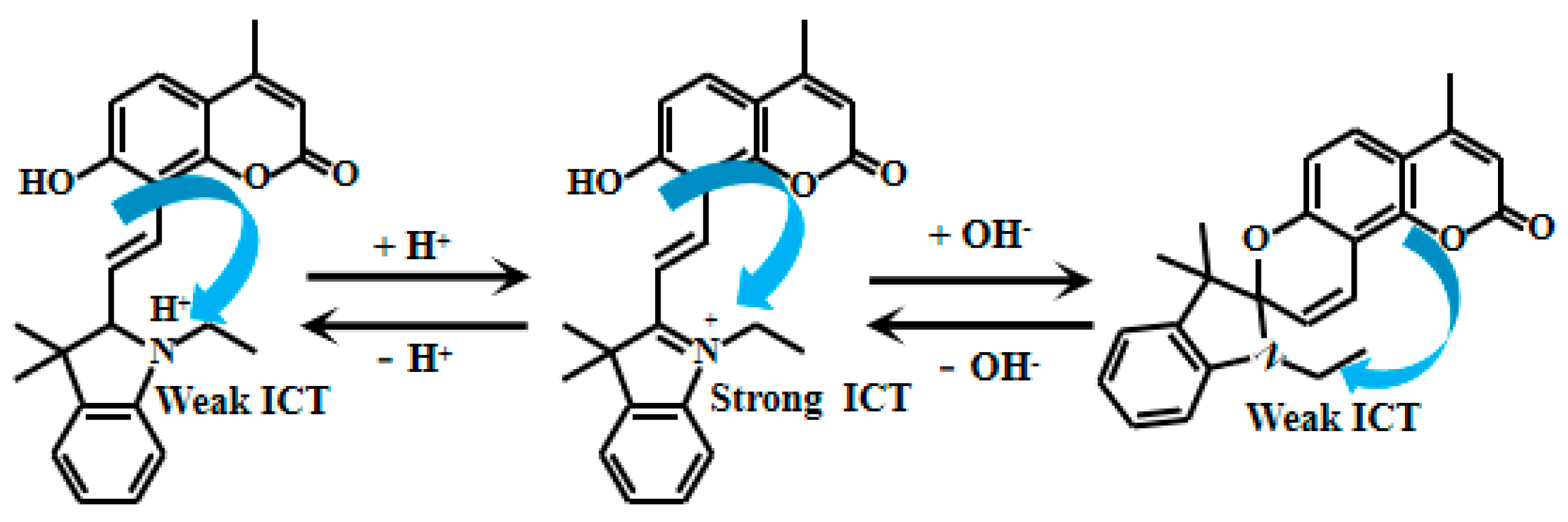
3.1. Strongly Alkaline Fluorescent Probes
3.2. Weakly Alkaline Fluorescent Probes
3.3. Wide-Range Alkaline Fluorescent Probes
4. Near-Neutral Fluorescent Probes
5. Wide-Range pH Fluorescent Probes
6. Summary and Outlook
7. Future Work
- (1)
- Probe enhancement strategies: Key strategies for advancing fluorescent probes include optimizing ICT efficiency through stronger electron donor/acceptor groups or nano-assemblies with signal amplification capabilities, and designing universal probes operable across broad pH ranges (e.g., pH 0–14) or specialized probes with ultra-high sensitivity at extreme values (e.g., pH > 13 or pH < 1).
- (2)
- Hysteresis characteristics of pH sensors: To enhance sensor reversibility and long-term stability, optimize sensing membrane design—including functional groups and cross-linking density—while analyzing OH− ion accumulation kinetics. Additionally, explore surface modification or buffering agents to minimize OH− retention and improve stability.
- (3)
- Stability: Structural modification of probe molecules (such as introducing large steric hindrance groups, oxidation/reduction resistant groups) or encapsulating them in stable carrier materials (such as silica and metal organic framework (MOFs)) to enhance their long-term photostability and chemical stability in specific harsh environments, such as strong acids/bases and highly reactive oxygen species.
- (4)
- Multi-functionality of sensors: In addition to pH detection, fluorescent sensors can also be used to detect other environmental parameters such as temperature, humidity, and ion concentration. In the future, sensor design based on multifunctional fluorescent materials can be explored to achieve simultaneous detection of multiple parameters and improve the application value of sensors.
- (5)
- Mechanical performance of sensors: The deployment of fiber optic sensors in mechanically demanding environments necessitates enhanced durability—a currently understudied aspect. Future work must: (a) engineer organic–inorganic hybrid coatings with gradient modulus, optimizing interfacial binding energy through MD simulations to ensure structural integrity under extreme stress; and (b) develop multi-scale mechanical models quantifying deformation-induced optical signal drift, enabling rational anti-interference design.
- (6)
- Frontier biomedical applications: Engineer a theranostic probe by merging the detection function of this sensor with photodynamic therapy (PDT) or photothermal therapy (PTT) capabilities, enabling concurrent diagnosis and treatment.
Author Contributions
Funding
Institutional Review Board Statement
Informed Consent Statement
Data Availability Statement
Conflicts of Interest
References
- Hussain, K.; Lone, S.M.; Masoodi, K.Z.; Balkhi, S.M. Chapter 6-pH (negative logarithm of hydrogen ion concentration). In Techniques for Biochemical Analysis; Hussain, K., Lone, S.M., Masoodi, K.Z., Balkhi, S.M., Eds.; Academic Press: Cambridge, MA, USA, 2024; pp. 37–40. [Google Scholar] [CrossRef]
- Noor, M.; Lyana, S.; Ramli, N.; Zolkefli, N.; Mustapha, N.A.; Hassan, M.A.; Maeda, T. Survivability of Alcaligenaceae and Chromatiaceae as palm oil mill effluent pollution bioindicators under fluctuations of temperature, pH and total suspended solid. J. Biosci. Bioeng. 2021, 132, 174–182. [Google Scholar] [CrossRef]
- Tarazona, J.V. Pollution, water. In Encyclopedia of Toxicology, 4th ed.; Wexler, P., Ed.; Academic Press: Oxford, UK, 2024; pp. 809–815. [Google Scholar] [CrossRef]
- Hou, J.; Ren, W.X.; Li, K.; Seo, J.; Sharma, A.; Yu, X.; Kim, J.S. Fluorescent bioimaging of pH: From design to applications. Chem. Soc. Rev. 2017, 46, 2076–2090. [Google Scholar] [CrossRef] [PubMed]
- Chao, L.; Aodeng, G.; Ga, L.; Ai, J. Research progress of organic small probes sensitive to tumor microenvironment. J. Mol. Struct. 2025, 1322, 140617. [Google Scholar] [CrossRef]
- Podder, A.; Won, M.; Kim, S.; Verwilst, P.; Maiti, M.; Yang, Z.; Qu, J.; Bhuniya, S.; Kim, J.S. A two-photon fluorescent probe records the intracellular pH through ‘OR’ logic operation via internal calibration. Sens. Actuators B. Chem. 2018, 268, 195–204. [Google Scholar] [CrossRef]
- Zhu, W.; Ma, Z.; Zhang, L.; Yang, C.; Xu, F.; Yu, J. Natural anthocyanin-based wearable colorimetric pH indicator with high color fastness. Dye. Pigment. 2025, 239, 112789. [Google Scholar] [CrossRef]
- Duan, M.; Li, G.; Shen, J.; Dai, R.; Li, X.; Liu, Z.; Jia, F. A CRISPR/Cas12a biosensor for portable and accessible detection of Salmonella typhimurium via multi-indicator pH millidisc colorimetry and smartphone imaging platform. Biosens. Bioelectron. 2025, 286, 117611. [Google Scholar] [CrossRef]
- Viscusi, G.; Lamberti, E.; Angilè, F.; Di Stasio, L.; Gerardi, C.; Giovinazzo, G.; Vigliotta, G.; Gorrasi, G. Smart pH-sensitive indicators based on rice starch/pectin/alginate loading Lambrusco pomace extract and curcumin to track the freshness of pink shrimps. Int. J. Biol. Macromol. 2025, 291, 139085. [Google Scholar] [CrossRef] [PubMed]
- Norouzi, M.; Ahmadi, E.; Mohamadnia, Z. Stimuli-responsive fluorescent polymer nanoparticles: Versatile applications in rapid colorimetric and fluorometric detection of cyanide in blood plasma, pH sensing, paper-based sensors, and intelligent artworks. J. Photochem. Photobiol. A Chem. 2024, 452, 115552. [Google Scholar] [CrossRef]
- Wockenfus, A.M.; Koch, C.D.; Conlon, P.M.; Sorensen, L.D.; Cambern, K.L.; Chihak, A.J.; Zmolek, J.A.; Petersen, A.E.; Burns, B.E.; Lieske, J.C.; et al. Discordance between urine pH measured by dipstick and pH meter: Implications for methotrexate administration protocols. Clin. Biochem. 2013, 46, 152–154. [Google Scholar] [CrossRef]
- Tantray, J.A.; Mansoor, S.; Wani, R.F.C.; Nissa, N.U. Chapter 3-pH meter: Its use and calibration. In Basic Life Science Methods; Tantray, J.A., Mansoor, S., Wani, R.F.C., Nissa, N.U., Eds.; Academic Press: Cambridge, MA, USA, 2023; pp. 9–10. [Google Scholar] [CrossRef]
- Afsahi, A.; Ahmadi-hamedani, M.; Khodadi, M. Comparative evaluation of urinary dipstick and pH-meter for cattle urine pH measurement. Heliyon 2020, 6, 03316. [Google Scholar] [CrossRef]
- Liu, Z.; Liu, J.; Li, B.; Zhang, Y.; Xing, X.-H. Focusing on the process diagnosis of anaerobic fermentation by a novel sensor system combining microbial fuel cell, gas flow meter and pH meter. Int. J. Hydrogen Energy 2014, 39, 13658–13664. [Google Scholar] [CrossRef]
- Nagasaki, H.; Asaka, T.; Iwami, K.; Umeda, N.; Yamamoto, C.; Hara, Y.; Masuda, A. Non-Destructive Measurement of Acetic Acid and Its Distribution in a Photovoltaic Module during Damp Heat Testing Using pH-Sensitive Fluorescent Dye Sensors. Sensors 2022, 22, 2520. [Google Scholar] [CrossRef]
- Kriksunov, L.B.; Macdonald, D.D. Development of glass pH sensors for use at temperatures of 200–250 °C. Sens. Actuators B Chem. 1994, 22, 201–204. [Google Scholar] [CrossRef]
- Liao, Y.; Chou, J. Preparation and characteristics of ruthenium dioxide for pH array sensors with real-time measurement system. Sens. Actuators B Chem. 2008, 128, 603–612. [Google Scholar] [CrossRef]
- Kreider, K.G.; Tarlov, M.J.; Cline, J.P. Sputtered thin-film pH electrodes of platinum, palladium, ruthenium, and iridium oxides. Sens. Actuators B Chem. 1995, 28, 167–172. [Google Scholar] [CrossRef]
- Wang, M.; Yao, S.; Madou, M. A long-term stable iridium oxide pH electrode. Sens. Actuators B Chem. 2002, 81, 313–315. [Google Scholar] [CrossRef]
- Huang, W.; Cao, H.; Deb, S.; Chiao, M.; Chiao, J.C. A flexible pH sensor based on the iridium oxide sensing film. Sens. Actuators A Phys. 2011, 169, 1–11. [Google Scholar] [CrossRef]
- Du, R.; Hu, R.; Huang, R.; Lin, C. In Situ Measurement of Cl− Concentrations and pH at the Reinforcing Steel/Concrete Interface by Combination Sensors. Anal. Chem. 2006, 78, 3179–3185. [Google Scholar] [CrossRef] [PubMed]
- Ensafi, A.A.; Kazemzadeh, A. Optical pH Sensor Based On Chemical Modification of Polymer Film. Microchem. J. 1999, 63, 381–388. [Google Scholar] [CrossRef]
- Babbar, A.; Kumar, R.; Dhawan, V.; Ranjan, N.; Sharma, A. Additive Manufacturing of Polymers for Tissue Engineering: Fundamentals, Applications, and Future Advancements; CRC Press: Boca Raton, FL, USA, 2022. [Google Scholar]
- Schäferling, M.; Ondrus, V. The Art of Fluorescence Imaging with Chemical Sensors: The Next Decade 2012–2022. Chemosensors 2024, 12, 31–33. [Google Scholar] [CrossRef]
- Shah, S.R.; Modi, C.D.; Singh, S.; Mori, D.D.; Soniwala, M.M.; Prajapati, B.G. Recent Advances in Additive Manufacturing of Polycaprolactone-Based Scaffolds for Tissue Engineering Applications: A Comprehensive Review. Regen. Eng. Transl. Med. 2024, 11, 1–20. [Google Scholar] [CrossRef]
- Schäferling, M. The Art of Fluorescence Imaging with Chemical Sensors. Angew. Chem. Int. Ed. 2012, 51, 3532–3554. [Google Scholar] [CrossRef]
- Fan, L.; Bao, Y. Review of fiber optic sensors for corrosion monitoring in reinforced concrete. Cem. Concr. Compos. 2021, 120, 104029. [Google Scholar] [CrossRef]
- Min, R.; Liu, Z.; Pereira, L.; Yang, C.; Sui, Q.; Marques, C. Optical fiber sensing for marine environment and marine structural health monitoring: A review. Opt. Laser Technol. 2021, 140, 107082. [Google Scholar] [CrossRef]
- Hosseinlou, R.; Dargahi, M.; Vanashi, A.K. Alkaline range pH sensor based on chitosan hydrogel: A novel approach to pH sensing. Int. J. Biol. Macromol. 2024, 279, 135199. [Google Scholar] [CrossRef]
- Liu, Y.; Yan, W.; Li, J.; Wang, Y.; Zhang, G.; Shi, L.; Zhang, C.; Shuang, S.; Dong, C. Polarity, pH and HClO triple-response fluorescent probe and its application in inflammation models. J. Mol. Liq. 2025, 422, 126953. [Google Scholar] [CrossRef]
- Zhang, T.; Wang, J.; Wang, F.; Wu, L.; Wang, C.; Zeng, J.; Xu, Y.; Xie, W.; Lu, X. Sulfonium perchlorate based near-infrared fluorescent probe targeting lysosome for pH imaging in living cells and tumor-bearing mice. Spectrochim. Acta Part A Mol. Biomol. Spectrosc. 2025, 329, 125558. [Google Scholar] [CrossRef]
- Zhao, H.Y.; Liu, Y.T.; Yao, S.Y.; Zhao, K.Y.; Zhang, Q.Q.; Zou, Y.L.; Zhao, L.X. A multifunctional near-infrared fluorescent probe with a large Stokes shift for monitoring of Al3+ and pH in biological vivo and its smartphone application. Microchem. J. 2025, 208, 112592. [Google Scholar] [CrossRef]
- Valeur, B.; Leray, I. Design principles of fluorescent molecular sensors for cation recognition. Coord. Chem. Rev. 2000, 205, 3–40. [Google Scholar] [CrossRef]
- Zhao, X.; Xiao, J.; Tang, D.; Zhou, L.; Qin, Z.; Wang, Z. Visualizing and tracking pH fluctuation in living systems and real food samples with a novel dual-site modulated reversible ratiometric fluorescent probe. J. Mol. Struct. 2025, 1331, 141647. [Google Scholar] [CrossRef]
- Yang, Y.; Shen, Z.; Shen, Z.; Meng, Z.; Gong, S.; Liang, Y.; Wang, Z.; Wang, S. A novel carbazole-pyrimidine-based dual mode fluorescent probe for detection of acidic and basic pH in biological systems. Spectrochim. Acta Part A Mol. Biomol. Spectrosc. 2025, 330, 125709. [Google Scholar] [CrossRef]
- Chalupczok, S.; Kurzweil, P.; Hartmann, H.; Schell, C. The Redox Chemistry of Ruthenium Dioxide: A Cyclic Voltammetry Study—Review and Revision. Int. J. Electrochem. 2018, 2018, 1273768. [Google Scholar] [CrossRef]
- Popoola, L.T.; Yusuff, A.S.; Aderibigbe, T.A. Assessment of natural groundwater physico-chemical properties in major industrial and residential locations of Lagos metropolis. Appl. Water Sci. 2019, 9, 191. [Google Scholar] [CrossRef]
- Johan, P.D.; Ahmed, O.H.; Omar, L.; Hasbullah, N.A. Phosphorus Transformation in Soils Following Co-Application of Charcoal and Wood Ash. Agronomy 2021, 11, 2010. [Google Scholar] [CrossRef]
- Webb, B.A.; Chimenti, M.; Jacobson, M.P.; Barber, D.L. Dysregulated pH: A perfect storm for cancer progression. Nat. Rev. Cancer 2011, 11, 671–677. [Google Scholar] [CrossRef] [PubMed]
- Ng, J.F.; Ahmed, O.H.; Omar, L.; Jalloh, M.B.; Kwan, Y.M.; Musah, A.A.; Chowdhury, A.J.K. Improving pH buffering capacity of an acid soil to regulate nutrient retention and mitigate water pollution using Calciprill and sodium silicate. Desalination Water Treat. 2024, 319, 100491. [Google Scholar] [CrossRef]
- Adeleke, R.; Nwangburuka, C.; Oboirien, B. Origins, roles and fate of organic acids in soils: A review. S. Afr. J. Bot. 2017, 108, 393–406. [Google Scholar] [CrossRef]
- Jie, Y.; Yan, Z.; Yuan, M.Z.; Xin, L.M.; Fei, Z.; Fa, W.S.; Long, W.Z.; Qin, Y.Y. Pyrimidin-2-amine derivative-grafted cellulose ratiometric fluorescent probe for pH detection in extremely acidic media. Cellulose 2021, 28, 10441–10455. [Google Scholar] [CrossRef]
- Xu, Y.; Jiang, Z.; Xiao, Y.; Bi, F.-Z.; Miao, J.-Y.; Zhao, B.-X. A new fluorescent pH probe for extremely acidic conditions. Anal. Chim. Acta 2014, 820, 146–151. [Google Scholar] [CrossRef] [PubMed]
- Li, D.; Zhang, P.; Guo, X.; Zhang, T.; Ran, X.; Zhang, T.; Xiao, H.; Shu, W. A new fluorescent probe for detection of hydrazine and extremely acidic pH in different modes. Tetrahedron 2024, 159, 134007. [Google Scholar] [CrossRef]
- Wan, Q.; Chen, S.; Shi, W.; Li, L.; Ma, H. Lysosomal pH Rise during Heat Shock Monitored by a Lysosome-Targeting Near-Infrared Ratiometric Fluorescent Probe. Angew. Chem. Int. Ed. 2014, 53, 10916–10920. [Google Scholar] [CrossRef]
- Pan, W.; Liu, M.; Zhang, S.; Wang, Y.; Yang, X. Methanol poisoning with bilateral basal ganglia necrosis and hemorrhage without visual impairment: A case report. Am. J. Emerg. Med. 2025, 93, e235–e237. [Google Scholar] [CrossRef]
- Jangjou, A.; Moqadas, M.; Mohsenian, L.; Kamyab, H.; Chelliapan, S.; Alshehery, S.; Ali, M.A.; Dehbozorgi, F.; Yadav, K.K.; Khorami, M.; et al. Awareness raising and dealing with methanol poisoning based on effective strategies. Environ. Res. 2023, 228, 115886. [Google Scholar] [CrossRef]
- Hu, Z.; Li, R.; Zhang, X.; Chen, Z. A pH-responsive NIR fluorescent probe for precise cancer phototheranostics. Sens. Actuators B Chem. 2025, 440, 137877. [Google Scholar] [CrossRef]
- Kashiwada, A.; Taoka, N.; Chijimi, Y.; Noguchi, K.; Shigematsu, K.; Miura, M.; Suzuki, T. Weakly acidic pH-responsive liposomal content release induced by histidine-modified agents. Org. Biomol. Chem. 2024, 22, 2844–2850. [Google Scholar] [CrossRef]
- Wang, Q.; Chen, L.; An, Z.; Zhang, S.; Jin, L.; Ai, T.; Dai, H.; Wang, L.; Lu, H.; Yang, X.-F. Learning from drugs: An edaravone-inspired design of a coumarin-based fluorescent probe for dual-channel imaging of hydroxyl radicals and pH in complex biosystems. Sens. Actuators B Chem. 2025, 441, 137946. [Google Scholar] [CrossRef]
- Xu, Y.; Duan, R.; Liu, H.; Xia, C.; Duan, G.; Ge, Y. Preparation of a novel pH-responsive fluorescent probe based on an imidazo [1,2-a]indole fluorophore and its application in detecting extremely low pH in saccharomyces cerevisiae. J. Fluoresc. 2021, 31, 1–7. [Google Scholar] [CrossRef]
- Li, X.; Meng, Z.; Gong, S.; Liang, Y.; Zhang, Y.; Yang, Y.; Xu, X.; Wang, Z.; Wang, S. Development of a novel ratiometric fluorescent probe for real-time monitoring of acidic pH and its applications in food samples and biosystem. Microchem. J. 2024, 204, 111169. [Google Scholar] [CrossRef]
- Feng, G.; Li, Z.; Zhai, P.; Ying, M.; Xu, Z.; Yang, C.; Wang, X.; Dong, B.; Yong, K.-T.; Xu, G. In-situ construction of fluorescent probes for lysosomes imaging and lysosomal pH ratiometric detection based on bioorthogonal fluorescence. Sens. Actuators B Chem. 2022, 371, 132577. [Google Scholar] [CrossRef]
- Hong, M.; Liu, A.; Xu, Y.; Xu, D. Synthesis and properties of three novel rhodamine-based fluorescent sensors for Hg2+. Chin. Chem. Lett. 2016, 27, 989–992. [Google Scholar] [CrossRef]
- Song, J.; Liu, S.; Zhao, N.; Zhao, L. A new fluorescent probe based on metallic deep eutectic solvent for visual detection of nitrite and pH in food and water environment. Food Chem. 2023, 398, 133935. [Google Scholar] [CrossRef]
- Liu, Y.; Zhang, Y.; Wang, J.; Yang, A.; Zhao, Y.; Zhou, A.; Xiong, R.; Huang, C. Colorimetric/spectral dual-mode analysis of sensitive fluorescent probe based on 2,3,3-trimethyl-3H-benzo[e]indole detection of acid pH. Bioorganic Chem. 2022, 124, 105792. [Google Scholar] [CrossRef]
- Chaghaghazardi, M.; Kashanian, S.; Nazari, M.; Omidfar, K.; Joseph, Y.; Rahimi, P. Nitrogen and sulfur co-doped carbon quantum dots fluorescence quenching assay for detection of mercury (II). Spectrochim. Acta Part A Mol. Biomol. Spectrosc. 2023, 293, 122448. [Google Scholar] [CrossRef]
- Vimer, C.; Yu, S.; Ghandehari, M. Probing pH Levels in Civil Engineering Materials. J. Mater. Civ. Eng. 2009, 21, 51–57. [Google Scholar] [CrossRef]
- Dang, V.H.; François, R. Influence of long-term corrosion in chloride environment on mechanical behaviour of RC beam. Eng. Struct. 2013, 48, 558–568. [Google Scholar] [CrossRef]
- Tariq, A.; Baydoun, J.; Remy, C.; Ghasemi, R.; Lefevre, J.P.; Mongin, C.; Dauzères, A.; Leray, I. Fluorescent molecular probe based optical fiber sensor dedicated to pH measurement of concrete. Sens. Actuators B Chem. 2021, 327, 128906. [Google Scholar] [CrossRef]
- Tariq, A.; Ghasemi, R.; Lefevre, J.P.; Mongin, C.; Dauzères, A.; Leray, I. Ratiometric fiber optic fluorescent pH sensor for hydroxide diffusion measurements in concrete. Sens. Actuators B Chem. 2024, 405, 135297. [Google Scholar] [CrossRef]
- Xu, W.; Fan, L.; Ma, Z.; Zhao, X.; Huang, L.; Chen, J. Development of optical fiber sensors for high-alkaline pH monitoring based on silica fluorescent nanoparticles. Measurement 2025, 253, 117803. [Google Scholar] [CrossRef]
- Huang, H.; Wang, X.; Zhou, G.; Qian, C.; Zhou, Z.; Wang, Z.; Yang, Y. A novel ratiometric fluorescent sensor from modified coumarin-grafted cellulose for precise pH detection in strongly alkaline conditions. Int. J. Biol. Macromol. 2024, 262, 130066. [Google Scholar] [CrossRef] [PubMed]
- Flinck, M.; Kramer, S.H.; Pedersen, S.F. Roles of pH in control of cell proliferation. Acta Physiol. 2018, 223, 13068. [Google Scholar] [CrossRef] [PubMed]
- Li, X.; Hu, Y.; Li, X.; Ma, H. Mitochondria-immobilized near-infrared ratiometric fluorescent pH probe to evaluate cellular mitophagy. Anal. Chem. 2019, 91, 11409–11416. [Google Scholar] [CrossRef]
- Yue, Z.; Zhai, S.; Wei, B.; Zhao, B.; Lin, Z. A ratiometric fluorescent probe based on FRET mechanism for pH and its cell imaging. Dye. Pigment. 2025, 233, 112544. [Google Scholar] [CrossRef]
- Fu, M.; Li, Q.; Chen, X.; Xie, Z.; Hu, J. pH-Activated NIR fluorescent probe for sensitive mitochondrial viscosity detection. Org. Biomol. Chem. 2025, 23, 3314–3319. [Google Scholar] [CrossRef] [PubMed]
- Kong, X.; Zhao, H.; Tang, Y. Synthesis and Optical Properties of 1,3-Bis(Arylimino) isoindoline-4,7-diols. Tetrahedron 2023, 144, 133546. [Google Scholar] [CrossRef]
- Chen, Y.; Zhou, B.; Liu, F.; Miao, J.; Zhao, B.; Lin, Z. A quinoline-salt-based fluorescent probe for precise monitoring of pH changes on mitochondria and water. Sens. Actuators B Chem. 2022, 373, 132732. [Google Scholar] [CrossRef]
- Liu, W.; Tian, X.; Gong, S.; Meng, Z.; Liang, Y.; Wang, Z.; Wang, S. A novel coumarin-derived fluorescent probe for real-time detection of pH in living zebrafish and actual food samples. J. Mol. Struct. 2024, 1299, 137141. [Google Scholar] [CrossRef]
- Zhang, C.; Guo, J.; Zhang, Y.; Liu, C. A naphthalimide-based bifunctional fluorescent probe for detecting CN− and alkaline pH and its application in food samples and living cells imaging. Microchem. J. 2024, 206, 111594. [Google Scholar] [CrossRef]
- Xu, K.; Zhang, C.; Li, M.; Gong, S.; Zhang, Y.; Wang, X.; Wang, Z.; Wang, S. A myrtenal-based colorimetric and fluorescent probe for reversibly monitoring alkaline pH and bioimaging in living cells and zebrafish. J. Photochem. Photobiol. A Chem. 2022, 430, 113962. [Google Scholar] [CrossRef]
- Dhawa, T.; Hazra, A.; Barma, A.; Pal, K.; Karmakar, P.; Roy, P. 4-Methyl-2,6-diformylphenol based biocompatible chemosensors for pH: Discrimination between normal cells and cancer cells. RSC Adv. 2020, 10, 15501–15513. [Google Scholar]
- Zhang, Z.; Wang, X.; Shi, L.; Lang, Y.; Gao, Y.; Bai, R.; Zhang, S.; Xi, J.; Han, C.; Zhang, X. A novel aggregation-induced emission fluorescent probe for sensitive detection of pH changes in seafood freshness monitoring. Tetrahedron 2025, 181, 134700. [Google Scholar] [CrossRef]
- Guan, L.; Hu, W.; Zhang, S.; Ai, Y.; Liang, Q. A dual-response fluorescent probe Rh-O-QL for simultaneous monitoring of NAD(P)H and pH during mitochondrial autophagy. Chem. Commun. 2025, 61, 7799–7802. [Google Scholar] [CrossRef] [PubMed]
- Du, Y.; Zhao, H.; Peng, X.; Zhou, X.; Yang, X.; Li, Y.; Yan, M.; Cui, Y.; Sun, G. A novel phenanthroline [9,10-d] imidazole-based fluorescent sensor for Hg2+ with “turn-on” fluorescence response. J. Photochem. Photobiol. A Chem. 2023, 439, 114604. [Google Scholar] [CrossRef]
- Han, H.; Liu, M.; Zhang, W.; Sun, L.; Ma, X.; Qiao, H.; Sun, S.; Yang, J.; Chai, X.; Wu, Z.; et al. The development of logic gate-based fluorescent probes that respond to intracellular hydrogen peroxide and pH in tandem. Talanta 2024, 270, 125526. [Google Scholar] [CrossRef] [PubMed]
- Li, F.; Dong, P.; Sun, S.; Zhai, S.; Zhao, B.; Lin, Z. A near-infrared fluorescent probe for simultaneous detection of pH and viscosity. Spectrochim. Acta Part A Mol. Biomol. Spectrosc. 2024, 318, 124486. [Google Scholar] [CrossRef] [PubMed]
- Mei, H.; Gu, X.; Wang, M.; Chen, J.; Yang, X.; Liu, X.; Xu, K. A tri-response colorimetric-fluorescent probe for pH and lysosomal imaging. Sens. Actuators B Chem. 2022, 370, 132425. [Google Scholar] [CrossRef]
- Liu, Y.; Hou, Y.; Yao, S.; Zhao, K.; Cui, H.; Zhang, M.; Wang, H.; Zou, Y.; Zhao, L. A novel dual-responsive NIR fluorescent probe based on isophorone: Real-time detection of Al3+ in vivo and colorimetric sensing of pH through smartphone. J. Environ. Chem. Eng. 2025, 13, 115620. [Google Scholar] [CrossRef]


| Probe | Core Fluorophore | Mechanism | Excitation Wavelength (nm) | Emission Wavelength (nm) | Ratio | pH Response Range | Applications | Ref. |
|---|---|---|---|---|---|---|---|---|
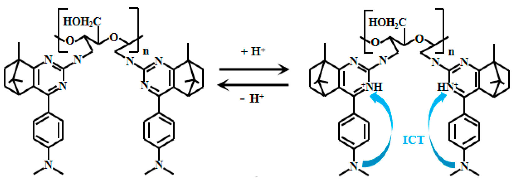
| 1 | Camphor | ICT | 355 | 416, 486 | I486/I416 | 1.04–2.35 | Fabrication of hybrid cellulose hydrogel, and measurement of vinegar and fruit acids | [42] |
| 2 | Coumarin | PET | 385 | 460 | - | 3.06–0.80 | Fluorescent imaging of E. coli | [43] |
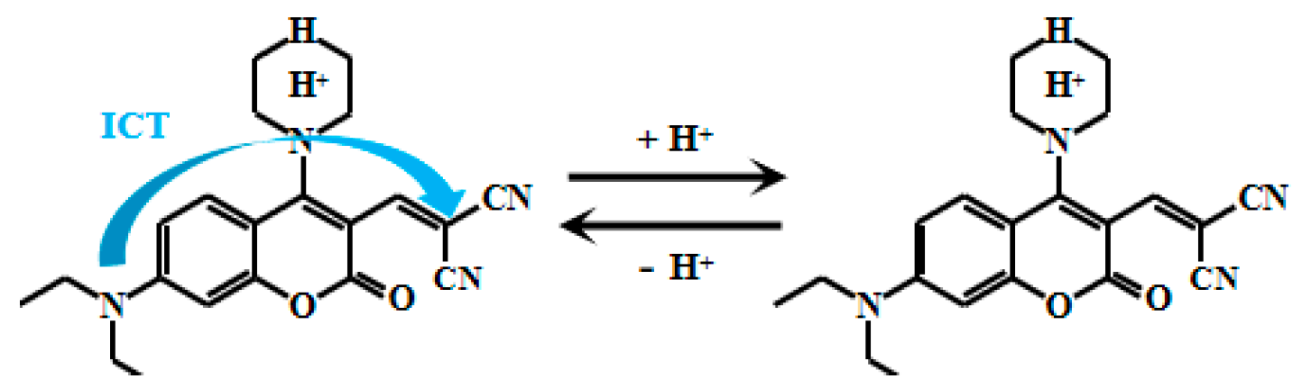
| 3 | Coumarin | ICT | 365 | 563 | - | 1.3–1.8 | Response to N2H4 | [44] |
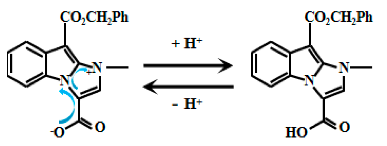
| 4 | Imidazole [1, 2-A] indole | ICT | 350 | 450 | - | 3.2–4.4 | Fluorescent imaging of saccharomyces cerevisiae | [51] |
| 5 | Pyrazole, pyridine | ICT | - | 487, 585 | I585/I487 | 3.0–5.5 | Freshness assessment of pork and chicken breast, fluorescent imaging in HeLa cells and zebrafish | [52] |
| 6 | Rhodamine | FRET | 365 | 480, 590 | I590/I480 | 3.9–5.3 | Lysosomal pH detection in MCF-7 cells | [53] |
| 7 | Rhodamine B | ICT | 526 | 585 | - | 5.0–6.0 | Fluorescent imaging of TM3 cells | [54] |
| 8 | Fe doped carbon dots | Static quenching (SQE) | 410 | 475 | - | 2.0–7.0 | Response to NO2− | [55] |
| 9 | Benzindole | ICT | 436 | 418, 545 | I545/I418 | 2.1–7.4 | Freshness assessment of food samples (milk, shrimp, and scallops). Fluorescent imaging in zebrafish and onion epidermal cells | [56] |
| 10 | N,S-CQDs | - | 365 | 420 | - | 2.01–5.11 | Gastric fluid pH Measurement | [57] |
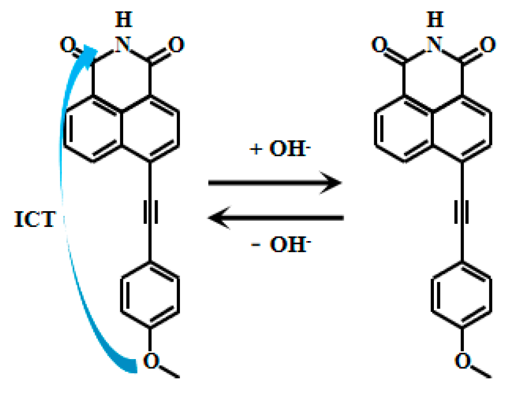
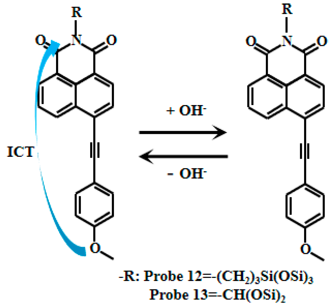
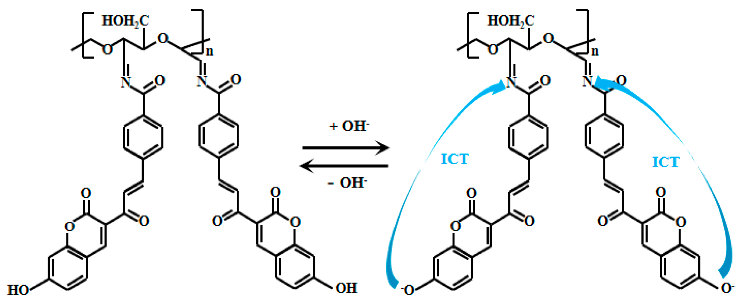
| 11 | Naphthylimide | ICT | 405 | 525 | - | 10.5–12.5 | Fabrication of PVA hydrogel for pH detection in concrete | [60,61] |
| 12 & 13 | Naphthylimide | ICT | 365 | 520&530 | - | 9.0–13.0 | Measurement of concrete simulated pore fluid pH | [62] |
| 14 | Coumarin | ICT | 365 | 459, 577 | I459/I577 | 9.0–13.0 | Environmental water samples, pH test strips | [63] |
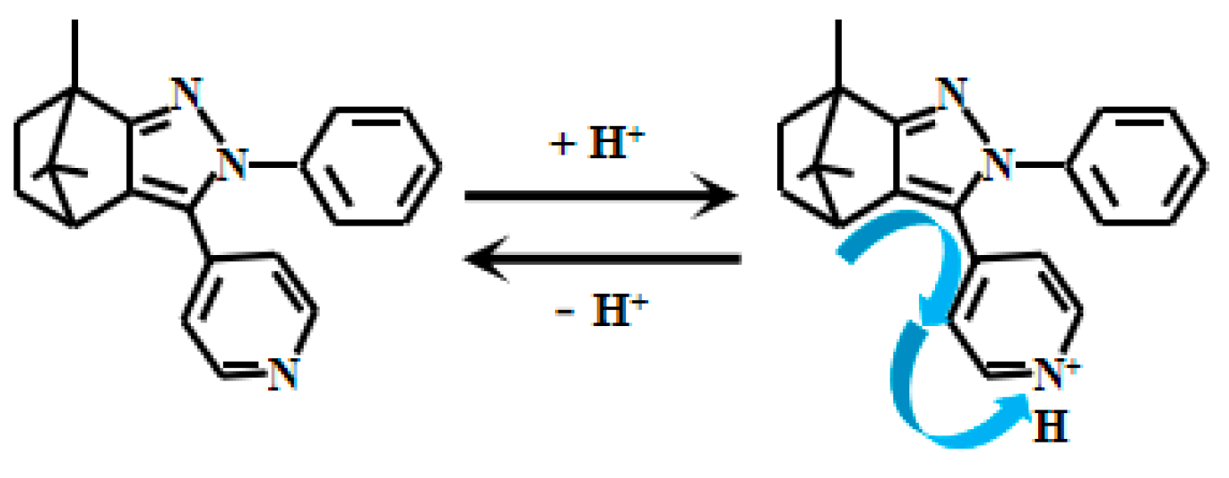
| 15 | Benzo[d]imidazole | ICT | - | 607 | - | 7.0–9.30 | - | [68] |
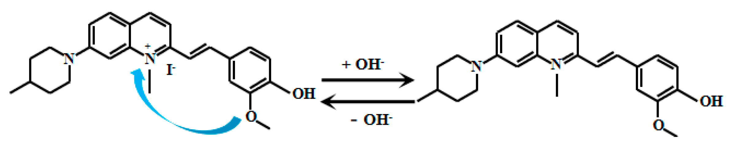
| 16 | Quinoline | ICT | 488 | 630 | - | 6.50–10.00 | Mitochondrial pH detection in HeLa, HepG2, and L-O2 cells | [69] |
| 17 | Coumarin | PET | 400 | 476 | - | 6.50–9.98 | Fluorescent imaging in zebrafish | [70] |
| 18 | Naphthalimide | ICT | 380 | 479 | - | 7.4–12.0 | Fabrication of test strips for fluorescent imaging in HeLa cells, and detection in real water/food samples | [71] |
| 19 | Myristaldehyde | ICT | 400 | 580 | - | 7.45–12.89 | Fluorescent imaging in HeLa cells and zebrafish | [72] |
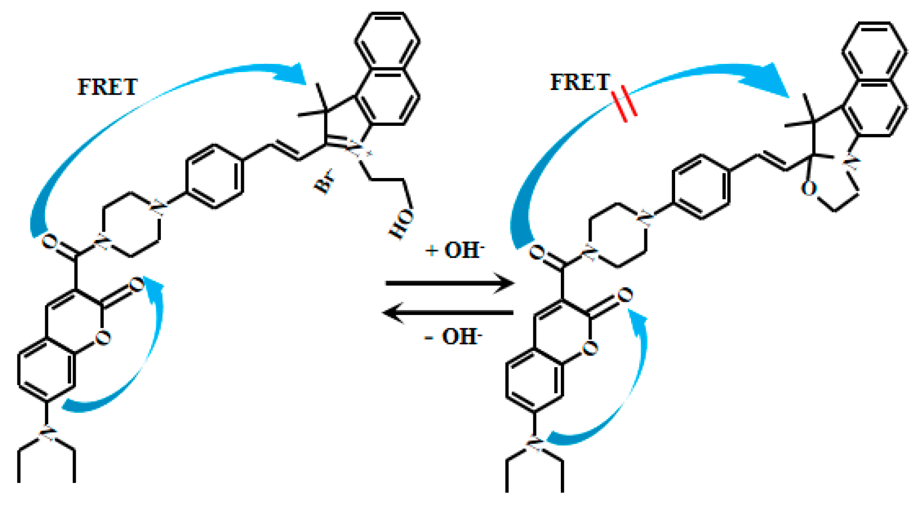
| 20 | Coumarin | FRET | 420 | 485, 608 | I485/I608 | 6.0–8.0 | Mitochondrial pH detection in HeLa cells | [66] |
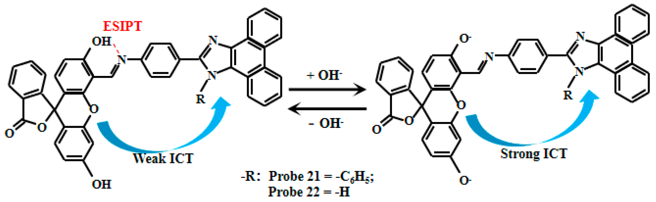
| 21 & 22 | Fluorescein | ESIPT, ICT | 320 | 518 & 516 | - | 5.40–7.30 6.50–8.00 | - | [76] |
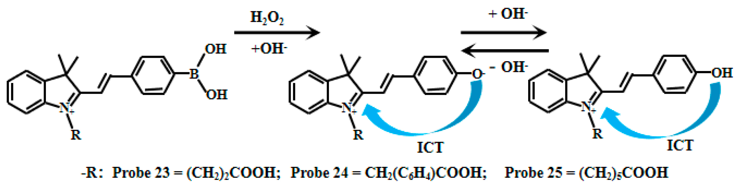
| 23 & 24 & 25 | Hemicyanine | ICT | 460 | 530, 560 | I530/I560 | 6.47, 6.47 & 6.40 | Fluorescent imaging in HeLa cells with H2O2 response | [77] |
| 26 | Benzothiazole | ICT | 600 | 672 nm, 715 nm | I672/I715 | 6.2–7.8 | pH detection in real water/human serum, freshness assessment of pork/chicken breast, fluorescent imaging in HeLa cells with viscosity response | [78] |
| 27 | Coumarin | PET | 545 | 650 | - | 2.00–6.65, 6.65–12.00 | Lysosomal fluorescent imaging in HeLa and SH-SY5Y cells | [79] |
| 28 | 8-Hydroxyquinoline | ICT | 365 | 665, 710 | I710/I665 | 5.0–13.0 | - | [80] |
Disclaimer/Publisher’s Note: The statements, opinions and data contained in all publications are solely those of the individual author(s) and contributor(s) and not of MDPI and/or the editor(s). MDPI and/or the editor(s) disclaim responsibility for any injury to people or property resulting from any ideas, methods, instructions or products referred to in the content. |
© 2025 by the authors. Licensee MDPI, Basel, Switzerland. This article is an open access article distributed under the terms and conditions of the Creative Commons Attribution (CC BY) license (https://creativecommons.org/licenses/by/4.0/).
Share and Cite
Xu, W.; Ma, Z.; Tian, Q.; Chen, Y.; Jiang, Q.; Fan, L. A Review of Fluorescent pH Probes: Ratiometric Strategies, Extreme pH Sensing, and Multifunctional Utility. Chemosensors 2025, 13, 280. https://doi.org/10.3390/chemosensors13080280
Xu W, Ma Z, Tian Q, Chen Y, Jiang Q, Fan L. A Review of Fluorescent pH Probes: Ratiometric Strategies, Extreme pH Sensing, and Multifunctional Utility. Chemosensors. 2025; 13(8):280. https://doi.org/10.3390/chemosensors13080280
Chicago/Turabian StyleXu, Weiqiao, Zhenting Ma, Qixin Tian, Yuanqing Chen, Qiumei Jiang, and Liang Fan. 2025. "A Review of Fluorescent pH Probes: Ratiometric Strategies, Extreme pH Sensing, and Multifunctional Utility" Chemosensors 13, no. 8: 280. https://doi.org/10.3390/chemosensors13080280
APA StyleXu, W., Ma, Z., Tian, Q., Chen, Y., Jiang, Q., & Fan, L. (2025). A Review of Fluorescent pH Probes: Ratiometric Strategies, Extreme pH Sensing, and Multifunctional Utility. Chemosensors, 13(8), 280. https://doi.org/10.3390/chemosensors13080280