

Characterization of E-Cadherin, SSEA-1, MSI-1, and SOX-2 Expression and Their Association with Pale Cells in Adenomyosis


 ,
, 

Abstract
1. Introduction
2. Materials and Methods
2.1. Patient and Sample Collection
2.2. Immunohistochemistry
2.3. Immunofluorescence
2.4. Immunoelectron Microscopy
2.5. Statistical Analysis
3. Results
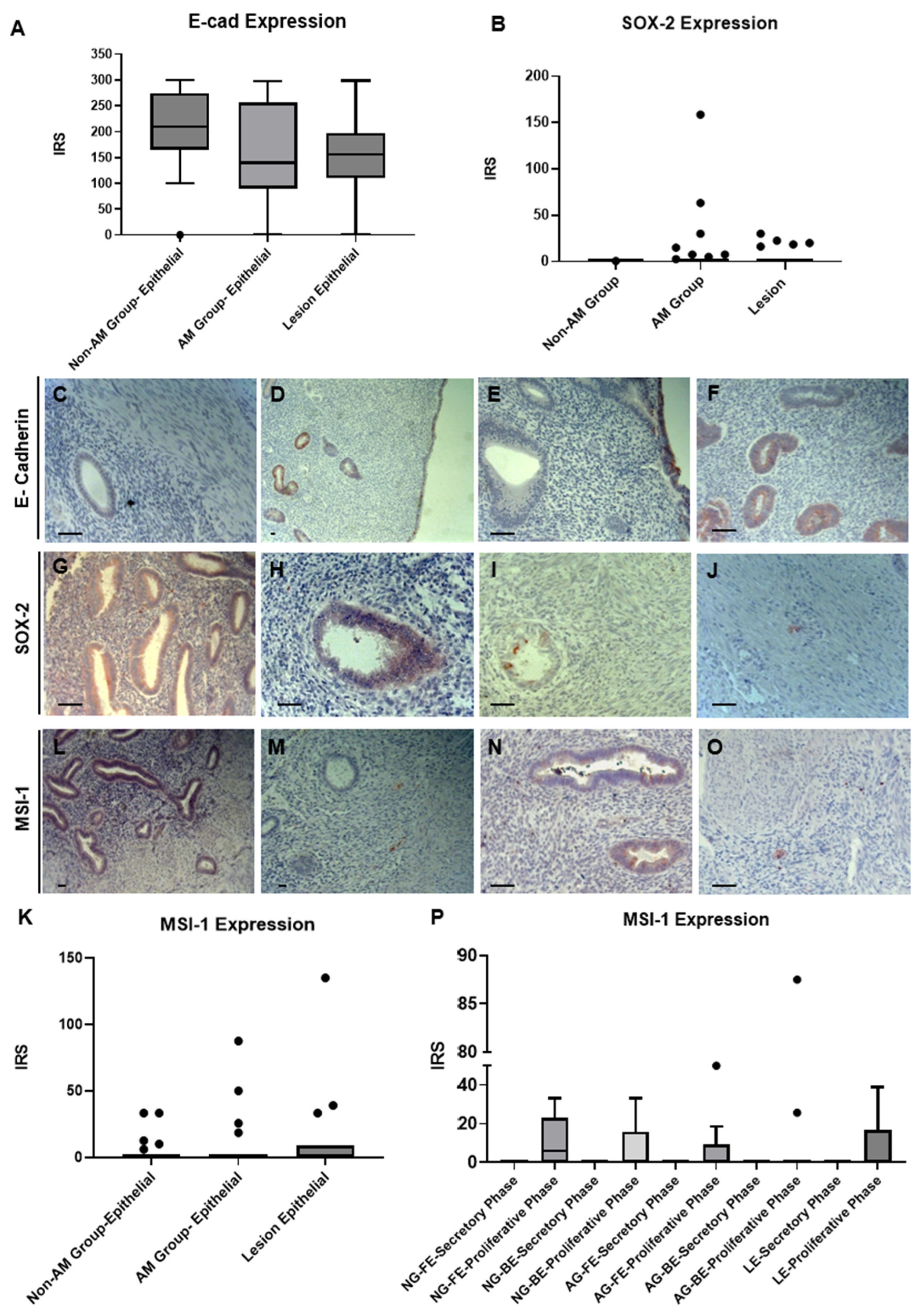
3.1. Immuno-Expression of E-Cadherin
3.2. Immuno-Expression of SOX-2
3.3. Immuno-Expression of MSI-1
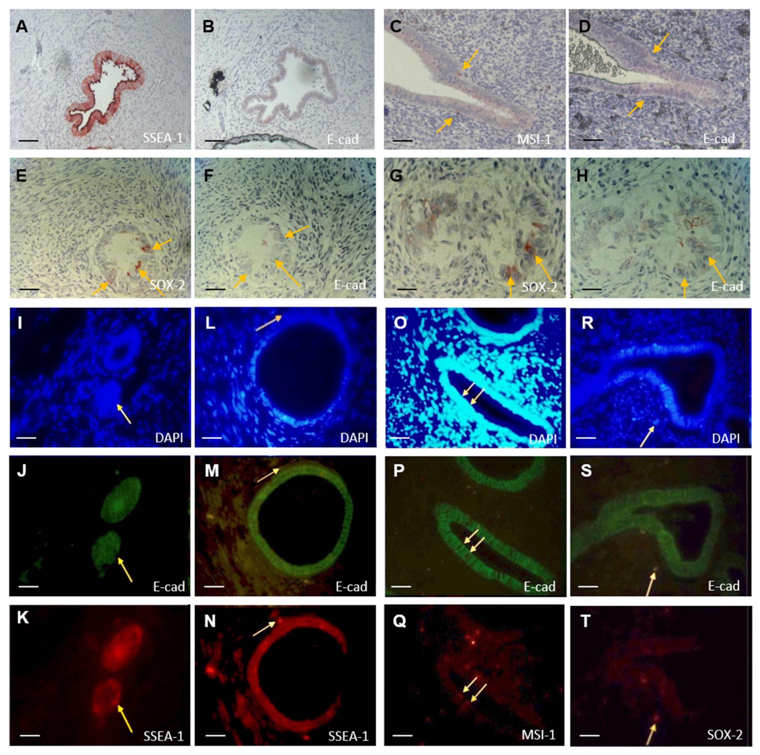
3.4. SSEA-1 Is Upregulated in AM Lesions
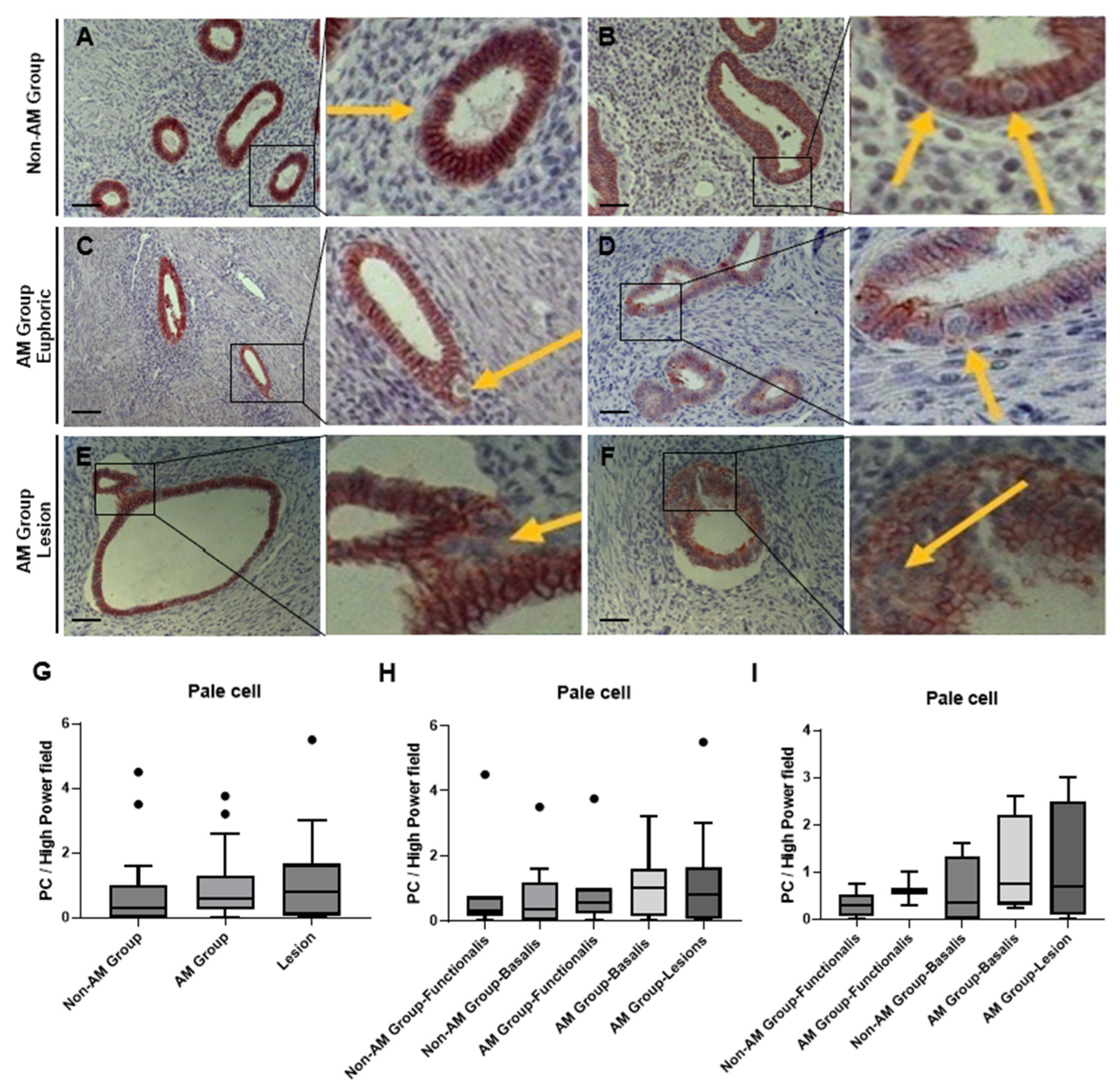
3.5. Phenotypic Characterization of Pale Cells by Light Microscopy
3.6. Immuno-Labeling of Ultrastructurally Identified Pale Cells Using Transmission Electron Microscopy
3.7. Colocalization of E-Cadherin Negative and SOX-2/MSI-1/SSEA-1 Positive Cells
4. Discussion
Supplementary Materials
Author Contributions
Funding
Institutional Review Board Statement
Informed Consent Statement
Data Availability Statement
Conflicts of Interest
References
- Sammour, A.; Pirwany, I.; Usubutun, A.; Arseneau, J.; Tulandi, T. Correlations between extent and spread of adenomyosis and clinical symptoms. Gynecol. Obstet. Investig. 2002, 54, 213–216. [Google Scholar] [CrossRef] [PubMed]
- Bird, C.C.; McElin, T.W.; Manalo-Estrella, P. The elusive adenomyosis of the uterus--revisited. Am. J. Obstet. Gynecol. 1972, 112, 583–593. [Google Scholar] [CrossRef] [PubMed]
- Vannuccini, S.; Petraglia, F. Recent advances in understanding and managing adenomyosis. F1000Research 2019, 8. [Google Scholar] [CrossRef]
- Gordts, S.; Grimbizis, G.; Campo, R. Symptoms and classification of uterine adenomyosis, including the place of hysteroscopy in diagnosis. Fertil. Steril. 2018, 109, 380–388.e381. [Google Scholar] [CrossRef] [PubMed]
- Li, J.J.; Chung, J.P.W.; Wang, S.; Li, T.C.; Duan, H. The Investigation and Management of Adenomyosis in Women Who Wish to Improve or Preserve Fertility. Biomed. Res. Int. 2018, 2018, 6832685. [Google Scholar] [CrossRef] [PubMed]
- De Graaff, A.A.; D’Hooghe, T.M.; Dunselman, G.A.; Dirksen, C.D.; Hummelshoj, L.; Simoens, S. The significant effect of endometriosis on physical, mental and social wellbeing: Results from an international cross-sectional survey. Hum. Reprod. 2013, 28, 2677–2685. [Google Scholar] [CrossRef]
- Hudelist, G.; Fritzer, N.; Thomas, A.; Niehues, C.; Oppelt, P.; Haas, D.; Tammaa, A.; Salzer, H. Diagnostic delay for endometriosis in Austria and Germany: Causes and possible consequences. Hum. Reprod. 2012, 27, 3412–3416. [Google Scholar] [CrossRef]
- Moradi, M.; Parker, M.; Sneddon, A.; Lopez, V.; Ellwood, D. Impact of endometriosis on women’s lives: A qualitative study. BMC Women’s Health 2014, 14, 123. [Google Scholar] [CrossRef]
- Fourquet, J.; Sinaii, N.; Stratton, P.; Khayel, F.; Alvarez-Garriga, C.; Bayona, M.; Ballweg, M.L.; Flores, I. Characteristics of women with endometriosis from the USA and Puerto Rico. J. Endometr. Pelvic Pain Disord. 2015, 7, 129–135. [Google Scholar] [CrossRef]
- Gargett, C.E. Uterine stem cells: What is the evidence? Hum. Reprod. Update 2007, 13, 87–101. [Google Scholar] [CrossRef]
- Padykula, H.A.; Coles, L.G.; McCracken, J.A.; King, N.W., Jr.; Longcope, C.; Kaiserman-Abramof, I.R. A zonal pattern of cell proliferation and differentiation in the rhesus endometrium during the estrogen surge. Biol. Reprod. 1984, 31, 1103–1118. [Google Scholar] [CrossRef] [PubMed]
- Gargett, C.E.; Schwab, K.E.; Deane, J.A. Endometrial stem/progenitor cells: The first 10 years. Hum. Reprod. Update 2016, 22, 137–163. [Google Scholar] [CrossRef] [PubMed]
- Schwab, K.E.; Gargett, C.E. Co-expression of two perivascular cell markers isolates mesenchymal stem-like cells from human endometrium. Hum. Reprod. 2007, 22, 2903–2911. [Google Scholar] [CrossRef] [PubMed]
- Vannuccini, S.; Tosti, C.; Carmona, F.; Huang, S.J.; Chapron, C.; Guo, S.W.; Petraglia, F. Pathogenesis of adenomyosis: An update on molecular mechanisms. Reprod. Biomed. Online 2017, 35, 592–601. [Google Scholar] [CrossRef]
- García-Solares, J.; Donnez, J.; Donnez, O.; Dolmans, M.M. Pathogenesis of uterine adenomyosis: Invagination or metaplasia? Fertil. Steril. 2018, 109, 371–379. [Google Scholar] [CrossRef]
- Yi, K.W.; Kim, S.H.; Ihm, H.J.; Oh, Y.S.; Chae, H.D.; Kim, C.H.; Kang, B.M. Increased expression of p21-activated kinase 4 in adenomyosis and its regulation of matrix metalloproteinase-2 and -9 in endometrial cells. Fertil. Steril. 2015, 103, 1089–1097.e1082. [Google Scholar] [CrossRef]
- Li, T.; Li, Y.G.; Pu, D.M. Matrix metalloproteinase-2 and -9 expression correlated with angiogenesis in human adenomyosis. Gynecol. Obstet. Investig. 2006, 62, 229–235. [Google Scholar] [CrossRef]
- Ibrahim, M.G.; Chiantera, V.; Frangini, S.; Younes, S.; Kohler, C.; Taube, E.T.; Plendl, J.; Mechsner, S. Ultramicro-trauma in the endometrial-myometrial junctional zone and pale cell migration in adenomyosis. Fertil. Steril. 2015, 104, 1475–1483.e1471–e1473. [Google Scholar] [CrossRef]
- He, Y.L.; Ding, N.; Qi, Y.F.; Li, Y.; Xiang, Y.; Qian, T.Y.; Liu, H.; Lin, C.Y.; Yuan, L.; Zhou, H.L.; et al. Visualising the boundary sharpness of uterine zonal structures using high-resolution T2-weighted images during the menstrual cycle. Clin. Radiol. 2019, 74, 81.e19–81.e24. [Google Scholar] [CrossRef]
- Götte, M.; Wolf, M.; Staebler, A.; Buchweitz, O.; Kelsch, R.; Schüring, A.N.; Kiesel, L. Increased expression of the adult stem cell marker Musashi-1 in endometriosis and endometrial carcinoma. J. Pathol. 2008, 215, 317–329. [Google Scholar] [CrossRef]
- Poncelet, C.; Leblanc, M.; Walker-Combrouze, F.; Soriano, D.; Feldmann, G.; Madelenat, P.; Scoazec, J.Y.; Daraï, E. Expression of cadherins and CD44 isoforms in human endometrium and peritoneal endometriosis. Acta Obstet. Gynecol. Scand. 2002, 81, 195–203. [Google Scholar] [CrossRef] [PubMed]
- Gaetje, R.; Kotzian, S.; Herrmann, G.; Baumann, R.; Starzinski-Powitz, A. Nonmalignant epithelial cells, potentially invasive in human endometriosis, lack the tumor suppressor molecule E-cadherin. Am. J. Pathol. 1997, 150, 461–467. [Google Scholar] [PubMed]
- Chen, Y.J.; Li, H.Y.; Huang, C.H.; Twu, N.F.; Yen, M.S.; Wang, P.H.; Chou, T.Y.; Liu, Y.N.; Chao, K.C.; Yang, M.H. Oestrogen-induced epithelial-mesenchymal transition of endometrial epithelial cells contributes to the development of adenomyosis. J. Pathol. 2010, 222, 261–270. [Google Scholar] [CrossRef]
- Bartley, J.; Jülicher, A.; Hotz, B.; Mechsner, S.; Hotz, H. Epithelial to mesenchymal transition (EMT) seems to be regulated differently in endometriosis and the endometrium. Arch. Gynecol. Obstet. 2014, 289, 871–881. [Google Scholar] [CrossRef]
- Valentijn, A.J.; Palial, K.; Al-Lamee, H.; Tempest, N.; Drury, J.; Von Zglinicki, T.; Saretzki, G.; Murray, P.; Gargett, C.E.; Hapangama, D.K. SSEA-1 isolates human endometrial basal glandular epithelial cells: Phenotypic and functional characterization and implications in the pathogenesis of endometriosis. Hum. Reprod. 2013, 28, 2695–2708. [Google Scholar] [CrossRef]
- Tempest, N.; Baker, A.M.; Wright, N.A.; Hapangama, D.K. Does human endometrial LGR5 gene expression suggest the existence of another hormonally regulated epithelial stem cell niche? Hum. Reprod. 2018, 33, 1052–1062. [Google Scholar] [CrossRef]
- Nguyen, H.P.T.; Xiao, L.; Deane, J.A.; Tan, K.S.; Cousins, F.L.; Masuda, H.; Sprung, C.N.; Rosamilia, A.; Gargett, C.E. N-cadherin identifies human endometrial epithelial progenitor cells by in vitro stem cell assays. Hum. Reprod. 2017, 32, 2254–2268. [Google Scholar] [CrossRef]
- Oh, S.J.; Shin, J.H.; Kim, T.H.; Lee, H.S.; Yoo, J.Y.; Ahn, J.Y.; Broaddus, R.R.; Taketo, M.M.; Lydon, J.P.; Leach, R.E.; et al. β-Catenin activation contributes to the pathogenesis of adenomyosis through epithelial-mesenchymal transition. J. Pathol. 2013, 231, 210–222. [Google Scholar] [CrossRef]
- Wang, X.Y.; Yin, Y.; Yuan, H.; Sakamaki, T.; Okano, H.; Glazer, R.I. Musashi1 modulates mammary progenitor cell expansion through proliferin-mediated activation of the Wnt and Notch pathways. Mol. Cell. Biol. 2008, 28, 3589–3599. [Google Scholar] [CrossRef]
- Chen, Y.Z.; Wang, J.H.; Yan, J.; Liang, Y.; Zhang, X.F.; Zhou, F. Increased expression of the adult stem cell marker Musashi-1 in the ectopic endometrium of adenomyosis does not correlate with serum estradiol and progesterone levels. Eur. J. Obstet. Gynecol. Reprod. Biol. 2014, 173, 88–93. [Google Scholar] [CrossRef]
- Lu, X.; Lin, F.; Fang, H.; Yang, X.; Qin, L. Expression of a putative stem cell marker Musashi-1 in endometrium. Histol. Histopathol. 2011, 26, 1127–1133. [Google Scholar] [CrossRef] [PubMed]
- Colitti, M.; Farinacci, M. Expression of a putative stem cell marker, Musashi 1, in mammary glands of ewes. J. Mol. Histol. 2009, 40, 139–149. [Google Scholar] [CrossRef] [PubMed]
- Hapangama, D.K.; Drury, J.; Da Silva, L.; Al-Lamee, H.; Earp, A.; Valentijn, A.J.; Edirisinghe, D.P.; Murray, P.A.; Fazleabas, A.T.; Gargett, C.E. Abnormally located SSEA1+/SOX9+ endometrial epithelial cells with a basalis-like phenotype in the eutopic functionalis layer may play a role in the pathogenesis of endometriosis. Hum. Reprod. 2019, 34, 56–68. [Google Scholar] [CrossRef] [PubMed]
- Götte, M.; Wolf, M.; Staebler, A.; Buchweitz, O.; Kiesel, L.; Schüring, A.N. Aberrant expression of the pluripotency marker SOX-2 in endometriosis. Fertil. Steril. 2011, 95, 338–341. [Google Scholar] [CrossRef]


| Group | Age | Diagnosis | Cycle | GPA | Symptoms |
|---|---|---|---|---|---|
| AM Group | 44 | Adenomyosis uteri | p | 000 | dysmenorrhea |
| 47 | Adenomyosis uteri | p | NI | - | |
| 42 | Adenomyosis uteri | p | 102 | defecation problems | |
| 43 | Endometriosis | p | 321 | lower abdominal pain | |
| 36 | Adenomyosis uteri | p | 422 | - | |
| 43 | Adenomyosis uteri | p | 413 | dysmenorrhea, hypermenorrhea | |
| 47 | Endometriosis | * | 220 | dysmenorrhea, hypermenorrhea | |
| 50 | Adenomyosis uteri | s | 321 | hypermenorrhea | |
| 37 | Adenomyosis uteri | p | 000 | dysmenorrhea | |
| 47 | Adenomyosis uteri | p | 110 | hypermenorrhea | |
| 30 | Adenomyosis uteri | s | 000 | dysmenorrhea, hypermenorrhea | |
| 37 | Adenomyosis uteri | p | 000 | hypermenorrhea, lower abdominal pain | |
| 41 | Adenomyosis uteri | s | 954 | dysmenorrhea, hypermenorrhea | |
| 34 | Adenomyosis uteri | a | 000 | - | |
| 37 | Adenomyosis uteri | p | NI | - | |
| 44 | Adenomyosis uteri | s | 000 | hypermenorrhea | |
| 36 | Adenomyosis uteri | p | 413 | dysmenorrhea, hypermenorrhea | |
| 41 | Adenomyosis uteri | p | 220 | dyspareunia, hypermenorrhea | |
| 41 | Adenomyosis uteri | p | 000 | - | |
| 41 | Adenomyosis uteri | p | 220 | dysmenorrhea | |
| 30 | Adenomyosis uteri | p | NI | dysmenorrhea | |
| 39 | Adenomyosis uteri and endometriosis | * | 000 | - | |
| 47 | Endometriosis | a | 110 | dysmenorrhea, dyspareunia, hypermenorrhea | |
| 45 | Adenomyosis uteri and endometriosis | * | 000 | dysmenorrhea, lower abdominal pain | |
| 45 | Endometriosis | * | 000 | dysmenorrhea, dyspareunia | |
| Non-AM Group | 46 | Uterus myomatous | s | 120 | lower abdominal pain |
| 47 | Uterus myomatous | p | NI | postmenopausal syndrome | |
| 37 | Uterus myomatous | s | 211 | - | |
| 45 | Vascular anomaly | s | 211 | dysmenorrhea | |
| 48 | Uterus myomatous | p | 000 | hypermenorrhea | |
| 50 | Uterus myomatous | p | NI | - | |
| 43 | Uterus myomatous | s | 000 | dyschezia | |
| 50 | Uterus myomatous | * | 220 | micturation disorders | |
| 32 | Cervical intraepithelial neoplasia III | p | 541 | - | |
| 44 | Uterus myomatous | p | 330 | hypermenorrhea | |
| 17 | Uterus didelphys | p | 000 | - | |
| 48 | Uterus myomatous | p | 220 | lower abdominal pain, hypermenorrhea | |
| 41 | Chronic endocervicitis | s | 220 | - | |
| 50 | Uterus myomatous | s | NI | - | |
| 44 | Uterus myomatous | s | NI | - |
| Marker | Eutopic Epithelial Cells | Ectopic Epithelial Cells | Stromal Cells | Myometrial Cells |
|---|---|---|---|---|
| E-cadherin Non-AM | +++ | # | − | − |
| E-cadherin AM | ++ | ++ | − | − |
| SOX-2 Non-AM | − | # | − | − |
| SOX-2 AM | − | − | − | − |
| MSI-1Non-AM | − | # | − | − |
| MSI-1 AM | − | − | − | − |
| SSEA-1 Non-AM | + | # | + | − |
| SSEA-1 AM | + | ++ | − | − |
| Lesion | Basalis | Functionalis | |
|---|---|---|---|
| Centered | 9/22 | 11/18 | 9/13 |
| Eccentric | 8/22 | 4/18 | 3/13 |
| Luminal | 5/22 | 3/18 | 1/13 |
| Total Occurrence | 22/25 | 18/21 | 13/14 |
Disclaimer/Publisher’s Note: The statements, opinions and data contained in all publications are solely those of the individual author(s) and contributor(s) and not of MDPI and/or the editor(s). MDPI and/or the editor(s) disclaim responsibility for any injury to people or property resulting from any ideas, methods, instructions or products referred to in the content. |
© 2024 by the authors. Licensee MDPI, Basel, Switzerland. This article is an open access article distributed under the terms and conditions of the Creative Commons Attribution (CC BY) license (https://creativecommons.org/licenses/by/4.0/).
Share and Cite
Tian, J.; Hoffmann, V.; Ibrahim, M.G.; Hansen, U.; Schüring, A.N.; Velho, R.V.; Mechsner, S.; Götte, M. Characterization of E-Cadherin, SSEA-1, MSI-1, and SOX-2 Expression and Their Association with Pale Cells in Adenomyosis. Biomolecules 2024, 14, 1355. https://doi.org/10.3390/biom14111355
Tian J, Hoffmann V, Ibrahim MG, Hansen U, Schüring AN, Velho RV, Mechsner S, Götte M. Characterization of E-Cadherin, SSEA-1, MSI-1, and SOX-2 Expression and Their Association with Pale Cells in Adenomyosis. Biomolecules. 2024; 14(11):1355. https://doi.org/10.3390/biom14111355
Chicago/Turabian StyleTian, Jingjun, Veronika Hoffmann, Mohamed Gamal Ibrahim, Uwe Hansen, Andreas N. Schüring, Renata Voltolini Velho, Sylvia Mechsner, and Martin Götte. 2024. "Characterization of E-Cadherin, SSEA-1, MSI-1, and SOX-2 Expression and Their Association with Pale Cells in Adenomyosis" Biomolecules 14, no. 11: 1355. https://doi.org/10.3390/biom14111355
APA StyleTian, J., Hoffmann, V., Ibrahim, M. G., Hansen, U., Schüring, A. N., Velho, R. V., Mechsner, S., & Götte, M. (2024). Characterization of E-Cadherin, SSEA-1, MSI-1, and SOX-2 Expression and Their Association with Pale Cells in Adenomyosis. Biomolecules, 14(11), 1355. https://doi.org/10.3390/biom14111355