Article

Curcuma aromatica and Curcuma comosa Extracts and Isolated Constituents Provide Protection against UVB-Induced Damage and Attenuate Matrix Metalloproteinase-1 Expression in HaCaT Cells

by
Wachirachai Pabuprapap
1,
Wongnapa Nakyai
1,*,
Waraluck Chaichompoo
1,
Nattharika Pheedee
1,
Saowanee Phetkeereerat
1,
Jarupa Viyoch
2,
Boon-ek Yingyongnarongkul
1,
Vachiraporn Ajavakom
1,
Apiwat Chompoosor
1,
Pawinee Piyachaturawat
3 and
Apichart Suksamrarn
1
1
Department of Chemistry and Center of Excellence for Innovation in Chemistry, Faculty of Science, Ramkhamhaeng University, Bangkok 10240, Thailand
2
Department of Pharmaceutical Technology and Center of Excellence for Innovation in Chemistry, Faculty of Pharmaceutical Sciences, Naresuan University, Phitsanulok 65000, Thailand
3
Department of Physiology, Faculty of Science, Mahidol University, Bangkok 10400, Thailand
*
Author to whom correspondence should be addressed.
Cosmetics 2022, 9(1), 23; https://doi.org/10.3390/cosmetics9010023
Submission received: 11 January 2022 / Revised: 5 February 2022 / Accepted: 8 February 2022 / Published: 11 February 2022
(This article belongs to the Special Issue Green Ingredients in Cosmetics and Food (Volume II))

Abstract

:
Ultraviolet-B (UVB) exposure is one of the primary extrinsic factors causing skin photoaging. It stimulates inflammatory responses and arrests the cell cycle. Matrix metalloproteinase-1 (MMP-1) secreted by keratinocytes is one of the important extracellular matrixes to attenuate UVB-induced skin aging via collagen degradation. Curcuma aromatica (CA) and Curcuma comosa (CC), the herbaceous plants in the Zingiberaceae family, are commonly used in Thai traditional women’s medicines. The present work was aimed to investigate the potential of the CA and CC extracts and their isolated compounds to attenuate UVB-induced MMP-1 and cell cycle arrest in HaCaT keratinocytes. Total phenolic contents and antioxidant capacities of the extracts were determined. CC extract contains more phenolic components and provides more potent antioxidant activities than CA extract. HaCaTs were pretreated with the extracts or their isolated constituents 14 for 24 h and then repeatedly exposed to UVB at 100 mJ/cm2 10 times. Both extracts and compounds 14 effectively reduce UVB-induced MMP-1 levels in HaCaT cells and restore cell cycle arrest. This is the first report on the potential of CA and CC extracts in reducing UVB-induced MMP-1 expression and regulating cell proliferation in HaCaT cells. Thus, CA and CC extracts might be used as alternative natural agents to prevent UVB-induced skin photoaging.

1. Introduction

Skin aging is a complicated biological process affected by both intrinsic and extrinsic influences. Intrinsic factors for example genetics, cellular metabolism, hormone, and metabolic processes, are considered to be the root cause of skin aging. Up to the age of 50, intrinsic changes occurred in people aged skin represents generalized atrophy with minimal structural changes resulting in slow deterioration. In contrast, UV exposure, one of the most well-known extrinsic factors, may influentially cause the physical damage to the skin responsible for skin photoaging [1,2,3,4]. The three types of UV radiation including UVA (320–400 nm), UVB (290–320 nm) and UVC (100–290 nm) can be harmful to human beings. However, the latter group does not reach the skin, since it is filtered by atmospheric ozone [5]. Although long-wavelength UVA radiation is considered to penetrate the skin reaching the deeper dermal layers, short-wavelength UVB exposure with more energy radiation is principally absorbed by the epidermis layer, which contains the most abundant keratinocyte cells [6,7]. UVB is responsible for the majority of UV radiation that directly penetrates the skin, which causes skin pigmentation and wrinkles, as well as inducing the synthesis of activator protein-1 (AP-1), activating nuclear factor kappa-light-chain-enhancer of activated B cells (NF-κB), generating reactive oxygen species (ROS) on cell surface receptors, such mitogen-activated kinases (MAPK) stimulating the induction and also the expression of matrix metalloproteinases (MMPs) [8]. Thus, because of this damage it is well-established that UVB is more genotoxic than UVA. Matrix metalloproteinases, a family of matrix metallopeptidases or calcium-dependent zinc-containing endopeptidases, degrade proteins and enzymes in the extracellular matrix (ECM) [9,10]. MMP-1, commonly known as a rate-limiting collagenase, is an enzyme that breaks down collagen, the most common protein in dermal connective tissue. Collagen breakdown and degradation are hallmarks of photoaging, so an increase in MMP-1 expression or activity is an indication of UV-induced skin damage [11,12,13,14].
Curcuma aromatica Salisb. (CA) belongs to the Zingiberaceae family, which is widely distributed in China, Japan, and southeastern Asia [15]. The rhizomes of CA are commonly used in oriental traditional medicines for tonic, carminative purposes, and externally in combinations with astringents, bitters, aromatics to treat bruises, skin eruptions, and infections, and to improve complexion, promoting blood circulation to eliminate blood stasis [16]. They have also been reported to possess various pharmacological activities including wound healing, anti-microbial, anti-angiogenic, anthelmintic, anti-tumor, cytoprotective, anti-inflammatory and antioxidant activities [17]. The extract of the rhizome of CA has been found to contain sesquiterpenoids and diterpenoids [18,19]. Curcuma comosa Roxb. (CC) is a phytoestrogen-producing plant belonging to the Zingiberaceae family [20]. The rhizomes of CC have been used to treat flatulence and gynecologic diseases, such as premenstrual syndrome, irregular menstruation, and uterine discomfort. In Southeast Asia, this plant is used as an active ingredient in a variety of traditional women’s medicines. It possesses estrogenic-like properties and is commonly used by postmenopausal women [21,22,23]. Previous phytochemical investigations of this plant revealed the presence of a rich source of diarylheptanoids [24]. Moreover, it has also been reported for various pharmacological activities including lipid-lowering, anti-adipogenic, bone sparing in ovariectomized rats, enhancing the expansion of hematopoietic stem cells for treatment of hematological disorders, antioxidant and anti-inflammatory activities [25,26,27,28,29,30,31].
Although CA and CC are known to possess several biological activities, the knowledge of their skin anti-aging potentials is still ambiguous and no previous studies have been performed to investigate the ability of CA and CC extracts to preserve against UVB-induced human HaCaT cells. Therefore, the present study of the effect of CA and CC extracts together with their isolated constituents on UVB-induced MMP-1 expression, cell proliferation and cell cycle profile in HaCaT cells can be potentially beneficial to the application for skin anti-aging products.

2. Materials and Methods

2.1. Reagents and Chemicals

All chemicals and solvents were used as received from Merck (Darmstadt, Germany), TCI (Tokyo, Japan), or Sigma-Aldrich (St. Louis, MO, USA). 3-(4,5-Dimethylthiazol-2-yl)-5-(3-carboxymethoxyphenyl)-2-(4-sulfophenyl)-2H-tetrazolium (MTS) (Abcam, Cambridge, MA, USA), Dulbecco’s Modified Eagle’s Medium (DMEM) (Gibco, Waltham, MA, USA), Penicillin-Streptomycin (Gibco, Waltham, MA, USA), Amphotericin B (Gibco, Waltham, MA, USA), Trypsin-EDTA (Gibco, Waltham, MA, USA), Fetal Bovine Serum (FBS) (Gibco, Waltham, MA, USA), Matrix Metalloproteinase-1 (MMP-1) Human ELISA kit (Abcam, Cambridge, MA, USA) and Muse® Cell Cycle kit (Merck Millipore, Darmstadt, Germany) were used. The extraction and isolation solvents were analytical grade and the HPLC mobile phases were HPLC grade.

2.2. Plant Materials

The rhizomes of Curcuma aromatica were collected from Sawang Daen Din district, Sakon Nakhon Province, Thailand, and the rhizomes of Curcuma comosa were collected from Kamphaeng Saen district, Nakhon Pathom Province, Thailand in January 2020. The plant species were identified by Assoc. Prof. Nopporn Dumrongsiri, Ramkhamhaeng University. The voucher specimens are deposited at the Faculty of Science, Ramkhamhaeng University, Thailand (Apichart Suksamrarn, Nos. 095 and 099, respectively).

2.3. Preparation of Plant Extracts

The fresh rhizomes of C. aromatica and C. comosa were separately sliced, air-dried, milled, and macerated with 95% (v/v) ethanol at room temperature for 72 h. The extraction procedure for each of the plant materials was repeated five times. The combined solution was filtered and the solvent was evaporated in vacuo at 40–45 °C. The residues were freeze-dried to remove the remaining solvent and the extracts were kept at −20 °C until further experiments.

2.4. Total Phenolic Content

The total phenolic content of the extract was determined by using Folin–Ciocalteu assay as previously reported [32].
A volume of 10 μL of the extract was mixed with 10 μL of the Folin–Ciocalteu reagent and 130 μL of deionized water and was neutralized with 100 μL of 7% (w/v) sodium carbonate solution. The reaction mixture was incubated in dark at room temperature for 30 min. The absorbance will be determined at the wavelength of 734 nm by a microplate spectrophotometer (Sunrise, Opfikon, Switzerland). The total phenolic contents are expressed as gallic acid equivalent per gram of extract (μg GAE/mg dry weight) for the standard curve.

2.5. Antioxidant Assays

The antioxidant capacities were measured by 2,2-diphenyl-1-picrylhydrazyl (DPPH), ferric ion reducing antioxidant power (FRAP), 2,2ʹ-azino-bis-(3-ethylbenzothiazoline-6-sulfonic acid) (ABTS) and nitric oxide (NO) radical scavenging methods as previously reported [32].

2.5.1. DPPH Free Radical Scavenging Assay

A solution of 0.2 mM DPPH (150 μL) was incubated with 75 μL of the sample dissolved in ethanol for 30 min in the dark. The absorbance was recorded against a blank at the wavelength of 517 nm. L-Ascorbic acid was used as a positive control. The inhibition percentage was calculated using equation (1) and the capacity to scavenge the DPPH radical by 50% (IC50) was calculated from the dose–effect curves by linear regression.
% Inhibition = [(Acontrol − Asample)/Acontrol] × 100
where Asample is the absorbance of the sample and Acontrol is the absorbance of reagent solution mixed with ethanol (blank).

2.5.2. FRAP Radical Scavenging Assay

Briefly, the solution of 10 mM of 2,4,6-tripyridyl-S-triazine (TPTZ) in 40 mM HCl, FeCl3·6H2O (20 mM) and acetate buffer (pH 3.6) were mixed in the ratio of 1:1:10 to obtain the working FRAP reagent. The reaction mixture was incubated at 37 °C for 10 min. The test sample (50 μL) was mixed with 150 μL of working FRAP solution for 10 min at room temperature. The absorbance of the colored product was measured at the wavelength of 593 nm. L-Ascorbic acid was used as a positive standard. The inhibition percentage was calculated using equation (1) and the FRAP activity to scavenge the radical by 50% (IC50) was calculated from the dose–effect curves by linear regression.

2.5.3. ABTS Radical Scavenging Assay

The stock solutions which are 7 mM ABTS solution and 2.4 mM potassium persulfate were freshly prepared. These stock solutions were then mixed in equal quantities and allowing them to react for 16–18 h in the dark. The solution was diluted by mixing 1 mL ABTS•+ solution with 10 mL ethanol to obtain an absorbance of less than 0.99 ± 0.01 units at 734 nm. The test sample (15 μL) was allowed to react with 150 μL of the freshly prepared ABTS•+ solution and the absorbance was taken at 734 nm after 10 min. The ABTS scavenging capacity of the extract will be compared with those of L-ascorbic acid and percentage inhibition calculated using equation (1).

2.5.4. NO Radical Scavenging Assay

Briefly, 10 mM sodium nitroprusside (SNP) solution (60 µL) was freshly prepared in phosphate buffer saline (pH 7.4) and was mixed with 60 µL of the test sample. The mixture was incubated at room temperature for 120 min. Then, 120 µL of Griess reagent (a mixture of 1% (w/v) sulfanilamide in 2.5% (v/v) H3PO4 and 0.1% (w/v) N-(1-naphthyl)ethylenediamine dihydrochloride (NED) in 2.5% (v/v) H3PO4) was added to the mixed solution, which was incubated at room temperature for 10 min. The incubated solution was measured for absorbance at 550 nm. L-Ascorbic acid was used as a positive control. The percentage of NO radical scavenging activity in a similar manner of DPPH assay.

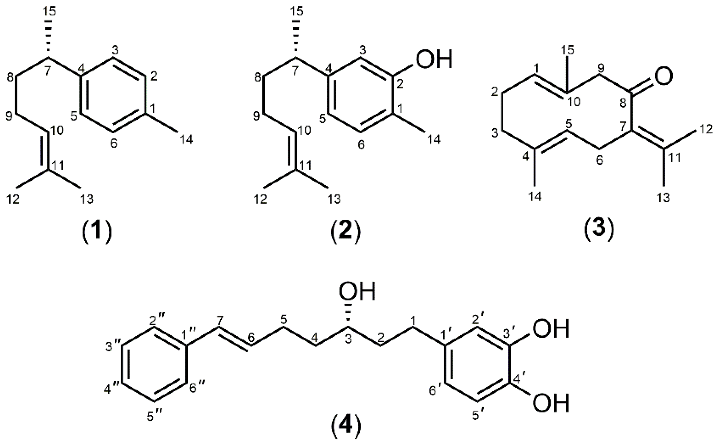
2.6. Isolation of Marker Compounds from Crude Extracts

The extract of CA was subjected to silica column chromatography, eluting with n-hexane, n-hexane:EtOAc, and EtOAc by increasing the more polar solvent to give (–)-curcumene (1), (–)-xanthorrhizol (2) and germacrone (3) (Figure 1). The spectroscopic (1H and 13C NMR, 2D NMR, and mass spectra) data were in agreement with their structures and were consistent with those of the reported values [33,34,35].
(–)-Curcumene (1). Colorless oil; α D 25 –29.1 (c 3.81, CHCl3); 1H NMR (400 MHz, CDCl3) δ 7.11 (2H, d, J = 8.4 Hz, H-3, H-5), 7.08 (2H, d, J = 8.4 Hz, H-2, H-6), 5.11 (1H, br t, 7.1 Hz, H-10), 2.67 (1H, m, H-7), 2.33 (3H, s, CH3-14), 1.88 (2H, m, H-9), 1.68 (3H, br s, CH3-12), 1.60 (2H, m, H-8), 1.54 (3H, br s, CH3-13), 1.23 (3H, d, J = 6.9 Hz, CH3-15); 13C NMR (100 MHz, CDCl3) δ 144.6 (C-1), 135.0 (C-4), 131.2 (C-11), 128.9 (C-3, C-5), 126.8 (C-2, C-6), 124.5 (C-10), 39.0 (C-7), 38.4 (C-8), 26.1 (C-9), 25.6 (CH3-12), 22.4 (CH3-15), 20.9 (CH3-14), 17.6 (CH3-13); HR-TOFMS (ES) m/z 201.1696 (calcd for C15H21, 201.1648).
(–)-Xanthorrhizol (2). Pale yellow oil; α D 25 –44.2 (c 2.82, CHCl3); 1H NMR (400 MHz, CDCl3) δ 7.01 (1H, d, J = 7.6 Hz, H-6), 6.66 (1H, dd, J = 7.6, 1.4 Hz, H-5), 6.59 (1H, d, J = 1.4 Hz, H-3), 5.07 (1H, br t, J = 7.1 Hz, H-10), 2.59 (1H, m, H-7), 2.20 (3H, s, CH3-14), 1.86 (2H, m, H-9), 1.66 (3H, br s, CH3-12), 1.57 (2H, m, H-8), 1.52 (3H, br s, CH3-13), 1.18 (3H, d, J = 6.9 Hz, CH3-15); 13C NMR (100 MHz, CDCl3) δ 153.5 (C-2), 147.2 (C-4), 131.4 (C-11), 130.7 (C-6), 124.4 (C-10), 120.7 (C-1), 119.4 (C-5), 113.5 (C-3), 39.0 (C-7), 38.3 (C-8), 26.1 (C-9), 25.6 (CH3-12), 22.3 (CH3-14), 17.6 (CH3-13), 15.2 (CH3-15); HR-TOFMS (ES) m/z 217.1594 (calcd for C15H21O, 217.1597).
Germacrone (3). White amorphous solid; 1H NMR (400 MHz, CDCl3) δ 4.95 (1H, br d, J = 11.0 Hz, H-1), 4.68 (1H, br dd, J = 11.3, 2.7 Hz, H-5), 3.38 (1H, d, J = 10.5 Hz, H-9b), 2.92 (1H, d, J = 10.5 Hz, H-9a), 2.91 (1H, br d, J = 12.5 Hz, H-6b), 2.82 (1H, br dd, J = 12.5, 2.8 Hz, H-6a), 2.32 (1H, partially overlapping signal, H-2b), 2.13 (1H, partially overlapping signal, H-3b), 2.08 (1H, partially overlapping signal, H-3a), 2.05 (1H, partially overlapping signal, H-2a), 1.74 (3H, s, CH3-12), 1.69 (3H, s, CH3-13), 1.59 (3H, d, J = 2.7 MHz, CH3-14), 1.41 (3H, br s, CH3-15); 13C NMR (100 MHz, CDCl3) δ 207.8 (C-8), 137.2 (C-11), 134.9 (C-4), 132.6 (C-1), 129.4 (C-10), 126.6 (C-7), 125.3 (C-5), 55.8 (C-9), 38.0 (C-3), 29.2 (C-6), 24.0 (C-2), 22.3 (CH3-13), 19.8 (CH3-12), 16.7 (CH3-14), 15.5 (CH3-15); HR-TOFMS (ES+) m/z 241.1530 (calcd for C15H22ONa, 241.1562).
Column chromatography of the CC extract eluting with the isocratic solvent system of n-hexane:EtOAc (3:2) gave (3S)-1-(3,4-dihydroxyphenyl)-7-phenyl-(6E)-6-hepten-3-ol (4). The spectroscopic (1H and 13C NMR, 2D NMR, and mass spectra) data were in agreement with the structure and were consistent with those of the reported values [24].
(3S)-1-(3,4-Dihydroxyphenyl)-7-phenyl-(6E)-6-hepten-3-ol (4). White amorphous solid; α D 24 –19.9 (c 0.77, CHCl3); 1H NMR (400 MHz, CDCl3) δ 7.30 (2H, td, J = 8.5, 1.8 Hz, H-3˝, H-5˝), 7.26 (2H, dd, J = 8.5, 1.6 Hz, H-2˝, H-6˝), 7.18 (1H, tdd, J = 8.5, 1.8, 1.6 Hz, H-4˝), 6.74 (1H, d, J = 8.1 Hz, H-5’), 6.68 (1H, d, J = 1.9 Hz, H-2’), 6.59 (1H, dd, J = 8.1, 1.9 Hz, H-6’), 6.38 (1H, br d, J = 15.8 Hz, H-7), 6.19 (1H, dt, J = 15.8, 6.9 Hz, H-6), 5.52 (1H, br s, 3’-OH), 5.31 (1H, br s, 4’-OH), 3.68 (1H, m, H-3), 2.65 (1H, m, H-1b), 2.55 (1H, m, H-1a), 2.30 (1H, m, H-5), 1.73 (1H, m, H-2), 1.63 (1H, m, H-4); 13C NMR (100 MHz, CDCl3) δ 143.5 (C-4’), 141. 7 (C-3’), 137.5 (C-1’), 134.9 (C-1˝), 130.3 (C-7), 130.2 (C-6), 128.5 (C-2˝, C-6˝), 126.9 (C-4˝), 125.9 (C-3˝, C-5˝), 120.6 (C-6), 115.4 (C-2’), 115.3 (C-5’), 71.0 (C-3), 39.0 (C-2), 36.9 (C-4), 31.3 (C-1), 29.2 (C-5); HR-TOFMS (ES+) m/z 321.1469 (calcd for C19H22O3Na, 321.1461).

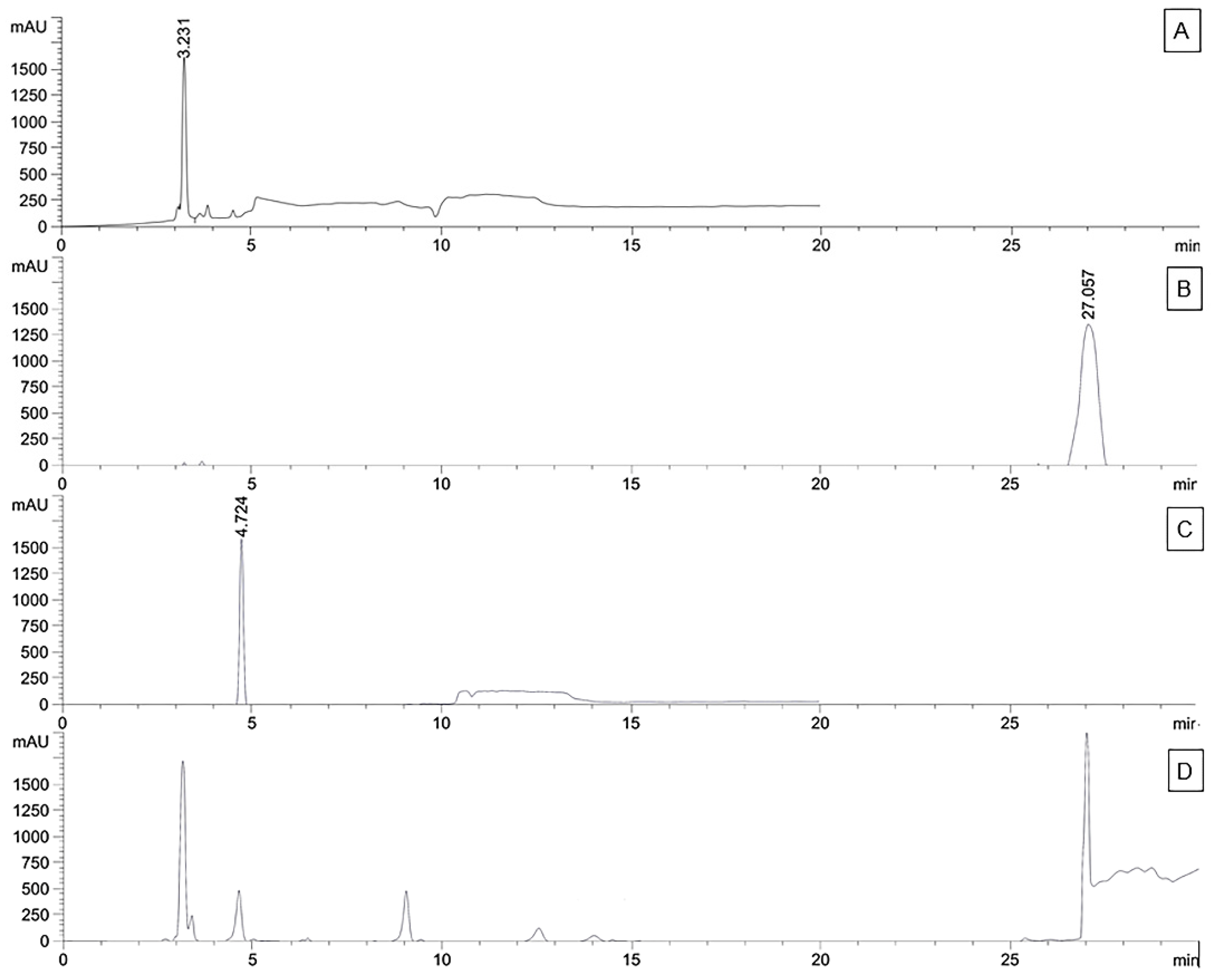
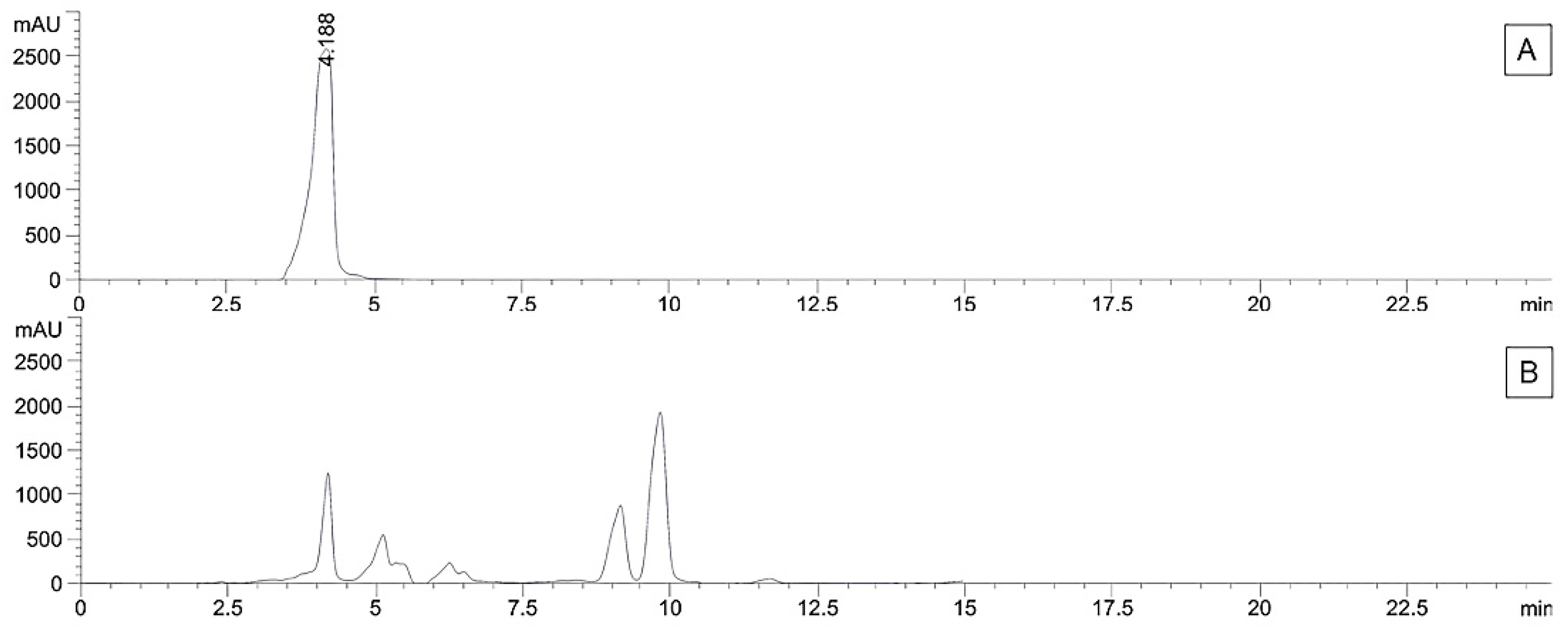
2.7. Quantification of Marker Compounds in Crude Extracts using High-Performance Liquid Chromatography-Diode Array (HPLC-DAD)

The purity of isolated marker compounds 14 was determined by 1H NMR and HPLC techniques. The quantity of marker compounds in the extract was determined using Agilent 1200 series HPLC system equipped with a G1311A pump and a G1315B DAD detector (Agilent Technologies, CA, USA).
For CA, the extract and each of the marker compounds 13 were accurately weighed and separately dissolved in isopropanol, filtered through a 0.22 µm nylon filter and the solution of each sample was injected into Phenomenex Luna Silica (2) (250 × 4.60 mm × 5 µm) column (Phenomenex, CA, USA) at a column temperature of 25 °C. The mobile phase consisted of n-hexane (solvent A) and isopropanol (solvent B). The elution program was optimized as follows: 0.0–6.0 min, 0.5% B, flow rate 1.0 mL/min; 6.0–6.3 min, 0.5% B, flow rate 1.8 mL/min; 6.3-19.0 min, 1% B, flow rate 1.8 mL/min; 19.0–30.0 min, 0.5% B, flow rate 1.0 mL/min. The injected volume was 50 µL and monitored at 214 nm.
For CC, the extract and marker compound 4 were prepared in a similar manner to those of the CA extract analysis, except that acetonitrile has been used in place of isopropanol. The analytical column was a 250 × 4.60 mm × 5 µm Phenomenex Luna reverse phase C18 column (Phenomenex, CA, USA) at a column temperature of 25 °C. Mobile phase A was 0.5% (v/v) acetic acid in water and mobile phase B was acetonitrile. The gradient elution program was optimized as follows: 0.0–5.0 min, 70% B; 5.0–10.0 min, 75% B; 10–15.0 min, 80% B; 15.0–25.0 min, 90% B. The analysis was carried out at a constant flow rate of 1.0 mL min−1. The injected volume was 50 µL and was monitored at 254 nm. The amount of marker compounds was calculated by using the peak integration of each compound using a calibration curve generated from isolated pure marker compounds. All experiments were performed in triplicate.

2.8. Validation of HPLC Analysis

Validation of HPLC analysis was performed using the purified marker compounds. Calibration curves were conducted from the peak areas of the standards 14 versus their seven different concentrations in the range of 3.91–250.00 µg/mL (13) and 1.95–125.00 µg/mL (4). The linearity of the calibration curves was evaluated using correlation coefficients (R2). The limit of detection (LOD) and the limit of quantification (LOQ) were determined as signal-to-noise (S/N) ratios of 3 and 10, respectively. The precision of the method was determined via a repeatability test within one day (intra-day) and an intermediate precision over three days (inter-day). Intra-day analysis was performed by analyzing the same standard solutions at three different concentrations (7.81, 31.25 and 125.00 µg/mL for compounds 13 and 3.91, 15.63 and 62.50 µg/mL for compound 4) in a single day and inter-day analysis was achieved by analyzing standard solutions at three different concentrations on three consecutive days, respectively. Precisions are given as relative standard deviations (RSDs) (where % RSD = (standard deviation / mean) × 100). The accuracy of the method was determined by the recovery test. Recovery was calculated as follows: % recovery = ((detected concentration − initial concentration)/spiked concentration) × 100.

2.9. Cell Line and Culture Conditions

The HaCaT cells were purchased from CLS Cell Lines Service GmbH (Eppelheim, Germany). They were maintained in Dulbecco’s modified Eagle’s medium (DMEM) (GIBCO, Paisley, UK) supplemented with 10% (v/v) fetal bovine serum (GIBCO, Paisley, UK) and 1% (v/v) penicillin/streptomycin (GIBCO, Paisley, UK). Cells were grown at 37 °C in a humidified 5% CO2 atmosphere. When cells (from passages 16 to 25) reached 70-80% confluence, they were trypsinized, planted at different densities according to the parameters studied, and incubated for 24 h.

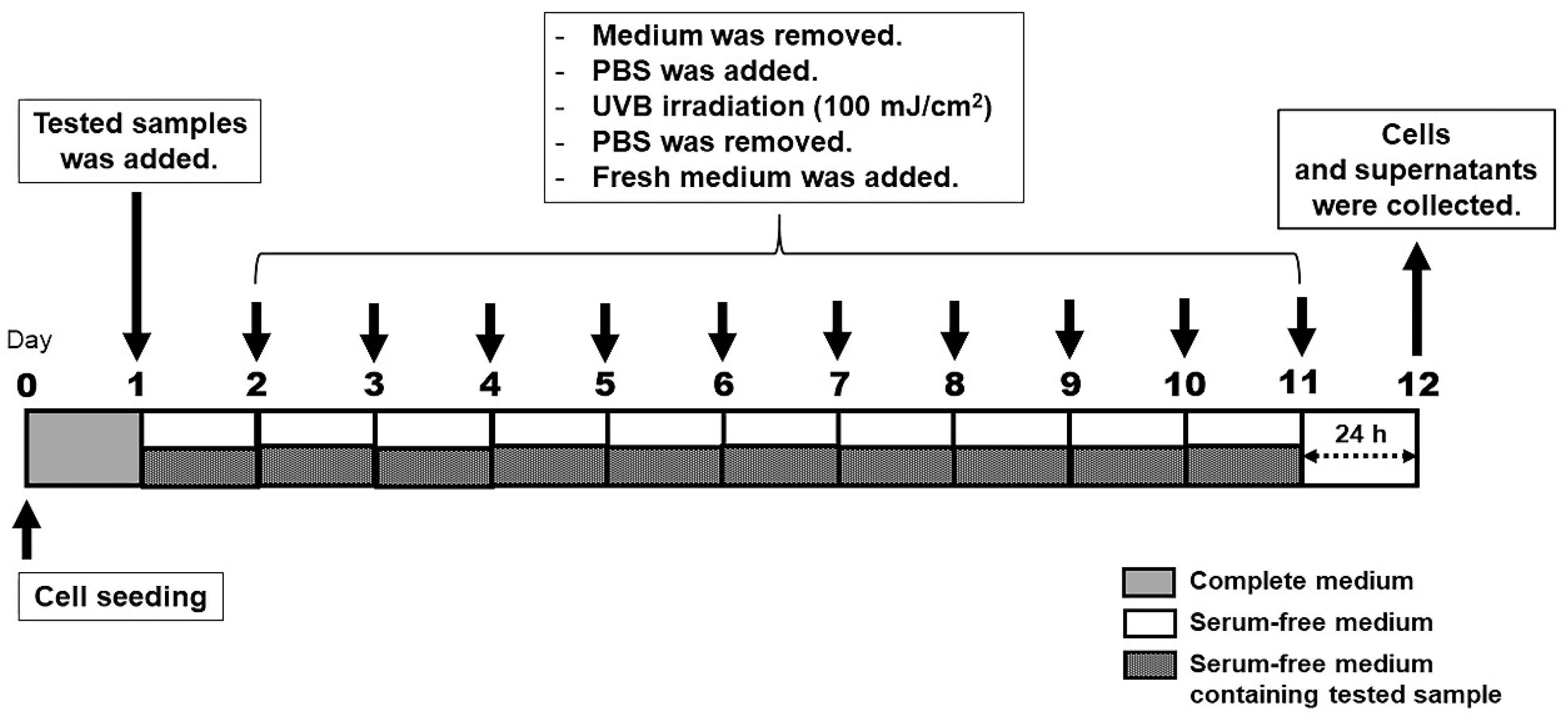
2.10. UV Irradiation

The HaCaT cells (5 × 10⁵ cell dish−1) were seeded into a 60 mm Petri dish and were incubated in cultured media for 24 h. Prior to UVB exposure, cells were washed with PBS. Cells were exposed to various intensities of UVB (50, 100, 200, and 400 mJ cm−2) under a thin layer of PBS. Philips TL 20W/01 UVB Narrowband TL lamps with an emission spectrum between 290 and 315 nm (peak, 310–315 nm) were used as the UVB source. The UVB source was positioned at 30 cm above the cell culture dishes. Sentry UV meter (model ST513, New Taipei City, Taiwan) was used to measure UV irradiance. Immediately after UVB irradiation, PBS was replaced with serum-free media for 24 h. Cell dishes were then repeatedly UVB-irradiated 10 times. The protocol for UVB irradiation is illustrated in Figure 2. After 24 h of the last irradiation, the culture media was removed and MTS solution was added into the culture dishes and further incubated for 4 h. The cell viabilities were quantified by the absorbance at 493 nm using a microplate spectrophotometer. The absorbance of the non-UVB-exposed cells was adjusted to 100% viability. Cell morphology was observed using an inverted microscope.

2.11. Cell Viability

To determine the effect of non-toxic concentrations of CA and CC extracts, cell viability was evaluated by MTS assay. The HaCaT cells (2 × 10⁴ cells/well) were seeded into 96-well plates and were maintained until 80% confluency, and the cells were pre-treated with serum-free medium or serum-free medium containing the extracts for 24 h. The cells were washed with PBS and irradiated (or non-irradiated for the control group) with UVB under a thin layer of PBS. After irradiation, the cells were then placed in a fresh serum-free medium and continuedly incubated for 24 h. After incubation, the culture media was removed and MTS solution was added into culture plates and further incubated for 4 h. The cell viabilities were quantified by the absorbance at 493 nm using a microplate spectrophotometer. The absorbance of the non-UVB-exposed cells was adjusted to 100% viability. Cell morphology was observed using an inverted microscope.

2.12. Cell Proliferation

To determine the effect of CA and CC extracts and their isolated constituents on cell proliferation, the HaCaTs were evaluated by trypan blue exclusion assays. Briefly, the cells were seeded into a 60 mm Petri dish at 5 × 10⁵ cells/dish and then incubated in a humidified atmosphere (37 °C, 5% CO2) for 24 h. Cells were then repeatedly UVB-irradiated 10 times as described in Figure 2. After 24 h of the last UVB exposure, cells were trypsinized and stained with 0.4% trypan blue solution (GIBCO, Paisley, UK). Enumeration of viable cells was carried out under a phase-contrast microscope with a hemocytometer.

2.13. Enzyme-Linked Immunosorbent Assay (ELISA)

The release of MMP-1 in UVB-irradiated HaCaT cells was analyzed by the ELISA technique. The HaCaT cells (5 × 10⁵ cells/dish) were pre-incubated in a 60 mm Petri dish for 24 h, and the cells were pre-treated with serum-free medium or serum-free medium containing the extracts or the isolated compounds for 24 h. Cell dishes were washed with PBS before UVB exposure (100 mJ/cm2) under a thin layer of PBS. Immediately after UVB irradiation, the cells were treated with a serum-free medium and further incubated for another 24 h. Cell dishes were then repeatedly UVB-irradiated 10 times. The protocol for cell treatment and irradiation is shown in Figure 2. Supernatant from each well was collected after 24 h of the last irradiation and analyzed for its MMP-1 contents by the manufacturer’s instructions of the ELISA kit.

2.14. Cell-cycle by Flow Cytometry

The HaCaT cells were maintained and repeatedly UVB-irradiated 10 times as described in Figure 2. After 24 h of the last UVB-irradiation and the removal of supernatants, the cells were washed twice with PBS, fixed and permeabilized with ice-cold 70% (v/v) ethanol, and washed with PBS. The cultures were then resuspended in Guava cell-cycle reagent. The cell-cycle analyses were performed using a Guava easyCyte 8HT flow cytometer (Merck Millipore, Darmstadt, Germany). The percentage of cells in the different stages of the cell cycle was assessed by a flow cytometer and analyzed with Guava Cell-Cycle software. The obtained results were expressed as the percentage of cells in the different stages of the cell cycle in treated cultures compared to that present in the control cultures.

2.15. Statistical Analysis

The results are presented as means ± SD, and statistical analyses were performed using a Student’s t-test by using GraphPad Prism version 7.00 for Windows software (La Jolla, CA, USA). p-Values < 0.05 were considered to indicate a statistically significant.

3. Results and Discussion

3.1. Extraction and Isolation

In preliminary screening for anti-photoaging agents, it was found that the ethanolic extracts of CA and CC exerted cytoprotective activity against UVB irradiation in human HaCaT keratinocytes by inducing the expression of MMP-1. This result prompts us to investigate phytochemical constituents from both CA and CC extracts, in order to clarify their anti-MMP-1 expression.
Briefly, the air-dried powder of the CA and CC rhizomes was extracted by the alcoholic maceration method. The yield of the ethanolic extract of CA was 10.8% and that of the CC extract was 9.6%. The chromatographic investigation of the ethanol extract of the rhizomes of CA led to the isolation of three major compounds, (–)-curcumene (1), (–)-xanthorrhizol (2) and germacrone (3). Additionally, the investigation of the ethanolic extract of the CC rhizomes resulted in the isolation of the major diarylheptanoid 4 [24] as the active constituent. Their structures were identified based on spectroscopic data and by comparisons with those of the reported values. The purity of isolated compounds 14 was more than 95%, determined by 1H NMR and HPLC techniques. The extracts and these compounds were then used for further experiments.

3.2. Antioxidant Activities of CA and CC Extracts

Oxidative stress is a pathological state, in which the reactive oxygen species (ROS) lead to oxidative modification of biomacromolecules, tissue injury, and accelerated cellular aging and death [36]. Excessive UVB exposure is a primary environmental condition that contributes to the development of skin disorders. Particularly, UVB exposure produces ROS, which damages DNA. This damage eventually leads to skin inflammation, photoaging, and photocarcinogenesis. The incident can be prevented by applying anti-oxidative agents that reduce the damage [37].
The total phenolic contents and IC50 of the antioxidant activities of the ethanol extracts of CA and CC are shown in Table 1. Total phenolic contents of CA and CC extracts were determined by using Folin–Ciocalteu method, which was 90.89 and 186.99 µg GAE/mg, respectively. The antioxidant potency of CC extract obtained by NO assay (IC50 0.26 µg/mL) showed higher potency than those obtained by ABTS (IC50 2.67 µg/mL), DPPH (IC50 21.22 µg/mL) and FRAP (IC50 32.47 µg/mL) assays, respectively, whereas CA extracts indicated antioxidant property against ABTS (IC50 19.17 µg/mL), DPPH (IC50 78.77 µg/mL), FRAP (IC50 86.85 µg/mL) and NO (IC50 159.90 µg/mL) assays. In cellular metabolism of the living body, free radical ROS and reactive nitrogen species (RNS) including hydroxyl, superoxide and nitric oxide radicals and non-free radical species including hydrogen peroxide, singlet oxygen and peroxynitrite are present. Various assays are used to investigate the antioxidant capacity of the tested samples. In general, DPPH and ABTS methods use a single-electron transfer mechanism to determine the antioxidant capacity of the tested compounds because they are simple and widely used approaches for determining antioxidant activity. FRAP assay evaluates via the reduction of ferric iron (Fe3+) to ferrous iron (Fe2+) mechanism, while NO is another free radical that reacts with superoxide anion radical to form peroxynitrite, which can effectively detect by using Griess’s reagent [38]. The differences in the antioxidant properties of both crude extracts were related to their chemical constituents and phenol concentration that are responsible for the antioxidant potential [39]. The CC extract mainly contains phenolic components, such as diarylheptanoids, which can play an important role in neutralizing free radical species. This causes the CC extract to show higher activity than the CA extract that contains a rich source of terpenoids, which are non-phenolic compounds. However, both CA and CC extracts exhibited less activity than the reference compound, L-ascorbic acid, which is known to be an outstanding antioxidant.

3.3. Validation and Quantitative Analysis of Marker Compounds by using HPLC

The linearity for compounds (14) was established by plotting the peak area (y) versus the concentration (x) of each analytical marker, as demonstrated by the equations shown in Table 2. All the standard curves showed good linearity (R2 > 0.999) within the tested ranges [40]. The LOD and LOQ for all the analytes are summarized in Table 2. To assess repeatability, the solutions of the same sample were analyzed. The precision was evaluated on inter-day and intra-day variations by performing three different concentrations with each triplicated experiment of the standard marker. The RSDs were all less than 2%, which indicates that the method has good repeatability [41]. The accuracy was studied by spiking markers in the analytical sample. The percent recovery was found to be in the range of 95–105%, which remarks that the accuracy of method analysis is acceptable [41]. The established method was identified as a suitable method for quantitative analysis of these samples.
To explicate the quantity of chemical constituents responsible for the anti-MMP-1 activity in the extracts, the sesquiterpenoids 13 in the CA extract and the diarylheptanoid 4 in the CC extract were quantified by using HPLC/DAD. The ethanol extract of CA contained compound 1 (192.76 ± 10.59 µg/mg), compound 2 (168.04 ± 15.55 µg/mg) and compound 3 (40.91 ± 0.85 µg/mg). These sesquiterpenoids are mainly phytochemical constituents in CA extract by our preliminary investigation. In addition, the diarylheptanoid 4 (128.07 ± 16.38 µg mg−1) was also quantified as the active compound of CC ethanolic extract. The HPLC chromatograms of compounds 14 are shown in Figure 3 and Figure 4.

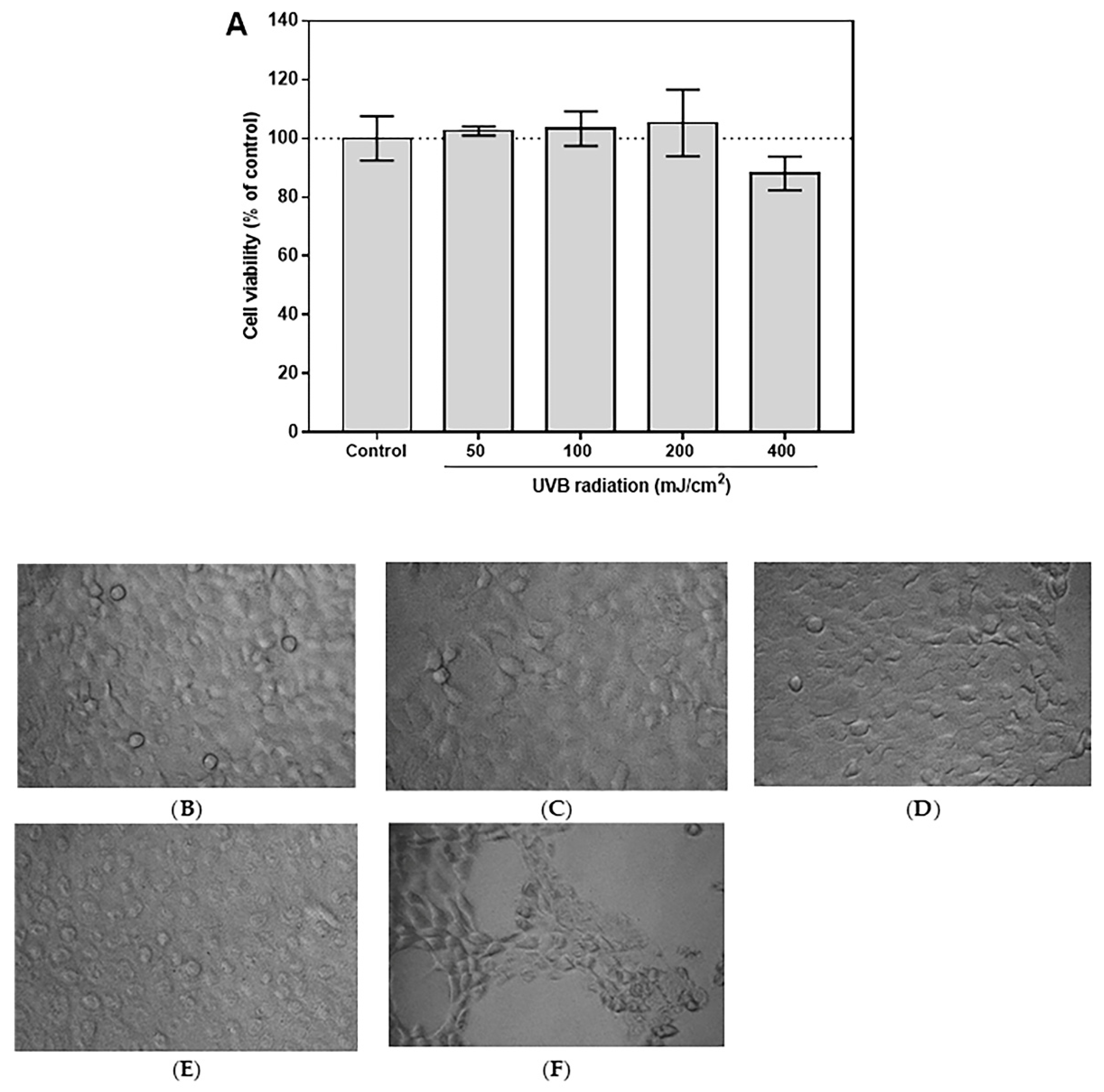
3.4. Selection of Optimal UVB Dose

To investigate the optimal UVB dose, the HaCaT cells were irradiated with dose-dependent of UVB at 50, 100, 200, and 400 mJ/cm2. The results showed that most of the tested UVB doses (50, 100, and 200 mJ/cm2) did not affect HaCaT viability (Figure 5A). However, the typical morphology of the non-UVB-irradiated group (Figure 5B) partially changed at the UVB irradiation dose of 200 mJ/cm2 (Figure 5E), whereas a dose of 100 mJ/cm2 or less did not change the morphology of cells (Figure 5C,D). In addition, the latter maximum dose of 400 mJ/cm2 did injure cells, leading to cell death (Figure 5F). Therefore, a UVB dose of 100 mJ/cm2 was selected for further experiments.

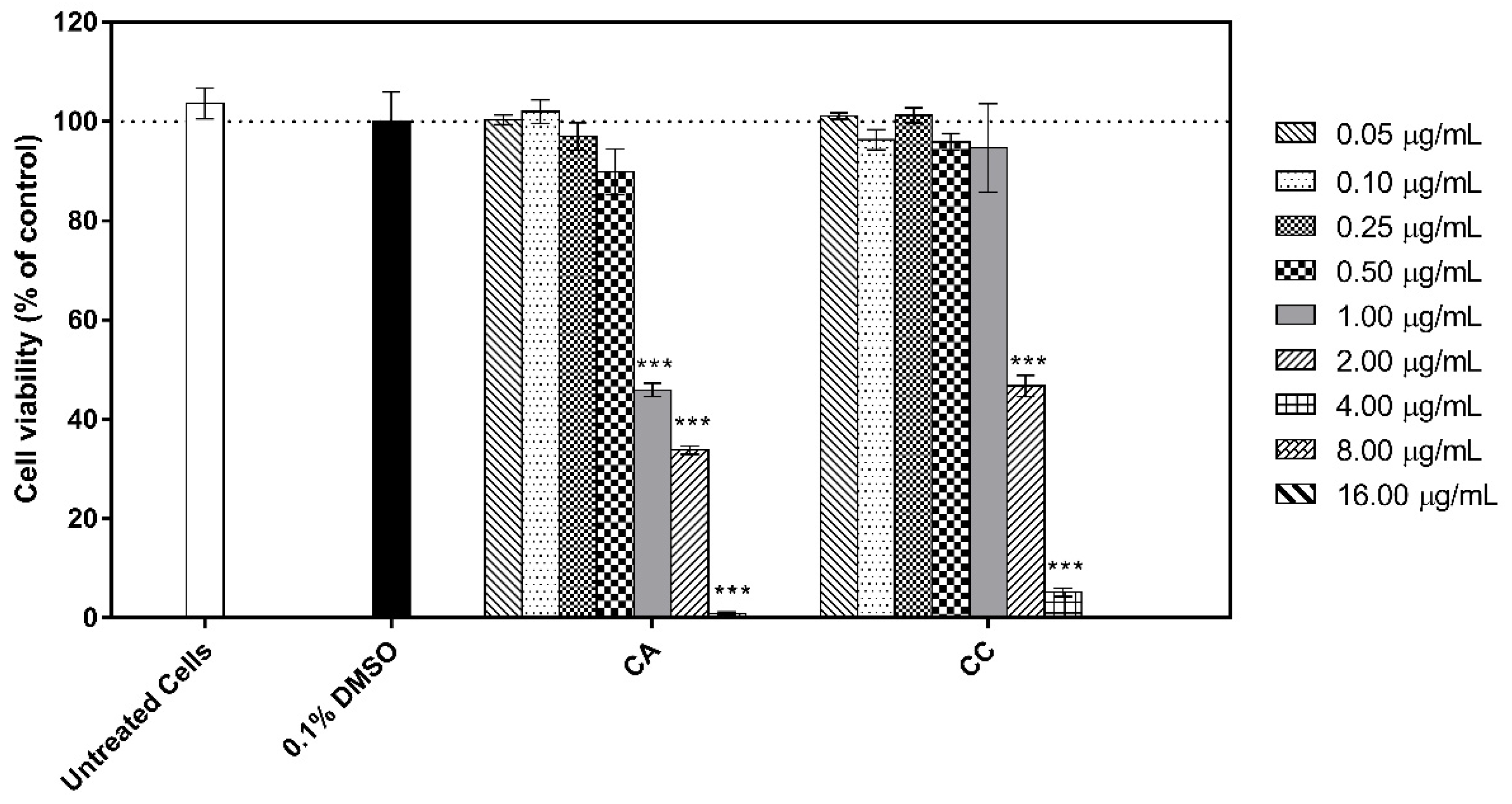
3.5. Cytotoxicity of the Extracts to Human HaCat Keratinocytes

To assess the effects of CA and CC extracts on cell viability, the MTS assay, according to the previous study [42] with some modifications was used in this investigation. The MTS tetrazolium compound is reduced by viable cells to produce a colored formazan dye that is soluble in cell culture medium in this colorimetric assay. In metabolically active cells, NAD(P)H-dependent dehydrogenase enzymes are responsible for this conversion. A change in the amount of formazan generated in response to an increase or reduction of viable cells indicates the degree of cytotoxicity. Compared with cells treated with 0.1% (v/v) DMSO, both extracts did not significantly affect the viability of HaCaT keratinocyte cells (>80% viability) at the examined concentrations of 0.05–0.50 µg mL−1 for CA and 0.05–1.00 µg/mL for CC after incubation for 24 h. However, the higher concentration of both extracts showed cytotoxicity against HaCaT cells (Figure 6). Thus, the concentrations of 0.50 µg/mL for CA and 1.00 µg/mL for CC were chosen for further experiments.

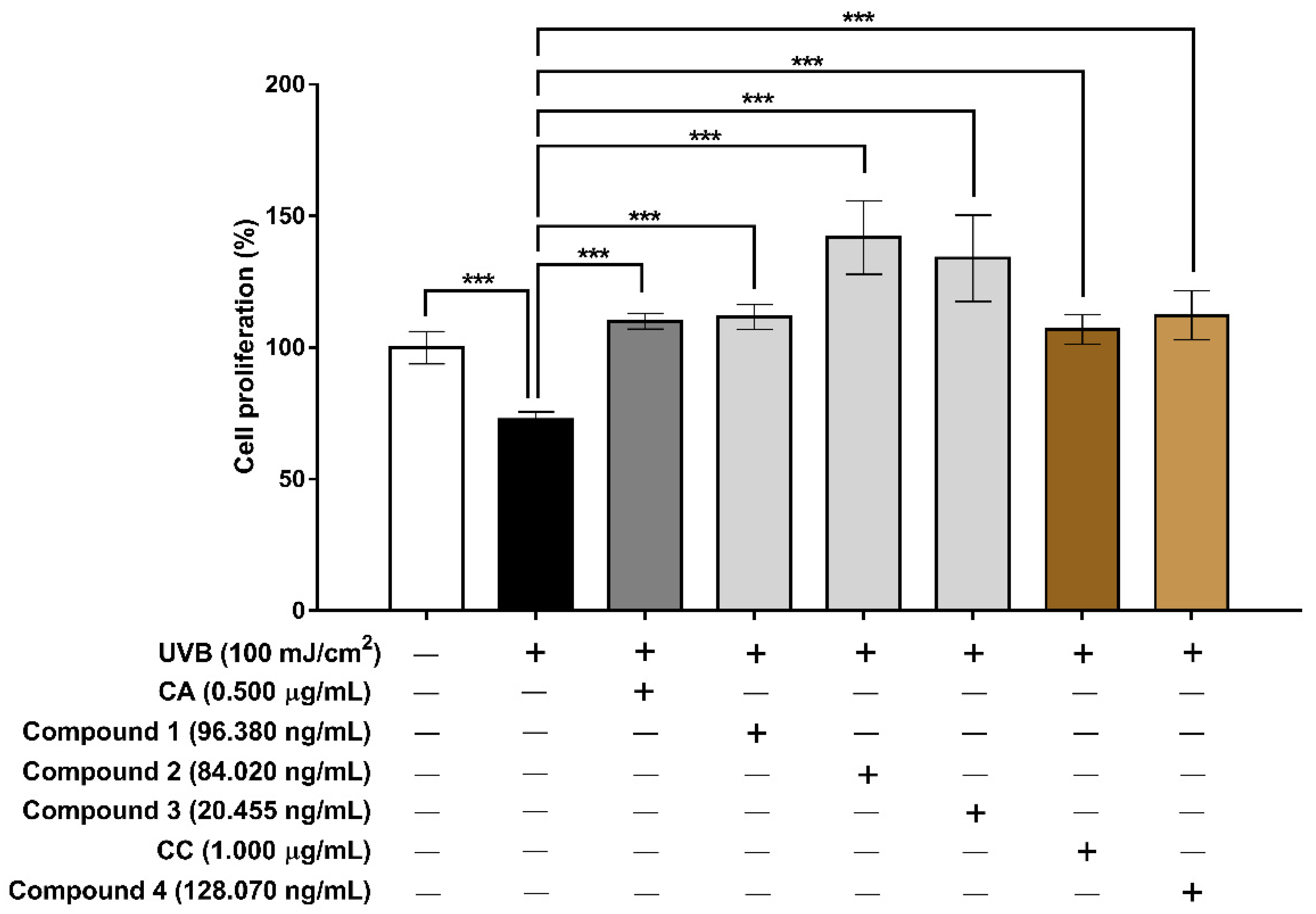
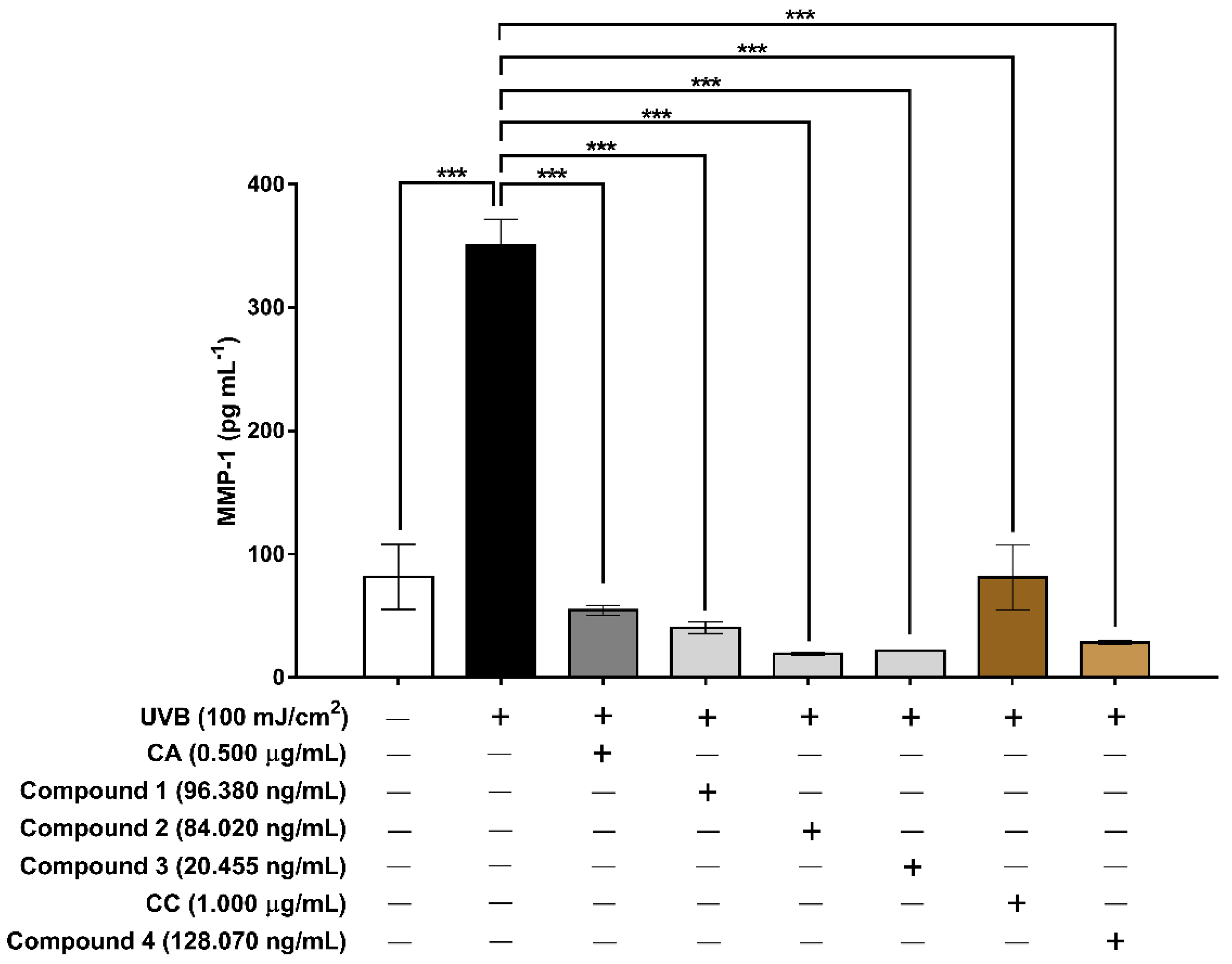
3.6. Evaluation of the Extracts and Isolated Constituents on Cell Proliferation and MMP-1 after UVB Irradiation in Human HaCaT Keratinocyte Cells

To test the effects of the CA and CC extracts together with their isolated compounds on cell proliferation, HaCaT cells were pre-treated with the serum-free medium containing the tested sample and were then repeatedly UVB-irradiated (100 mJ/cm2 × 10 times). The cell treatment and irradiation protocol are demonstrated in Figure 2. A concentration of CA and CC extracts was used based on cytotoxic results (0.50 and 1.00 µg/mL, respectively); whereas a dose of isolated compounds 14 was treated with an equivalent amount in crude extracts, which was then quantified by the HPLC technique. After 24 h of the tenth UVB-irradiation, the total number of viable cells was then counted using trypan blue staining and a hemocytometer. In the UVB-treated control group, cell numbers significantly decreased compared with the untreated group (Figure 7). Pre-treatment with the CA and CC extracts and their isolated compounds 14 before repeated UVB irradiation markedly enhanced the cell proliferation compared with the UVB-irradiated control group (1.47–1.95-folds of control). These results suggested that the CA and CC extracts and their isolated compounds could prevent UVB-inhibited human keratinocyte cells proliferation.
To investigate the effects of the CA and CC extracts and their isolated compounds 14 on the level of MMP-1 in UVB-irradiated human HaCaT keratinocytes cells, the cells were pre-treated with the tested samples for 24 h before repeated UVB irradiation (100 mJ/cm2 × 10 times) (Figure 8). The amount of MMP-1 secreted into the culture medium was analyzed. The results showed that the level of MMP-1 secretion dramatically increased in repeatedly UVB-irradiated keratinocytes (control group) compared with non-irradiated keratinocytes (4.29-fold of non-UVB-irradiated cells), but treatment with 0.5 µg/mL of the CA extract markedly inhibited UV-induced MMP-1 (6.42-fold of control). Furthermore, the 1.0 µg/mL of CC extract decreased the level of MMP-1 (4.31-fold of control) (Figure 8). Pre-treatment with the CA and CC extracts and their isolated compounds 14 after UVB irradiation decreased the level of MMP-1 produced by keratinocytes compared with the UVB-irradiated group (8.68 to 18.18-folds of control) (Figure 8). Thus, it could be concluded that compounds 14 play a critical role in human keratinocytes by preventing the UVB-induced overexpression of MMP-1. Our study on human keratinocyte cells demonstrated that MMP-1 expression was activated in UVB-irradiated HaCaT cells and a significant reduction in MMP-1 expression in cells pre-treated with the CA and CC extracts and their constituents were observed. Therefore, the prevention of UVB-induced skin damage caused by collagen breakdown could be, at least in the past, attributed to a reduction in the overexpression of MMP-1. In addition, our finding is consistent with other reports demonstrating that (–)-xanthorrhizol (2), isolated from Curcuma xanthorrhiza, significantly reduced MMP-1 expression in a dose-dependent manner [43], while germacrone (3), also isolated from the same plant species, inhibited protein expression levels of MMP-1, MMP-2, and MMP-3 in human keratinocytes and UVB-induced upregulation of the mRNA [44]. However, the molecular mechanism by which the CA and CC extracts reduce MMP-1 expression requires further study.

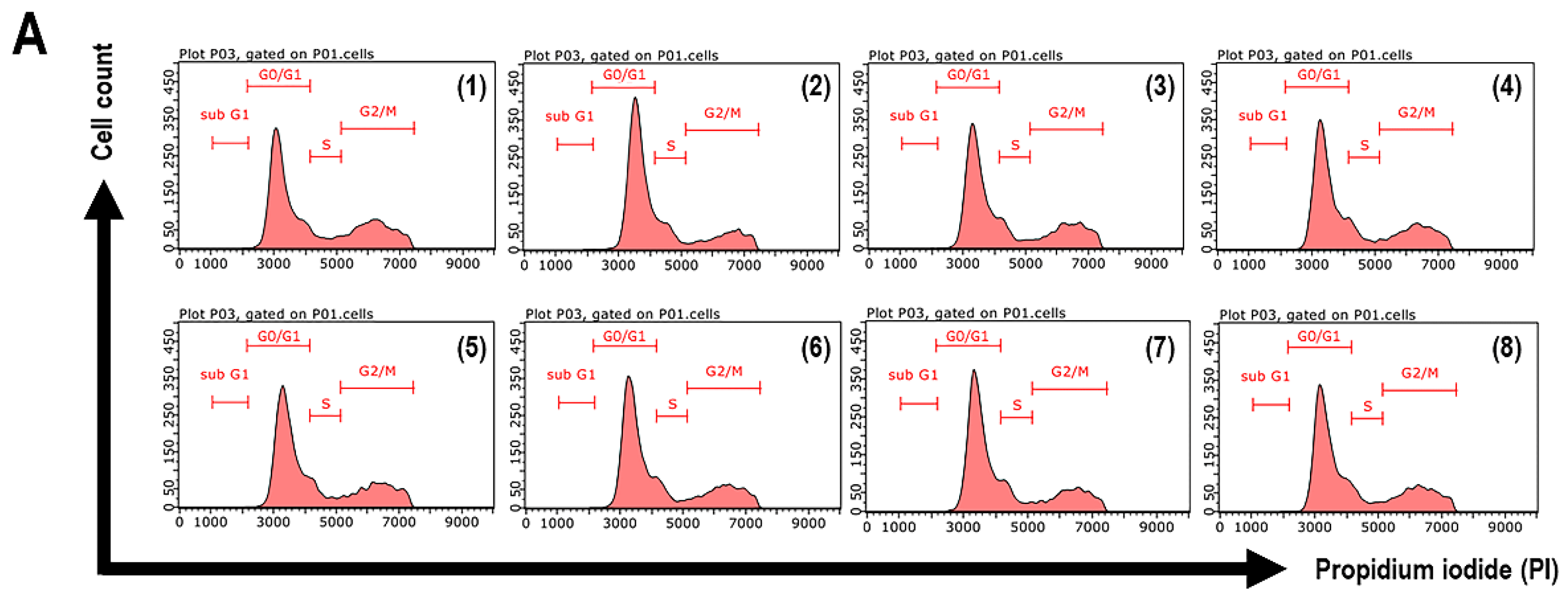
3.7. Cell Cycle Arrest of UVB-Exposed Cells to the Extracts and Isolated Constituents

Cell cycle arrest is one of the most typical events that occur during cell development, which consists of four stages including G1, S, G2, and M phases. The synthesis of enzymes essential for DNA replication is a hallmark of the G1 phase. DNA is duplicated during the S phase to form two identical sets of chromosomes. The G2 phase is primarily concerned with the creation of microtubules, which are necessary during the mitotic phase of cell division. During the interphase, there is an increase in cell volume (G1, S, and G2 phases). The parent cell is divided into two daughter cells by nuclear and cytoplasmic divisions and the production of a new cell membrane during the M phase, which sequentially includes prophase, metaphase, anaphase, and telophase. The G1/S transition is one of the two key checkpoints in the cell cycle, with the other occurring at the G2/M transition. It controls the cell cycle and is responsible for the initiation and completion of DNA replication. DNA ploidy and protein analyses are commonly used in cell cycle studies after fluorescent staining or labeled nucleic acid incorporation. The cell-cycle reagent contains the dye propidium iodide (PI), which could penetrate cell membranes, bind covalently to DNA, and emit red fluorescence [45,46,47,48].
To investigate the possibility that the cell cycle was suppressed by the CA and CC extracts and their constituents 14, results from cell cycle disruption, flow cytometric analyses stained with PI were performed. Cell cycle progression was examined after 24 h of the tenth of 100 mJ/cm2 UVB irradiation of human HaCaT keratinocytes treated with the CA and CC extracts and isolated compounds 14. The histograms generated were analyzed with InCyte Software for Guava easyCyte HT Systems, to determine the proportion of cells in G0/G1, S, and G2/M for each sample. The gating histogram and cell cycle distribution of cell cycle were illustrated in Figure 9A,B. Table 3 presents the percentages of cells in the cell cycle phases (G0/G1, S, and G2/M). The results revealed that repeated UVB exposure induces an increase in the percentages of cells in the G0/G1 and S phases. On the other hand, the repeated UVB exposure exhibited a decrease in the percentages of cells in the G2/M phase at 24 h post-UVB irradiation. Interestingly, pre-treatment of UVB-exposed cells to the CA and CC extracts resulted in a decrease of cell populations compared with the UVB-treated control group in the G0/G1 phase (–2.7% and –1.2% at 24 h after the last UVB irradiation, respectively) and the S phase (–2.8% and –2.6%, respectively), associated with increases of cells in the G2/M phase (+5.6% and +3.8%, respectively). In addition, the percentages of the sub-G1 phase were less than 0.2% (data not shown) the same as the non-UVB-treated group, since both extracts did not induce cell death at the tested concentration. Furthermore, the isolated constituents of both extracts also showed the percentages of cell populations in G0/G1, S, and G2/M in a similar trend with their extracts. It seemed probable that compounds 14 play an important protective role in the CA and CC extracts against UVB-induced cell growth inhibition. As in other investigations, UVB has been used to perform cell cycle arrest [49]. In this study, the cell populations in the G0/1 and S phases were significantly increased after UVB irradiation, indicating cells were arrested at the G0/G1 and S phases. Our results imply that the CA and CC extracts, and their isolated constituents 14 restored cell cycle arrest to levels similar to those of the non-UVB irradiated group at low concentrations after 24 h of UVB irradiation, indicating that the CA and CC extracts and their constituents could rescue UVB-induced cell cycle arrest. Consequently, their treatment at the tested concentrations did not induce cell death in HaCaT cells. Therefore, the CA and CC extracts along with their constituents showed a photoprotective effect against UVB-induced cell growth inhibition. However, the mechanism of its action is required.

4. Conclusions

In conclusion, we demonstrated that C. aromatica and C. comosa extracts, and their isolated constituents had photoprotective effects against UVB-induced HaCaT keratinocyte cells, which did not induce cell death. They increased cell proliferation, inhibited MMP-1 overexpression and restore cell cycle arrest in UVB-irradiated HaCaTs. C. aromatica and C. comosa extracts and their isolated constituents may serve as potential candidates for the prevention of skin photoaging. However, further studies are necessary to elucidate the signaling pathways and molecular mechanisms involved in the anti-photoaging activity of these agents.

Author Contributions

Conceptualization, W.N.; methodology, W.N.; software, W.P.; validation, W.N. and W.P.; formal analysis, W.P. and W.N.; investigation, W.P., W.N., W.C., N.P. and S.P.; resources, W.N., A.S. and B.-e.Y.; data curation, W.P., W.N., W.C. and A.S.; writing—original draft preparation, W.P.; writing—review and editing, W.N., A.S., J.V., V.A., W.C., N.P. and S.P.; visualization, W.P.; supervision, A.S., J.V., B.-e.Y., V.A., A.C. and P.P.; project administration, W.N.; funding acquisition, A.S. and W.N. All authors have read and agreed to the published version of the manuscript.

Funding

This research was funded by the Agricultural Research Development Agency (Public Organization), grant number CRP6305030500 and The Thailand Research Fund, grant number DBG6180030.

Institutional Review Board Statement

Not applicable.

Informed Consent Statement

Not applicable.

Data Availability Statement

Not applicable.

Acknowledgments

This work was supported by the Agricultural Research Development Agency (Public Organization) (grant number CRP6305030500). W.P. thanks the scholarship from the Science Achievement Scholarship of Thailand, Ministry of Higher Education, Science, Research and Innovation. Partial supports from Ramkhamhaeng University, The Thailand Research Fund (grant number DBG6180030) and the Center of Excellence for Innovation in Chemistry (PERCH-CIC), Ministry of Higher Education, Science, Research and Innovation are also gratefully acknowledged.

Conflicts of Interest

The authors declare no conflict of interest.

References

  1. Ganceviciene, R.; Liakou, A.I.; Theodoridis, A.; Makrantonaki, E.; Zouboulis, C.C. Skin anti-aging strategies. Dermatoendocrinology 2012, 4, 308–319. [Google Scholar] [CrossRef] [PubMed] [Green Version]
  2. Cevenini, E.; Invidia, L.; Lescai, F.; Salvioli, S.; Tieri, P.; Castellani, G.; Franceschi, C. Human models of aging and longevity. Expert Opin. Biol. Ther. 2008, 8, 1393–1405. [Google Scholar] [CrossRef] [PubMed]
  3. Farage, M.A.; Miller, K.W.; Elsner, P.; Maibach, H.I. Intrinsic and extrinsic factors in skin ageing: A review. Int. J. Cosmet. Sci. 2008, 30, 87–95. [Google Scholar] [CrossRef] [PubMed]
  4. Song, J.H.; Piao, M.J.; Han, X.; Kang, K.A.; Kang, H.K.; Yoon, W.J.; Ko, M.H.; Lee, N.H.; Lee, M.Y.; Chae, S.; et al. Anti-wrinkle effects of Sargassum muticum ethyl acetate fraction on ultraviolet B-irradiated hairless mouse skin and mechanistic evaluation in the human HaCaT keratinocyte cell line. Mol. Med. Rep. 2016, 14, 2937–2944. [Google Scholar] [CrossRef] [Green Version]
  5. Krutmann, J.; Bouloc, A.; Sore, G.; Bernard, B.A.; Passeron, T. The skin aging exposome. J. Dermatol. Sci. 2017, 85, 152–161. [Google Scholar] [CrossRef] [Green Version]
  6. Pambianchi, E.; Hagenberg, Z.; Pecorelli, A.; Grace, M.; Therrien, J.-P.; Lila, M.A.; Valacchi, G. Alaskan bog blueberry (Vaccinium uliginosum) extract as an innovative topical approach to prevent UV-induced skin damage. Cosmetics 2021, 8, 112. [Google Scholar] [CrossRef]
  7. Morocho-Jácome, A.L.; Freire, T.B.; de Oliveira, A.C.; de Almeida, T.S.; Rosado, C.; Velasco, M.V.R.; Baby, A.R. In vivo SPF from multifunctional sunscreen systems developed with natural compounds–A review. J. Cosmet. Dermatol. 2021, 20, 729–737. [Google Scholar] [CrossRef]
  8. Kwon, K.-R.; Alam, M.B.; Park, J.-H.; Kim, T.-H.; Lee, S.-H. Attenuation of UVB-induced photo-aging by polyphenolic-rich Spatholobus suberectus stem extract via modulation of MAPK/AP-1/MMPs signaling in human keratinocytes. Nutrients 2019, 11, 1341. [Google Scholar] [CrossRef] [Green Version]
  9. Verma, R.P.; Hansch, C. Matrix metalloproteinases (MMPs): Chemical-biological functions and (Q)SARs. Bioorg. Med. Chem. 2007, 15, 2223–2268. [Google Scholar] [CrossRef]
  10. Jabłońska-Trypuć, A.; Matejczyk, M.; Rosochacki, S. Matrix metalloproteinases (MMPs), the main extracellular matrix (ECM) enzymes in collagen degradation, as a target for anticancer drugs. J. Enzyme Inhib. Med. Chem. 2016, 31, 177–183. [Google Scholar] [CrossRef] [Green Version]
  11. Dong, K.K.; Damaghi, N.; Picart, S.D.; Markova, N.G.; Obayashi, K.; Okano, Y.; Masaki, H.; Grether-Beck, S.; Krutmann, J.; Smiles, K.A.; et al. UV-induced DNA damage initiates release of MMP-1 in human skin. Exp. Dermatol. 2008, 17, 1037–1044. [Google Scholar] [CrossRef]
  12. Kim, C.; Ryu, H.C.; Kim, J.H. Low-dose UVB irradiation stimulates matrix metalloproteinase-1 expression via a BLT2-linked pathway in HaCaT cells. Exp. Mol. Med. 2010, 42, 833–841. [Google Scholar] [CrossRef] [PubMed] [Green Version]
  13. Ryu, J.Y.; Na, E.J. MMP expression alteration and MMP-1 production control by syringic acid via AP-1 mechanism. Biomed. Dermatol. 2018, 2, 15. [Google Scholar] [CrossRef] [Green Version]
  14. Pittayapruek, P.; Meephansan, J.; Prapapan, O.; Komine, M.; Ohtsuki, M. Role of matrix metalloproteinases in photoaging and photocarcinogenesis. Int. J. Mol. Sci. 2016, 17, 868. [Google Scholar] [CrossRef] [PubMed] [Green Version]
  15. Kuroyanagi, M.; Ueno, A.; Ujiie, K.; Sato, S. Structures of sesquiterpenes from Curcuma aromatica Salisb. Chem. Pharm. Bull. 1987, 35, 53–59. [Google Scholar] [CrossRef] [Green Version]
  16. Al-Reza, S.M.; Rahman, A.; Sattar, M.A.; Rahman, M.O.; Fida, H.M. Essential oil composition and antioxidant activities of Curcuma aromatica Salisb. Food Chem. Toxicol. 2010, 48, 1757–1760. [Google Scholar] [CrossRef]
  17. Ahmed, S.; Ansari, S.H.; Ali, M.; Bhatt, D.; Ansari, F. Phytochemical and biological investigations on Curcuma aromatica. Pharmacog. Rev. 2008, 2, 151–156. [Google Scholar]
  18. Pintatum, A.; Maneerat, W.; Logie, E.; Tuenter, E.; Sakavitsi, M.E.; Pieters, L.; Berghe, W.V.; Sripisut, T.; Deachathai, S.; Laphookhieo, S. In vitro anti-inflammatory, antioxidant, and cytotoxic activities of four Curcuma species and the isolation of compounds from Curcuma aromatica rhizome. Biomolecules 2020, 10, 799. [Google Scholar] [CrossRef]
  19. Sun, W.; Wang, S.; Zhao, W.; Wu, C.; Guo, S.; Gao, H.; Tao, H.; Lu, J.; Wang, Y.; Chen, X. Chemical constituents and biological research on plants in the genus Curcuma. Crit. Rev. Food Sci. Nutr. 2016, 57, 1451–1523. [Google Scholar] [CrossRef]
  20. Soontornchainaksaeng, P.; Jenjittikul, T. Chromosome number variation of phytoestrogen-producing Curcuma (Zingiberaceae) from Thailand. J. Nat. Med. 2010, 64, 370–377. [Google Scholar] [CrossRef]
  21. Piyachaturawat, P.; Ercharuporn, S.; Suksamrarn, A. Uterotrophic effect of Curcuma comosa in rats. Int. J. Pharmacogn. 1995, 33, 334–338. [Google Scholar] [CrossRef]
  22. Piyachaturawat, P.; Ercharuporn, S.; Suksamrarn, A. Estrogenic activity of Curcuma comosa extract in rats. Asia Pac. J. Pharmacol. 1995, 10, 121–126. [Google Scholar]
  23. Winuthayanon, W.; Piyachaturawat, P.; Suksamrarn, A.; Ponglikitmongkol, M.; Arao, Y.; Hewitt, S.C.; Korach, K.S. Diarylheptanoid phytoestrogens isolated from the medicinal plant Curcuma comosa: Biological actions in vitro and in vivo indicate estrogen receptor-dependent mechanisms. Environ. Health Persp. 2009, 117, 1155–1161. [Google Scholar] [CrossRef] [PubMed] [Green Version]
  24. Suksamrarn, A.; Ponglikitmongkol, M.; Wongkrajang, K.; Chindaduang, A.; Kittidanairak, S.; Jankam, A.; Yingyongnarongkul, B.; Kittipanumat, N.; Chokchaisiri, R.; Khetkam, P.; et al. Diarylheptanoids, new phytoestrogens from the rhizomes of Curcuma comosa: Isolation, chemical modification and estrogenic activity evaluation. Bioorg. Med. Chem. 2008, 16, 6891–6902. [Google Scholar] [CrossRef]
  25. Piyachaturawat, P.; Charoenpiboonsin, J.; Toskulkao, C.; Suksamrarn, A. Reduction of plasma cholesterol by Curcuma comosa extract in hypercholesterolaemic hamsters. J. Ethnopharmacol. 1999, 66, 199–204. [Google Scholar] [CrossRef]
  26. Sutjarit, N.; Sueajai, J.; Boonmuen, N.; Sornkaew, N.; Suksamrarn, A.; Tuchinda, P.; Zhu, W.; Weerachayaphorn, J.; Piyachaturawat, P. Curcuma comosa reduces visceral adipose tissue and improves dyslipidemia in ovariectomized rats. J. Ethnopharmacol. 2017, 215, 167–175. [Google Scholar] [CrossRef]
  27. Tantikanlayaporn, D.; Wichit, P.; Weerachayaphorn, J.; Chairoungdua, A.; Chuncharunee, A.; Suksamrarn, A.; Piyachaturawat, P. Bone sparing effect of a novel phytoestrogen diarylheptanoid from Curcuma comosa Roxb. in ovariectomized rats. PLoS ONE 2013, 8, e78739. [Google Scholar] [CrossRef]
  28. Tanhuad, N.; Thongsa-Ad, U.; Sutjarit, N.; Yoosabai, P.; Panvongsa, W.; Wongniam, S.; Suksamrarn, A.; Piyachaturawat, P.; Anurath-apan, U.; Borwornpinyo, S.; et al. Ex vivo expansion and functional activity preservation of adult hematopoietic stem cells by a diarylheptanoid from Curcuma comosa. Biomed. Pharm. 2021, 143, 112102. [Google Scholar] [CrossRef]
  29. Chuaicharoen, P.; Charaslertrangsi, T.; Chuncharunee, A.; Suksamrarn, A.; Piyachaturawat, P. Non-phenolic diarylheptanoid from Curcuma comosa protects against thioacetamide-induced acute hepatotoxicity in mice. Pharm. Sci. Asia 2020, 47, 74–85. [Google Scholar] [CrossRef]
  30. Jantaratnotai, N.; Utaisincharoen, P.; Piyachaturawat, P.; Chongthammakun, S.; Sanvarinda, Y. Inhibitory effect of Curcuma comosa on NO production and cytokine expression in LPS-activated microglia. Life Sci. 2006, 78, 571–577. [Google Scholar] [CrossRef]
  31. Sodsai, A.; Piyachaturawat, P.; Sophasan, A.; Suksamrarn, A.; Vongsakul, M. Suppression by Curcuma comosa Roxb. of pro-inflammatory cytokine secretion in phorbol-12-myristate-13-acetate stimulated human mononuclear cells. Int. Immunopharmacol. 2007, 7, 524–531. [Google Scholar] [CrossRef] [PubMed]
  32. Nakyai, W.; Pabuprapap, W.; Sroimee, W.; Ajavakom, V.; Yingyongnarongkul, B.; Suksamrarn, A. Anti-acne vulgaris potential of the ethanolic extract of Mesua ferrea L. flowers. Cosmetics 2021, 8, 107. [Google Scholar] [CrossRef]
  33. Ehara, T.; Tanikawa, S.; Ono, M.; Akita, H. Synthesis of (R)-curcumene and (R)-xanthorrizol based on 1,2-aryl migration via phenonium ion. Chem. Pharm. Bull. 2007, 55, 1361–1364. [Google Scholar] [CrossRef] [PubMed] [Green Version]
  34. Chavan, S.P.; Thakkar, M.; Kalkote, U.R. An enantiospecific synthesis of (+)-isoparvifolinone and (−)-parvifoline. Tetrahedron Lett. 2007, 48, 535–537. [Google Scholar] [CrossRef]
  35. Barrero, A.F.; Herrador, M.M.; López-Pérez, J.-L.; Arteaga, J.F.; Catalán, J. New pathways in transannular cyclization of germacrone [germacra-1(10),4,7(11)-trien-8-one]: Evidence regarding a concerted mechanism. Org. Lett. 2009, 11, 4782–4785. [Google Scholar] [CrossRef]
  36. Demirci-Çekiça, S.; Özkan, G.; Avan, A.N.; Uzunboy, S.; Çapanoğlu, E.; Apak, R. Biomarkers of oxidative stress and antioxidant defense. J. Pharm. Biomed. Anal. 2022, 209, 114477. [Google Scholar] [CrossRef]
  37. Tiraravesit, N.; Yakaew, S.; Rukchay, R.; Luangbudnark, W.; Viennet, C.; Humbert, P.; Viyoch, J. Artocarpus altilis heartwood extract protects skin against UVB in vitro and in vivo. J. Ethnopharmacol. 2015, 175, 153–162. [Google Scholar] [CrossRef]
  38. Kwon, S.H.; Wang, Z.; Hwang, S.H.; Kang, Y.-H.; Lee, J.-Y.; Lim, S.S. Comprehensive evaluation of the antioxidant capacity of Perilla frutescens leaves extract and isolation of free radical scavengers using step-wise HSCCC guided by DPPH-HPLC. Int. J. Food Prop. 2017, 20, 921–934. [Google Scholar] [CrossRef] [Green Version]
  39. Apak, R.; Özyürek, M.; Güçlü, K.; Çapanoğlu, E. Antioxidant activity/capacity measurement. 1. classification, physicochemical principles, mechanisms, and electron transfer (ET)-based assays. J. Agric. Food Chem. 2016, 64, 997–1027. [Google Scholar] [CrossRef]
  40. Kuntasal, O.O.; Karman, D.; Wang, D.; Tuncel, S.G.; Tuncel, G. Determination of volatile organic compounds in different microenvironments by multibed adsorption and short-path thermal desorption followed by gas chromatographic-mass spectrometric analysis. J. Chromatogr. A 2005, 1099, 43–54. [Google Scholar] [CrossRef]
  41. Huber, L. Validation and Qualification in Analytical Laboratories, 2nd ed.; Informa Healthcare USA, Inc.: New York, NY, USA, 2007; pp. 125–154. [Google Scholar]
  42. Behzadi, M.A.; Choi, A.; Duehr, J.; Feyznezhad, R.; Upadhyay, C.; Schotsaert, M.; Palese, P.; Nachbagauer, R. A cross-reactive mouse monoclonal antibody against rhinovirus mediates phagocytosis in vitro. Sci. Rep. 2020, 10, 9750. [Google Scholar] [CrossRef] [PubMed]
  43. Oh, H.-I.; Shim, J.-S.; Gwon, S.-H.; Kwon, H.-J.; Hwang, J.-K. The effect of xanthorrhizol on the expression of matrix metalloproteinase-1 and type-I procollagen in ultraviolet-irradiated human skin fibroblasts. Phytother. Res. 2009, 23, 1299–1302. [Google Scholar] [CrossRef] [PubMed]
  44. Park, J.-H.; Mohamed, M.A.A.; Jung, Y.-J.; Shrestha, S.; Lee, T.H.; Lee, C.-H.; Han, D.; Kim, J.; Baek, N.-I. Germacrane sesquiterpenes isolated from the rhizome of Curcuma xanthorrhiza Roxb. inhibit UVB-induced upregulation of MMP-1, -2, and -3 expression in human keratinocytes. Arch. Pharm. Res. 2014, 38, 1752–1760. [Google Scholar] [CrossRef] [PubMed]
  45. Zhang, C.; Zhang, J.; Li, X.; Sun, N.; Yu, R.; Zhao, B.; Yu, D.; Cheng, Y.; Liu, Y. Huaier aqueous extract induces hepatocellular carcinoma cells arrest in S phase via JNK signaling pathway. Evid. Based Complement. Altern. Med. 2015, 171356, 1–11. [Google Scholar] [CrossRef] [PubMed] [Green Version]
  46. Xu, N.; Chen, X.; Rui, J.; Yu, Y.; Gu, D.; Ruan, J.J.; Ruan, B.H. Cell growth measurement. In Cell Growth; IntechOpen: London, UK, 2020. [Google Scholar] [CrossRef] [Green Version]
  47. Tiraravesit, N.; Humbert, P.; Robin, S.; Tissot, M.; Viennet, C.; Viyoch, J. Artocarpin-enriched (Artocarpus altilis) heartwood extract provides protection against UVB-induced mechanical damage in dermal fibroblasts. Photochem. Photobiol. 2017, 93, 1232–1239. [Google Scholar] [CrossRef]
  48. Nakyai, W.; Saraphanchotiwitthaya, A.; Viennet, C.; Humbert, P.; Viyoch, J. An in vitro model for fibroblast photoaging comparing single and repeated UVA irradiations. Photochem. Photobiol. 2017, 93, 1462–1471. [Google Scholar] [CrossRef]
  49. Deng, M.; Li, D.; Zhang, Y.; Zhou, G.; Liu, W.; Cao, Y.; Zhang, W. Protective effect of crocin on ultraviolet B-induced dermal fibroblast photoaging. Mol. Med. Rep. 2018, 18, 1439–1446. [Google Scholar] [CrossRef] [Green Version]
Figure 1. The structures of marker compounds of C. aromatica (13) and C. comosa (4).
Cosmetics 09 00023 g001
Figure 2. Schematic illustration of the protocol of repeated UVB irradiation.
Cosmetics 09 00023 g002
Figure 3. HPLC chromatograms of (A) compound 1, (B) compound 2, (C) compound 3 and (D) crude extract of CA.
Cosmetics 09 00023 g003
Figure 4. HPLC chromatograms of (A) compound 4 and (B) crude extract of CC.
Cosmetics 09 00023 g004
Figure 5. Effect of UVB irradiation on HaCaT viability. Cells were irradiated with various UVB intensities. They were then incubated in a culture medium for 24 h. (A) Cytotoxicity was conducted by MTS assay of non-UVB-irradiated and UVB-irradiated cells (100% of control). Data are presented as means ± S.D. Morphologies of (B) non-UVB-irradiated cells or cells irradiated with UVB at a dose of (C) 50 mJ/cm2, (D) 100 mJ/cm2, (E) 200 mJ/cm2, and (F) 400 mJ/cm2.
Cosmetics 09 00023 g005
Figure 6. Cytotoxicity of CA and CC extracts against human HaCaT keratinocytes. Data are presented as the means ± SD. *** p < 0.05 compared to the control (0.1% DMSO).
Cosmetics 09 00023 g006
Figure 7. Effects of the CA and CC extracts and isolated constituents on cell proliferation by UVB-induction. Human skin keratinocytes were pre-treated with the tested samples for 24 h before repeated UVB irradiation (100 mJ/cm2 × 10 times). Cells were harvested for 24 h after the tenth of UVB irradiation. The percentage of cell proliferation was determined via the trypan blue assay. Data are presented as the means ± SD. *** p < 0.05, using unpaired Student’s t-test.
Cosmetics 09 00023 g007
Figure 8. Effects of the CA and CC extracts and isolated constituents on MMP-1 production by UVB-induction. Human skin keratinocytes were pre-treated with the tested samples for 24 h before repeated UVB irradiation (100 mJ/cm2 × 10 times). Cell-free supernatants were collected for ELISA for 24 h after the tenth irradiation. Data are presented as the means ± SD. *** p < 0.05, using unpaired Student’s t-test.
Cosmetics 09 00023 g008
Figure 9. Cell cycle arrested following CA and CC extracts and isolated compounds treatments for 48 h. 0.1% DMSO was used as a control. The cell cycle was analyzed using flow cytometry. Human keratinocytes were pre-treated with the tested samples for 24 h. The cells were harvested, stained with propidium iodide (PI) for 24 h after the tenth UVB irradiation (100 mJ cm−2) and DNA content was quantified by flow cytometry. (A) Data shows the gating histogram showing the percentage of cells in the G0/G1, S and G2/M phases of the cell cycle obtained after flow cytometry analysis, where (1) = non-UVB-treated cells, (2) = UVB-treated cells, (3) = CA extract, (4) = compound 1, (5) = compound 2, (6) = compound 3, (7) = CC extract and (8) = compound 4. For each sample, 500,000 cells were acquired. (B) Cell cycle distribution pre-treated compounds treatments. Data are presented as the means ± SD. Asterisks (*) denote significant differences from the UVB-treated group (p < 0.05).
Cosmetics 09 00023 g009aCosmetics 09 00023 g009b
Table 1. Total phenolic contents and antioxidant activities of CA and CC extracts.
SampleTotal Phenolic Contents
(µg GAE/mg)
Antioxidant Activities (IC50, µg/mL)
DPPHABTSFRAPNO
CA extract90.89 ± 0.1478.77 ± 1.0219.17 ± 0.0786.85 ± 1.11159.90 ± 1.04
CC extract186.99 ± 0.0121.22 ± 1.012.67 ± 1.0232.47 ± 1.040.26 ± 1.04
L-Ascorbic acid 1-5.15 ± 1.040.86 ± 1.032.06 ± 1.560.04 ± 1.04
The results were expressed as mean ± standard deviation (SD) of triplicate experiments. (n = 3) 1 L-Ascorbic acid was used as the positive control.
Table 2. Validation of HPLC analysis of marker compounds of CA (13) and CC (4).
CompoundsLinearityCorrelation Coefficient (R²)LOD
(µg/mL)
LOQ
(µg/mL)
% RSD% Recovery
Inter-Day
(n = 9)
Intra-Day
(n = 9)
1y = 91.53x + 427.860.99901.484.720.45 ± 0.090.63 ± 0.3098.93 ± 1.06
2y = 89.04x + 272.380.99920.892.851.17 ± 0.160.58 ± 0.39101.79 ± 1.48
3y = 98.72x + 560.040.99920.421.341.30 ± 0.350.65 ± 0.10100.43 ± 0.35
4y = 174.91x + 165.030.99970.050.150.98 ± 0.181.53 ± 0.5999.13 ± 0.60
The letters y and x are peak area and concentration, respectively.
Table 3. Cell cycle distribution of the CA and CC extracts and isolated compounds 14 treated HaCaT cells at 24 h after the tenth UVB exposure (100 mJ/cm2).
Sample% of Total Cell Cycle Population in:
G0/G1SG2/M
Non-UVB-treated cells59.64 ± 2.017.96 ± 0.0532.05 ± 2.09
UVB-treated cells64.47 ± 0.7913.73 ± 0.1121.17 ± 0.81
CA extract61.73 ± 1.2410.93 ± 1.2826.77 ± 2.52
Compound 162.28 ± 0.4210.03 ± 0.5427.15 ± 0.81
Compound 261.25 ± 0.5411.34 ± 1.1726.83 ± 1.29
Compound 362.69 ± 0.869.76 ± 0.1026.95 ± 0.96
CC extract63.31 ± 0.3711.15 ± 0.5624.98 ± 0.22
Compound 463.35 ± 0.1910.40 ± 1.2925.74 ± 2.53
The results were expressed as mean ± standard deviation (SD) of triplicate experiments. (n = 3).
Publisher’s Note: MDPI stays neutral with regard to jurisdictional claims in published maps and institutional affiliations.

