Review

Recent Advances on Topical Application of Ceramides to Restore Barrier Function of Skin

Department of Pharmaceutical Technology, Faculty of Pharmacy, Istanbul University, 34116 Beyazıt, Turkey
*
Author to whom correspondence should be addressed.
Cosmetics 2019, 6(3), 52; https://doi.org/10.3390/cosmetics6030052
Submission received: 30 June 2019 / Revised: 16 August 2019 / Accepted: 17 August 2019 / Published: 20 August 2019
(This article belongs to the Special Issue Topical Pharmaceutical Products and Cosmetics)

Abstract

:
Human skin is the largest organ of the body and is an effective physical barrier keeping it from environmental conditions. This barrier function of the skin is based on stratum corneum, located in the uppermost skin. Stratum corneum has corneocytes surrounded by multilamellar lipid membranes which are composed of cholesterol, free fatty acids and ceramides (CERs). Alterations in ceramide content of the stratum corneum are associated with numerous skin disorders. In recent years, CERs have been incorporated into conventional and novel carrier systems with the purpose of exogenously applying CERs to help the barrier function of the skin. This review provides an overview of the structure, function and importance of CERs to restore the barrier function of the skin following their topical application.

1. Skin Barrier

Human skin is the largest organ of the body, accounting for 16% of total weight and having an area of 1.8 m2. It prevents excessive water loss from the body and helps to maintain electrolyte balance and involves some metabolic processes that are related to immune system. It also protects the organism against microorganisms, ultraviolet radiation, toxic substances and mechanical damage, creating a physical barrier between environmental conditions and the body. Basically, the human skin is a multi-layered membrane. From deep to superficial, these layers are (Figure 1) [1]:
Hypodermis (subcutaneous or subcutis): It consists of essentially loose connective tissue, blood vessels and fat cells. This layer is 2–7 mm in thickness and has important functions such as storing lipids, isolating the body and regulating temperature.
Dermis: It enables a critical support to the epidermis. The fibroblasts are mainly cell types in the dermis which is about 1 mm in thickness. It produces collagen, enabling strength and endurance to the dermis, elastin fibers providing elasticity and flexibility to the skin and proteoglycans maintaining skin hydration. In addition, inflammatory reactions initiate in this layer via macrophages and mast cells residing in the dermis. The hair follicles, pilosebaceous units, apocrine and endocrine glands also exist in this layer [2,3]
Epidermis (upper skin): The epidermis is the outermost layer of the skin, and predominantly consists of keratinocytes arranged in several layers. It generates a physical and functional barrier between the human body and the environment as a result of cornification or keratinization, which is reformation of terminally differentiating keratinocytes into corneocytes in order to make up the stratum corneum [4,5]. Additionally, it contains some nerve endings for the perception of pain, but it does not have any blood vessels or lymphatic system.
The thickness of the epidermis varies from 50 to 150 µm and is composed of five layers. From deep to superficial, these layers are [6,7]:
  • Basal layer (stratum germinativum)
  • Prickle layer (stratum spinosum)
  • Granular layer (stratum granulosum)
  • Stratum lucidum
  • Stratumcorneum.

1.1. Barrier Function of Stratum Corneum

The stratum corneum consists of a lipid matrix and 15–20 layers of flattened dead cells (corneocytes) embedded in this lipid matrix. This structure with 10–20 µm in thickness, which is called a “brick and mortar” model, constitutes a major barrier function of the stratum corneum (Figure 1). In this barrier layer, the ‘‘bricks’’ are terminally differentiated keratinocytes (corneocytes) composed mostly offilaggrin and a dense network of keratin macrofibrils, which is a protein that helps to keep the skin hydrated by preventing water evaporation and absorbing water. The corneocytes are surrounded by an insoluble cornified cell envelope which results in keratinization and is composed of a monolayer of ceramides. These cells are held together in the lipid matrix by corneodesmosomes, modified desmosomes from the uppermost nucleated epidermal layers. Also, intracellular humectants (natural moisturizing factors) that are essential for stratum corneum hydration, barrier homeostasis and desquamation localize in the corneocytes [8,9].
The ‘‘mortar’’ includes intercellular lipids arranged into lamellar layers consisting of cholesterol, free fatty acids, approximately ceramides (CERs) and sterol/wax esters. The intercellular lipid lamellar matrix of stratum corneum is essential for maintaining skin barrier function and preventing transepidermal water loss (TEWL) [1,10].
Besides the structures of the stratum corneum, the tight junctions attached to lateral walls of keratinocytes in the stratum granulosum have presented a secondary functional barrier in the skin. Also, these complex cell-cell junctions which are affected chemical, microbial or immunological barriers influence the stratum corneum barrier [11,12].

1.2. Relationship of Skin pH, Transepidermal Water Loss (TEWL) and Stratum Corneum Barrier

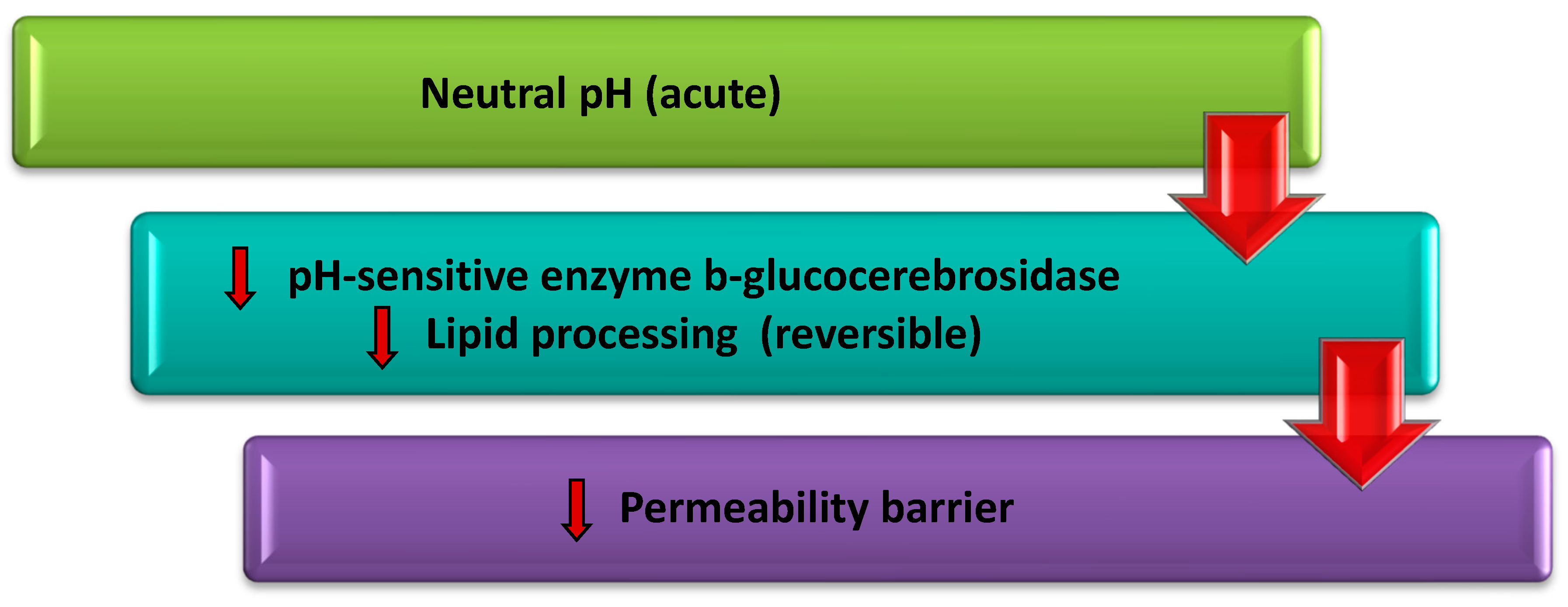
Skin pH: The “acid mantle” of the stratum corneum has been known since the 1920s [13]. In recent years, development of new technologies and animal models have implied that stratum corneum acidity is essential in many epidermal functions such as epidermal permeability barrier, epidermal anti-microbial barrier, integrity and cohesion of the stratum corneum. In addition, it has been reported that the barrier regeneration proceeds at an acidic skin pH, and conversely delays at a neutral skin pH [14], because b-glucocerebrosidase activity and lipid secretion through basal cell layers decrease reversibly in neutral skin pH, so the intercellular lipid lamellar matrix could not generate for an effective permeability barrier (Figure 2) [15].
Transepidermal Water Loss (TEWL): It has well known for many years that the stratum corneum prevents TEWL and hydrates corneocytes for continuity of enzymatic reactions enabling desquamation and its effective barrier characteristics [16,17]. The water acts as a plasticizer that imparts an elastic property to corneocyte proteins. When the water is deprived of the skin, the corneocyte proteins become more fragile and the skin tends to crack with the mechanical stress. Thus, the natural barrier of the stratum corneum is damaged with a decrease of skin hydration and an increase of TEWL [16]. However, this correlated relationship between skin hydration and TEWL only occurs in some skin disorders such as irritation and psoriasis. In the case of aged skin, the skin effectively maintains its barrier function despite the decrease of skin hydration, without a significant increase of TEWL values. Conversely, occlusion after hydration results in increased hydration and TEWL values, and damage of barrier function [15]. Considering these data, skin hydration and TEWL values are not indicators of stratum corneum integrity.

1.3. Major Lipids of Stratum Corneum

The major lipids of human stratum corneum are cholesterol, free fatty acids, and CERs in an approximately 1:1:1 ratio. This equimolar ratio is highly important to maintain epidermal homeostasis because of alterations in the lipid content of the stratum corneum resulting in a disturbed barrier function [18].

1.3.1. Cholesterol

Cholesterol is a lipid with low molecular weight that plays a role in regulating the desquamation process [19]. In the final stage of the keratinization process, unsaturated fatty acids (mostly oleic acid) are transferred into the cholesterol, which is the only large sterol in the human stratum corneum, to produce cholesterol esters. These cholesterol esters settle into the intercellular lipid lamellar matrix of the stratum corneum [20].

1.3.2. Free Fatty Acids

The free fatty acids in the human stratum corneum are principally straight-chain saturated derivates in the range of 14 and 28 carbons in length. These are mostly ≥20 carbons in length, with the fatty acids consisting of 22 and 24 carbons being the most abundant. The free fatty acids take part in the structure of CERs, reacting with long-chain sphingoid [21].

1.3.3. Ceramides

Structure and Physicochemical Characteristics of Ceramides

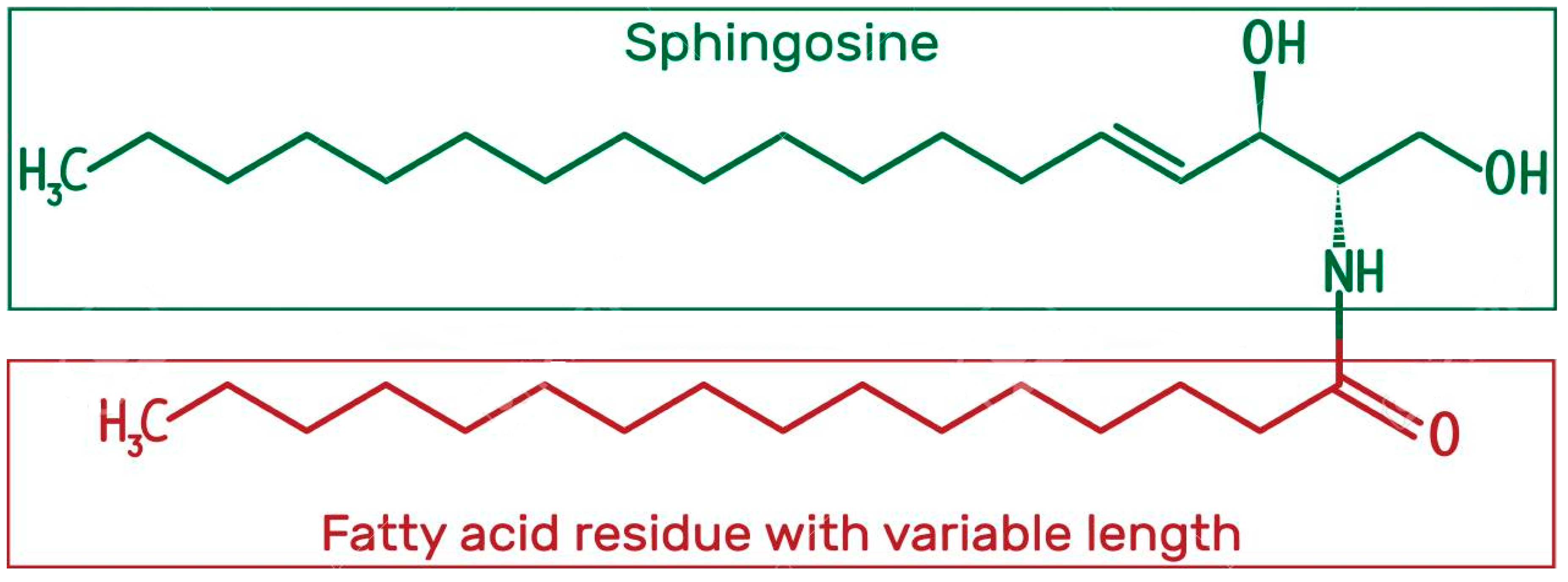
CERs are predominant lipid components of the stratum corneum and comprise 30–40% of the stratum corneum lipids by mass. They are composed of long-chain sphingoid bases (dihydrosphingosine, sphingosine, phytosphingosine or 6-hydroxysphingosine) which are linked to long chain free fatty acids (non-hydroxy fatty acids, α-hydroxy fatty acids or ester-linked ω-hydroxy fatty acids) via amide bonds (Figure 3). Hence, the head groups in CERs include hydroxyl groups that can form inter- and intramolecular hydrogen bonds. The number of hydroxyl groups in the head group of CERs is important for integrity of the stratum corneum’s barrier function. Also, CERs exhibit the heterogeneity in terms of chain length (16–30 carbons) and degree of unsaturation (predominantly saturated) and hydroxylation pattern. The chain length of fatty acids in the CERs generally is 24–26 carbons, but there are slightly available fatty acids comprising 16–18 carbons [22], and their chain length affects the skin permeability and barrier function of the stratum corneum [23].
Considering the chemical structure of CERs, they are highly lipophilic compounds because the ratio of long-chain fatty acids to the hydrophilic head part is high. As such, CERs are poorly water-soluble compounds [24]. Also, CERs are compounds with high molecular weight [25]. Because of the aforementioned physicochemical characteristics of CERs, the percutaneous absorption of these compounds is limited when topically applied to the skin. Besides this issue, CERs exhibit polymorphic characteristics which resulted in some questions during the fabrication of ceramide formulations [26].
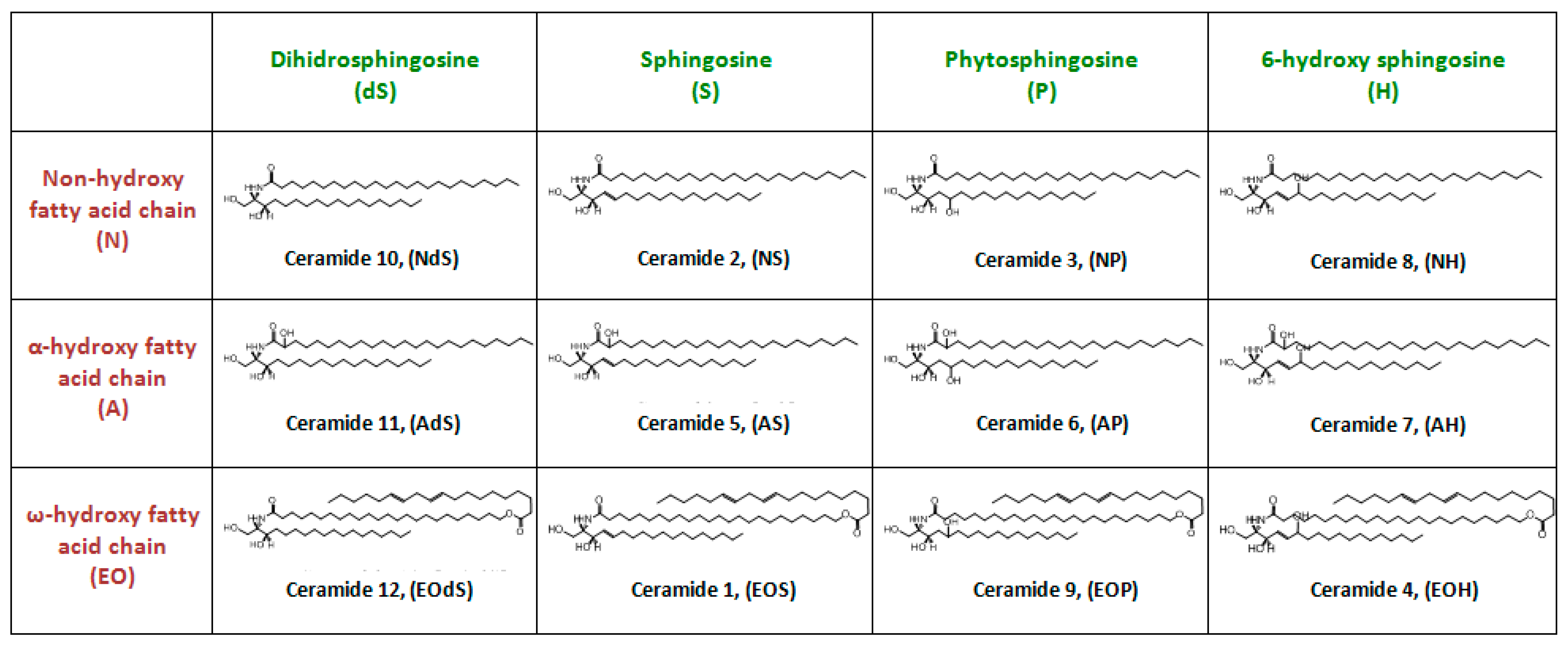
There are an available 12 types of extracted CERs in the human stratum corneum, which are derived from the aforementioned types of fatty acids and sphingoid bases, differing from one another based on the composition of the head group or esterification of fatty acids (Figure 4) [27,28].

Biosynthesis of Ceramides in the Skin

The production of CERs could be from different metabolic pathways. These are the de novo biosynthesis pathway, SMase pathway (activation of sphingomyelinases) and salvage pathway [29]. In the de novo pathway, ceramide biosynthesis occurs in the endoplasmic reticulum of corneocytes in the stratum basale. For the synthesis of CERs, L-serine amino acid and palmitoyl CoA are primarily condensed via serine palmitoyl transferase. The first long-chains of CERs form and these molecules are named as 3-ketodihydrosphingosine. 3-ketodihydrosphingosine undergoes a reduction reaction for conversion to dihydrosphingosine. Then, dihydrosphingosine is converted to dihydroceramide by acylation. Finally, dihydroceramide is catalyzed by dihydroceramide desaturase to synthesize CERs [30,31]. The salvage pathway occurs in the acidic subcellular compartments, such as the late endosomes and the lysosomes. In these compartments, sphingosine-1-phosphate is converted to sphingosine via sphingosine kinase. Then, sphingosine is converted to CERs by ceramide synthase. SMase pathway occurs in the plasma membrane and the endosomal/lysosomal compartments. CERs are formed by hydrolysis of sphingomyelin by SMase [29,31].
On the other hand, salvage and SMase pathways are reversible metabolic processes, therefore they behavelike a control mechanism to keep constant the ceramide, fatty acid and cholesterol ratio. In this way, CERs are taken under the control and reach the upper layer of the skin even through the stratum corneum. They harmonize to corneocyte about forming the "brick-mortar" model and organization occurs regularly thanks to their head-tail structure [29].

Location and Position of Ceramides in the Skin

The lateral and lamellar organization of lipids is crucial for the barrier function of the skin. The lateral organization is the arrangement of the lipids vertically to the plane of lamellar lipid organization. There are three possible organizations in intercellular spaces: (i) fluid organization, which is the most disordered and highly permeable, (ii) hexagonal organization, which is less dense and medium permeable, and (iii) orthorhombic organization, which is a highly ordered state and exhibits low permeability [32,33,34]. In the orthorhombic organization, highly ordered state is based on Van der Waals interactions in the tails and hydrogen bonds in the head groups of the lipids [35,36] and the ceramide molecules are organized with width and length as 0.41 nm and 0.37 nm in per 0.182 nm2 area, respectively [37]. Besides, in the hexagonal organization, the ceramide molecules are arranged in an order having equal width and length as 0.41 nm in per 0.194 nm2 area, respectively. Also, the width and length are angled as 120° in a hexagonal structure because of weakened interactions, as distinct from orthorhombic organization. These structural diversities make the hexagonal structure less constricted, probably causing changes in skin barrier function. Nevertheless, the hexagonal structure in regular order provides a sufficient barrier function rather than a fluid organization that the ceramide molecules could be disorganized in the skin [38].
In the lamellar organization, the lipids which are ordered as bilayers are arranged with different periodicity phases. If the lipids repeat in per 6 nm interval, the alignment is identified as a short periodicity phase (SPP). On the other hand, it is named as a long periodicity phase (LPP) if the lipids recur in per 13 nm interval [34,38].

Role of Ceramides in the Skin Barrier and Skin Disorders

The morphology of ceramide holding the corneocytes and attaining to intercellular matrix knits the skin and remain the skin integrity. The ordered alignment of lipid forms a closed system to prevent TEWL and makes the stratum corneum more impermeable. As a result, changes in the amount and organization of the stratum corneum CERs cause skin disorders with barrier defects.
Atopic Dermatitis: Atopic dermatitis is a dermatological disorder which is characterized by dry skin, pruritus, increased TEWL, and decreased skin barrier function. Matsumoto et al. [39] reported that the ceramide I, long-chain ceramide, decreased by 52% in atopic dermatitis. In addition, ceramide V in non-lesionedparts of the skin were raised, and ceramide I and ceramide III were reduced in lesioned parts of the skin [40,41]. Moreover, the metabolism pathways such as ceramidase are overactive in the epidermis with atopic dermatitis [42]. Besides, Chermprapai et al. [43] determined that free fatty acids were reduced as well as CERs in the atopic dermatitis.
Ichthyosis: As much as atopic dermatitis, free fatty acid ratio reduces in contradistinction for CERs ratio. Also, orthorhombic organization and LPP, having crucial roles in the skin barrier function, degenerate inichthyosis patients [44]. In Dorfman-Chanarin syndrome, ichthyosis is also shown as a symptom with descending w-OH-acylceramide and w-O-acylceramide [45]. Additionally, ceramide synthase 3 enzyme synthesizing the ceramide 3 has gene damage in a patient with congenital ichthyosis [46].
Psoriasis: In another skin disorder in which barrier defects occurs, Motta et al. [42] revealed the relation between ceramide composition and TEWL in psoriatic and healthy skin. According to results, ceramide I, III, IV, V, and VI reduced TEWL increased in all psoriatic scales.
Acne: It is considered that altered ceramide values is also effective on acne. Because of high-level TEWL in acne, altered ceramide could have a role in this point. Pappas et al. [47] examined the correlation between them. However, TEWL is not negatively correlated with symptoms even though all subclasses of ceramide are negatively correlated with TEWL. Therefore, they considered the ceramide composition of healthy skin and skin with acne according to season. As a result, decreased ceramide aggravates the symptoms, especially in winter months. Seasonal changes in the amount of ceramide are exposed by increasing TEWL. On the other hand, healthy skin has high-level ceramide VI and VIII providing adaptation of environmental conditions in winter months.
Gaucher Disease: As a genetic disorder, Gaucher disease arising from β-glucocerebrosidase enzyme deficiency is also related to the ceramide biosynthesis and metabolism pathway. On glucosylceramide breaking down to glucose and ceramide, β-glucocerebrosidase has a role with hydrolyzation. After this stage, glucosylceramide is diminished and a ceramide molecule is exposed. However, in patients with Gaucher disease, this metabolism pathway loses the function because of β-glucocerebrosidase deficiency. Unlike the aforementioned disorders, a decrease in the amount of ceramide in Gaucher disease is originated from this point [48].
Dry Skin: The lipid envelope, LPP, and orthorhombic organization keep the moisturizing balance under control and reduce TEWL. In this way, even though the skin is not moisturized by supplementary products, hydration content is preserved in skin layers. With decreasing ceramide levels in the skin, the barrier function of lipid envelopes becomes incapacitated. Compared to dry and normal skin, ceramide I, II, III, IV, V and VI diminish with dryness, while CERs are at a high level in normal skin [39].

2. Topical Applications of Ceramides

As mentioned earlier, decreased levels of stratum corneum CERs have been shown to be a major effect of the occurrence of skin disorders. As a result of this, the researchers have thought that these disorders could be improved by replacing decreased CERs levels. Therefore, topical applications including conventional and novel carrier systems have been formulated by diverse researchers [49,50,51,52].

2.1. Conventional Carrier Systems

Several commercial lotions, creams, and moisturizers (e.g., Eucerin Smoothing Repair Dry Skin Lotion, Eucerin Eczema Relief Body Creme, CeraVe Moisturizing Lotion, CeraVe Suncare Sunscreen Face SPF 30) [53] based on CERs 1 and 3 have been formulated in the market. However, there is confusion related to skin permeability and efficacy of CERs after topical application via conventional carrier systems. Spada et al. [49] reported that the phytosphingosine, ceramide I, ceramide III and ceramide VI-II in a simplified cream have a crucial role in the hydration of skin and reducing TEWL values compared to placebo data. Draelos et al. [54] and Neubert et al. [50] also suggested that CERs in their conventional formulations could be efficient to restore the skin barrier in skin disorders. However, glycyrrhetinic acid (an anti-inflammatory compound) and ethoxydiglycol (a penetration enhancer) were used to improve therapeutic efficacy and skin permeation of CERs in these studies, respectively. Hence, these results also could be due to the aforementioned constituents, because Chang et al. [55] revealed that emulsion including ceramide, niacin, and arginine-sodium-PCA complex could not create a notable clinical response. Similarly, Zhang et al. [56] showed via tape stripping and IR imaging studies that ceramide 1 and 3 in suspension could not penetrate through the stratum corneum despite the presence of oleic acid.
This distinction could be based on whether the skin is healthy or dry. Aoki et al. [51] observed in the fluorescent light after 12 hours following application that ceramide 2 NBD in emulsion formulating with water, Tween 20 and octyldodecanol was able to permeate to all layers of dry skin while remaining in the upper layer of healthy skin. The researchers considered the relationship between the amounts of endogenous ceramide in healthy/dry skin and penetration capability. It is clear that dry skin consisting of low endogenous ceramide is convenient for exogenous ceramide passing. Nakaune-Lijima et al. [57] also demonstrated via TEM images following application that the emulsions containing ceramide I, ceramide IX-S and ceramide IX-L in ratio 0.3%, cholesterol, 1,3 butylene glycol and aqueous lecithin were able to restore the damaged LPP formation of stratum corneum lipids.

2.2. Novel Carrier Systems

Conventional carrier systems have been used for skin delivery of various compounds for many years. However, these systems are inappropriate for skin delivery of large molecular structure and highly lipophilic compounds, due to the complex nature of the skin. Hence, the researchers have developed novel carrier systems such as vesicular systems, microemulsions, and nanoparticles to overcome these disadvantages [58,59]. In this context, the microemulsions are widely studied to increase the penetration of CERs through the stratum corneum [50,52]. Considering the literature review, we assert that these microemulsion formulations are the most permeable preparations for topical application of CERs among other novel carrier systems. This situation could be based on directly improving penetration through the stratum corneum due to the high surfactant content of formulations and indirectly increasing solubility (thereby thermodynamic activity) of CERs via microemulsions. Moreover, some researchers have suggested that droplet size of microemulsions and nanoemulsions including CERs is crucial for their penetration through the stratum corneum [50]. Therefore, Su et al. [60] studied the relation of droplet size and temperature changes in the production of o/w nanoemulsions. The other group indicated that oil phase type affects the droplet size of ceramide 3b nanoemulsion. In the study, the largest droplet size occurred in the formulation with avocado oil/oleic acid while the smallest droplet size obtained in the formulation with squalene [61].
In the literature, another option is ceramide-based liposomes with high membrane fluidity, high fusion activity, and comprising stratum corneum lipids to facilitate the penetration of CERs to the skin. These liposomes were composed of C-8 ceramide/cholesterol/linolenic acid/cholesterol sulfate = 45/5/5/45 (w/w%) [62]. Also, a human-type nanoceramide dispersion called Astalift Jelly Aquarysta was developed to improve ceramide localization in the stratum corneum. According to the results of in vivo tape stripping studies, the localization of human-type nano CERs in the stratum corneum was determined to be 9 times greater than that of its reference [63].
Apart from microemulsions, nanoemulsions, and liposomes, novel carrier systems for topical delivery of CERs are nanoparticles and microparticles. These systems have aimed at controlled and targeted delivery of CERs via nanoparticles [64,65,66]. The penetration percentage of nanoparticles was 60% while the microemulsion was 92%, in the study of Tessema et al. [64]. They obtained in a controlled manner delivery for nanoparticle formulations. Similarly, Jung et al. [65] designed a new chitosan-coated ceramide/PLGA nanoparticle to target the main region with dermatitis by releasing ceramide, particularly in the acidic state. Kim et al. [66] also prepared a microparticle with pseudo ceramide PC 104, stearic acid and cholesterol. They found that the microparticles with ceramide PC 104 are able to repair barrier function and obstruct TEWL in atopic dermatitis.

3. Conclusions

The skin is an effective barrier mainly due to the structure of the stratum corneum, its outermost layer. The stratum corneum is composed of keratinized cells (corneocytes) embedded in the lipid matrix. CERs are the main lipids in the stratum corneum, which play a key role on the barrier function. Alterations of CERs or stratum corneum lipid composition results in skin disorders with barrier defects such as atopic dermatitis, psoriasis and ichthyoses. In recent years, CERs have been incorporated into conventional and novel carrier systems with the purpose of topical delivery of CERs to repair the barrier function of the skin. However, the penetration of CERs into deeper layers of the skin is also a controversial debate because of their high molecular weight and highly lipophilic character. Thus, in recent years, researchers have focused on the development of their novel carrier systems such as microemulsions in order to improve the potential of topical delivery of CERs through the skin. However, these carrier systems could not be feasible for improvement of barrier defects in skin disorders such as atopic dermatitis, psoriasis and ichthyoses due to the high surfactant content of the microemulsions.
As a conclusion, there is a debate about the efficacy of CERs following topical application. Hence, the potential topical delivery of CERs needs to be clarified and there is a challenge to develop skin-friendly formulation contents for topical application of CERs.

Author Contributions

M.K.: researched the literature. H.Ş.B.: implemented the review of the literature and drafted the manuscript. E.K.: contributed to the planning, writing and editing of the manuscript. S.G.: was involved in planning and supervised the paper. All authors provided critical feedback and helped shape the manuscript.

Funding

This research received no external funding.

Conflicts of Interest

The authors declare no conflict of interest.

References

  1. Aoki, M.; Ogai, K.; Kobayashi, M.; Minematsu, T.; Nakatani, T.; Okuwa, M.; Sanada, H.; Sugama, J. Comparison of ceramide retention in the stratum corneum between dry skin and normal skin using animal model with fluorescent imaging method. Skin Res. Technol. 1999, 25, 158–164. [Google Scholar] [CrossRef] [PubMed]
  2. Bleck, O.; Abeck, D.; Ring, J.; Hoppe, U.; Vietzke, J.P.; Wolber, R.; Brandt, O.; Schreiner, V. Two Ceramide Subfractions Detectable in Cer(AS) Position by HPTLC in Skin Surface Lipids of Non-Lesional Skin of Atopic Eczema. J. Investig. Dermatol. 1999, 113, 894–900. [Google Scholar] [CrossRef] [PubMed] [Green Version]
  3. Bouwstra, J.A.; Gooris, G.S. The Lipid Organisation in Human Stratum Corneum and Model Systems. Open Dermatol. J. 2010, 4, 10–13. [Google Scholar] [CrossRef]
  4. Bouwstra, J.A.; Gooris, G.S.; Dubbelaar, F.E.; Ponec, M. Phase behavior of skin barrier model membranes at pH 7.4. Cell Mol. Biol. 2002, 46, 979–992. [Google Scholar]
  5. Brandner, J.; Zorn-Kruppa, M.; Yoshida, T.; Moll, I.; Beck, L.; De Benedetto, A. Epidermal tight junctions in health and disease. Tissue Barriers 2015, 3, e974451. [Google Scholar] [CrossRef] [PubMed]
  6. Brandner, J.M. Importance of Tight Junctions in Relation to Skin Barrier Function. In Skin Barrier Function; Karger Publishers: Basel, Switzerland, 2016; Volume 49, pp. 27–37. [Google Scholar]
  7. Champagne, A.M.; Pigg, V.A.; Allen, H.C.; Williams, J.B. Presence and persistence of a highly ordered lipid phase state in the avian stratum corneum. J. Exp. Biol. 2018, 221, jeb176438. [Google Scholar] [CrossRef] [PubMed] [Green Version]
  8. Chang, A.L.S.; Chen, S.C.; Osterberg, L.; Brandt, S.; Von Grote, E.C.; Meckfessel, M.H. A daily skincare regimen with a unique ceramide and filaggrin formulation rapidly improves chronic xerosis, pruritus, and quality of life in older adults. Geriatr. Nurs. 2018, 39, 24–28. [Google Scholar] [CrossRef] [PubMed]
  9. Chermprapai, S.; Broere, F.; Gooris, G.; Schlotter, Y.M.; Rutten, V.P.M.G.; Bouwstra, J.A. Altered lipid properties of the stratum corneum in Canine Atopic Dermatitis. Biochim. Biophys. Acta Biomembr. 2018, 1860, 526–533. [Google Scholar] [CrossRef] [PubMed]
  10. Choe, C.; Lademann, J.; Darvin, M.E. A depth-dependent profile of the lipid conformation and lateral packing order of the Raman microscopy. Analyst 2016, 141, 1981–1987. [Google Scholar] [CrossRef]
  11. Choe, C.; Schleusener, J.; Lademann, J.; Darvin, M.E. In vivo confocal Raman microscopic determination of depth pro fi les of the stratum corneum lipid organization in fl uenced by application of various oils. J. Dermatol. Sci. 2017, 87, 183–191. [Google Scholar] [CrossRef]
  12. Choi, M.J.; Maibach, H.I. Role of Ceramides in Barrier Function of Healthy and Diseased Skin. Am. J. Clin. Dermatol. 2005, 6, 215–223. [Google Scholar] [CrossRef] [PubMed]
  13. Cox, R.M.; Munoz-Garcia, A.; Jurkowitz, M.S.; Williams, J.B. beta-Glucocerebrosidase activity in the stratum corneum of house sparrows following acclimation to high and low humidity. Physiol. Biochem.Zool. 2008, 81, 97–105. [Google Scholar] [CrossRef] [PubMed]
  14. Di Nardo, A.; Wertz, P.; Giannetti, A.; Seidenari, S. Ceramide and cholesterol composition of the skin of patients with atopic dermatitis. Acta Derm. Venereol. 1998, 78, 27–30. [Google Scholar] [CrossRef] [PubMed] [Green Version]
  15. Draelos, Z.D.; Raymond, I. The Efficacy of a Ceramide-based Cream in Mild-to-moderate Atopic Dermatitis. J. Clin. Aesthetic Dermatol. 2018, 11, 30–32. [Google Scholar]
  16. Eckl, K.M.; Tidhar, R.; Thiele, H.; Oji, V.; Hausser, I.; Brodesser, S.; Preil, M.L.; Önal-Akan, A.; Stock, F.; Müller, D.; et al. Impaired Epidermal Ceramide Synthesis Causes Autosomal Recessive Congenital Ichthyosis and Reveals the Importance of Ceramide Acyl Chain Length. J. Investig. Dermatol. 2013, 133, 2202–2211. [Google Scholar] [CrossRef] [PubMed] [Green Version]
  17. Elias, P.M. Epidermal lipids, barrier function, and desquamation. J. Investig.Dermatol. 2003, 80, 44–49. [Google Scholar] [CrossRef] [PubMed]
  18. Falluel-Morel, A.; Aubert, N.; Vaudry, D.; Desfeux, A.; Allais, A.; Burel, D.; Basille, M.; Vaudry, H.; Laudenbach, V.; Gonzalez, B.J. Interactions of PACAP and Ceramides in the Control of Granule Cell Apoptosis During Cerebellar Development. J. Mol. Neurosci. 2008, 36, 8–15. [Google Scholar] [CrossRef] [PubMed]
  19. Gaul, E.; Underwood, G.B. Relation of dew point & barometric pressure to chapping of normal skin. J. Investig. Dermatol. 2001, 19, 9–19. [Google Scholar]
  20. Gaur, M.; Dobke, M.; Lunyak, V.V. Mesenchymal Stem Cells from Adipose Tissue in Clinical Applications for Dermatological Indications and Skin Aging. Int. J. Mol. Sci. 2017, 18, 208. [Google Scholar] [CrossRef]
  21. Haftek, M. Epidermal barrier disorders and corneodesmosome defects. Cell Tissue Res. 2015, 360, 483–490. [Google Scholar] [CrossRef]
  22. Hwang, K.; Kim, H.; Kim, D.J. Thickness of skin and subcutaneous tissue of the free flap donor sites: A histologic study. Microsurgery 2016, 36, 54–58. [Google Scholar] [CrossRef] [PubMed]
  23. Janssens, M.; Van Smeden, J.; Gooris, G.S.; Bras, W.; Portale, G.; Caspers, P.J.; Vreeken, R.J.; Hankemeier, T.; Kezic, S.; Wolterbeek, R.; et al. Increase in short-chain ceramides correlates with an altered lipid organization and decreased barrier function in atopic eczema patients. J. Lipid Res. 2012, 53, 2755–2766. [Google Scholar] [CrossRef] [PubMed] [Green Version]
  24. Jung, S.M.; Yoon, G.H.; Lee, H.C.; Jung, M.H.; Yu, S.I.; Yeon, S.J.; Min, S.K.; Kwon, Y.S.; Hwang, J.H.; Shin, H.S. Thermodynamic Insights and Conceptual Design of Skin-Sensitive Chitosan Coated Ceramide/PLGA Nanodrug for Regeneration of Stratum Corneum on Atopic Dermatitis. Sci. Rep. 2015, 5, 18089. [Google Scholar] [CrossRef] [PubMed]
  25. Kahraman, E.; Güngör, S.; Özsoy, Y. Potential enhancement and targeting strategies of polymeric and lipid-based nanocarriers in dermal drug delivery. Ther. Deliv. 2017, 8, 967–985. [Google Scholar] [CrossRef] [PubMed]
  26. Khazanov, E.; Priev, A.; Shillemans, J.P.; Barenholz, Y.; Khazanov, E.; Priev, A.; Shillemans, J.P.; Barenholz, Y. Physicochemical and Biological Characterization of Ceramide-Containing Liposomes: Paving the Way to Ceramide Therapeutic Application Physicochemical and Biological Characterization of Ceramide-Containing Liposomes: Paving the Way to Ceramide Therapeutic. Langmuir 2008, 24, 6965–6980. [Google Scholar] [CrossRef] [PubMed]
  27. Kim, B.E.; Leung, D.Y. Significance of Skin Barrier Dysfunction in Atopic Dermatitis. Allergy Asthma Immunol. Res. 2018, 10, 207–215. [Google Scholar] [CrossRef]
  28. Kim, D.H.; Park, W.R.; Kim, J.H.; Cho, E.C.; An, E.J.; Kim, J.W.; Oh, S.G. Fabrication of pseudo-ceramide-based lipid microparticles for recovery of skin barrier function. Colloids Surf. B Biointerfaces 2012, 94, 236–241. [Google Scholar] [CrossRef]
  29. Matsumoto, M.; Umemoto, N.; Sugiura, H.; Uehara, M. Difference in ceramide composition between “dry” and “normal” skin in patients with atopic dermatitis. Acta Derm-Venereol 1999, 79, 246–247. [Google Scholar] [CrossRef]
  30. Mauro, T.M. SC pH: Measurement, Origins, and Functions. In Skin Barrier; Elias, P.M., Feingold, K.R., Eds.; Taylor & Francis Group: New York, NY, USA, 2006; pp. 223–229. [Google Scholar]
  31. Meckfessel, M.H.; Brandt, S. The structure, function, and importance of ceramides in skin and their use as therapeutic agents in skin-care products. J. Am. Acad. Dermatol. 2014, 71, 177–184. [Google Scholar] [CrossRef]
  32. Mojumdar, E.H.; Gooris, G.S.; Barlow, D.J.; Lawrence, M.J.; Demé, B.; Bouwstra, J.A. Skin Lipids: Localization of Ceramide and Fatty Acid in the Unit Cell of the Long Periodicity Phase. Biophys. J. 2015, 108, 2670–2679. [Google Scholar] [CrossRef] [Green Version]
  33. Mojumdar, E.H.; Kariman, Z.; Kerckhove, L.; Van Gooris, G.S.; Bouwstra, J.A. Biochimica et Biophysica Acta The role of ceramide chain length distribution on the barrier properties of the skin lipid membranes. Biochim. Biophys. Acta BBA 2014, 1838, 2473–2483. [Google Scholar] [CrossRef] [PubMed]
  34. Nafisi, S.; Maibach, H.I. Nanotechnology in Cosmetics. In Cosmetic Science and Technology: Theoretical Principles and Applications; Elsevier: Amsterdam, The Netherlands, 2017; pp. 337–369. [Google Scholar]
  35. Nakaune-Iijima, A.; Sugishima, A.; Omura, G.; Kitaoka, H.; Tashiro, T.; Kageyama, S.; Hatta, I. Topical treatments with acylceramide dispersions restored stratum corneum lipid lamellar structures in a reconstructed human epidermis model. Chem. Phys. Lipids 2018, 215, 56–62. [Google Scholar] [CrossRef] [PubMed]
  36. Nemes, Z.; Steinert, P.M. Bricks and mortar of the epidermal barrier. Exp. Mol. Med. 1999, 31, 5–19. [Google Scholar] [CrossRef] [PubMed]
  37. Neubert, R.H.; Sonnenberger, S.; Dobner, B.; Gray, C.W., Jr.; Barger, K.N.; Sevi-Maxwell, K.; Sommer, E.; Wohlrab, J. Controlled Penetration of a Novel Dimeric Ceramide into and across the Stratum Corneum Using Microemulsions and Various Types of Semisolid Formulations. Ski. Pharmacol. Physiol. 2016, 29, 130–134. [Google Scholar] [CrossRef] [PubMed]
  38. Oltulu, P.; Ince, B.; Kökbudak, N.; Fındık, S.; Kilinc, F. Measurement of epidermis, dermis, and total skin thicknesses from six different body regions with a new ethical histometric technique. Turk. J. Plast. Surg. 2018, 26, 56–61. [Google Scholar] [CrossRef]
  39. Pappas, A.; Kendall, A.C.; Brownbridge, L.C.; Batchvarova, N.; Nicolaou, A. Seasonal changes in epidermal ceramides are linked to impaired barrier function in acne patients. Exp. Dermatol. 2018, 27, 833–836. [Google Scholar] [CrossRef] [PubMed] [Green Version]
  40. Pilgram, G.; Van Der Meulen, J.; Gooris, G.; Koerten, H.; Bouwstra, J. The influence of two azones and sebaceous lipids on the lateral organization of lipids isolated from human stratum corneum. Biochim. Biophys. Acta BBA Biomembr. 2001, 1511, 244–254. [Google Scholar] [CrossRef] [Green Version]
  41. Pilgram, G.S.; Vissers, D.C.; Van Der Meulen, H.; Koerten, H.K.; Pavel, S.; Lavrijsen, S.P.; Bouwstra, J.A. Aberrant Lipid Organization in Stratum Corneum of Patients with Atopic Dermatitis and Lamellar Ichthyosis. J. Investig. Dermatol. 2001, 117, 710–717. [Google Scholar] [CrossRef] [Green Version]
  42. Qurt, M.; Esentürk, İ.; Birteksöz Tan, S.; Erdal, M.S.; Araman, A.; Güngör, S. Voriconazole and sertaconazole loaded colloidal nano-carriers for enhanced skin deposition and improved topical fungal treatment. J. Drug Deliv. Sci. Technol. 2018, 48, 215–222. [Google Scholar] [CrossRef]
  43. Rabionet, M.; Gorgas, K.; Sandhoff, R. Ceramide synthesis in the epidermis. Biochim. Biophys. Acta BBA Mol. CellBiol. Lipids 2014, 1841, 422–434. [Google Scholar] [CrossRef]
  44. Rawlings, A.; Voegeli, R. Stratum corneum proteases and dry skin conditions. Cell Tissue Res. 2013, 351, 217–235. [Google Scholar] [CrossRef] [PubMed]
  45. Rawlings, A.V. Skin Waxes: Their Composition, Properties, Structures and Biological Significance. In Waxes: Chemistry, MolecularBiology & Functions; Hamilton, R.J., Ed.; The Oily Press: Dundee, UK, 2004; pp. 221–256. [Google Scholar]
  46. Roger, M.; Fullard, N.; Costello, L.; Bradbury, S.; Markiewicz, E.; O’Reilly, S.; Darling, N.; Ritchie, P.; Määttä, A.; Karakesisoglou, I.; et al. Bioengineering the microanatomy of human skin. J. Anat. 2019, 234, 438–455. [Google Scholar] [CrossRef] [PubMed]
  47. Sahle, F.F.; Wohlrab, J.; Neubert, R.H. Controlled penetration of ceramides into and across the stratum corneum using various types of microemulsions and formulation associated toxicity studies. Eur. J. Pharm. Biopharm. 2014, 86, 244–250. [Google Scholar] [CrossRef] [PubMed]
  48. Schade, H. Zur physikalischen chemie der hautoberflache. Arch. Dermatol. Syph. 2008, 154, 690–716. [Google Scholar] [CrossRef]
  49. Schmitt, T.; Lange, S.; Sonnenberger, S.; Dobner, B.; Demé, B.; Neubert, R.H.; Gooris, G.; Bouwstra, J.A. Determination of the influence of C24 D/(2R)-and L/(2S)-isomers of the CER[AP] on the lamellar structure of stratum corneum model systems using neutron diffraction. Chem. Phys. Lipids 2017, 209, 29–36. [Google Scholar] [CrossRef]
  50. Školová, B.; Janůšová, B.; Zbytovská, J.; Gooris, G.; Bouwstra, J.; Berka, P.; Roh, J.; Palát, K.; Hrabálek, A.; Slepička, P.; et al. Ceramides in the Skin Lipid Membranes: Length Matters. Langmuir 2013, 29, 15624–15633. [Google Scholar]
  51. Van Smeden, J.; Bouwstra, J.A. Stratum Corneum Lipids: Their Role for the Skin Barrier Function in Healthy Subjects and Atopic Dermatitis Patients. Environ. Factors Ski. Dis. 2016, 49, 8–26. [Google Scholar]
  52. Sondari, D.; Haryono, A.; Harmami, S.B.; Randy, A. Influence of Palmitoyl Pentapeptide and Ceramide III B on the Droplet Size of Nanoemulsion. In Southeast Asian International Advances in Micro/Nano-Technology; SPIE Press: Bellingham, WA, USA, 2010; Volume 7743, p. 77430D. [Google Scholar]
  53. Spada, F.; Barnes, T.M.; Greive, K.A. Skin hydration is significantly increased by a cream formulated to mimic the skin’s own natural moisturizing systems. Clin. Cosmet. Investig. Dermatol. 2018, 11, 491–497. [Google Scholar] [CrossRef]
  54. Strugar, T.L.; Kuo, A.; Seite, S.; Lin, M.; Lio, P. Connecting the Dots: From Skin Barrier Dysfunction to Allergic Sensitization, and the Role of Moisturizers in Repairing the Skin Barrier. J. Drugs Dermatol. 2019, 18, 581. [Google Scholar]
  55. Su, R.; Yang, L.; Wang, Y.; Yu, S.; Guo, Y.; Deng, J.; Zhao, Q.; Jin, X. Formulation, development, and optimization of a novel octyldodecanol-based nanoemulsion for transdermal delivery of ceramide IIIB. Int. J. Nanomed. 2017, 12, 5203–5221. [Google Scholar] [CrossRef]
  56. Tashiro, T.; Nakaune, A.; Kosugi, T.; Arakawa, J.; Mori, H.; Serizawa, S.; Suzuki, K.; Mori, F.; Orikasa, A.; Nakamura, Y. Development of functional cosmetics “ASTALIFT JELLY AQUARYSTA”. Fugifilm Res. Dev. 2011, 56, 1–4. [Google Scholar]
  57. Tessema, E.N.; Gebre-Mariam, T.; Paulos, G.; Wohlrab, J.; Neubert, R.H. Delivery of oat-derived phytoceramides into the stratum corneum of the skin using nanocarriers: Formulation, characterization and in vitro and ex-vivo penetration studies. Eur. J. Pharm. Biopharm. 2018, 127, 260–269. [Google Scholar] [CrossRef] [PubMed]
  58. Gebre-Mariam, T.; Wohlrab, J.; Tessema, E.N.; Neubert, R.H. Potential Applications of Phyto-Derived Ceramides in Improving Epidermal Barrier Function. Ski. Pharmacol. Physiol. 2017, 30, 115–138. [Google Scholar] [Green Version]
  59. Tokudome, Y.; Saito, Y.; Sato, F.; Kikuchi, M.; Hinokitani, T.; Goto, K. Preparation and characterization of ceramide-based liposomes with high fusion activity and high membrane fluidity. Colloids Surf. B Biointerfaces 2009, 73, 92–96. [Google Scholar] [CrossRef] [PubMed]
  60. Uchida, Y.; Cho, Y.; Moradian, S.; Kim, J.; Nakajima, K.; Crumrine, D.; Park, K.; Ujihara, M.; Akiyama, M.; Shimizu, H.; et al. Neutral Lipid Storage Leads to Acylceramide Deficiency, Likely Contributing to the Pathogenesis of Dorfman–Chanarin Syndrome. J. Investig. Dermatol. 2010, 130, 2497–2499. [Google Scholar] [CrossRef] [PubMed]
  61. Vavrova, K.; Kovacık, A.; Opalka, L. Ceramides in the skin barrier. Eur. Pharm. J. 2017, 64, 1–8. [Google Scholar] [CrossRef]
  62. Wertz, P.W. Biochemistry of Human Stratum Corneum Lipids. In Skin Barrier; Elias, P.M., Feingold, K.R., Eds.; Taylor & Francis Group: New York, NY, USA, 2006; pp. 33–42. [Google Scholar]
  63. Wertz, P.W.; Swartzendruber, D.C.; Madison, K.C.; Downing, D.T. The composition and morphology of epidermal cyst lipids. J. Investig. Dermatol. 2007, 89, 419–425. [Google Scholar] [CrossRef]
  64. Williams, A. Transdermal and Topical Drug Delivery: From Theory to Clinical Practice; Pharmaceutical Press: London, UK, 2003. [Google Scholar]
  65. Yu, H.; Valerio, M.; Bielawski, J. Fenretinide Inhibited de novo Ceramide Synthesis and Pro-inflammatory Cytokines Induced by A. actinomycetemcomitans. J. Lipid Res. 2012, 54, 189–201. [Google Scholar] [CrossRef]
  66. Zhang, Q.; Flach, C.R.; Mendelsohn, R.; Mao, G.; Pappas, A.; Mack, M.C.; Walters, R.M.; Southall, M.D. Topically applied ceramide accumulates in skin glyphs. Clin. Cosmet. Investig. Dermatol. 2015, 8, 329–337. [Google Scholar]
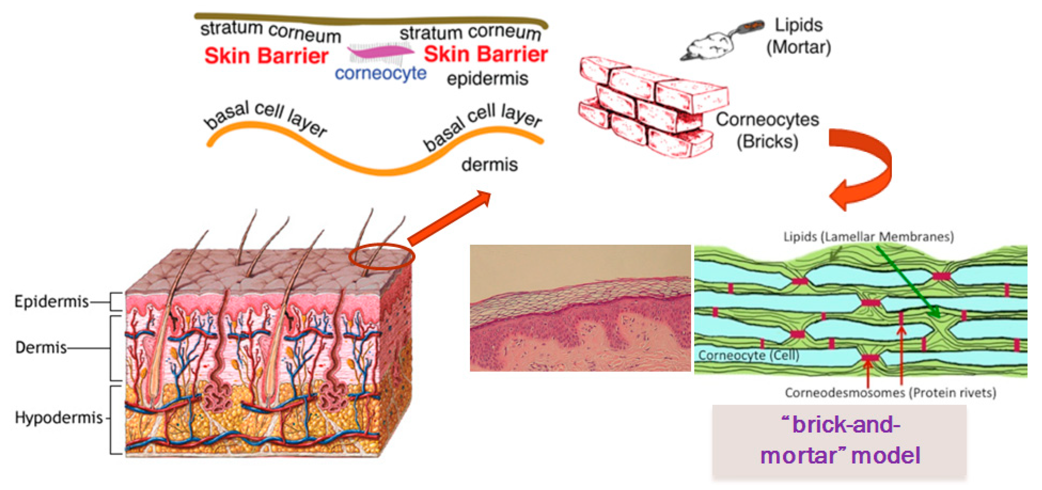
Figure 1. Structure of the stratum corneum.
Cosmetics 06 00052 g001
Figure 2. The effect of skin pH change on permeability barrier.
Cosmetics 06 00052 g002
Figure 3. Ceramide structure.
Cosmetics 06 00052 g003
Figure 4. The identified ceramide types in the human stratum corneum.
Cosmetics 06 00052 g004