Article

Physicochemical and In Vitro Biological Characterization of Usnea barbata Extract in Karanja Oil for Potential Applications in Skincare

by
Mihaela Afrodita Dan
1,†,
Emma Adriana Ozon
1,†,
Denisa Margina
1,
Marina Ionela Nedea
1,
Claudia Maria Guțu
1,
Anca Ungurianu
1,
George Mihai Nițulescu
1,
Violeta Popovici
2,*,
Adina Magdalena Musuc
3,*,
Veronica Bratan
3,
Mihai Anastasescu
3,
Ioana Cristina Marinas
4,
Daniela Luiza Baconi
1,
Andreea Letitia Arsene
1,
Dumitru Lupuliasa
1 and
Eugen Tarta
5
1
Faculty of Pharmacy, “Carol Davila” University of Medicine and Pharmacy, 020945 Bucharest, Romania
2
Center for Mountain Economics, “Costin C. Kiritescu” National Institute of Economic Research (INCE-CEMONT), Romanian Academy, 725700 Vatra-Dornei, Romania
3
Institute of Physical Chemistry—Ilie Murgulescu, Romanian Academy, 060021 Bucharest, Romania
4
Research Institute of the University of Bucharest (ICUB), 050095 Bucharest, Romania
5
Faculty of Medicine, “Carol Davila” University of Medicine and Pharmacy, 050474 Bucharest, Romania
*
Authors to whom correspondence should be addressed.
These authors contributed equally to this work.
Cosmetics 2026, 13(4), 174; https://doi.org/10.3390/cosmetics13040174
Submission received: 29 April 2026 / Revised: 30 June 2026 / Accepted: 2 July 2026 / Published: 5 July 2026
(This article belongs to the Section Cosmetic Formulations)

Abstract

Plant extracts in vegetable oils are foundational and eco-responsible for skin care, combining their emollient properties with other additional benefits, derived from their antioxidant, antimicrobial and UV-absorbing activity. The present research conducted a complex investigation of Usnea barbata extract in Karanja oil (KO), aiming for its further incorporation into various cosmetic formulations. The lichen extract (UBKO) was obtained through cold maceration. Phytochemical screening was performed using the Folin–Ciocalteu method and Graphite Furnace Atomic Absorption Spectrophotometry (GFAAS). Physicochemical properties were evaluated via Fourier Transform Infrared Spectroscopy (FTIR) and Atomic Force Microscopy (AFM). The rheological behavior and oxidative stability of the oil samples, UBKO and KO, were also investigated. UBKO had a slightly lower density (0.827 vs. 0.955) and pH (4.22 vs. 4.86) than KO, and a slightly higher oxidative resistance, quantified as the induction period (IP) value (6.45 vs. 6.00). The total phenolic-equivalent content (TPC, µg GAE/mL oil sample) was significantly greater in UBKO than in KO (567.16 ± 14.96 vs. 433.26 ± 22.96, p = 0.001). The values of minimum inhibitory concentration (MIC, mg/mL) indicated significantly higher antibacterial effect against S. aureus and antifungal effect against C. albicans for UBKO than KO (9.62 ± 2.87 vs. 31.25 ± 18.75, p = 0.049, and, respectively, 5.06 ± 1.68 vs. 37.50 ± 12.50, p = 0.01). Finally, our results showed that UBKO had an estimated sun-protective factor (SPF) of 30.9, slightly higher than 29.8 for the base oil formulation, KO; these findings represent baseline in vitro UV-absorbing trends. All of these results suggest that U. barbata extract in Karanja oil may exhibit complementary bioactive properties with potential applications in skincare.

1. Introduction

Modern skincare science is increasingly oriented toward multi-active botanical formulations that deliver broad-spectrum efficacy while reducing reliance on synthetic chemical agents [1]. Natural products may exhibit complementary benefits when combined, making botanical extracts and vegetable oils attractive multifunctional ingredients in skincare formulations [2,3].
Karanja oil (KO) is a non-edible vegetable oil extracted from the seeds of the Pongamia pinnata (L.) Pierre, also known as Millettia pinnata (L.) Panigrahi, a tree native to India. Its main constituents are triglycerides and unsaturated fatty acids: oleic acid (44–71%), linoleic acid (10–18%), palmitic acid, and stearic acid [4]; it also contains three phenolic constituents (pongamol, karanjin, and cycloart-23-ene-3β,25-diol) with potential UVA and UVB sunscreen activities [5]. Karanja oil is described as a high-polarity active oil and is traditionally used for various chronic skin conditions [6]. Patents claim the use of Karanja oil in sunscreen formulations [7]. Dhule et al. incorporated Karanja oil into a herbal antifungal spray [8]. Karanjin, isolated from seed oil, can be used in cosmetic products such as soaps, body oils, and shampoos [9]. Recent studies report that its incorporation into modern formulations may benefit psoriasis and acne [10,11].
In the plant world, lichens are symbiotic organisms that produce specific, highly bioactive secondary metabolites. A widely studied lichen representative, Usnea barbata (L.) F.H. Wigg, belonging to the Parmeliaceae family, is available online as an extract at https://cosmileeurope.eu/inci/detail/16750/usnea-barbata-extract/, accessed on 10 June 2026. The selection of the lichen U. barbata as the active phytochemical solute was also based on both evolutionary and formulation considerations. It often thrives in extremely high-altitude environments with persistent exposure to intense solar ultraviolet radiation. Thus, U. barbata has evolved by overproducing specialized secondary metabolites, which act as natural, endogenous photoprotective screens and potent antioxidants [12,13]. Modern chemical analysis has identified numerous secondary metabolites from Usnea species [14]: usnic acid is the predominant bioactive constituent, followed by depsides and depsidones (barbatic acid, diffractaic acid, evernic acid) and phenolic acids [15]. Engel et al. demonstrated that U. barbata standardized extract can be a promising ingredient for the development of UV-protective skin care products or anti-inflammatory topical preparations [16].
Usnic acid was loaded into electrospun fibers and hydrogel membranes to optimize wound-healing efficiency [17,18]. Currently, it is used in various topical products as a fragrance, preservative, and antimicrobial agent, including deodorants, anti-acne treatments, creams, powders, toothpastes, mouthwashes, shampoos, and sunscreens. Its capacity to absorb UV light has attracted attention for photoprotection, with the (+)-enantiomer showing photoprotective and photostable properties similar to those of octocrylene in cosmetic formulations [19].
Our team analyzed the composition of U. barbata collected from the same unpolluted area in the Calimani Mountains at 900 m altitude, as documented in earlier studies. U. barbata extracts were prepared using various solvents—ethanol, methanol, ethyl acetate, and acetone—through different methods such as maceration and Soxhlet extraction. A comprehensive elemental analysis was conducted on dried lichen collected in 2020. Twenty-three metals were examined using inductively coupled plasma mass spectrometry (ICP-MS), revealing that native U. barbata contains all metals below the permissible limits for medicinal plants. Usnic acid (a dibenzofuran derivative) and other phenolic secondary metabolites, including phenolic acids (caffeic, p-coumaric acid, ellagic acid, chlorogenic acid, cinnamic acid, and gallic acid), were identified and quantified via UHPLC. Additionally, usnic acid was isolated from the dry U. barbata extract in ethyl acetate using semi-preparative chromatography. We have also extracted U. barbata in Canola oil, and a UHPLC method was validated for determining usnic acid content in U. barbata oil extract. The antimicrobial, antioxidant, and cytotoxic properties of various U. barbata extracts have been previously studied [20].
Both cosmetic ingredients (Karanja oil and U. barbata) are already used in skincare formulations. INCI decoder, the new INCI application (available online at https://incidecoder.com/, accessed on 10 June 2026), indicates that Karanja oil (used at 3–5% concentration) and U. barbata can serve as natural preservatives in cosmetic products, due to their antimicrobial activity. Additionally, the literature provides substantial evidence for antimicrobial, anti-inflammatory, antioxidant, and photoprotective activities—yet they act through distinct molecular mechanisms, exhibit different chemical polarities (lipophilic oil versus lichen-acid complex), and target complementary portions of the UV spectrum [16,21,22,23,24,25]. However, no literature reports the experimental approach of their combination.
In this context, we used Karanja oil (KO) to extract secondary metabolites from Usnea barbata, since vegetable oils are highly effective, eco-friendly solvents owing to their distinctive chemical and physical characteristics [26]. Then, we investigated the physicochemical and pharmacological properties of the oil lichen extract in KO (UBKO) compared with the extraction solvent. We hypothesized that dissolving the secondary metabolites of U. barbata (rich in lipophilic polyphenols and usnic acid) into pure KO would yield a complementary combination. The KO furanoflavonoids (karanjin and pongamol) serve as the primary optical shield, actively absorbing UVA/UVB photons. At the same time, the co-extracted lichen phenolics act as a secondary metabolic sink, systematically quenching the reactive oxygen species (ROS) that escape physical filtration.
Conventional products typically depend on synthetic organic UV filters like avobenzone, oxybenzone, or octocrylene, which face increasing restrictions from regulatory and health agencies due to their links to contact dermatitis, hormonal disruption, and environmental harm, such as coral reef bleaching [27,28,29,30,31,32,33,34,35,36,37,38] (Conventional Sunscreens: UV Filters & Research, available at https://spflist.com/conventional-sunscreens/ accessed on 10 June 2025). In contrast, our formulation utilizes a self-filtering, multi-targeted natural structure based on an active vehicle concept [39]. Instead of merely incorporating a lichen extract into an inert base, the extraction solvent (Karanja oil) serves as a broad-spectrum UV absorber [40]. It is thereby hypothesized to contribute as a preliminary in vitro UV-attenuating barrier, while concurrently suggesting a supportive, putative role in mitigating oxidative stress through an antioxidant cascade. However, it must be noted that these distinct conceptual mechanisms remain to be validated in advanced cellular or tissue-engineered skin models.
The novelty of our study lies in a complex physicochemical and pharmacological characterization of UBKO, a green extract with a specific vehicle–solute interaction, for potential incorporation into various cosmetic formulations.

2. Materials and Methods

2.1. Materials and Equipment

Karanja oil (KO), extracted from the seeds of Pongamia pinnata by cold pressing, was supplied by Fagron-Hellas (Trikala, Greece). The oil is highly pure and suitable for cosmetic uses. Its composition offers oxidative stability, a non-greasy feel, and good compatibility with human skin (Karanja seed oil, available at https://www.fao.org/4/x5043e/x5043e0e.htm, accessed on 9 June 2026).
In March 2024, U. barbata thalli were collected from the Călimani Mountains in Romania (47°28′ N, 25°13′ E at 900 m altitude). The freshly collected lichens were cleaned of debris and dried at 18–25 °C in a herbal room away from sunlight. For long-term preservation, dried samples were stored under similar conditions. The identification was performed by the Department of Pharmaceutical Botany at the Faculty of Pharmacy, Carol Davila University of Medicine and Pharmacy, using standard techniques. A voucher specimen is housed at the Herbarium of the Pharmacognosy Department, Faculty of Pharmacy, Carol Davila University of Medicine and Pharmacy (UBL 3/2024, Ph-UMFCD).
All chemicals, solvents, and reagents used were of high quality, ensuring reliable results. We sourced Dimethyl sulfoxide (DMSO), sodium carbonate (Na2CO3), and Folin–Ciocalteu reagent from Sigma-Aldrich Chemie GmbH (Schnelldorf, Germany). For our experiments, we also used high-purity 69% nitric acid (trace grade) and prepared standard stock solutions containing 1000 mg/L H3AsO4 and Pb(NO3)2 in 0.5 M nitric acid. Additionally, we obtained 30% H2O2, 96% ethanol, methanol, crystal violet, acetic acid, and ultrapure deionized water from Merck Millipore (Burlington, MA, USA).
Microbial strains were acquired from the American Type Culture Collection (ATCC): Staphylococcus aureus ATCC 25923, Escherichia coli ATCC 25922, and Candida albicans ATCC 10231. Culture media came from two suppliers. Tryptic Soy Agar for bacteria was obtained from Sigma-Aldrich Merck (Dartmouth, Germany), while RPMI 1640 for fungi was supplied by American Biorganics (Buffalo, NY, USA).

2.2. U. barbata Extract in Karanja Oil

The oil lichen extract (UBKO) was prepared through 3-month cold maceration, as described earlier [25,41]. About 20 g of this material was precisely measured with a Kern analytical balance and placed in a 1000 mL brown glass container, to which 500 mL of KO was added (dried lichen/solvent ratio w/v = 1:25). The dried thallus of U. barbata was ground using a laboratory knife mill and standardized with a standard sieve. The particle size distribution was controlled by passing the material through successive sieves with 2.5 mm (DIN 1171) and 1.2 mm (DIN 117) mesh for homogenization [41].
Moisture content after drying: Prior to extraction, the lichen material was air-dried in a dark, well-ventilated room until it reached a constant weight. The residual moisture content was determined by loss-on-drying and was found to be below 5%, within the acceptable pharmaceutical limit to prevent accidental water-induced hydrolysis in the lipophilic medium.
The maceration was conducted in hermetically sealed, amber glass containers that were sterilized beforehand, stored in a dark, temperature-controlled environment (21–22 °C), manually shaken daily, and inspected periodically. No macroscopic or biochemical signs of spoilage were observed.
After this period, UBKO was filtered through cotton-mesh gauze into a brown vessel with a sealed lid and stored in a plant room away from sunlight.
Extraction yield (%) and efficiency evaluation: Since the extraction was performed directly into a lipid vehicle (Karanja oil) rather than a volatile organic solvent, the traditional mass-balance extraction yield (dry weight of extract/dry weight of plant) could not be directly determined by evaporation. Therefore, we appreciated the extraction efficiency by measuring the total phenolic content (TPC) transferred to the oil phase.
Control of microbial contamination during the 3-month maceration: To ensure microbiological safety during the extended 3-month maceration, multiple barriers were in place:
-
Pure Karanja oil is a lipophilic vehicle, with a thermodynamic state that fundamentally inhibits the growth and proliferation of bacteria, yeasts, and molds.
-
Both Karanja oil (due to karanjin) and Usnea barbata (due to usnic acid) possess well-documented, broad-spectrum antimicrobial and antifungal properties, acting as a self-preserving system.
The UBKO density (g/mL) was slightly lower than that of KO (0.827 < 0.955).
The pH values for KO and UBKO were measured with a pH meter (P601, CONSORT nv, Turnhout, Belgium). Equal volumes (1 mL) of UBKO and KO were transferred to 10 mL volumetric flasks, and 4 mL of deionized hot water (80 °C) was added. The mixtures were shaken vigorously for 5 min and then filtered. The pH value of the resulting aqueous extract was measured with the calibrated pH meter at 25 °C. UBKO has a lower pH value than KO (4.22 < 4.86).

2.3. Total Reducing Capacity (Folin–Ciocalteu Assay)

To establish a uniform system, 2 mL of each sample (KO and UBKO) was dissolved in 8 mL of polyethylene glycol 400 (PEG 400), ensuring consistent oil dispersion and enhanced solubilization of phenolic compounds in the reaction medium. Initial testing revealed that PEG 400 alone led to phase separation during the Folin–Ciocalteu assay, causing heterogeneous samples and unreliable spectrophotometric readings. Therefore, samples in PEG 400 were further diluted 1:1 (v/v) with DMSO. This PEG 400/DMSO co-solvent system improved sample miscibility and stability during analysis [42]. Briefly, 50 μL of each sample was combined with 50 μL of Folin–Ciocalteu (FC) reagent, 450 μL of distilled water, and 500 μL of 7% (w/v) Na2CO3 solution. The mixtures were mixed thoroughly and incubated in the dark for 50 min to allow the formation of the characteristic blue complex resulting from the reduction of the FC reagent by compounds present in the samples. Following incubation, samples were centrifuged at 10,000 rpm for 5 min to remove insoluble particles. Absorbance was measured at 765 nm using a UV–Vis FlexStation 3 spectrophotometer (Molecular Devices, San Jose, CA, USA).
This method showed good linearity within the concentration range of 25–250 µg/mL gallic acid (R2 = 0.9965), with the calibration equation y = 0.0027x − 0.0261. The detection and quantification limits (LOD and LOQ) were 9.88 µg/mL and 29.94 µg/mL, respectively. All measurements were conducted in triplicate, with matrix interference controlled using DMSO and PEG 400/DMSO blanks. This approach allowed us to distinguish the solvent system’s contribution from that of the oil samples and to highlight PEG 400’s influence on the measured reducing capacity. Results are expressed as gallic acid equivalents (GAE) per mL or per g, representing the total phenolic-equivalent content (TPC). For results in μg GAE/g, the conversion from μg GAE/mL was based on the system’s density. The assay used PEG 400 alone as a control to clearly illustrate the oils’ composition [41].

2.4. FTIR Spectra

FTIR spectra were obtained using an FTIR spectrometer (FT/IR-4200 apparatus, from JASCO, Tokyo, Japan). The spectral experiments were obtained in the wavelength range between 4000 and 400 cm−1, with a 4 cm−1 resolution. All spectra were recorded at ambient temperature and are presented in transmittance values [41].

2.5. AFM Analysis

For Atomic Force Microscopy (AFM) analysis, KO and UBKO (20 µL each) were diluted in 2 mL of 96% ethanol. Then, the samples were further deposited on a clean glass substrate and heated at 20 °C for 30 min. This additional step aimed to ensure solvent evaporation and proper sample adhesion. AFM analysis was performed in enhanced contrast mode [41]. The resulting line scans illustrated the surface profiles of both studied samples.

2.6. Heavy Metals Content

Heavy metals, arsenic (As) and lead (Pb), were detected using Graphite Furnace Atomic Absorption Spectrophotometry (GFAAS) as previously described. The measurements were carried out with a SOLAAR 6M (Thermo Electron Inc., Waltham, MA, USA) atomic absorption spectrometer equipped with a deuterium lamp for background correction. The specific wavelengths used were 193.7 nm for As and 283.3 nm for Pb. Concentrations of both metals in each oil sample were determined based on their respective regression lines, which relate absorbance to concentration. All measurements were performed in duplicate [41].

2.7. Rheological Measurements

Rheological measurements were conducted using a B One Plus rotational viscometer (Lamy Rheology, Champagne-au-Mont-d’Or, France), which offers a full-scale accuracy of ±1% and a repeatability of 2%. The instrument is equipped with seven spindles (RV1–RV7), each designed to operate within a defined viscosity range. For the present study, spindle RV3 was selected and immersed in 50 mL of each oil sample for viscosity determination. Measurements were carried out at rotational speeds of 50, 100, 150, 200, and 250 rpm, with each reading recorded after 10 s of stabilization. All analyses were performed under controlled temperature conditions of 22 °C [41]. The spreading properties of the samples were evaluated using an extensometer (Epsilon Technology Corp., Jackson, WY, USA). A fixed volume of 0.5 g of oil was carefully deposited at the center of the lower glass plate, after which a second glass plate (50 g) was placed on top to initiate spreading. The system was subsequently subjected to incremental loading by adding weights of 50, 100, 200, and 500 g. After a 1-min relaxation period for each applied load, the diameter of the spread sample was recorded. The area of the spreading was then calculated using the formula πr2.

2.8. Oxidative Stability

The Velp OXITEST reactor (Velp Scientifica Srl, Usmate Velate, MB, Italy), which speeds up oxidation under carefully controlled and consistent conditions, was used to evaluate the oxidative stability of UBKO and KO by measuring the induction period [43], as previously described [41].

2.9. Antioxidant Activity

2.9.1. DPPH Assay

We evaluated the radical-scavenging ability of UBKO using the 2,2-diphenyl-1-picrylhydrazyl (DPPH) method, as previously described [44]. The DPPH method relies on the discoloration of a solution containing the DPPH free radical (Sigma-Aldrich Inc., Burlington, MA, USA) when it interacts with antioxidants in the tested sample. For each sample, multiple dilutions in methanol were prepared (1:1, 1:5, 1:10, and 1:25) to examine the dose-dependent variability of the antioxidant effect.
Each sample was combined with the DPPH working solution at a 1:2 ratio, starting with an optical density (OD) value of around 0.8.
We performed an assessment by measuring the decrease in OD at 517 nm over one hour. Additionally, we conducted endpoint measurements at 5, 30, and 60 min to analyze the decrease in OD:
ΔOD (%) = 100 × (ODDPPH − ODDPPH + sample)/ODDPPH
where ODDPPH corresponds to methanol + DPPH working solution. This was used to demonstrate the antioxidant effect of the tested oil samples.

2.9.2. ABTS Assay

The 2,2′-Azino-bis(3-ethylbenzothiazoline-6-sulfonic acid) (ABTS) method assesses a sample’s capacity to neutralize the ABTS+∙ free radical. The stock solution, containing 7 mM ABTS and 2.45 mM K2S2O8, was stored in the dark at 4 °C for 16 h to produce the green ABTS+∙ radical. This was diluted with distilled water to a final optical density of 0.8 at 714 nm, creating the working solution. Oil samples, both undiluted and diluted at ratios of 1:1, 1:5, 1:10, and 1:25, were mixed with the working solution at a 1:3 ratio, and their absorbance was measured at 714 nm after 10 min. A control sample was prepared using ethanol instead of the oil. The results are expressed as the percentage decrease in optical density (% ΔOD) [44].
ΔOD (%) = 100 × (ODblank − ODsample)/ODblank
This was considered to express the antioxidant effect of the tested oil samples [44].

2.10. Antibacterial and Antifungal Activity

To assess the antimicrobial activity of KO and UBKO against the most common pathogens (S. aureus ATCC 25923, E. coli ATCC 25922, and C. albicans ATCC 10231), we first determined their minimum inhibitory concentration (MIC). Subsequently, using sub-MICs (MIC/2 and MIC/4), we evaluated the oil samples’ ability to inhibit microbial adhesion to an inert substrate (AC, expressed as a percentage) [45].

2.10.1. Assessment of the Minimum Inhibitory Concentrations

To assess the inhibitory activity of the oil samples, a 20% Tween 80 (T80) solution in ethanol was used to dissolve the bioactive compounds, facilitating their dispersion in the aqueous culture medium. All microbiological assays included vehicle control wells using the Tween 80–ethanol system [45].
Suspensions with an optical density of 0.5 McFarland for bacteria and 1 McFarland for fungi were prepared. The following procedures were carried out: 100 μL of TSA or RPMI 1640 was distributed into each of the 96-well plates, followed by 100 μL of a 100 mg/mL solution of the compounds under investigation in the first well. A binary serial dilution scheme was then performed up to the tenth well. Subsequently, 20 μL of suspension from the strains designated for analysis was added, ranging from well 1 to well 12. Well 11 was with vehicle control (T80), while well 12 was designated for negative control. Absorbance measurements were conducted at 620 nm utilizing a Thermo Scientific™ Multiskan™ GO Microplate Spectrophotometer (Thermo Fisher Scientific, Waltham, MA, USA) [44].

2.10.2. Assessment of How Oil Samples Affect Microbial Adherence to the Inert Substrate

The impact of UBKO and KO on biofilm formation was examined using the method previously outlined [45]. The adhesion level of the tested strains to an inert substrate was measured at sub-MICs (MIC/2 and MIC/4) after fixing with methanol and staining with a 1% crystal violet solution in 33% acetic acid [45].
The adherence capacity (AC%) of the microbial strains was ascertained through the utilization of the subsequent formula.
AC (%) = 100 × (Asample − Ablank)/(Acontrol − Ablank)
where Asample is the absorbance at 490 nm of the tested oils, while Acontrol represents the absorbance at 490 nm of the negative control, represented by untreated microbial strains [44].

2.11. Sunscreen Properties

Volumes of 1 mL of the studied oil sample were transferred to a volumetric flask (100 mL) and diluted with ethanol to volume. The samples were then ultrasonicated for 5 min. The SPF values of the oil samples were measured using a spectrophotometer, Perkin-Elmer Lambda 35 (Perkin-Elmer Inc., Waltham, MA, USA). The experiments were made in transmission mode. The samples were placed in microcuvettes with a 10 mm light path, and the absorption values were recorded over the range of 290 to 320 nm, at 5 nm intervals. Each measurement was performed three times, and the average value was used. SPF values were calculated using the Mansur equation [44] (Equation (1)).
S P F = C F × 320 290 E E λ × I λ × A b s λ
where CF = correction factor (10), I(λ) = intensity of solar light at wavelength λ, EE(λ) = erythemogenic effect of radiation at wavelength λ, and Abs (λ) = absorbance of wavelength λ by the preparation solution. The value of E E λ × I λ is constant.

2.12. Statistical Analysis

Most measurements were carried out three times to ensure consistent results, and the findings are presented as averages with their standard deviations. We analyzed the data using SigmaXL v.11.03/2025 (SigmaXL Inc., Kitchener, Ontario, Canada) and Microsoft Excel v.16.0 19328 (Microsoft Corporation, Redmond, WA, USA). To identify significant differences among variables (p < 0.05), we used One-Way ANOVA and Means Matrix, 2-Sample t-test, and Kruskal–Wallis test.

3. Results

3.1. Total Reducing Capacity (Folin Ciocalteu Assay)

The results obtained by the Folin–Ciocalteu assay (Table 1) show significant differences in the total phenolic-equivalent content (TPC, µg GAE/mL) between the KO and UBKO. Karanja oil had an appreciable capacity to reduce FC reagent; U. barbata extraction in KO significantly increased the total reducing capacity (567.16 vs. 433.26 µg GAE/mL in the oil sample; p = 0.001; Table 1). The oil samples emulsified with PEG 400 had a higher capacity to reduce FC reagent, with significant differences between KO and UBKO, because the Folin–Ciocalteu assay is based on an electron-transfer (redox) mechanism and exhibits known interferences with non-phenolic reducing compounds (Table 1).
The positive value obtained for the PEG 400 control (260.49 ± 0.27 µg GAE/mL) was due to the reducing capacity of the terminal hydroxyl groups of the short polymer, enhanced by possible traces of aldehyde impurities or reducing sugars from the raw material, typical of low-molecular-weight materials [46]. Therefore, the results provide an estimate of the total reducing capacity of the oil samples (KO and UBKO). Since the method also responds to other reducing compounds, the obtained values do not represent a specific measurement of phenolic secondary metabolites’ concentration.

3.2. FTIR Analysis

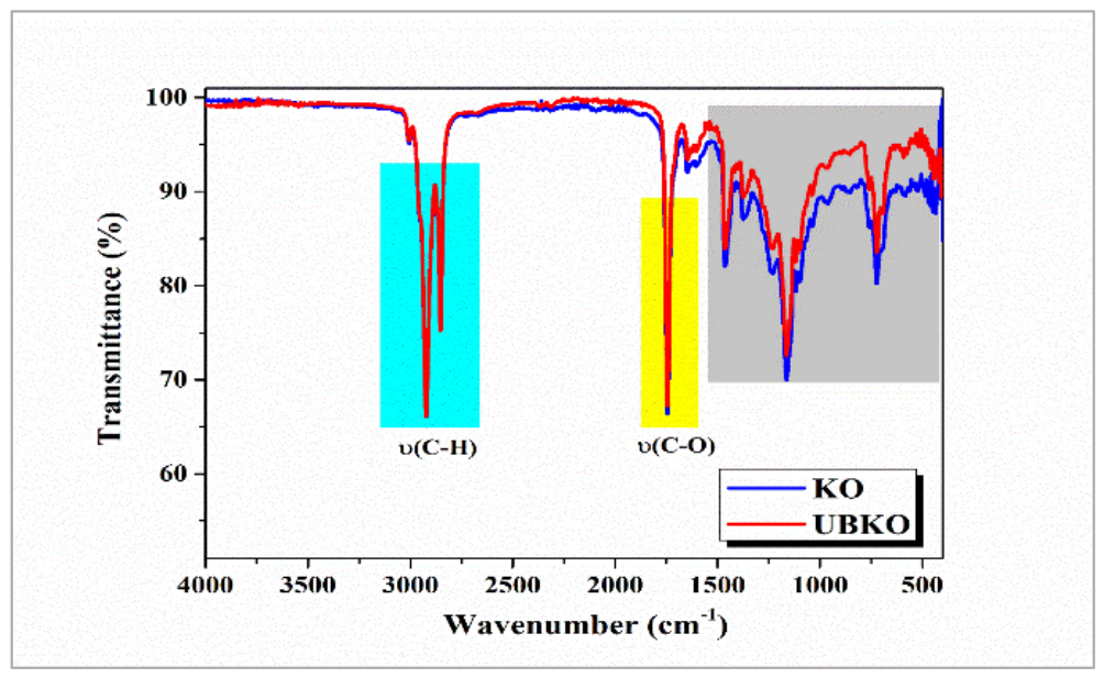
The IR spectrum of KO reveals molecular structures rich in saturated aliphatic chains, evidenced by C–H stretching vibrations at 2922.6 cm−1 and 2853.2 cm−1, which are typical of methylene (–CH2) groups in alkane derivatives (Figure 1, blue line) [47]. The C–H bending vibrations are observed at 1463.7 cm−1 and 1376.0 cm−1, confirming the presence of alkyl chains. The absorption at 722.2 cm−1 corresponds to the rocking vibration of methylene groups, supporting the existence of long-chain aliphatic backbones. The weak band at 3006.5 cm−1 indicates a =C–H stretching vibration, but the high transmittance suggests a low proportion of double carbon–carbon bonds compared to the saturated (single-bonded) carbon atoms in the oil components (Figure 1, blue line). The strong band at 1743.3 cm−1 confirms the presence of ester groups assigned to the carbonyl (–C=O) stretch vibration. The bands observed at 1230.4 cm−1, 1162.9 cm−1, and 1116.6 cm−1 are assigned to the C–O stretching vibrations. The free acids are below the detection limit, as shown by the absence of the characteristic hydroxyl-stretching broad O–H band from the range of 3500 to 2500 cm−1. The IR spectrum suggests that triglycerides may be the most abundant components (Figure 1, blue line). The IR spectrum of UBKO shows some differences in the 1600–400 cm−1 region, but the spectral shapes are similar (Figure 1, red line). These differences in the fingerprint region suggest slight structural alterations, such as changes in the chemical environment, cation substitution, or various lattice vibrations/degrees of ordering between the two oil samples, thereby maintaining a pattern similar to the KO. This similarity, together with the absence of significant band shifts or new absorption bands, demonstrates that U. barbata metabolites extracted in KO do not undergo chemical interactions, but only physical associations.

3.3. AFM Analysis

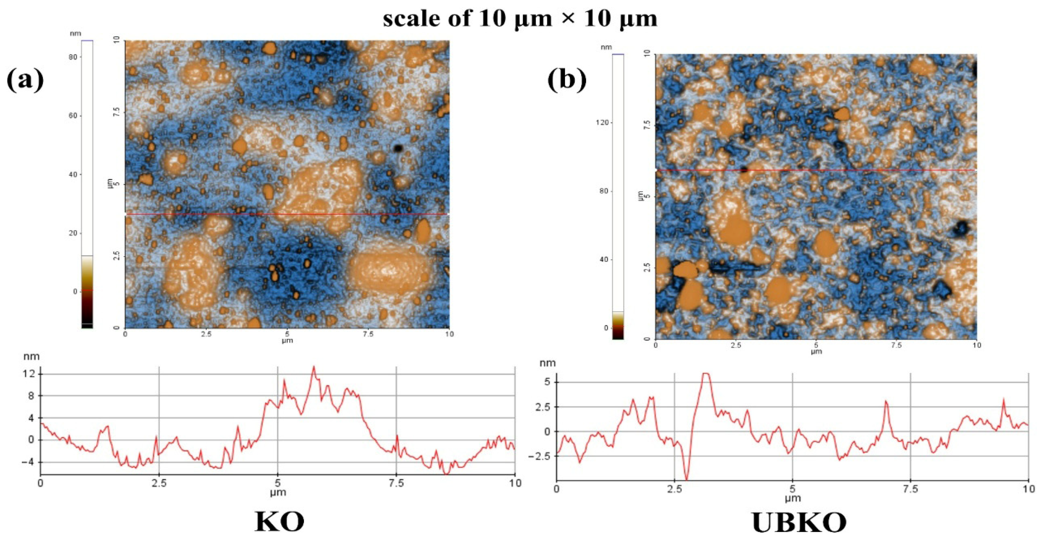
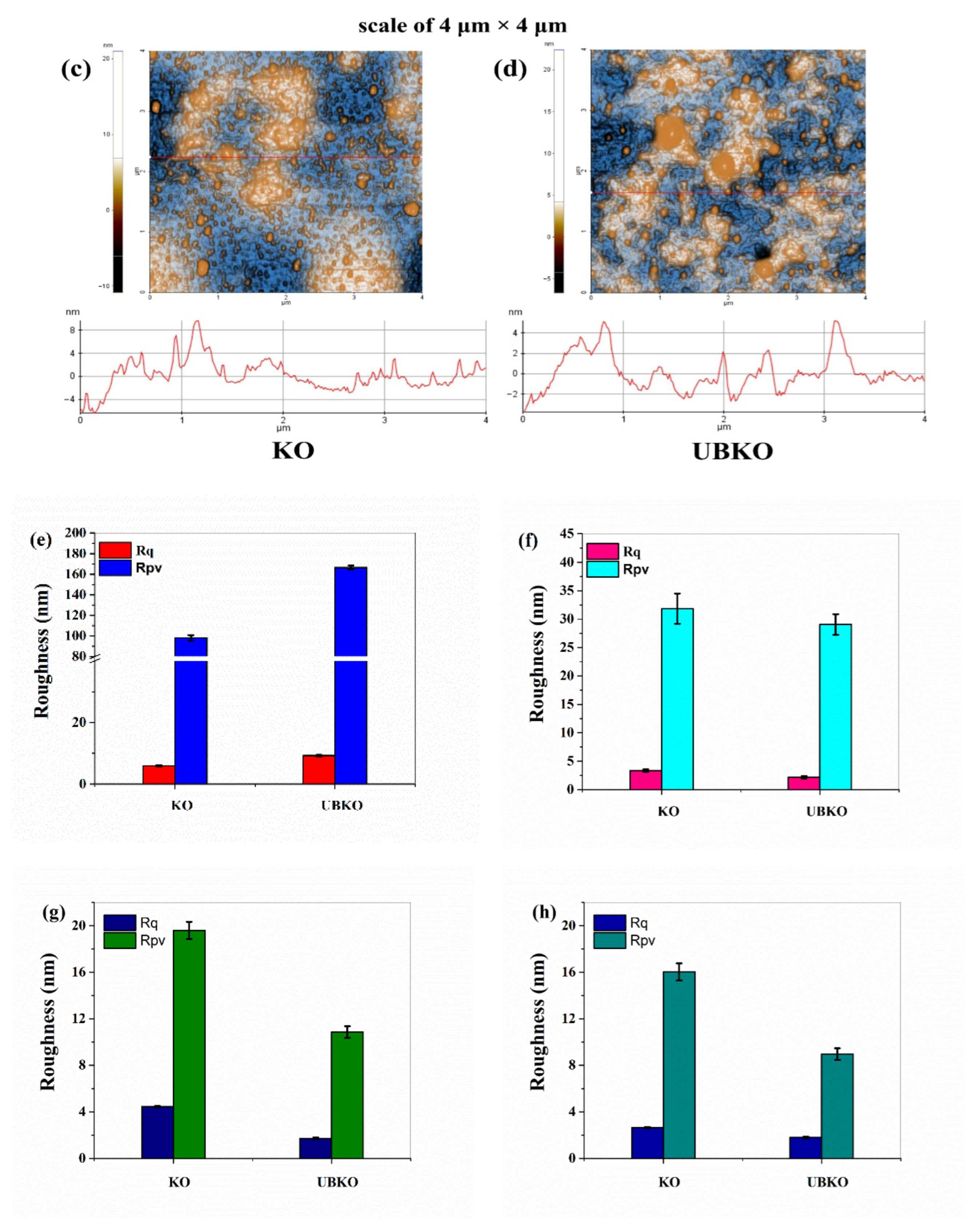
The AFM images of KO and UBKO are shown in Figure 2. Figure 2a displays the scan over a (10 × 10) µm2 area for KO, while Figure 2b shows a similar scan for UBKO. Figure 3c,d present scans over a (4 × 4) µm2 area for KO and UBKO, respectively. The characteristic line-scan profiles are indicated at the position marked by the red line. Additionally, Figure 2e–h show histograms of the peak-to-valley (Rpv) and roughness (Rq) parameters.
AFM analysis shown in Figure 2 highlights several morphological differences between KO and UBKO at both examined scales of (10 × 10) µm2 and (4 × 4) µm2. KO exhibits a mostly uniform surface, characterized by larger structures or aggregates that are sporadically dispersed. This suggests the presence of microdomains on the surface, leading to a vertical red line scale variation of approximately 17 nm (from −5 to 12 nm), specific to its lipid composition (Figure 2a). In contrast, UBKO displays a much more heterogeneous surface, characterized by smaller structures that are more numerous and evenly distributed (Figure 2b). This results in lower variations of around 9 nm (from −3 to 6), as indicated by the red line scan in Figure 2b. These formations suggest changes in the surface morphology and structural organization of the oil matrix following extract incorporation. The AFM topography of both compounds is also confirmed at a smaller scale (4 × 4) µm2, where the morphological details become more evident (Figure 2c,d).
The analyzed roughness parameters, Rpv (peak-to-valley parameter) and RMS (root mean square) roughness (Rq), respectively, exhibit lower values in the case of KO despite the presence of larger aggregates. This observation confirms its smooth and homogeneous nature (Figure 2e,f). The KO sample is characterized by an Rq of 5.9 nm and an Rpv of approximately 98.1 nm across the entire scanned area of (10 × 10) µm2 (Figure 2e). The relatively low density of KO (0.9548 g/cm3) indicates a less dense molecular association, typical of lipid systems. This KO structure facilitates molecular mobility, thereby supporting the formation of larger, yet sporadically distributed, aggregates that contribute to a relatively smooth surface.
In contrast, the U. barbata phytochemicals extracted in Karanja oil resulted in much higher roughness parameter values across the entire scanned area and along the corresponding red line profile (Figure 2e–h). This indicates a noticeable increase in the roughness and local height variations. UBKO is characterized by an Rq of about 9.3 nm and a Rpv of about 166.6 nm. Lichen constituents can induce surface reorganization, possibly by forming smaller nanoscale structures, thereby increasing surface roughness.

3.4. Heavy Metals Content

The arsenic content (μg/g) is higher in UBKO compared to KO (0.203 versus 0.131), while the lead content is lower (0.056 versus 0.065). The data are presented in both μg/L and μg/g in Table 2. The arsenic levels in both oil samples stay within the range considered safe for cosmetic or unrefined vegetable oils. These findings align with recent literature, which reports arsenic concentrations in vegetable oils typically between 0.05 and 0.20 mg/kg. Both oil samples exhibit similar lead levels (0.065 and 0.056 μg/g), which are well below the maximum permissible limit of 0.100 μg/g established by Regulation (EC) No. 1881/2006 for lead in vegetable oils and fats [48].

3.5. Rheological Properties

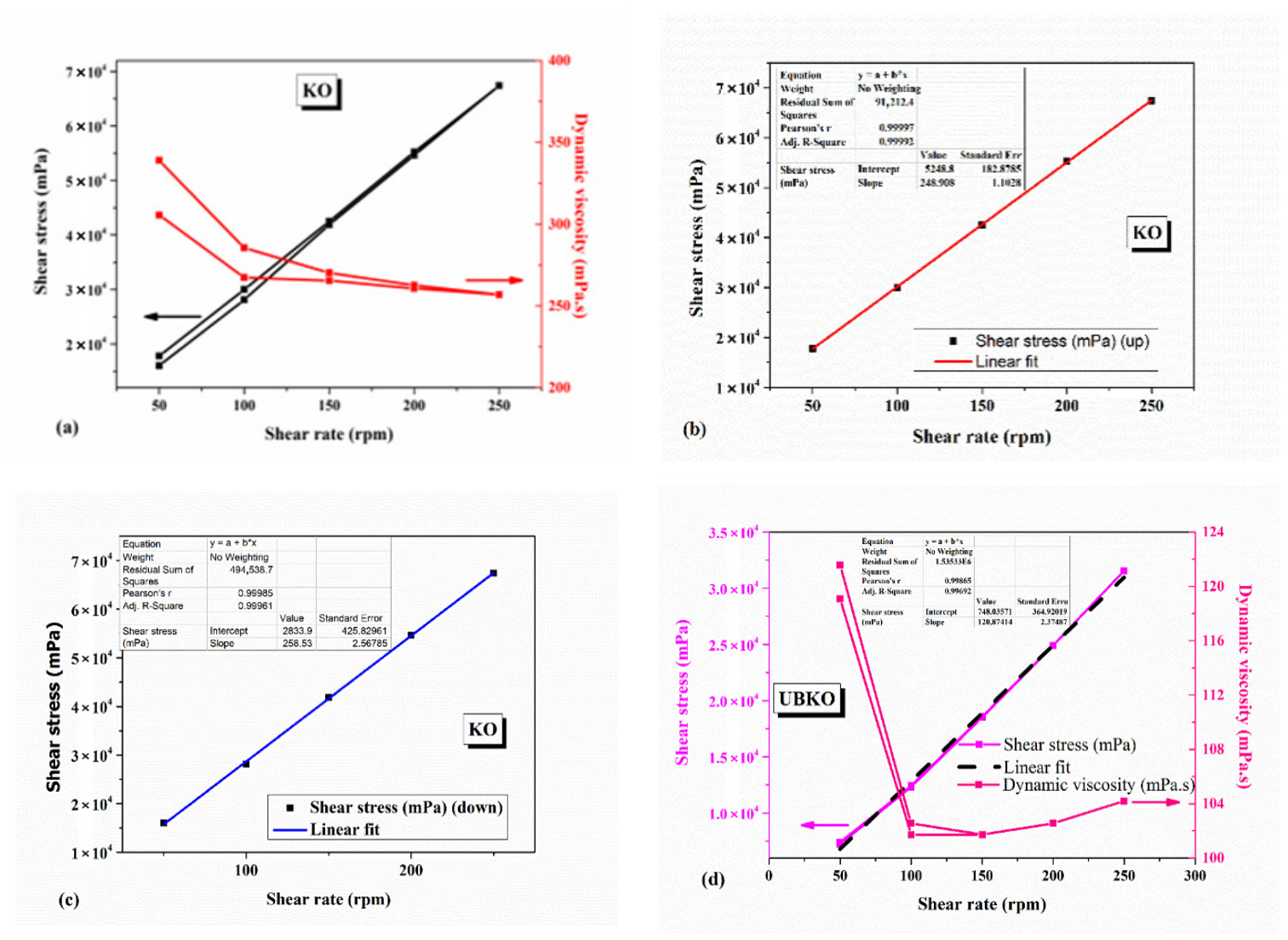
The rheological curves of KO and UBKO, along with the linear fit equations for shear stress, are shown in Figure 3. We examined the rheological behavior of KO by looking at how shear stress (mPa) relates to shear rate (rpm) (see Figure 3a–c). A clear linear relationship was found across the explored range, which suggests Newtonian behavior—meaning shear stress increases directly with shear rate.
The experimental data were fitted using a linear regression model of the form (Equation (2))
y = a + b x
where y is the shear stress (τ), x is the shear rate ( γ ˙ ), and slope (b) represents the dynamic viscosity (η), according to Equation (3) [49].
η = τ γ ˙
The high correlation coefficient (r2 ≈ 0.999) confirms a strong linear dependence between shear stress and shear rate. Although a rapid decrease in dynamic viscosity was observed with increasing or decreasing shear rate, in the initial range of 50 to 100 rpm (at low shear rates), the KO exhibits shear-thinning behavior. Subsequently, the dynamic viscosity ranges from approximately 250 to 260 mPa·s at higher shear rates, indicating a transition to Newtonian behavior. This variation can be attributed to the low torque values (0.115–0.485 mN·m) recorded during measurements. Additionally, the composition of higher fatty acids influences KO’s shear-thinning behavior, leading to non-Newtonian behavior at lower shear rates due to molecular interactions involving monounsaturated fatty acids, which result in higher dynamic viscosity at low shear rates. Therefore, the linear shear stress–shear rate relationship is considered a more reliable parameter for determining KO’s fluid behavior. Overall, these results demonstrate that the analyzed KO sample behaves as a Newtonian fluid within the tested shear rate range.
The rheological behavior of UBKO was assessed by analyzing the relationship between shear stress and shear rate (Figure 3d). A linear correlation was observed throughout the entire tested shear rate range, and the dynamic viscosity remained mostly constant between 100 and 250 shear rates, confirming that the UBKO also displays Newtonian behavior, similar to KO. Linear regression was conducted using the same model as before (Equation (2)). Additionally, the high correlation coefficient (r2 ≈ 0.997) supports the strong linear relationship between shear stress and shear rate and indicates no yield stress. The nearly constant dynamic viscosity suggests that UBKO’s flow resistance is independent of shear rate, a hallmark of Newtonian fluids.
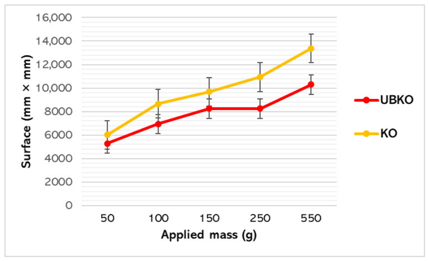
Initially, when the upper plate was applied, the stretching surface of UBKO was smaller than that of KO, indicating its spreadability (Figure 4).
Later, when more weights are added, UBKO’s extensibility remains lower than KO’s (7800.88 ± 1851.54 compared to 9727.16 ± 2723.88, p > 0.05; Figure 4). The spreadability of the samples correlates with their viscosity behavior, suggesting that KO will likely spread quickly and evenly over the skin without needing much pressure. Additionally, extensibility indicates KO’s behavior within various fluid or semi-solid systems. The results show that KO can spread uniformly across different carriers with minimal energy input. Formulation cohesiveness influences extensibility, and KO maintains its structural integrity and flexibility. Gobi et al. demonstrated that viscoelastic properties significantly impact adhesion [50].
A robust link between spreading and viscoelasticity is observed in UBKO, much like in KO. Nonetheless, the minor variations observed imply that lichen components form new interparticle bonds within the oily system, as reflected in the changes in rheological properties.

3.6. Oxidative Stability

Oxidative stability was quantitatively assessed by the induction period (IP), recorded in hours. This is the time point where the tangent lines drawn before and after the inflection point intersect. The IP represents how long it takes for oxidation to initiate, potentially resulting in noticeable rancidity or a sharp rise in oxidation rate.
Generally, a longer IP correlates with higher oxidation resistance and shelf life [43]. Our results show that UBKO had slightly greater oxidative stability (IP = 6.45) than KO (IP = 6).

3.7. Antioxidant Activity

To assess the antioxidant activity of KO and UBKO accurately, samples were diluted with methanol at ratios of 1:1, 1:5, 1:10, and 1:25. These were then compared to a solvent-based standard to analyze their dose–response behavior.
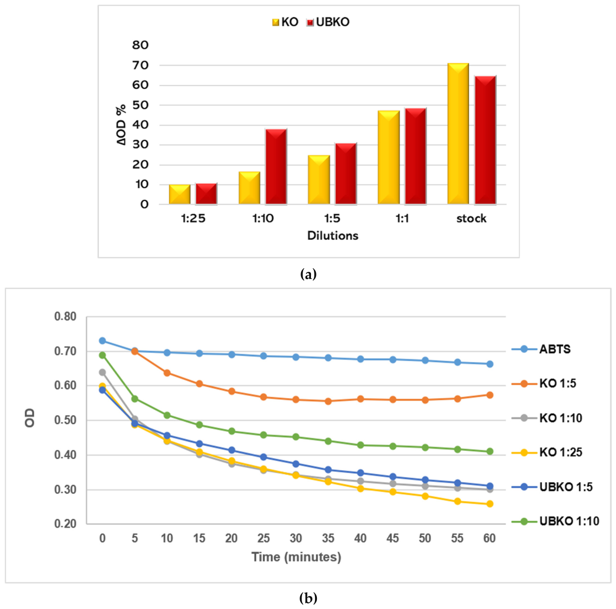
The in vitro radical-scavenging ability of KO and UBKO, evaluated with the DPPH method, was determined using both kinetic and endpoint assays (Figure 5a).
The KO sample stock solution reported the strongest DPPH radical-scavenging activity (ΔOD = 70.92%), higher than UBKO (ΔOD = 64.64%). UBKO dilutions exhibited greater antioxidant capacity than KO, which generally decreased with dilution grade from 1:1 to 1:25 (Figure 5a).
Results from the ABTS method are shown in Figure 5b. We were unable to assess the KO and UBKO undiluted samples or the lowest dilutions because an emulsification process rendered them unsuitable for reading, as the ABTS working solution was prepared with water as the solvent, which is incompatible with the hydrophilic antioxidants in the evaluated samples. Therefore, results were presented alongside the dynamics of the ABTS solution over the same time span (Figure 5b). The evaluated dilutions ranged from 1:5 to 1:25. The radical-scavenging activity of both oil samples increased over time, as indicated by decreases in OD values (Figure 5b).
The results revealed that KO has antioxidant capacity, with statistically significant differences among the tested dilutions. UBKO also exhibited antiradical activity, though variation across the tested dilutions was less pronounced. Overall, both samples showed reasonable free radical-scavenging activity over the 1:5 to 1:25 dilution range.

3.8. Antibacterial and Antifungal Activity

The antimicrobial activity assessment targeted two aspects: (1) establishing the minimum inhibitory concentration (MIC) and (2) assessing KO and UBKO’s capacity to prevent bacterial and fungal biofilm formation.
To assess the inhibitory activity of the oil samples, a 20% Tween 80 (T80) solution in ethanol was employed for solubilization, facilitating the dispersion of bioactive compounds in the aqueous culture medium. Tween 80 also served as a positive control in the microbiological assays.
All determined MIC values (mg/mL) shown in Table 3 were considerably higher than those of conventional antibiotics’ values (mg/mL) reported by Rankovic et al. for the same ATC bacterial and fungal lines [51]. UBKO had the highest inhibitory activity against S. aureus and C. albicans, and the lowest antibacterial efficacy against E. coli (Table 3).
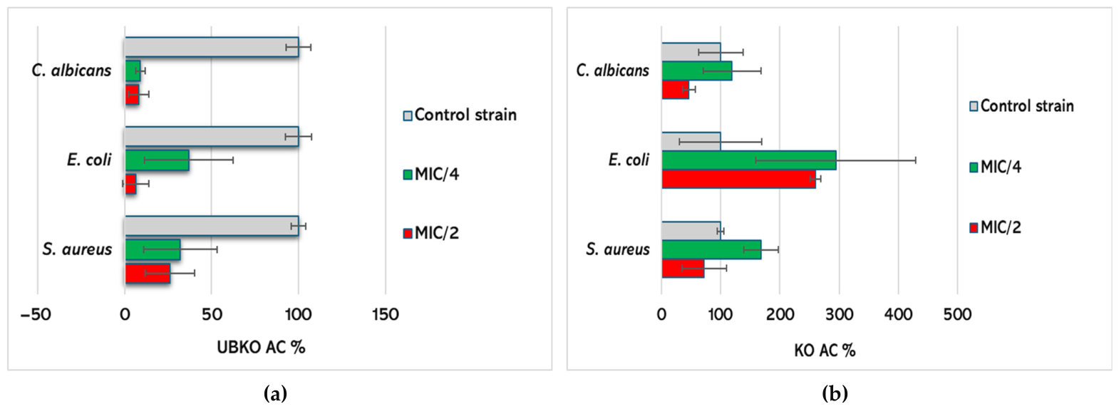
The influence of KO and UBKO at sub-MICs (MIC/2 and MIC/4) on microbial adherence compared to the negative control (untreated microorganisms) is illustrated in Figure 6.
The results show that UBKO inhibited the adherence of all bacterial and fungal strains at both sub-MICs tested, and the effect was directly proportional to the concentration tested. The AC values for all pathogens were significantly lower than those of negative controls (p < 0.05, Figure 6a). The highest antiadherence capacity was observed against C. albicans, with very little difference in AC (%) values at both sub-MICs (MIC/2 vs. MIC/4, 7.89 vs. 8.49; Figure 6a). Higher AC values were recorded on S. aureus (31.99 vs. 26.62, Figure 6a). UBKO at MIC/2 showed an E. coli adherence inhibition almost 6 times higher than MIC/4 (6.19 vs. 36.77, Figure 6a).
Contrariwise, KO at MIC/4 stimulated biofilm formation for all pathogens; the highest adherence capacity was observed in E. coli, followed by S. aureus and C. albicans (294.52 vs. 167.91 and 119.33, Figure 6b). At MIC/2, KO considerably inhibited the adherence of C. albicans and S. aureus (46.55% and 72.24%, respectively), while its stimulatory effect on E. coli remained high and recorded a very low diminution (AC = 260.30 at MIC/2 vs. 294.52 at MIC/4, Figure 6b).

3.9. UV Absorption

SPF values serve as a standard for evaluating the efficacy of sunscreen formulations against UV-B radiation. SPF value represents the ratio of UV energy required to achieve a minimum erythematous dose (MED) in protected skin relative to unprotected skin. The absorbance values of the KO and UBKO samples obtained using the UV spectrophotometric method are shown in Table 4.
The SPF value of KO is 29.8, and for UBKO is 30.9. It was observed that extracting U. barbata bioactive constituents into Karanja oil increased the SPF by approximately 1 unit. These results show that U. barbata extraction did not significantly change the estimated SPF value under the tested conditions. However, the formulation kept UV-absorbent and antioxidant properties that could be important for photoprotective uses. The results suggest that the KO and UBKO can be considered for inclusion in the sunscreen formulations, together with other ingredients.

4. Discussion

Our study proposes a novel combination of two known bioactive natural products (U. barbata extract in Karanja oil) with potential benefits in skincare. The research had a broad experimental scope, integrating physicochemical and biological characterization relevant to cosmetic applications.
The selection of a 3-month maceration period at room temperature for UBKO preparation was determined by the unique mass-transfer kinetics present in a highly viscous lipid medium. According to fundamental diffusion principles, as modeled by the Stokes–Einstein relation, the molecular diffusion coefficient of a solute is inversely proportional to the dynamic viscosity of the extraction solvent. While volatile organic phase extractions using low-viscosity media such as acetone or ethanol reach equilibrium within hours, unrefined vegetable oils are composed of bulky, long-chain fatty acid triglycerides with dynamic viscosities nearly two orders of magnitude higher at 20 °C [52]. As a result, the mass transfer of lipophilic phytocompounds across the dense, fibrous cortical and medullary layers of the U. barbata thallus is structurally limited. To ensure complete exhaustion of the vegetal matrix without using thermal acceleration—which would compromise the photostability of the furanoflavonoids and cause oxidative degradation of the oil’s unsaturated lipid profile—the extraction period was extended to 3 months [25,53]. This kinetic compensation ensured maximum passive solute saturation while preserving the intact biological functionality of the photoprotective constituents.
Both oil samples (UBKO and KO) were investigated concomitantly to compare the properties of U. barbata oil extract with those of Karanja oil. Our previous research on U. barbata underlines the complex design of the present study. Phenolic-equivalent content accounts for all bioactive constituents of U. barbata and Karanja oil, with or without phenolic structure. In KOs extracted from Pongamia tree seeds harvested from two different zones in India, Watti et al. reported 13.5–15.2 mg GAE/g [54]. Our results indicate lower TPC levels in both KO (407.57 µg GAE/g) and UBKO (530.08 µg GAE/g), probably due to differences in the KO origin and preparation. However, it must be acknowledged that the chemical standardization of the UBKO remains our priority in further studies. Precisely identifying bioactive components using high-performance liquid chromatography (HPLC) or gas chromatography–mass spectrometry (GC-MS) is an important direction for future work.
The potential complementary effects of the formulation components arise from the possible physical association between the phenolic compounds in the extract and the bioactive constituents of KO, which, together, could enhance UBKO’s pharmacological potential. One of the targeted screenings was focused on the most critical toxic elements rather than a comprehensive multi-element analysis. Arsenic (As) and lead (Pb) were chosen as representatives of heavy metals because of their toxicological significance, regulatory importance, and known affinity for plant- and lichen-based matrices. Previous analysis of dry lichen revealed a Pb concentration of 1.296 µg/g, with arsenic undetected. The heavy metal levels in UBKO and KO are below the maximum permissible limits, indicating adherence to European safety standards. These findings align with recent literature, which reports typical Pb levels in vegetable oils ranging from 10 to 80 µg/kg. This supports the method’s validity and excludes external contamination in both oil samples. The HNO3/H2O2 mineralization technique using the Ethos Easy microwave system, combined with GFAAS detection, proved effective, reproducible, and sensitive for trace heavy metal measurement in vegetable oils. The WHO considers arsenic and lead priority contaminants due to their cumulative, carcinogenic, and neurotoxic effects and emphasizes the absence of a safe exposure level, especially for lead. Lichens like U. barbata are well-known bioaccumulators of atmospheric pollutants, with arsenic and lead being among the most frequently reported metals. From an analytical standpoint, both elements can be transferred into lipid matrices and precisely measured at trace levels using GF-AAS. In contrast, metals such as Fe, Zn, and Cu are either essential, naturally abundant, or less relevant to safety in this context.
Accelerated oxidative stability tests were conducted under extreme thermal stress (at 90 °C) to induce rapid degradation. According to fundamental lipid oxidation kinetics (the Arrhenius equation and Van’t Hoff’s rule), the rate of lipid auto-oxidation approximately doubles for every 10 °C increase in temperature [55]. Consequently, a 7.5% relative increase in the IP at an elevated temperature translates into an exponential extension of shelf life by several months under ambient storage conditions (20–25 °C) [56]. Because the rate of triglyceride auto-oxidation decreases exponentially as temperature approaches ambient storage levels (20–25 °C), this 7.5% relative improvement in thermal oxidative resistance translates to a substantial extension of the product’s commercial shelf life, effectively delaying rancidity and the formation of secondary malodorous decomposition products [57] Regarding the oxidative stability of the formulations, the pure KO vehicle showed a baseline Induction Period (IP) of 6.00 h, which increased to 6.45 h with the addition of U. barbata metabolites. Although this absolute increase of 0.45 h is modest under accelerated, high-temperature testing conditions (90 °C), its practical and commercial significance is reasonable when viewed in terms of lipid degradation kinetics.
This baseline stabilization also has significant in-use relevance. When applied as a topical sunscreen film, the unsaturated fatty acids of KO are susceptible to immediate photo-oxidation under direct sunlight. The dissolved polyphenols and (+)-usnic acid from U. barbata serve as efficient, sacrificial, chain-breaking antioxidants. By trapping alkylperoxyl radicals, the lichen metabolites preserve the structural integrity of the furanoflavonoid carrier during the critical hours of skin sun exposure. Importantly, this preservation is achieved without the addition of controversial synthetic stabilizers such as butylated hydroxytoluene (BHT) or butylated hydroxyanisole (BHA), maintaining the entirely natural, eco-friendly, and non-irritating profile of the photoprotective system.
Excessive UV radiation can promote the growth of stress-resistant microorganisms [58,59,60]. Therefore, the antimicrobial activity of KO and UBKO was tested against common pathogens, including S. aureus, E. coli, and C. albicans. Our findings showed that UBKO exhibits moderate and dose-dependent antimicrobial and antibiofilm effects against S. aureus and C. albicans. These results align with the existing literature, which confirms its antibacterial activity, especially against Gram-positive bacteria [24,51,61,62,63,64]. In UBKO, usnic acid is an antibacterial agent against Gram-positive bacteria, with its dibenzofuran structure disrupting RNA and DNA synthesis and inhibiting DNA replication and elongation [65]. At this stage, these preliminary antimicrobial properties should be regarded only as a supporting characteristic that may contribute to the overall microbiological safety and secondary protection of the further topical formulations.
Karanja oil-specific phenolic constituents (karanjin, pongamol, and cycloart-23-ene-3β,25-diol) act through a different route—their bactericidal activity is primarily due to inhibition of bacterial cell membrane synthesis [22]. The antibacterial and antifungal activities of Pongame Oiltree seeds are well documented in the literature. Additional studies confirmed inhibitory effects on S. aureus, E. coli, and C. albicans [66,67,68,69]. Devidas et al. reported that unsaturated fatty acids (linoleic, oleic, linolenic) in the oil also independently contribute to antimicrobial activity through membrane-disruptive mechanisms [9]. Thus, the UBKO anti-staphylococcal and anti-candida efficacies are significantly higher than those of KO. Moreover, the KO biofilm-formation stimulation across all pathogens at MIC/4 can be explained. All our results suggest a potential complementary effect of triterpenoids and flavonoids, which could represent a new approach for treating bacterial infections [70].
Phenolic metabolites and unsaturated fatty acids also contribute to the antioxidant and sunscreen properties of UBKO and KO. UBKO’s higher TPC, derived from lichen secondary metabolites, gives it higher DPPH-radical scavenging activity, better oxidative stability, and a greater photoprotective effect than KO.
KO shows higher activity in the stock solution. The apparent discrepancy between the DPPH and ABTS assays may be due to their slightly different solvent affinities. The DPPH assay is better suited for measuring lipophilic antioxidant compounds, while the ABTS assay can detect both hydrophilic and lipophilic antioxidant activity. Since KO is an oil-based sample, its higher antioxidant activity in the undiluted sample may be more effectively measured using the DPPH method. Additionally, the scavenging activity in both assays increased with concentration, confirming the dose-dependent antioxidant potential of the tested oils. We did not use any standard, as we compared our results with those obtained with the specific radical scavenging reagents. Nevertheless, a comparative evaluation of KO and UBKO under identical experimental conditions still provides relevant information on their relative free radical scavenging capacities. We acknowledge the inherent limitations of the ABTS assay for evaluating antioxidant activity in oil matrices, as solvent polarity and matrix effects may influence radical-scavenging measurements. Furthermore, the absence of a standard antioxidant reference precludes direct comparison of antioxidant capacity with established benchmark compounds. Consequently, the ABTS and DPPH results are considered comparative measurements obtained under the applied experimental conditions rather than absolute indicators of antioxidant potential. Accordingly, the interpretation of the antioxidant behavior of KO and UBKO is based solely on the obtained results.
Before in vivo testing in humans, in vitro testing can be used as a formulation tool to identify novel filters, optimize combinations of existing ones, and prescreen protective formulas [71]. The aim of our study was to determine whether UBKO can be considered a bioactive ingredient in green cosmetic formulations, which are increasingly desired in modern society. The Mansur method used in the present study is an in vitro spectrophotometric technique based on a dilute solution [72]. It mathematically demonstrates the chemical filtering capacity of UV radiation; however, it does not simulate the behavior on viable human skin, such as film adhesion or photostability, nor does it account for cellular biological mechanisms. Nevertheless, it is a method on which the standard protocol is based. The prevailing international standard is ISO 24443:2021, a standardized testing method used in the cosmetics industry to evaluate the in vitro UVA protection provided by sunscreen products. Currently, ISO 24443:2021 is one of the most widely used in vitro methods for determining sunscreen UVA photoprotection, available online on https://www.iso.org/obp/ui/en/#iso:std:iso:24443:ed-2:v2:en, accessed on 10 June 2026. A slight increase in the estimated SPF value of UBKO compared with the KO sample using the Mansur spectrophotometric assay was observed. Still, the Mansur method provides only a preliminary estimation of UVB protection based on spectrophotometric measurements [73,74,75]. Given their baseline spectrophotometric profiles, KO and UBKO may be considered supporting ingredients in sunscreen formulations.
The photoprotective potential of UBKO could result from complementary mechanisms that combine chemical UV absorption with advanced biological cytoprotection. Karanja oil is rich in furanoflavonoids, mainly karanjin and pongamol. These natural phytocompounds act as UVA and UVB filters, absorbing radiation directly in the 290–400 nm range, which explains the calculated high baseline SPF of 30. UV radiation generates reactive oxygen species (ROS), which cause lipid peroxidation and cellular DNA damage. Usnea barbata, with its high content of usnic acid and polyphenols, functions as a secondary antioxidant [76,77,78]. It neutralizes UV-induced ROS, prevents secondary oxidative stress, and inhibits inflammatory cascades such as cyclooxygenase-2 and pro-inflammatory cytokines, serving as a biologically active cellular protector (bio-photoprotector) [76,79]. Although the in vitro spectrophotometric Mansur method is widely accepted and highly reproducible for evaluating Sun Protection Factor (SPF), it primarily measures a formulation’s passive UV-filtering capacity [80,81]. In our study, the SPF value of 30 achieved by UBKO can be attributed to a complementary contribution of the lichen metabolites. Karanja oil inherently provides a robust structural shield against both UVB and UVA radiation due to its high concentration of furanoflavonoids, such as karanjin and pongamol, which contain chromophores that dissipate absorbed photons [4]. At the same time, adding Usnea barbata metabolites during the extraction process provides an important secondary layer of biological photoprotection. Standardized lichen metabolites, especially (+)-usnic acid and related polyphenols, have strong radical scavenging abilities. When exposed to ultraviolet radiation, these compounds reduce UV-induced cellular damage by neutralizing reactive oxygen species (ROS) and by lowering pro-inflammatory pathways, such as TNF-alpha and NF-kB expression, which cause solar erythema [70]. So, the UBKO-based further formulations not only act as a passive ultraviolet barrier but also as a bioactive, cytoprotective system that can reduce both optical penetration and oxidative stress at the biomolecular level [19,82]. Additionally, the passive screening results obtained with the Mansur method (SPF = 30) should be correlated with UBKO’s free radical-scavenging ability, as assessed via DPPH and ABTS methods, because UV radiation triggers intracellular oxidative stress, initiating a cascade of singlet oxygen and hydroxyl radicals that can overwhelm the skin’s natural defenses. However, our findings represent baseline in vitro UV-absorbing trends, and further in vivo verification is needed to determine their actual photoprotective relevance.
Beyond bioactivity, an important aspect of this lichen extract in KO is its physicochemical compatibility. Usnic acid is lipophilic; it is nearly insoluble in water but dissolves easily in oils and organic solvents [53]. Karanja oil, rich in oleic acid, naturally solubilizes lipophilic polyphenols. Hence, KO could serve as an ideal carrier for usnic acid, potentially enhancing its skin bioavailability by promoting penetration, as demonstrated with U. barbata extract in Canola oil [83]. Lipid-based systems address the instability and poor solubility of lipophilic actives. Nanostructured lipid carriers co-encapsulating flavonoids can achieve complementary antimicrobial effects against multidrug-resistant pathogens.
Major concerns center on usnic acid as a well-known contact allergen that can trigger type IV hypersensitivity, mainly documented through patch tests in patients exposed to natural cosmetics or occupational lichen contact [84]. Its growing presence in botanical personal care products warrants greater clinical caution [85]. Therefore, topical formulations containing usnic acid should undergo additional testing in patients with dermatitis on the axillae, face, or sun-exposed areas using botanical products. However, lichen additives are increasingly causing allergic contact dermatitis, with usnic acid and oak moss absolute (Evernia prunastri) among the most common culprits [85,86,87]. Usnic acid functions as a classic low-molecular-weight hapten. Skin sensitization occurs through covalent binding of these chemicals to proteins, a process known as haptenation [88]. This involves two phases: (1) first, sensitization, in which the hapten penetrates the stratum corneum, interacts with keratinocytes, and triggers the release of proinflammatory cytokines; (2) second, elicitation, in which re-exposure activates effector T cells. The hapten–carrier complex becomes a new self-antigen, stimulating both B and T cells when presented by HLA molecules, leading to a delayed-type (type IV) hypersensitivity response that generally appears 48–72 h after re-exposure [89,90]. However, Karanja oil’s emollient fatty acids can help counteract the potential drying or irritating effects of usnic acid at higher concentrations, improving tolerability—especially in sensitive-skin formulations. A rigorous toxicological characterization, including in vivo skin-irritation tests, will be conducted in our future research, aiming to incorporate UBKO into various topical formulations.

5. Conclusions

Extracting Usnea barbata metabolites into Pongamia pinnata (Karanja) seed oil significantly enhances its properties. The present study showed noticeable improvements in phenolic-equivalent content and antimicrobial activity (especially against S. aureus and C. albicans), along with a slight increase in SPF and oxidative stability. Our results suggest that UBKO could serve as a multifunctional ingredient in sunscreen formulations.
Complex biological tests are needed to confirm UBKO’s photoprotective potential and other skincare benefits. Additional experiments should investigate the effects of usnic acid and karanjin on S. aureus and other bacterial and fungal pathogens that cause skin infections, to better understand their interactions. Further research should also assess whether Karanja oil improves usnic acid delivery compared to water-based systems and include ex vivo experiments using inflammatory skin models, comparing formulations with both ingredients to each ingredient alone.
Moreover, advanced dermatological compatibility testing, toxicological assessments, acute dermal irritation, skin sensitization, phototoxicity, and in vitro cytotoxicity are required for the development and optimization of cosmetic formulations.

Author Contributions

Conceptualization, E.A.O., V.P. and D.L.; methodology, M.A.D., E.A.O., D.M., M.I.N., C.M.G., G.M.N., V.P., A.M.M., I.C.M., D.L.B., A.L.A. and D.L.; software, V.P.; validation, E.A.O. and D.L.; formal analysis, V.P.; investigation, M.A.D., E.A.O., D.M., M.I.N., C.M.G., A.U., G.M.N., V.P., A.M.M., V.B., M.A., I.C.M., D.L.B., A.L.A., D.L. and E.T.; resources, M.A.D., E.A.O., D.M., M.I.N., C.M.G., A.U., G.M.N., V.P., A.M.M., V.B., M.A., I.C.M., D.L.B., A.L.A., D.L. and E.T.; data curation, M.A.D., E.A.O., D.M., M.I.N., C.M.G., A.U., G.M.N., V.P., A.M.M., V.B., M.A., I.C.M., D.L.B., A.L.A., D.L. and E.T.; writing—original draft preparation, E.A.O., V.P. and A.M.M.; writing—review and editing, E.A.O., V.P. and A.M.M.; visualization, M.A.D., E.A.O., D.M., M.I.N., C.M.G., A.U., G.M.N., V.P., A.M.M., V.B., M.A., I.C.M., D.L.B., A.L.A., D.L. and E.T.; supervision, E.A.O. and D.L.; project administration, D.L.; funding acquisition, M.A.D. and E.T. All authors have read and agreed to the published version of the manuscript.

Funding

This research was supported by Carol Davila University of Medicine and Pharmacy, Bucharest, Romania.

Institutional Review Board Statement

The study did not involve human subjects. Microbial strains were acquired from the American Type Culture Collection (ATCC): Staphylococcus aureus ATCC 25923, Escherichia coli ATCC 25922, and Candida albicans ATCC 10231. Culture media came from two suppliers. Tryptic Soy Agar for bacteria was obtained from Sigma-Aldrich Merck (Dartmouth, Germany), while RPMI 1640 for fungi was supplied by American Biorganics (Buffalo, NY, USA). Therefore, approval from the Institutional Review Board is not applicable.

Informed Consent Statement

The study did not involve human subjects. Microbial strains were acquired from the American Type Culture Collection (ATCC): Staphylococcus aureus ATCC 25923, Escherichia coli ATCC 25922, and Candida albicans ATCC 10231. Culture media came from two suppliers. Tryptic Soy Agar for bacteria was obtained from Sigma-Aldrich Merck (Dartmouth, Germany), while RPMI 1640 for fungi was supplied by American Biorganics (Buffalo, NY, USA). Therefore, the need for informed consent is not applicable.

Data Availability Statement

The original contributions presented in this study are included in the article. Further inquiries can be directed to the corresponding authors.

Acknowledgments

During the preparation of this manuscript, the authors used Grammarly Premium (https://app.grammarly.com/, accessed on 10 June 2026) to improve their English language proficiency and to reformulate the description of technological processes, given that previous studies used the same techniques and shared inherent similarities. The authors have reviewed and edited the output and take full responsibility for the content of this publication.

Conflicts of Interest

The authors declare no conflicts of interest.

Abbreviations

The following abbreviations are used in this manuscript:
AFMAtomic Force Microscopy
FTIRFourier Transform Infrared spectroscopy
GFAASGraphite Furnace Atomic Absorption Spectrophotometry
KOKaranja oil
UBKOUsnea barbata extract in KO
DPPH2,2-diphenyl-1-picrylhydrazyl
ABTS2,2′-azino-bis(3-ethylbenzothiazoline-6-sulfonic acid)
UHPLCUltra-High-Performance Liquid Chromatography
ODOptical density
ΔODDecrease in optical density
SPFSun Protection Factor
ACAdhesion capacity
MICMinimum inhibitory concentration
ROSReactive oxygen species
TRPM8Transient Receptor Potential Cation Channel Subfamily M Member 8
TNFTumor Necrosis Factor-Alpha

References

  1. Rybczyńska-Tkaczyk, K.; Grenda, A.; Jakubczyk, A.; Kiersnowska, K.; Bik-Małodzińska, M. Natural Compounds with Antimicrobial Properties in Cosmetics. Pathogens 2023, 12, 320. [Google Scholar] [CrossRef] [PubMed]
  2. Pop, R.-A.; Săplăcan, Z.; Alt, M.-A. Social Media Goes Green—The Impact of Social Media on Green Cosmetics Purchase Motivation and Intention. Information 2020, 11, 447. [Google Scholar] [CrossRef]
  3. Gǎlbǎu, C.-Ş.; Irimie, M.; Neculau, A.E.; Dima, L.; Pogačnik da Silva, L.; Vârciu, M.; Badea, M. The Potential of Plant Extracts Used in Cosmetic Product Applications—Antioxidants Delivery and Mechanism of Actions. Antioxidants 2024, 13, 1425. [Google Scholar] [CrossRef] [PubMed]
  4. Sharma, A.; Kaushik, N.; Rathore, H. Karanja (Milletia pinnata (L.) Panigrahi): A Tropical Tree with Varied Applications. Phytochem. Rev. 2020, 19, 643–658. [Google Scholar] [CrossRef]
  5. Gore, V.K.; Satyamoorthy, P. Determination of Pongamol and Karanjin in Karanja Oil by Reverse Phase Hplc. Anal. Lett. 2000, 33, 337–346. [Google Scholar] [CrossRef]
  6. Al Muqarrabun, L.M.R.; Ahmat, N.; Ruzaina, S.A.S.; Ismail, N.H.; Sahidin, I. Medicinal Uses, Phytochemistry and Pharmacology of Pongamia pinnata (L.) Pierre: A Review. J. Ethnopharmacol. 2013, 150, 395–420. [Google Scholar] [CrossRef] [PubMed]
  7. Bernoud, T.; Ramiandrasoa, P. Photoprotective Composition. Google Patents WO2014016349A1, 30 January 2014. [Google Scholar]
  8. Mohini Shekhar Dhule, M.S.D.; Sukanya Vilas Lad, S.V.L.; Krishna Khamkar, K.K.; Kakade, R. A Study on the Preparation and Evaluation of an Herbal Antifungal Spray. Int. J. Pharm. Res. Appl. 2025, 10, 669–679. [Google Scholar] [CrossRef]
  9. Devidas, T.B.; Vyas, A.; Sridhar, K.; Chawla, P.; Bains, A.; Sharma, M. Valorization of Pongame Oiltree (Millettia pinnata) Seed and Seed Oil: A Promising Source of Phytochemicals and Its Applications. Waste Biomass Valorization 2024, 15, 5705–5717. [Google Scholar] [CrossRef]
  10. Shiven, A.; Alam, A.; Dewangan, H.K.; Shah, K.; Alam, P.; Kapoor, D.N. Optimisation and in-Vivo Evaluation of Extracted Karanjin Loaded Liposomal Topical Formulation for Treatment of Psoriasis in Tape-Stripped Mouse Model. J. Microencapsul. 2024, 41, 345–359. [Google Scholar] [CrossRef] [PubMed]
  11. Ansari, S.A.; Qadir, A.; Warsi, M.H.; Mujeeb, M.; Aqil, M.; Mir, S.R.; Sharma, S. Ethosomes-Based Gel Formulation of Karanjin for Treatment of Acne Vulgaris: In Vitro Investigations and Preclinical Assessment. 3 Biotech 2021, 11, 456. [Google Scholar] [CrossRef] [PubMed]
  12. Goga, M.; Elečko, J.; Marcinčinová, M.; Ručová, D.; Bačkorová, M.; Bačkor, M. Lichen Metabolites: An Overview of Some Secondary Metabolites and Their Biological Potential. In Reference Series in Phytochemistry; Springer Science and Business Media B.V.: Cham, Switzerland, 2020; pp. 175–209. [Google Scholar]
  13. Fernández-Moriano, C.; Pilar Gómez-Serranillos, M.; Crespo, A.; Gó Mez-Serranillos, M.P. Antioxidant Potential of Lichen Species and Their Secondary Metabolites. A Systematic Review. Pharm. Biol. 2016, 54, 1–17. [Google Scholar] [CrossRef] [PubMed]
  14. Salgado, F.; Albornoz, L.; Cortéz, C.; Stashenko, E.; Urrea-Vallejo, K.; Nagles, E.; Galicia-Virviescas, C.; Cornejo, A.; Ardiles, A.; Simirgiotis, M.; et al. Secondary Metabolite Profiling of Species of the Genus Usnea by UHPLC-ESI-OT-MS-MS. Molecules 2018, 23, 54. [Google Scholar] [CrossRef] [PubMed]
  15. Paliya, B.S.; Bajpai, R.; Jadaun, V.; Kumar, J.; Kumar, S.; Upreti, D.K.; Singh, B.R.; Nayaka, S.; Joshi, Y.; Singh, B.N. The Genus Usnea: A Potent Phytomedicine with Multifarious Ethnobotany, Phytochemistry and Pharmacology. RSC Adv. 2016, 6, 21672–21696. [Google Scholar] [CrossRef]
  16. Engel, K.; Schmidt, U.; Reuter, J.; Weckesser, S.; Simon-Haarhaus, B.; Schempp, C.M. Usnea barbata Extract Prevents Ultraviolet-B Induced Prostaglandin E2 Synthesis and COX-2 Expression in HaCaT Keratinocytes. J. Photochem. Photobiol. B 2007, 89, 9–14. [Google Scholar] [CrossRef] [PubMed]
  17. Araújo, E.; Pereira, E.; da Costa, M.; da Silva, N.; de Oliveira, H. Bactericidal Activity of Usnic Acid-Loaded Electrospun Fibers. Recent Pat. Nanotechnol. 2016, 10, 252–257. [Google Scholar] [CrossRef] [PubMed]
  18. Zhao, X.; Shi, Y.; Niu, S.; Wei, X.; Liu, T.; Yang, M.; Wu, M.; Gao, G.; Ma, T.; Li, G. Enhancing Wound Healing and Bactericidal Efficacy: A Hydrogel Membrane of Bacterial Cellulose and Sanxan Gel for Accelerating the Healing of Infected Wounds. Adv. Healthc. Mater. 2024, 13, 2303216. [Google Scholar] [CrossRef] [PubMed]
  19. Galanty, A.; Popiół, J.; Paczkowska-Walendowska, M.; Studzińska-Sroka, E.; Paśko, P.; Cielecka-Piontek, J.; Pękala, E.; Podolak, I. (+)-Usnic Acid as a Promising Candidate for a Safe and Stable Topical Photoprotective Agent. Molecules 2021, 26, 5224. [Google Scholar] [CrossRef] [PubMed]
  20. Popovici, V.; Matei, E.; Cozaru, G.C.; Bucur, L.; Gîrd, C.E.; Schröder, V.; Ozon, E.A.; Sarbu, I.; Musuc, A.M.; Atkinson, I.; et al. Formulation and Development of Bioadhesive Oral Films Containing Usnea barbata (L.) F. H. Wigg Dry Ethanol Extract (F-UBE-HPC) with Antimicrobial and Anticancer Properties for Potential Use in Oral Cancer Complementary Therapy. Pharmaceutics 2022, 14, 1808. [Google Scholar] [PubMed]
  21. Sangwan, S.; Rao, D.V.; Sharma, R.A. A Review on Pongamia pinnata (L.) Pierre: A Great Versatile Leguminous Plant. Nat. Sci. 2010, 8, 130–139. [Google Scholar]
  22. Baswa, M.; Rath, C.C.; Dash, S.K.; Mishra, R.K. Antibacterial Activity of Karanj (Pongamia pinnata) and Neem (Azadirachta indica) Seed Oil: A Preliminary Report. Microbios 2001, 105, 183–189. [Google Scholar] [PubMed]
  23. Dwivedi, D.; Dwivedi, M.; Malviya, S.; Singh, V. Evaluation of Wound Healing, Anti-Microbial and Antioxidant Potential of Pongamia pinnata in Wistar Rats. J. Tradit. Complement. Med. 2017, 7, 79–85. [Google Scholar] [CrossRef] [PubMed]
  24. Živković, M.; Kukić-Marković, J.; Milenković, M.; Nikolić, G.; Savić, I.; Zlatković, S.; Kundaković, T. The Antimicrobial Activity of Usnea Barbata Ethanol Extract. Planta Med. 2009, 75, PJ77. [Google Scholar] [CrossRef]
  25. Basiouni, S.; Fayed, M.A.; Tarabees, R.; El-Sayed, M.; Elkhatam, A.; Töllner, K.R.; Hessel, M.; Geisberger, T.; Huber, C.; Eisenreich, W.; et al. Characterization of Sunflower Oil Extracts from the Lichen Usnea barbata. Metabolites 2020, 10, 353. [Google Scholar] [CrossRef] [PubMed]
  26. Yara-Varón, E.; Li, Y.; Balcells, M.; Canela-Garayoa, R.; Fabiano-Tixier, A.S.; Chemat, F. Vegetable Oils as Alternative Solvents for Green Oleo-Extraction, Purification and Formulation of Food and Natural Products. Molecules 2017, 22, 1474. [Google Scholar] [CrossRef] [PubMed]
  27. Butt, S.T.; Christensen, T. Toxicity and Phototoxicity of Chemical Sun Filters. Radiat. Prot. Dosim. 2000, 91, 283–286. [Google Scholar] [CrossRef]
  28. Vejerano, E.P.; Khushboo, K.; Vejerano, J.; Ballejipalli, S.K. Exposure of Selected Sunscreens to Artificial Sunlight Generates Persistent Free Radicals. Environ. Sci. Technol. Lett. 2025, 12, 1554–1560. [Google Scholar] [CrossRef] [PubMed]
  29. Harshada, M.S.; Bhakti, Y.S.; Chaitali, R.D.; Saurabh, S.P.; Ajay, D. More Review on Formulation and Evaluation of Sunscreen. Int. J. Adv. Res. Sci. Commun. Technol. 2024, 4, 203–213. [Google Scholar] [CrossRef]
  30. Keyes, E.; Werth, V.P.; Brod, B. Potential Allergenicity of Commonly Sold High SPF Broad Spectrum Sunscreens in the United States; from the Perspective of Patients with Autoimmune Skin Disease. Int. J. Womens Dermatol. 2019, 5, 227–232. [Google Scholar] [CrossRef] [PubMed]
  31. Ignasiak, M.T.; Houée-Levin, C.; Kciuk, G.; Marciniak, B.; Pedzinski, T. A Reevaluation of the Photolytic Properties of 2-Hydroxybenzophenone-Based UV Sunscreens: Are Chemical Sunscreens Inoffensive? ChemPhysChem 2015, 16, 628–633. [Google Scholar] [CrossRef] [PubMed]
  32. Meng, Q.; Yeung, K.; Chan, K.M. Toxic Effects of Octocrylene on Zebrafish Larvae and Liver Cell Line (ZFL). Aquat. Toxicol. 2021, 236, 105843. [Google Scholar] [CrossRef] [PubMed]
  33. Akerlof, K.L.; Loevenich, J.; Melena, S.; Lipsky, C.A. What’s Local Policy Got to Do with It? Testing the Efficacy of Federal Interventions to Protect Coastal Parks from Sunscreen Chemical Pollution in Hawaii and North Carolina. Mar. Policy 2023, 157, 105849. [Google Scholar] [CrossRef]
  34. Levine, A. Reducing the Prevalence of Chemical UV Filters from Sunscreen in Aquatic Environments: Regulatory, Public Awareness, and Other Considerations. Integr. Environ. Assess. Manag. 2021, 17, 982–988. [Google Scholar] [CrossRef] [PubMed]
  35. Levine, A. Sunscreen Use and Awareness of Chemical Toxicity among Beach Goers in Hawaii Prior to a Ban on the Sale of Sunscreens Containing Ingredients Found to Be Toxic to Coral Reef Ecosystems. Mar. Policy 2020, 117, 103875. [Google Scholar] [CrossRef]
  36. Akhiyat, S.; Olasz Harken, E.B. Update on Human Safety and Environmental Impact of Physical and Chemical Sunscreen Filters. Pract. Dermatol. 2019, 16, 48–52. [Google Scholar]
  37. Wheate, N.J. A Review of Environmental Contamination and Potential Health Impacts on Aquatic Life from the Active Chemicals in Sunscreen Formulations. Aust. J. Chem. 2022, 75, 241–248. [Google Scholar] [CrossRef]
  38. McDonald, K.A.; Lytvyn, Y.; Mufti, A.; Chan, A.-W.; Rosen, C.F. Review on Photoprotection: A Clinician’s Guide to the Ingredients, Characteristics, Adverse Effects, and Disease-Specific Benefits of Chemical and Physical Sunscreen Compounds. Arch. Dermatol. Res. 2022, 315, 735–749. [Google Scholar] [CrossRef] [PubMed]
  39. Lal, R.; Kannaujiya, V.K. Green Photoprotective Chemicals: Diversity, Recent Developments, Challenges and Therapeutic Significance. Total Environ. Microbiol. 2025, 1, 100047. [Google Scholar] [CrossRef]
  40. Montenegro, L.; Santagati, L.M. Use of Vegetable Oils to Improve the Sun Protection Factor of Sunscreen Formulations. Cosmetics 2019, 6, 25. [Google Scholar] [CrossRef]
  41. Dan, M.A.; Cioancă, O.; Popovici, V.; Musuc, A.M.; Nițulescu, G.M.; Anastasescu, M.; Ozon, E.A.; Marinas, I.C.; Guțu, C.M.; Baconi, D.L.; et al. Phytochemical Screening and Physicochemical Properties of Oil Extract of Usnea barbata, LFH Wigg from Călimani Mountains, Romania. Int. J. Mol. Sci. 2026, 27, 1903. [Google Scholar] [CrossRef] [PubMed]
  42. Dhiman, M.; Upmanyu, A.; Singh, D.; Juglan, K. Ultrasonic and Spectroscopic Investigations of Molecular Interactions in Binary Mixture of PEG-400 and DMSO at Different Temperatures. J. Serbian Chem. Soc. 2024, 89, 1363–1385. [Google Scholar] [CrossRef]
  43. Tsao, C.-H.; Chang, C.-W.; Ho, Y.-C.; Chuang, Y.-K.; Lee, W.-J. Application of OXITEST for Prediction of Shelf-Lives of Selected Cold-Pressed Oils. Front. Nutr. 2021, 8, 763524. [Google Scholar] [CrossRef] [PubMed]
  44. Dan, M.A.; Nedea, M.I.; Ozon, E.A.; Ungurianu, A.; Ungureanu, A.R.; Popovici, V.; Musuc, A.M.; Bratan, V.; Cazacincu, R.G.; Arsene, A.L.; et al. Antioxidant, Photoprotective, and Antimicrobial Potential of Oil Extract of Usnea barbata, LFH Wigg from Călimani Mountains, Romania. Molecules 2026, 31, 1324. [Google Scholar] [CrossRef] [PubMed]
  45. Nedea, M.I.; Bădiceanu, C.D.; Gheorghe-Barbu, I.; Marinaș, I.C.; Pericleanu, R.; Dragomir, R.I.; Dumbravă, A.Ș.; Dascălu, A.M.; Șerban, D.; Tudor, C.; et al. Antifungal and Antioxidant Activity of Thiourea Derivatives Against Nosocomial Candida Auris Strains Isolated in Romania. Molecules 2025, 30, 1675. [Google Scholar] [CrossRef] [PubMed]
  46. Šuran, J.; Cepanec, I.; Mašek, T.; Starčević, K.; Tlak Gajger, I.; Vranješ, M.; Radić, B.; Radić, S.; Kosalec, I.; Vlainić, J. Nonaqueous Polyethylene Glycol as a Safer Alternative to Ethanolic Propolis Extracts with Comparable Antioxidant and Antimicrobial Activity. Antioxidants 2021, 10, 978. [Google Scholar] [CrossRef] [PubMed]
  47. Anwar, F.; Tariq, M.; Nisar, J.; Ali, G.; Kanwal, H. Optimization of Biodiesel Yield from Non-Food Karanja Seed Oil: Characterization and Assessment of Fuel Properties. Sustain. Chem. Environ. 2023, 3, 100035. [Google Scholar] [CrossRef]
  48. Commission Regulation (EC). Commission Regulation (EC) No 118/2006 of 19 December 2006 Setting Maximum Levels for Certain Contaminants in Foodstuffs. Off. J. Eur. Union 2006, 364, 5–24. [Google Scholar]
  49. Pawar, R.V.; Hulwan, D.B. Rheological and Tribological Evaluation of Sustainable Biolubricants Derived from Jatropha and Karanja Oils for Crankshaft Bearing Applications. Tribol. Int. 2025, 211, 110875. [Google Scholar] [CrossRef]
  50. Gobi, R.; Ravichandiran, P.; Babu, R.S.; Yoo, D.J. Biopolymer and Synthetic Polymer-Based Nanocomposites in Wound Dressing Applications: A Review. Polymers 2021, 13, 1962. [Google Scholar] [CrossRef] [PubMed]
  51. Ranković, B.; Kosanić, M.; Stanojković, T.; Vasiljević, P.; Manojlović, N. Biological Activities of Toninia candida and Usnea barbata Together with Their Norstictic Acid and Usnic Acid Constituents. Int. J. Mol. Sci. 2012, 13, 14707–14722. [Google Scholar] [CrossRef] [PubMed]
  52. Sahasrabudhe, S.N.; Rodriguez-Martinez, V.; O’Meara, M.; Farkas, B.E. Density, Viscosity, and Surface Tension of Five Vegetable Oils at Elevated Temperatures: Measurement and Modeling. Int. J. Food Prop. 2017, 20, 1965–1981. [Google Scholar] [CrossRef]
  53. Ingólfsdóttir, K. Usnic Acid. Phytochemistry 2002, 61, 729–736. [Google Scholar] [CrossRef] [PubMed]
  54. Wati, M.; Rani, P.; Charan Singh, C.; Khabiruddin, M. Studies on Pharmacology and Phytochemistry of Pongamia pinnata. Int. J. Chem. Stud. 2018, 6, 2555–2559. [Google Scholar]
  55. Aktar, T.; Adal, E. Determining the Arrhenius Kinetics of Avocado Oil: Oxidative Stability under Rancimat Test Conditions. Foods 2019, 8, 236. [Google Scholar] [CrossRef] [PubMed]
  56. Rajagukguk, Y.V.; Islam, M.; Siger, A.; Fornal, E.; Tomaszewska-Gras, J. Oxidative Stability Assessment of Industrial and Laboratory-Pressed Fresh Raspberry Seed Oil (Rubus idaeus, L.) by Differential Scanning Calorimetry. Food Chem. Adv. 2023, 2, 100186. [Google Scholar] [CrossRef]
  57. Gharby, S.; Asbbane, A.; Nid Ahmed, M.; Gagour, J.; Hallouch, O.; Oubannin, S.; Bijla, L.; Goh, K.W.; Bouyahya, A.; Ibourki, M. Vegetable Oil Oxidation: Mechanisms, Impacts on Quality, and Approaches to Enhance Shelf Life. Food Chem. X 2025, 28, 102541. [Google Scholar] [CrossRef] [PubMed]
  58. Gilaberte, Y.; Piquero-Casals, J.; Schalka, S.; Leone, G.; Brown, A.; Trullàs, C.; Jourdan, E.; Lim, H.W.; Krutmann, J.; Passeron, T. Exploring the Impact of Solar Radiation on Skin Microbiome to Develop Improved Photoprotection Strategies. Photochem. Photobiol. 2025, 101, 38–52. [Google Scholar] [CrossRef] [PubMed]
  59. Patra, V.; Byrne, S.N.; Wolf, P. The Skin Microbiome: Is It Affected by UV-Induced Immune Suppression? Front. Microbiol. 2016, 7, 1235. [Google Scholar] [CrossRef] [PubMed]
  60. Duarte, M.; Pedrosa, S.S.; Khusial, P.R.; Madureira, A.R. Exploring the Interplay between Stress Mediators and Skin Microbiota in Shaping Age-Related Hallmarks: A Review. Mech. Ageing Dev. 2024, 220, 111956. [Google Scholar] [CrossRef] [PubMed]
  61. Idamokoro, E.M.; Masika, P.J.; Muchenje, V.; Falta, D.; Green, E. In-Vitro Antibacterial Sensitivity of Usnea barbata Lichen Extracted with Methanol and Ethyl-Acetate against Selected Staphylococcus Species from Milk of Cows with Mastitis. Arch. Anim. Breed. 2014, 57, 25. [Google Scholar] [CrossRef]
  62. Ivanovic, J.; Meyer, F.; Misic, D.; Asanin, J.; Jaeger, P.; Zizovic, I.; Eggers, R. Influence of Different Pre-Treatment Methods on Isolation of Extracts with Strong Antibacterial Activity from Lichen Usnea barbata Using Carbon Dioxide as a Solvent. J. Supercrit. Fluids 2013, 76, 1–9. [Google Scholar] [CrossRef]
  63. Zizovic, I.; Ivanovic, J.; Misic, D.; Stamenic, M.; Djordjevic, S.; Kukic-Markovic, J.; Petrovic, S.D. SFE as a Superior Technique for Isolation of Extracts with Strong Antibacterial Activities from Lichen Usnea barbata, L. J. Supercrit. Fluids 2012, 72, 7–14. [Google Scholar] [CrossRef]
  64. Madamombe, I.T.; Afolayan, A.J. Evaluation of Antimicrobial Activity of Extracts from South African Usnea barbata. Pharm. Biol. 2003, 41, 199–202. [Google Scholar] [CrossRef]
  65. Sepahvand, A.; Studzińska-Sroka, E.; Ramak, P.; Karimian, V. Usnea Sp.: Antimicrobial Potential, Bioactive Compounds, Ethnopharmacological Uses and Other Pharmacological Properties; A Review Article. J. Ethnopharmacol. 2021, 268, 113656. [Google Scholar] [CrossRef] [PubMed]
  66. Vidhale, D.K.; Mora, V.R.; Pasam, S.; Kotha, A.; Konda, U. Narender Boggula Critical Review on Pongamia pinnata: A Versatile Medicinal Plant. Asian J. Pharm. Res. Dev. 2024, 12, 138–146. [Google Scholar] [CrossRef]
  67. Sajid, Z.I.; Anwar, F.; Shabir, G.; Rasul, G.; Alkharfy, K.M.; Gilani, A.-H. Antioxidant, Antimicrobial Properties and Phenolics of Different Solvent Extracts from Bark, Leaves and Seeds of Pongamia pinnata (L.) Pierre. Molecules 2012, 17, 3917–3932. [Google Scholar] [CrossRef] [PubMed]
  68. Asaiya, A.; Raj, D.; Prajapati, C. Antibacterial Effect of Pongamia pinnata Leaf Extract against Some Human Pathogenic Bacteria. J. Plant Pathol. Microbiol. 2021, 12, 565. [Google Scholar]
  69. Chithra, B.K.; Anand, P.P.; Pushpalatha, E.; Vardhanan, Y.S.; Shareef, K.P.M. Bio-Augmentative Antibacterial Activity of Pongamia pinnata(L.) Gall Extract with Lemongrass Oil Formulation against Selected Gram-Negative Bacteria. Curr. Sci. 2024, 127, 1219–1226520. [Google Scholar] [CrossRef]
  70. Wrońska, N.; Szlaur, M.; Zawadzka, K.; Lisowska, K. The Synergistic Effect of Triterpenoids and Flavonoids—New Approaches for Treating Bacterial Infections? Molecules 2022, 27, 847. [Google Scholar] [CrossRef] [PubMed]
  71. Zarkogianni, M.; Nikolaidis, N. Determination of Sun Protection Factor (SPF) and Stability of Oil-in-Water Emulsions Containing Greek Red Saffron (Crocus sativus L.) as a Main Antisolar Agent. Int. J. Adv. Res. Chem. Sci. 2016, 3, 1–7. [Google Scholar] [CrossRef]
  72. Mansur, M.J.d.S.; Breder, M.N.R.; Mansur, M.C.D. Azulay Determinação do Fator de Proteção Solar por Espectrofotometria. An. Bras. Dermatol. 1986, 61, 121–124. [Google Scholar]
  73. Hulankova, R. Methods for Determination of Antimicrobial Activity of Essential Oils In Vitro—A Review. Plants 2024, 13, 2784. [Google Scholar] [CrossRef] [PubMed]
  74. Lamera, E.; Mennai, I.; Slougui, N.; Bensouici, C.; Hazmoune, H.; Boulebd, H.; Akkal, S.; Nieto, G. Chemical Constituents, In Silico Studies and In Vitro Antioxidant, Enzyme Inhibitory and Antibacterial Activities of the Algerian Tamarix Boveana Essential Oil and Extracts. Plants 2025, 14, 2497. [Google Scholar] [CrossRef] [PubMed]
  75. Hermund, D.B.; Torsteinsen, H.; Vega, J.; Figueroa, F.L.; Jacobsen, C. Screening for New Cosmeceuticals from Brown Algae Fucus Vesiculosus with Antioxidant and Photo-Protecting Properties. Mar. Drugs 2022, 20, 687. [Google Scholar] [CrossRef] [PubMed]
  76. Nichols, J.A.; Katiyar, S.K. Skin Photoprotection by Natural Polyphenols: Anti-Inflammatory, Antioxidant and DNA Repair Mechanisms. Arch. Dermatol. Res. 2010, 302, 71–83. [Google Scholar] [PubMed]
  77. Kolbe, L. Biological Cell Protection by Natural Compounds, a Second Line of Defense Against Solar Radiation. In Skin Stress Response Pathways; Springer International Publishing: Cham, Switzerland, 2016; pp. 361–378. [Google Scholar]
  78. Heidrich, A.; Höfer, M.; Böhm, V. Protective Effects of Natural Lipophilic Antioxidants on UV-Induced Lipid Oxidation in Liposomes and Their Enhancement. Antioxidants 2025, 14, 1450. [Google Scholar] [CrossRef] [PubMed]
  79. Aslan Engin, T. Exploring the Antioxidant and Protective Effects of Usnic Acid: Opportunities and Challenges. Front. Life Sci. Relat. Technol. 2025, 6, 53–59. [Google Scholar] [CrossRef]
  80. Reis Mansur, M.C.P.P.; Leitão, S.G.; Cerqueira-Coutinho, C.; Vermelho, A.B.; Silva, R.S.; Presgrave, O.A.F.; Leitão, Á.A.C.; Leitão, G.G.; Ricci-Júnior, E.; Santos, E.P. In Vitro and in Vivo Evaluation of Efficacy and Safety of Photoprotective Formulations Containing Antioxidant Extracts. Rev. Bras. Farmacogn. 2016, 26, 251–258. [Google Scholar] [CrossRef]
  81. Reis-Mansur, M.C.P.P.; Firmino Gomes, C.C.; Nigro, F.; Ricci-Júnior, E.; de Freitas, Z.M.F.; dos Santos, E.P. Nanotechnology as a Tool for Optimizing Topical Photoprotective Formulations Containing Buriti Oil (Mauritia flexuosa) and Dry Aloe Vera Extracts: Stability and Cytotoxicity Evaluations. Pharmaceuticals 2023, 16, 292. [Google Scholar] [CrossRef] [PubMed]
  82. Varol, M.; Tay, T.; Candan, M.; Türk, A.; Koparal, A.T. Evaluation of the Sunscreen Lichen Substances Usnic Acid and Atranorin. Biocell 2015, 39, 25–31. [Google Scholar]
  83. Popovici, V.; Bucur, L.; Gîrd, C.E.; Rambu, D.; Calcan, S.I.; Cucolea, E.I.; Costache, T.; Ungureanu-Iuga, M.; Oroian, M.; Mironeasa, S.; et al. Antioxidant, Cytotoxic, and Rheological Properties of Canola Oil Extract of Usnea barbata (L.) Weber Ex F. H. Wigg from Călimani Mountains, Romania. Plants 2022, 11, 854. [Google Scholar] [CrossRef] [PubMed]
  84. Baczewska, I.; Hawrylak-Nowak, B.; Zagórska-Dziok, M.; Ziemlewska, A.; Nizioł-Łukaszewska, Z.; Borowski, G.; Dresler, S. Towards the Use of Lichens as a Source of Bioactive Substances for Topical Applications. Molecules 2024, 29, 4352. [Google Scholar] [CrossRef] [PubMed]
  85. Sheu, M.; Simpson, E.L.; Law, S.V.; Storrs, F.J. Allergic Contact Dermatitis from a Natural Deodorant: A Report of 4 Cases Associated with Lichen Acid Mix Allergy. J. Am. Acad. Dermatol. 2006, 55, 332–337. [Google Scholar] [CrossRef] [PubMed]
  86. Mitchell, J.C.; Champion, R.H. Human Allergy to Lichens. Bryologist 1965, 68, 116. [Google Scholar] [CrossRef]
  87. Mitchell, J.C. Allergy to Lichens: Allergic Contact Dermatitis from Usnic Acid Produced by Lichenized Fungi. Arch. Dermatol. 1965, 92, 142–146. [Google Scholar] [CrossRef]
  88. Wang, H.; Xuan, M.; Huang, C.; Wang, C. Advances in Research on Bioactivity, Toxicity, Metabolism, and Pharmacokinetics of Usnic Acid In Vitro and In Vivo. Molecules 2022, 27, 7469. [Google Scholar] [CrossRef] [PubMed]
  89. Chu, M.-T.; Chang, W.-C.; Pao, S.-C.; Hung, S.-I. Delayed Drug Hypersensitivity Reactions: Molecular Recognition, Genetic Susceptibility, and Immune Mediators. Biomedicines 2023, 11, 177. [Google Scholar] [CrossRef] [PubMed]
  90. Marwa, K.; Goldin, J.; Kondamudi, N.P. Type IV Hypersensitivity Reaction; StatPearls Publishing: Treasure Island, FL, USA, 2026. [Google Scholar]
Figure 1. Overlapped IR spectra of KO (blue line) and UBKO (red line).
Cosmetics 13 00174 g001
Figure 2. AFM images together with the characteristic red scan line profiles for KO (a,c) and UBKO (b,d). The AFM images are scanned over areas of (10 × 10) µm2 (a,b) and (4 × 4) µm2 (c,d). Additionally, histograms of roughness (Rq) and peak-to-valley (Rpv) parameters are provided for the entire scanned regions. (e) (10 × 10) µm2 and (f) (4 × 4) µm2 and along the line scan for (g) 10 µm and for (h) 4 µm.
Cosmetics 13 00174 g002aCosmetics 13 00174 g002b
Figure 3. Rheological analysis of KO (a); Linear fit of Shear stress for KO (up mode) (b); Linear fit of Shear stress for KO (down mode) (c); Rheological analysis of UBKO (d). KO—Karanja oil; UBKO—U. barbata extract in Karanja oil.
Cosmetics 13 00174 g003
Figure 4. Spreadability analysis of UBKO and KO; KO—Karanja oil; UBKO—U. barbata extract in Karanja oil.
Cosmetics 13 00174 g004
Figure 5. Antioxidant capacity (%) expressed as decrease of optical density determined via the DPPH method at different dilutions of both oil samples (a) and ABTS method—OD dynamics in an hour for KO and UBKO (b); ΔOD (%)—decrease of optical density; OD—optical density; KO—Karanja oil; UBKO—U. barbata extract in Karanja oil.
Cosmetics 13 00174 g005
Figure 6. The influence of KO (a) and UBKO (b) on the adherence capacity of microbial strains; the negative controls were untreated microbial strains, and their adherence capacity was considered 100%. KO—Karanja oil; UBKO—U. barbata extract in Karanja oil. AC% = adherence capacity expressed as percentages.
Cosmetics 13 00174 g006
Table 1. Total reducing capacity of both oil samples, expressed as total phenolic-equivalent content (µg GAE/mL oil or µg GAE/g).
SampleTotal Phenolic-Equivalent Content (TPC)
µg GAE/mL Oil + PEG 400µg GAE/mL Oil Sampleµg GAE/g Oil + PEG 400µg GAE/g Oil Sample
KO 693.75 ± 22.96 a 433.26 ± 22.96 a 652.61 ± 20.28 a 407.57 ± 20.28 a
UBKO 827.66 ± 14.96 b 567.16 ± 14.96 b 773.55 ± 13.98 b 530.08 ± 13.98 b
PEG 400 260.49 ± 0.27 c - 229.23 ± 0.24 c -
KO—Karanja oil; UBKO—U. Barbata extract in Karanja oil; PEG 400—polyethylene glycol 400. In the same column, the values marked with different superscripts are significantly different. The TPC values reported for “oil + PEG 400” correspond to the complete emulsified system corrected only against the DMSO blank, whereas the “oil sample” values represent TPC values further corrected by subtraction of the PEG 400/DMSO blank contribution. The statistical analysis was performed using one-way ANOVA for correlation with PEG400 and an unpaired t-test with Welch’s correction for oil comparisons (p < 0.05). The normality and homogeneity of variances tests confirmed the appropriateness of using parametric methods.
Table 2. The As and Pb content in KO and UBKO.
MetalOil SampleSample WeightHeavy Metal Concentration
Gμg/Lμg/g
As KO 0.315 4.122 0.131
UBKO 0.318 6.450 0.203
Pb KO 0.315 2.049 0.065
UBKO 0.318 1.788 0.056
KO—Karanja oil; UBKO—U. barbata extract in Karanja oil.
Table 3. MIC values of oil samples and solvent control.
Microbial Cell LineUBKOKOT80
MIC (mg/mL)
S. aureus ATCC 259239.62 ± 2.87 a31.25 ± 18.75 b50 ± 0.00
E.coli ATCC 2592250 ± 0.0037.50 ± 12.5037.5 ± 12.50
C. albicans ATCC 102315.06 ± 1.68 a37.50 ± 12.50 b50 ± 0.00
KO—Karanja oil; UBKO—U. barbata extract in Karanja oil, T80—Tween 80. In the same row, the values marked with different superscripts are significantly different (p < 0.05).
Table 4. The results for KO and UBPO absorption using the spectrophotometric method.
Wavelength (cm−1)EE(λ) × I(λ)Absorbance
KOUBKO
290 0.0150 2.80294 2.96479
295 0.0817 2.92969 3.06945
300 0.2874 2.98885 3.11607
305 0.3278 3.00255 3.10471
310 0.1864 2.97773 3.09127
315 0.0837 2.96323 3.03929
320 0.0180 2.91667 3.01506
KO—Karanja oil; UBKO—U. barbata extract in Karanja oil. EE(λ) represents the erythemogenic effect of radiation at wavelength λ; I(λ) indicates the solar light intensity at that wavelength; Abs(λ) denotes the absorbance of wavelength λ by the preparation solution.
Disclaimer/Publisher’s Note: The statements, opinions and data contained in all publications are solely those of the individual author(s) and contributor(s) and not of MDPI and/or the editor(s). MDPI and/or the editor(s) disclaim responsibility for any injury to people or property resulting from any ideas, methods, instructions or products referred to in the content.