


Abstract
Calcium hydroxyapatite (CaHA)-based dermal fillers have been shown to help counteract and potentially reverse certain aspects of skin aging. By applying isolated CaHA microspheres, we investigated the importance of the direct contact of dermal cells to microspheres and their role for the expression of extracellular matrix (ECM) components. To this end, human dermal fibroblasts were cultured in the presence of CaHA microspheres. Cell migration, cell–microsphere interaction, and CaHA dose-dependent effects on the expression of ECM proteins were examined using microscopy, mRNA and protein expression analysis. Our results indicated that fibroblasts established direct and close contact to CaHA microspheres. This interaction was associated with a time- and dose-dependent increase in ECM protein expression, including collagen-1, emilin-1, elastin, fibulin-5, fibronectin, and the proteoglycans—lumican and versican. These observations indicate that direct contact between fibroblasts and CaHA microspheres promotes ECM protein expression, suggesting a role for this interaction in supporting skin regeneration and counteracting age-related changes, potentially augmented in vivo by immunomodulatory effects.
1. Introduction
Dermal fillers used in aesthetic medicine serve to restore facial volume, smooth wrinkles and improve facial contours and skin texture. As such, they represent a non-surgical and minimally invasive option to counteract environmentally and aging-induced alterations of the skin or to augment facial features. Ideally, fillers affect aging processes by stimulating a regenerative healing response, as observed in healthy skin tissue, while largely avoiding the induction of inflammatory processes observed in damaged tissue during wound healing [1]. In accordance with these requirements, fillers were observed to stimulate the proliferation of dermal cells [2,3], to enhance procollagen and collagen synthesis [4,5], to stimulate the expression of elastin [3,5,6], and to promote angiogenesis [3,6,7]. Among the dermal fillers acting in these ways, calcium hydroxyapatite (CaHA) containing fillers are particularly notable due to their high biocompatibility and regenerative properties [8]. Within CaHA-based fillers, CaHA/CMC (Radiesse™, Merz North America, Inc. Franksville, WI, USA) stands out with homogeneously sized and smooth microspheres suspended in a carboxymethylcellulose (CMC) gel, which are entirely biodegradable [9] yet provide excellent in situ stability for up to 30 months [10].
Biostimulatory microspheres reverse aging processes in the skin by acting on both structural and physiological changes, including the production of extracellular matrix (ECM) components, as well as the functional activity of fibroblasts [11]. The altered expression of ECM proteins linked with the loss of a young and healthy skin appearance [12] comprises important proteins such as collagens, elastin and proteoglycans [8]. Collagen-1 is the predominant structural protein in the skin that, once organized into collagen fibers, provides mechanical strength and resistance to tensile forces. The proper formation and function of collagen fibers depend on a network of associated proteins that regulate collagen assembly, organization, and stability. For example, lumican, a major keratan sulfate proteoglycan, binds to collagen-1 and is critical in regulating collagen fibril assembly, diameter, and interfibrillar spacing, thereby modulating fibrillogenesis and affecting tissue strength and skin repair capacity [13]. Similarly, fibronectin acts as a scaffold protein during early collagen fibrillogenesis and is crucially involved in cell adhesion, cell migration, and wound healing [14]. While collagen fibers provide structural integrity and tensile strength to the skin, elastin and several other proteins act as their counterparts, providing elasticity and resilience and allowing for the tissue to undergo repeated cycles of stretch and recoil without loss of structural integrity [15]. The formation and function of elastin-rich fibers require the coordinated action of several elastin-associated proteins that regulate tropoelastin synthesis, fiber assembly, stabilization and integration within the ECM. In the course of these processes, the soluble precursor tropoelastin is secreted by fibroblasts and undergoes coacervation following extensive cross-linking by lysyl oxidase (LOX) enzymes to form insoluble elastin polymers [15]. In addition, scaffold proteins and a network of accessory proteins are required, including elastin microfibril interface-located protein 1 (emilin-1) for functional elastic fiber assembly and fibulin-5 regulating the deposition and alignment of tropoelastin onto microfibrils [16].
Calcium hydroxylapatite (CaHA) is a widely used biomaterial whose regenerative effects have been primarily characterized in clinical contexts. Clinical and histological observations indicate that CaHA implantation promotes tissue remodeling, including neocollagenesis and extracellular matrix (ECM) deposition [3,4,6,8]. These effects are commonly attributed to the activation of immune-mediated processes, such as the recruitment of macrophages and other innate immune cells, which can release cytokines and growth factors that stimulate fibroblast activity and matrix synthesis, although the inflammatory potential of CaHA microspheres has been described to be low compared to polymer-based particles [1,8].
In addition to these indirect immune-driven mechanisms, CaHA microspheres may directly interact with dermal resident cells, thereby modulating cellular behavior at a molecular level [17]. Potential mechanisms include mechanotransductive signaling induced by microsphere–cell contact, alterations in cytoskeletal organization, and activation of intracellular pathways involved in matrix production. Furthermore, CaHA surface properties may influence cell adhesion, integrin engagement, and downstream transcriptional regulation of ECM-related genes, including collagen, elastin, and proteoglycans.
Despite growing clinical evidence for CaHA-induced regeneration, the specific contribution of direct microsphere–cell interactions to these molecular responses remains poorly defined. To isolate and characterize these mechanisms, we performed experiments using a cell-based in vitro model that avoids the potential interfering effects of other tissue components, immune processes and other systemic events. This reductionist approach enables the investigation of CaHA-induced cellular signaling, gene expression, and ECM-related responses in the absence of immune system-derived confounders, thereby providing mechanistic insight into the direct molecular effects of CaHA microspheres on dermal cells.
2. Materials and Methods
2.1. Isolation of CaHA Spheres
CaHA microspheres of CaHA/CMC dermal filler (Radiesse ™, Merz North America, Franksville, WI, USA) were isolated through intensive washing with milliQ water (Millipore, Merck; Darmstadt, Germany). For this purpose, CaHA filler material was diluted tenfold with milliQ water, carefully vortexed, and then sedimented by centrifugation at 4975 g. Following multiple repeats of this procedure, the polymeric gel carrier appeared to be completely removed and the isolated CaHA microspheres were diluted as needed for the experiments.
2.2. Cell Culture
Normal human dermal fibroblasts (NHDFs obtained from PromoCell; Heidelberg, Germany) were cultured at 37 °C in T75 Nunc flasks (Thermo Fisher Scientific; Darmstadt, Germany) with normal fibroblast growth medium 2 plus SupplementMix (PromoCell), additionally supplemented with vitamin C (0.32 mg/mL; Sigma; Taufkirchen, Germany), penicillin (100 units/mL; Cytiva; Wilmington, DE, USA), and streptomycin (100 µg/mL; Cytiva). This medium will be referred to as standard medium.
2.3. SEM (Scanning Electron Microscopy)
For SEM experiments, NHDFs were seeded in 12-well plates (Costar, Corning; New York, NY, USA) in normal growth medium and co-cultured with CaHA microspheres at 1 and 5 mg/mL. After 7 days of incubation, the cells were fixed with 4% paraformaldehyde (PFA; Morphisto; Offenbach, Germany), followed by an exchange of fixing solution and further PFA incubation overnight (o/n). Then, cells were washed multiple times with phosphate-buffered saline (PBS) and dehydrated by incubation in ethanol solutions with increasing concentrations from 30% to 99%. Finally, the samples were dried o/n using hexamethyldisilazan (Sigma) and then coated with a thin gold layer. Cell images were obtained with a scanning electron microscope (PhenomTM pure EM; PhenomWorld; Eindhoven, The Netherlands), equipped with a backscatter electron detector, with the acceleration voltage set to 5 kV. For quantification of NHDFs interacting with the CaHA microspheres, ImageJ (Java 1.8.0_172 (64 bit) was used. For this purpose, a random image section comprising 2036 × 2048 pixels was selected, and a total of 300 microspheres were inspected and assessed to either form direct contact with cells or being devoid of cell material on their surface.
2.4. Tracking Cell Movement
For the migration assay, NHDFs were seeded in 24-well plates; 50 nM Mitotracker Red (Thermo Fisher Scientific) was added to the culture for 45 min to visualize living cells, before the solution was replaced by growth medium containing CaHA microspheres (1 mg/mL). Living cells were monitored in CellInsight CX7 LED Pro HCS Platform (Thermo Fisher Scientific) under constant conditions (5% CO2, 37 °C). Images of CaHA-treated cells were taken at 1 h intervals. Cell movement and CaHA microsphere translocation was analyzed using ImageJ software [18].
2.5. Immunofluorescence
Immunofluorescence images were obtained with NHDFs cultured in glass-bottom culture slides (ibidi µ-Slide 8-well; Ibidi; Gräfelfing, Germany) in standard medium for 7 days, with CaHA microspheres added at 1, 3, and 5 mg/mL. After incubation, the cells were fixed with −20 °C methanol (MeOH) or with 4% PFA + 0.5% Triton X100, followed by multiple wash steps with PBS. This was followed by incubation with blocking solution (ROTI Immunoblock; Carl Roth; Karlsruhe, Germany) at room temperature (RT) and subsequent incubation with the primary antibody o/n at 4 °C. Antibodies directed against lumican, emilin-1, fibronectin, collagen-1, collagen-3 (all from Abcam; Cambridge, UK), versican (Thermo Fisher Scientific), and fibrillin-1 (Sigma) were used. After multiple washes with PBS, samples were further incubated with Alexa Fluor™ 488 goat anti-rabbit (2 mg/mL; Thermo Fisher Scientific) and Alexa Fluor™ 568 goat anti-mouse (2 mg/mL; Thermo Fisher Scientific) secondary antibodies, stained with DAPI (2 µg/mL; Thermo Fisher Scientific) to visualize cell nuclei and then analyzed using an inverted Axio Observer 7 microscope (Carl Zeiss, Jena, Germany) with Zen Blue (Carl Zeiss) microscopy software or using InCell Analyzer 2200 (GE Healthcare, Düsseldorf, Germany). Control stainings of ECM markers (emilin-1, fibulin-5, versican, lumican, fibrilin-1, fibronectin) for not previously used antibodies were done with NHDFs in the absence of CaHA microspheres as indicated in Supplementary Figure S2.
2.6. Western Blot
For Western blot analysis of protein expression, NHDFs were seeded in multi-well plates in normal growth medium with CaHA microspheres for 7 days, briefly washed with PBS and then scraped off the bottom and lysed using NP-40 cell lysis buffer (Invitrogen; Darmstadt, Germany) with added protease inhibitors (Thermo Fisher Scientific) and 0.1% SDS (Biochemica; Sauerlach, Germany). Non-lysed cell material was then sedimented by centrifugation at (13,000× g) and the supernatants removed and stored in Eppendorf tubes (Eppendorf; Hamburg, Germany) at −20 °C.
Prior to the Western blot analysis, samples were heated to 95 °C for 5 min and cooled on ice. Then samples were loaded into a microplate and separated with a ProteinSimple® Next Generation Western Protein Analyzer (Abby, Biotechne; Wiesbaden, Germany). Antibodies targeted fibulin-5 and lumican, and total amounts of proteins were detected using chemiluminescence. Protein data analysis was conducted using the software package “Compass for Simple Western” (version 6.3.0), with the results normalized to the total amount of protein loaded.
2.7. mRNA Isolation, cDNA Synthesis and qPCR
Isolation and quantification of RNA and the synthesis of cDNA were done using commercial kits (Qiagen; Hilden, Germany, Agilent; Santa Clara, CA, USA) following the manufacturer’s instructions. Real-time PCR reactions were performed in triplicates on an Applied Biosystems 7500 Real-Time PCR System using predesigned TaqMan Real-Time–PCR Assays with primers for GAPDH, versican, lumican, and elastin (Thermo Fisher Scientific). The mRNA levels were normalized to the expression of the housekeeping gene GAPDH, and relative expression levels were calculated as ∆∆Ct.
Used primers in this study: ELN, Hs00355783_m1; LUM, Hs00929860_m1; VCAN, Hs00171642_m1; GAPDH, Hs02786624_g1 (Thermo Fisher Scientific).
2.8. String Analysis
STRING network analysis involving protein–protein interactions and clustering to identify functional groupings was performed using the MCL (Markov Cluster Algorithm). Inflation parameter was set to 3 to identify natural clusters based on the stochastic flow of the information network. Network visualization was done for displaying strong intra-cluster connections (solid lines) and dotted inter-cluster links, indicating functional associations within and between the clusters.
2.9. Statistics
Statistical analyses were performed using one-way ANOVA followed by multiple-comparison post hoc test with single pooled variance. Where appropriate, unpaired two-tailed t-tests were used for pairwise comparisons. p-values < 0.05 were considered statistically significant. All experiments were conducted at least in triplicates. Statistical analyses were conducted in GraphPad Prism 11 (GraphPad Software, San Diego, CA, USA).
3. Results
3.1. Fibroblasts Move Towards Co-Cultured CaHA Microspheres
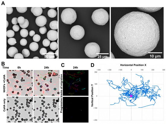
Separating CaHA-containing dermal filler from their gel carrier material through extensive washing, we obtained CaHA microspheres of rather uniform size, with >93% of microspheres within the range of 25 to 45 µm, and of spherical shape with a smooth surface (Figure 1A) as already shown before. To investigate whether NHDFs directly interact with the surface of CaHA microspheres, fluorescence-labelled NHDFs were co-cultured with CaHA microspheres. As NHDFs were seeded at low density rather than confluency, migration of individual cells was anticipated. If cells adhered to CaHA microspheres, accumulation of cells around the microspheres would be expected. Conversely, in the absence of sufficient adhesion, cells would be expected to ignore or avoid them. Interestingly, NHDFs not only migrated across the CaHA microspheres, but microsphere displacement was also observed in the presence of cells (Video S1). This suggests that cells remained motile while attached to the microspheres over the 24 h observation period as indicated by the translocation of CaHA microspheres (Figure 1B, Video S1). In contrast, microspheres incubated under identical conditions but in the absence of cells remained static throughout the entire observation period (Figure 1B, bottom). Consistent with these observations, progressive microsphere translocation tracks were detected only in the microsphere–cell condition (Figure 1C), mirroring the trajectories illustrated in Figure 1D.
Figure 1.
NHDFs adhere and interact with CaHA microspheres. (A) SEM images of CaHA microspheres after isolation from CaHA/CMC dermal filler: CaHA microspheres are spherically shaped and display a smooth surface. (B) Light microscopic image of microspheres cultured in the presence (upper panel) or absence (lower panel) of NHDFs at timepoint 0 h and 24 h. (C) Extensive CaHA microsphere trajectories in the presence of cells (upper panel) and the lack of trajectories in the absence of cells (lower panel). (D) CaHA trajectories in the presence of NHDFs (blue track, n = 15 microspheres) and their static position in the absence of cells (magenta track, n = 10 microspheres). Scale bars are indicated.
3.2. Fibroblasts Display Direct Contact with CaHA Microspheres
A direct fibroblast interaction with CaHA microspheres on the surface level has never been fully addressed. We therefore used scanning electron microscopy (SEM) imaging to provide an in-depth visual approach for the direct interaction between NHDFs and CaHA microspheres 7 days post-addition.
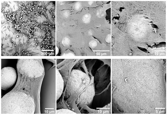
The overview revealed that fibroblasts were not only present in regions containing microspheres but also exhibited strong adhesion and extensive spreading on and around the CaHA microspheres. At low magnification, numerous microspheres were embedded within a dense cellular network, indicating successful cell–material interaction. Fibroblasts formed interconnected layers, creating a three-dimensional matrix that partially enveloped the microspheres (Figure 2, upper panel).
Figure 2.
Scanning electron microscopy (SEM) images of fibroblasts co-cultured with calcium hydroxyapatite (CaHA) microspheres for 7 days. SEM images show microspheres surrounded by cells and newly formed ECM. Scale bars are indicated.
At higher magnification, fibroblasts displayed elongated and flattened morphologies with well-developed cytoplasmic and protrusion-like cell extensions bridging adjacent microspheres. Fine filamentous structures, consistent with ECM deposition, were observed spanning between cells and microspheres, suggesting active-matrix production and remodeling. Close-up images revealed intimate contact between fibroblast membranes and the microsphere surfaces. This scaffold-like arrangement showed the deposited matrix components in the vicinity of the CaHA microspheres and the established contact with the microsphere surface. The cell surfaces appeared textured and covered with microfilament-like structures, indicating strong attachment and cellular activity. The CaHA microspheres were partially coated with cellular material, implying progressive integration into the fibroblast-generated matrix (Figure 2, bottom panel). Quantitative analysis based on overview images indicated that approximately 90% of the CaHA microspheres were surrounded by newly formed ECM after 7 days, highlighting their favorable biocompatibility and potential role in stimulating tissue regeneration by matrix deposition (Figure S1).
3.3. CaHA Microsphere-to-Cell Contact Stimulates Deposition of ECM Components
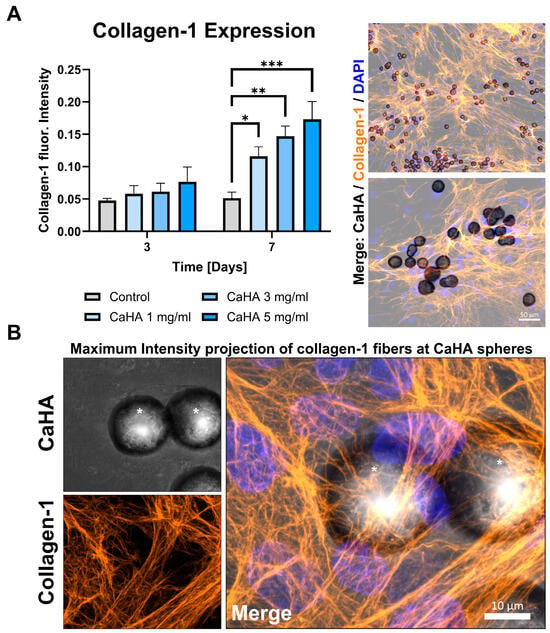
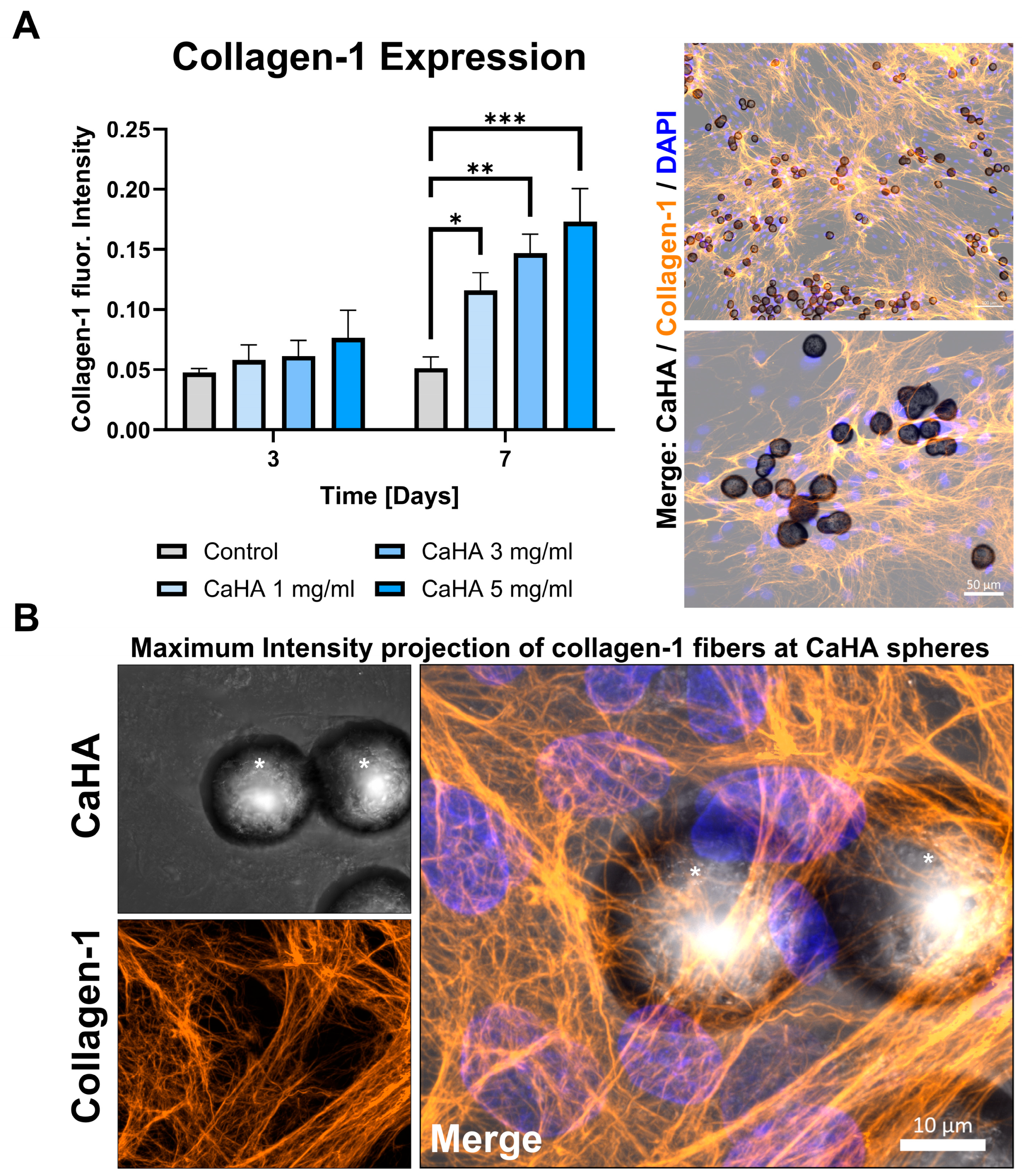
To further investigate our idea that the newly formed structures represent protein fibers and constitute components of the extracellular matrix, we set out to identify their molecular nature. Accordingly, immunofluorescence staining and Western blot analysis were performed to identify specific ECM proteins and assess their organization. One of the most extensively documented effects of dermal fillers is the stimulation of collagen synthesis [8]. Consistent with this, we observed a concentration-dependent increase in collagen type I expression in the presence of 1, 3, and 5 mg/mL CaHA microspheres, with levels significantly exceeding those of untreated controls (Figure 3A). Notably, the collagen fiber network was predominantly formed around and in close proximity to the CaHA microspheres by fibroblasts directly interacting with these spheres (Videos S2 and S3). In these regions, collagen fibers appeared markedly denser than in surrounding areas, indicating enhanced collagen deposition and improved fiber organization in the presence of the microspheres (Figure 3A,B).
Figure 3.
CaHA microspheres stimulate collagen type I production in NHDFs. (A) Quantitative analysis of collagen type I fluorescence intensity in NHDFs cultured in the absence (control) or presence of CaHA microspheres (1, 3, and 5 mg/mL) for 3 and 7 days. While only minor changes were detected on day 3, a significant, concentration-dependent increase in collagen type I expression was observed on day 7 in CaHA-treated cultures compared with untreated controls, as indicated by asterisks (* p < 0.05; ** p < 0.01; *** p < 0.005). Statistical analysis was performed using two-way ANOVA followed by Sídák’s multiple comparisons test. Data are presented as mean ± SEM from n = 3 independent experiments. Representative immunofluorescence images show collagen type I (orange), CaHA microspheres (black) and cell nuclei (blue), illustrating enhanced collagen deposition in the presence of CaHA microspheres. (B) Maximum intensity projection demonstrates dense collagen fiber networks forming in close proximity to CaHA microspheres. Collagen fibers appear highly organized and concentrated around microspheres (asterisks), indicating localized stimulation of collagen synthesis by NHDFs interacting with CaHA. Scale bars: 50 µm (A) and 10 µm (B).
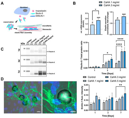
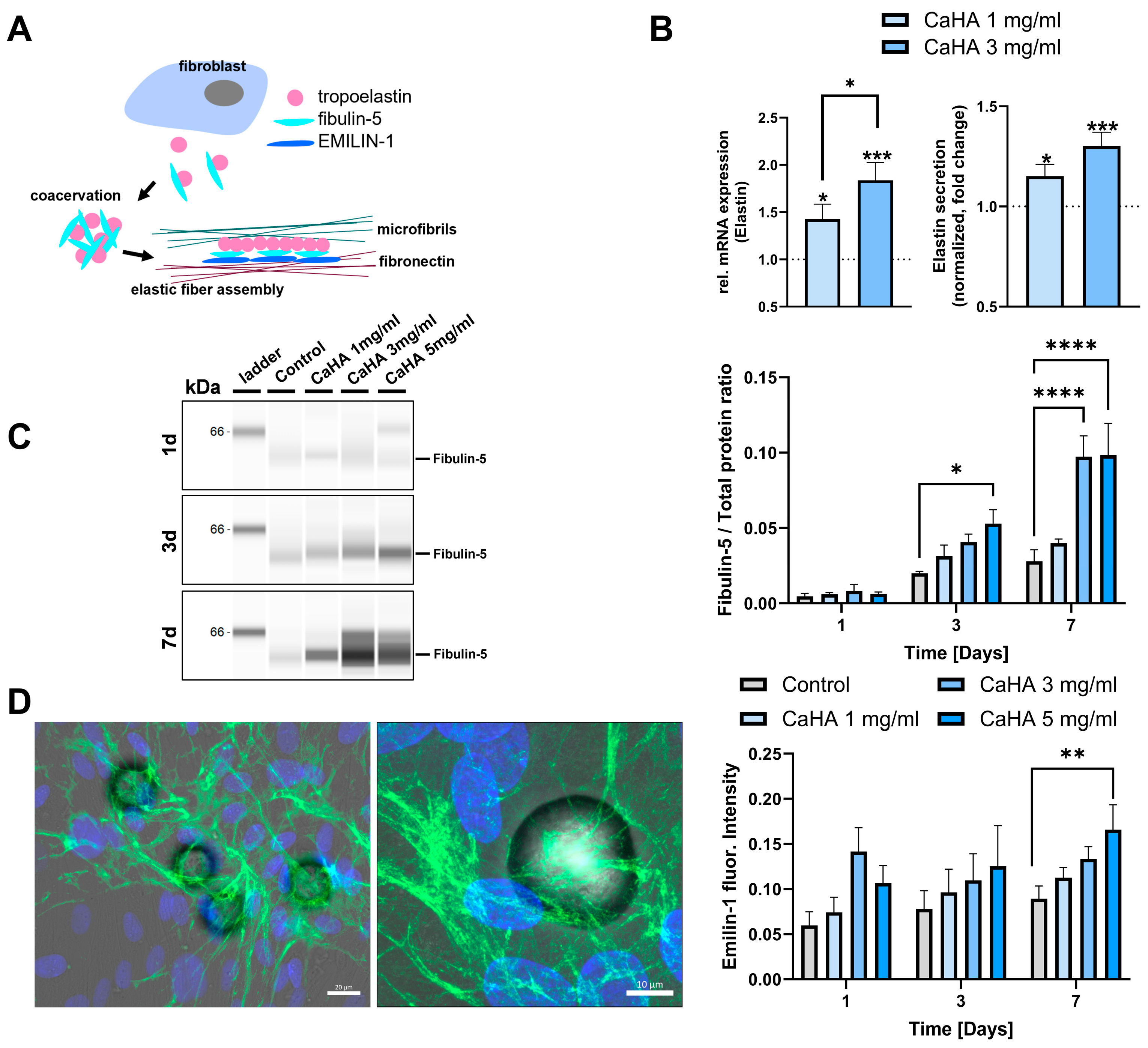
We next examined the expression of elastin and elastin-related structure proteins, the expression of which appears to be vital for skin aging and rejuvenation [8,19]. This applies in particular for the elastin precursor tropoelastin, which requires the scaffold proteins emilin-1 and fibulin-5 for functional elastic fiber formation after lysyl oxidase-mediated coacervation of tropoelastin, as depicted in Figure 4A [16]. Again, a CaHA dose-dependent increase in elastin mRNA expression, accompanied by enhanced elastin secretion, was observed in NHDFs (Figure 4B). Subsequently, the potential effects of CaHA on the elastin scaffolding proteins fibulin-5 and emilin-1 were evaluated. Protein levels of fibulin-5 were increased in a dose- and time-dependent manner by CaHA microspheres, as assessed by Western blot analysis (Figure 4C). Similar to fibulin-5, emilin-1 was found to be upregulated by CaHA microspheres in a time and dose-dependent manner. Interestingly, the strongest emilin-1 fluorescence signal was observed in NHDFs located in close proximity to CaHA microspheres (Figure 4D, Video S4). In addition to emilin-1 and fibulin-5, lysyl oxidase-like protein 2 (LOXL2) was found to be upregulated by CaHA treatment of NHDFs using Western blot (Figure S3B). Together, these results indicate that CaHA treatment not only upregulates elastin and its precursor, tropoelastin, but also enhances the expression and activity of proteins required for functional elastic fiber assembly.
Figure 4.
CaHA microspheres enhance elastogenic responses and elastic fiber assembly in fibroblasts. (A) Schematic illustration of the interplay between the elastin precursor tropoelastin and scaffold proteins emilin-1 and fibulin-5, required for functional elastic fiber formation. (B) Relative mRNA expression of tropoelastin and secretion of elastin is upregulated by CaHa at 1 and 3 mg/mL. (C) Representative Western blot analysis and quantification showing enhanced fibulin-5 expression over the time course of 7 d indicating significant fibulin-5 increase at 3 d for 5 mg/mL and at 7 d for 3 and 5 mg/mL compared to untreated controls. (D) Emilin-1 expression is increased in the presence of CaHA microspheres with quantification indicating a dose- and time-dependent increase in emilin-1 expression by CaHA microspheres. Data are presented as mean ± SEM from n = 3 independent experiments. Statistical analysis was performed using one-way or two-way ANOVA followed by Sídák’s multiple comparisons test (* p < 0.05; ** p < 0.01; *** p < 0.005, **** p < 0.0001).
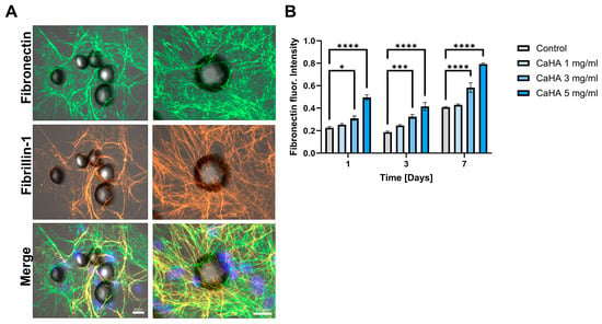
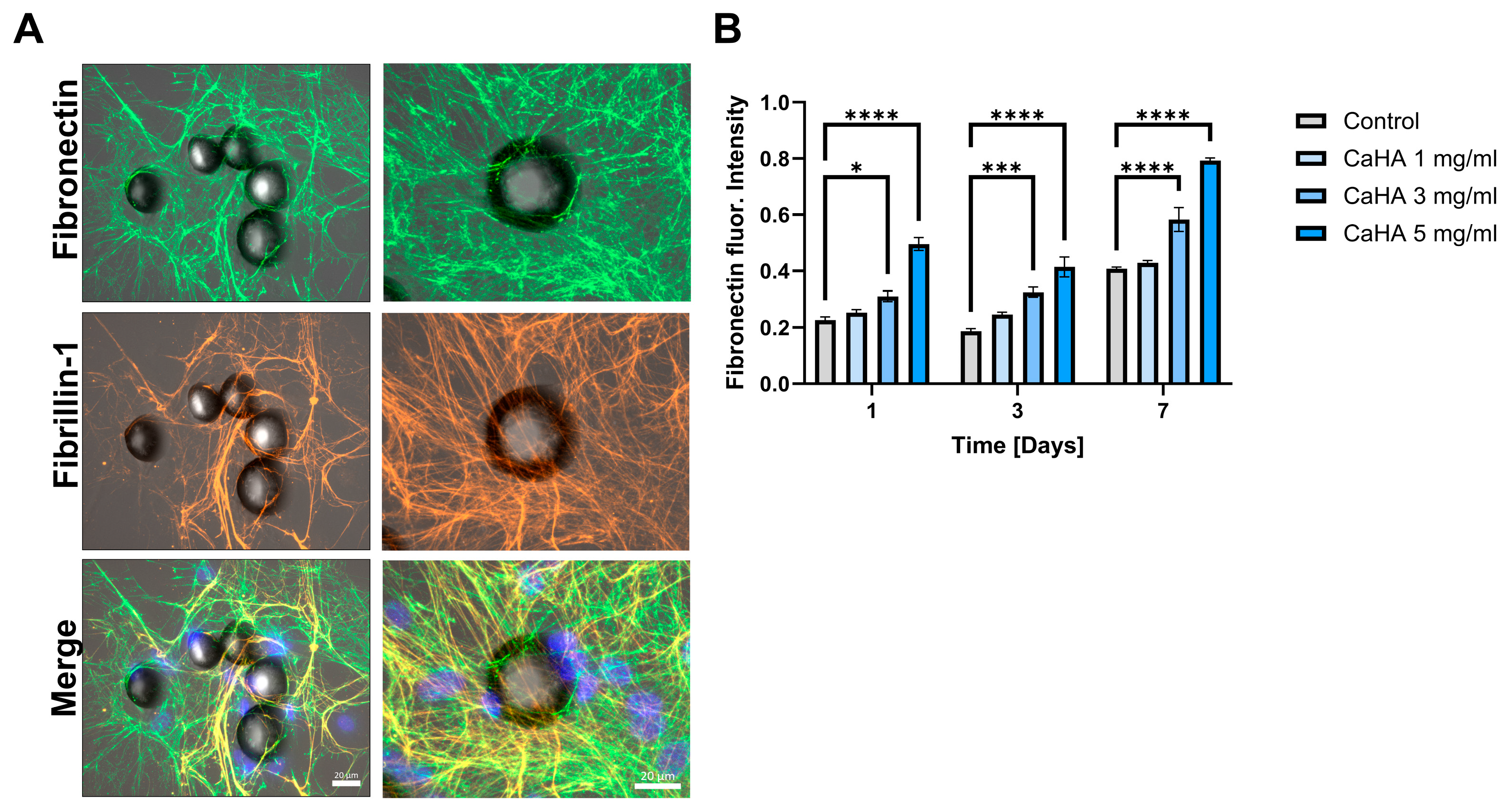
Similar data were obtained when following the expression of fibronectin, an ECM protein required for the assembly of fibrillins, the major matrix components of multifunctional microfibrils [20]. As depicted in Figure 5, fibronectin expression was closely linked with that of fibrillin-1 in the vicinity of microspheres and interacting cells and followed a dose- and time-dependent pattern as assessed by immunofluorescence.
Figure 5.
(A) Fluorescence images of NHDFs after 7 d of co-culture with CaHA-microspheres, stained for fibronectin and fibrillin-1. (B) Quantification of the dose- and time-dependent increase in fibronectin expression mediated by CaHA microspheres. Data are presented as mean ± SEM from n = 3 independent experiments. Statistical analysis was performed using two-way ANOVA followed by Sídák’s multiple comparisons test (* p < 0.05; *** p < 0.005; **** p < 0.0001).
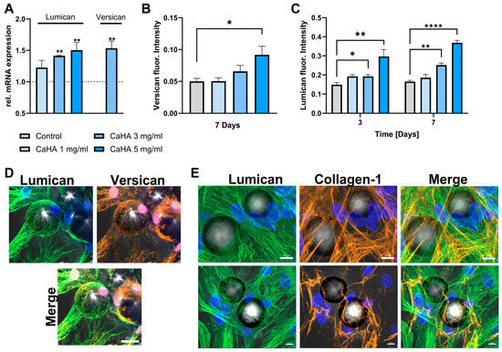
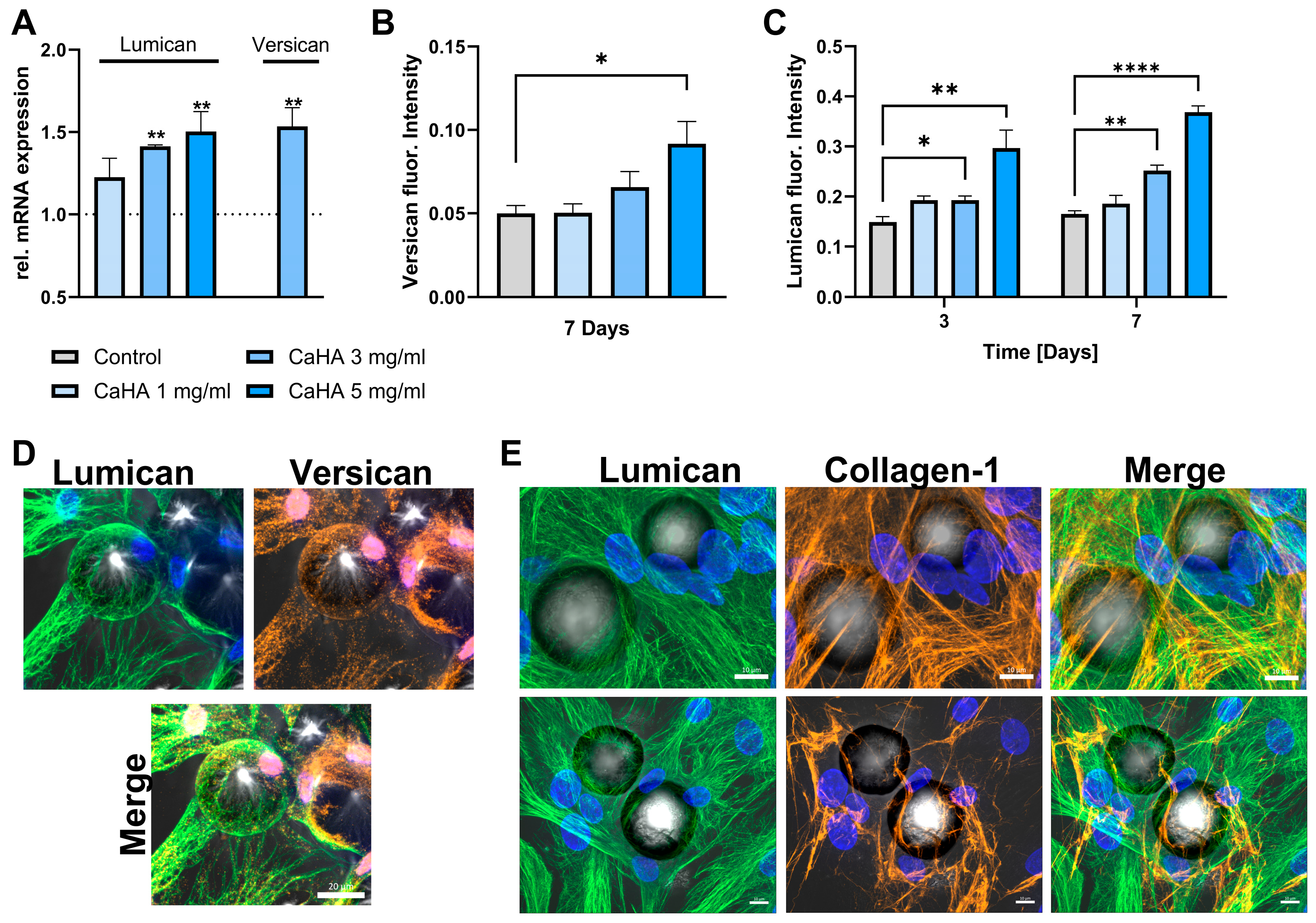
Finally, we investigated the impact of CaHA microspheres on the expression of proteoglycans, important structural ECM components known to be depleted in aging skin [21,22]. The mRNA expression of lumican, a regulator of fibrillogenesis in the skin [23], was increased by the presence of 1, 3 and 5 mg/mL CaHA microspheres, whereas versican, which contributes to elastogenesis [24], was significantly upregulated by 3 mg/mL CaHA microspheres (Figure 6A). Fluorescence imaging revealed a CaHA dose-dependent increase in versican expression after 7 days of culture, which was statistically significant in the presence of 5 mg/mL CaHA microspheres. Additionally, lumican expression increased at 3 and 5 mg/mL CaHA microspheres, reaching statistical significance after both 3 and 7 days of co-culturing cells with microspheres (Figure 6A,C and Figure S3C). Images obtained by maximum intensity projection showed that both lumican and versican were clearly enriched around CaHA microspheres (Figure 6D). Finally, the co-staining for lumican and collagen-1 revealed co-localization of both proteins surrounding the cells located near CaHA microspheres (Figure 6E, Video S5). STRING network analysis of the investigated ECM proteins highlighted their close functional interactions and their involvement in extracellular matrix organization and connective tissue functions (Figure S4). In addition, a network clustering for elastic fiber formation associated proteins (red) and fibrillar collagen formation (green) was done (Figure S3). To investigate CaHA-induced fibroblast mechanosensing, we analyzed the expression of mechanosensitive receptors and found that the ion channel Piezo-1 was upregulated following CaHA treatment (Figure S3A, borderline significance).
Figure 6.
(A) Co-cultivation of NHDFs with CaHA microspheres increases mRNA expression of lumican and versican in a dose dependent manner. (B) Protein expression of versican and lumican (C) is increased by CaHA microspheres in a dose- and time-dependent manner. (D) Maximum intensity projection of fluorescence images showing the enrichment of lumican and versican surrounding CaHA microspheres. (E) Co-localization of collagen-1 with lumican, a collagen-1 stabilizing proteoglycan. Data are presented as mean ± SEM from n = 3 independent experiments using one-way or two-way ANOVA followed by Sídák’s multiple comparisons test (* p < 0.05; ** p < 0.01; **** p < 0.0001).
4. Discussion
The current study supports the concept that direct cell-to-CaHA contact, likely to involve mechanotransduction-mediated cell signaling, is a major contributor to the beneficial skin regenerative effects of CaHA-based dermal filler like Radiesse®. Dermal fibroblasts rapidly established direct and close contact to CaHA microsphere within several hours, a process presumably supported by the smooth surface of the microspheres [25,26]. With this contact established, cells showed enhanced production of ECM proteins, including collagen-1, elastin, emilin-1, fibulin-5, fibronectin, as well as proteoglycans lumican and versican. For most of these proteins, the extent of expression positively correlated with the concentration of CaHA microspheres in the culture medium. This was demonstrated by a dose- and/or time-dependent increase in mRNA or protein expression, and corresponding cellular expression patterns assessed by fluorescence imaging.
These biostimulatory effects primarily targeted cells in the vicinity of the microspheres, highlighting the importance of mechanical interaction rather than systemic inflammatory signaling. The delicate fiber network on the CaHA microspheres, composed of newly synthesized ECM components, suggests active cellular responses driven by mechanotransduction and cellular attachment proteins such as integrins. Interestingly, Liu et al. demonstrated that key adhesion proteins, including vinculin and integrins αV and β1, were upregulated following CaHA/HA injections in rats at one month post-implantation. CaHA microspheres sintered at both low (300 °C) and high (800 °C) temperatures exhibited comparable effects on the upregulation of vinculin and integrins [27]. However, only microspheres sintered at 300 °C showed effective Ca2+ release under in vitro conditions, supporting calcium-mediated signaling as a mechanism for the observed effects. This finding suggests that the upregulation of adhesion-associated integrins by microspheres sintered at high temperature may occur independently of calcium signaling.
Notably, under in vivo conditions, CaHA microspheres are reported to degrade through phagocytosis by multinucleated macrophages [28,29]. Within these cellular structures, CaHA degradation may result in substantially higher local calcium release, potentially sufficient to support calcium-mediated signaling pathways involved in adhesion protein upregulation [30]. The induction of ECM expression in response to mechanical stimuli thus highlights the dynamic interplay between cellular mechanosensing and extracellular matrix remodeling. This supports the notion that fibroblast–ECM contact is an important driver of skin aging processes, and that restoring those interactions may contribute to skin rejuvenation [11]. These beneficial effects are likely further supported by the smooth surface and homogeneous size of the CaHA microspheres. Indeed, comparison with irregularly shaped poly-L-lactic acid-based microspheres (PLLA) suggests a potential advantage of CaHA in promoting more favorable ECM-related and immune-related responses [31,32]. Accordingly, CaHA microspheres induced only a relatively limited inflammatory response, which appeared less pronounced than that observed with alternative materials. At the same time, CaHA microspheres enhanced collagen-1 biosynthesis to a significantly greater extent than PLLA-based particles [31,32].
The enhanced expression of multiple ECM proteins induced by CaHA microspheres not only confirms previous findings but also extends their relevance. Notably, collagen synthesis was observed under in vitro conditions, in the absence of immune or systemic influences, suggesting that direct interaction with CaHA microspheres is sufficient to trigger expression of collagen and other important ECM players. A similar effect was observed for elastin synthesis. Elastogenesis predominantly occurs during early development and is largely absent in adult tissues. With ageing and continuous exposure to damaging environmental stressors, elastic fibers progressively degrade [15,33]. Consistently, emilin-1 expression has been reported to decrease with age, contributing to impaired elastin fiber formation [34]. Following mechanical damage, the production of new elastin fibers is triggered by inflammatory processes associated with wound healing. However, these newly formed fibers are often functionally compromised and spatially disorganized, leading to stiff and inelastic scar tissue [35]. In contrast, CaHA microspheres promoted elastin synthesis together with increased expression of elastin-associated scaffold proteins, including emilin-1, fibulin-5, and LOXL2, which are essential for proper elastic fiber assembly [16]. As visualized here for emilin-1, its expression was primarily detectable along fibrous structures in cells surrounding the microspheres. Thus, our findings not only align with previous studies focusing on elastin in mouse models and human skin [36] but also provide new evidence that direct interaction with CaHA microspheres is sufficient to induce these effects under in vitro conditions, largely independent of systemic cues.
Similar observations were made for fibronectin, a protein essential for in vivo collagen assembly [14], which co-localized with fibrillin-1 on fibers and, independently, exhibited a dose- and time-dependent increase in cultures containing CaHA microspheres. Furthermore, fibrillin-1 has been shown to be required for proper ECM deposition of emilin-1 and emilin-2 [19]. It may also interact with fibulin-5 and other fibulins, acting as an adaptor for tropoelastin [37], highlighting the complexity of the protein interactions involved.
Further complementing this picture, we observed that the proteoglycans lumican and versican exhibited a dose- and time-dependent responses to CaHA microspheres. Accordingly, both proteins appeared enriched in the vicinity of the microspheres, and lumican co-localized with collagen-1 on fibrous structures. Lumican has been associated with multiple functions in the ECM, including collagen fibrillogenesis [23,38] and wound healing [39], and has been reported to be reduced in aging skin [21,22]. However, its specific role in skin regeneration and its response to dermal filler application remain unclear. While previous studies investigating human skin following CaHA dermal filler injection reported an overall increase in proteoglycans, likely including lumican, decorin and versican [34,40], our findings directly demonstrate an upregulation of lumican expression. This observation suggests that lumican may contribute to ECM remodeling and skin regeneration following CaHA-based treatment. A similar role may be proposed for versican, which is critical for maintaining skin elasticity by linking microfibrils to the hyaluronan-rich matrix, thereby supporting ECM integrity and viscoelastic properties [41]. In addition, versican has been implicated in tissue remodeling processes, including the regulation of cellular adhesion and proliferation [42]. In the context of solar elastosis, degradation by MMP-12 disrupts hyaluronan-binding capacity of versican, leading to pathological alterations in photoaged dermis [41]. Thus, the observed increase in versican expression in response to CaHA microspheres suggests that versican may support the organization of collagen and elastin networks and potentially contribute to fibroblast protection against UV-induced apoptosis.
Combining the investigated ECM proteins in a STRING interaction network highlighted the close functional association between proteins involved in extracellular matrix architecture and connective tissue organization (Figure S3). STRING analysis predicts both direct and indirect protein–protein associations based on known and predicted functional interactions. The observed dense interaction network indicates that the investigated proteins do not act independently but rather form an interconnected regulatory and structural matrix system. In particular, proteins associated with elastic fiber assembly, including ELN, FBLN5, LOX, LOXL2, EMILIN1, and VCAN, formed a highly interconnected cluster, supporting their coordinated role in elastic fiber maturation, cross-linking, and stabilization. In parallel, COL1A1, COL3A1, and LUM clustered as fibrillar collagen-associated proteins, reflecting their established roles in collagen fibrillogenesis and extracellular matrix tensile strength. Importantly, bridging proteins such as FN1 and FBN1 linked both clusters, suggesting functional crosstalk between collagen fibril organization and elastic fiber formation. Thus, the STRING mapping not only visualizes protein connectivity, but also emphasizes the biological interdependence of ECM pathways involved in tissue integrity, mechanics, and remodeling.
Limitations
This study is limited by its reliance on two-dimensional (2D) in vitro cell culture experiments with short-term observation periods. Consequently, the findings may not fully recapitulate the complex biological environment encountered in vivo, nor do they entirely reflect the behavior of CaHA microspheres and the complete dermal filler formulation in human subjects under clinical conditions. In particular, 2D cultures lack the three-dimensional tissue architecture, mechanical constraints, and long-term material–tissue interactions that are known to influence cellular responses and extracellular matrix remodeling.
A major limitation of this experimental approach is the absence of an immune component. Clinical outcomes associated with CaHA implantation are influenced by immune-mediated processes, including macrophage recruitment, foreign body responses, and cytokine-driven paracrine signaling. These immune cells play a critical role in orchestrating tissue regeneration and ECM turnover, and their exclusion from the current model prevents assessment of the dynamic crosstalk between immune cells and dermal fibroblasts that are likely to contribute to CaHA-induced regenerative effects in vivo.
Furthermore, the simplified in vitro system does not account for additional factors such as vascularization, filler degradation kinetics, or biomechanical forces present in human skin. As a result, while the present model is well suited to isolate and study direct microsphere–cell interactions at a molecular level, its translational relevance to clinical outcomes remains inherently limited. The CaHA concentrations used in this study (1, 3, and 5 mg/mL) do not directly correspond to in vivo conditions. In clinical practice, CaHA is administered as microspheres suspended in a carrier gel rather than as a defined concentration in solution. Therefore, direct comparison between in vitro and in vivo exposure is not straightforward.
Building on the mechanistic insights generated in this study, future research will focus on translating these findings toward more physiologically relevant models. Key areas will include the incorporation of immune cell co-culture systems, three-dimensional skin equivalents, and long-term exposure paradigms, as well as validation in preclinical and clinical settings. Such approaches will be essential to bridge the gap between in vitro observations and the complex regenerative responses observed following CaHA treatment in patients.
5. Conclusions
In summary, our study supports the concept that direct contact between fibroblasts and CaHA microspheres significantly contributes to aspects of skin regeneration. Fibroblasts establish direct contact with CaHA microspheres that subsequently trigger the enhanced expression of key ECM proteins. These effects occur in the absence of inflammatory processes, thereby promoting the restoration of structural and functional characteristics associated with youthful skin. Given the complexity of the molecular pathways involved, the proteins identified likely represent components of a broader and highly interconnected interaction network, the precise mechanisms and relationships of which remain to be further elucidated.
Supplementary Materials
The following supporting information can be downloaded at: https://www.mdpi.com/article/10.3390/cosmetics13030156/s1; Figure S1: Quantification of microsphere–cell contact. Figure S2: Fluorescence images of ECM protein expression of NHDFs without CaHA-treatment. Figure S3: Protein expression of piezo-1, LOXL2 and lumican post CaHA microsphere treatment. Figure S4: MCL clustering analysis in STRING. Video S1: Translocation of CaHA-microspheres in the presence of NHDFs. Video S2: Z-stack video of collagen-1 expression of NHDFs in close proximity to CaHA microspheres. Video S3: 3D projection of the z-stack shown in Video S2. Video S4: Z-stack video of emilin-1 and collagen-1 expression of NHDFs in close proximity to CaHA microspheres. Video S5: Z-stack video of lumican and collagen-1 expression of NHDFs in close proximity to CaHA microspheres.
Author Contributions
Conceptualization, Y.D., C.K., K.R. and C.H., methodology, Y.D., C.K., K.R., D.S., C.W. and C.H.; software, Y.D., C.K., K.R. and C.H., validation, Y.D., C.K. and C.H.; formal analysis, Y.D., C.K., K.R. and C.H.; writing, Y.D., C.K., K.R., T.H. and C.H. All authors have read and agreed to the published version of the manuscript.
Funding
No external funding was received for the conduct of this research. Merz Aesthetics GmbH provided financial support for medical writing and editorial assistance.
Institutional Review Board Statement
This work did not require approval from an institutional ethics committee, as it did not involve human participants, patient data, or animal experimentation. Nevertheless, all procedures and analysis were conducted in accordance with internationally accepted scientific and ethical standards, ensuring integrity, transparency, and responsible research practices.
Informed Consent Statement
This study did not involve human participants or animal subjects; therefore, ethical approval and informed consent were not required.
Data Availability Statement
The data that support the findings of this study are available from the corresponding author upon reasonable request.
Acknowledgments
Medical writing assistance was provided by Gerhard Krumschnabel (G.K.) (www.medical-writing.at (accessed on 3 February 2025)) under the direction of the authors.
Conflicts of Interest
Y.D., C.K., K.R., D.S., C.W., T.H. and C.H. are/were employees of Merz Aesthetics. The funders had no role in the design of the study; in the collection, analyses or interpretation of data; in the writing of the manuscript; or in the decision to publish the results. All authors contributed to the development and review of this work and agreed with the content.
References
- Nowag, B.; Schäfer, D.; Hengl, T.; Corduff, N.; Goldie, K. Biostimulating Fillers and Induction of Inflammatory Pathways: A Preclinical Investigation of Macrophage Response to Calcium Hydroxylapatite and Poly-L Lactic Acid. J. Cosmet. Dermatol. 2024, 23, 99–106. [Google Scholar] [CrossRef] [Scilit] [PubMed]
- Yanatma, I.; Sarac, G.; Gul, M.; Gul, S.; Kapicioglu, Y. Comparison of Polycaprolactone and Calcium Hydroxylapatite Dermal Fillers in a Rat Model. Dermatol. Ther. 2021, 34, e14716. [Google Scholar] [CrossRef] [Scilit] [PubMed]
- Yutskovskaya, Y.; Kogan, E.; Leshunov, E. A Randomized, Split-Face, Histomorphologic Study Comparing a Volumetric Calcium Hydroxylapatite and a Hyaluronic Acid-Based Dermal Filler. J. Drugs Dermatol. 2014, 13, 1047–1052. [Google Scholar] [PubMed]
- Coleman, K.M.; Voigts, R.; DeVore, D.P.; Termin, P.; Coleman, W.P. Neocollagenesis after Injection of Calcium Hydroxylapatite Composition in a Canine Model. Dermatol. Surg. 2008, 34, S53–S55. [Google Scholar] [CrossRef] [Scilit] [PubMed]
- Fan, Y.; Choi, T.H.; Chung, J.H.; Jeon, Y.K.; Kim, S. Hyaluronic Acid-Cross-Linked Filler Stimulates Collagen Type 1 and Elastic Fiber Synthesis in Skin through the TGF-β/Smad Signaling Pathway in a Nude Mouse Model. J. Plast. Reconstr. Aesthetic Surg. 2019, 72, 1355–1362. [Google Scholar] [CrossRef] [Scilit] [PubMed]
- Yutskovskaya, Y.A.; Kogan, E.A. Improved Neocollagenesis and Skin Mechanical Properties after Injection of Diluted Calcium Hydroxylapatite in the Neck and Déćolletage: A Pilot Study. J. Drugs Dermatol. 2017, 16, 68–74. [Google Scholar] [PubMed]
- Rovatti, P.P.; Pellacani, G.; Guida, S. Hyperdiluted Calcium Hydroxylapatite 1: 2 for Mid and Lower Facial Skin Rejuvenation: Efficacy and Safety. Dermatol. Surg. 2020, 46, E112–E117. [Google Scholar] [CrossRef] [Scilit] [PubMed]
- Aguilera, S.B.; McCarthy, A.; Khalifian, S.; Lorenc, Z.P.; Goldie, K.; Chernoff, W.G. The Role of Calcium Hydroxylapatite (Radiesse) as a Regenerative Aesthetic Treatment: A Narrative Review. Aesthetic Surg. J. 2023, 43, 1063–1090. [Google Scholar] [CrossRef] [Scilit] [PubMed]
- Pavicic, T. Complete Biodegradable Nature of Calcium Hydroxylapatite after Injection for Malar Enhancement: An MRI Study. Clin. Cosmet. Investig. Dermatol. 2015, 8, 19–25. [Google Scholar] [CrossRef] [Scilit] [PubMed]
- Bass, L.S.; Smith, S.; Busso, M.; McClaren, M. Calcium Hydroxylapatite (Radiesse) for Treatment of Nasolabial Folds: Long-Term Safety and Efficacy Results. Aesthetic Surg. J. 2010, 30, 235–238. [Google Scholar] [CrossRef] [Scilit] [PubMed]
- Shin, S.H.; Lee, Y.H.; Rho, N.K.; Park, K.Y. Skin Aging from Mechanisms to Interventions: Focusing on Dermal Aging. Front. Physiol. 2023, 14, 1195272. [Google Scholar] [CrossRef] [Scilit] [PubMed]
- Zarbafian, M.; Fabi, S.G.; Dayan, S.; Goldie, K. The Emerging Field of Regenerative Aesthetics-Where We Are Now. Dermatol. Surg. 2022, 48, 101–108. [Google Scholar] [CrossRef] [Scilit] [PubMed]
- Chen, S.; Birk, D.E. The Regulatory Roles of Small Leucine-Rich Proteoglycans in Extracellular Matrix Assembly. FEBS J. 2013, 280, 2120–2137. [Google Scholar] [CrossRef] [Scilit] [PubMed]
- Paten, J.A.; Martin, C.L.; Wanis, J.T.; Siadat, S.M.; Figueroa-Navedo, A.M.; Ruberti, J.W.; Deravi, L.F. Molecular Interactions between Collagen and Fibronectin: A Reciprocal Relationship That Regulates De Novo Fibrillogenesis. Chem 2019, 5, 2126–2145. [Google Scholar] [CrossRef] [Scilit]
- Baumann, L.; Bernstein, E.F.; Weiss, A.S.; Bates, D.; Humphrey, S.; Silberberg, M.; Daniels, R. Clinical Relevance of Elastin in the Structure and Function of Skin. Aesthetic Surg. J. Open Forum 2021, 3, ojab019. [Google Scholar] [CrossRef] [Scilit] [PubMed]
- Nonaka, R.; Onoue, S.; Wachi, H.; Sato, F.; Urban, Z.; Starcher, B.C.; Seyama, Y. DANCE/Fibulin-5 Promotes Elastic Fiber Formation in a Tropoelastin Isoform-Dependent Manner. Clin. Biochem. 2009, 42, 713–721. [Google Scholar] [CrossRef] [Scilit] [PubMed]
- Nowag, B.; Casabona, G.; Kippenberger, S.; Zöller, N.; Hengl, T. Calcium Hydroxylapatite Microspheres Activate Fibroblasts through Direct Contact to Stimulate Neocollagenesis. J. Cosmet. Dermatol. 2023, 22, 426–432. [Google Scholar] [CrossRef] [Scilit] [PubMed]
- Schneider, C.A.; Rasband, W.S.; Eliceiri, K.W. NIH Image to ImageJ: 25 Years of Image Analysis. Nat. Methods 2012, 9, 671–675. [Google Scholar] [CrossRef] [Scilit] [PubMed]
- Schiavinato, A.; Marcous, F.; Zuk, A.V.; Keene, D.R.; Tufa, S.F.; Mosquera, L.M.; Zigrino, P.; Mauch, C.; Eckes, B.; Francois, K.; et al. New Insights into the Structural Role of EMILINs within the Human Skin Microenvironment. Sci. Rep. 2024, 14, 30345. [Google Scholar] [CrossRef] [Scilit] [PubMed]
- Sabatier, L.; Chen, D.; Fagotto-Kaufmann, C.; Hubmacher, D.; McKee, M.D.; Annis, D.S.; Mosher, D.F.; Dieter, R.P. Fibrillin Assembly Requires Fibronectin. Mol. Biol. Cell 2009, 20, 846–858. [Google Scholar] [CrossRef] [Scilit] [PubMed]
- Lee, D.H.; Oh, J.H.; Chung, J.H. Glycosaminoglycan and Proteoglycan in Skin Aging. J. Dermatol. Sci. 2016, 83, 174–181. [Google Scholar] [CrossRef] [Scilit] [PubMed]
- McCabe, M.C.; Hill, R.C.; Calderone, K.; Cui, Y.; Yan, Y.; Quan, T.; Fisher, G.J.; Hansen, K.C. Alterations in Extracellular Matrix Composition during Aging and Photoaging of the Skin. Matrix Biol. Plus 2020, 8, 100041. [Google Scholar] [CrossRef] [Scilit] [PubMed]
- Chakravarti, S.; Magnuson, T.; Lass, J.H.; Jepsen, K.J.; LaMantia, C.; Carroll, H. Lumican Regulates Collagen Fibril Assembly: Skin Fragility and Corneal Opacity in the Absence of Lumican. J. Cell Biol. 1998, 141, 1277–1286. [Google Scholar] [CrossRef] [Scilit] [PubMed]
- Keire, P.A.; Bressler, S.L.; Mulvihill, E.R.; Starcher, B.C.; Kang, I.; Wight, T.N. Inhibition of Versican Expression by SiRNA Facilitates Tropoelastin Synthesis and Elastic Fiber Formation by Human SK-LMS-1 Leiomyosarcoma Smooth Muscle Cells in Vitro and in Vivo. Matrix Biol. 2016, 50, 67–81. [Google Scholar] [CrossRef] [Scilit] [PubMed]
- Baxter, L.C.; Frauchiger, V.; Textor, M.; Ap Gwynn, I.; Richards, R.G.; Bongrand, P.; Brunette, D. Fibroblast and Osteoblast Adhesion and Morphology on Calcium Phosphate Surfaces. Eur. Cell Mater. 2002, 4, 1–17. [Google Scholar] [CrossRef] [Scilit] [PubMed]
- Juhl, O.J.; Merife, A.B.; Zhang, Y.; Lemmon, C.A.; Donahue, H.J. Hydroxyapatite Particle Density Regulates Osteoblastic Differentiation Through β-Catenin Translocation. Front. Bioeng. Biotechnol. 2021, 8, 591084. [Google Scholar] [CrossRef] [Scilit] [PubMed]
- Liu, S.; Song, L.; Huang, S.; Liu, Z.; Xu, Y.; Wang, Z.; Qiu, H.; Wang, J.; Chen, Z.; Xiao, Y.; et al. Hydroxyapatite Microspheres Encapsulated within Hybrid Hydrogel Promote Skin Regeneration through the Activation of Calcium Signaling and Motor Protein Pathway. Bioact. Mater. 2025, 50, 287–304. [Google Scholar] [CrossRef] [Scilit] [PubMed]
- Zerbinati, N.; D’Este, E.; Parodi, P.C.; Calligaro, A. Microscopic and Ultrastructural Evidences in Human Skin Following Calcium Hydroxylapatite Filler Treatment. Arch. Dermatol. Res. 2017, 309, 389–396. [Google Scholar] [CrossRef] [Scilit] [PubMed]
- Sheikh, Z.; Brooks, P.J.; Barzilay, O.; Fine, N.; Glogauer, M. Macrophages, Foreign Body Giant Cells and Their Response to Implantable Biomaterials. Materials 2015, 8, 5671–5701. [Google Scholar] [CrossRef] [Scilit] [PubMed]
- van Loghem, J. Calcium Hydroxylapatite in Regenerative Aesthetics: Mechanistic Insights and Mode of Action. Aesthetic Surg. J. 2025, 45, 393–403. [Google Scholar] [CrossRef] [Scilit]
- Kunzler, C.; Hartmann, C.; Nowag, B.; Shah, R.; El-Banna, R.; Backfisch, S.; Schafer, D.; Hengl, T.; Hagedorn, N. Comparison of Physicochemical Characteristics and Biostimulatory Functions in Two Calcium Hydroxyapatite-Based Dermal Fillers. J. Drugs Dermatol. 2023, 22, 910–916. [Google Scholar] [CrossRef] [PubMed]
- McCarthy, A.D.; Hartmann, C.; Durkin, A.; Shahriar, S.; Khalifian, S.; Xie, J. A Morphological Analysis of Calcium Hydroxylapatite and Poly-l-Lactic Acid Biostimulator Particles. Ski. Res. Technol. 2024, 30, e13764. [Google Scholar] [CrossRef] [Scilit] [PubMed]
- Gardeazabal, L.; Izeta, A. Elastin and Collagen Fibres in Cutaneous Wound Healing. Exp. Dermatol. 2024, 33, e15052. [Google Scholar] [CrossRef] [Scilit] [PubMed]
- Kondo, S.; Tohgasaki, T.; Shiga, S.; Nishizawa, S.; Ishiwatari, S.; Ishikawa, S.; Takeda, A.; Sakurai, T. Elastin Microfibril Interface-Located Protein 1 and Its Catabolic Enzyme, Cathepsin K, Regulate the Age-Related Structure of Elastic Fibers in the Skin. J. Cosmet. Dermatol. 2022, 21, 4796–4804. [Google Scholar] [CrossRef] [Scilit] [PubMed]
- Sivaraman, B.; Bashur, C.A.; Ramamurthi, A. Advances in Biomimetic Regeneration of Elastic Matrix Structures. Drug Deliv. Transl. Res. 2012, 2, 323–350. [Google Scholar] [CrossRef] [Scilit] [PubMed]
- Amiri, M.; Meçani, R.; Niehot, C.D.; Phillips, T.; Kolb, J.; Daughtry, H.; Muka, T. Skin Regeneration-Related Mechanisms of Calcium Hydroxylapatite (CaHA): A Systematic Review. Front. Med. 2023, 10, 1195934. [Google Scholar] [CrossRef] [Scilit] [PubMed]
- El-Hallous, E.; Sasaki, T.; Hubmacher, D.; Getie, M.; Tiedemann, K.; Brinckmann, J.; Bätge, B.; Davis, E.C.; Reinhardt, D.P. Fibrillin-1 Interactions with Fibulins Depend on the First Hybrid Domain and Provide an Adaptor Function to Tropoelastin. J. Biol. Chem. 2007, 282, 8935–8946. [Google Scholar] [CrossRef] [Scilit] [PubMed]
- Chen, D.; Smith, L.R.; Khandekar, G.; Patel, P.; Yu, C.K.; Zhang, K.; Chen, C.S.; Han, L.; Wells, R.G. Distinct Effects of Different Matrix Proteoglycans on Collagen Fibrillogenesis and Cell-Mediated Collagen Reorganization. Sci. Rep. 2020, 10, 19065. [Google Scholar] [CrossRef] [Scilit] [PubMed]
- Karamanou, K.; Perrot, G.; Maquart, F.X.; Brézillon, S. Lumican as a Multivalent Effector in Wound Healing. Adv. Drug Deliv. Rev. 2018, 129, 344–351. [Google Scholar] [CrossRef] [Scilit] [PubMed]
- Goldie, K.; Casabona, G.; Corduff, N.; McCarthy, A.D.; Riegel, K. Regeneration of an Extracellular Matrix Ecosystem Following Subcutaneous Injection with Different Radiesse Dilutions: A Histologic and Ultrasound Study. J. Cosmet. Dermatol. 2026, 25, e70930. [Google Scholar] [CrossRef] [Scilit]
- Hasegawa, K.; Yoneda, M.; Kuwabara, H.; Miyaishi, O.; Itano, N.; Ohno, A.; Zako, M.; Isogai, Z. Versican, a Major Hyaluronan-Binding Component in the Dermis, Loses Its Hyaluronan-Binding Ability in Solar Elastosis. J. Investig. Dermatol. 2007, 127, 1657–1663. [Google Scholar] [CrossRef] [Scilit] [PubMed]
- Ruiz Martínez, M.A.; Peralta Galisteo, S.; Castán, H.; Morales Hernández, M.E. Role of Proteoglycans on Skin Ageing: A Review. Int. J. Cosmet. Sci. 2020, 42, 529–535. [Google Scholar] [CrossRef] [Scilit] [PubMed]
Disclaimer/Publisher’s Note: The statements, opinions and data contained in all publications are solely those of the individual author(s) and contributor(s) and not of MDPI and/or the editor(s). MDPI and/or the editor(s) disclaim responsibility for any injury to people or property resulting from any ideas, methods, instructions or products referred to in the content. |
© 2026 by the authors. Licensee MDPI, Basel, Switzerland. This article is an open access article distributed under the terms and conditions of the Creative Commons Attribution (CC BY) license.