1. Introduction
Follicular unit extraction (FUE) has become the dominant donor harvesting technique in modern hair transplantation due to its ability to harvest follicular units without linear scarring and its flexibility in terms of donor access, session scheduling, and postoperative aesthetics; these characteristics are consistent with patients’ increasing preference for shorter hairstyles and less noticeable surgical scars [
1,
2]. As FUE volumes and session sizes expand globally, donor site management has evolved from a purely technical intraoperative concern to a central determinant of long-term surgical success and patient satisfaction. In parallel, recent reviews highlight the ongoing practice gaps in emerging technologies and protocol standardization in hair transplantation, underlining the need for reproducible donor preservation frameworks [
3].
Unlike the recipient region, where density can be redistributed, the donor region constitutes a limited and non-renewable source of follicular units. Suboptimal donor selection (e.g., harvesting grafts from miniaturized areas or diffuse unpatterned alopecia (DUPA) and overly aggressive extraction strategies can lead to irreversible donor depletion, visible thinning (“moth-eaten” appearance), hypopigmented punctate scarring, and limited reserve for future sessions [
2,
4]. These risks are further amplified in high-demand scenarios, such as young patients with evolving hair styles, patients requesting very large graft numbers, and those seeking multiple treatments over time. In FUE, donor site complications are not uncommon in real-world practice and include hypopigmentation, textural changes, folliculitis, sensory impairment, and scar patterns that become more pronounced as surrounding hair is kept short. Importantly, many donor site complications can be prevented through careful preoperative donor assessment, conservative extraction density planning, appropriate punch selection, and disciplined distribution of extractions in a safe donor area [
4,
5]. Accordingly, modern donor management is best conceptualized as a long-term conservation strategy: prioritizing donor aesthetics, preserving reserve for future loss progression, and balancing immediate cosmetic goals with lifetime graft availability. Recent literature increasingly emphasizes that optimizing donor recovery and minimizing long-term donor morbidity requires not only surgical technique but also structured preoperative and postoperative assessments. A systematic review focusing on donor site interventions in FUE highlights the increasing interest in evidence-based strategies for donor site recovery and complication prevention, while underlining the heterogeneity of reported methods and outcomes among studies [
6]. In parallel, current surgical reviews have called for standardized donor assessment and harvesting principles to reduce overharvesting and maximize lifetime use of the donor site [
1,
5].
Accordingly, current donor management should be considered not as a technical intraoperative detail, but as a long-term preservation strategy. Preserving donor aesthetics, maintaining future surgical flexibility, and aligning graft allocation with expected alopecia progression are central to sustainable practice [
4,
6]. This review synthesizes current evidence and surgical principles to provide a structured clinical framework for donor site preservation in FUE.
Materials and Methods
This article was prepared as a structured narrative review. A literature search was conducted in PubMed/MEDLINE, Scopus, and Google Scholar for studies and reviews addressing follicular unit extraction donor site assessment, safe donor zone concepts, miniaturization, extraction density, overharvesting, donor site complications, repeated session scheduling, long hair FUE, and robotic FUE systems. Combinations of terms such as “follicular unit extraction”, “FUE donor area,” “safe donor zone”, “overharvesting”, “donor depletion”, “white dot scarring”, “shock loss”, “robotic FUE”, and “long hair FUE” were used.
English-language articles considered clinically relevant were included, with particular emphasis on recent reviews, expert consensus statements, practice guidelines, and representative clinical trials. Case reports and older landmark technical articles were also retained where they contributed important fundamental concepts or practical details. Due to heterogeneity in study design, outcome definitions, and reporting methods, a quantitative synthesis was not attempted [
7,
8]. Instead, existing evidence was synthesized narratively and combined with practice-oriented interpretation to create a structured clinical framework for donor site preservation in FUE.
2. Donor Site Biology and Safe Donor Site
The biological basis for donor site conservation in FUE relies on the principle of “donor dominance,” first established in strip surgery and subsequently adapted to follicular unit extraction. Follicles harvested from the occipital and parietal scalp are generally resistant to androgen-mediated miniaturization and retain their growth characteristics when transplanted to recipient sites. However, this resistance is not absolute but relative and may vary depending on age, pattern progression, and individual susceptibility [
1,
2].
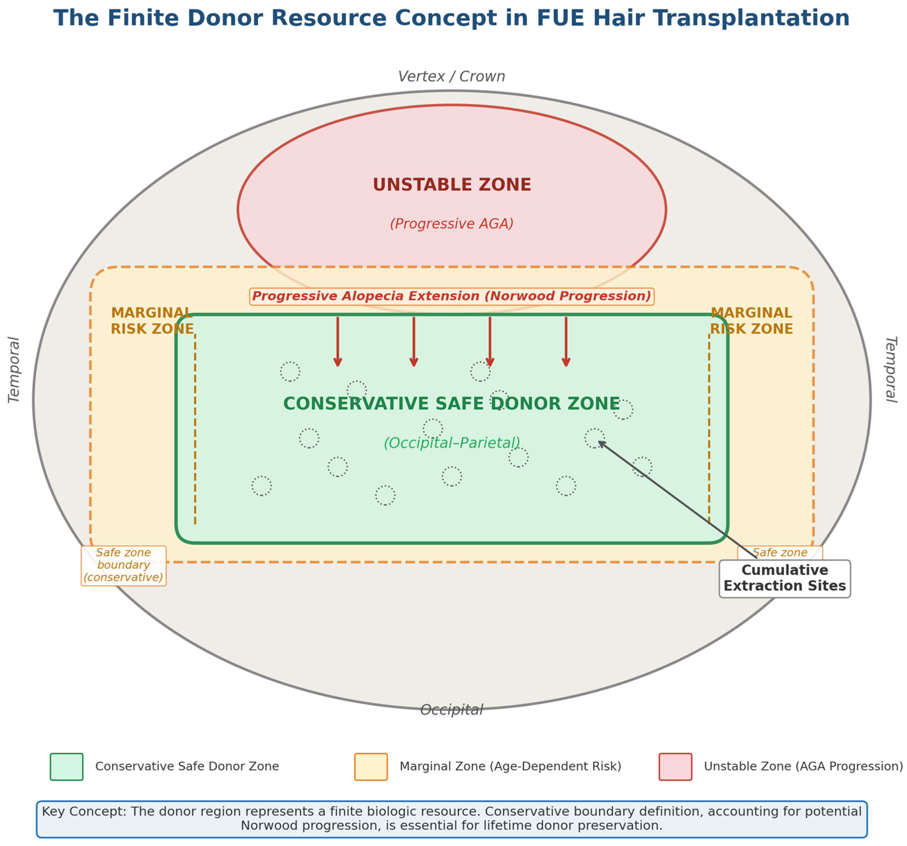
The probabilistic nature of the safe donor site and its relationship to Norwood progression is schematically shown (see
Figure 1).
2.1. Safe Donor Zone: Concept and Limitations
Traditionally, the “safe donor zone” (SDZ) encompasses the mid-occipital scalp and extends laterally into the parietal regions within boundaries considered relatively stable throughout life. In FUE, the ability to harvest extensively over a wide area can create the illusion of an unlimited donor source. In reality, the SDZ is a probabilistic construct rather than a fixed anatomical region. Long-term progression of androgenetic alopecia (AGA), particularly in young patients or those with a strong family history of advanced Norwood patterns, can encroach upon presumed stable zones [
2,
5].
Diffuse unpatterned alopecia (DUPA) constitutes a critical contraindication for aggressive donor harvesting. In DUPA, miniaturization extends to the occipital and parietal zones, rendering conventional SDZ boundaries unreliable. Failure to detect this pattern preoperatively can lead to a shortened graft lifespan and noticeable thinning of the donor area over time [
2].
Gender differences should also be considered when defining a safe donor area. In men with androgenetic alopecia, donor planning is generally based on the expected Norwood progression and the possibility of exposure of seemingly stable marginal areas over time. In women, donor planning can be more complex because female-pattern hair loss may present with diffuse thinning and, in some patients, occipital involvement. Accordingly, the “safe donor area” should not be treated the same in men and women; instead, it should be individualized through trichoscopic assessment of occipital stability, miniaturization rate, and overall pattern distribution. In both sexes, the severity and rate of progression of androgenetic alopecia directly influence how conservative donor margins should be [
2,
5,
9].
2.2. Follicular Unit Density and Miniaturization
Basal follicular unit density in the donor area varies greatly among individuals and is influenced by gender, ethnicity, and hair phenotype. In practice, clinically significant donor capacity depends not only on follicular unit density but also on various qualitative factors that determine “coverage value,” such as terminal-vellus ratio (percentage of miniaturization), hair shaft diameter, hair curl or wave pattern, and hair–skin color contrast. These characteristics affect both the number of available transplantable units and how cosmetically hidden the extraction-related changes are. Importantly, increased miniaturization in the donor area, especially when widespread or extending beyond expected stable areas, may indicate donor instability and decreased long-term graft durability. Therefore, trichoscopic assessment of miniaturization and donor heterogeneity should be routinely included in preoperative planning, especially in young patients and those with rapidly progressing patterns of androgenetic alopecia [
1,
5].
2.3. Regional Variability in Donor Area
The occipital scalp generally provides the most robust graft source; however, parietal and supra-auricular regions generally show slightly lower density and may be more prone to thinning with age. In high-graft sessions, excessive extension to lateral regions increases the risk of visible patchiness, especially in patients who prefer short hairstyles [
4,
5].
Furthermore, excessive vertical extension beyond conservative SDZ boundaries may compromise long-term stability if future hair loss progression exposes these harvested areas. Accordingly, donor mapping should account for potential Norwood progression rather than just the current appearance.
2.4. Vascular and Healing Considerations
The scalp typically has a rich vascular network that contributes to rapid healing after FUE. However, repeated extraction in dense areas can disrupt local microvascular nutrition and contribute to delayed healing, hypopigmentation, or textural irregularities. Emerging literature on donor site healing emphasizes that extraction density, punch diameter, and spatial distribution significantly affect postoperative tissue healing and scar visibility [
4,
6]. Recent donor site studies using quantitative trichoscopy further highlight that donor remodeling and post-harvest changes are measurable and clinically relevant, supporting systematic donor monitoring over time [
10].
Although FUE is often described as “scarless,” if extraction thresholds are exceeded, the cumulative effect of thousands of micro-wounds can lead to clinically noticeable donor changes. In conclusion, donor site preservation should be conceptualized not only as technical precision during extraction but also as biologically informed resource management throughout the patient’s life.
2.5. Long-Term Biological Planning
Modern FUE planning increasingly requires predictive graft allocation strategies that account for progressive androgenetic alopecia and the likelihood of future surgical intervention. Conservative graft harvesting in early sessions preserves the ability to address subsequent pattern evolution, density improvement, or scar revision. In contrast, aggressive early extraction may compromise long-term adaptation and limit corrective options [
1,
2].
Taken as a whole, understanding the biological variability and limitations of the donor site is fundamental to safe and sustainable FUE practice.
Section 3 addresses preoperative donor assessment and risk stratification strategies that translate these biological principles into clinical decision-making.
3. Preoperative Donor Assessment and Risk Classification
Effective donor site protection begins before surgery. In FUE, the ability to widely harvest hair from a large scalp surface can mask early instability of the “safe donor” area, and donor depletion is often a planning error rather than a technical failure. Accordingly, current donor assessment should be approached as a structured risk classification process integrating clinical history, scalp examination, trichoscopy/densitometry, and long-term progression prediction [
11,
12]. To make donor assessment functional, a structured risk classification matrix is proposed to guide extraction planning and long-term budgeting (
Table 1).
3.1. Clinical History and Progression Prediction
A focused history should include the following features: (i) age and rate of onset of hair loss, (ii) family history of advanced hair loss, (iii) previous medical treatment and compliance, (iv) previous procedures (including number of extractions if known), and (v) concomitant diseases affecting wound healing or scar risk. Progression prediction is important, especially in young patients and those with strong familial Norwood progression, as future losses may reveal over-harvested areas or uncover previously hidden extraction patterns [
2,
13]. Wherever possible, surgeons should incorporate “lifetime donor budgeting” into consent discussions, framing grafts as a limited rather than an unlimited resource [
5,
14].
3.2. Donor Mapping and Defining the Conservative Graft Harvesting Area
While the concept of a safe donor zone (SDZ) remains fundamental, current practices increasingly emphasize individualized donor mapping. Particularly in patients with early-stage alopecia, conservative margin selection is recommended, as the “permanent zone” may be narrower than anticipated in some phenotypes. Practical approaches standardizing donor zone measurement have been proposed to reduce unwanted graft harvesting from potentially unstable margins [
12]. The International Hair Restoration Practice Guideline also emphasizes systematic donor density assessment at multiple points and avoiding over-graft harvesting that could lead to visible thinning [
15]. Similarly, an international expert consensus statement highlighted the importance of standardized perioperative counseling and postoperative care to reduce variability in outcomes and reporting [
16].
3.3. Physical Examination: Density, Diameter, and Miniaturization
Donor quality is not solely dependent on follicular unit density. Preoperative assessment should integrate quantitative and qualitative parameters, including follicular unit density at multiple occipital and parietal points, number of hairs per follicular unit, hair strand diameter, curl pattern, and hair–skin contrast, as these collectively determine the optical effect of extraction and the expected cosmetic “coverage value.” Miniaturization screening in the donor area is equally important, as high donor miniaturization can reflect instability (e.g., diffuse thinning phenotypes) and increase the risk of both apparent donor depletion and decreased long-term graft durability. In routine practice, handheld densitometry provides estimates of follicular unit density and hair counts, while trichoscopy improves the detection of miniaturization patterns and helps identify latent inflammatory or autoimmune mimics that may compromise donor dominance. Since the cosmetic results of excessive hair harvesting are often delayed, conservative extraction planning is usually necessary in patients with borderline density, higher donor miniaturization, or uncertain long-term stability [
5,
11,
12].
3.4. Trichoscopy and Exclusion of High-Risk Phenotypes (DUPA, Inflammatory Mimics)
Trichoscopy is particularly valuable in differentiating patterned hair loss from formations that can mimic or coexist with androgenetic alopecia (e.g., alopecia areata incognita, fibrosing alopecia with patterned distribution). These conditions can alter the prognosis and increase the risk of unpredictable hair loss or long-term instability if surgical intervention is performed unnoticed [
11]. Widespread irregular alopecia (DUPA), characterized by widespread miniaturization including the occipital and parietal regions, remains a critical contraindication for aggressive donor harvesting, as it increases the risk of both donor depletion and graft failure over time due to loss of donor dominance [
2].
A related practical consideration is that occipital involvement may be more common than previously thought in some women with female-pattern hair loss; trichometric comparisons of occipital and mid-scalp parameters may help identify patients in whom “safe donor” assumptions require more scrutiny [
9].
3.5. Risk Classification and Consent: Aligning Goals with Donor Reality
A structured donor risk discussion should clearly link the patient’s goals (desired number of grafts, density expectations, hairstyle preferences) to donor constraints and long-term planning. Given the visibility of donor changes in short hairstyles, patients should be informed that FUE is not “scarless” and that hypopigmented punctate scarring and donor thinning may be cosmetically significant if extraction density is excessive or distribution is not properly spread [
5,
6]. In cases where donor stability is uncertain (young age, rapid progression, borderline miniaturization), priority should be given to conservative planning or staged procedures along with appropriate medical treatment optimization [
13,
15]. In summary, donor evaluation in FUE should be treated as a repeatable, multi-parameter assessment aimed not only at maximizing current graft yield but also at preserving donor aesthetics and leaving a reserve for future needs.
In our clinical practice, we take a conservative and longitudinal approach to donor management. We routinely assess donor density, miniaturization, hair shaft diameter, hair–skin contrast, and previous graft harvesting history before determining extraction targets. For higher-risk patients, including younger individuals, patients with borderline donor density, or those exhibiting signs of widespread instability, we prefer phased planning and repeated evaluation instead of aggressive single-session graft harvesting. Graft harvesting is performed using manual or motorized FUE with punch sizes typically within the range of 0.7 and 0.9 mm, selected according to hair characteristics and graft integrity requirements. Our practical priority is widespread distribution, prevention of local clustering, and preservation of donor flexibility for future procedures. This approach is consistent with current recommendations emphasizing individualized donor mapping, conservative extraction, and reassessment before repeated procedures [
5,
12,
15].
The next section will address extraction strategies and delivery principles that translate these biological principles into clinical decision-making.
4. Harvesting Strategies and Extraction Distribution Principles
Translating donor biology and preoperative assessment into safe surgical practice requires disciplined harvesting strategies. In FUE, donor preservation is influenced not only by the total number of grafts extracted, but also by punch diameter, cutting rates, spatial distribution, session planning, and cumulative lifetime extraction intensity. Importantly, visible donor loss is more often the result of excessive local extraction intensity or poor distribution than absolute graft numbers [
5,
6].
Where high-level comparative evidence is limited, the practical recommendations in this review reflect a combination of published literature, expert consensus, and clinically based surgical principles aimed at supporting conservative donor management.
4.1. Punch Diameter and Cutting Risk
Punch size selection directly affects both graft integrity and donor site scarring. Smaller punches (e.g., 0.7–0.9 mm) can reduce scar visibility but can increase cutting rates if misaligned with the follicular angle or hair fold. On the other hand, larger staples may reduce the cutting rate but may increase the risk of hypopigmented macules and textural irregularities [
1,
4].
Modern manual, motorized, and robotic FUE systems aim to optimize graft integrity while minimizing donor site morbidity; however, variability in device parameters and the lack of standardized reporting frameworks continue to limit direct comparison between techniques [
8,
17]. Comparative clinical analyses of robotic and conventional FUE have shown measurable differences in workflow efficiency, cutting rates, and operative metrics, underlining the need for standardized outcome reporting [
18]. In parallel, image-guided robotic platforms continue to evolve, including integrated systems designed to support both harvesting and implantation workflows [
19]. Accordingly, punch selection should be individualized based on hair strand thickness, curl pattern, and surgeon experience, rather than a uniform device preference.
Robotic FUE systems represent an evolving area of donor harvesting. Current studies suggest that robotic systems can improve procedure standardization and some operative metrics in certain settings; however, the evidence is heterogeneous and depends on device generation, operator experience, and reported endpoints [
2,
8,
18].
4.2. Extraction Intensity Thresholds
A fundamental principle in donor site conservation is to limit extraction intensity per session and cumulatively over a lifetime. While absolute thresholds vary across publications, conservative guidelines generally recommend avoiding extractions of more than 15–20% of follicular units from a defined area in a single session and careful spacing to minimize clustering [
5,
15]. Quantitative donor site analyses also demonstrate that extraction rates can vary significantly in real-world practice; In one clinical study, the number of hairs extracted represented approximately one-third of the baseline donor density (range ~28.9–42.8%), which reinforces the need for careful extraction planning and individualized donor budgeting [
20].
Hair transplantation beyond conservative thresholds increases the likelihood of visible “moth-eaten” thinning, reduces camouflage in short hairstyles, delays healing with hypopigmentation, and reduces the reserve for future procedures. Even if the total number of grafts appears acceptable, local over-transplantation can compromise optical density and donor aesthetics.
In high-graft sessions, strategic distribution between the occipital and parietal regions instead of dense harvesting from a single band helps preserve optical density [
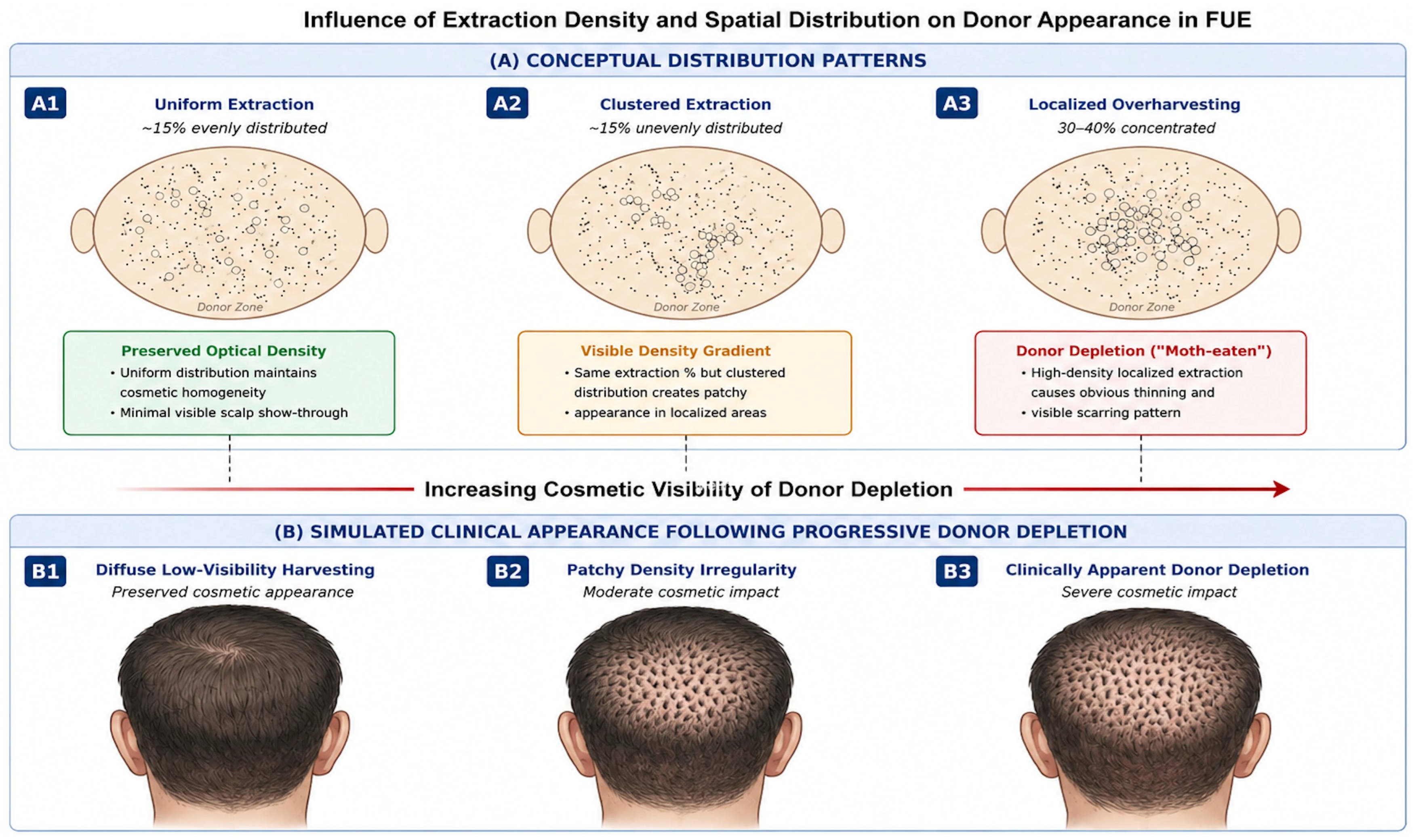
14]. The cosmetic effects of extraction density and spatial distribution are shown in
Figure 2, and it is emphasized that clustering increases visible thinning even at the same extraction percentages.
4.3. Spatial Distribution: Patterned and Diffuse Harvesting
The visual appearance of the donor area depends not only on how many grafts are extracted but also on how they are distributed. Diffuse, evenly spaced extractions reduce the risk of detectable patterning, while clustered harvesting can create visible density gradients even if the overall extraction percentage appears acceptable [
4,
6]. Although computer-aided mapping and grid-based marking techniques have been proposed to standardize extraction spacing, robust comparative data remain limited [
8,
19,
21,
22]. In practice, awareness of spacing by the disciplined surgeon and avoidance of repetitive transitions within the same micro-region are critical.
A rigid millimeter spacing cannot be universally applied because the cosmetic effect of harvesting depends on the initial density, hair diameter, curl, hair–skin contrast, and punch size. The most important principles in practice are even distribution, prevention of local clustering, and minimizing repeated harvesting from immediately adjacent follicular units within the same micro-region [
4,
21]. Grid-based marking or systematic zone rotation can help maintain even spacing in high-volume sessions.
4.4. Mega Sessions and Cumulative Donor Burden
The increase in “mega sessions” (3000–5000+ grafts) has increased pressure on donor management strategies. While large sessions may be appropriate in selected patients with high baseline density and stable donor sites, aggressive early harvesting may compromise long-term resilience. Especially in younger patients, multi-stage planning should prioritize staged extraction rather than maximizing initial depletion [
1,
2].
Importantly, cumulative donor burden should be considered not only per session but over the patient’s lifetime. Repeated procedures without comprehensive donor reassessment may increase the risk of visible thinning or scarring, particularly at the parietal margins [
14].
Procedure time is another practical factor contributing to donor site morbidity. While a universal time threshold has not been established, long, large-session FUE can increase cumulative tissue manipulation, surgeon fatigue, repeated passes, and overall tissue stress; therefore, procedure time should be guided by a non-traumatic technique rather than a fixed numerical target [
1,
23]. Repeated harvesting should be based not only on patient requests but also on donor reassessment. Before additional sessions, donor density, miniaturization, scar visibility, and remaining reserve should be reassessed to reduce the risk of cumulative depletion [
14,
15].
4.5. Technical Factors Affecting Healing
Extraction angle alignment, depth control, and non-traumatic graft removal affect donor site healing. Excessive torque, repeated puncture attempts, and large puncture diameters can increase local tissue trauma and inflammatory response, potentially affecting pigmentation outcome and scar visibility [
4,
6].
Although FUE is generally described as minimally invasive, the cumulative micro-injuries occurring in thousands of sites represent a significant tissue burden. Careful technique during surgery, temperature control, and avoidance of areas of intense trauma remain key elements in preserving donor tissue.
5. Overharvesting and Donor Depletion
One of the most significant long-term risks of FUE is excessive tissue harvesting. Unlike linear excision, where tissue removal is spatially defined and immediately noticeable, FUE allows for gradual extraction over a large surface area. While this flexibility is advantageous, it also leads to cumulative tissue loss that may become clinically apparent months or years after surgery. Donor tissue loss typically manifests as decreased optical density, uneven thinning, parietal-occipital gradients, or patchy transparency highlighted by short hairstyles [
24].
The etiology of donor depletion is multifactorial. Excessive local extraction density remains the dominant factor, especially when removal is concentrated within narrow bands. Overextension beyond conservative safe zone boundaries, failure to account for progressive androgenetic alopecia, and inadequate reassessment before repeated sessions further increase the risk. Importantly, depletion is usually the result of cumulative inadequate decisions made over multiple procedures, not a single major session [
25].
Clinical signs of early depletion include asymmetrical thinning in the extraction area, increased visibility of the scalp under bright light, and textural irregularities detectable upon close inspection. In more advanced cases, even if the recipient area appears satisfactory, the transparency of the donor area can compromise the aesthetic outcome. When significant depletion occurs, restoration options become limited to the donor reserve, which is the constraint that is not preserved [
2,
26].
Treatment depends on the remaining follicular availability. In selected cases where density is preserved in adjacent areas, limited redistribution grafting can partially correct visible gradients. However, when the reserve is insufficient, non-surgical camouflage techniques become primary. Scalp micropigmentation has emerged as a valuable adjunct in donor depletion, improving the perception of cosmetic density and reducing the contrast between hypopigmented extraction areas and the surrounding scalp. Its role is particularly important in patients with short hair or darker phototypes. In conclusion, prevention through conservative planning, uniform distribution, and long-term donor budgeting remains the most effective strategy. Technical modifications such as long-hair FUE have been suggested to improve real-time visualization of donor density and help avoid overharvesting, particularly in patients who prioritize donor aesthetics [
27].
Long-hair FUE has been proposed as an adjunct method for preserving the donor area because the preserved hair length can provide better immediate visualization of the donor area’s appearance and assist surgeons in assessing optical density during hair harvesting. Potential advantages include improved intraoperative camouflage assessment and a reduced risk of cosmetically significant excessive hair harvesting in selected patients. However, long-hair FUE is technically more challenging, can be slower, and may complicate visualization and extraction efficiency depending on hair characteristics and operator experience [
27].
6. Scarring Minimization in FUE
While FUE eliminates the linear scarring characteristic of strip excision, it creates thousands of small circular scars that determine long-term donor aesthetics. The common misconception that FUE is “scar-free” has contributed to underestimating the risk of donor scarring in high-density extractions.
The most common scarring manifestation is hypopigmented punctate macules (“white dot scarring”), the visibility of which depends on punch diameter, extraction density, healing biology, and skin phototype. Larger punches increase scar diameter, while excessively small punches can increase incision and repeated attempts, paradoxically worsening local trauma. Depth control and alignment with the follicular angle are equally critical; excessive penetration can enlarge the scar and increase the inflammatory response.
Tissue alterations represent a more subtle but clinically significant consequence of high-density harvesting. When dermal support is reduced through excessive cumulative extraction, minor contour irregularities or changes in scalp elasticity may develop. These changes are particularly pronounced in shaved donor areas.
Donor area telogen effluvium (“donor shock loss”) has also been described following intense trauma. It typically manifests as temporary thinning at or adjacent to the extraction site and resolves within a few months. Its occurrence enhances the donor area’s biological susceptibility to cumulative mechanical stress. Therefore, minimizing scarring requires a multidimensional approach: appropriate staple selection, uniform spatial distribution, adherence to conservative extraction percentages, and non-traumatic technique. Scar visibility is rarely attributable solely to staple size; rather, it reflects the interaction between extraction intensity and tissue response.
7. Donor Site Complications in FUE: Prevention and Management
Although FUE prevents linear scarring, it is not a “scarless” method. Donor morbidity ranges from temporary postoperative effects (pain, edema, erythema) to long-term aesthetic sequelae (hypopigmented lesions, tissue changes, donor depletion) and less common inflammatory or infectious complications. Current reviews highlight that most donor site complications can be prevented with conservative planning, disciplined extraction distribution, and meticulous technique, while also noting that reported complication rates and definitions vary considerably between studies [
4,
6,
7]. Recent complication-focused syntheses, classifying donor events as early postoperative reactions, pigmentation scarring, depletion, cysts/folliculitis, and less common inflammatory conditions, have reinforced the need for structured prevention pathways [
7]. Donor site morbidity can be mechanistically classified, allowing for targeted prevention strategies as summarized in
Table 2.
7.1. Early Postoperative Events and Wound Healing
Common early findings at the donor site include pain, edema, erythema, crusting, and transient pruritus. These usually resolve spontaneously and respond to routine postoperative care (gentle cleaning, topical moisturizers, avoidance of friction, and adherence to follow-up appointments). A recent systematic review focusing on donor site healing interventions highlights the diversity of postoperative practices (dressings, topical agents, procedural aids) and notes that outcomes and reporting remain heterogeneous, limiting definitive comparative results [
6].
Prevention (high-yield): minimize repeated punctures in the same micro-area, avoid excessive puncture depth/torque, maintain consistent intervals, and respect conservative extraction intensity thresholds [
4,
23].
Postoperative donor site care significantly contributes to wound healing and scar quality. Patients should be advised to keep the donor area clean, avoid rubbing and scratching, follow gentle washing instructions, and avoid early mechanical trauma and excessive sun exposure during the initial healing phase. Standardized preoperative and postoperative care can help reduce variability in healing outcomes [
6,
16,
23].
7.2. Hypopigmented Spots and Visible “White Dot” Scars
Hypopigmented punctate scars are among the most visible long-term donor site sequelae of FUE, particularly in patients with darker skin phototypes or those who keep their hair very short. Scar visibility is affected by punch diameter, cumulative extraction density, depth/angle control, and healing biology [
4,
13].
Prevention: smaller punch selection when appropriate, homogeneous distribution of extractions, and avoidance of high-density harvesting within a narrow band.
Management: patient counseling (especially in those who prefer short hair), camouflage strategies (hair styling, scalp micropigmentation), and in selected cases, corrective approaches depending on donor reserve (e.g., targeted grafting to depleted areas) [
23].
7.3. Donor Site Effluvium (Donor “Shock Loss”)
Localized telogen effluvium in the donor site has been defined after graft harvesting and may present as transient diffuse thinning in or adjacent to the extraction site. It typically begins within weeks and resolves within months; however, its incidence is uncertain due to limited formal reporting [
7,
28].
Prevention: Avoid areas of intense trauma, minimize repeated passing and excessive pulling, and inform patients with pre-existing miniaturization or marginal donor density about the possibility of transient thinning.
Management: Reassure, document, optimize medical treatment as needed, and follow up to confirm healing [
28].
7.4. Folliculitis, Cysts, and Inflammatory Complications
Folliculitis may occur in both recipient and donor sites during the postoperative period. Follicular trauma, obstruction, bacterial overgrowth, or improper postoperative hygiene in the donor area may be the cause. Most cases are mild and respond to local care and short-term topical or systemic treatment depending on severity; however, persistent or atypical presentations require culture and evaluation for unusual organisms or inflammatory dermatoses [
4,
7]. Epidermoid/epithelial cysts and delayed inflammatory nodules have also been described and may require drainage or excision in selected cases [
7].
7.5. Sensory Symptoms and Neurovascular Considerations
Transient sensory disturbance (numbness, dysesthesia) may occur due to localized nerve irritation or edema. Permanent symptoms are rare but should warrant re-evaluation for localized nerve damage, scar attachment, or secondary inflammation. Conservative graft harvesting, avoiding deep puncture, and respecting anatomical planes reduce the risk [
4,
23].
7.6. Integrated Prevention Strategy in High-Volume Practice
Effective prevention of donor site morbidity in FUE requires a structured and disciplined approach integrating preoperative planning, intraoperative technique, and long-term follow-up. In high-volume settings where procedural efficiency and large graft numbers are frequently emphasized, strict adherence to donor preservation principles is particularly important to prevent cumulative depletion.
Preoperatively, conservative donor margin mapping and lifetime graft budgeting form the basis of prevention. Extraction goals should be aligned with expected long-term androgenetic progression rather than immediate cosmetic goals. Patients with borderline donor density, increased miniaturization, or young age require particularly careful planning, often preferring staged procedures over aggressive single-session harvesting. During surgery, prevention relies on maintaining an even spatial distribution of extractions, avoiding clustering within narrow bands, and respecting conservative extraction thresholds per region. Even if the total graft count appears acceptable, excessive localized extraction increases the risk of apparent density gradients and long-term optical thinning. Punch size selection, angle alignment with follicular outflow, and minimizing repeated attempts within the same micro-region further reduce cumulative tissue trauma.
Postoperatively, systematic documentation of extraction sites, punch diameter, and estimated density reduction allows for more reliable planning of future sessions. Reassessment of donor stability and recalculation of remaining reserve are essential before any repeated procedures. Repeated harvesting without updated assessment represents one of the most frequent contributors to progressive donor depletion.
In summary, donor area preservation should be treated not as a technical detail of FUE, but as a continuous management strategy extending beyond a single surgical session. In sustainable hair restoration practice, moderation and structured planning are as important as surgical precision.
8. Long-Term Donor Management and Multi-Session Planning
Long-term donor tissue preservation in FUE can be conceptualized as a cumulative structural phenomenon where successive extractions progressively alter the baseline density and optical coverage. Even if individual sessions appear conservative, repeated graft harvesting without reassessment can lead to clinically significant donor tissue damage [
7,
14].
Accordingly, long-term management should integrate lifetime graft allocation, reassessment before repeated procedures, proactive delivery strategies, and defined mitigation pathways [
20].
8.1. Lifetime Donor Budgeting and Staged Restoration Planning
A practical “lifetime donor budgeting” approach links expected alopecia progression and hair style preferences to a conservative extraction plan; this plan aims to preserve reserve for future improvements (temples, crown, scars, density revisions) and reduce the risk of visible donor thinning as patterns evolve [
5,
14]. This is particularly important for younger patients and those seeking large graft numbers (“mega sessions”), where aggressive early harvesting can compromise long-term resilience [
2,
14]. If there is a possibility of future loss, phased planning can reduce the need for “overbuying” intensity early on and support a more adaptable long-term design.
8.2. Mandatory Donor Reassessment Before Repeated Sessions
Repeated procedures should not be planned solely based on previous graft counts or surgical notes. Instead, donor reassessment should be addressed mandatorily, including updated trichoscopy/densitometry, miniaturization scanning, and a renewed donor map [
11,
12]. Standardized approaches to defining donor margins can reduce accidental graft harvesting from unstable margins and facilitate consistent documentation across multiple sessions [
12]. In practice, the donor “risk profile” can change with age, adherence to medical treatment, and evolving alopecia patterns; this makes reassessment central to preventing progressive donor depletion [
7].
8.3. Avoiding Cosmetic Donor Depletion in Serial FUE
The dominant technical cause of long-term donor aesthetic loss is excessive local extraction density over time; this can result from repeated tissue harvesting from the same band or failure to achieve homogeneous distribution. Therefore, long-term donor preservation benefits from: (i) broad and evenly distributed tissue harvesting within conservative limits; (ii) avoiding repeated passes over the same micro-regions; and (iii) clear documentation of puncture diameter, extraction density strategy, and tissue distribution plan to support reproducibility in future sessions [
6,
14]. Recent evidence syntheses also highlight heterogeneity in donor site outcome reporting and postoperative practices, reinforcing the value of standardized documentation when assessing cumulative donor morbidity [
6].
8.4. Combining Harvesting Methods and Alternative Strategies in Cases of Limited Donor Sources
In patients with advanced hair loss or a lifelong high graft requirement, FUE alone may limit the total number of harvestable grafts before visible depletion occurs. A 2024 technical review describes an approach that combines FUE with linear strip excision using a modified “FUE-Linear Ellipse” method to optimize graft yield while aiming to reduce the risk of long-term donor depletion. While broader comparative evidence is still limited, this study supports the concept that offering multiple donor harvesting methods can preserve lifelong options for selected patients [
26].
Another real-world strategy is to harvest grafts from outside the classic “safe zone” to avoid over-harvesting of a narrow band and to homogenize the cosmetic appearance of the donor in short hairstyles. However, grafts from more marginal areas may be more sensitive to androgens. Practical placement strategies for such grafts have been proposed with the aim of maximizing benefit while minimizing long-term risk; these strategies emphasize careful labeling/separation during surgery and deliberate placement in low-priority areas [
29]. These approaches should be carefully framed and supported by clear counseling on potential long-term durability differences [
2,
29].
8.5. Repair and Camouflage of Donor Morbidity
When donor depletion or “white spot” scarring becomes visible, management often shifts from optimization to mitigation. Options include conservative medical stabilization of ongoing loss (where appropriate), targeted redistribution/repair grafting in carefully selected cases with remaining reserve, and camouflage techniques such as scalp micropigmentation (SMP). Recent clinical technical analysis and case series support SMP as a cosmetically effective approach for localized alopecia patterns and offer procedural parameters that can help standardize outcomes [
30]. Previous baseline studies also identify SMP as a non-surgical option to reduce the visibility of scarring or thinning and support its role as an adjunct corrective strategy [
31]. In cases of donor morbidity, SMP may be particularly valuable when additional harvesting is not recommended.
8.6. Practical Long-Term Framework for High-Volume FUE Practice
In high-volume FUE procedures, long-term donor conservation should be implemented based not only on informal surgical judgment but also on a structured and reproducible clinical framework. This begins with comprehensive preoperative donor mapping and systematic miniaturization screening, both initially and before any repeated procedures, to ensure objective verification of donor stability [
11,
12].
In parallel, lifetime donor budgeting should be explicitly integrated into the surgical plan. Graft allocation should reflect anticipated androgenetic progression, projected future needs, and the patient’s hairstyle preferences, rather than focusing solely on immediate cosmetic goals [
5,
14].
During surgery, conservation relies on maintaining a uniform harvest distribution strategy at conservative density thresholds. Extraction patterns, punch diameter, and regional graft allocation should be documented in a standardized manner to support reproducibility and allow for informed planning in subsequent sessions [
6,
7].
Before any repeat sessions, mandatory donor reassessment is required. Updated trichoscopy or densitometry, assessment of intermittent miniaturization, and recalculation of remaining donor reserve should guide revised long-term planning [
7,
12]. Repeated harvesting without structured reassessment significantly contributes to cumulative donor depletion.
Finally, clinics should establish a mitigation pathway for donor morbidity; This pathway should include conservative management criteria, limited repair grafting when reserve permits, and camouflage strategies such as scalp micropigmentation when further graft harvesting is not feasible [
30,
31].
Through this integrated approach, donor management transforms from a session-specific technical assessment into a longitudinal conservation strategy consistent with the progressive nature of androgenetic alopecia.
9. Structured Clinical Framework for Donor Preservation in FUE
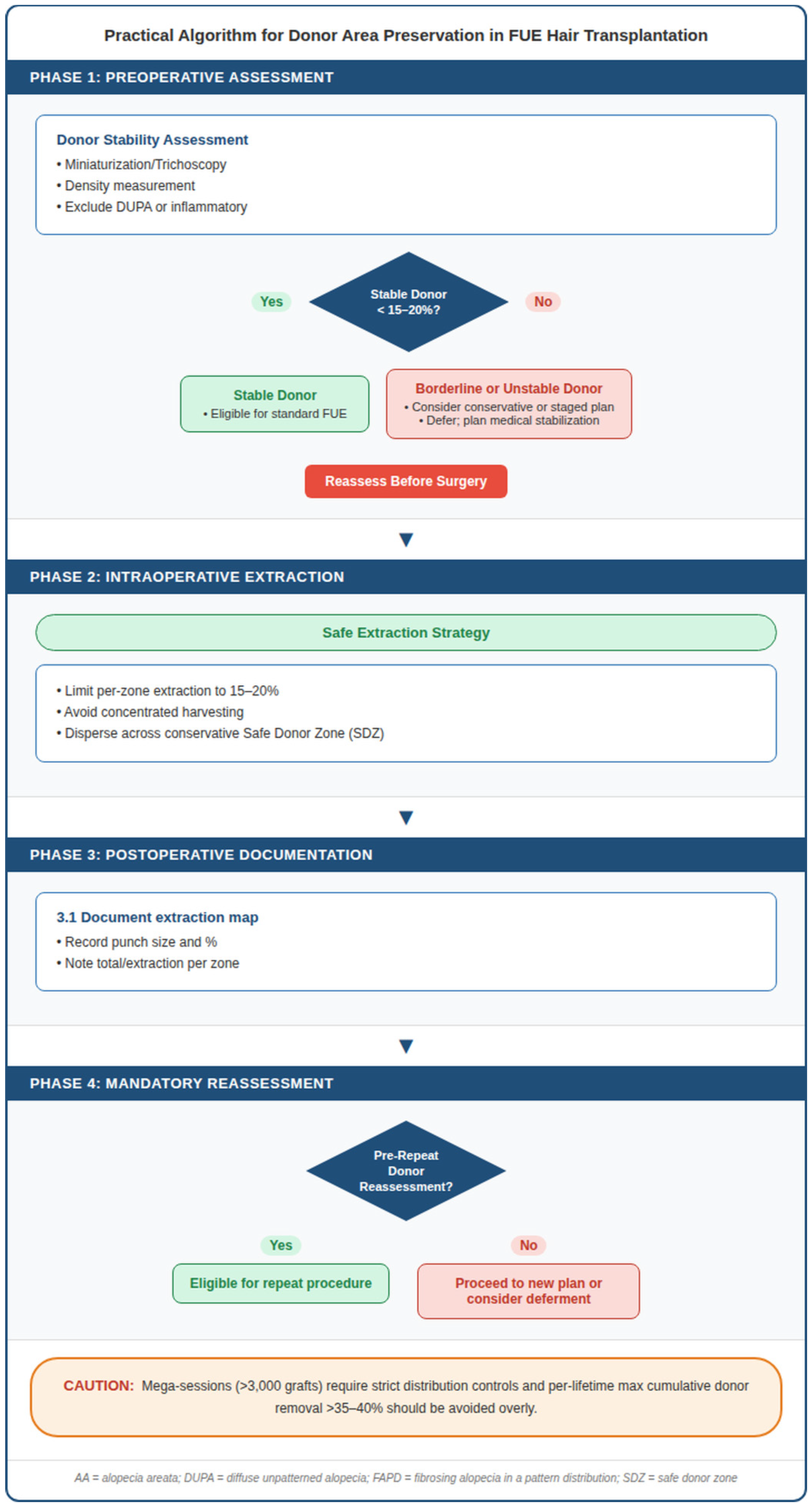
To put these principles into practice, the donor tissue preservation process in FUE can be structured as a sequential and repeatable clinical process encompassing preoperative risk stratification, intraoperative tissue removal control, and mandatory reassessment before repeated interventions.
Preoperatively, donor stability should be confirmed through trichoscopic assessment, density measurement, and conservative margin mapping. Patients exhibiting features consistent with significant miniaturization or diffuse irregular alopecia in the donor area should be carefully evaluated or surgery should be postponed [
11].
Intraoperatively, the extraction density should remain within conservative limits for each defined area. While precise numerical thresholds vary in the literature, cumulative removal exceeding approximately 15–20% in a confined area is often associated with an increased risk of visible thinning [
5,
15]. Uniform spatial distribution is essential to preserve optical density.
Postoperatively, documenting the extraction patterns, punch size, and harvested areas facilitates reliable planning for future sessions. Prior to any repeated procedure, re-evaluation of the donor should be mandatory to readjust lifetime donor budgeting.
This structured framework reinforces that donor protection is a central determinant of sustainable FUE practice, not a technical afterthought.
A structured clinical algorithm integrating preoperative risk stratification, intraoperative extraction control, and mandatory re-evaluation before repeated sessions is proposed (see
Figure 3).
This structured framework reinforces the idea that donor tissue preservation is not a procedural detail, but a long-term surgical responsibility that is integral to sustainable FUE practice.
10. Conclusions
Sustainable donor site management is fundamental to long-term success in follicular unit extraction. FUE minimizes linear scar formation and increases procedural flexibility; however, when extraction strategies are inadequately distributed or planned, the donor site becomes vulnerable to cumulative depletion, hypopigmentation, and textural changes.
Durable outcomes are achieved not through maximum graft harvesting, but through disciplined extraction strategies that respect progressive alopecia patterns and preserve future surgical adaptability. The structured clinical framework proposed in this review supports donor integrity in both routine and high-volume surgical settings by translating these principles into reproducible practice.