Article

Ex Vivo Assessment of Heat and Humidity Effects on Human Skin and Potential Protection by Kombucha Tea Extract


1
BIOMECA, Bâtiment Adénine, 60 Avenue Rockefeller, 69008 Lyon, France
2
LVMH Recherche, Life Science Department, 185 Avenue de Verdun, 45800 Saint Jean de Braye, France
3
CELLOMET, ADERA, 146 rue Léo Saignat, 33076 Bordeaux, France
4
INSERM U1211, 33076 Bordeaux, France
*
Author to whom correspondence should be addressed.
Cosmetics 2026, 13(3), 114; https://doi.org/10.3390/cosmetics13030114
Submission received: 24 March 2026 / Revised: 25 April 2026 / Accepted: 30 April 2026 / Published: 6 May 2026
(This article belongs to the Section Cosmetic Formulations)

Abstract

Human skin homeostasis relies on the delicate equilibrium between epidermal stem cell renewal, dermoepidermal junction (DEJ) architecture, and environmental interactions. With aging and exposure to external stressors, this equilibrium becomes disrupted, leading to reduced regenerative capacity. In this study, we established an ex vivo human skin model to examine the impact of dry and tropical (hot and humid) environmental conditions on epidermal homeostasis and to evaluate the protective potential of Kombucha tea extract, a fermented tea known for its antioxidant and regenerative properties. Histological analyses revealed that tropical conditions induced pronounced epidermal thickening (+157%) and disruption of the normal undulating architecture of the DEJ. Atomic force microscopy demonstrated a loss of mechanical contrast between dermal papillae and epidermal ridges, indicative of junctional flattening (−61 and −81%). At the molecular level, heat and humidity upregulated a stem cell marker (+85%) and collagen VII (+39%), reflecting an adaptive but potentially destabilizing activation of basal keratinocytes and matrix reorganization. Topical application of Kombucha tea extract counteracted these effects. Together, these results highlight the sensitivity of epidermal stem cell niches to heat and humidity stress and identify Kombucha tea extract as a promising bioactive agent to preserve epidermal homeostasis under challenging climatic conditions.

Graphical Abstract

1. Introduction

Human skin homeostasis refers to the dynamic equilibrium of cellular, molecular and structural processes that preserve the integrity, barrier function, and overall health of the skin in response to internal and external stimuli. Epidermal homeostasis is maintained through the proliferation of keratinocytes in the basal layer, where cells are anchored to the underlying basement membrane. Within this layer, epidermal stem cells, organized in a patterned, non-random distribution, present unlimited self-renewal capacity [1,2]. These stem cells are located along the dermoepidermal junction (DEJ), which exhibits a distinctive undulating architecture formed by epidermal rete ridges interdigitating with the papillary dermis, resulting in a variable number of epidermal strata [3,4]. Some studies have localized epidermal stem cells to the tips of dermal papillae [5,6,7], whereas others have identified them along the epidermal ridges [8,9]. These differing localizations may reflect the presence of stem cells at distinct transitional states [10]. Epidermal stem cells can be distinguished from other keratinocytes by their high expression of β1 integrins and melanoma chondroitin sulfate proteoglycan (MCSP), along with the absence of terminal differentiation markers [6,7,11,12].
Skin homeostasis and structure are progressively altered with age. Skin aging is a multifactorial process that includes intrinsic aging as well as extrinsic aging, which is driven by environmental exposures such as ultraviolet radiation, air pollution, and fluctuations in temperature and humidity. Intrinsic aging is characterized by morphological changes, notably the reduction in dermal papillae height and flattening of the DEJ. Several studies have reported that rete ridge height significantly declines with age, with older adults showing markedly reduced papillae height compared to youngest individuals [13,14]. Although the number of epidermal stem cells remains stable, cells from older donors display reduced proliferative capacity, diminished stemness potential, and decreased expression of markers such as MCSP [13,14,15]. Consequently, aging epidermal stem cells proliferate less efficiently and progressively lose function, leading to impaired tissue resilience, weakened barrier function, epidermal flattening, and reduced biomechanical properties [4].
Extrinsic factors, particularly heat and humidity, further impact epidermal homeostasis and accelerate skin aging. Proper skin hydration depends on the integrity of the stratum corneum and the function of its protective barrier, which are supported by tight junctions and lipids such as ceramides. Environmental conditions significantly alter these processes in vivo [16,17] and in vitro [18,19,20,21,22], albeit through distinct mechanisms. Dry environments promote transepidermal water loss, causing stratum corneum dehydration, keratinocyte hyperproliferation and epidermal thickening (hyperkeratosis). This state disrupts desquamation, leading to corneocyte accumulation, rough skin, and barrier dysfunction, which increases susceptibility to irritants. In contrast, excessive humidity can overhydrate the stratum corneum, destabilizing the lipid matrix, weakening barrier integrity, and heightening skin permeability and infection risk. Overhydration can also alter desquamation, resulting in conditions such as maceration. Heat exposure has also emerged as a critical factor in extrinsic aging, a process referred to as thermal skin aging [23]. Heat shock modulates the signaling pathways controlling matrix metalloproteinases (MMPs) in dermal fibroblasts [24,25,26], while elevated temperatures promote reactive oxygen species (ROS) generation in keratinocytes, thereby accelerating natural aging processes [27,28]. Infrared radiation and chronic heat exposure also contribute to premature skin aging by inducing angiogenesis, inflammatory cell infiltration, extracellular matrix degradation via MMP activation, and alterations in structural proteins [29]. Furthermore, heat stress can damage mitochondria and induce oxidative stress in mesenchymal stem cells (MSCs), impairing their proliferative, differentiation and immunomodulatory functions [30]. Collectively, intrinsic aging and environmental stressors synergistically drive the morphological and functional decline of the skin.
In this study, we first investigated the effects of environmental variations (heat and humidity) on the mechanical properties and activity of basal stem cells in the human epidermis. We also evaluated the potential protective effect of Kombucha tea, produced by fermenting tea with a symbiotic culture of bacteria and yeast, commonly referred to as the “tea fungus” [31]. The selection of kombucha tea extract was mechanistically justified rather than empirically driven. Unlike conventional antioxidants that primarily target oxidative stress, kombucha tea extract uniquely addresses the stem cell-mediated regenerative deficit induced by heat and humidity stress through the concurrent modulation of Hedgehog and ROBO signaling pathways. This positions it as a highly specific and innovative candidate for counteracting environmental stress-induced DEJ alterations, clearly distinguishing it from other natural skincare agents. Our findings highlight both the influence of environmental conditions on epidermal homeostasis and the potential protective role of Kombucha tea in maintaining dermal–epidermal junction integrity.

2. Materials and Methods

2.1. Human Skin Samples

Human skin tissue explants were collected with informed consent from patients undergoing surgical procedures, following the ethical guidelines. Normal human skin samples were obtained from the surgical discard of anonymous healthy patients in accordance with ethical guidelines (French Bioethics law of 2004) and declared to the French research ministry (Declaration no. DC-2025-7230), delivered to LVMH Recherche. The donor was healthy Caucasian female 39-year-old abdominoplasty. After collection, the hypodermis was carefully removed using scissors and a scalpel. The skin was used for fibroblasts extraction or cut into 1 cm-diameter round pieces, flash-frozen in liquid nitrogen, and stored at −80 °C. Frozen samples were embedded in OCT embedding matrix (Sakura 4583, Torrance, CA, USA), and 16 µm cryosections were prepared using a Cryostat (LEICA CM3050S, Paris, France) at −20 °C. The sections were mounted on SuperFrost Plus slides (Fisher, Strasbourg, France) and stored at −20 °C.

2.2. Kombucha Tea Extract

Kombucha is a fermented tea produced through a microbiological process involving a symbiosis between yeasts (primarily of the Saccharomyces genus) and bacteria (Bacterium Xylinum). The bacteria create a polysaccharide matrix in which the yeasts reside. Key microorganisms involved include various species such as Saccharomyces ludwigii, Schizosaccharomyces pombe, Acetobacter ketogenum, and Pichia fermentans, among others. The kombucha extract used in association in the present invention is the product marketed by SEDERMA (Montigny-le-Bretonneux, France) under the trade reference KOMBUCHKA™. The process for obtaining this extract is notably described in the French patent FR 2 843 023. This fermentation process gives rise to a complex and diverse matrix of bioactive compounds. Kombucha is notably rich in polyphenols and flavonoids, including catechins, quercetin, and kaempferol, which confer potent antioxidant and anti-inflammatory properties. Fermentation also generates a broad spectrum of organic acids, such as glucuronic acid, gluconic acid, acetic acid, lactic acid, and D-saccharic acid-1,4-lactone (DSL), known for their antimicrobial, detoxifying, and skin pH-balancing activities. Additionally, Kombucha contains water-soluble vitamins (B1, B6, B12, and Vitamin C), free amino acids, and microbial metabolites, including exopolysaccharides and short-chain fatty acids, which collectively contribute to its barrier-reinforcing and pro-regenerative potential.

2.3. Environmental Stress Exposure

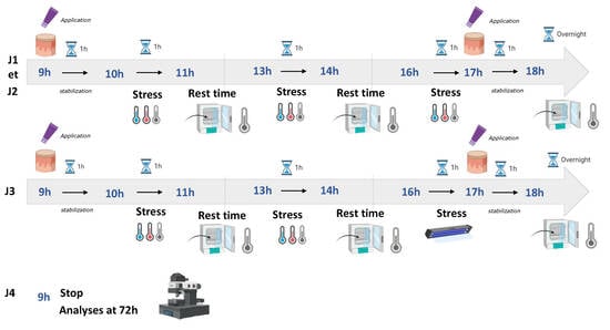
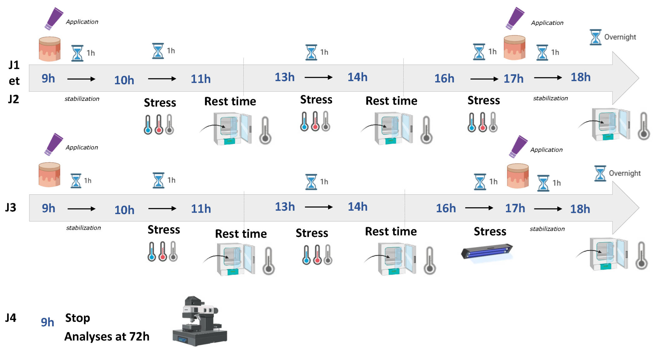
Skin explants were grown in control conditions and submitted to 1 h stress 4 times a day over 72 h, as described in Figures S1 and S2. Treated samples were incubated with 3% of Kombucha tea extract every day.
Tropical environment: temperature: 30–35 °C; relative humidity: 70–90%; exposure: humid heat.
Dry environment: temperature: 20–25 °C; relative humidity: 10–30%; exposure: dry atmosphere, potentially air-conditioned, low ambient humidity.
Control condition: temperature: 22–24 °C; relative humidity: 40–60%; environment: thermoregulated, controlled hygrometry.
The experiment was conducted over a total duration of 72 h. On days 1 and 2, three stress sessions were applied daily (10:00–11:00, 13:00–14:00, and 16:00–17:00), each followed by a recovery phase. Active compounds were applied from 09:00 to 10:00 and form 17:00 to 18:00. On day 3, the last stress session (16:00–17:00) was replaced by a UV exposure, followed by application of active compounds (17:00–18:00). The experiment was terminated the following day at 09:00. Temperature and humidity were continuously monitored throughout the experiment to ensure controlled environmental conditions. The dry condition was maintained within a range of approximately 22–25 °C and 25–35% relative humidity, while the tropical condition ranged from approximately 30–35 °C and 70–90% relative humidity. Control conditions were maintained within intermediate physiological ranges. Epidermal thickness was quantified using 18 independent measurement zones per condition.

2.4. Immunofluorescence Assays

Cryosections were thawed and rehydrated in 1X PBS for 10 min. Sections were then fixed in 4% formaldehyde (Sigma, Saint-Quentin-Fallavier, France) and diluted in 1X PBS for 20 min. Following three washes with 1X PBS, sections were blocked with 5% BSA (Sigma, Saint-Quentin-Fallavier, France) in 1X PBS for 2 h at room temperature (RT). Primary antibodies, diluted in 5% Bovine Serum Albumin (BSA), were applied overnight (O/N) at 4 °C. The primary antibodies used included mouse anti-MCSP (NG2) at a 1:100 dilution (Santa cruz_SCs-80003,Heidelberg, Germany) and mouse anti-collagen VII (Santa cruz_SCs-33710, (Heidelberg, Germany). After three washes with 1X PBS, sections were incubated with secondary antibodies for 2 h at RT.
BSA included anti-goat CF555 (1:800, Sigma_Sab4600072, Saint-Quentin-Fallavier, France), anti-mouse Alexa Fluor488 (1:800, Thermofisher_A28175, Strasbourg, France), anti-mouse IgG2a Alexa Fluor555 (1:200, Invitrogen A-21137, Strasbourg, France), and anti-mouse IgG1 Alexa Fluor488 (1:200, Invitrogen A-21121, Strasbourg, France). Nuclei were stained with DAPI (1 µg/mL, Sigma, Saint-Quentin-Fallavier, France) for 20 min at RT. Finally, sections were washed three times with 1X PBS and stored in 1X PBS at 4 °C. Mounting was performed using a 1X PBS-50% glycerol solution (Euromedex, Souffelweyersheim, France) as the mounting medium between the slide and coverslip, which were sealed with nail varnish and stored at 4 °C.

2.5. Image Acquisition and Analysis

Fluorescence imaging was conducted using a ZEISS LSM880 confocal microscope (CIQLE-Centre d’Imagerie Quantitative Lyon Est, Lyon 8, Lyon, France) equipped with a 40× 1.3 Oil Plan-Apochromat objective. Image analysis was performed using ImageJ, 1.54s software.
For each marker, 9 independent confocal measurement zones per condition were analyzed.

2.6. Atomic Force Microscopy (AFM) Measurements

AFM measurements were conducted using a Resolve Bioscope (Bruker Nano Surface, Santa Barbara, CA, USA) mounted on an inverted optical DMI8 microscope (Leica, Paris, France). A conical attached to a flexible cantilever with a spring constant of 0.35 N/m (DNP-10A, Bruker AFM probes, Bruker, MA, USA) was utilized for the measurements. Prior to each experiment, the deflection sensitivity of the cantilever was calibrated, and the spring constant was determined using the thermal tuning method. Data acquisition was performed with Nanoscope software (version 9.1.R.3) in AFM QNM (Quantitative Nanomechanical Mapping) mode. Force measurements were conducted in 1X PBS on human cryosections that had been fixed with 4% formaldehyde and immunolabeled for MCSP and collagen VII. Force volume acquisitions were carried out over a 25 × 25 μm area at three distinct locations: (1) at the level of MCSP-positive cells and putative ISCs above the DP; (2) at the level of MCSP-negative cells above the RR; and (3) in the reticular dermis. This 25 × 25 µm scan size allowed for the characterization of the mechanical properties of both basal epidermal cells and the ECM composing the DEJ and papillary dermis. Each force volume included 4096 measurement points, with each point corresponding t an indentation force curve from which the elastic modulus (Ea) was extracted. The quantification of the elastic modulus from the raw force curves was performed using the Sneddon mathematical model within BioMeca Analysis processing software. Finally, stiffness maps were reconstructed from the elastic modulus values, allowing for the extraction of the stiffness. All measurements were performed on standardized regions of interest (ROIs) defined on cryosections, with ROI dimensions determined according to the scale bars provided in the corresponding figures. ROIs were randomly selected across the cryosection areas to minimize sampling bias. For each condition, an average of 593 force/distance curves were acquired. These measurements were distributed across 9 independent measurement zones per punch biopsies to ensure spatial representativeness. Data were averaged per region prior to analysis to avoid overestimation of technical replicates.

2.7. Proteome Analysis

Primary human skin fibroblasts were seeded (100.000 cells) in Costar® 6-well clear TC-treated Multiple Well Plates, individually wrapped and sterile (Cat: 3516, Saint-Quentin-Fallavier, France), in the presence of DMEM, low glucose, and pyruvate (Gibco: 31885023, Strasbourg, France). After attachment, in the treated group, 1% and 3% Kombucha tea extract were added for 48h. Treated cells were then lysed in RIPA buffer (Sigma-aldrich, D4641, Saint-Quentin-Fallavier, France). The lysate was sonicated, and after centrifugation (300× g, 10 min), soluble proteins were isolated in the supernatant. Protein concentration was determined using the BCA method (A53225—Thermo fisher scientific, Strasbourg, France) using incubation for 15 min at 65 °C. Finally, nLC-MS/MS analysis/Label-Free Quantitative. Data Analysis was performed as described in (Figure S3).

2.8. Statistical Analysis

Data are presented as mean ± standard deviation (SD). Statistical significance was determined based on the normality of the data, assessed using the Shapiro–Wilk test. For normally distributed data, either the Student t-test or one-way or two-way analysis of variance (ANOVA) was employed using Prism software (version 10, GraphPad Software, San Diego, CA, USA). In cases where the data did not meet normality assumptions, the Mann–Whitney or Wilcoxon test was applied. Differences between means were deemed statistically significant at (* p ≤ 0.05, ** p ≤ 0.01, *** p ≤ 0.001, **** p ≤ 0.0001) and considered a trend at p ≤ 0.1, with # noted.

3. Results

3.1. Experimental Setup for Exposing Human Skin Explants to Challenging Environmental Conditions and Evaluating the Effects of Kombucha Tea Extract

We established a protocol to expose human skin explants to challenging environmental conditions for 72 h (see Materials and Methods and Figures S1 and S2). Both heat and humidity effects were investigated. Under control conditions, temperature was maintained between 20 and 25 °C and relative humidity around 50%. To mimic dry environmental conditions, the temperature was kept constant, while humidity was maintained below 30%. To produce tropical conditions, the temperature was raised above 32 °C, with humidity exceeding 70% (Figure S2). Skin explants were cultured under one of these three conditions and treated, or not treated, with Kombucha tea extract at several time points through the exposure period to sustain its biological effect (see Materials and Methods and Figure S1).
We hypothesized that Kombucha tea could exert a protective effect on human skin, particularly under stressful environmental conditions. Previous studies have reported several health-promoting properties of Kombucha, including anticancer activity, liver protection and immune stimulation [32,33,34,35,36,37,38].
To gain preliminary insight into its cellular mechanisms, we first assessed the effect of Kombucha tea extract on the proteome of human dermal fibroblasts. Cells were treated with 1% or 3% concentrations of the extract, and proteomic profiling revealed signatures consistent with enhanced proliferation (Figure S3). At 1%, Kombucha extract significantly altered the expression of 352 proteins (p < 0.05) and modulated 56 canonical signaling pathways (Z score > 1), as determined by Ingenuity Pathway Analysis. This low concentration promoted fibroblast proliferation, as evidenced by top-ranked pathways associated with mitosis and DNA synthesis (Z score > 4). The proliferative response may be mediated through activation of the Hedgehog and RUNX1-3 signaling pathways, both implicated in the maintenance of stem cell populations [39,40,41]. At the higher concentration (3%), Kombucha tea extract induced broader effects, altering the expression of 387 proteins and modulating 101 canonical pathways. Activation of the Hedgehog signaling pathway was again identified among the top-ranking changes, along with activation of ROBO receptor signaling, known to mediate stem/progenitor responses in non-skin systems, and potentially interacting with Hedgehog for regenerative processes [42,43,44]. Overall, these findings demonstrate that Kombucha tea extract stimulates fibroblast proliferation through modulation of key regenerative signaling pathways. Based on these results, we hypothesize that Kombucha tea could also benefit other skin cell types.
We therefore analyzed human skin explants to assess epidermal morphometry, MSCP expression, and the ridge-to-papilla stiffness ratio—a biomechanical indicator of DEJ flattening—under normal, dry and tropical conditions, with or without topical application of a Kombucha-containing formulation. It should be noted that the present findings are based on a single-donor ex vivo model and are therefore exploratory in nature, intended to provide a mechanistic basis for future studies involving larger and more diverse donor cohorts.

3.2. Epidermal Morphometry Is Altered Under Tropical Conditions and Preserved by Kombucha Tea Extract

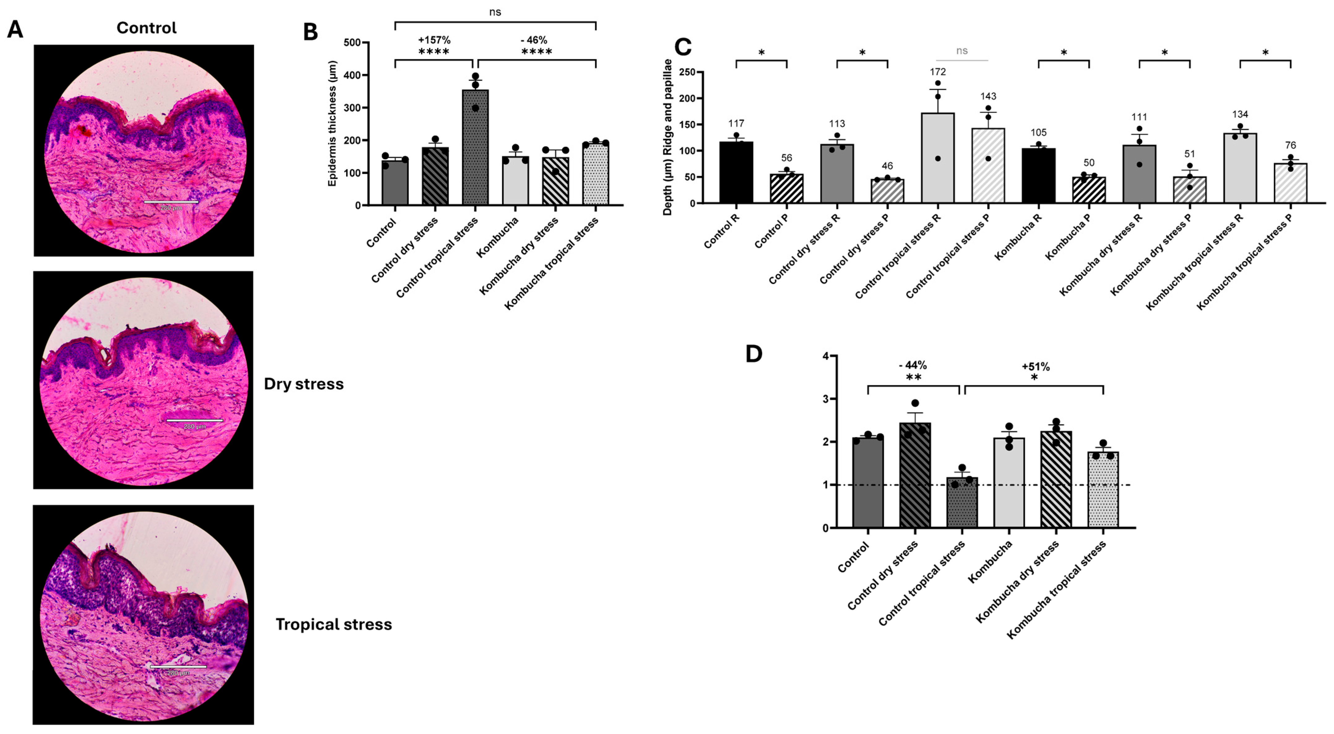
To evaluate the global impact of challenging environmental conditions on human skin homeostasis, we analyzed epidermal thickness on hematoxylin–eosin (HE)-stained cross sections (Figure 1A). This staining distinctly highlights epidermal stratification and the organization of dermal collagen fibers.
Under normal conditions, the epidermis displayed a typical multilayered organization (Figure 1B). Exposure to a tropical climate (high temperature and humidity) induced a marked increase in epidermal thickness (+157%), whereas dry conditions resulted in a more moderate, not significant thickening (+29%). When epidermal thickness was assessed in conjunction with nuclei count, no increase in cell number was observed, thereby ruling out true hyperproliferation as an explanatory mechanism. The increase in epidermal thickness is therefore more likely to reflect hydration-induced swelling of the stratum corneum rather than a genuine proliferative response. Topical application of Kombucha tea extract modulated some of these responses. Under both normal and dry conditions, Kombucha treatment did not significantly affect epidermal thickness. However, under tropical conditions, Kombucha tea markedly mitigated epidermal hyperplasia (**** p ≤ 0.0001), restoring epidermal thickness to levels comparable to those observed under normal conditions.
To assess potential flattening of the DEJ, a structural hallmark of stress and reduced self-renewal capacity, we quantified the papillary relief on HE sections by measuring the depths of epidermal ridges and dermal papillae and calculating the ridge-to-papilla ratio (Figure 1C,D). Under dry conditions, papillary architecture remained largely preserved, with ridge depth (~120 µm) exceeding papilla depth (~50 µm), reflecting a normal undulating DEJ and a ridge-to-papilla ratio greater than 2. In contrast, exposure to tropical conditions significantly reduced this depth difference (ridge and papilla both ~150 µm), resulting in a ratio close to 1—indicative of pronounced dermoepidermal flattening. Notably, treatment with Kombucha tea extract restored ridge-papilla asymmetry (average difference: 57 µm vs. 29 µm; ridge-to-papilla ratio ≈ 1.7 under tropical stress), suggesting that Kombucha preserves DEJ integrity and mitigates stress-induced architectural degradation.
Together, these findings demonstrate that tropical conditions profoundly disrupt epidermal architecture and dermoepidermal morphology, leading to thickening and loss of papillary relief loss—features reminiscent of early skin aging and stress-induced remodeling. Topical Kombucha tea extract effectively counteracted these effects, reinforcing dermoepidermal stability, preserving epidermal microrelief, and maintaining structural resilience under tropical stress.

3.3. AFM Analysis Reveals Environmental Stress–Induced Loss of Mechanical Contrast at the DEJ, Mitigated by Kombucha Tea Extract

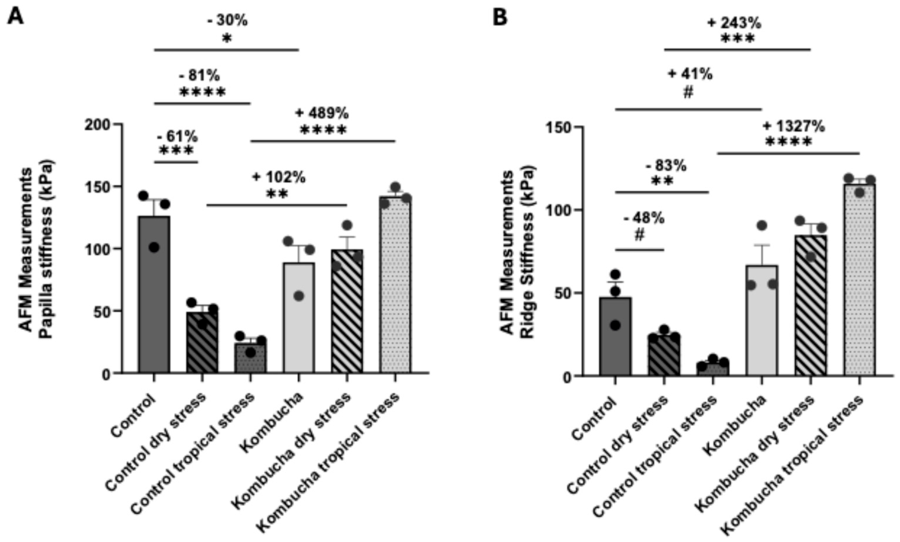
To further investigate the mechanical consequences of environmental stress on the DEJ, we analyzed the stiffness of both epidermal ridges and dermal papillae using atomic force microscopy (AFM). This approach enabled assessment of the mechanical balance across the DEJ under different environmental conditions. Under physiological conditions, dermal papillae typically display higher stiffness than the overlying epidermal ridges, generating a native mechanical contrast essential for DEJ integrity. Alteration of this balance—either through a decrease in papilla stiffness or an increase in ridge stiffness—results in a more homogeneous stiffness distribution between ridge and papilla regions, reflecting a reduction in mechanical contrast. Such homogenization is often associated with tissue remodeling, stress responses, or early signs of dermoepidermal destabilization, including junctional flattening. The intrinsic mechanical contrast between dermal papillae and epidermal ridges plays a crucial role in maintaining the basal stem cell pool, particularly in papillary areas where stiffness guides cell adhesion, fate, and renewal [45]. Excessive uniformity in stiffness, through apparently stabilizing, can thus compromise the spatial organization and regenerative potential of epidermal stem cell niches.
We observed that both dry and tropical stress induced a marked reduction in papilla stiffness (−61 and −81%, respectively; *** p ≤ 0.001 and **** p ≤ 0.0001; Figure 2A), while ridge stiffness decreased more moderately under dry stress (−48 and −83%, respectively; trend p ≤ 0.1 (#) and ** p ≤ 0.01, respectively; Figure 2B). This imbalance reflected a loss of mechanical contrasts and impaired undulation of the DEJ. Kombucha tea treatment increased both ridge and papilla stiffness, suggesting its ability to counteract these mechanical alterations under environmental stress. Kombucha application restored the mechanical contrast between ridges and papillae, indicating the recovery of DEJ topography and improved epidermal–dermal coupling.
Collectively, these findings indicate that Kombucha tea preserves mechanical contrast and dermoepidermal organization under environmental stress, thereby maintaining the structural and functional integrity of the epidermal regenerative compartment. This protective effect may help sustain efficient dermoepidermal exchanges and preserve epidermal renewal capacity under challenging climatic conditions.

3.4. Environmental Stress Alters MSCP and Collagen VII Expression, Normalized by Kombucha Tea Extract

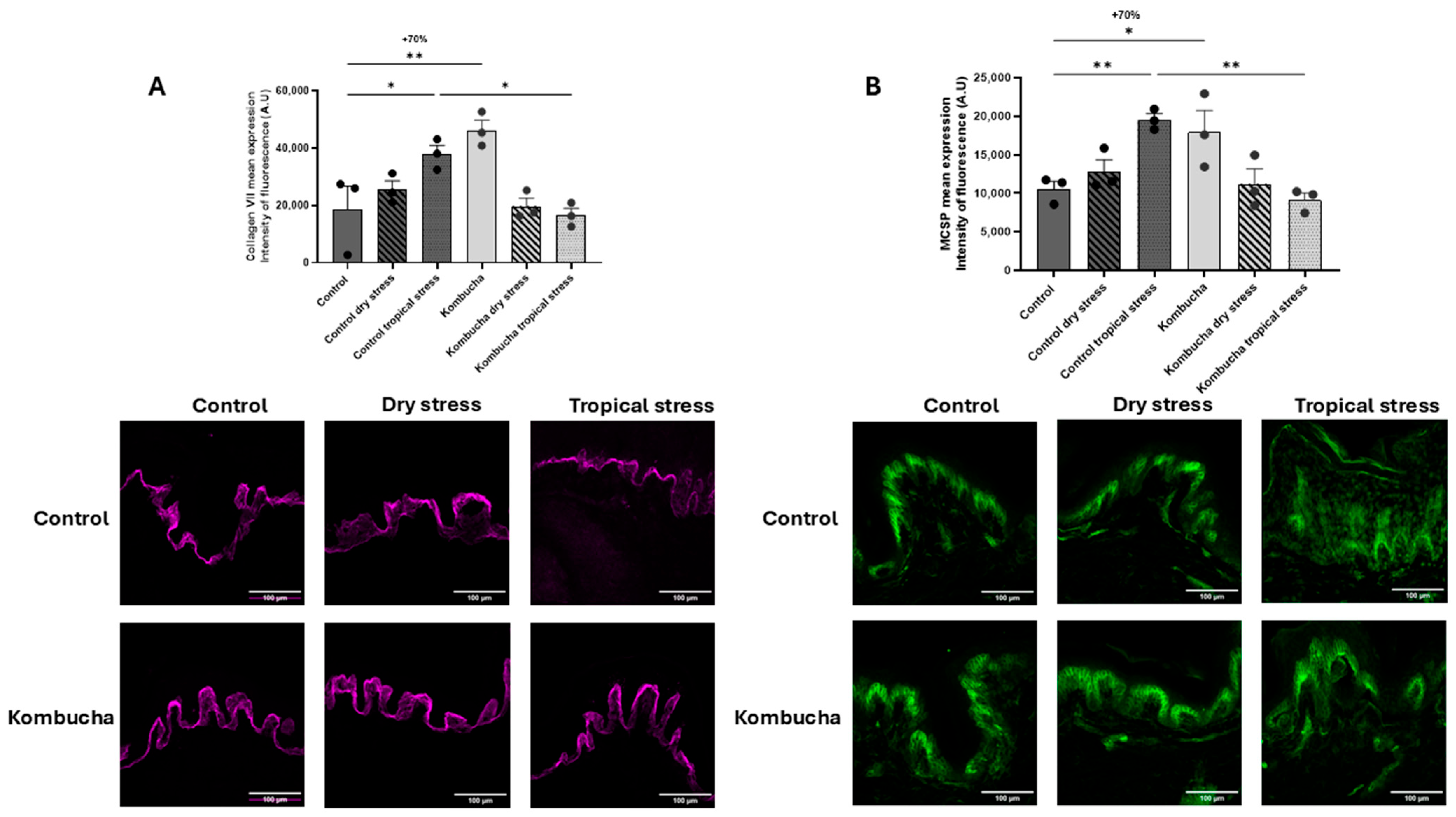
To confirm the impact of environmental variations at the molecular level in both epidermal and dermal compartments, we analyzed the expression of MSCP and collagen VII in human skin by fluorescence microscopy. MSCP proteoglycan is a key marker of epidermal stem cells. Indeed, highly proliferative basal keratinocytes expressing elevated levels of MSCP exhibit enhanced vitality and robust self-renewal capability, whereas a decrease in its expression reflects a loss of stemness potential [6,7,14,46]. Collagen VII, the main structural component of anchoring fibrils, plays a critical role in maintaining skin integrity by securing adhesion between the DEJ and the underlying connective tissue. This anchorage ensures the structural integrity of the skin and its resistance to external stress [47]. Together, MSCP and collagen VII represent essential molecular determinants of epidermal regeneration and dermoepidermal cohesion.
Exposure to a dry environment did not significantly modify MSCP or collagen VII expression (Figure 3A,B). In contrast, tropical stress (high heat and humidity) significantly increased MSCP and collagen VII expression, indicating stimulation of basal keratinocyte activity and extracellular matrix remodeling in response to environmental stress.
We then examined the effects of the Kombucha tea extract on the expression of these two markers. Under normal conditions, Kombucha application increased MSCP levels by approximately by 70% (** p ≤ 0.01) and significantly upregulated collagen VII (** p ≤ 0.01), consistent with a playing role in enhancing epidermal renewal and supporting DEJ architecture. Under dry conditions, where neither MSCP nor collagen VII were significantly affected, Kombucha treatment did not induce any notable changes. Conversely, in tropical conditions, Kombucha application significantly reduced MSCP (** p ≤ 0.01) and collagen VII (*** p ≤ 0.001) expression compared with untreated samples, restoring them to physiological levels similar to those observed under normal environmental conditions.
Collectively, these findings indicate that Kombucha tea helps maintain epidermal homeostasis under heat and humidity by preserving epidermal stem cell function and DEJ cohesion. Through its ability to normalize MSCP expression and support appropriate collagen VII levels, Kombucha may prevent stem cell exhaustion and protect the structural integrity of epidermal niche, thereby enhancing skin resilience in challenging climatic environments.

4. Discussion

Human skin continuously adapts to environmental fluctuations such as changes in temperature and humidity, which strongly influence epidermal morphology, hydration, and barrier function. In this study, we found that both tropical (high temperature and humidity) and dry environments induced measurable structural alterations, including increased epidermal thickness and modifications of the DEJ. These changes were accompanied by a reduction in mechanical contrast between epidermal ridges and dermal papillae, reflecting a disturbed homeostasis and early signs of extrinsic aging [45].
Several studies have shown that humidity influences the balance between keratinocyte proliferation and differentiation. Low humidity stimulates DNA synthesis and keratinocyte proliferation in murine skin and in vitro models [21]. In reconstructed human epidermis, exposure to dry air results in a thicker stratum corneum (SC) with altered differentiation markers and barrier-related gene expression, indicating an active adaptive response [19]. More recently, Izutsu-Matsumoto et al. [48] demonstrated that low humidity impairs terminal differentiation and alters tight junction protein organization in human skin [48]. In our study, keratinocyte proliferation remained unchanged, suggesting that the observed thickening results predominantly from hydration-driven mechanisms. Indeed, hydration-induced swelling of corneocytes and increased water content in the extracellular spaces can substantially increase SC thickness without requiring enhanced proliferation [18,49,50]. Such swelling-related effects could likely account for much of the epidermal enlargement seen under varying humidity conditions.
Environmental stressors, especially those amplified by global warming, such as heat waves, have profound effects on skin integrity. Heat stress induces oxidative stress, inflammation, and extracellular matrix remodeling [29]. Seasonal climate strongly influences the stratum corneum (SC) lipid composition and skin hydration. Rogers et al. reported reduced lipid content and poorer barrier function in winter compared to warmer months {Rogers, 1996 #47}. Conversely, high humidity and temperature increase sweat production and transepidermal water loss (TEWL) [51]. Pore density and TEWL also fluctuate seasonally [52], reinforcing that hydration status reflects dynamic environmental adaptation. Moderate heat shock (45–60 °C) stimulates procollagen I and III expression in dermal fibroblasts [53] and induces MMP-1 and MMP-3 via ERK/JNK activation and an autocrine IL-6 loop [25], illustrating how heat contributes to dermal skin aging. Consistent with this, we observed increased collagen VII expression under tropical conditions, likely reflecting an attempt to reinforce DEJ anchorage under mechanical load. Likewise, the elevated MSCP expression suggests a transient activation of basal keratinocytes turnover as a regenerative response. However, humidity and heat rarely act alone; they often co-occur with pollutants, allergens, or microbial challenges. As noted by Guarnieri et al., relative humidity may synergize with other stressors to exacerbate irritation and inflammation [54], underscoring the importance of maintaining controlled ambient conditions for skin health. Both MSCP modulation and the reduced stiffness of epidermal ridges and dermal papillae under environmental stress reflect a loss of mechanical heterogeneity, a key feature that maintains DEJ integrity and stabilizes epidermal stem cell niches. The mechanical contrast between the epidermis and papillary dermis ensures proper anchorage and governs stem cell behavior through mechanotransduction. Collagen VII, whose expression increased under tropical stress, may likely contribute to this compensatory anchorage reinforcement.
These findings align with recent mechanobiological studies showing that interfollicular epidermal stem cells and their underlying DEJ exhibit a specific mechanical signature—higher stiffness than neighboring basal keratinocytes—which is progressively lost during aging [4]. This niche stiffness depends on strong adhesion complexes of intergins and hemidesmosomes [6,12,55,56] and a specialized cytoskeletal organization. Aging reduces integrin and MSCP expression [14], leading to cytoskeletal remodeling that decreases cellular stiffness [57,58,59,60,61], including changes in keratin intermediate filaments [62,63,64]. Age-related softening of the dermal substrate further contributes, as increasing substrate stiffness is known to promote actin organization and cell stiffening [65,66,67,68,69].
Kombucha tea extract significantly mitigated the deleterious effects of environmental stress. It stabilized epidermal thickness within normal ranges, preserved ridge–papilla asymmetry, and maintained papillary stiffness, particularly under tropical conditions. These findings indicate improved DEJ microarchitecture and enhanced preservation of the stem cell niche. Kombucha’s bioactivity likely arises from its combination of polyphenols, organic acids, vitamins, and fermentation-derived metabolites, known for antioxidant, anti-inflammatory, and extracellular matrix-protective effects [32,33,34,35,36,37,38].
Our observation of Hedgehog signaling pathway modulation—implicated in keratinocyte proliferation and pigmentation [70]—suggests that Kombucha may engage in the regenerative signaling routes also used during stress adaptation. Few cosmetic ingredients demonstrate protective activity against thermal stress. Compounds such as epigallocatechin-3-gallate (EGCG) and Dunaliella salina extracts modulate MMP expression in heat-shocked skin cells [71,72], while apple mint leaf provides antioxidant protection and anti-inflammatory effects by suppressing ROS, MMPs, and IL-8, and downregulating MAPK pathways [73]. Our findings indicate that Kombucha tea is a promising natural active agent for mitigating climate-induced morphological and mechanical deterioration of the epidermis.
The present study has several limitations that should be acknowledged and carefully considered when interpreting the findings. First, skin explants were derived from a single donor, which represents the most significant constraint of this work. Although three independent punch biopsies were analyzed per condition and time point to ensure intra-individual reproducibility, the absence of inter-individual variability data prevents any generalization of the results. Second, the ex vivo nature of the model, while offering a valuable and ethically relevant alternative to in vivo experimentation, inherently lacks the systemic biological context of living tissue, including vascular supply, immune cell recruitment, and hormonal influences, all of which may modulate the skin’s response to environmental stress in vivo. Third, the short duration of stress exposure employed in this study may not fully recapitulate the chronic and cumulative effects of prolonged heat and humidity exposure encountered in real-life conditions. Fourth, the absence of functional outcome measures, such as transepidermal water loss (TEWL), skin hydration, or mechanical resilience assessments, limits the translational relevance of the molecular and histological findings reported here. Collectively, these limitations underscore the exploratory and hypothesis-generating nature of the present study, and future investigations incorporating multi-donor cohorts, longer exposure durations, in vivo validation, and functional endpoints will be essential to confirm and extend the findings reported here.

5. Conclusions

Overall, this work provides new insights into how temperature and humidity perturb epidermal structure, hydration, and mechanobiology, generating early aging-like features. Kombucha tea extract effectively counteracts these alterations by preserving DEJ integrity, maintaining mechanical contrast, and supporting stem cell niche function. These findings highlight the importance of targeting both molecular and mechanobiological pathways—particularly those linked to adhesion, cytoskeletal dynamics, and Hedgehog-related signaling—to bolster skin resilience under environmental stress.

Supplementary Materials

The following supporting information can be downloaded at https://www.mdpi.com/article/10.3390/cosmetics13030114/s1: Figure S1. Experimental protocol for the biomechanical characterization of epidermal basal stem cells. Figure S2. Experimental protocol for skin explant exposure under simulated tropical and arid environmental conditions. Figure S3. Proteome remodeling induced by Kombucha tea extract in primary human dermal fibroblasts.

Author Contributions

Conceptualization, A.-L.B., J.C., K.L. and T.P.-J.; methodology, J.C. and G.R. formal analysis, R.R., J.C., T.P.-J. and G.R.; investigation, M.B. and N.D.A.; writing—original draft preparation, A.-L.B. writing—review and editing, J.C., A.-L.B., R.R. and C.N.; supervision, A.-L.B.; project administration, C.N. and K.P.; funding acquisition, K.P. All authors have read and agreed to the published version of the manuscript.

Funding

This research received no external funding.

Institutional Review Board Statement

This study was performed on biopsy obtained from surgical residues after written informed consent from the donor in full respect of the Declaration of Helsinki and the article L.1243-4 of the French Public Health Code. The latter does not require any prior authorization by an ethics committee for sampling and using surgical residues.

Informed Consent Statement

Not applicable.

Data Availability Statement

The original contributions presented in this study are included in the article/Supplementary Material. Further inquiries can be directed to the corresponding author(s).

Acknowledgments

The authors thank Ingrid Masse for her contribution to the drafting and editing of the manuscript.

Conflicts of Interest

Kristell Lazou, Carine Nizard, Anne-Laure Bulteau, Tiphaine Pele-Joly, and Karl Pays are employed by LVMH recherche. Julien Chlasta and Gaël Runel are employed by Biomeca. The authors declare that the research was conducted in the absence of any commercial or financial relationships that could be construed as a potential conflict of interest. The funders had no role in the design of the study; in the collection, analyses or interpretation of data; in the writing of the manuscript; or in the decision to publish the results.

References

  1. Blanpain, C.; Fuchs, E. Epidermal homeostasis: A balancing act of stem cells in the skin. Nat. Rev. Mol. Cell Biol. 2009, 10, 207–217. [Google Scholar] [CrossRef] [PubMed]
  2. Watt, F.M. Epidermal stem cells: Markers, patterning and the control of stem cell fate. Philos. Trans. R. Soc. B Biol. Sci. 1998, 353, 831–837. [Google Scholar] [CrossRef]
  3. Lawlor, K.T.; Kaur, P. Dermal Contributions to Human Interfollicular Epidermal Architecture and Self-Renewal. Int. J. Mol. Sci. 2015, 16, 28098–28107. [Google Scholar] [CrossRef] [PubMed]
  4. Roig-Rosello, E.; Rousselle, P. The Human Epidermal Basement Membrane: A Shaped and Cell Instructive Platform That Aging Slowly Alters. Biomolecules 2020, 10, 1607. [Google Scholar] [CrossRef]
  5. Jensen, K.B.; Watt, F.M. Single-cell expression profiling of human epidermal stem and transit-amplifying cells: Lrig1 is a regulator of stem cell quiescence. Proc. Natl. Acad. Sci. USA 2006, 103, 11958–11963. [Google Scholar] [CrossRef]
  6. Jones, P.H.; Harper, S.; Watt, F.M. Stem cell patterning and fate in human epidermis. Cell 1995, 80, 83–93. [Google Scholar] [CrossRef]
  7. Legg, J.; Jensen, U.B.; Broad, S.; Leigh, I.; Watt, F.M. Role of melanoma chondroitin sulphate proteoglycan in patterning stem cells in human interfollicular epidermis. Development 2003, 130, 6049–6063. [Google Scholar] [CrossRef]
  8. Inoue, K.; Aoi, N.; Sato, T.; Yamauchi, Y.; Suga, H.; Eto, H.; Kato, H.; Araki, J.; Yoshimura, K. Differential expression of stem-cell-associated markers in human hair follicle epithelial cells. Lab. Investig. 2009, 89, 844–856. [Google Scholar] [CrossRef]
  9. Webb, A.; Li, A.; Kaur, P. Location and phenotype of human adult keratinocyte stem cells of the skin. Differentiation 2004, 72, 387–395. [Google Scholar] [CrossRef]
  10. Wang, S.; Drummond, M.L.; Guerrero-Juarez, C.F.; Tarapore, E.; MacLean, A.L.; Stabell, A.R.; Wu, S.C.; Gutierrez, G.; That, B.T.; Benavente, C.A.; et al. Single cell transcriptomics of human epidermis identifies basal stem cell transition states. Nat. Commun. 2020, 11, 4239. [Google Scholar] [CrossRef] [PubMed]
  11. Ghali, L.; Wong, S.-T.; Tidman, N.; Quinn, A.; Philpott, M.P.; Leigh, I.M. Epidermal and hair follicle progenitor cells express melanoma-associated chondroitin sulfate proteoglycan core protein. J. Investig. Dermatol. 2004, 122, 433–442. [Google Scholar] [CrossRef]
  12. Jones, P.H.; Watt, F.M. Separation of human epidermal stem cells from transit amplifying cells on the basis of differences in integrin function and expression. Cell 1993, 73, 713–724. [Google Scholar] [CrossRef] [PubMed]
  13. Baqar, A. Dermal papillae; the histomorphological changes in the height of dermal papillae of human skin, in different age groups. Prof. Med. J. 2019, 26, 208–212. [Google Scholar] [CrossRef]
  14. Giangreco, A.; Goldie, S.J.; Failla, V.; Saintigny, G.; Watt, F.M. Human skin aging is associated with reduced expression of the stem cell markers beta1 integrin and MCSP. J. Investig. Dermatol. 2010, 130, 604–608. [Google Scholar] [CrossRef]
  15. Zobiri, O.; Deshayes, N.; Rathman-Josserand, M. Evolution of the clonogenic potential of human epidermal stem/progenitor cells with age. Stem Cells Cloning 2012, 5, 1–4. [Google Scholar] [CrossRef][Green Version]
  16. Jang, S.I.; Han, J.; Lee, M.; Seo, J.; Kim, B.J.; Kim, E. A study of skin characteristics according to humidity during sleep. Ski. Res. Technol. 2019, 25, 456–460. [Google Scholar] [CrossRef] [PubMed]
  17. Tsukahara, K.; Hotta, M.; Fujimura, T.; Haketa, K.; Kitahara, T. Effect of room humidity on the formation of fine wrinkles in the facial skin of Japanese. Ski. Res. Technol. 2007, 13, 184–188. [Google Scholar] [CrossRef] [PubMed]
  18. Bouwstra, J.A.; de Graaff, A.; Gooris, G.S.; Nijsse, J.; Wiechers, J.W.; van Aelst, A.C. Water distribution and related morphology in human stratum corneum at different hydration levels. J. Investig. Dermatol. 2003, 120, 750–758. [Google Scholar] [CrossRef]
  19. Cau, L.; Pendaries, V.; Lhuillier, E.; Thompson, P.R.; Serre, G.; Takahara, H.; Méchin, M.-C.; Simon, M. Lowering relative humidity level increases epidermal protein deimination and drives human filaggrin breakdown. J. Dermatol. Sci. 2017, 86, 106–113. [Google Scholar] [CrossRef]
  20. Denda, M.; Sato, J.; Masuda, Y.; Tsuchiya, T.; Koyama, J.; Kuramoto, M.; Elias, P.M.; Feingold, K.R. Exposure to a dry environment enhances epidermal permeability barrier function. J. Investig. Dermatol. 1998, 111, 858–863. [Google Scholar] [CrossRef]
  21. Denda, M.; Sato, J.; Tsuchiya, T.; Elias, P.M.; Feingold, K.R. Low humidity stimulates epidermal DNA synthesis and amplifies the hyperproliferative response to barrier disruption: Implication for seasonal exacerbations of inflammatory dermatoses. J. Investig. Dermatol. 1998, 111, 873–878. [Google Scholar] [CrossRef]
  22. Shinohara, K.; Hara-Chikuma, M. Low humidity altered the gene expression profile of keratinocytes in a three-dimensional skin model. Mol. Biol. Rep. 2022, 49, 7465–7474. [Google Scholar] [CrossRef]
  23. Seo, J.Y.; Chung, J.H. Thermal aging: A new concept of skin aging. J. Dermatol. Sci. Suppl. 2006, 2, S13–S22. [Google Scholar] [CrossRef]
  24. Chen, Z.; Seo, J.Y.; Kim, Y.K.; Lee, S.R.; Kim, K.H.; Cho, K.H.; Eun, H.C.; Chung, J.H. Heat modulation of tropoelastin, fibrillin-1, and matrix metalloproteinase-12 in human skin in vivo. J. Investig. Dermatol. 2005, 124, 70–78. [Google Scholar] [CrossRef]
  25. Park, C.-H.; Lee, M.J.; Ahn, J.; Kim, S.; Kim, H.H.; Kim, K.H.; Eun, H.C.; Chung, J.H. Heat shock-induced matrix metalloproteinase (MMP)-1 and MMP-3 are mediated through ERK and JNK activation and via an autocrine interleukin-6 loop. J. Investig. Dermatol. 2004, 123, 1012–1019. [Google Scholar] [CrossRef]
  26. Shin, M.H.; Seo, J.-E.; Kim, Y.K.; Kim, K.H.; Chung, J.H. Chronic heat treatment causes skin wrinkle formation and oxidative damage in hairless mice. Mech. Ageing Dev. 2012, 133, 92–98. [Google Scholar] [CrossRef] [PubMed]
  27. Park, G.; Oh, M.S. Acceleration of heat shock-induced collagen breakdown in human dermal fibroblasts with knockdown of NF-E2-related factor 2. BMB Rep. 2015, 48, 467–472. [Google Scholar] [CrossRef]
  28. Shin, M.H.; Moon, Y.J.; Seo, J.-E.; Lee, Y.; Kim, K.H.; Chung, J.H. Reactive oxygen species produced by NADPH oxidase, xanthine oxidase, and mitochondrial electron transport system mediate heat shock-induced MMP-1 and MMP-9 expression. Free Radic. Biol. Med. 2008, 44, 635–645. [Google Scholar] [CrossRef]
  29. Cho, S.; Shin, M.H.; Kim, Y.K.; Seo, J.-E.; Lee, Y.M.; Park, C.-H.; Chung, J.H. Effects of infrared radiation and heat on human skin aging in vivo. J. Investig. Dermatol. Symp. Proc. 2009, 14, 15–19. [Google Scholar] [CrossRef]
  30. Shimoni, C.; Goldstein, M.; Ribarski-Chorev, I.; Schauten, I.; Nir, D.; Strauss, C.; Schlesinger, S. Heat Shock Alters Mesenchymal Stem Cell Identity and Induces Premature Senescence. Front. Cell Dev. Biol. 2020, 8, 565970. [Google Scholar] [CrossRef] [PubMed]
  31. Chen, C.; Liu, B.Y. Changes in major components of tea fungus metabolites during prolonged fermentation. J. Appl. Microbiol. 2000, 89, 834–839. [Google Scholar] [CrossRef]
  32. Battikh, H.; Chaieb, K.; Bakhrouf, A.; Ammar, E. Antibacterial and Antifungal Activities of Black and Green Kombucha Teas. J. Food Biochem. 2013, 37, 231–236. [Google Scholar] [CrossRef]
  33. Conney, A.H.; Lu, Y.-P.; Lou, Y.-R.; Huang, M.-T. Inhibitory effects of tea and caffeine on UV-induced carcinogenesis: Relationship to enhanced apoptosis and decreased tissue fat. Eur. J. Cancer Prev. 2002, 11, S28–S36. [Google Scholar] [PubMed]
  34. Ioannides, C.; Yoxall, V. Antimutagenic activity of tea: Role of polyphenols. Curr. Opin. Clin. Nutr. Metab. Care 2003, 6, 649–656. [Google Scholar] [CrossRef] [PubMed]
  35. Park, A.M.; Dong, Z. Signal transduction pathways: Targets for green and black tea polyphenols. J. Biochem. Mol. Biol. 2003, 36, 66–77. [Google Scholar]
  36. Villarreal-Soto, S.A.; Beaufort, S.; Bouajila, J.; Souchard, J.-P.; Renard, T.; Rollan, S.; Taillandier, P. Impact of fermentation conditions on the production of bioactive compounds with anticancer, anti-inflammatory and antioxidant properties in kombucha tea extracts. Process Biochem. 2019, 83, 44–54. [Google Scholar] [CrossRef]
  37. Villarreal-Soto, S.A.; Beaufort, S.; Bouajila, J.; Souchard, J.-P.; Taillandier, P. Understanding Kombucha Tea Fermentation: A Review. J. Food Sci. 2018, 83, 580–588. [Google Scholar] [CrossRef]
  38. Wang, Y.; Ji, B.; Wu, W.; Wang, R.; Yang, Z.; Zhang, D.; Tian, W. Hepatoprotective effects of kombucha tea: Identification of functional strains and quantification of functional components. J. Sci. Food Agric. 2014, 94, 265–272. [Google Scholar] [CrossRef]
  39. Abe, Y.; Tanaka, N. Roles of the Hedgehog Signaling Pathway in Epidermal and Hair Follicle Development, Homeostasis, and Cancer. J. Dev. Biol. 2017, 5, 12. [Google Scholar] [CrossRef]
  40. Chuang, L.S.H.; Matsuo, J.; Douchi, D.; Bte Mawan, N.A.; Ito, Y. RUNX3 in Stem Cell and Cancer Biology. Cells 2023, 12, 408. [Google Scholar] [CrossRef]
  41. Rozen, E.J.; Ozeroff, C.D.; Allen, M.A. RUN(X) out of blood: Emerging RUNX1 functions beyond hematopoiesis and links to Down syndrome. Hum. Genom. 2023, 17, 83. [Google Scholar] [CrossRef] [PubMed]
  42. Borrell, V.; Cárdenas, A.; Ciceri, G.; Galcerán, J.; Flames, N.; Pla, R.; Nóbrega-Pereira, S.; García-Frigola, C.; Peregrín, S.; Zhao, Z.; et al. Slit/Robo signaling modulates the proliferation of central nervous system progenitors. Neuron 2012, 76, 338–352. [Google Scholar] [CrossRef]
  43. Harburg, G.; Compton, J.; Liu, W.; Iwai, N.; Zada, S.; Marlow, R.; Strickland, P.; Zeng, Y.A.; Hinck, L. SLIT/ROBO2 signaling promotes mammary stem cell senescence by inhibiting Wnt signaling. Stem Cell Rep. 2014, 3, 385–393. [Google Scholar] [CrossRef] [PubMed]
  44. Park, J.-S.; Cho, R.; Kang, E.-Y.; Oh, Y.-M. Effect of Slit/Robo signaling on regeneration in lung emphysema. Exp. Mol. Med. 2021, 53, 986–992. [Google Scholar] [CrossRef] [PubMed]
  45. Miny, S.; Runel, G.; Chlasta, J.; Lapart, J.-A.; Bonod, C. Putative interfollicular stem cells of skin epidermis possess a specific mechanical signature that evolves during aging. bioRxiv 2025. [Google Scholar] [CrossRef]
  46. Ji, Y.; Kumar, R.; Gokhale, A.; Chao, H.-P.; Rycaj, K.; Chen, X.; Li, Q.; Tang, D.G. LRIG1, a regulator of stem cell quiescence and a pleiotropic feedback tumor suppressor. Semin. Cancer Biol. 2022, 82, 120–133. [Google Scholar] [CrossRef]
  47. Breitkreutz, D.; Koxholt, I.; Thiemann, K.; Nischt, R. Skin basement membrane: The foundation of epidermal integrity—BM functions and diverse roles of bridging molecules nidogen and perlecan. Biomed. Res. Int. 2013, 2013, 179784. [Google Scholar] [CrossRef]
  48. Izutsu-Matsumoto, Y.; Okano, Y.; Masaki, H.; Tokudome, Y.; Iwabuchi, T. Effect of low humidity on the barrier functions of keratinocytes in a reconstructed human epidermal model. Int. J. Cosmet. Sci. 2025, 47, 523–534. [Google Scholar] [CrossRef]
  49. Rawlings, A.V.; Matts, P.J. Stratum corneum moisturization at the molecular level: An update in relation to the dry skin cycle. J. Investig. Dermatol. 2005, 124, 1099–1110. [Google Scholar] [CrossRef]
  50. Warner, R.R.; Stone, K.J.; Boissy, Y.L. Hydration disrupts human stratum corneum ultrastructure. J. Investig. Dermatol. 2003, 120, 275–284. [Google Scholar] [CrossRef]
  51. Kim, S.; Park, J.W.; Yeon, Y.; Han, J.Y.; Kim, E. Influence of exposure to summer environments on skin properties. J. Eur. Acad. Dermatol. Venereol. 2019, 33, 2192–2196. [Google Scholar] [CrossRef]
  52. Song, E.J.; Lee, J.A.; Park, J.J.; Kim, H.J.; Kim, N.S.; Byun, K.S.; Choi, G.S.; Moon, T.K. A study on seasonal variation of skin parameters in Korean males. Int. J. Cosmet. Sci. 2015, 37, 92–97. [Google Scholar] [CrossRef] [PubMed]
  53. Dams, S.D.; de Liefde-van Beest, M.; Nuijs, A.M.; Oomens, C.W.J.; Baaijens, F.P.T. Heat shocks enhance procollagen type I and III expression in fibroblasts in ex vivo human skin. Skin Res. Technol. 2011, 17, 167–180. [Google Scholar] [CrossRef]
  54. Guarnieri, G.; Olivieri, B.; Senna, G.; Vianello, A. Relative Humidity and Its Impact on the Immune System and Infections. Int. J. Mol. Sci. 2023, 24, 9456. [Google Scholar] [CrossRef]
  55. Sonnenberg, A.; Calafat, J.; Janssen, H.; Daams, H.; van der Raaij-Helmer, L.M.; Falcioni, R.; Kennel, S.J.; Aplin, J.D.; Baker, J.; Loizidou, M. Integrin alpha 6/beta 4 complex is located in hemidesmosomes, suggesting a major role in epidermal cell-basement membrane adhesion. J. Cell Biol. 1991, 113, 907–917. [Google Scholar] [CrossRef]
  56. Yu, X.; Miyamoto, S.; Mekada, E. Integrin alpha 2 beta 1-dependent EGF receptor activation at cell-cell contact sites. J. Cell Sci. 2000, 113, 2139–2147. [Google Scholar] [CrossRef]
  57. Broussard, J.A.; Yang, R.; Huang, C.; Nathamgari, S.S.P.; Beese, A.M.; Godsel, L.M.; Hegazy, M.H.; Lee, S.; Zhou, F.; Sniadecki, N.J.; et al. The desmoplakin-intermediate filament linkage regulates cell mechanics. Mol. Biol. Cell 2017, 28, 3156–3164. [Google Scholar] [CrossRef] [PubMed]
  58. Efremov, Y.M.; Dokrunova, A.A.; Efremenko, A.V.; Kirpichnikov, M.P.; Shaitan, K.V.; Sokolova, O.S. Distinct impact of targeted actin cytoskeleton reorganization on mechanical properties of normal and malignant cells. Biochim. Biophys. Acta 2015, 1853, 3117–3125. [Google Scholar] [CrossRef]
  59. Kasza, K.E.; Rowat, A.C.; Liu, J.; Angelini, T.E.; Brangwynne, C.P.; Koenderink, G.H.; Weitz, D.A. The cell as a material. Curr. Opin. Cell Biol. 2007, 19, 101–107. [Google Scholar] [CrossRef]
  60. Wang, Y.; Xu, C.; Jiang, N.; Zheng, L.; Zeng, J.; Qiu, C.; Yang, H.; Xie, S. Quantitative analysis of the cell-surface roughness and viscoelasticity for breast cancer cells discrimination using atomic force microscopy. Scanning 2016, 38, 558–563. [Google Scholar] [CrossRef] [PubMed]
  61. Xu, W.; Mezencev, R.; Kim, B.; Wang, L.; McDonald, J.; Sulchek, T. Cell stiffness is a biomarker of the metastatic potential of ovarian cancer cells. PLoS ONE 2012, 7, e46609. [Google Scholar] [CrossRef]
  62. Homberg, M.; Ramms, L.; Schwarz, N.; Dreissen, G.; Leube, R.E.; Merkel, R.; Hoffmann, B.; Magin, T.M. Distinct Impact of Two Keratin Mutations Causing Epidermolysis Bullosa Simplex on Keratinocyte Adhesion and Stiffness. J. Investig. Dermatol. 2015, 135, 2437–2445. [Google Scholar] [CrossRef] [PubMed]
  63. Ramms, L.; Fabris, G.; Windoffer, R.; Schwarz, N.; Springer, R.; Zhou, C.; Lazar, J.; Stiefel, S.; Hersch, N.; Schnakenberg, U.; et al. Keratins as the main component for the mechanical integrity of keratinocytes. Proc. Natl. Acad. Sci. USA 2013, 110, 18513–18518. [Google Scholar] [CrossRef]
  64. Seltmann, K.; Fritsch, A.W.; Käs, J.A.; Magin, T.M. Keratins significantly contribute to cell stiffness and impact invasive behavior. Proc. Natl. Acad. Sci. USA 2013, 110, 18507–18512. [Google Scholar] [CrossRef]
  65. Byfield, F.J.; Reen, R.K.; Shentu, T.-P.; Levitan, I.; Gooch, K.J. Endothelial actin and cell stiffness is modulated by substrate stiffness in 2D and 3D. J. Biomech. 2009, 42, 1114–1119. [Google Scholar] [CrossRef] [PubMed]
  66. Discher, D.E.; Mooney, D.J.; Zandstra, P.W. Growth factors, matrices, and forces combine and control stem cells. Science 2009, 324, 1673–1677. [Google Scholar] [CrossRef]
  67. Emig, R.; Knodt, W.; Krussig, M.J.; Zgierski-Johnston, C.M.; Gorka, O.; Groß, O.; Kohl, P.; Ravens, U.; Peyronnet, R. Piezo1 Channels Contribute to the Regulation of Human Atrial Fibroblast Mechanical Properties and Matrix Stiffness Sensing. Cells 2021, 10, 663. [Google Scholar] [CrossRef]
  68. Engler, A.J.; Sen, S.; Sweeney, H.L.; Discher, D.E. Matrix elasticity directs stem cell lineage specification. Cell 2006, 126, 677–689. [Google Scholar] [CrossRef]
  69. Solon, J.; Levental, I.; Sengupta, K.; Georges, P.C.; Janmey, P.A. Fibroblast adaptation and stiffness matching to soft elastic substrates. Biophys. J. 2007, 93, 4453–4461. [Google Scholar] [CrossRef]
  70. Zhang, L.; Zeng, H.; Jiang, L.; Fu, C.; Zhang, Y.; Hu, Y.; Zhang, X.; Zhu, L.; Zhang, F.; Huang, J.; et al. Heat promotes melanogenesis by increasing the paracrine effects in keratinocytes via the TRPV3/Ca2+/Hh signaling pathway. iScience 2023, 26, 106749. [Google Scholar] [CrossRef] [PubMed]
  71. Joo, J.-H.; Seok, J.H.; Hong, I.-k.; Kim, N.K.; Choi, E. Inhibitory Effects of Dunaliella salina Extracts on Thermally-Induced Skin Aging. J. Soc. Cosmet. Sci. Korea 2016, 42, 57–64. [Google Scholar] [CrossRef]
  72. Kim, H.-S.; Montana, V.; Jang, H.-J.; Parpura, V.; Kim, J.-a. Epigallocatechin gallate (EGCG) stimulates autophagy in vascular endothelial cells: A potential role for reducing lipid accumulation. J. Biol. Chem. 2013, 288, 22693–22705. [Google Scholar] [CrossRef] [PubMed]
  73. Son, D.; Kim, M.; Woo, H.; Park, D.; Jung, E. Anti-Thermal Skin Aging Activity of Aqueous Extracts Derived from Apple Mint (Mentha suaveolens Ehrh.) in Human Dermal Fibroblasts. Evid. Based Complement. Altern. Med. 2018, 2018, 4595982. [Google Scholar] [CrossRef]
Figure 1. Skin morphology assessment following environmental stress exposure. (A) Skin morphology assessment by Hematoxylin & Eosin (H & E) staining following environmental stress exposure. (B) Quantitative assessment of skin thickness after stress and treatment with 3% Kombucha tea extract. (C) Environmental stress-induced structural remodeling of the dermal–epidermal junction (DEJ). Kombucha treatment (3%) maintains a significant height difference between papillae and ridges under both control and dry conditions, indicating a good preservation of the dermoepidermal junction relief in these environments. (D) Ratio between ridge and papillae depth (* p ≤ 0.05, ** p ≤ 0.01, **** p ≤ 0.0001).
Cosmetics 13 00114 g001
Figure 2. Atomic Force Microscopy (AFM) analysis of skin mechanical properties. (A) AFM measurement at the papilla level. (B) AFM measurement at the ridge level. Effect of the Kombucha tea extract at 3%. (* p ≤ 0.05, ** p ≤ 0.01, *** p ≤ 0.001, **** p ≤ 0.0001, and significance is limited at p ≤ 0.1 (#).).
Cosmetics 13 00114 g002
Figure 3. Effect of Kombucha tea extract on the expression of molecular markers involved in basal stem cell anchorage at the dermal–epidermal junction (DEJ). (A) Representative images and quantification of collagen 7 immunofluorescent staining. (B) Representative images and quantification of MCSP. Effect of the Kombucha tea extract at 3%. (* p ≤ 0.05, ** p ≤ 0.01).
Cosmetics 13 00114 g003
Disclaimer/Publisher’s Note: The statements, opinions and data contained in all publications are solely those of the individual author(s) and contributor(s) and not of MDPI and/or the editor(s). MDPI and/or the editor(s) disclaim responsibility for any injury to people or property resulting from any ideas, methods, instructions or products referred to in the content.