Article

Harnessing miRNA Milk-Derived Exosomes for Hair Loss Disorders: In Vitro Modulation of WNT Signaling and Dermal Papilla Proliferation


by
Daniela Pinto
1,*,
Giorgia Mondadori
1,
Monica Cozzi
1,
Piero Tesauro
2,
Martin Hintersteiner
3,
Raúl López Domínguez
4,
Esperanza de Santiago Rodríguez
4,
Giammaria Giuliani
1 and
Fabio Rinaldi
1
1
Giuliani SpA, 20129 Milan, Italy
2
Studio Tesauro, 20124 Milan, Italy
3
Evobiotix, CH 6815 Melide, Switzerland
4
GENYO, Centre for Genomics and Oncological Research, University of Granada, Andalusian Regional Government, Genomics Unit, PTS Granada, Avenida de la Ilustración 114, 18016 Granada, Spain
*
Author to whom correspondence should be addressed.
Cosmetics 2026, 13(1), 38; https://doi.org/10.3390/cosmetics13010038
Submission received: 23 December 2025 / Revised: 26 January 2026 / Accepted: 29 January 2026 / Published: 10 February 2026

Abstract

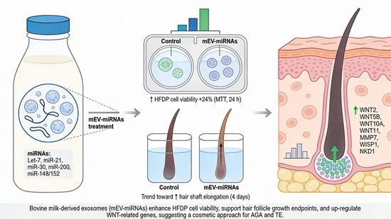
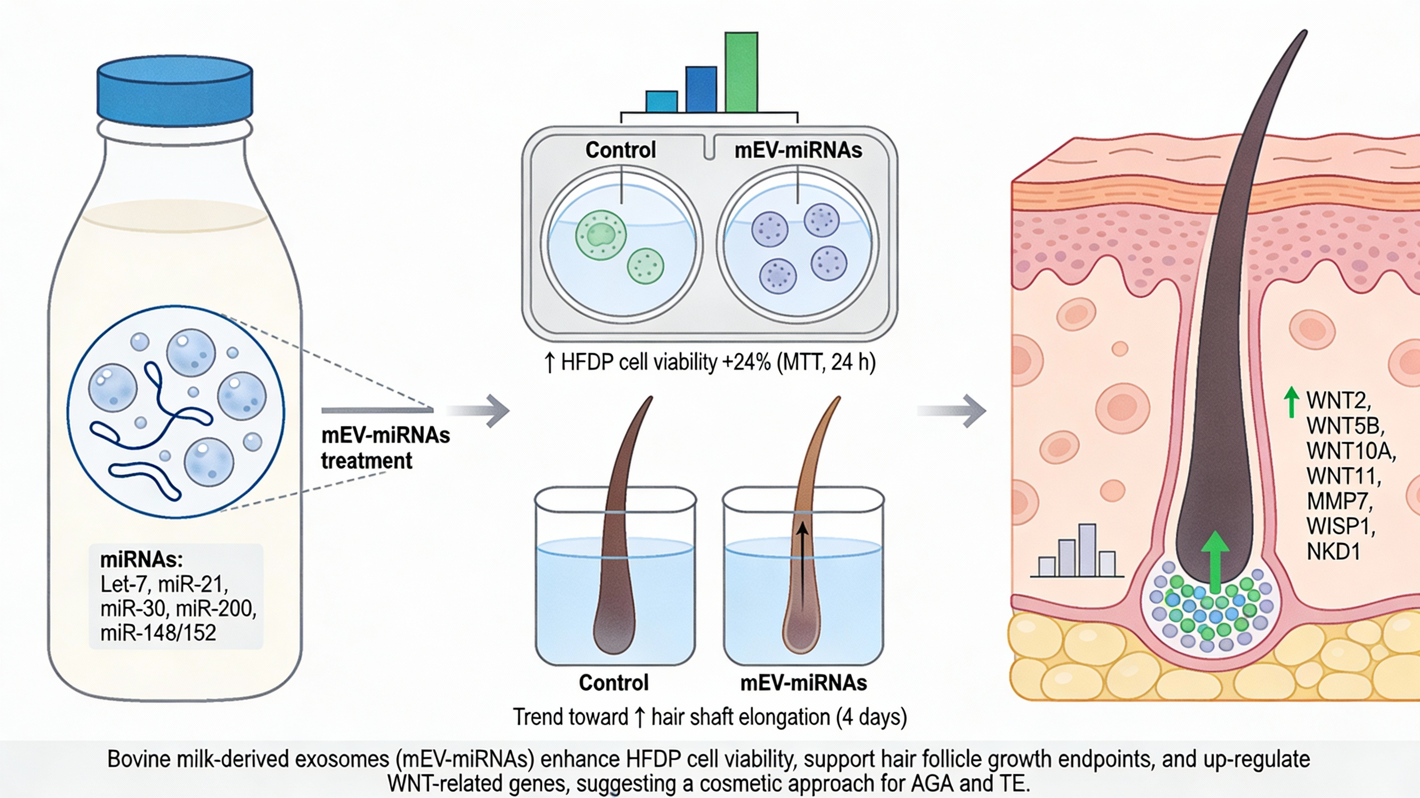
Androgenetic alopecia (AGA) and telogen effluvium (TE) are common hair loss disorders characterized by dysregulated hair follicle cycling and impaired dermal papilla cell function. Emerging evidence indicates that exosomes are key mediators of intercellular communication, largely through their microRNA (miRNA) cargo. Milk-derived exosomes (Mi-Exos) represent an accessible and biologically active source of regulatory miRNAs with potential relevance for hair disorders. This study evaluated the in vitro effects of bovine milk-derived exosomes (MEV-miRNAs) on human hair follicles. MEV-miRNAs were enriched in miRNA families (Let-7, miR-21, miR-30, miR-200, and miR-148/152) previously implicated in hair follicle regulation. Viability/metabolic activity of hair follicle dermal papilla (HFDP) cells was assessed, and human hair follicles were cultured ex vivo to measure shaft elongation and modulation of the WNT signaling pathway by qRT-PCR. MEV-miRNAs significantly increased HFDP cell viability after 24 h compared with controls. Human hair follicles showed a non-significant trend toward increased elongation following treatment. Gene expression analysis revealed significant up-regulation of key WNT pathway components, including WNT2, WNT5B, WNT10A, WNT11, MMP7, WISP1, and NKD1, indicating modulation of WNT-associated pathways implicated in hair follicle growth and cycling. Overall, MEV-miRNAs exhibit positive modulatory effects on signaling pathways, supporting their potential as a novel therapeutic strategy for AGA and TE.

Graphical Abstract

1. Introduction

Androgenetic alopecia (AGA) and telogen effluvium (TE) are the most prevalent forms of hair loss worldwide, posing a significant clinical challenge in dermatology. AGA, characterized by progressive thinning of hair predominantly in genetically predisposed individuals, is influenced by hormonal and genetic factors that primarily act through the androgen receptor and intricate signaling cascades, such as the Wnt/β-catenin, TGF-β, bone morphogenetic protein (BMP), and Hedgehog signaling pathways [1,2,3]. TE is characterized by diffuse hair loss that occurs when a significant number of hair follicles transition prematurely into the telogen phase, often triggered by physiological or psychological stressors, hormonal changes, or nutritional deficiencies [4,5,6].
Recent advancements have highlighted the significance of extracellular vesicles, particularly exosomes, in mediating intercellular communication and influencing hair follicle biology [7,8], and above all, the role of the microRNAs (miRNAs) contained in extracellular vesicles such as exosomes.
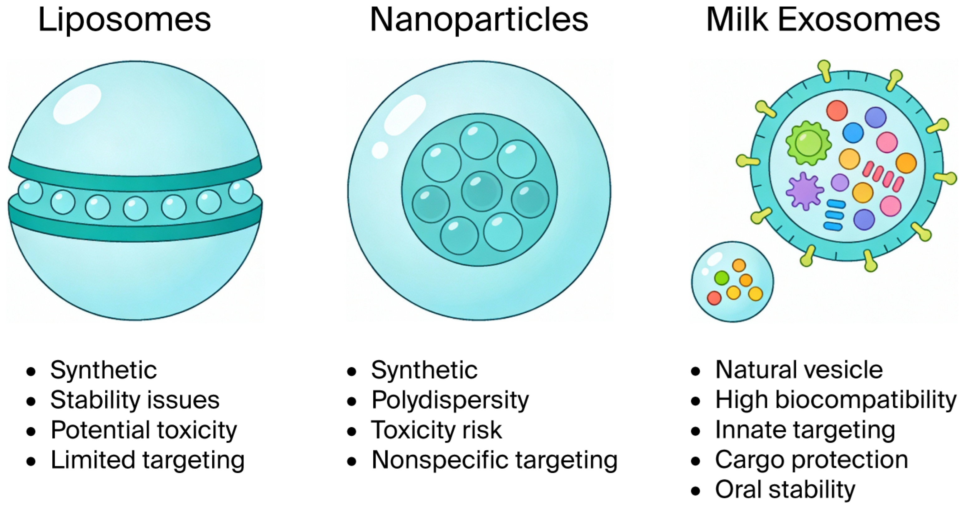
Exosomes are a type of extracellular vesicle that originate from the invagination of endosomal membranes, which are then released into the extracellular space after fusion with the plasma membrane [9]. Their main role appears to be intercellular communication [10]. In fact, exosomes carry a wide variety of molecules in their lumen, including proteins, lipids, and nucleic acids. Among these, miRNAs are particularly relevant. Exosomes containing miRNA can influence the functions and behavior of recipient cells by regulating gene expression [9,11]. Several cell types can release exosomes, and therefore, they can be found in numerous biological fluids [12]. A biological fluid particularly rich in exosomes is milk. Milk-derived exosomes (Mi-Exos) are important for certain developmental processes in newborns, including immune competence. Such effects have also been attributed to the large number of immunomodulatory miRNAs [13] in Mi-Exos. Mi-Exos have shown protective effects against inflammatory disorders, such as necrotizing enterocolitis in premature infants, but it is believed that their protective roles may also extend to dermatological disorders [14]. Compared to liposomes and polymeric nanoparticles, exosomes possess high circulation capacity, superior penetration, cell membrane similarity, and degradation resistance (Figure 1).
Among the various bioactive molecules contained within exosomes, miRNAs play a pivotal role in regulating gene expression and various cellular processes, including proliferation, differentiation, and apoptosis, all of which are critical in hair follicle dynamics [9,11,15,16]. Notably, Mi-Exos have emerged as a compelling avenue for research due to their relatively abundant composition of specific miRNAs and their demonstrated efficacy in various therapeutic contexts [2,7].
The notion that miRNAs derived from milk exosomes could potentially modulate hair follicle activity holds promise, particularly regarding alopecia. A recent study demonstrated that colostrum-derived exosomes promote hair regeneration by influencing the transition from telogen to anagen, suggesting a critical role for these extracellular vesicles in hair follicle cycling [7]. Furthermore, mechanistic insights into how exosomal miRNAs can activate pathways such as the Wnt/β-catenin pathway have led to speculation about their therapeutic potential for treating AGA and related hair loss disorders [8,17].
Given this background, the present study aims to elucidate the in vitro effects of miRNAs derived from milk exosomes on hair loss disorders, specifically targeting AGA and TE. This work aims to provide foundational data and is poised to pave the way for future translational applications in dermatological therapeutics.

2. Materials and Methods

2.1. Exosomes Isolation and miRNA Characterization

Lyophilized powders containing exosomes derived from bovine milk (mEV-miRNAs) were obtained from Evobiotix SA, Melide, Switzerland, and produced according to patent No. WO 2023/067490 A1.
Stock solutions were obtained by resuspending the dried exosomes in distilled water and subsequent sterile filtration (0.22 µm).

2.2. RNA Extraction and Sequencing

Total RNA was extracted from six samples of lyophilized mEV-miRNAs, 15 mg each, using the miRNeasy Serum/Plasma Kit (QIAGEN, Hilden, Germany), following the manufacturer’s instructions. RNA concentration was determined using both a NanoDrop spectrophotometer and the Qubit Broad Range (BR) assay (Thermo Fisher Scientific, Waltham, MA, USA).
Small RNA libraries were prepared using the QIAseq miRNA UDI Library Kit (QIAGEN). For each sample, 250 ng of total RNA was used as input material. Briefly, 3′ and 5′ adapters were sequentially ligated to mature miRNAs, followed by reverse transcription with the incorporation of unique molecular identifiers (UMIs). Adapter and primer dilutions were optimized as follows: the 3′ adapter was diluted 1:5, the 5′ adapter 1:2.5, and the reverse transcription primer 1:5. Library amplification was performed using 22 PCR cycles, and unique dual indexes (UDIs) were applied to enable sample multiplexing.
Final libraries were quality-controlled and sequenced on an Illumina NextSeq 500 platform using paired-end sequencing (2 × 150 bp). Sequencing was performed at an average depth of approximately 25 million reads per sample.

2.3. Bioinformatic Analysis of Small RNA-Seq Data

Raw sequencing data in FASTQ format were processed using a standardized small RNA pipeline. Initial quality control was performed with FastQC to assess read quality, adapter contamination, and length distribution. Adapter sequences were trimmed using Cutadapt, and reads with low quality or incorrect length were discarded.
Unique molecular identifiers (UMIs) were extracted from each read to eliminate PCR duplicates and improve quantification accuracy. Reads lacking valid UMIs were removed, as indicated by the percentage of lost reads in QC metrics.
High-quality reads were aligned to the Bos taurus reference genome using Bowtie (a short-read aligner optimized for small RNAs). miRNA annotation was performed against the miRBase database (version 22.1) using the GeneGlobe/QIAGEN pipeline. Reads mapping to known miRNA loci were quantified, and the percentage of miRNA-mapped reads per sample was calculated.
Counts were normalized across samples (e.g., reads per million, RPM). Stringent filters were applied to retain high-confidence miRNAs based on read counts and annotation quality.
Ranked lists of miRNAs were generated for each sample (Top 20, Top 50, Top 100). Analysis was performed at the Centre for Genomics and Oncological Research (GENYO, Granada, Spain).

2.4. Hair Follicle Dermal Papilla Cell Culture and Viability Assay (MTT Assay)

A primary cell culture of Hair Follicle dermal papilla (HFDP) cells was obtained from PromoCell (GmbH Sickingenstr, 63/65 69126, Heidelberg, Germany). It was cultivated in 25 cm2 cell culture flasks in HFDPC medium (PromoCell) and incubated in a humified 5% CO2 atmosphere at 37 °C.
An MTT assay was performed according to the method described in [18], with slight modifications. Briefly, HFDP cells were seeded in 96-well plates at a density of 5 × 10⁴ cell per well and incubated at 37 °C in 5% CO2. After overnight incubation, the medium was removed from the wells, and the cells were incubated with the mEV-miRNAs diluted in HFDPC medium at about 6 × 10⁹ particles/mL. Untreated cells were used as a control. Cells were then incubated for 24 h at 37 °C in 5% CO2. After the incubation period, the medium was removed, and 100 µL of MTT reagent (0.05 mg/mL) was applied to the cells. The plate was incubated for 3 h in the dark at 37 °C with 5% CO2. Afterward, the MTT solution was aspirated, and 100 µL of dimethyl sulfoxide (DMSO) was added to dissolve the purple formazan product. The solution was shaken in the dark for 15 min at room temperature. The absorbance of the solutions was read at 570 nm and 630 nm (as a reference) in a microplate reader (BioTek Instruments Inc., Bad Friedrichshall, Germany). The experiment was performed in triplicate. Data were expressed as the cell viability percentage, compared to control cells, as per the following formula: % cell viability/ctrl = (Abs sample/Abs ctrl) × 100.

2.5. Human Hair Follicle Cultivation and Elongation Measurement

Human hair follicles were obtained from Studio Piero Tesauro (Milan, Italy), and the study was approved by the Ethical Independent Committee for Clinical, not pharmacological, investigation in Genoa (Italy) and in accordance with the ethical standards of the 1964 Declaration of Helsinki. All volunteers signed an informed consent form. Within a day of excision, hair follicles from three different donors were isolated and cultivated in vitro in William’s E medium supplemented with L-glutamine (2 mM), insulin (2 µg/mL), hydrocortisone (2 ng/mL), and antibiotic solution (penicillin/streptomycin 100 U/mL) in 24-well plates with 400 µL of medium and incubated in a humified, 5% CO2 atmosphere at 37 °C.
After overnight incubation, the basal medium of human hair follicles was substituted with basal medium enriched with mEV-miRNAs 6 × 10¹⁰ particles/mL; hair follicles cultivated in basal medium were used as a control. The length of each hair follicle was measured with an eyepiece with a micrometer on day 0 and every 24–48 h until the end of incubation on day 4. Elongation is expressed in mm.

2.6. Hair Follicle Taqman Array Analysis of WNT Signaling Pathway

At the end of the incubation period, RNA was extracted from hair follicles according to the method described by Chomczynski and Mackey [19]. Complementary DNA (cDNA) was synthesized via reverse transcriptase using the commercial kit “PrimeScriptTM RT Reagent Kit (perfect Real Time)” (TakaraBioInc., Kyoto, Japan). cDNA was then amplified in a TaqMan™ Array 96-Well Plate (Study Name Human WNT Signaling Pathway, Appliedbiosystems Thermo Fisher Scientific 4391524, Waltham, MA USA) using the TaqMan™ Fast Advanced Master Mix (Appliedbiosystems Thermo Fisher Scientific 4444557), and amplification was measured using qRT-PCR. GAPDH was used as a housekeeping gene. Each biological replicate was run in duplicate, and the resulting data were analyzed using the 2−ΔΔCt method [20].

2.7. Statistical Analysis

Student’s t-test was used for MTT assay, elongation, and qRT-PCR analyses (GraphPad Prism v. 10.4.1, GraphPad Software Inc., Boston, MA, USA). Each gene was analyzed using an individual unpaired t-test (with Welch’s correction) comparing mEV-miRNA-treated samples with the corresponding control, and no formal multiple-comparison correction was applied. p-values equal to or less than 0.05, 0.01, and 0.001 were considered significant.

3. Results

3.1. Identifying the miRNA Fingerprint of mEV-miRNAs

The miRNA fingerprint of the mEV-miRNA preparations was determined via RNA extraction, deep sequencing, and bioinformatics analysis. After filtering a list of the topmost abundant miRNAs, a consensus was identified across all samples (Table 1). This list of core miRNAs, present in all bovine milk EV samples, is therefore similar to a miRNA fingerprint of bovine milk-derived extracellular vesicles.

3.2. Viability/Metabolic Activity on HFDP Cells

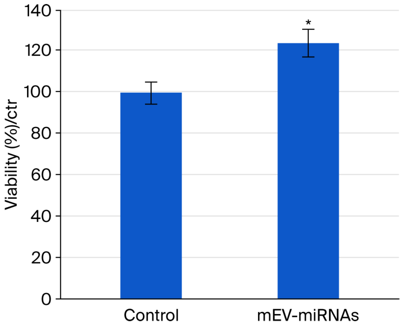
The viability assay showed that cultivating HFDP cells with medium enriched with mEV-miRNAs for 24 h significantly increased their viability (+24.27%) compared to control cells (Figure 2). These data suggest hair growth-promoting effects of mEV-miRNAs.

3.3. Elongation of Human Hair Follicles

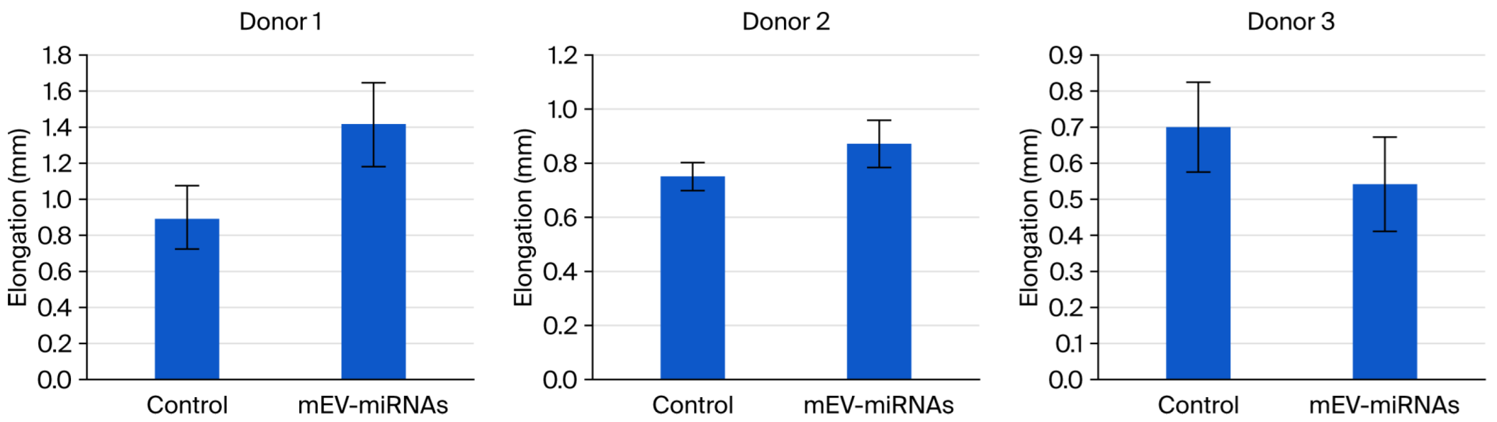
Cultivating human hair follicles in a medium enriched with mEV-miRNAs and measuring their elongation further showed a trend of growth promotion. Indeed, the experiment performed on the first two donors showed a tendency to up-regulate their shaft elongation, although the data were not statistically significant (Figure 3).

3.4. Differentially Expressed Genes in the WNT Signaling Pathway

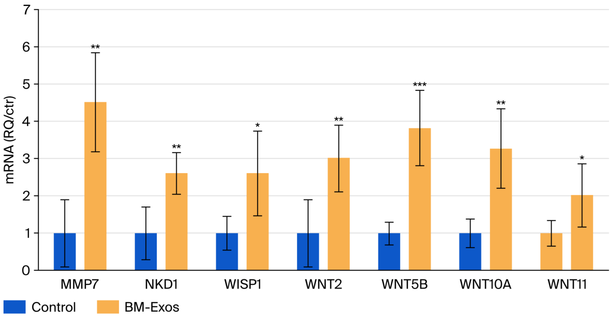
The TaqMan™ Array Human WNT Pathway 96-well Plate (Applied Biosystems™, Waltham, MA, USA) includes 92 assays targeting genes linked to WNT signaling and 4 assays for endogenous reference genes. This array focuses on genes belonging to the WNT family of proteins, which are involved in cell-to-cell communication and are key modulators of the WNT pathway, as well as WNT-associated genes involved in pathway regulation, downstream signaling, and development. Figure 4 shows the average value of three biological replicates of significantly regulated genes compared to control samples.

4. Discussion

In this study, we investigated the potential of miRNAs contained in mEV-miRNAs to influence hair-related endpoints, providing preliminary support for their use in conditions such as AGA and TE. Recent studies have acknowledged that exosomes from various sources can promote hair growth by enhancing cell proliferation and regulating gene expression in hair follicle dermal papilla (HFDP) cells [1,4]. For example, recent research has explored the potential of exosomes from different sources for hair growth and the treatment of hair loss. Exosomes derived from Leuconostoc holzapfelii isolated from the human scalp successfully increased HFDP cell proliferation and migration and modulated HFDP cell gene expression in vitro [21]. Kim et al. [7] showed that exosomes derived from colostrum induced proliferation of HFDP cells and rescued dihydrotestosterone-induced arrest of follicle development in vitro. In addition, they showed that the same exosomes stimulated dorsal hair regrowth in mice at levels comparable to those of minoxidil. Another study also showed that exosomes from bovine colostrum (but not those from mesenchymal stem cells) increased the growth of hair follicles cultivated in vitro [22].
Most importantly, miRNAs have been shown to be essential for hair follicle development in embryos and are necessary for postnatal growth, as they regulate proliferation and apoptosis [23]. For example, in hair follicle stem cells, miR-205 is reported to promote stem cell expansion during early HF development. In contrast, miR-125b is considered a repressor of HF stem cell differentiation required for anagen onset [23].
In the HF hair matrix, miR-31 promotes proliferation by modulating the activity of BMP and FGF signaling pathways, as well as via changes in the expression of structural proteins (keratin 16 and keratin 17). On the contrary, miR-214 inhibits hair growth by targeting the Wnt and Shh signaling pathways. miR-24 and miR-205 are involved in hair follicle differentiation and protection from hair follicle regression (catagen)-associated apoptosis, respectively [23].
Specific miRNAs have also been implicated in hair loss disease, including AGA and TE [24,25]
In this study, we used exosomes derived from bovine milk (Bos taurus). This source was chosen primarily for its mammalian origin and consequent conservation of key signaling pathways, as well as for its accessibility and scalability. More specifically, the miRNAs derived from Bos taurus were verified as orthologs of Homo sapiens miRNAs (https://mirgenedb.org/ accessed on 8 September 2025), allowing them to influence the function and behavior of human recipient cells.
The miRNA content characterization showed that the MEV-miRNAs mainly contained miRNAs from five families: Let-7 family (Bta-Let-7-P2a3_5p, Bta-Let-7-P2b1_5p, Bta-Let-7P1d_5p, and Bta-Let-7-P2c3_5p); miR-21 family (Bta-miR-21_5p); miR-30 family (Bta-miR-30-P1b_5p, Bta-miR-30-P1d_5p); miR-200 family (Bta-miR-8-P1a_3p, Bta-miR-8-P1b_3p); and miR-148/152 Family (Bta-Mir-148-P1_3p).
Previous studies have shown that some of these were highly expressed in hair follicles of Mus musculus, such as miR-21 and many Let-7 family members [26]. miR-21 controls hair follicle growth in cashmere goats, being highly expressed in the telogen phase [27], and an in vitro study has shown that a long non-coding RNA (lncRNA) may arrest the progression of AGA by down-regulating miRNA-21 and TGF-β1 in Hair Follicle Stem cells (HFSC) and, at the same time, up-regulating the WNT/β catenin pathway [28]. The role of miRNAs belonging to the Let-7 family in the regulation of the hair follicle growth cycle in cashmere goats has been investigated in a few studies, suggesting that miR-let7a plays a regulatory function in this cycle, particularly its targets IGF-1R, C-myc, and FGF5 proteins [29]. miR-let-7b has been studied in alpaca instead, where it seems to negatively regulate TGFβR I, which in turn inhibits the elongation of hair in the dorsal region of alpaca [30]. Another study by Liu et al. [31] showed that miR-let-7b regulates the growth of alpaca via down-regulation of ectodysplasin A. Some miRNAs belonging to the miR30 family have been linked to hair pathologies; for instance, miR-30b/d was found to be one of the most significantly associated genes in alopecia areata (AA), as its expression is markedly reduced in hair follicles of AA patients [32].
The results of the present study indicate that the miRNAs contained within these mEV-miRNAs significantly enhanced the viability/metabolic activity of HFDP cells after 24 h of incubation, demonstrating a clear difference compared to the untreated controls. Notably, while we observed a tendency toward increased elongation of human hair follicles upon treatment with mEV-miRNAs, this effect did not reach statistical significance; however, the observed trend is consistent with previous reports linking exosomal delivery of miRNAs to accelerated hair follicle growth [33]. It is important to notice that the time during which we measured hair follicle elongation was limited to four days; it is possible that longer incubation periods may allow observation of significantly increased elongation.
The analysis of genes linked to the WNT signaling pathway of hair follicles cultivated in the presence of mEV-miRNAs showed significantly increased expression of WNT2, WNT5B, WNT10A, and WNT11. It has previously been shown that WNT2 and WNT10A are important for hair follicle growth and development, as activators of the canonical WNT pathway. WNT2 is involved in hair follicle development and plays a pivotal role in hair follicle morphogenesis and hair length regulation [34], whereas WNT10A plays an important role in the development of ectodermal appendages. It was shown to be switched on during early anagen in bulge hair follicle stem cells, suggesting that it is required for regenerating the outer root sheath during the human hair cycle [35,36]. Previous studies have shown that a WNT10A variant is associated with short anagen hair (SAH), and patients with a loss-of-function mutation in WNT10A suffer from alopecia or hypotrichosis [36,37]. WNT5B seems to be a ligand of the non-canonical WNT pathway and is often considered an antagonist to β-catenin signaling [38], although some studies have found an association between WNT5B mRNA up-regulation and hair growth [39]. Reddy et al. [40] showed that WNT11 is expressed in certain cell types of the outer root sheath and dermal sheath, and they suggest it as a candidate for the signals that control the movements of outer root sheath cells during anagen. Another study showed that WNT11 was significantly up-regulated in anagen hair and down-regulated in catagen hair of cashmere goats [41].
Another significantly up-regulated gene in hair follicles cultivated with mEV-miRNAs was MMP7, which encodes matrilysin. This protein is a direct target of the WNT signaling pathway and appears to regulate extracellular matrix remodeling in hair follicles [42,43]. WNT1-inducible signaling pathway protein 1 (WISP1) is a secreted multicellular protein that activates a variety of downstream signaling pathways of various biological processes, from cell proliferation, adhesion, and differentiation to wound healing and tissue repair [44]. Finally, NKD1 was also significantly up-regulated in this study, consistent with engagement of the WNT cascade and suggesting activation of endogenous negative-feedback circuits that constrain signaling amplitude, highlighting the need for caution when inferring net pathway activation from this marker alone [45].
Future investigations should include more detailed in vitro analysis to distinguish canonical from non-canonical WNT signaling, alongside in vivo validation, and to dissect the contribution of specific miRNAs to the observed effects. In addition, longer culture periods and larger donor cohorts will be required to better capture cumulative follicle responses. Finally, formal testing of WNT–β-catenin pathway dependence—for example, through pharmacological inhibition or genetic modulation—will be essential to consolidate the mechanistic interpretation.
An additional mechanistic aspect concerns the miR-200 family, which is prominently represented in our mEV-miRNA profile. Das et al. [46] recently showed that miR-200s are enriched in WNT-active matrix cells but largely absent from the upper follicle, the niche where sebaceous and stem cell lineages emerge, and that ectopic miR-200 in this region amplifies WNT/β-catenin signaling, impairs SOX9 function, and curtails proliferative and lipogenic programs, ultimately preventing sebaceous gland formation while leaving hair shaft development intact. In light of our finding that mEV-miRNAs enhance the expression of several WNT ligands (WNT2, WNT5B, WNT10A, WNT11) and downstream effectors such as MMP7, WISP1 and NKD1 in human hair follicles, it is tempting to speculate that exosomal miR-200 could influence epithelial plasticity by favoring a WNT-high, SOX9-dependent milieu. Nonetheless, our in vitro model does not distinguish between follicular compartments nor interrogate SOX9, NFATC1 or PPARγ activity, so future work using spatially resolved WNT/β-catenin readouts and targeted manipulation of the miR-200–SOX9 axis in organ-cultured follicles will be required to determine whether this circuitry underlies the effects of mEV-miRNAs.

5. Conclusions

In conclusion, this study provides foundational in vitro evidence supporting the use of mEV-miRNAs and their miRNA cargo as a promising cosmetic candidate with potential translational relevance for hair loss disorders, including AGA and TE. The multifaceted effects observed, ranging from enhanced HFDP cell viability/metabolic activity to modulation of hair-relevant signaling pathways, underscore the potential of miRNA-based approaches in dermatological research, while also highlighting the need for comprehensive in vivo and safety studies.

Author Contributions

Conceptualization, D.P. and F.R.; methodology, D.P., G.M., M.H., R.L.D. and E.d.S.R.; investigation, G.M., M.C., P.T., R.L.D. and E.d.S.R.; resources, M.H. and G.G.; data analysis and curation, D.P., G.M., M.H., R.L.D. and E.d.S.R.; writing—original draft preparation, D.P., G.M. and M.H.; writing—review and editing, D.P. and F.R. All authors have read and agreed to the published version of the manuscript.

Funding

This research and the APC were funded by Giuliani SPA, Milan, Italy.

Institutional Review Board Statement

This study was conducted in accordance with the Declaration of Helsinki and approved by the Ethical Independent Committee for Clinical, not a pharmacological, investigation in Genoa (Italy), protocol code No. 2018/3, date of approval 27 February 2018.

Informed Consent Statement

Informed consent was obtained from all subjects involved in the study.

Data Availability Statement

The datasets used during and/or analyzed during the current study are available from the corresponding author upon reasonable request.

Conflicts of Interest

F.R. serves as a consultant for Giuliani S.P.A., G.G. is in the board of directors of Giuliani SpA and D.P., G.M. and M.C. are employed by Giuliani S.P.A. M.H. is CSO of EvoBiotiX SA. P.T. is member of the International Society of Hair Restoration Surgery (ISHRS). R.L.D. and E.D are employed at GENYO, Centre for Genomics and Oncological Research. All named authors meet the International Committee of Medical Journal Editors (ICMJE) criteria for authorship for this manuscript, take re-sponsibility for the integrity of the work as a whole, and have given final approval for the version to be published. The funders had no role in the design of the study; in the collection, analyses or in-terpretation of data; in the writing of the manuscript; or in the decision to publish the results.

References

  1. Kost, Y.; Muskat, A.; Mhaimeed, N.; Nazarian, R.S.; Kobets, K. Exosome therapy in hair regeneration: A literature review of the evidence, challenges, and future opportunities. J. Cosmet. Dermatol. 2022, 21, 3226–3231. [Google Scholar] [CrossRef] [PubMed]
  2. Tang, X.; Cao, C.; Liang, Y.; Han, L.; Tu, B.; Yu, M.; Wan, M. Adipose-Derived Stem Cell Exosomes Antagonize the Inhibitory Effect of Dihydrotestosterone on Hair Follicle Growth by Activating Wnt/β-Catenin Pathway. Stem Cells Int. 2023, 2023, 5548112. [Google Scholar] [CrossRef]
  3. Chen, X.; Liu, B.; Li, Y.; Han, L.; Tang, X.; Deng, W.; Lai, W.; Wan, M. Dihydrotestosterone Regulates Hair Growth Through the Wnt/β-Catenin Pathway in C57BL/6 Mice and In Vitro Organ Culture. Front. Pharmacol. 2020, 10, 1528. [Google Scholar] [CrossRef]
  4. Zhang, Y.; Huang, J.; Fu, D.; Liu, Z.; Wang, H.; Wang, J.; Qu, Q.; Li, K.; Fan, Z.; Hu, Z. Transcriptome Analysis Reveals an Inhibitory Effect of Dihydrotestosterone-Treated 2D- and 3D-Cultured Dermal Papilla Cells on Hair Follicle Growth. Front. Cell Dev. Biol. 2021, 9, 724310. [Google Scholar] [CrossRef]
  5. Cedirian, S.; Bruni, F.; Quadrelli, F.; Caro, G.; Fortuna, M.; Rossi, A.; Piraccini, B.M.; Starace, M. Clinical study on the efficacy and tolerability of a topical regenerative treatment in patients with telogen effluvium and mild androgenetic alopecia. J. Cosmet. Dermatol. 2023, 22, 3347–3351. [Google Scholar] [CrossRef] [PubMed]
  6. Rudnicka, L.; Kaczorowska, A. Treatment of diseases associated with non-cicatricial alopecia. Dermatology 2021, 108, 504–516. [Google Scholar]
  7. Kim, H.; Jang, Y.; Kim, E.H.; Jang, H.; Cho, H.; Han, G.; Song, H.K.; Kim, S.H.; Yang, Y. Potential of Colostrum-Derived Exosomes for Promoting Hair Regeneration Through the Transition From Telogen to Anagen Phase. Front. Cell Dev. Biol. 2022, 10, 815205. [Google Scholar] [CrossRef] [PubMed]
  8. Xia, Y.; Wei, K.; Hu, L.Q.; Zhou, C.R.; Lu, Z.B.; Zhan, G.S.; Pan, X.L.; Pan, C.F.; Wang, J.; Wen, W.; et al. Exosome-mediated transfer of miR-1260b promotes cell invasion through Wnt/β-catenin signaling pathway in lung adenocarcinoma. J. Cell. Physiol. 2020, 235, 6843–6853. [Google Scholar] [CrossRef]
  9. Zhang, J.; Li, S.; Li, L.; Li, M.; Guo, C.; Yao, J.; Mi, S. Exosome and exosomal microRNA: Trafficking, sorting, and function. Genom. Proteom. Bioinform. 2015, 13, 17–24. [Google Scholar] [CrossRef]
  10. Raposo, G.; Stoorvogel, W. Extracellular vesicles: Exosomes, microvesicles, and friends. J. Cell Biol. 2013, 200, 373–383. [Google Scholar] [CrossRef]
  11. Ge, Q.; Zhou, Y.; Lu, J.; Bai, Y.; Xie, X.; Lu, Z. miRNA in plasma exosome is stable under different storage conditions. Molecules 2014, 19, 1568–1575. [Google Scholar] [CrossRef]
  12. Kalluri, R.; LeBleu, V.S. The biology, function, and biomedical applications of exosomes. Science 2020, 367, eaau6977. [Google Scholar] [CrossRef] [PubMed]
  13. Torregrosa Paredes, P.; Gutzeit, C.; Johansson, S.; Admyre, C.; Stenius, F.; Alm, J.; Scheynius, A.; Gabrielsson, S. Differences in exosome populations in human breast milk in relation to allergic sensitization and lifestyle. Allergy 2014, 69, 463–471. [Google Scholar] [CrossRef] [PubMed]
  14. Pinto, D.; Giuliani, G.; Mondadori, G.; Rinaldi, F. Exosomes in Dermatological Research: Unveiling Their Multifaceted Role in Cellular Communication, Healing, and Disease Modulation. Cosmetics 2025, 12, 16. [Google Scholar] [CrossRef]
  15. Wang, J.; Ma, Y.; Li, T.; Li, J.; Yang, X.; Hua, G.; Cai, G.; Zhang, H.; Liu, Z.; Wu, K.; et al. MiR-199a-3p Regulates the PTPRF/β-Catenin Axis in Hair Follicle Development: Insights into the Pathogenic Mechanism of Alopecia Areata. Int. J. Mol. Sci. 2023, 24, 17632. [Google Scholar] [CrossRef]
  16. de Rivero Vaccari, J.P.; Sawaya, M.E.; Brand, F.; Nusbaum, B.P.; Bauman, A.J.; Bramlett, H.M.; Dietrich, W.D.; Keane, R.W. Caspase-1 level is higher in the scalp in androgenetic alopecia. Dermatol. Surg. 2012, 38, 1033–1039. [Google Scholar] [CrossRef]
  17. Hu, S.; Li, Z.; Lutz, H.; Huang, K.; Su, T.; Cores, J.; Dinh, P.U.C.; Cheng, K. Dermal exosomes containing miR-218-5p promote hair regeneration by regulating β-catenin signaling. Sci. Adv. 2020, 6, eaba1685. [Google Scholar] [CrossRef]
  18. Mosmann, T. Rapid colorimetric assay for cellular growth and survival: Application to proliferation and cytotoxicity assays. J. Immunol. Methods 1983, 65, 55–63. [Google Scholar] [CrossRef]
  19. Chomczynski, P.; Mackey, K. Short technical reports. Modification of the TRI reagent procedure for isolation of RNA from polysaccharide- and proteoglycan-rich sources. Biotechniques 1995, 19, 942–945. [Google Scholar]
  20. Vigetti, D.; Viola, M.; Karousou, E.; Rizzi, M.; Moretto, P.; Genasetti, A.; Clerici, M.; Hascall, V.C.; De Luca, G.; Passi, A. Hyaluronan-CD44-ERK1/2 regulate human aortic smooth muscle cell motility during aging. J. Biol. Chem. 2008, 283, 4448–4458. [Google Scholar] [CrossRef]
  21. Yoon, Y.C.; Ahn, B.H.; Min, J.W.; Lee, K.R.; Park, S.H.; Kang, H.C. Stimulatory Effects of Extracellular Vesicles Derived from Leuconostoc holzapfelii That Exists in Human Scalp on Hair Growth in Human Follicle Dermal Papilla Cells. Curr. Issues Mol. Biol. 2022, 44, 845–866. [Google Scholar] [CrossRef]
  22. Ferruggia, G.; Zimbone, M.; Brundo, M.V. New technologies for hair follicle growth and regeneration: Comparison between different commercial products contain exosomes. bioRxiv 2024. bioRxiv:2024.11.17.623990. [Google Scholar]
  23. Andl, T.; Botchkareva, N.V. MicroRNAs (miRNAs) in the control of HF development and cycling: The next frontiers in hair research. Exp. Dermatol. 2015, 24, 821–826. [Google Scholar] [CrossRef]
  24. Paul, S.; Licona-Vázquez, I.; Serrano-Cano, F.I.; Frías-Reid, N.; Pacheco-Dorantes, C.; Pathak, S.; Chakraborty, S.; Srivastava, A. Current insight into the functions of microRNAs in common human hair loss disorders: A mini review. Hum. Cell 2021, 34, 1040–1050. [Google Scholar] [CrossRef] [PubMed]
  25. Rajar, U.D.M.; Fahim, A.; Qazi, S.; Kazi, N.; Maheshwary, N.; Rph, A.A. Platelet-Rich Plasma Therapy for Telogen Effluvium: A Comprehensive Evaluation of Efficacy and Safety. Dermis 2024, 4, 1–9. [Google Scholar] [CrossRef]
  26. Yi, R.; O’Carroll, D.; Pasolli, H.A.; Zhang, Z.; Dietrich, F.S.; Tarakhovsky, A.; Fuchs, E. Morphogenesis in skin is governed by discrete sets of differentially expressed microRNAs. Nat. Genet. 2006, 38, 356–362. [Google Scholar] [CrossRef]
  27. Xiang, B.; Li, Y.; Li, J.; Zhang, B.; Li, J.; Jiang, H.; Zhang, Q. MiR-21 regulated hair follicle cycle development in Cashmere goats by targeting FGF18 and SMAD7. Anim. Biotechnol. 2023, 34, 4695–4702. [Google Scholar] [CrossRef] [PubMed]
  28. Xiong, J.; Wu, B.; Hou, Q.; Huang, X.; Jia, L.; Li, Y.; Jiang, H. Comprehensive Analysis of LncRNA AC010789.1 Delays Androgenic Alopecia Progression by Targeting MicroRNA-21 and the Wnt/β-Catenin Signaling Pathway in Hair Follicle Stem Cells. Front. Genet. 2022, 13, 782750. [Google Scholar] [CrossRef]
  29. Ma, T.; Li, J.; Jiang, Q.; Wu, S.; Jiang, H.; Zhang, Q. Differential expression of miR-let7a in hair follicle cycle of Liaoning cashmere goats and identification of its targets. Funct. Integr. Genom. 2018, 18, 701–707. [Google Scholar] [CrossRef]
  30. Shen, Y.; Zhang, Y.; Liu, N.; Wang, H.-D.; Xie, J.-S.; Gao, S.-Y.; Cheng, J.-Q.; Yu, X.-J.; Wang, T.; Dong, C.-S.; et al. Let-7b promotes alpaca hair growth via transcriptional repression of TGFβR I. Gene 2016, 577, 32–36. [Google Scholar] [CrossRef]
  31. Liu, N.; Niu, S.; Cao, X.; Cheng, J.; Gao, S.; Yu, X.; Wang, H.; Dong, C.; He, X. Let-7b regulates alpaca hair growth by downregulating ectodysplasin A. Mol. Med. Rep. 2018, 17, 4688–4694. [Google Scholar] [CrossRef]
  32. Tafazzoli, A.; Forstner, A.J.; Broadley, D.; Hofmann, A.; Redler, S.; Petukhova, L.; Giehl, K.A.; Kruse, R.; Blaumeiser, B.; Böhm, M.; et al. Genome-Wide MicroRNA Analysis Implicates miR-30b/d in the Etiology of Alopecia Areata. J. Investig. Dermatol. 2018, 138, 549–556. [Google Scholar] [CrossRef]
  33. Yan, H.; Gao, Y.; Ding, Q.; Liu, J.; Li, Y.; Jin, M.; Xu, H.; Ma, S.; Wang, X.; Zeng, W.; et al. Exosomal Micro RNAs Derived from Dermal Papilla Cells Mediate Hair Follicle Stem Cell Proliferation and Differentiation. Int. J. Biol. Sci. 2019, 15, 1368–1382. [Google Scholar] [CrossRef]
  34. Liu, J.; Liu, B.; Mu, Q.; Liu, J.; Li, Y.; Gong, W.; Chahaer, T.; Song, Y.; Hai, E.; Wang, H.; et al. Melatonin promotes the proliferation of dermal papilla cells in cashmere goats via activation of chi-let-7d-5p/WNT2 axis. Genomics 2024, 116, 110961. [Google Scholar] [CrossRef]
  35. Benard, E.L.; Hammerschmidt, M. The fundamentals of WNT10A. Differentiation 2025, 142, 100838. [Google Scholar] [CrossRef]
  36. Hawkshaw, N.J.; Hardman, J.A.; Alam, M.; Jimenez, F.; Paus, R. Deciphering the molecular morphology of the human hair cycle: Wnt signalling during the telogen-anagen transformation. Br. J. Dermatol. 2020, 182, 1184–1193. [Google Scholar] [CrossRef] [PubMed]
  37. Cesarato, N.; Schwieger-Briel, A.; Gossmann, Y.; Henne, S.K.; Hillmann, K.; Frommherz, L.H.; Wehner, M.; Xiong, X.; Thiele, H.; Oji, V.; et al. Short anagen hair syndrome: Association with mono- and biallelic variants in WNT10A and a genetic overlap with male pattern hair loss. Br. J. Dermatol. 2023, 189, 741–749. [Google Scholar] [CrossRef] [PubMed]
  38. Suthon, S.; Perkins, R.S.; Bryja, V.; Miranda-Carboni, G.A.; Krum, S.A. WNT5B in Physiology and Disease. Front. Cell Dev. Biol. 2021, 9, 667581. [Google Scholar] [CrossRef]
  39. Zheng, M.; Choi, N.; Jang, Y.; Kwak, D.E.; Kim, Y.; Kim, W.S.; Oh, S.H.; Sung, J.H. Hair growth promotion by Necrostatin-1s. Sci. Rep. 2020, 10, 17622. [Google Scholar] [CrossRef]
  40. Reddy, S.; Andl, T.; Bagasra, A.; Lu, M.M.; Epstein, D.J.; Morrisey, E.E.; Millar, S.E. Characterization of Wnt gene expression in developing and postnatal hair follicles and identification of Wnt5a as a target of Sonic hedgehog in hair follicle morphogenesis. Mech. Dev. 2001, 107, 69–82. [Google Scholar] [CrossRef] [PubMed]
  41. Gong, G.; Fan, Y.; Yan, X.; Li, W.; Yan, X.; Liu, H.; Zhang, L.; Su, Y.; Zhang, J.; Jiang, W.; et al. Identification of Genes Related to Hair Follicle Cycle Development in Inner Mongolia Cashmere Goat by WGCNA. Front. Vet. Sci. 2022, 9, 894380. [Google Scholar] [CrossRef]
  42. Crawford, H.C.; Fingleton, B.; Gustavson, M.D.; Kurpios, N.; Wagenaar, R.A.; Hassell, J.A.; Matrisian, L.M. The PEA3 Subfamily of Ets Transcription Factors Synergizes with β-Catenin–LEF-1 To Activate Matrilysin Transcription in Intestinal Tumors. Mol. Cell. Biol. 2001, 21, 1370–1383. [Google Scholar] [CrossRef] [PubMed]
  43. Botchkarev, V.A.; Sharov, A.A. BMP signaling in the control of skin development and hair follicle growth. Differentiation 2004, 72, 512–526. [Google Scholar] [CrossRef] [PubMed]
  44. Deng, W.; Fernandez, A.; McLaughlin, S.L.; Klinke, D.J. WNT1-inducible signaling pathway protein 1 (WISP1/CCN4) stimulates melanoma invasion and metastasis by promoting the epithelial–mesenchymal transition. J. Biol. Chem. 2019, 294, 5261–5280. [Google Scholar] [CrossRef] [PubMed]
  45. Angonin, D.; Raay, T.J.V. NKD1 Functions as a Passive Antagonist of Wnt Signaling. PLoS ONE 2013, 8, e74666. [Google Scholar] [CrossRef]
  46. Das, A.; Fu, Y.C.; Li, H.; Wong, M.A.; Che, A.; Singh, A.; Han, J.; Bjerke, G.; Wang, D.; Yi, R. Coordinated inhibition of SOX9 and cell cycle progression by microRNA-200 restricts sebaceous gland fate specification. bioRxiv 2026. bioRxiv:2026.01.09.698672. [Google Scholar]
Figure 1. Milk exosomes versus conventional nanocarriers as advanced dermal delivery systems.
Cosmetics 13 00038 g001
Figure 2. The viability percentage of HFDP cells subjected to treatment with mEV-miRNAs 6 × 10⁹ particles/mL for 24 h. Columns represent the mean value of three independent analyses, and error bars represent SEM. Asterisks indicate a statistically significant difference compared to control (* = p < 0.05).
Cosmetics 13 00038 g002
Figure 3. Elongation of human hair follicles subjected to treatment with mEV-miRNAs 6 × 10¹⁰ particles/mL for 4 days. Columns represent the mean value of at least six independent measurements of different hair follicles, and error bars represent SEM.
Cosmetics 13 00038 g003
Figure 4. Gene expression analysis of WNT signaling pathway genes determined via qRT-PCR of hair follicles treated with mEV-miRNAs 6 × 10¹⁰ particles/mL for 4 days. Columns represent the mean value of three independent samples, and error bars represent SD. Asterisks indicate a significant difference compared to control hair follicles (* = p < 0.05; ** = p < 0.01; *** = p < 0.001).
Cosmetics 13 00038 g004
Table 1. A list of the top 25 most abundant miRNAs in mEV-miRNAs.
#miRNA
1Bta.Let.7.P2b2_5p
2Bta.Let.7.P1d_5p
3Bta.Mir.8.P1b_3p
4Bta.Mir.8.P2b_3p
5Bta.Mir.15.P2a_5p
6Bta.Mir.30.P1b_5p
7Bta.Mir.28.P2_5p
8Bta.Let.7.P2a3_5p
9Bta.Let.7.P2a1_5p
10Bta.Let.7.P2a2_5p
11Bta.Let.7.P2c3_5p
12Bta.Mir.26.P4_5p
13Bta.Mir.26.P1_5p
14Bta.Mir.320.P1b_3p
15Bta.Let.7.P1c_5p
16Bta.Mir.26.P2_5p
17Bta.Mir.191_5p
18Bta.Mir.423_5p
19Bta.Let.7.P2b1_5p
20Bta.Mir.30.P1a_5p
21Bta.Mir.21_5p
22Bta.Mir.320.P1a_3p
23Bta.Mir.29.P2d2_3p
24Bta.Mir.29.P2b_3p
25Bta.Let.7.P2c1_5p
Disclaimer/Publisher’s Note: The statements, opinions and data contained in all publications are solely those of the individual author(s) and contributor(s) and not of MDPI and/or the editor(s). MDPI and/or the editor(s) disclaim responsibility for any injury to people or property resulting from any ideas, methods, instructions or products referred to in the content.