Article

Effects of ε-Viniferin and ε-Viniferin-Enriched Extract from Vitis labruscana B. ‘Campbell Early’ Cell Cultures on Wound Healing and Epidermal Barrier Restoration in Human Skin Cells

1
Department of Genetics & Biotechnology, Graduate School of Biotechnology, College of Life Sciences, Kyung Hee University, Youngin 17104, Republic of Korea
2
iNGR Inc., Rm.205, 88 Sanupdanji-gil, Pungsan-eup, Andong-si 36618, Gyeongsangbuk-do, Republic of Korea
*
Authors to whom correspondence should be addressed.
These authors contributed equally to this work.
Cosmetics 2025, 12(5), 181; https://doi.org/10.3390/cosmetics12050181
Submission received: 27 June 2025 / Revised: 1 August 2025 / Accepted: 20 August 2025 / Published: 25 August 2025
(This article belongs to the Section Cosmetic Dermatology)

Abstract

Skin wound healing and barrier restoration are complex, tightly regulated processes critical for maintaining skin integrity, particularly in aged or compromised skin. This study investigated the wound healing efficacy and epidermal barrier-restoring effects of ε-Viniferin, a bioactive resveratrol dimer, and Vino Chocolate™, a grape flower-derived extract from Vitis labruscana ‘Campbell Early’ cell cultures enriched with ε-Viniferin. An HPLC analysis confirmed a high concentration of ε-Viniferin (547.58 ppm) in the cell culture-derived extract. In vitro assays conducted on HaCaT keratinocytes and HDFn fibroblasts demonstrated that the treatment with ε-Viniferin and Vino Chocolate™ significantly enhanced fibroblast migration. ELISA analyses showed that both treatments induced a dose-dependent increase in pro-collagen type I (COL1A1), with ε-Viniferin at 1 ppm demonstrating superior efficacy compared to TGF-β1. Additionally, these compounds notably suppressed the expression of matrix metalloproteinases MMP-1 and MMP-3, displaying effects comparable to or greater than retinoic acid. The Western blot analysis further revealed an increased filaggrin expression in keratinocytes, suggesting an improved epidermal barrier function. Collectively, these results indicate that ε-Viniferin and Vino Chocolate™ effectively promote extracellular matrix remodeling, modulate inflammatory responses, and enhance epidermal barrier integrity. These findings highlight their potential as multifunctional bioactive agents for cosmeceutical applications and emphasize the advantages of plant cell culture technology as a sustainable, innovative platform for advanced skincare ingredient development.

1. Introduction

Skin, the largest organ of the human body, serves as a critical physical barrier protecting internal tissues against environmental stressors, dehydration, and pathogen invasion. It is also pivotal as the first line of innate immune defense [1,2]. The effective restoration of skin integrity post-injury is vital for maintaining homeostasis and preventing infection, making wound healing an essential target within both clinical dermatology and cosmetic research. Wound healing is a tightly orchestrated, multifaceted process involving four overlapping phases: hemostasis, inflammation, proliferation, and tissue remodeling [3,4]. During these phases, keratinocytes facilitate re-epithelialization, and dermal fibroblasts synthesize extracellular matrix (ECM) components, managing the ECM turnover [5]. Disruption or delays in these cellular events can impair healing, fueling interest in bioactive compounds that support or accelerate tissue repair [6].
Skin regeneration is further modulated by molecular markers such as matrix metalloproteinases (MMPs), filaggrin, and pro-collagen. MMPs, notably MMP-1, -3, and -9, are zinc-dependent enzymes responsible for degrading type I and III collagens and other ECM constituents, resulting in compromised skin elasticity and delayed wound closure [7,8]. Excessive MMP expression, often induced by oxidative stress or UV exposure, contributes significantly to skin aging and impaired healing. In contrast, filaggrin, a crucial epidermal protein, reinforces barrier function by aiding keratin aggregation and natural moisturizing factor formation, whereas pro-collagen synthesis is fundamental for restoring the dermal matrix integrity [9,10,11]. Evaluating these molecular mechanisms is thus critical for assessing the wound healing and anti-aging potential of cosmetic bioactives. Plant-derived natural products have gained prominence as potential wound healing agents due to their antioxidant, anti-inflammatory, and regenerative properties [12,13,14].
Plant cell culture technology, particularly suspension cultures, presents a sustainable, consistent, and controlled method for producing bioactive secondary metabolites with minimal environmental impact and batch-to-batch variability [15,16]. Optimized elicitation techniques, such as using methyl jasmonate and stevioside in grape flower-derived cell cultures, enhance the accumulation of targeted stilbenes like viniferin [17]. Conditioned media from these cultures have also demonstrated the efficient bioconversion of trans-resveratrol into viniferin derivatives through extracellular peroxidase activity, presenting an eco-friendly approach to bioactive production [18].
These attributes make plant cell cultures an attractive platform for cosmeceutical and pharmaceutical development, particularly for skin repair, barrier restoration, and anti-aging formulations [19]. Vitis vinifera extracts, extensively studied for their antioxidant and protective effects, validate their use in skincare formulations [20,21]. Here, we explore the potential of Vitis labruscana B. ‘Campbell Early’, an East Asian table grape cultivar, renowned for its polyphenolic richness, including resveratrol analogs, yet relatively less investigated compared to Vitis vinifera. Emerging research indicates Campbell Early may offer comparable or superior bioactive potential, especially in antioxidant efficacy and skin health [22]. Of particular interest is ε-Viniferin, a potent resveratrol dimer known for its antioxidant and anti-inflammatory properties, capable of modulating aging-related cellular pathways [23,24,25].
In particular, the use of grape flower-derived plant cell cultures represents a novel and underexplored approach in dermocosmetic research. Unlike leaf or fruit tissues commonly employed in plant biotechnology, floral tissues often exhibit distinct metabolic profiles and an enhanced biosynthetic capacity for producing secondary metabolites such as stilbenoids and flavonoids [26]. Specifically, suspension cultures established from the Vitis labruscana ‘Campbell Early’ flower tissue enable the efficient and scalable production of ε-Viniferin and exosome-like vesicles under controlled conditions, free from seasonal or environmental fluctuations [17]. These cultures also offer a sustainable alternative to whole-plant harvesting, contributing to a reduced ecological footprint and the greater reproducibility of bioactive compound yields [27]. While grape-derived bioactives have been widely studied in leaf or skin tissues, the use of floral cell cultures for regenerative skin applications remains largely unexplored, presenting new opportunities for innovation in cosmeceutical development [28].
Although the biological effects of resveratrol and its monomers are well-characterized [29,30], few studies have assessed ε-Viniferin’s wound healing properties [31]. Moreover, direct comparative analyses of this purified compound against complex grape flower cell culture extracts remain unexplored, despite the potential synergistic or broad bioactivities from their diverse phytochemical profiles [31,32,33]. Additionally, novel microbial studies identified Brevundimonas vitisensis, an endophytic bacterium from grape tissues, capable of producing melanin-like pigments such as pyomelanin, enriching the functional understanding of grape-derived bioactives [34].
This study aims to evaluate and compare the wound healing activities of ε-Viniferin and grape flower cell culture extracts on human keratinocytes and dermal fibroblasts. While ε-Viniferin is anticipated to show distinct biological activities, the extract may provide multifunctional benefits due to its complex composition. This comparative study addresses gaps in the current literature and highlights the practical and commercial viability of plant cell culture-derived bioactives as regenerative cosmetic ingredients [35,36,37].

2. Materials and Methods

2.1. Preparation of ε-Viniferin and Vino Chocolate™

ε-Viniferin (≥98% purity; CAS No. 25913-28-8) was obtained from Wako Pure Chemical Industries, Ltd. (Osaka, Japan). Vino Chocolate™, a viniferin-enriched extract derived from grape flower (Vitis labruscana ‘Campbell Early’) cell cultures, was provided by iNGR Inc. (Andong, Republic of Korea). This extract was produced via a proprietary multi-stage plant cell culture process specifically optimized to enhance viniferin biosynthesis. Initially, callus tissue was induced from grape flower explants and subsequently transitioned from solid media to liquid suspension cultures under carefully optimized conditions. The suspension cultures were then scaled up using bioreactors with strictly controlled environmental and nutritional parameters (Figure 1).

2.2. Cell Culture

Normal human dermal fibroblasts (HDFn) and HaCaT cells, an immortalized human keratinocyte cell line, were obtained from the American Type Culture Collection (ATCC, Manassas, VA, USA). Cells were cultured in Dulbecco’s Modified Eagle Medium (DMEM; Welgene, Gyeongsan, Republic of Korea) supplemented with 10% fetal bovine serum (FBS; Welgene, Gyeongsan, Republic of Korea) and 1% penicillin/streptomycin (Welgene, Gyeongsan, Republic of Korea). The cells were maintained at 37 °C in a humidified incubator with 5% CO2.

2.3. Cell Viability

Cell viability was assessed using the EZ-Cytox WST-1 assay kit (Daeil Lab Service Co. Ltd., Seoul, Republic of Korea) according to the manufacturer’s protocol. Cells were seeded at a density of 5 × 10³ cells/well in 96-well plates and incubated for 24 h. After incubation, cells were treated with ε-Viniferin (0.001, 0.01, 0.1, 0.5, 1, 5, and 10 ppm) and Vino Chocolate™ (0.001, 0.01, 0.1, 0.5, 1, 5, and 10 ppm) for 24, 48, and 72 h. Following treatment, the media was replaced with fresh DMEM containing 10% EZ-Cytox reagent, and the absorbance was measured at 450 nm using a microplate reader (Tecan, Mannedorf, Switzerland).

2.4. High-Performance Liquid Chromatography (HPLC) Analysis

For HPLC analysis, grape callus culture medium was processed by transferring 1 mL of the sample into a 1.5 mL microcentrifuge tube and centrifuging at 13,000 rpm for 5 min. A 200 μL aliquot of the supernatant was then mixed with 200 μL of ethyl acetate, vortexed, and centrifuged at 13,000 rpm for another 5 min. The upper organic layer was collected and evaporated using a vacuum concentrator (Micro Vac, Eyela, Tokyo, Japan). This extraction step was repeated, and the combined organic layers were evaporated to dryness. The residue was reconstituted in 200 μL ethanol (Daejung, Siheung, Republic of Korea) for HPLC analysis.
HPLC was performed using a YL9100 series system (Young In Chromass, Anyang, Republic of Korea) equipped with a vacuum degasser, binary pump, column compartment, and UV/V detector. The mobile phase consisted of water (solvent A) and acetonitrile (solvent B), with gradient conditions outlined in Table 1. The detection wavelength was set at 305 nm, and samples (5 μL) were manually injected using a syringe (Hamilton, Reno, NV, USA), rinsed thoroughly with ethanol before and after injection.

2.5. Wound Healing Assay

HDFn cells were seeded at 3 × 10⁵ cells/well in 6-well plates and incubated under 5% CO2 conditions. After reaching confluence, a scratch was created using a 200 µL pipette tip, and the cells were washed twice with Dulbecco’s phosphate-buffered saline (DPBS). Cells were treated with ε-Viniferin (0.1, 0.5, 1 ppm) or Vino Chocolate™ (0.1, 0.5, 1 ppm) in serum-free DMEM, and DMEM containing 2% FBS served as a positive control. Images of the cells were taken post-treatment, and wound closure was analyzed using ImageJ software (ImageJ 1.51j8, RRID:SCR_003070, https://imagej.net/ij (accessed on 10 April 2025)).

2.6. Enzyme-Linked Immunosorbent Assay (ELISA)

HDFn cells were seeded at 1.5 × 10⁴ cells/well in 48-well plates. After 24 h of treatment with ε-Viniferin (0.1, 0.5, 1 ppm) or Vino Chocolate™ (0.1, 0.5, 1 ppm), supernatants were collected for ELISA analysis of pro-collagen type I alpha 1 (COL1A1), using a commercial ELISA kit (R&D Systems, Minneapolis, MN, USA) according to the manufacturer’s instructions. Similarly, after 24 h of treatment with ε-Viniferin (0.1, 0.5, 1, 5 ppm) or Vino Chocolate™ (0.1, 0.5, 1, 5 ppm), MMP-1 and MMP-3 levels in the supernatants were measured using commercial ELISA kits (R&D Systems, MN, USA) following the manufacturer’s instructions. Protein content was measured using a BCA Protein Assay Kit (Thermo Scientific, Rockford, IL, USA), with bovine serum albumin as the standard.

2.7. Western Blot

HaCaT cells were seeded at 1.5 × 10⁵ cells/well in 6-well plates. After 24 h of incubation, cells were treated with ε-Viniferin (1, 5 ppm) or Vino Chocolate™ (1, 5 ppm) for 24, 48, and 72 h. Cells were washed twice with DPBS and lysed in RIPA buffer supplemented with a protease inhibitor cocktail (Sigma-Aldrich, St. Louis, MO, USA) and 1 mM PMSF. Equal amounts of protein were separated on NuPAGE™ 12% Bis-Tris gels (Invitrogen, Carlsbad, CA, USA) and transferred to PVDF membranes. After blocking with 5% BSA in TBS-T, membranes were incubated overnight with anti-filaggrin and anti-β-actin antibodies (1:1000, Santa Cruz Biotechnology, Santa Cruz, CA, USA), followed by incubation with horseradish peroxidase-conjugated anti-mouse secondary antibodies (1:10,000, Bio-Rad, Hercules, CA, USA). Protein bands were detected using a WEST-ZOL® Plus Western Blot Detection System (INtRON Biotechnology, Seoul, Republic of Korea) and visualized with SuperSignal™ West Pico PLUS Chemiluminescent Substrate (Thermo Scientific, Waltham, MA, USA) using a ChemiDoc XRS (Bio-Rad) and FluorChem E (ProteinSimple, San Jose, CA, USA).

2.8. Statistical Analysis

Data was analyzed using SPSS software. SPSS® Package Program ver. 20 (IBM, Armonk, NY, USA). ANOVA was conducted, and significant differences between groups were assessed using Tukey’s test at a significance level of p < 0.05.

3. Results

3.1. Identification of ε-Viniferin in Vino Chocolate™ by HPLC

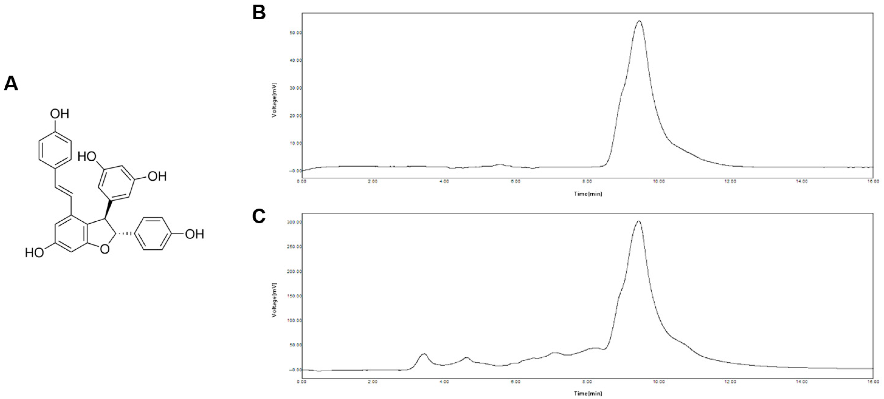
The chemical structure of ε-Viniferin is illustrated in Figure 2A. The high-performance liquid chromatography (HPLC) analysis was conducted to confirm the presence of ε-Viniferin in the grape flower-derived extract, Vino Chocolate™. The ε-Viniferin standard displayed a single, distinct peak at approximately 9.5 min, indicative of a high purity (Figure 2B). The chromatogram of Vino Chocolate™ (Figure 2C) revealed a prominent peak at the same retention time, confirming that ε-Viniferin is a major constituent of the extract. Additionally, several smaller peaks were observed between 3 and 8 min, suggesting the presence of other phytochemicals co-extracted alongside ε-Viniferin. The substantial intensity of the 9.5 min peak underscores the high relative abundance of ε-Viniferin in the Vino Chocolate™ extract. The quantitative HPLC analysis demonstrated ε-Viniferin to be present at approximately 547.58 ppm, whereas resveratrol was detected at a significantly lower concentration of 3.50 ppm, and δ-viniferin was not detected.

3.2. Effect of ε-Viniferin and Vino Chocolate™ on Cell Viability in HaCaT and HDFn Cells

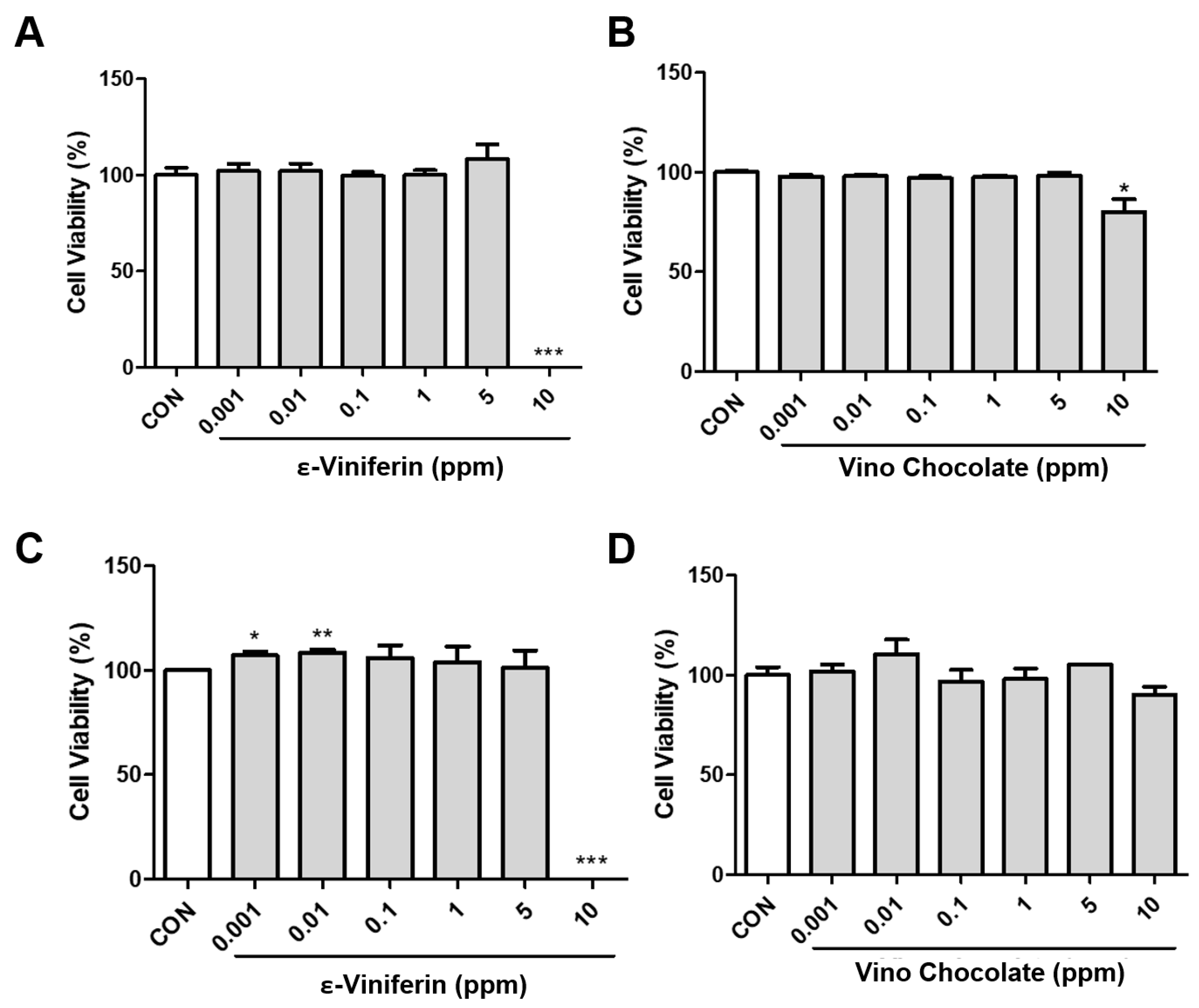
To determine suitable concentrations for subsequent experiments, we initially evaluated the cytotoxicity of ε-Viniferin and Vino Chocolate™ in HaCaT keratinocytes and HDFn fibroblasts using the WST-1 cell viability assay. Cells were treated with various concentrations (0.001, 0.01, 0.1, 0.5, 1, 5, and 10 ppm) for 24, 48 (Figure S1), and 72 h (Figure 3). Significant cytotoxicity was observed at the highest tested concentration (10 ppm), particularly at later time points. Based on these findings, a concentration of 5 ppm was selected as the maximum dose for subsequent experiments.

3.3. ε-Viniferin and Vino Chocolate™ Promote Wound Healing in Human Dermal Fibroblasts

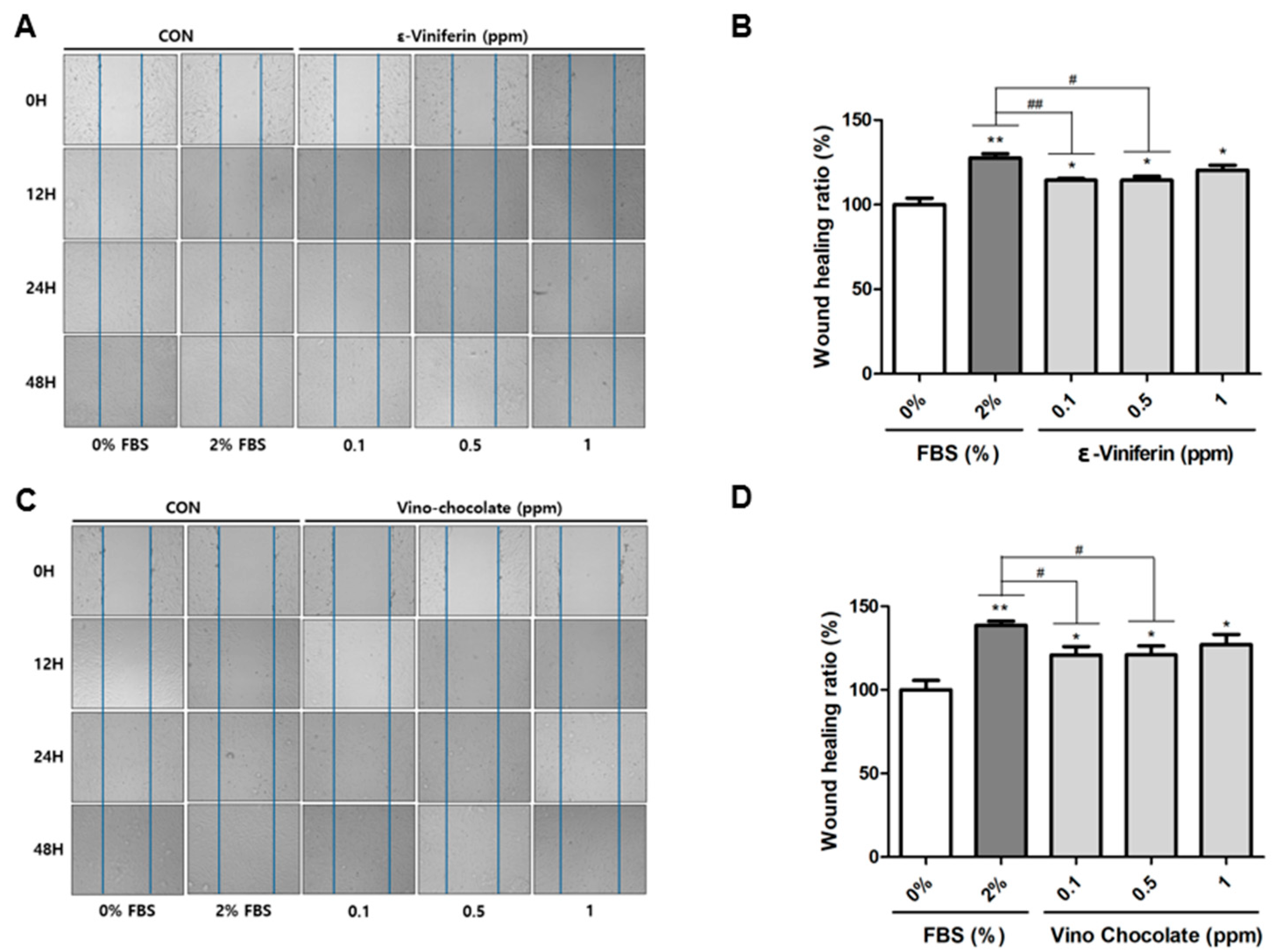
Skin fibroblasts are essential for maintaining skin homeostasis and play a central role in tissue repair. Age-related declines in the fibroblast migratory capacity are associated with impaired wound healing, as evidenced by a reduced regenerative ability in aged skin. To evaluate the effects of ε-Viniferin and Vino Chocolate™ on fibroblast migration, a scratch wound assay was performed using human dermal fibroblasts (HDFn). Cells cultured in a medium supplemented with 2% fetal bovine serum (FBS) served as a positive control, while the FBS-free medium was used for treatment groups. The cell migration was assessed at 0, 12, 24, and 48 h following the treatment with either ε-Viniferin or Vino Chocolate™ (Figure 4A,C).
The treatment with ε-Viniferin resulted in a concentration-dependent enhancement of the wound closure, with 0.1, 0.5, and 1 ppm all significantly promoting fibroblast migration (Figure 4B). When the wound healing ratio of the untreated control (0% FBS) was set at 100%, the positive control (2% FBS) achieved 127%. Notably, the 1 ppm ε-Viniferin treatment reached approximately 120% wound closure, closely matching the positive control.
Similarly, the Vino Chocolate™ treatment enhanced the cell migration in a concentration-dependent manner across all tested doses (Figure 4D). The positive control group showed a 138% healing ratio, whereas the 1 ppm Vino Chocolate™ achieved an approximately 127% wound closure, indicating a comparable efficacy to the FBS-stimulated positive control. The statistical analysis revealed no significant difference in the wound healing efficacy between the 1 ppm ε-Viniferin or Vino Chocolate™ groups and the 2% FBS-treated positive control (p > 0.05), further supporting the comparable regenerative potential of these treatments.
These results suggest that both ε-Viniferin and Vino Chocolate™ may support skin regenerative processes by promoting fibroblast migration in vitro, indicating their potential utility as functional cosmetic ingredients.

3.4. ε-Viniferin and Vino Chocolate™ Enhance Pro-Collagen Type I Synthesis in Human Dermal Fibroblasts

Skin aging is characterized by both a quantitative reduction and structural disorganization of collagen fibers. To assess the effects of ε-Viniferin and Vino Chocolate™ on collagen biosynthesis, HDFn cells were treated for 24 h, and levels of pro-collagen type I alpha 1 (COL1A1) were quantified using the ELISA. The transforming growth factor-beta 1 (TGF-β1), a well-known inducer of extracellular matrix production, was used as a positive control.
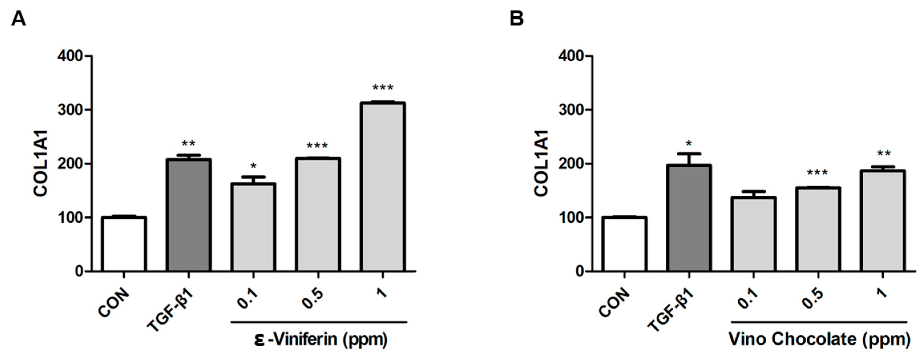
The treatment with both ε-Viniferin and Vino Chocolate™ resulted in a dose-dependent increase in pro-collagen synthesis (Figure 5). Specifically, the 1 ppm ε-Viniferin elevated COL1A1 levels to approximately 212.34% relative to the untreated control, surpassing the effect of TGF-β1 (Figure 5A). Vino Chocolate™ also significantly enhanced the pro-collagen expression, with a maximal increase of 86.82% at 1 ppm compared to the control (Figure 5B).
These findings suggest that ε-Viniferin and Vino Chocolate™ support collagen-related cellular activities in dermal fibroblasts, indicating their potential as functional ingredients in skincare formulations.

3.5. ε-Viniferin and Vino Chocolate™ Suppress MMP-1 Expression in Human Dermal Fibroblasts

The excessive expression of matrix metalloproteinase-1 (MMP-1) disrupts the extracellular matrix (ECM) integrity by degrading type I collagen, thereby impairing the wound healing process. Retinoic acid (RA), a well-established inhibitor of MMP-1, was used as a positive control to evaluate the anti-degradative and wound-repairing effects of the test compounds [38,39].
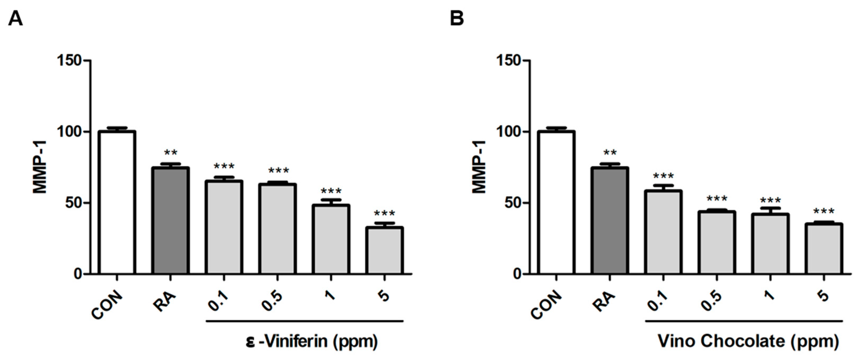
The ε-Viniferin treatment significantly downregulated the MMP-1 expression in HDFn cells in a dose-dependent manner (Figure 6A). At 5 ppm, MMP-1 levels were reduced by approximately 50% compared to the untreated control, showing an even greater inhibition than the RA-treated group.
Similarly, Vino Chocolate™ also suppressed the MMP-1 expression in a concentration-dependent manner (Figure 6B). Inhibition was detectable from 0.1 ppm and became more pronounced with increasing concentrations. At 5 ppm, MMP-1 levels were comparable to or even lower than those observed in the RA group.
These results suggest that both ε-Viniferin and Vino Chocolate™ may support ECM stability by modulating the MMP-1 expression, indicating potential benefits in skin care formulations targeting visible signs of aging.

3.6. ε-Viniferin and Vino Chocolate™ Suppress MMP-3 Expression in Human Dermal Fibroblasts

Matrix metalloproteinase-3 (MMP-3) contributes to the degradation of multiple extracellular matrix (ECM) components, including proteoglycans, laminin, and fibronectin, thereby compromising dermal structural integrity and delaying wound repair [40,41]. In addition, MMP-3 acts as an upstream activator of other MMPs, such as MMP-1 and MMP-9, amplifying matrix degradation and inflammatory responses in chronic wounds [42]. Retinoic acid (RA), a known downregulator of MMP-3 expression, was used as a positive control in this study.
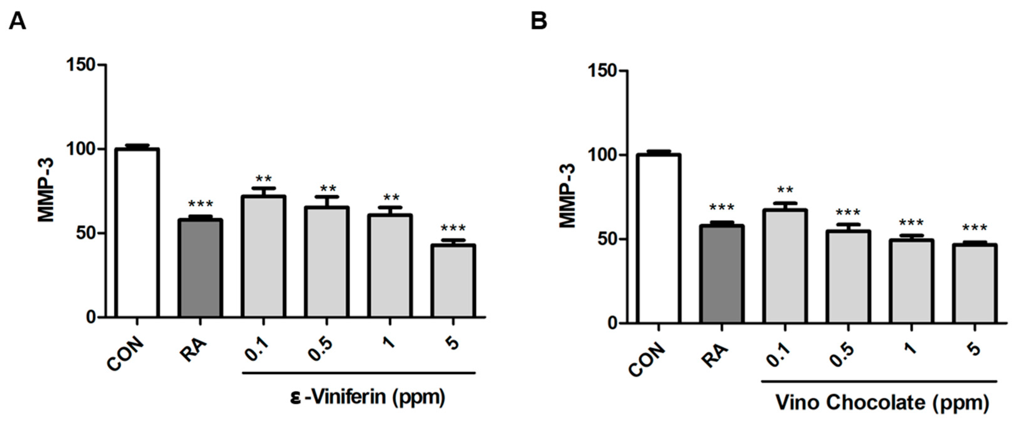
The treatment with ε-Viniferin significantly suppressed the MMP-3 expression in HDFn cells in a dose-dependent manner (Figure 7A). At 5 ppm, ε-Viniferin reduced MMP-3 levels below those observed in the RA-treated group, indicating a potent inhibitory activity. Similarly, the Vino Chocolate™ treatment led to the marked suppression of the MMP-3 expression at all tested concentrations (Figure 7B). Inhibition was evident starting from 0.1 ppm and became more pronounced with increasing doses, with 5 ppm achieving levels comparable to or lower than those induced by RA.
These results indicate that both ε-Viniferin and Vino Chocolate™ may contribute to ECM maintenance by modulating the MMP-3 expression, suggesting their potential utility in advanced skincare formulations.

3.7. ε-Viniferin and Vino Chocolate™ Enhanced Filaggrin Protein Levels in Human Keratinocytes

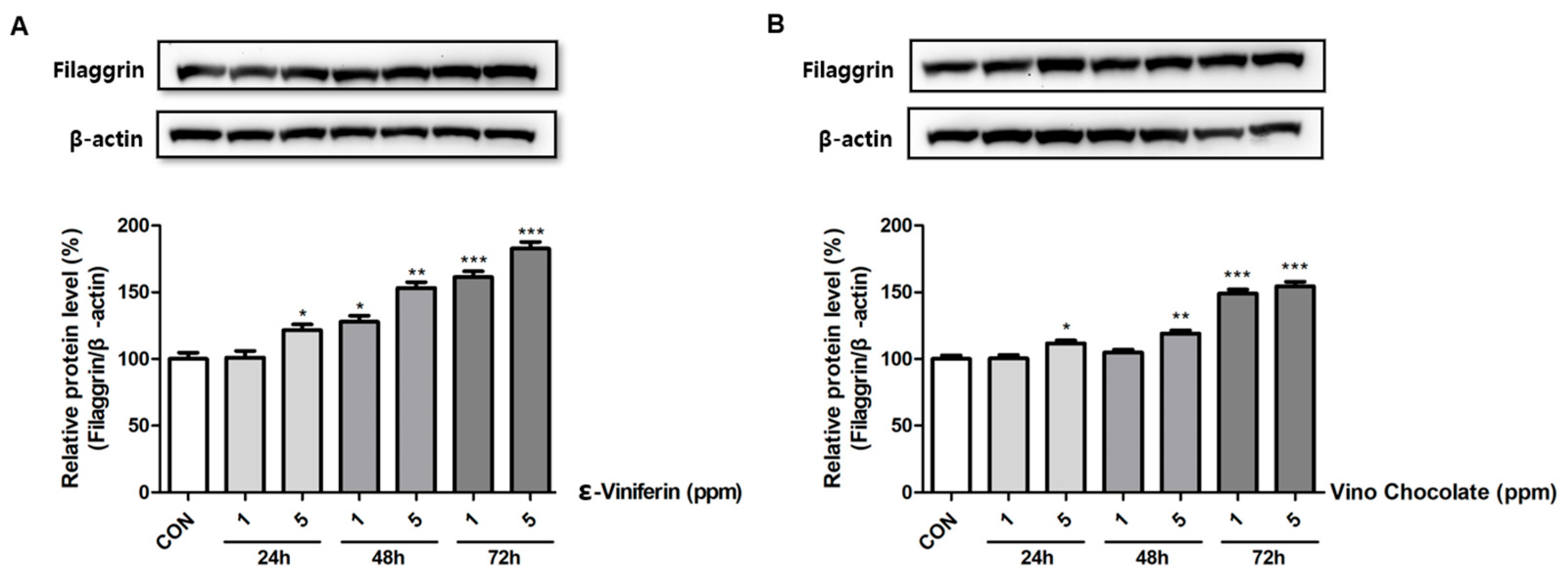
Filaggrin is a critical epidermal protein involved in skin barrier restoration and re-epithelialization during wound healing [43]. To investigate whether ε-Viniferin and Vino Chocolate™ promote epidermal repair, HaCaT keratinocytes were treated with 1 and 5 ppm of each compound for 24, 48, and 72 h, and filaggrin protein levels were assessed by the Western blot analysis. The ε-Viniferin treatment induced a time- and dose-dependent upregulation of the filaggrin expression, reaching its peak at 5 ppm after 72 h (Figure 8A). A similar trend was observed following the Vino Chocolate™ treatment, which also significantly upregulated the filaggrin expression, particularly at the highest dose and longest incubation period (Figure 8B).
The densitometric analysis confirmed that both compounds increased the filaggrin expression in keratinocytes, suggesting a potential utility in formulations targeting skin barrier maintenance and recovery support.

4. Discussion

This study highlights the wound healing and skin-regenerative potential of ε-Viniferin and Vino Chocolate™, a grape flower-derived plant cell culture extract enriched in ε-Viniferin. Using in vitro assays on human dermal fibroblasts (HDFn) and keratinocytes (HaCaT), both treatments significantly promoted essential regenerative processes, including fibroblast migration, collagen synthesis, the suppression of matrix metalloproteinases (MMPs), and the enhancement of the epidermal barrier function.
In the initial cell viability assay (Figure 3), ε-Viniferin and Vino Chocolate™ maintained cell viability up to 5 ppm, confirming their safety for further experimentation. In the wound healing assay (Figure 4), both compounds significantly promoted fibroblast migration in a dose-dependent manner. At 1 ppm, their wound closure efficacy was comparable to or even exceeded that of the 2% FBS positive control. These findings underscore the re-epithelialization potential of both agents and are consistent with previous reports on the stimulatory effects of resveratrol derivatives on dermal fibroblast activity and tissue remodeling [44,45].
Pro-collagen type I (COL1A1), the precursor of the most abundant structural collagen in human dermis, was significantly upregulated upon treatment with both ε-Viniferin and Vino Chocolate™ (Figure 5). Notably, ε-Viniferin at 1 ppm induced a 212.34% increase in COL1A1 production compared to the untreated control, surpassing even the effect of TGF-β1. Vino Chocolate™ also enhanced collagen synthesis, albeit to a lesser extent than ε-Viniferin. These findings suggest that the stilbene dimer ε-Viniferin may promote dermal matrix remodeling by activating TGF-β/Smad signaling pathways and concurrently inhibiting collagen-degrading enzymes such as matrix metalloproteinases [46]. Matrix metalloproteinases (MMPs), particularly MMP-1 and MMP-3, play a central role in ECM degradation and are known to be upregulated in photoaged or chronically wounded skin [7,47]. In our study, both ε-Viniferin and Vino Chocolate™ markedly suppressed the MMP-1 and MMP-3 expression in a concentration-dependent manner (Figure 6 and Figure 7), with the highest inhibition observed at 5 ppm—surpassing even the effect of retinoic acid.
Matrix metalloproteinases (MMPs), particularly MMP-1 and MMP-3, play a central role in ECM degradation and are known to be upregulated in photoaged or chronically wounded skin [7,47]. In our study, both ε-Viniferin and Vino Chocolate™ markedly suppressed the MMP-1 and MMP-3 expression in a concentration-dependent manner (Figure 6 and Figure 7), with the highest inhibition observed at 5 ppm—surpassing even the effect of retinoic acid.
Notably, MMP-3, which acts as an upstream activator of other MMPs including MMP-1 and MMP-9, exhibited a heightened sensitivity to both treatments. This suggests that the test compounds may not only preserve ECM structural integrity but also attenuate the cascade of matrix degradation and chronic inflammation associated with impaired wound healing [48].
Notably, these findings suggest that ε-Viniferin and Vino Chocolate™ may contribute to maintaining ECM stability by modulating MMP activity, offering potential benefits for cosmetic applications targeting age-related skin changes and environmental stress.
The Western blot analysis demonstrated a significant upregulation of the filaggrin protein expression in HaCaT cells treated with both ε-Viniferin and Vino Chocolate™ (Figure 8). The effect was most pronounced at 5 ppm after 72 h of treatment. Filaggrin, a key structural protein involved in the terminal differentiation of keratinocytes and epidermal barrier formation [7], was strongly induced by both compounds. These results suggest that ε-Viniferin and Vino Chocolate™ may help support skin barrier function, particularly under conditions that mimic aging-related or environmentally induced stress.
The superior performance of ε-Viniferin observed across multiple assays may be attributed to its well-defined molecular structure and specific biological activity. In contrast, Vino Chocolate™, being a complex botanical extract, may exert its effects via a broader spectrum of phytochemicals acting synergistically. The high-performance liquid chromatography (HPLC) analysis confirmed a substantial ε-Viniferin content of 547.58 ppm (Figure 2), while additional minor peaks suggest the presence of other bioactive compounds, collectively supporting its multifunctional profile.
While this study primarily focused on the wound healing efficacy of Vino Chocolate™, a grape flower-derived plant cell culture extract enriched with ε-Viniferin, we acknowledge that the extract contains a variety of other polyphenolic compounds that may also contribute to its overall bioactivity. Given the complexity of botanical extracts, it is plausible that synergistic or additive interactions among these constituents, including flavonoids and stilbenes beyond ε-Viniferin, may play a role in enhancing biological outcomes. Future studies involving comprehensive metabolomic profiling and bioactivity-guided fractionation will be essential to elucidate the contribution of these additional components and their potential synergistic mechanisms. Such investigations may provide deeper insight into the full therapeutic potential of Vino Chocolate™ and support the rational design of optimized multifunctional cosmeceutical formulations.
Furthermore, the utilization of grape flower-derived plant cell cultures represents an environmentally sustainable and industrially scalable strategy for sourcing functional bioactives for skincare applications. This biotechnological approach ensures consistent quality, enhanced safety, and improved metabolite accumulation through elicitation techniques, offering a reliable alternative to traditional plant extraction methods [49].
Collectively, our findings indicate that both ε-Viniferin and Vino Chocolate™ represent promising multifunctional bioactives for skin regeneration. Their demonstrated ability to enhance fibroblast migration, stimulate collagen synthesis, inhibit MMP activity, and upregulate epidermal barrier proteins positions them as strong candidates for inclusion in advanced cosmeceutical formulations. To better understand the mechanisms behind these bioactivities, further in vivo and mechanistic studies focusing on key signaling pathways such as MAPK, NF-κB, and TGF-β are needed [50,51]. These pathways are critical because MAPK is involved in cell proliferation and migration, NF-κB regulates inflammatory responses, and TGF-β plays a central role in collagen synthesis and extracellular matrix remodeling [52,53,54].

5. Conclusions

In summary, both ε-Viniferin and Vino Chocolate™, an ε-Viniferin and exosome-enriched extract derived from grape flower cell cultures, demonstrated significant wound healing and skin-regenerative effects in human keratinocytes and fibroblasts. These effects included enhanced fibroblast migration, increased pro-collagen type I synthesis, the suppression of the MMP-1 and MMP-3 expression, and the upregulation of filaggrin, a key marker of epidermal barrier function. The well-defined molecular efficacy of ε-Viniferin and the synergistic activity of Vino Chocolate™ collectively underscore their potential as multifunctional bioactive ingredients in cosmeceutical applications. Given their promising in vitro efficacy and favorable biocompatibility profile, further in vivo and clinical investigations may help clarify their potential role in supporting skin regeneration through advanced cosmetic formulations.

Supplementary Materials

The following supporting information can be downloaded at https://www.mdpi.com/article/10.3390/cosmetics12050181/s1: Figure S1: ε-Viniferin and Vino Chocolate are non-toxic to cells from 1 ppm to 5 ppm.

Author Contributions

Conceptualization, J.H. and M.Y.; methodology, D.K., J.L. and G.K.; software, J.H., D.K. and G.K.; validation, D.K., J.L. and G.K.; formal analysis, D.K. and J.L.; investigation, D.K., J.L. and K.L.; resources, J.L. and G.K.; data curation, D.K. and J.L.; writing—original draft preparation, D.K. and J.L.; writing—review and editing, J.H., D.K. and J.L.; visualization, D.K., J.L. and K.L.; supervision, J.H. and J.P.; project administration, J.H. and M.Y. All authors have read and agreed to the published version of the manuscript.

Funding

This work was supported in part by the Bio & Medical Technology Development Program (RS-2024-00440478) of the National Research Foundation (NRF) funded by the Korean government (MSIT) and by additional support through the Innopolis Foundation through the ‘2024 project for Rapid Support of Sustainable Growth in the Jeonbuk Innopolis’, funded by the Ministry of science and ICT (RS-2024-IN245096).

Institutional Review Board Statement

Not applicable.

Informed Consent Statement

Not applicable.

Data Availability Statement

The data presented in this study are available on request from the corresponding author.

Conflicts of Interest

The authors Jimin Lim, Gisol Kim, Jaeho Pyee, and Minkyoung You are employees of iNGR Co., Ltd. Among them, Jimin Lim played a major role in both the experimental procedures and manuscript preparation, while Gisol Kim contributed to a portion of the experimental work. The remaining authors declare that the research was conducted in the absence of any commercial or financial relationships that could be construed as potential conflicts of interest.

Abbreviations

The following abbreviations are used in this manuscript:
HDFnHuman Dermal Fibroblast, Neonatal
HaCaTHigh Sensitivity of Human Epidermal Keratinocytes
ECMExtracellular Matrix
MMPsMatrix Metalloproteinases
COL1A1Pro-collagen Type I Alpha 1
ELISAEnzyme-Linked Immunosorbent Assay
HPLCHigh-Performance Liquid Chromatography
RARetinoic Acid
TGF-β1Transforming Growth Factor-Beta 1

References

  1. Proksch, E.; Brandner, J.M.; Jensen, J.M. The skin: An indispensable barrier. Exp. Dermatol. 2008, 17, 1063–1072. [Google Scholar] [CrossRef]
  2. Nestle, F.O.; Di Meglio, P.; Qin, J.Z.; Nickoloff, B.J. Skin immune sentinels in health and disease. Nat. Rev. Immunol. 2009, 9, 679–691. [Google Scholar] [CrossRef] [PubMed]
  3. Gurtner, G.C.; Werner, S.; Barrandon, Y.; Longaker, M.T. Wound repair and regeneration. Nature 2008, 453, 314–321. [Google Scholar] [CrossRef] [PubMed]
  4. Eming, S.A.; Krieg, T.; Davidson, J.M. Inflammation in wound repair: Molecular and cellular mechanisms. J. Investig. Dermatol. 2007, 127, 514–525. [Google Scholar] [CrossRef]
  5. Werner, S.; Grose, R. Regulation of wound healing by growth factors and cytokines. Physiol. Rev. 2003, 83, 835–870. [Google Scholar] [CrossRef]
  6. Martin, P. Wound healing--aiming for perfect skin regeneration. Science 1997, 276, 75–81. [Google Scholar] [CrossRef]
  7. Quan, T.; Qin, Z.; Xia, W.; Shao, Y.; Voorhees, J.J.; Fisher, G.J. Matrix-degrading metalloproteinases in photoaging. J. Investig. Dermatol. Symp. Proc. 2009, 14, 20–24. [Google Scholar] [CrossRef]
  8. Kim, J.; Lee, C.W.; Kim, E.K.; Lee, S.J.; Park, N.H.; Kim, H.S.; Kim, H.K.; Char, K.H.; Jang, Y.P.; Kim, J.W. Inhibition effect of Gynura procumbens extract on UV-B-induced matrix-metalloproteinase expression in human dermal fibroblasts. J. Ethnopharmacol. 2011, 137, 427–433. [Google Scholar] [CrossRef]
  9. Rawlings, A.V.; Harding, C.R. Moisturization and skin barrier function. Dermatol. Ther. 2004, 17, 43–48. [Google Scholar] [CrossRef]
  10. Vičanová, J.; Boelsma, E.; Mommaas, A.M.; Kempenaar, J.A.; Forslind, B.; Pallon, J.; Egelrud, T.; Koerten, H.K.; Ponec, M. Normalization of epidermal calcium distribution profile in reconstructed human epidermis is related to improvement of terminal differentiation and stratum corneum barrier formation. J. Investig. Dermatol. 1998, 111, 97–106. [Google Scholar] [CrossRef] [PubMed]
  11. Moloney, S.J.; Edmonds, S.H.; Giddens, L.D.; Learn, D.B. The hairless mouse model of photoaging: Evaluation of the relationship between dermal elastin, collagen, skin thickness and wrinkles. Photochem. Photobiol. 1992, 56, 505–511. [Google Scholar] [CrossRef]
  12. Mukherjee, P.K.; Verpoorte, R.; Suresh, B. Evaluation of in-vivo wound healing activity of Hypericum patulum leaf extract on different wound model in rats. J. Ethnopharmacol. 2000, 70, 315–321. [Google Scholar] [CrossRef]
  13. Choi, S.W.; Son, B.W.; Son, Y.S.; Park, Y.I.; Lee, S.K.; Chung, M.H. The wound-healing effect of a glycoprotein fraction isolated from aloe vera. Br. J. Dermatol. 2001, 145, 535–545. [Google Scholar] [CrossRef]
  14. Pandey, K.B.; Rizvi, S.I. Plant polyphenols as dietary antioxidants in human health and disease. Oxid. Med. Cell. Longev. 2009, 2, 270–278. [Google Scholar] [CrossRef]
  15. Rao, S.R.; Ravishankar, G.A. Plant cell cultures: Chemical factories of secondary metabolites. Biotechnol. Adv. 2002, 20, 101–153. [Google Scholar] [CrossRef]
  16. Kolewe, M.E.; Gaurav, V.; Roberts, S.C. Pharmaceutically active natural product synthesis and supply via plant cell culture technology. Mol. Pharm. 2008, 5, 243–256. [Google Scholar] [CrossRef]
  17. Jeong, Y.J.; Park, S.H.; Park, S.-C.; Kim, S.; Kim, Y.-G.; Kim, S.W.; Lee, J.; Ryu, Y.B.; Jeong, J.C.; Kim, C.Y. Induced Extracellular Production of Stilbenes in Grapevine Cell Culture Medium by Elicitation with Methyl Jasmonate and Stevioside. Bioresour. Bioprocess. 2020, 7, 38. [Google Scholar] [CrossRef]
  18. Park, S.H.; Jeong, Y.J.; Park, S.-C.; Kim, S.; Kim, Y.-G.; Shin, G.; Jeong, H.J.; Ryu, Y.B.; Lee, J.; Lee, O.R.; et al. Highly Efficient Bioconversion of trans-Resveratrol to δ-Viniferin Using Conditioned Medium of Grapevine Callus Suspension Cultures. Int. J. Mol. Sci. 2022, 23, 4403. [Google Scholar] [CrossRef] [PubMed]
  19. Bapat, V.A.; Kishor, P.B.K.; Jalaja, N.; Jain, S.M.; Penna, S. Plant Cell Cultures: Biofactories for the Production of Bioactive Compounds. Agronomy 2023, 13, 858. [Google Scholar] [CrossRef]
  20. Russo, C.; Maugeri, A.; Albergamo, A.; Dugo, G.; Navarra, M.; Cirmi, S. Protective Effects of a Red Grape Juice Extract against Bisphenol A-Induced Toxicity in Human Umbilical Vein Endothelial Cells. Toxics 2023, 11, 391. [Google Scholar] [CrossRef] [PubMed]
  21. Piazza, S.; Fumagalli, M.; Khalilpour, S.; Martinelli, G.; Magnavacca, A.; Dell’Agli, M.; Sangiovanni, E. A Review of the Potential Benefits of Plants Producing Berries in Skin Disorders. Antioxidants 2020, 9, 542. [Google Scholar] [CrossRef]
  22. Kim, H.J.; Chang, M.J.; Choi, Y.H.; Chung, Y.S. Antioxidant and antiobesity activities of seed extract from Campbell Early grape (Vitis labrusca × Vitis vinifera). J. Food Biochem. 2011, 35, 1201–1209. [Google Scholar] [CrossRef]
  23. Sy, B.; Krisa, S.; Richard, T.; Courtois, A. Resveratrol, ε-Viniferin, and Vitisin B from Vine: Comparison of Their In Vitro Antioxidant Activities and Study of Their Interactions. Molecules 2023, 28, 7521. [Google Scholar] [CrossRef] [PubMed]
  24. Gabaston, J.; Cantos-Villar, E.; Biais, B.; Waffo-Teguo, P.; Renouf, E.; Corio-Costet, M.-F.; Richard, T.; Mérillon, J.-M. Stilbenes from Vitis vinifera L. Waste: A Sustainable Tool for Controlling Plasmopara viticola. J. Agric. Food Chem. 2017, 65, 2711–2718. [Google Scholar] [CrossRef]
  25. Subedi, L.; Lee, T.H.; Wahedi, H.M.; Baek, S.H.; Kim, S.Y. Resveratrol-Enriched Rice Attenuates UVB-ROS-Induced Skin Aging via Downregulation of Inflammatory Cascades. Oxid. Med. Cell. Longev. 2017, 2017, 8379539. [Google Scholar] [CrossRef] [PubMed]
  26. Ramakrishna, A.; Ravishankar, G.A. Influence of abiotic stress signals on secondary metabolites in plants. Plant Signal. Behav. 2011, 6, 1720–1731. [Google Scholar] [CrossRef]
  27. Murthy, H.N.; Lee, E.J.; Paek, K.Y. Production of secondary metabolites from cell and organ cultures: Strategies and approaches for biomass improvement and metabolite accumulation. Plant Cell Tissue Organ Cult. 2014, 118, 1–16. [Google Scholar] [CrossRef]
  28. Eibl, R.; Meier, P.; Stutz, I.; Schildberger, D.; Hühn, T.; Eibl, D. Plant cell culture technology in the cosmetics and food industries: Current state and future trends. Appl. Microbiol. Biotechnol. 2018, 102, 8661–8675. [Google Scholar] [CrossRef]
  29. Pezzuto, J.M. Resveratrol: Twenty Years of Growth, Development and Controversy. Biomol. Ther. 2019, 27, 1–14. [Google Scholar] [CrossRef]
  30. Gambini, J.; Inglés, M.; Olaso-González, G.; Lopez-Grueso, R.; Bonet-Costa, V.; Gimeno-Mallench, L.; Mas-Bargues, C.; Abdelaziz, K.M.; El Alami, M.; Borras, C.; et al. Properties of resveratrol: In vitro and in vivo studies about metabolism, bioavailability, and biological effects in animal models and humans. Oxid. Med. Cell. Longev. 2015, 2015, 837042. [Google Scholar] [CrossRef]
  31. Morazzoni, P.; Vanzani, P.; Santinello, S.; Gucciardi, A.; Zennaro, L.; Miotto, G.; Ursini, F. Grape Seeds Proanthocyanidins: Advanced Technological Preparation and Analytical Characterization. Antioxidants 2021, 10, 418. [Google Scholar] [CrossRef]
  32. Sangiovanni, E.; Di Lorenzo, C.; Piazza, S.; Manzoni, Y.; Brunelli, C.; Fumagalli, M.; Magnavacca, A.; Martinelli, G.; Colombo, F.; Casiraghi, A.; et al. Vitis vinifera L. Leaf Extract Inhibits In Vitro Mediators of Inflammation and Oxidative Stress Involved in Inflammatory-Based Skin Diseases. Antioxidants 2019, 8, 134. [Google Scholar] [CrossRef]
  33. Thring, T.S.A.; Hili, P.; Naughton, D.P. Anti-collagenase, anti-elastase and anti-oxidant activities of extracts from 21 plants. BMC Complement. Altern. Med. 2009, 9, 27. [Google Scholar] [CrossRef]
  34. Jiang, L.; Jeon, D.; Kim, J.; Lee, C.W.; Peng, Y.; Seo, J.; Lee, J.H.; Paik, J.H.; Kim, C.Y.; Lee, J. Pyomelanin-Producing Brevundimonas vitisensis sp. nov., Isolated from Grape (Vitis vinifera L.). Front. Microbiol. 2021, 12, 733612. [Google Scholar] [CrossRef] [PubMed]
  35. Espinosa-Leal, C.A.; Puente-Garza, C.A.; García-Lara, S. In vitro plant tissue culture: Means for production of biological active compounds. Planta 2018, 248, 1–18. [Google Scholar] [CrossRef] [PubMed]
  36. Sharifi-Rad, J.; Sureda, A.; Tenore, G.C.; Daglia, M.; Sharifi-Rad, M.; Valussi, M.; Tundis, R.; Loizzo, M.R.; Ademiluyi, A.O.; Sharifi-Rad, R.; et al. Plants of the genus Zingiber as potential sources of bioactive phytochemicals: From tradition to pharmacy. Molecules 2017, 22, 2145. [Google Scholar] [CrossRef] [PubMed]
  37. Zhang, Y.; Wang, Y.; Li, Y.; Wang, Y.; Zhang, Y.; Wang, Y. Angelica dahurica Ethanolic Extract Improves Impaired Wound Healing by Activating Angiogenesis in Diabetes. PLoS ONE 2017, 12, e0177862. [Google Scholar] [CrossRef] [PubMed]
  38. Lateef, H.; Stevens, M.J.; Varani, J. All-trans-retinoic acid suppresses matrix metalloproteinase activity and increases collagen synthesis in diabetic human skin in organ culture. Am. J. Pathol. 2004, 165, 167–174. [Google Scholar] [CrossRef]
  39. Fisher, G.J.; Talwar, H.S.; Lin, J.; Lin, P.; McPhillips, F.; Wang, Z.; Li, X.; Wan, Y.; Kang, S.; Voorhees, J.J. Retinoic acid inhibits UV-induced MMP-1 production in human skin in vivo. J. Clin. Investig. 1998, 101, 1432–1440. [Google Scholar] [CrossRef]
  40. Madlener, M. Differential expression of matrix metalloproteinases and their physiological inhibitors in acute murine skin wounds. Arch. Dermatol. Res. 1998, 290 (Suppl. 1), S24–S29. [Google Scholar] [CrossRef]
  41. Caley, M.P.; Martins, V.L.C.; O’Toole, E.A. Metalloproteinases and wound healing. Adv. Wound Care 2015, 4, 225–234. [Google Scholar] [CrossRef] [PubMed]
  42. Madlener, M.; Parks, W.C.; Werner, S. Matrix metalloproteinases and their physiological inhibitors are differentially expressed during excisional skin wound repair. Exp. Cell Res. 1998, 242, 201–210. [Google Scholar] [CrossRef]
  43. Kurokawa, I.; Mizutani, H.; Kusumoto, K.; Nishijima, S.; Tsujita-Kyutoku, M.; Shikata, N.; Tsubura, A. Cytokeratin, filaggrin, and p63 expression in reepithelialization during human cutaneous wound healing. Wound Repair Regen. 2006, 14, 38–45. [Google Scholar] [CrossRef]
  44. Kaleci, B.; Koyuturk, M. Efficacy of Resveratrol in the Wound Healing Process by Reducing Oxidative Stress and Promoting Fibroblast Cell Proliferation and Migration. Dermatol. Ther. 2020, 33, e14357. [Google Scholar] [CrossRef]
  45. Lin, M.-H.; Hung, C.-F.; Sung, H.-C.; Yang, S.-C.; Yu, H.-P.; Fang, J.-Y. The Bioactivities of Resveratrol and Its Naturally Occurring Derivatives on Skin. J. Food Drug Anal. 2021, 29, 15–38. [Google Scholar] [CrossRef] [PubMed]
  46. Kim, H.J.; Chang, E.J.; Cho, S.H.; Chung, S.K.; Park, H.D. Protective Effects of Vitis amurensis Rupr. Stem Extract against UVB-Induced Damage in Human Dermal Fibroblasts via Regulation of the TGF-β/Smad Pathway. J. Food Biochem. 2021, 45, e13597. [Google Scholar] [CrossRef]
  47. Krutmann, J.; Bouloc, A.; Sore, G.; Bernard, B.A.; Passeron, T. The Skin Aging Exposome. J. Dermatol. Sci. 2017, 85, 152–161. [Google Scholar] [CrossRef]
  48. Zhao, R.; Liang, H.; Clarke, E.; Jackson, C.; Xue, M. Inflammation in Chronic Wounds. Int. J. Mol. Sci. 2016, 17, 2085. [Google Scholar] [CrossRef]
  49. Ramirez-Estrada, K.; Vidal-Limón, H.; Hidalgo, D.; Moyano, E.; Goleniosiewski, M.; Cusidó, R.M.; Palazón, J. Elicitation, an Effective Strategy for the Biotechnological Production of Bioactive High-Added-Value Compounds in Plant Cell Factories. Molecules 2016, 21, 182. [Google Scholar] [CrossRef]
  50. Pignet, A.-L.; Schellnegger, M.; Hecker, A.; Kohlhauser, M.; Kotzbeck, P.; Kamolz, L.-P. Resveratrol-Induced Signal Transduction in Wound Healing. Int. J. Mol. Sci. 2021, 22, 12614. [Google Scholar] [CrossRef]
  51. Jia, Y.; Shao, J.-H.; Zhang, K.-W.; Zou, M.-L.; Teng, Y.-Y.; Tian, F.; Chen, M.-N.; Yuan, Z.-D.; Wu, J.-J.; Guo, Z.-R. Emerging Effects of Resveratrol on Wound Healing: A Comprehensive Review. Molecules 2022, 27, 6736. [Google Scholar] [CrossRef]
  52. Liu, X.; Chen, B.; Liu, X.; Zhang, X.; Wu, J. Interplay between MAPK signaling pathway and autophagy in skin aging: Mechanistic insights and therapeutic implications. Front. Cell Dev. Biol. 2025, 13, 1625357. [Google Scholar] [CrossRef] [PubMed]
  53. Pohlers, D.; Brenmoehl, J.; Löffler, I.; Müller, C.K.; Leipner, C.; Schultze-Mosgau, S.; Stallmach, A.; Kinne, R.W.; Wolf, G. TGF-β and fibrosis in different organs—Molecular pathway imprints. Biochim. Biophys. Acta (BBA)-Mol. Basis Dis. 2009, 1792, 746–756. [Google Scholar] [CrossRef] [PubMed]
  54. Giarratana, A.O.; Prendergast, C.M.; Salvatore, M.M.; Capaccione, K.M. TGF-β signaling: Critical nexus of fibrogenesis and cancer. J. Transl. Med. 2024, 22, 594. [Google Scholar] [CrossRef] [PubMed]
Figure 1. Schematic representation of the preparation of ε-Viniferin and Vino Chocolate™ extract from Vitis labruscana B. ‘Campbell Early’. ε-Viniferin is a purified resveratrol dimer, whereas Vino Chocolate™ is a viniferin-enriched extract produced through a multi-stage plant cell suspension culture process involving callus induction, bioreactor scale-up, and bioactive enhancement.
Cosmetics 12 00181 g001
Figure 2. (A) Chemical structure of ε-Viniferin. (B) HPLC chromatogram of ε-Viniferin standard showing a single, sharp peak at approximately 9.5 min. (C) HPLC chromatogram of Vino Chocolate™, highlighting a major peak at the same retention time (~9.5 min), confirming the presence of ε-Viniferin. Several minor peaks observed between 3 and 8 min indicate the presence of additional co-extracted phytochemicals.
Cosmetics 12 00181 g002
Figure 3. Effects of ε-Viniferin and Vino Chocolate™ on the viability of HaCaT (A,B) and HDFn (C,D) cells. Cells were treated with ε-Viniferin (A,C) or Vino Chocolate™ (B,D) at concentrations ranging from 0.001 to 10 ppm for 72 h. Cell viability was assessed using the WST-1 assay. Data are presented as mean ± SD (n = 3). Statistical analysis was performed using one-way ANOVA. * p < 0.05, ** p < 0.01, and *** p < 0.001 vs. untreated control.
Cosmetics 12 00181 g003
Figure 4. Effects of ε-Viniferin and Vino Chocolate™ on wound healing. The cell migration of HDFn cells was assessed using a scratch assay after the treatment with different concentrations of each compound. (A,C) Microscopic images of scratched cells at 0, 12, 24, and 48 h after treatment with ε-Viniferin (A) and Vino Chocolate™ (C). The blue line indicates the initial wound margin. (B,D) The quantification of the wound closure corresponding to images in (A) and (C), respectively. Data are presented as the mean ± SD (n = 3). The statistical analysis was performed using a one-way ANOVA. * p < 0.05, and ** p < 0.01vs. 0% FBS control and # p < 0.05, and ## p < 0.01 vs. 2% FBS positive control.
Cosmetics 12 00181 g004
Figure 5. Effects of ε-Viniferin and Vino Chocolate™ on pro-collagen type I (COL1A1) expression in human dermal fibroblasts. HDFn cells were treated with ε-Viniferin (A) or Vino Chocolate™ (B) for 24 h, and COL1A1 levels were quantified using the ELISA. Transforming growth factor-β1 (TGF-β1, 10 ng/mL) was used as a positive control. Data are expressed as percentages relative to the untreated control and shown as the mean ± SD (n = 3). * p < 0.05, ** p < 0.01, and *** p < 0.001 vs. the untreated control.
Cosmetics 12 00181 g005
Figure 6. Inhibitory effects of ε-Viniferin and Vino Chocolate™ on MMP-1 expression in HDFn cells. HDFn cells were treated with ε-Viniferin (A) or Vino Chocolate™ (B) at concentrations of 0.1, 0.5, 1, and 5 ppm for 24 h. Retinoic acid (RA, 1 μM) was used as a positive control. MMP-1 levels were quantified by ELISA and expressed as percentages relative to the untreated control (CON). Both compounds significantly reduced MMP-1 expression in a dose-dependent manner. Data are presented as mean ± SD (n = 3). ** p < 0.01 and *** p < 0.001 vs. control.
Cosmetics 12 00181 g006
Figure 7. Inhibitory effects of ε-Viniferin and Vino Chocolate™ on MMP-3 expression in HDFn cells. HDFn cells were treated with ε-Viniferin (A) or Vino Chocolate™ (B) at concentrations of 0.1, 0.5, 1, and 5 ppm for 24 h. Retinoic acid (RA, 1 μM) was used as a positive control. MMP-3 levels were measured by the ELISA and are expressed as percentages relative to the untreated control (CON). Both treatments significantly reduced MMP-3 expression in a dose-dependent manner. Data are presented as the mean ± SD (n = 3). ** p < 0.01 and *** p < 0.001 vs. control.
Cosmetics 12 00181 g007
Figure 8. ε-Viniferin (A) and Vino Chocolate™ (B) upregulate filaggrin expression in HaCaT keratinocytes. HaCaT cells were treated with ε-Viniferin or Vino Chocolate™ at concentrations of 1 and 5 ppm for 24, 48, and 72 h. Filaggrin protein levels were analyzed by the Western blot, and β-actin was used as a loading control. Bar graphs represent the densitometric quantification of the filaggrin expression normalized to β-actin. Data are presented as the mean ± SD (n = 3). * p < 0.05, ** p < 0.01, and *** p < 0.001 vs. untreated control.
Cosmetics 12 00181 g008
Table 1. HPLC Pump Gradient Program Settings.
StepTime (min)Flow Rate (mL/min)%A%B
100.55842
2110.55842
3120.50100
4140.50100
5150.55842
6160.55842
Disclaimer/Publisher’s Note: The statements, opinions and data contained in all publications are solely those of the individual author(s) and contributor(s) and not of MDPI and/or the editor(s). MDPI and/or the editor(s) disclaim responsibility for any injury to people or property resulting from any ideas, methods, instructions or products referred to in the content.