Article

Effect of SMART DNA Therapy Retix.C Application on Skin Microbiome

by
Dorota Sobolewska-Sztychny
1,2,
Karolina Wódz
3 and
Aleksandra Lesiak
1,2,*
1
Dermoklinika Medical Center, 90-436 Lodz, Poland
2
Department of Dermatology, Pediatric Dermatology and Oncology, Laboratory of Autoinflammatory, Genetic and Rare Skin Disorders, Medical University of Lodz, 90-419 Lodz, Poland
3
Laboratory of Molecular Biology, Vet-Lab Brudzew, 62-720 Brudzew, Poland
*
Author to whom correspondence should be addressed.
Cosmetics 2025, 12(5), 178; https://doi.org/10.3390/cosmetics12050178
Submission received: 4 July 2025 / Revised: 19 August 2025 / Accepted: 20 August 2025 / Published: 22 August 2025
(This article belongs to the Special Issue Feature Papers in Cosmetics in 2025)

Abstract

Background: The skin microbiome plays a key role in maintaining skin health, and its composition can be influenced by cosmetic treatments. This study aimed to investigate the effects of SMART DNA Therapy treatment on facial skin microbiome composition, with specific focus on changes in commensal and pathogenic bacterial populations following multi-component anti-aging intervention. Methods: This clinical study included 10 Caucasian female participants aged 28–50 years (Clinical trial registration number: 0406/2023). Each participant received three Retix.C SMART DNA THERAPY treatments at 14-day intervals over 6 weeks. The protocol included three phases: chemical peeling with ferulic acid, peptide microinjections for DNA repair, and home-care products with antioxidants. Bacterial samples were collected from forehead and cheek skin before treatment and 2 weeks after the final treatment. Samples were analyzed using bacterial culture and PCR methods. Results: After treatment, the skin microbiome showed beneficial changes with increased numbers of helpful bacteria and elimination of harmful bacteria: complete removal of Cutibacterium acnes and Staphylococcus aureus was observed, Staphylococcus epidermidis and other beneficial bacteria increased on both forehead and cheek areas. Overall bacterial diversity decreased, and participants exhibited more similar microbiome patterns after treatment. Conclusions: SMART DNA Therapy treatment successfully modified the skin microbiome by increasing protective bacteria and eliminating pathogenic species. The treatment may support skin health through microbiome modulation and the potential antioxidant effects of its active ingredients, although these were not directly assessed in this study.

1. Introduction

The cutaneous microbiome plays a critical role in maintaining skin homeostasis, barrier function, and immune regulation [1,2,3]. Dominated by commensal organisms such as Staphylococcus epidermidis, Cutibacterium acnes, and various Corynebacterium species, this complex microbial ecosystem provides protection against pathogenic colonization, produces antimicrobial peptides, and modulates local immune responses [4,5,6]. Disruption of microbiome composition has been associated with inflammatory skin conditions, compromised barrier integrity, and accelerated aging processes [7,8,9]. Topical interventions, including cosmeceutical treatments and dermatological procedures, can significantly influence cutaneous microbial communities [10,11]. Chemical peels, bioactive compounds, and peptide-based formulations may alter the skin’s microenvironment through changes in pH, lipid composition, and cellular turnover rates, consequently affecting microbial growth patterns and species distribution [12,13].
Understanding these interactions is essential for optimizing therapeutic outcomes while maintaining beneficial microbial populations. Modern anti-aging therapies increasingly incorporate multi-component approaches that combine antioxidants, peptides, and growth factors to address cellular damage and promote regeneration [14]. However, the impact of such comprehensive treatments on skin microbiome composition remains poorly understood.
While these interventions effectively target cellular repair mechanisms and barrier enhancement, their effects on commensal and pathogenic bacterial populations require systematic investigation to ensure therapeutic benefits do not compromise microbial homeostasis. Previous research by Schagen [15] has demonstrated that topical antioxidant peptides, particularly carnosine and N-acetylcarnosine, exhibit significant protective effects in cosmetic applications, with clinical studies showing enhanced antioxidant capacity and protective effects against oxidative damage.
These findings support the premise that antioxidant peptide formulations can modulate skin cellular responses and potentially influence the cutaneous microenvironment that supports beneficial microbial populations. The SMART DNA Therapy protocol represents a multi-phase treatment combining advanced chemical peeling with ferulic acid-based formulations, peptide microinjections targeting DNA repair pathways, and specialized home-care products containing antioxidant complexes. This comprehensive approach aims to address multiple aspects of skin aging while promoting cellular protection and regeneration. Given the protocol’s multi-component nature and direct skin contact, its potential influence on cutaneous microbiome warrants comprehensive evaluation. The present study investigates the effects of SMART DNA Therapy treatment on facial skin microbiome composition, with a specific focus on changes in commensal and pathogenic bacterial populations. We hypothesize that this multi-component therapeutic intervention will selectively modulate microbiome composition, potentially enhancing beneficial bacterial populations while reducing pathogenic species, thereby supporting both anti-aging objectives and microbiome health.
Unlike many traditional anti-aging treatments that rely solely on topical antioxidant formulations or single-step chemical peels, SMART DNA Therapy utilizes a structured, three-phase protocol. It combines a ferulic acid-based chemical peel, DNA-repair-targeted peptide microinjections, and antioxidant-rich home-care products. The inclusion of peptides mimicking FOXO3 protein function and advanced radical scavengers enables both cellular protection and direct modulation of skin physiology. This integrative approach offers potentially superior efficacy not only in reversing signs of aging, but also in reshaping the skin microenvironment, making it a novel candidate for influencing the cutaneous microbiome.

2. Materials and Methods

2.1. Cosmetic Products Used in the Study

The following commercial products were used in the study:
  • Retix.C SMART DNA AOX Peel (brand: Retix.C, manufacturer: URGO Sp. z o.o., Warsaw, Poland)—chemical peel containing ferulic acid as the main active ingredient (exact concentration is proprietary information of the manufacturer). Product purchased directly from the manufacturer.
  • Retix.C SMART DNA Meso Peptide Cocktail (Retix.C, URGO Sp. z o.o., Warsaw, Poland)—sterile solution for microinjection containing a proprietary blend of peptides and ferulic acid (exact concentration is proprietary information of the manufacturer). Product purchased from the manufacturer.
In addition, the study used a cosmetic product applied by the participants in self-use:
  • Ferulic Triple-C SPF Cream (Retix.C, URGO Sp. z o.o., Warsaw, Poland)—antioxidant sunscreen containing vitamin C derivatives and ferulic acid (exact concentration is proprietary information of the manufacturer). Provided for home use.
All products were registered in the EU CPNP system. All products were provided by URGO Sp. z o.o. (Warsaw, Poland) for research use within the framework of the funded project.

2.2. Study Design

The Retix.C SMART DNA THERAPY treatment protocol consisted of three sequential phases. The initial phase involved the application of an advanced chemical peel formulated to provide potent anti-aging stimulation and ameliorate signs of exogenous skin aging. The peel formulation was composed of ferulic acid as the primary active component, enhanced by a synergistic peptide and transforming growth factor β2 (TGF-β2), which demonstrated an identical metabolic pathway to retinoids. This preliminary step facilitated enhanced penetration of subsequent active compounds delivered via the microinjection technique while conferring prophylactic protection against future manifestations of skin aging. The second phase comprised the administration of a sterile microinjection cocktail containing a proprietary blend of peptides synergistically enhanced with ferulic acid. This formulation provided comprehensive cellular protection against DNA damage induced by both exogenous and endogenous factors through peptide-mediated neutralization of cytotoxic radicals. The key peptide component mimicked the biological activity of forkhead box O3 (FOXO3) protein, thereby upregulating gene expression within DNA repair pathways. In vitro studies demonstrated complete radical scavenging activity (100% ABTS inhibition), confirming the potent antioxidant properties of this formulation. The third phase involved the daily home-care application of Retix.C Ferulic Triple C serum. Optimal post-treatment recovery required strict photoprotection through consistent application and reapplication of broad-spectrum sunscreen products, supplemented with antioxidant-containing formulations. The Retix.C Ferulic Triple C SPF cream provided comprehensive solar radiation protection through a dual mechanism of action: UV radiation absorption coupled with antioxidant activity, thereby preventing reactive oxygen species-mediated cascade reactions and ensuring cellular protection. This study was conducted in accordance with Good Clinical Practice (GCP) guidelines and was approved by the Bioethics Committee at the Medical University of Lodz (approval number: RNN/109/23, dated 18 April 2023). Clinical trial registration number: 0406/2023.

2.3. Selection of Study Group

This clinical study was conducted in a cohort of 10 female participants aged 28–50 years. Participants were classified according to Fitzpatrick skin phototype classification as follows: Type II (n = 8) and Type III (n = 2).

2.3.1. Inclusion Criteria

Eligible participants were required to be in good general health with no clinically significant medical conditions as determined by physician evaluation, and to provide written informed consent for study participation. To ensure study integrity, participants were required to have no relation to the research team and could not be medical students. Comprehensive medical histories were obtained from all enrolled subjects following informed consent procedures.

2.3.2. Exclusion Criteria

Subjects were excluded if they presented with any of the following: pregnancy or lactation, known hypersensitivity or allergic reactions to any study product components, active febrile conditions or inflammatory states, current or recent oral isotretinoin or retinoid therapy, active viral or bacterial infections, active inflammatory dermatoses, autoimmune disorders, chronic systemic diseases or dermatological conditions, compromised skin integrity including irritation, damage, solar erythema, open wounds or active infections, history of abnormal scarring or keloid formation, or recent dermatological procedures such as eyebrow depilation, cosmetic tattooing, or permanent makeup applications.

2.4. Treatment Schedule

Each participant received three Retix.C SMART DNA THERAPY treatments administered at 14-day intervals over a 6-week study period. Throughout the inter-treatment intervals, participants were instructed to follow a standardized home-care regimen consisting of twice-daily application of Ferulic Triple C serum and Renewal TGF Cream following facial cleansing with Dual Action Gentle Cleanser, supplemented by daily photoprotection using Ferulic Triple C SPF cream.
Microbiological sampling was conducted at baseline (prior to each treatment session) and at 2 weeks post-final treatment using standardized procedures. Duplicate swab specimens were obtained for aerobic bacterial and fungal culture, as well as for anaerobic bacterial detection. Skin surface scrapings were collected for microscopic analysis, and additional swab specimens were obtained for polymerase chain reaction (PCR) analysis.

2.5. Effectiveness Evaluation

The study cohort comprised 10 Caucasian female participants aged 28–50 years (mean age 39.5 years). Participants were classified according to Fitzpatrick skin phototype classification as follows: Type II (n = 8) and Type III (n = 2).

Microbiological Sampling and Analysis

Microbiological sampling focused primarily on bacterial populations, with additional screening for fungal organisms. Bacterial sampling was performed from standardized anatomical sites on the forehead and cheeks. A defined 4.4 × 4.4 cm area within each designated region was systematically sampled using sterile swabs pre-moistened with sterile saline solution, employing a standardized Z-pattern sampling technique. Duplicate specimen sets were collected to enable separate detection of aerobic bacteria and fungi versus anaerobic bacteria. Skin surface scrapings were obtained from a 4 cm2 area using sterile disposable scalpel blades (No. 15), with collected material subsequently transferred using cotton-tipped applicators moistened with 0.9% sodium chloride solution. Additional swab specimens were collected specifically for polymerase chain reaction (PCR) analysis (Figure 1).
All samples were cultured onto Columbia Agar with 5% Sheep Blood and CNA Agar (both from Graso, Starogard Gdański, Poland). Selective Sabouraud Agar (SABGC) and Brilliance Candida Agar (both from OXOID, Thermo Fisher Scientific, Waltman, MA, USA) were used for the presumptive identification of Candida spp. Plates were incubated at 37 °C for 24 h under aerobic conditions.
Bacterial isolates were initially identified based on colony morphology, hemolysis type, and catalase and oxidase reactions. Phenotypic confirmation of Staphylococcus aureus was performed using the PASTOREX™ STAPH-PLUS rapid agglutination test (Bio-Rad, Hercules, CA, USA). Final identification of isolated strains was carried out with the VITEK 2 Compact system using GP, GN, ANC, CBC, and YST cards (bioMérieux, Craponne, France).
A real-time PCR method based on the detection of genes specific for E. coli, S. aureus (both from Primerdesign, Chandler’s Ford, United Kingdom Staphylococcus spp. (Ingenetix, Wien, Austria) was used. DNA was extracted from bacterial cells using an automated magnetic isolation method (MagnifiQ™ Pathogen kit, A&A Biotechnology, Gdansk, Poland) and Nucleic Acid Purification System—Auto-Pure 96 (Hangzhou Allsheng Instruments, Wuxi, China) and real-time PCR using Applied 7500FAST (ThermoFisher, Waltham, MA, USA). PCR conditions: 50 cycles: 95 °C for 10 s and 60 °C for 1 min. for E. coli, S. aureus, 95 °C for 2 min., 45 cycles: 95 °C for 5 s and 60 °C for 1 min. for Staphylococcus spp. and 95 °C for 2 min. Fluorescence detection in channels FAM (target) and HEX (internal control).
Bacterial presence was described using semi-quantitative terms such as “few” (light growth; ≤10 colonies), “moderately numerous” (moderate growth; 11–99 colonies) and “numerous” (heavy growth; ≥100 colonies), based on relative abundance observed at each timepoint. These descriptors were not derived from quantitative colony counts or molecular thresholds. Absence in culture or PCR does not confirm eradication but only indicates that the bacteria were not detected at the time of sampling. Given the small sample size and exploratory nature of this study, formal statistical testing was not performed. The primary goal was to observe general microbiome trends before and after treatment.

3. Results

To evaluate treatment effects on the cutaneous microbiome, comparative analysis of microbial communities from forehead and cheek regions was performed. Baseline relative abundance of bacterial taxa demonstrated consistent distribution patterns across participants. The predominant microbial populations belonged to the genus Staphylococcus, including S. epidermidis, S. hominis subsp. hominis, S. aureus, S. capitis, S. intermedius, and S. lugdunensis (Table 1).
Bacterial species distribution patterns on forehead skin before and after treatment are shown in Figure 2.
Additional taxa identified included Streptococcus (S. mitis), Micrococcus (M. luteus), Dermacoccus spp., Kocuria (K. kristinae), Corynebacterium (C. tuberculostearicum, C. amycolatum, C. striatum), Dermabacter (D. hominis), Cutibacterium (C. acnes), and Pseudomonas (P. aeruginosa).
Similar patterns were observed for cheek skin samples before (A) and after (B) treatment (Figure 3).
Post-treatment microbiome analysis revealed reduced microbial diversity across both forehead and cheek regions (Table 1). Furthermore, increased inter-participant similarity in microbiome composition was observed following treatment completion. Complete eradication of obligate anaerobic bacteria (C. acnes) and facultative anaerobic bacteria (S. aureus) was documented post-treatment. Similar elimination was observed for M. restricta, while C. albicans persisted in post-treatment samples. PCR analysis confirmed the absence of Mycoplasma spp. and Chlamydia spp. in all samples throughout the study period. Figure 2 and Figure 3B collectively visualize the microbiological shifts observed in both regions and support the compositional trends described in Table 1. No adverse events or skin irritations were observed during the treatment period, confirming the safety profile of the intervention.
Changes in bacterial presence before and after treatment are illustrated in Figure 2 and Figure 3. Figure 2 shows results for the forehead area, while Figure 3 presents corresponding findings for the cheek. In both regions, a reduction in S. aureus and C. acnes was observed, accompanied by a relative increase in S. epidermidis abundance. These findings are consistent with the trends described in Table 1.

4. Discussion

This study demonstrated beneficial modulation of the cutaneous microbiome following treatment, characterized by increased abundance of commensal bacteria and elimination of pathogenic organisms. These compositional changes may support cutaneous health by enhancing colonization resistance, promoting immune balance, and improving skin barrier function. Consequently, this therapeutic intervention functions primarily as a microbiome-modulating treatment. It may also act as an antioxidant-promoting modality based on the properties of its components, such as ferulic acid and peptides. However, antioxidant activity was not directly assessed in this study [3,15,16,17,18]. Such microbiome alterations may also contribute to cutaneous protection, based on mechanisms described in the literature—such as the role of Staphylococcus epidermidis in modulating antioxidant and immune pathways [16]. S. epidermidis is known to produce antimicrobial peptides (AMPs) and to regulate keratinocyte signaling, contributing to redox balance, epidermal differentiation, and barrier repair [16,19]. In parallel, the observed reduction or elimination of pathogenic species such as S. aureus and C. acnes may help decrease local inflammation and reactive oxygen species (ROS) production, both of which are associated with skin barrier disruption and premature aging [3,18]. Finally, the establishment of a more balanced and uniform commensal microbiota across participants may reinforce barrier function and innate immune responses through enhanced microbial–epithelial interactions, contributing to skin homeostasis and resilience [16]. Within the immunomodulatory context, the cutaneous microbiome assumes critical significance. As the largest organ system, the skin serves dual functions as a protective barrier and interface mediating host-environment interactions. Commensal microorganisms, particularly S. epidermidis, provide protection against pathogenic colonization while modulating immune system functionality. Previous research by Severn and Horswill [16] has demonstrated that S. epidermidis actively coordinates skin response to injury and maintains barrier integrity through multiple mechanisms, including stimulation of neutrophil CXCL10 signaling and recruitment of type I interferon-producing plasmacytoid dendritic cells. These findings support our observed enhancement of beneficial S. epidermidis populations following treatment. The observed post-treatment increase in S. epidermidis abundance carries significant immunological implications. This species produces immunomodulatory molecules, including antimicrobial peptides (AMPs), which enhance barrier integrity and stimulate host defense responses. AMP production, regulated through microbiome–keratinocyte interactions, represents a critical component of anti-infectious defense mechanisms [19,20,21,22].
Furthermore, commensal bacteria such as S. epidermidis modulate immune homeostasis by promoting immune tolerance through regulatory T cell (Treg) development. These cells are essential for preventing excessive inflammatory and autoimmune responses. The cutaneous microbiome thus maintains the delicate balance between immune activation and tolerance, fundamental to both local and systemic health [16,19,23,24]. The documented elimination of pathogenic species, including Staphylococcus aureus and Cutibacterium acnes, following treatment demonstrates therapeutic efficacy in microbiome remodeling toward a more favorable composition. As extensively characterized by Severn and Horswill [16], S. aureus represents a significant pathogenic threat due to its inflammatory potential and ability to disrupt skin barrier function, while S. epidermidis provides colonization resistance through direct antimicrobial action and immune modulation [16]. Furthermore, the selective preservation and enhancement of S. epidermidis populations is particularly significant given that Nakatsuji et al. [25] identified specific strains capable of producing bioactive molecules with tumor-suppressive properties, suggesting that our therapeutic intervention may favor protective microbial phenotypes. While the observed reduction in microbiome diversity might initially suggest an unfavorable outcome, the concurrent increase in beneficial commensal organisms, particularly S. epidermidis, indicates selective microbiome modulation rather than indiscriminate reduction. The enhanced inter-participant similarity in microbiome composition suggests treatment-induced stabilization and homogenization, which may confer immunomodulatory benefits through the establishment of a more predictable and beneficial microbial ecosystem [16,25,26].
While these findings are promising, the study’s limitations include the small sample size and the absence of a parallel untreated control group. The pre-post design enabled intra-subject comparison, but future randomized studies with control arms are warranted to confirm the observed microbiome shifts and better isolate the effects of individual treatment components.

5. Conclusions

This study demonstrated that Retix.C SMART DNA THERAPY treatment produced beneficial modulation of the cutaneous microbiome, characterized by increased abundance of commensal bacterial populations and elimination of pathogenic organisms. These compositional changes may support cutaneous health by enhancing colonization resistance, promoting immune balance, and improving skin barrier function. Therefore, this therapeutic intervention represents a microbiome-modulating treatment and may have antioxidant potential due to its active ingredients, such as ferulic acid and peptides. However, since antioxidant activity was not directly evaluated in this study, further research is needed to confirm this effect.

Author Contributions

Conceptualization, D.S.-S. and A.L.; methodology, D.S.-S., K.W. and A.L.; software, D.S.-S. and K.W.; validation, D.S.-S. and K.W.; formal analysis, A.L.; investigation, D.S.-S., K.W. and A.L.; resources, A.L.; data curation, D.S.-S.; writing—original draft preparation, D.S.-S.; writing—review and editing, D.S.-S. and A.L.; visualization, D.S.-S. and K.W.; supervision, A.L.; project administration, D.S.-S. and A.L.; funding acquisition, A.L. All authors have read and agreed to the published version of the manuscript.

Funding

The study was funded by the Medical University of Lodz, grant no. 503/1-064-01/503-51-001-19-00 and URGO Sp. z o.o., Aleje Jerozolimskie 142B, 02-305 Warsaw, Poland.

Institutional Review Board Statement

The study was conducted in accordance with the Declaration of Helsinki, and approved by the Bioethics Committee of the Medical University of Lodz (protocol code: RNN/109/23 and date of approval: 18 April 2023). The tested product was registered in the European Commission CPNP database under registration number 3973035.

Informed Consent Statement

Informed consent was obtained from all subjects involved in the study.

Data Availability Statement

The datasets used and/or analyzed during the current study are available from the corresponding author on reasonable request.

Conflicts of Interest

The authors declare no conflicts of interest.

Abbreviations

The following abbreviations are used in this manuscript:
AMPantimicrobial peptide
FOXO3forkhead box O3
PCRpolymerase chain reaction
SPFsun protection factor
TGF-β2transforming growth factor β2

References

  1. Smythe, P.; Wilkinson, H.N. The skin microbiome: Current landscape and future opportunities. Int. J. Mol. Sci. 2023, 24, 3950. [Google Scholar] [CrossRef]
  2. Zhang, X.E.; Zheng, P.; Ye, S.Z.; Ma, X.; Liu, E.; Pang, Y.-B.; He, Q.-Y.; Zhang, Y.-X.; Li, W.-Q.; Zeng, J.-H.; et al. Microbiome: Role in inflammatory skin diseases. J. Inflamm. Res. 2024, 17, 1057–1082. [Google Scholar] [CrossRef]
  3. Lee, H.J.; Kim, M. Skin barrier function and the microbiome. Int. J. Mol. Sci. 2022, 23, 13071. [Google Scholar] [CrossRef]
  4. Fournière, M.; Latire, T.; Souak, D.; Feuilloley, M.G.J.; Bedoux, G. Staphylococcus epidermidis and Cutibacterium acnes: Two major sentinels of skin microbiota and the influence of cosmetics. Microorganisms 2020, 8, 1752. [Google Scholar] [CrossRef]
  5. Joshi, A.A.; Vocanson, M.; Nicolas, J.F.; Wolf, P.; Patra, V. Microbial derived antimicrobial peptides as potential therapeutics in atopic dermatitis. Front. Immunol. 2023, 14, 1125635. [Google Scholar] [CrossRef]
  6. Harris-Tryon, T.A.; Grice, E.A. Microbiota and maintenance of skin barrier function. Science 2022, 376, 940–945. [Google Scholar] [CrossRef]
  7. Myers, T.; Bouslimani, A.; Huang, S.; Hansen, S.T.; Clavaud, C.; Azouaoui, A.; Ott, A.; Gueniche, A.; Bouez, C.; Zheng, Q.; et al. A multi-study analysis enables identification of potential microbial features associated with skin aging signs. Front. Aging 2024, 4, 1304705. [Google Scholar] [CrossRef]
  8. Woo, Y.R.; Kim, H.S. Interaction between the microbiota and the skin barrier in aging skin: A comprehensive review. Front. Physiol. 2024, 15, 1322205. [Google Scholar] [CrossRef]
  9. De Pessemier, B.; Grine, L.; Debaere, M.; Maes, A.; Paetzold, B.; Callewaert, C. Gut-skin axis: Current knowledge of the interrelationship between microbial dysbiosis and skin conditions. Microorganisms 2021, 9, 353. [Google Scholar] [CrossRef]
  10. SanMiguel, A.J.; Meisel, J.S.; Horwinski, J.; Zheng, Q.; Grice, E.A. Topical antimicrobial treatments can elicit shifts to resident skin bacterial communities and reduce colonization by Staphylococcus aureus competitors. Antimicrob. Agents Chemother. 2017, 61, e00774-17. [Google Scholar] [CrossRef]
  11. Hwang, B.K.; Lee, S.; Myoung, J.; Hwang, S.J.; Lim, J.M.; Jeong, E.T.; Park, S.G.; Youn, S.H. Effect of the skincare product on facial skin microbial structure and biophysical parameters: A pilot study. Microbiologyopen 2021, 10, e1236. [Google Scholar] [CrossRef]
  12. Rendon, M.I.; Berson, D.S.; Cohen, J.L.; Roberts, W.E.; Starker, I.; Wang, B. Evidence and considerations in the application of chemical peels in skin disorders and aesthetic resurfacing. J. Clin. Aesthet. Dermatol. 2010, 3, 32–41. [Google Scholar]
  13. Baldwin, H.E.; Bhatia, N.C.; Friedman, A.; Prunty, T.; Martin, R.; Seite, S. The role of cutaneous microbiota harmony in maintaining a functional skin barrier. J. Drugs Dermatol. 2017, 16, 12–18. [Google Scholar] [CrossRef]
  14. Sorg, H.; Tilkorn, D.J.; Hager, S.; Hauser, J.; Mirastschijski, U. Skin wound healing: An update on the current knowledge and concepts. Eur. Surg. Res. 2017, 58, 81–94. [Google Scholar] [CrossRef]
  15. Schagen, S.K. Topical peptide treatments with effective anti-aging results. Cosmetics 2017, 4, 16. [Google Scholar] [CrossRef]
  16. Severn, M.M.; Horswill, A.R. Staphylococcus epidermidis and its dual lifestyle in skin health and infection. Nat. Rev. Microbiol. 2023, 21, 97–111. [Google Scholar] [CrossRef]
  17. Nakamizo, S.; Egawa, G.; Honda, T.; Nakajima, S.; Belkaid, Y.; Kabashima, K. Commensal bacteria and cutaneous immunity. Semin. Immunopathol. 2015, 37, 73–80. [Google Scholar] [CrossRef]
  18. Azzimonti, B.; Ballacchino, C.; Zanetta, P.; Cucci, M.A.; Monge, C.; Grattarola, M.; Dianzani, C.; Barrera, G.; Pizzimenti, S. Microbiota, oxidative stress, and skin cancer: An unexpected triangle. Antioxidants 2023, 12, 546. [Google Scholar] [CrossRef]
  19. Pastar, I.; O’Neill, K.; Padula, L.; Head, C.R.; Burgess, J.L.; Chen, V.; Garcia, D.; Stojadinovic, O.; Hower, S.; Plano, G.V.; et al. Staphylococcus epidermidis boosts innate immune response by activation of gamma delta T cells and induction of perforin-2 in human skin. Front. Immunol. 2020, 11, 550946. [Google Scholar] [CrossRef]
  20. Nguyen, H.L.T.; Trujillo-Paez, J.V.; Umehara, Y.; Yue, H.; Peng, G.; Kiatsurayanon, C.; Chieosilapatham, P.; Song, P.; Okumura, K.; Ogawa, H.; et al. Role of antimicrobial peptides in skin barrier repair in individuals with atopic dermatitis. Int. J. Mol. Sci. 2020, 21, 7607. [Google Scholar] [CrossRef]
  21. Simanski, M.; Erkens, A.S.; Rademacher, F.; Harder, J. Staphylococcus epidermidis-induced interleukin-1 beta and human beta-defensin-2 expression in human keratinocytes is regulated by the host molecule A20 (TNFAIP3). Acta Derm. Venereol. 2019, 99, 181–187. [Google Scholar] [CrossRef]
  22. Chessa, C.; Bodet, C.; Jousselin, C.; Wehbe, M.; Lévêque, N.; Garcia, M. Antiviral and immunomodulatory properties of antimicrobial peptides produced by human keratinocytes. Front. Microbiol. 2020, 11, 1155. [Google Scholar] [CrossRef]
  23. Leech, J.M.; Dhariwala, M.O.; Lowe, M.M.; Chu, K.; Merana, G.R.; Cornuot, C.; Weckel, A.; Ma, J.M.; Leitner, E.G.; Gonzalez, J.R.; et al. Toxin-triggered interleukin-1 receptor signaling enables early-life discrimination of pathogenic versus commensal skin bacteria. Cell Host Microbe 2019, 26, 795–809. [Google Scholar] [CrossRef]
  24. Singh, T.P.; Amorim, C.F.; Lovins, V.M.; Bradley, C.W.; Carvalho, L.P.; Carvalho, E.M.; Grice, E.A.; Scott, P. Regulatory T cells control Staphylococcus aureus and disease severity of cutaneous leishmaniasis. J. Exp. Med. 2023, 220, e20230035. [Google Scholar] [CrossRef]
  25. Nakatsuji, T.; Chen, T.H.; Butcher, A.M.; Trzoss, L.L.; Nam, S.-J.; Shirakawa, K.T.; Zhou, W.; Oh, J.; Otto, M.; Fenical, W.; et al. A commensal strain of Staphylococcus epidermidis protects against skin neoplasia. Sci. Adv. 2018, 4, eaao4502. [Google Scholar] [CrossRef]
  26. Dréno, B.; Araviiskaia, E.; Berardesca, E.; Gontijo, G.; Sanchez Viera, M.; Xiang, L.F.; Martin, R.; Bieber, T. Microbiome in healthy skin, update for dermatologists. J. Eur. Acad. Dermatol. Venereol. 2016, 30, 2038–2047. [Google Scholar] [CrossRef]
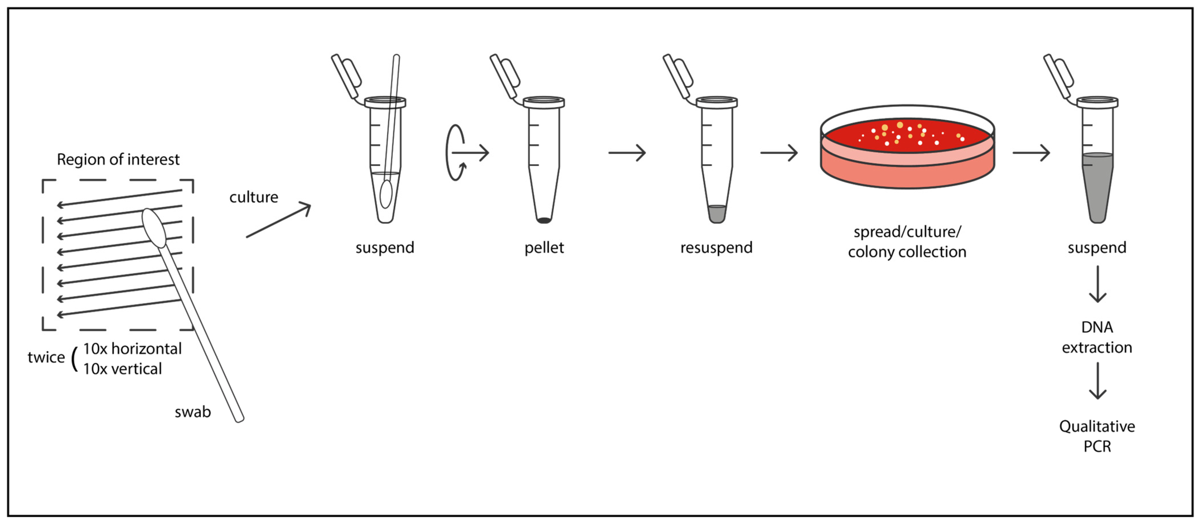
Figure 1. Schematic representation of the microbiological sampling and processing workflow used in the study.
Cosmetics 12 00178 g001
Figure 2. Detection of selected bacterial species on the forehead before (A) and after (B) treatment. Colors represent qualitative abundance categories: heavy growth (green), moderate growth (red), and light growth (blue). A post-treatment reduction in S. aureus and C. acnes was observed, with a relative increase in S. epidermidis across participants.
Cosmetics 12 00178 g002
Figure 3. Detection of selected bacterial species on the cheek before (A) and after (B) treatment. The data show reduced presence of potentially pathogenic bacteria and relative stabilization of commensal S. epidermidis levels.
Cosmetics 12 00178 g003
Table 1. Microbiome composition changes in forehead and cheek skin samples before and after Retix.C SMART DNA THERAPY treatment in 10 female participants. Bacterial abundance is reported qualitatively as numerous, moderately numerous, or few based on culture growth assessment. Abbreviations: S.—Staphylococcus; C.—Cutibacterium/Corynebacterium/Candida; M.—Micrococcus/Malassezia; P.—Pseudomonas; K.—Kocuria; D.—Dermabacter.
PatientBefore TreatmentAfter Treatment
1 foreheadS. epidermidis—numerous
S. hominis subsp. hominis—moderately numerous
S. aureus—a few
C. acnes—a few
Candida albicans—a few
S. epidermidis—numerous
S. hominis subsp. hominis—numerous
Candida albicans—a few
1 cheekS. epidermidis—numerous
S. hominis subsp. hominis—moderately numerous
C. acnes—a few
S. epidermidis—numerous
S. hominis subsp. hominis—numerous
2 foreheadS. epidermidis—numerous
S. hominis subsp. hominis—moderately numerous
Micrococcus spp.—moderately numerous
S. epidermidis—numerous
S. hominis subsp. hominis—numerous
Micrococcus spp.—moderately numerous
2 cheekS. epidermidis—numerous
S. hominis subsp. hominis—moderately numerous
Micrococcus spp.—moderately numerous
S. epidermidis—numerous
S. hominis subsp. hominis—numerous
3 foreheadS. epidermidis—numerous
S. hominis subsp. hominis—moderately numerous
Corynebacterium spp.—numerous
S. epidermidis—numerous
S. hominis subsp. hominis—numerous
Corynebacterium spp.—a few
3 cheekS. epidermidis—numerous
S. hominis subsp. hominis—moderately numerous
Corynebacterium spp.—moderately numerous
S. epidermidis—numerous
S. hominis subsp. hominis—moderately numerous
Corynebacterium spp.—moderately numerous
4 foreheadS. hominis subsp. hominis—moderately numerous Dermacoccus spp.—a few
Micrococcus spp.—a few
S. epidermidis—moderately numerous
S. hominis subsp. hominis—moderately numerous
Dermacoccus spp.—a few
4 cheekS. epidermidis—numerous
Dermacoccus spp.—a few
S. epidermidis—numerous
5 foreheadC. tuberculostearicum—a few
C. amycolatum—a few
S. capitis—moderately numerous
C. amycolatum—a few
5 cheekS. capitis—a few
S. mitis—a few
Micrococcus spp.—a few
S. capitis—numerous
Micrococcus spp.—a few
6 foreheadS. epidermidis—numerous
Streptococcus spp.—a few
C. acnes—a few
S. epidermidis—numerous
Streptococcus spp.—moderately numerous
C. albicans—a few
6 cheekS. epidermidis—numerous
Streptococcus spp.—a few
C. albicans—a few
S. epidermidis—numerous
S. lugdunensis—numerous
C. albicans—a few
7 foreheadS. epidermidis—numerousS. epidermidis—numerous
7 cheekS. epidermidis—numerous
P. aeruginosa—a few
S. epidermidis—numerous
8 foreheadS. epidermidis—numerous
D. hominis—moderately numerous
S. epidermidis—numerous
D. hominis—a few
8 cheekS. epidermidis—numerousS. epidermidis—numerous
9 foreheadS. epidermidis—numerous
S. intermedius—a few
C. acnes—moderately numerous
S. epidermidis—numerous
S. intermedius—numerous
9 cheekS. epidermidis—numerous
S. intermedius—a few
S. epidermidis—numerous
S. intermedius—moderately numerous
10 foreheadK. kristinae—moderately numerous
M. luteus—a few
C. striatum—numerous
M. restricta—a few
S. epidermidis—numerous
K. kristinae—moderately numerous M. luteus—a few
C. striatum—a few
10 cheekS. epidermidis —moderately numerous
M. luteus—a few
C. striatum—numerous
M. restricta—a few
S. epidermidis—numerous
K. kristinae—moderately numerous M. luteus—a few
Note: Qualitative abundance categories correspond to those used in Figure 2 and Figure 3—“numerous” (heavy growth; ≥100 colonies), “moderately numerous” (moderate growth; 11–99 colonies), and “few” (light growth; ≤10 colonies).
Disclaimer/Publisher’s Note: The statements, opinions and data contained in all publications are solely those of the individual author(s) and contributor(s) and not of MDPI and/or the editor(s). MDPI and/or the editor(s) disclaim responsibility for any injury to people or property resulting from any ideas, methods, instructions or products referred to in the content.