Article

Metagenomic Analysis of the Skin Microbiota of Brazilian Women: How to Develop Anti-Aging Cosmetics Based on This Knowledge?

by
Raquel Allen Garcia Barbeto Siqueira
1,
Ana Luiza Viana Pequeno
1,
Yasmin Rosa Santos
1,
Romualdo Morandi-Filho
2,
Alexandra Lan
3,
Edileia Bagatin
4,
Vânia Rodrigues Leite-Silva
5,6,
Newton Andreo-Filho
6 and
Patricia Santos Lopes
6,*
1
Programa de Pós-Graduação em Medicina Translacional, Departamento de Medicina, Escola Paulista de Medicina, Universidade Federal de São Paulo, São Paulo 04021-001, Brazil
2
A.C Camargo Cancer Center, São Paulo 01509-900, Brazil
3
Shanghai Pechoin Daily Chemical Corporation, Shanghai 200060, China
4
Departamento de Dermatologia, Escola Paulista de Medicina, Universidade Federal de São Paulo, UNIFESP, São Paulo 04021-001, Brazil
5
Therapeutics Research Centre, The University of Queensland Diamantina Institute, Translational Research Institute, Brisbane, QL 4102, Australia
6
Departamento de Ciências Farmacêuticas, Instituto de Ciências Ambientais, Químicas e Farmacêuticas, Universidade Federal de São Paulo, UNIFESP, Diadema 09913-030, Brazil
*
Author to whom correspondence should be addressed.
Cosmetics 2025, 12(4), 165; https://doi.org/10.3390/cosmetics12040165
Submission received: 13 May 2025 / Revised: 26 July 2025 / Accepted: 31 July 2025 / Published: 5 August 2025
(This article belongs to the Special Issue Feature Papers in Cosmetics in 2025)

Abstract

Metagenomic studies have provided deeper insights into the complex interactions between the skin and its microbiota. However, limited research has been conducted on the skin microbiota of Brazilian women. Given that Brazil ranks as the fourth-largest consumer of cosmetics worldwide, the development of new tools to analyze skin microbiota is crucial for formulating cosmetic products that promote a healthy microbiome. Skin samples were analyzed using the Illumina platform. Biometrology assessments were applied. The results showed pH variations were more pronounced in the older age group, along with higher transepidermal water loss values. Metagenomic analysis showed a predominance of Actinobacteria (83%), followed by Proteobacteria (7%), Firmicutes (9%) and Bacteroidetes (1%). In the older group (36–45 years old), an increase in Actinobacteria (87%) was observed and a decrease in Proteobacteria (6%). Moreover, the results differ from the international literature, since an increase in proteobacteria (13.9%) and a decrease in actinobacteria (46.7%) were observe in aged skin. The most abundant genus identified was Propionibacterium (84%), being the dominant species. Interestingly, previous studies have suggested a decline in Cutibacterium abundance with aging; although there is no significant difference, it is possible to observe an increasing trend in this genus in older skin. These studies can clarify many points about the skin microbiota of Brazilian women, and these findings could lead to the development of new cosmetics based on knowledge of the skin microbiome.

Graphical Abstract

1. Introduction

The skin microbiome is responsible for essential functions to achieve a homeostatic condition, overseeing the supervision of several processes such as the maintenance of physiological pH, composition of the lipid mantle, and formation of barrier protection. Understanding the dynamics between the skin microbiome and endogenous and exogenous factors is essential for supporting the development of effective cosmetic products. Brazil was the fourth country in the global ranking of the largest consumer markets and launchers of new products in 2024. The majority of these consumers are Brazilian women, for whom the use of cosmetics is a culturally common practice. The skin of Brazilian women, with its genetic and ethnic diversity, presents anatomical and physiological characteristics that make their skins unique, which results in different skin types, with varying phenotypic characteristics—white, black, brown, indigenous, or yellow, as classified by the Brazilian Institute of Geography and Statistics (IBGE)—and particularities in terms of thickness, sebum production and reactivity to solar radiation [1,2,3]. The tropical climate and high sun exposure are factors that can contribute to premature skin aging and an in-creased risk of dermatological diseases in Brazilian women [4]. The skin diversity of Brazilian women, combined with exogenous factors such as exposure to high UV levels and skincare routines, can directly influence the relationship with the skin microbiota [5,6]. The composition of the microbiota is essential for deepening the characteristics of the skin of Brazilian women, supported by the development of effective cosmetic products for a high population demand. However, only 1% of these microorganisms are cultivable in the laboratory, which makes their identification difficult [7]. Metagenomics is the most effective and the fastest way to establish important phylogenetic relationships and understand the microbiota of this population, producing personalized anti-aging cosmetics that could increase the presence of key microorganisms. In this sense, the objective of this work is to study the skin microbiota of Brazilian women and correlate it with biometrological parameters, generating results that can contribute to the development of new anti-aging formulations.

2. Materials and Methods

2.1. Study Design and Population

This research project was approved by the Research Ethics Committee at UNIFESP (CAAE: 74030523.0.0000.5505), and included 40 women, aged 25 to 45, with mild to moderate facial aging. Participants were recruited through the intranet/UNIFESP, the Dermatology Outpatient Clinic of EPM/UNIFESP, the University Hospital 2 (HU2), the NASF (Employee Health Assistance Center), or through spontaneous inquiry at HU2. Twenty participants were between 25 and 35 years old (group 1), and the other 20 were between 36 and 45 years old (group 2). These smaller age ranges were chosen with the aim of clarifying whether it is possible to stop the aging process before it begins, since the tropical climate and high sun exposure could contribute to premature skin aging. The inclusion criteria were according to the age, phototypes I–IV and Glogau scales I and II. Exclusion criteria were no acne or melasma and not signing the informed consent form. Regarding skincare routine, participants were advised to maintain their daily habits and they were instructed to wash their faces the previous night but not wash it in the morning before the skin assessment or use makeup or sunscreen.

2.2. Skin Biometrological Measurements

Clinical parameters including changes in the skin microbiome, evaluated via forehead swabs (Figure 1), were taken at the same location, marked, and photographed. Additional measurements of skin hydration included Corneometry (Corneometer™), Transepidermal Water Loss (TEWLmeter™), pH (pHmeter™) and oiliness (Sebumeter™), were measured with Derma Unit SSC 3 (Heidenheim an der Brenz, Germany) and Tewameter® TM300 (Cologne, Germany) For hydration analysis, 10 measurements were taken in the frontal region and the average was established. The oiliness was measured 3 times in the lateral and central region of the face. pH was also measured. Measurement using the TEWLmeter was carried out for 1 min and a half, and the average and standard deviation values were recorded for all the parameters.

2.3. Sample Collection and DNA Extraction

For microbiome sampling, participants did not wash their skin. They were instructed to wash their skin the day before the assessment. A sterile cotton swab from (ZymoBIOMICS®, Irvine, CA, USA), soaked in saline solution, was used. The swab was collected from the forehead, with a total of 10 zigzag strokes using light pressure. The swab was then placed in a preservative shield from the same company and kept at room temperature until further analysis. DNA was isolated from swabs using a ZymoBIOMICS® DNA Miniprep Kit (Zymo Research, Irvine, CA, USA). Due to the low biomass, this resulted in more concentrated DNA. Bacterial 16S ribosomal RNA gene targeted sequencing was performed using the Quick-16S™ NGS Library Prep Kit (Zymo Research, Irvine, CA, USA).

2.4. 16S rRNA Sequencing

The bacterial 16S primers amplified the V3–V4 region of the 16S rRNA gene. The final pooled library was cleaned with the Select-a-Size DNA Clean & Concentrator™ (Zymo Research, Irvine, CA, USA), then quantified with TapeStation® (Agilent Technologies, Santa Clara, CA, USA) and Qubit® (Thermo Fisher Scientific, Waltham, WA, USA), as showed at Figure 1. The sequencing was performed with 30% PhiX spike-in, 16S V3–V4 Primer Sequences (adapters not included)—341f (CCTACGGGDGGCWGCAG and CCTAYGGGGYGCWGCAG, 17 bp) and 806r (GACTANVGGGTMTCTAATCC, 24 bp). The forward primer 341f is a mixture of the two sequences listed. PCR parameters: 95 °C—binding of primers—10 min, 95 °C—denaturation—30 s, 55 °C—annealing—30 s, 72 °C—extension—3 min by 42 cycles. SYBR Green (Thermo Fisher Scientific Inc., Waltham, WA, USA) was used as dye. Using bioinformatics, the resulting FastQ files were then imported into Qiime2 software (version 2024.10), where primers were removed using the Cutadapt plugin. The DADA2 1.26.0 tool was used to correct base calling errors (denoising) and generate amplicon sequence variants (ASVs). Chimeric ASVs were eliminated using the UCHIME algorithm [8] and the VSEARCH tool, using the Silva database (version 138.1) as a reference. To increase the accuracy of taxonomic classification, the database was optimized: low-quality sequences and duplicates were removed, and only regions of interest were extracted based on the primers used to sequence the samples (in silico PCR). Then, the classifier was trained with the optimized database, and taxonomies were assigned to ASVs by the naive Bayes method, implemented with the Sklearn plugin. Non-bacterial ASVs were excluded. The final library was sequenced on Illumina® Nextseq™ (Thermo Fisher Scientific Inc., Waltham, WA, USA).

2.5. Statistical Analysis

First, we defined a minimum sequencing depth value of 35,728 reads for data normalization using ranked subsampling (SRS). The normalized data were used in the diversity analyses. The diversity within each sample was analyzed. This included alpha diversity as well as the diversity between samples (beta diversity). For alpha diversity, the ASV richness indexes, Shannon index, Gini-Simpson index and Faith’s phylogenetic diversity index were analyzed. For beta diversity, we used the weighted and unweighted UniFrac indexes, Bray–Curtis index and Jaccard index. An analysis was also performed of the relative abundance of the ASVs found. The Willcoxon test was used to compare the alpha diversity indices among the participants [9,10].

3. Results

3.1. In Vivo Cutaneous Biometrology

The skin condition of the participants was analyzed through the parameters of hydration, oiliness, pH and transepidermal water loss (Figure 2). The results show that there was no statistical difference; however, it is possible to note that skin oiliness is more variable in the younger group (G1). In addition, the pH variation in the older group (G2) may have an impact on the skin barrier function of those participating (Figure 2c,d). The results show a pH variation of 5.1 to 5.9 for G1 and 4.8 to 6.4 for G2. Transepidermal water loss was also analyzed (Figure 2e) and the results suggest that in G2 there is also a more pronounced tendency for transepidermal water loss, increasing the risk of tissue dehydration and compromising skin integrity.

3.2. Analysis of Skin Microbiota by Metagenomics

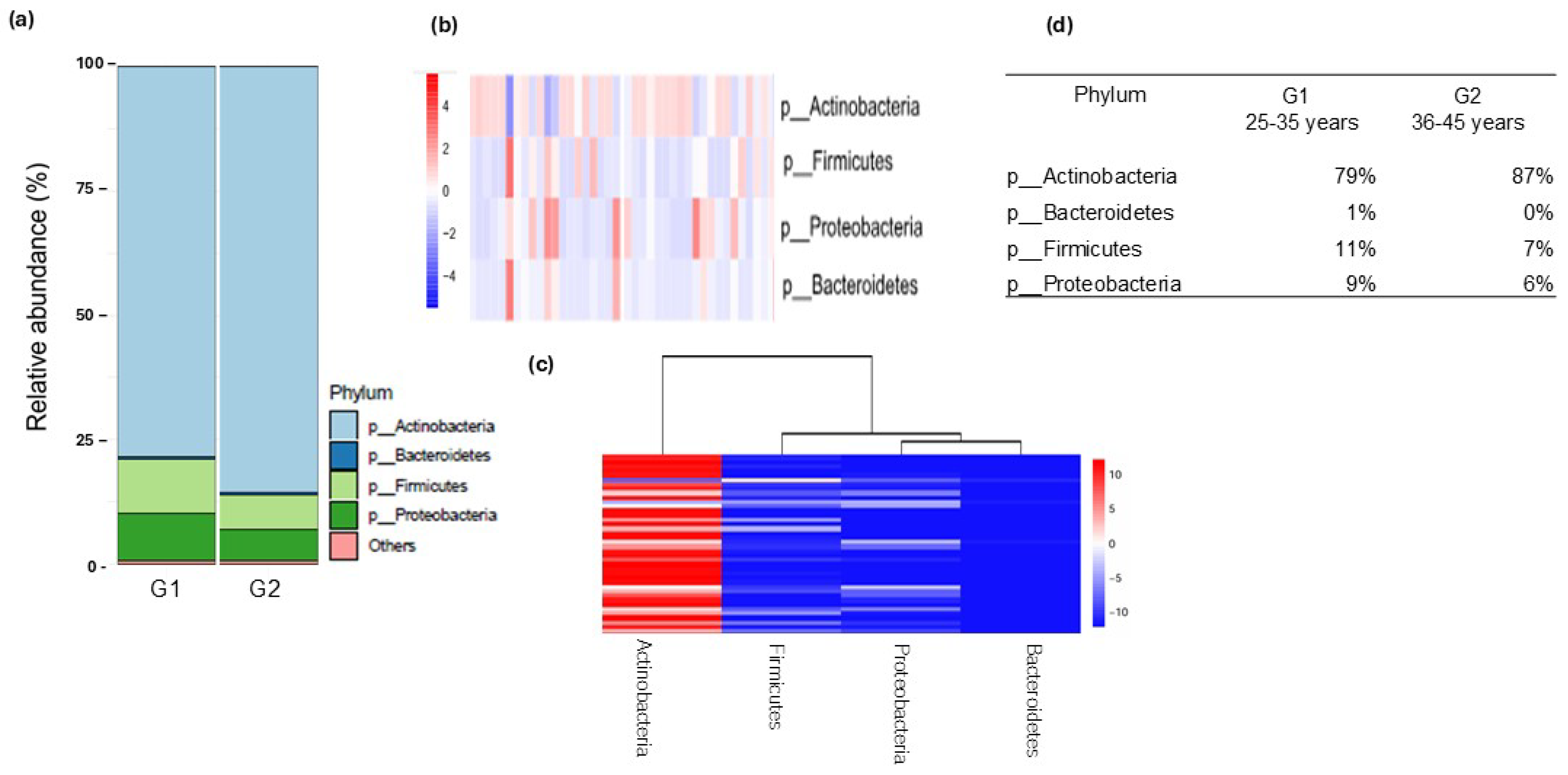
The material was subjected to metagenomic analysis and the taxonomic classification of kingdom to species was obtained using bioinformatics tools (DADA2, Qiime2). Taxa smaller than 1% were considered as “others”. The results indicate that two kingdoms were found throughout the study (Bacteria and Archea) with absolute predominance of the bacteria kingdom (Table S1). In addition, 20 phyla were found, of which the most predominant were 4: Actinobacteria (83%), Firmicutes (9%), Proteobacteria (7%) and Bacteroidetes (1%). The phyla were separated into groups according to age range (Figure 3a) and grouped in a heatmap by variability and abundance (Figure 3b,c, respectively). The results (Figure 3a) show an increase in G1 (25–35 years) of Firmicutes and Proteobacteria, but a decrease in the phylum Actinobacteria. In contrast, in G2 (36–45 years), an increase in the phylum Actinobacteria was observed and a decrease in Firmicutes and Proteobacteria. These results are very consistent when considering that the increase in Actinobacteria decreases other phyla and vice-versa. The table (Figure 3d) shows an abundance of 79% and 87% of Actinobacteria in groups G1 and G2, respectively, 1% of Bacteroidetes only in G1, 11% (G1) and 7% (G2) of Firmicutes, and 9% (G1) and 6% (G2) of Proteobacteria. The correlation between these results is discussed later.
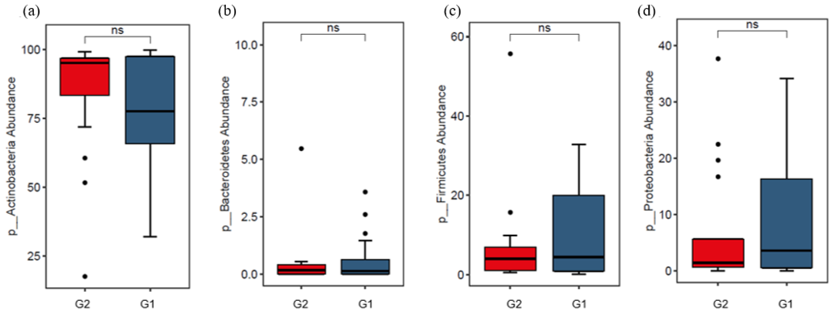
Then, a boxplot was performed to confirm whether the difference between the groups analyzed was statistical or just a trend (Figure 4). The results showed that there was no statistical difference between the groups (G1 and G2), probably due to the high standard deviation and presence of outliers, since there is a great individual variability among the participants. These results corroborate that shown in Figure 3b.
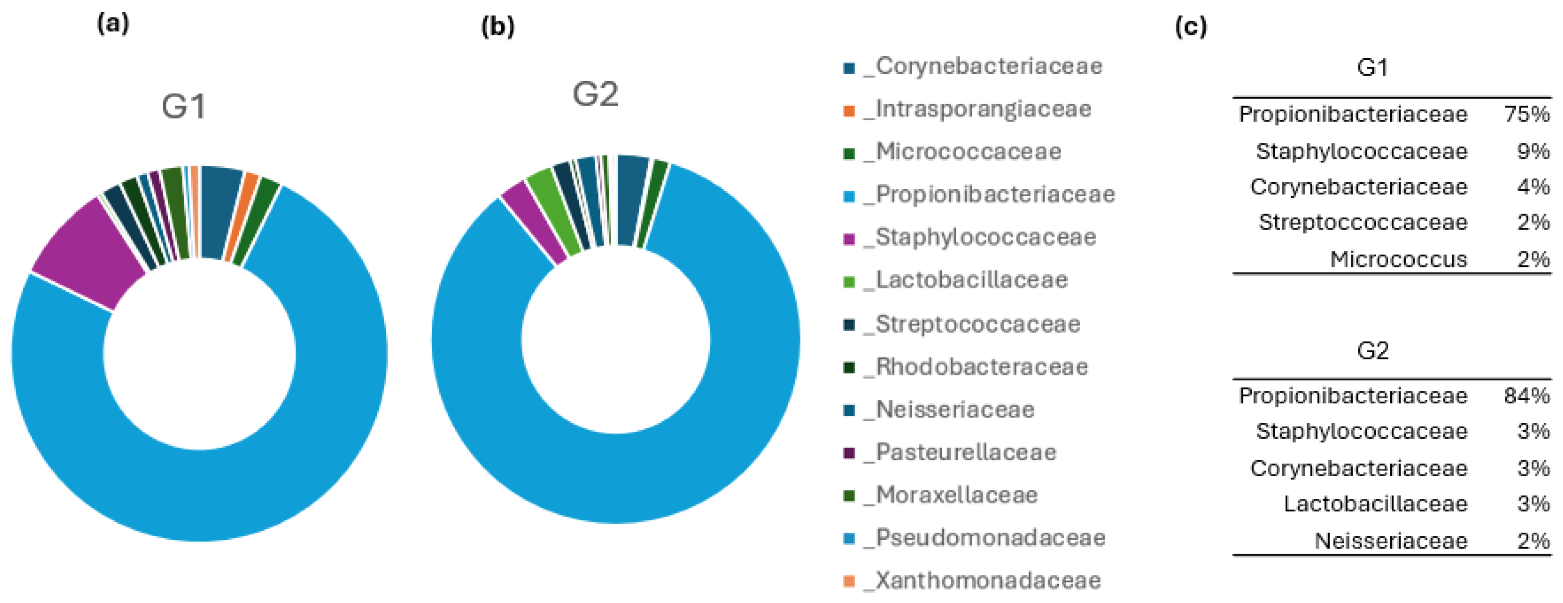
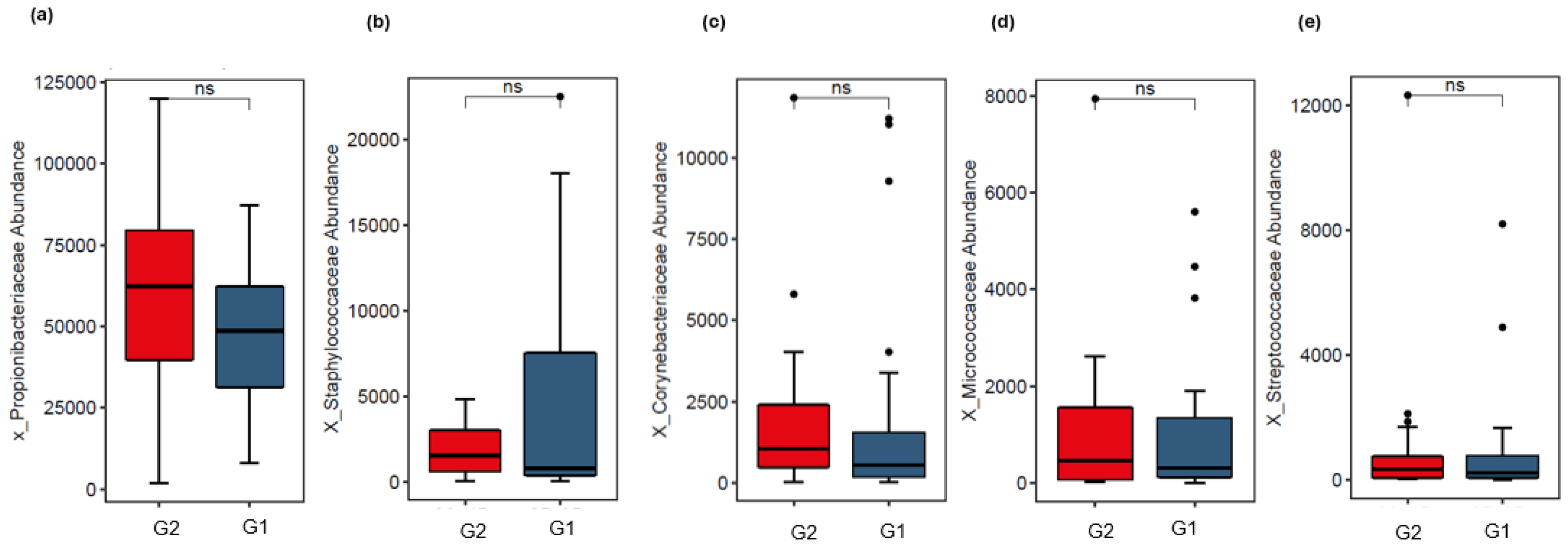
A total of 43 classes were found, with 5 being the most abundant: Actinobacteria (83%), Bacilli (9%), Alphaproteobacteria (3%), Betaproteobacteria (2%) and Gammaproteobacteria (3%). In addition, 90 orders were found, with 11 being the most abundant: Propionibacteriales (76%), Bacillales (9%), Lactobacillales (3%), Corynebacteriales (2%), Micrococcales (3%), Pseudomonadales (2%), Xanthomonadales (1%), Rhizobiales (1%), Rhodobacteriales (1%), Nesseriales (1%) and Pasteurellales (1%) (Table S1). A total of 169 families were also found, with 12 being the most abundant. When comparing the groups, a variation in composition was observed according to the age range (Figure 5), especially in the composition of the Propionibacteriaceae and Staphylococcaceae families, with 75% and 84% of the Propionibacteriaceae family found in Groups G1 and G2 and 9% and 3% of the Staphylococcaceae family in Groups G1 and G2, respectively (Figure 5c). Next, we analyzed whether the differences were statistical among the five most abundant families, and the data showed that there was no difference between the groups (Figure 6). However, these results are important in establishing trends in the families most abundant in the skin of Brazilian women, especially since there are no other studies in this regard.
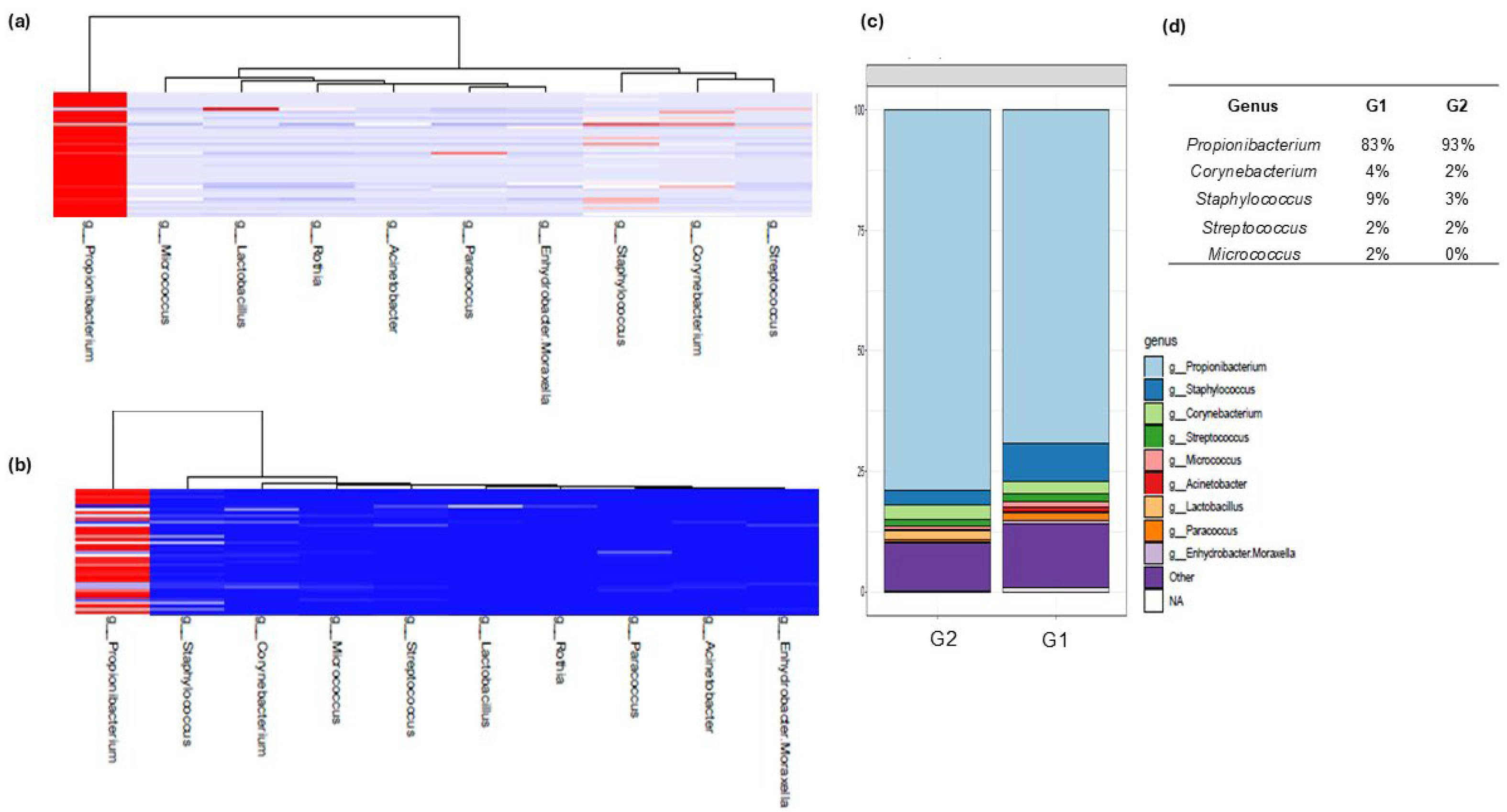
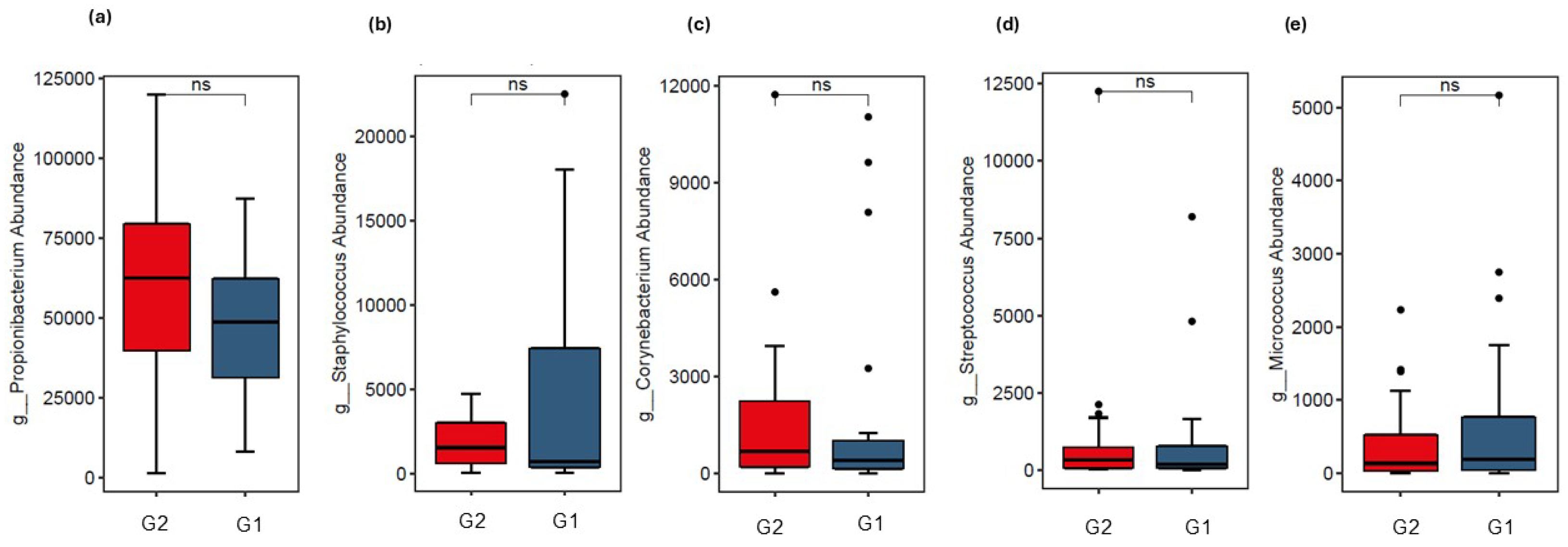
Next, the results of abundant generalizations were analyzed (Figure 7). In this study, 407 genera were found (Figure 8), with 5 being the most abundant: Propionibacterium—currently called Cutibacterium (84%), Staphylococcus (8%), Corynebacterium (4%), Streptococcus (2%) and Micrococcus (2%). Based on their low relative abundance, the other genera were not analyzed. When separated by group, we can observe the variability between the groups in the heatmap (Figure 8a) and when analyzed by abundance, the results indicate a predominance in G1 of Staphylococcus and lower abundance for Cutibacterium, when compared to G2 (Figure 8b), while in G2, an increase in Cutibacterium, Corynebacterium and Lactobacillus were observed. In short, younger skin expressed more of the genera Staphylococcus, Micrococcus and Corynebacterium and less of Cutibacterium (Figure 8d). In this sense, we analyzed whether the differences observed in gender were statistical, with the top 5. For this, we performed boxplots and the results indicate that there was no statistical variation, but a tendency towards a differentiated microbiological profile (Figure 9), except when we compared Cutibacterium to all other genera, when it is possible to note a statistical difference in both groups (Supplementary Material Figure S1). These results lead to a discussion about the fact that young skin expresses more Cutibacterium acnes than older skin, but this fact seems to be related only to a wider age range and not to the narrower one described in this work.
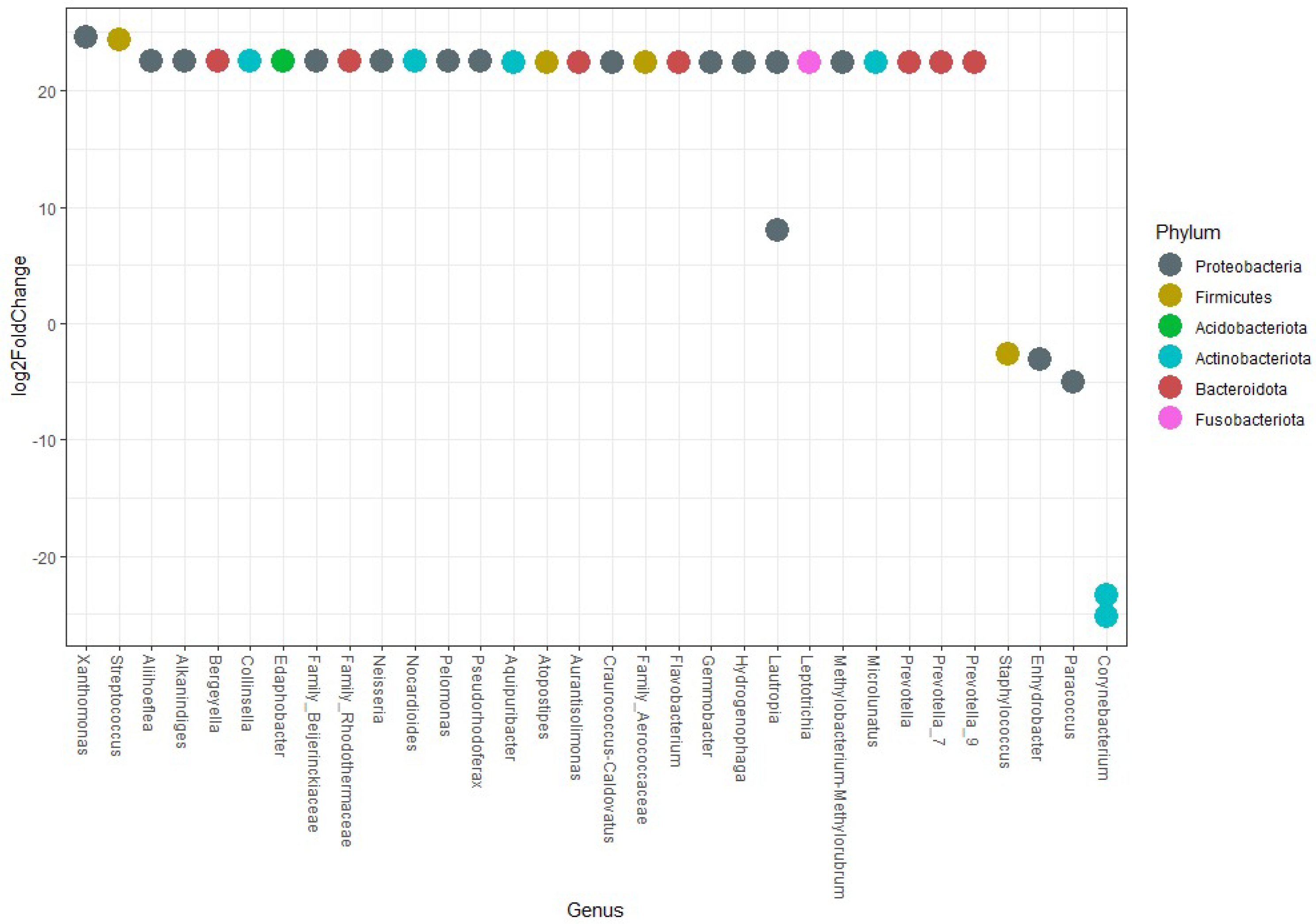
Then, using the Wald test and the Benjamini–Hochberg p-value adjustment, it is possible to observe that all genera illustrated in the graph showed statistical difference (p < 0.05) (Figure 10). Thus, all genera above the horizontal line 0 of the Y axis increased in participants from G2 in relation to G1, 28 in total. Likewise, the 4 genera below the same line decreased: Corynebacterium, Paracoccus, Enhydrobacter and Lautropia. Corynebacterium and Lautropia have two dots in the same column, showing that there were two possible different species of this genus that are differentially abundant.
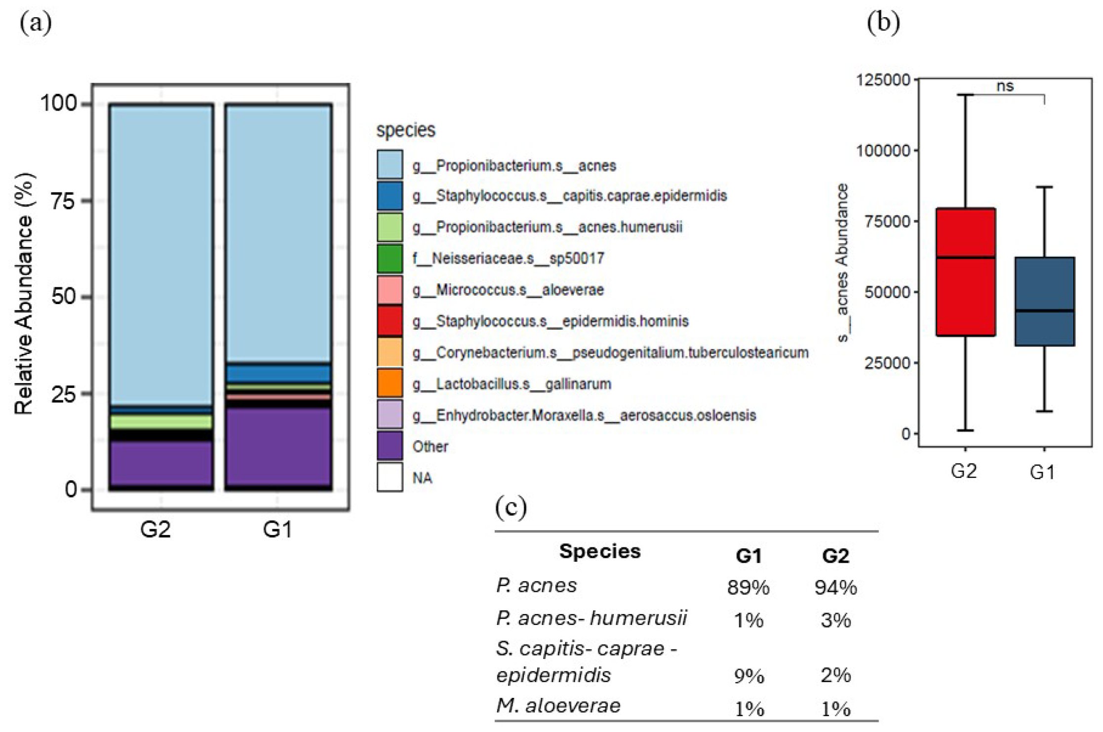
Next, the 395 species were analyzed. The results suggest an abundance of four species: P. acnes (C. acnes), P. acnes-humerusii, S. capitis-caprae-epidermidis and M. aloeverae (Figure 11a). The abundance of C. acnes, although not statistically significant, is noticeable as being more increased in the older group (G2) than in G1, which surprised us greatly. We can also observe that the younger group has an abundance of Staphylococcus capitis-caprae-epidermidis, while the older skin presented more C. acnes and P. acnes-humerusii (Figure 11c). However, it is possible to note that the “others” category was more abundant in the younger skin.
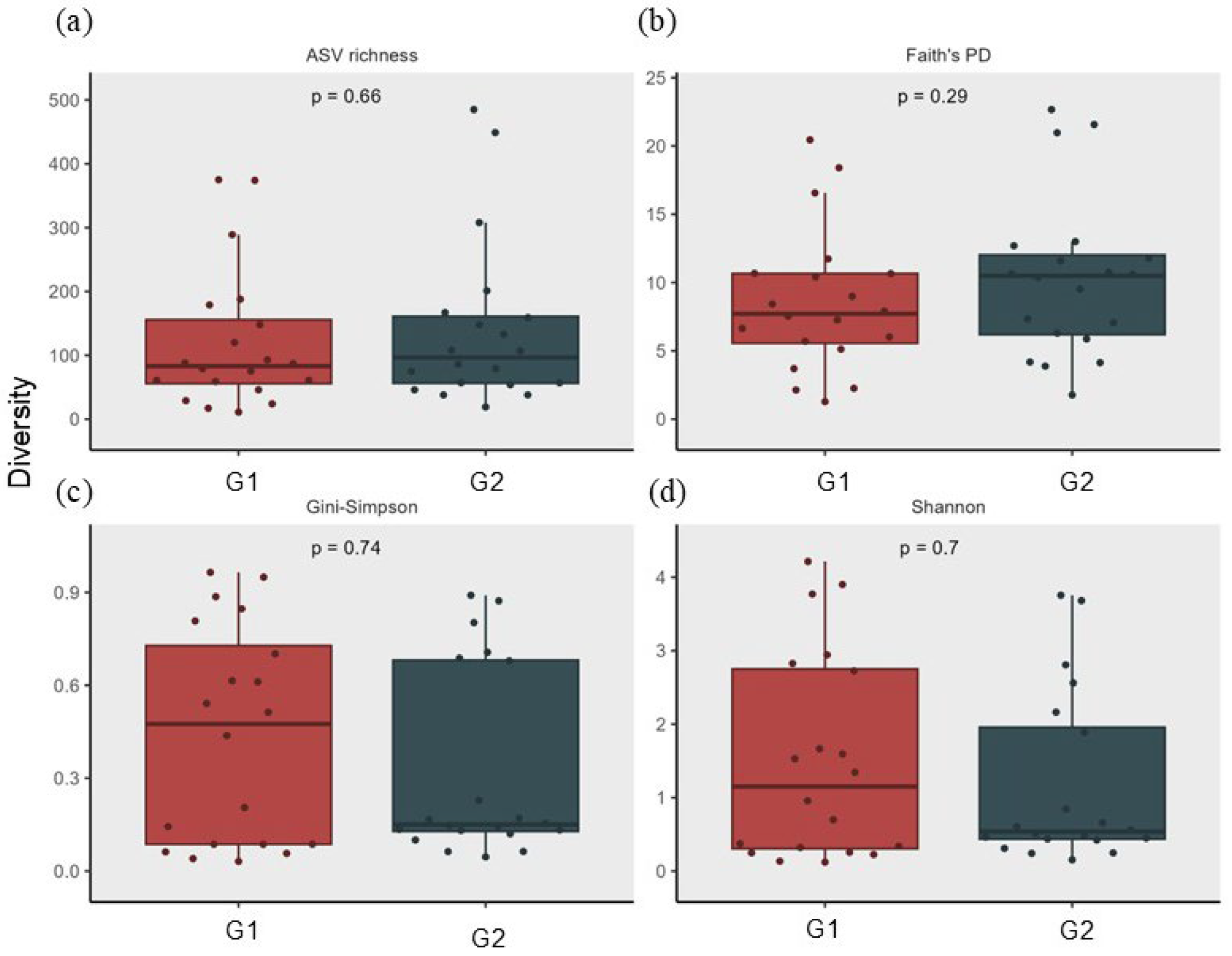
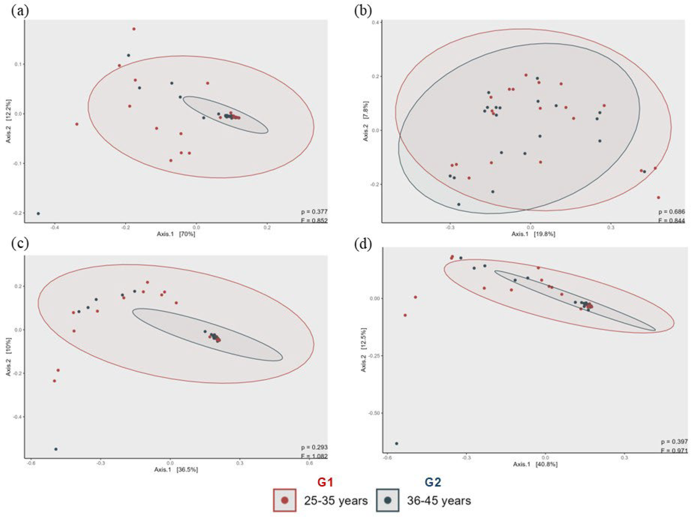
The alpha diversity of the species was analyzed using the Shannon index, Gini-Simpson index and Faith’s phylogenetic diversity index. The results showed no statistical differences between the groups analyzed (Figure 12). For beta diversity, we used the weighted and unweighted UniFrac indexes, Bray–Curtis index and Jaccard index. We also performed an analysis of the relative abundance of the ASVs found. The results show a microbiological diversity identified in the study, with the youngest group being the most abundant (Figure 13), corroborating the results of Figure 10, which show the increase in other species in the youngest group, but without statistical difference (p = 0.29):
Performing an individual analysis, we noticed that the most abundant bacteria on the skin were not present or had a very small quantity in some patients (Figure 14). Interestingly, in our study, nine participants (22.5%) did not present or presented very little abundance of C. acnes on the skin (five in G1 and four in G2), which aroused our curiosity.

4. Discussion

The results show variability between the groups analyzed (G1 and G2), and the biometrology data corroborates the metagenomics, especially regarding pH changes. G2 presented greater pH variation and increased transepidermal water loss. The pH variation favors the growth of some microorganisms, as well as the reduction in others, since the production of some lipids in the stratum corneum may be affected, since lipid synthesis is performed by pH-dependent enzymes. Thus, the skin barrier function may be compromised [11] and pH imbalances are related to some dermatological pathologies [12]. Pearson correlation was used to compare the transepidermal water loss (TEWL) and pH, and the results showed a positive correlation in both groups (*** p < 0.001), indicating that changes in pH increase TEWL. Furthermore, changes in pH can influence skin dehydration (Supplementary Materials, Figures S2 and S3).
An increase in Actinobacteria and a decrease in Proteobacteria are observed in aged skin. Most studies in the literature use very broad age ranges, so we chose to evaluate a smaller range, aiming to clarify whether we could stop the aging process before it begins, since the tropical climate and high sun exposure are factors that can contribute to premature skin aging. In this context the findings slightly diverge from a study conducted with women and men from Asia [13], which reported an increase in Proteobacteria and a decrease in Actinobacteria in aged skin, as well as another study with women from Western Europe [14]. Both these studies reported a wider age gap—17–37 years old and older adults of 54–76 years old. These differences may also be due to different ethnicity, skincare habits and nutrients, which were not considered for this study.
An increase of 28 genera was observed with significance when comparing G1 and G2 and a decrease of 4 genera, including Corynebacterium, which is linked to bacteria found in aging skin [13]. These results show that the microbiological profile of the skin of older Brazilian women was different to that of younger skin. At the species level, older skin showed a higher abundance of Cutibacterium acnes and a decrease in Staphylococcus capitis. The younger group, on the other hand, showed a higher abundance of Staphylococcus capitis and less Cutibacterium acnes. It is important to note that previous studies have shown that C. acnes is a bacterium found mainly in younger skin [13]. However, if we look at cutaneous biometric data, the younger population, which has oilier skin, should express much more of this bacterium than older skin, which tends to be drier [15]. We did not observe a significant change in alpha diversity since the ages were very similar. New studies are being conducted with a greater separation in ages to clarify these points. However, beta diversity showed that younger skin presents greater diversity (Figure 13).
It is important to note that with aging, microbial diversity increases, as reported by many authors [13,14,16] although some more abundant microorganisms of the skin tend to decrease like Lactobacillus and Cutibacterium. Pearson correlation on the abundance of C. acnes in groups G1 and G2 was analyzed, then the Score of Intrinsic and Extrinsic Skin Aging (SCINEXA) scale was used to correlate the abundance of C. acnes in both groups. The results suggest that when correlated with the SCINEXA aging scale, it had a negative correlation with the aging scale of group 2, but a positive correlation with the SCINEXA of group 1 (younger). This suggests that the abundance of C. acnes in older skin had a positive correlation with the aging scale of younger individuals, corroborating the hypothesis that C. acnes is present in the older group (See Supplementary Materials, Figures S4 and S5).
Interestingly, in our study, nine participants (22.5%) had no or very low abundance of C. acnes on their skin (Figure S1) which raised an important question: Why is the most abundant and most important bacteria on the skin not present in these participants? When correlating clinical data between groups—such as the Glogau and SCINEXA aging scales, comorbidities, medications, phototype, skincare routine, sunscreen use and age variation—no factor justified the change in the microbiological profile. In this sense, we hypothesized that the presence of certain lipids may influence the activity of C. acnes. Establishing a correlation between the lipids found in the skin and the microbiota is of utmost importance. C. acne releases fatty acids that inhibit the growth of pathogenic species; however, when produced in excess, it can lead to skin disorders. Specific lipids that promote balance between these microbial species could be the key to healthier, more youthful skin [17,18,19,20]. These data may be very impactful if we consider the importance that Brazil represents in the global cosmetics market and that there are variations in the composition of the skin regarding lipid composition and microbiota that are still unclear because there are no published studies involving the skin microbiota of Brazilian women.
Our study included only 40 individuals (20 individuals between 25 and 35 years old and 20 individuals between 36 and 45 years old), presenting, as another limitation, the recruitment of participants only from São Paulo, although it is a large city with great ethnic diversity. To help clarify this point we are conducting a new study with a larger age range. In addition, the use of microbiome sampling using swab and not film dressings with acrylic or urethane adhesive [21] or Stripping Discs on a polyester carrier sheet [22] could affect the results, even if most studies have been applying this technique [23,24,25].
Nevertheless, our results could complement those presented in the literature where a multi-omics profile during a human aging study reports that substantial dysregulation occurred at two major periods, being approximately 44 years and 60 years of chronological age [26]. The influence of exposome factors and lifestyle as well as ethnicity are indeed important variables and new metagenomic studies focused on these points may help to clarify the role of C. acnes in aging and thus stimulate the development of cosmetic products that favor the growth and rebalance of microbiota, providing more youthful and balanced skin.

5. Conclusions

In conclusion, these findings provide valuable insights into the microbiological profile of Brazilian women’s skin and may serve as a basis for the development of innovative anti-aging cosmetic products. Furthermore, the question remains: What makes Brazilian women’s skin unique compared to others? New lipidomic studies should be conducted to shed light on these questions, providing answers to this important question. In addition, the use of metagenomics could lead to more personalized products according to skin type and to an innovative formulation targeting the microbiome that addresses aging concerns, improving skin health and slowing the aging process.

Supplementary Materials

The following supporting information can be downloaded at: https://www.mdpi.com/article/10.3390/cosmetics12040165/s1, Table S1. Relative abundance of all taxons of the main groups found in the study. Figure S1. Statistical analysis comparing the abundance of Propionibacterium with Staphylococcus and Corynebacterium. Figure S2. Pearson correlation between transepidermal water loss (TEWL) and pH in group 1 and 2. Figure S3. Pearson correlation between Corneometer parameter (skin hydration) and oiliness in groups 1 and 2. Figure S4. Pearson correlation on the abundance of C. acnes in groups G1 and G2. Figure S5. Pearson correlation between C. acnes and Score of Intrinsic and Extrinsic Skin Aging (SCINEXA) scale in groups 1 and 2.

Author Contributions

Conceptualization, V.R.L.-S., N.A.-F., A.L. and P.S.L.; formal analysis, R.A.G.B.S.; funding acquisition, A.L., V.R.L.-S., N.A.-F. and P.S.L.; investigation, R.A.G.B.S., A.L.V.P., Y.R.S. and E.B.; project administration, P.S.L.; resources, N.A.-F.; software, R.M.-F.; supervision, P.S.L.; writing—original draft, R.A.G.B.S.; writing—review and editing, V.R.L.-S., N.A.-F. and P.S.L. All authors have read and agreed to the published version of the manuscript.

Funding

This research was funded by the companies Shanghai Pechoin Biotech Co., Ltd. and Solabia Biotecnologica LTDA and EMBRAPII—Brazilian Company of Research and Industrial Innovation grant number #PFSP2304.0022, UNIFESP SEI Process number nº 23089.003494/2023-51.

Institutional Review Board Statement

The study was conducted in accordance with the Declaration of Helsinki and approved at 12 December 2023 by the Research Ethics Committee at UNIFESP (CAAE: 74030523.0.0000.5505) for studies involving humans.

Informed Consent Statement

Informed consent for participation was obtained from all subjects involved in the study. Written informed consent has been obtained from the subjects to publish this paper.

Data Availability Statement

Data are contained within the article and Supplementary Materials.

Acknowledgments

The authors would like to thank Rafael Maia and Júlia Penha Maróstica for their technical support in data acquisition and CAPES and FAPESP for the Ph.D. scholarships.

Conflicts of Interest

Author Alexandra Lan was employed by Shanghai Pechoin Biotech Co., Ltd. The authors declare that this study received funding from the companies Shanghai Pechoin Biotech Co., Ltd. and Solabia Biotecnologica LTDA, and the EMBRAPII—Brazilian Company of Research and Industrial Innovation The funders had no role in the design of the study; in the collection, analyses, or interpretation of data; in the writing of the manuscript; or in the decision to publish the results.

Abbreviations

The following abbreviations are used in this manuscript:
IBGEBrazilian Institute of Geography and Statistics
G1Group 1 between 25 and 35 years old
G2Group 2 between 36 and 45 years old
ASVamplicon sequence variant
SCINEXAScore of Intrinsic and Extrinsic Skin Aging

References

  1. Costa, F. Características anatômicas da pele humana e suas variações étnicas. J. Dermatol. Bras. 2019, 43, 254–267. [Google Scholar]
  2. ABIHPEC. Panorama do Setor de Higiene Pessoal no Brasil; ABIHPEC: São Paulo, Brazil, 2024. [Google Scholar]
  3. IBGE. Censo Demográfico 2010: Características Gerais da População, Religião e Pessoas Com Deficiência; Instituto Brasileiro de Geografia e Estatística: Palácio da Fazenda, Brazil, 2010. [Google Scholar]
  4. Matsuo, T. Impacto da exposição solar na pele das brasileiras. Rev. Bras. Dermatol. 2017, 92, 659–667. [Google Scholar]
  5. Byrd, A.L.; Belkaid, Y.; Segre, J.A. The human skin microbiome. Nat. Rev. Microbiol. 2018, 16, 143–155. [Google Scholar] [CrossRef]
  6. Prescott, S.L.; Larcombe, D.L.; Logan, A.C.; West, C.; Burks, W.; Caraballo, L.; Levin, M.; Etten, E.; Van Horwitz, P.; Kozyrskyj, A.; et al. The skin microbiome: Impact of modern environments on skin ecology, barrier integrity, and systemic immune programming. World Allergy Organ. J. 2017, 10, 29. [Google Scholar] [CrossRef]
  7. Amann, R.I.; Ludwig, W.; Schleifer, K.H. Phylogenetic identification and in situ detection of individual microbial cells without cultivation. Microbiol. Rev. 1995, 59, 143–169. [Google Scholar] [CrossRef] [PubMed] [PubMed Central]
  8. UCHIME Improves Sensitivity and Speed of Chimera Detection|Bioinformatics|Oxford Academic [Internet]. Available online: https://academic.oup.com/bioinformatics/article/27/16/2194/255262 (accessed on 1 August 2025).
  9. Kers, J.G.; Saccenti, E. The Power of Microbiome Studies: Some Considerations on Which Alpha and Beta Metrics to Use and How to Report Results. Front. Microbiol. 2022, 12, 796025. [Google Scholar] [CrossRef] [PubMed]
  10. Wilcoxon Signed Ranks Test-An Overview|ScienceDirect Topics [Internet]. Available online: https://www.sciencedirect.com/topics/medicine-and-dentistry/wilcoxon-signed-ranks-test (accessed on 25 July 2025).
  11. Elias, P.M. Stratum corneum defensive functions: An integrated view. J. Investig. Dermatol. 2005, 125, 183–200. [Google Scholar] [CrossRef] [PubMed]
  12. Schmid-Wendtner, M.H.; Korting, H.C. The pH of the skin surface and its impact on the barrier function. Ski. Pharmacol. Physiol. 2006, 19, 296–302. [Google Scholar] [CrossRef] [PubMed]
  13. Jugé, R.; Rouaud-Tinguely, P.; Breugnot, J.; Servaes, K.; Grimaldi, C.; Roth, M.P.; Coppin, H.; Closs, B. Shift in skin microbiota of Western European women across aging. J. Appl. Microbiol. 2018, 125, 907–916. [Google Scholar] [CrossRef] [PubMed]
  14. Shibagaki, N.; Suda, W.; Clavaud, C.; Bastien, P.; Takayasu, L.; Iioka, E.; Kurokawa, R.; Yamashita, N.; Hattori, Y.; Shindo, C.; et al. Aging-related changes in the diversity of women’s skin microbiomes associated with oral bacteria. Sci. Rep. 2017, 7, 10567. [Google Scholar] [CrossRef] [PubMed] [PubMed Central]
  15. Bek-Thomsen, M.; Lomholt, H.B.; Scavenius, C.; Enghild, J.J.; Brüggemann, H. Proteome analysis of human sebaceous follicle infundibula extracted from healthy and acne-affected skin. PLoS ONE 2014, 9, e107908. [Google Scholar] [CrossRef] [PubMed] [PubMed Central]
  16. Howard, B.; Bascom, C.C.; Hu, P.; Binder, R.L.; Fadayel, G.; Huggins, T.G.; Jarrold, B.B.; Osborne, R.; Rocchetta, H.L.; Swift, D.; et al. Aging-Associated Changes in the Adult Human Skin Microbiome and the Host Factors that Affect Skin Microbiome Composition. J. Investig. Dermatol. 2022, 142, 1934–1946. [Google Scholar] [CrossRef] [PubMed]
  17. Elias, P.M.; Feingold, K.R. Lipids and the epidermal water barrier: Metabolism, regulation, and pathophysiology. Semin. Dermatol. 1992, 11, 176–182. [Google Scholar] [PubMed]
  18. Feingold, K.R. Thematic review series: Skin lipids. The role of epidermal lipids in cutaneous permeability barrier homeostasis. J. Lipid Res. 2007, 48, 2531–2546. [Google Scholar] [CrossRef] [PubMed]
  19. Knox, S.; O’Boyle, N.M. Skin lipids in health and disease: A review. Chem. Phys. Lipids 2021, 236, 105055. [Google Scholar] [CrossRef] [PubMed]
  20. Kurokawa, I.; Danby, F.W.; Ju, Q.; Wang, X.; Xiang, L.F.; Xia, L.; Chen, W.; Nagy, I.; Picardo, M.; Suh, D.H.; et al. New developments in our understanding of acne pathogenesis and treatment. Exp. Dermatol. 2009, 18, 821–832. [Google Scholar] [CrossRef] [PubMed]
  21. Ogai, K.; Shibata, K.; Takahashi, N.; Ogura, K.; Okamoto, S.; Sugama, J. Amplicon-based skin microbiome profiles collected by tape stripping with different adhesive film dressings: A comparative study. BMC Microbiol. 2021, 21, 54. [Google Scholar] [CrossRef]
  22. Perugini, P.; Grignani, C.; Condrò, G.; van der Hoeven, H.; Ratti, A.; Mondelli, A.; Colpani, A.; Bleve, M. Skin Microbiota: Setting up a Protocol to Evaluate a Correlation between the Microbial Flora and Skin Parameters. Biomedicines 2023, 11, 966. [Google Scholar] [CrossRef] [PubMed]
  23. Karoglan, A.; Paetzold, B.; Pereira de Lima, J.; Brüggemann, H.; Tüting, T.; Schanze, D.; Güell, M.; Gollnick, H. Safety and Efficacy of Topically Applied Selected Cutibacterium acnes Strains over Five Weeks in Patients with Acne Vulgaris: An Open-label, Pilot Study. Acta. Derm. Venereol. 2019, 99, 1253–1257. [Google Scholar] [CrossRef] [PubMed]
  24. Alan, M.; O’Neill, N.T.; Hayachi, A.; Williams, M.R.; Mills, R.H.; Gonzalez, D.J.; Gallo, R.L. Identification of a Human Skin Commensal Bacterium that Selectively Kills Cutibacterium acnes. J. Investig. Dermatol. 2020, 140, 1619–1628. [Google Scholar] [CrossRef]
  25. Robe, P.; Jarrin, C.; Zanchetta, C.; Dupont, J.; Chapuis, E.; Scandolera, A.; Auriol, D.; Reynaud, R. Rehabilitation of Skin Bacterial Counts to Assess the Short-Term Impact of Ingredients in Topical Applications—Presenting a Culture-Based Viability Score. Cosmetics 2023, 10, 50. [Google Scholar] [CrossRef]
  26. Shen, X.; Wang, C.; Zhou, X.; Zhou, W.; Hornburg, D.; Wu, S. Nonlinear dynamics of multi-omics profiles during human aging. Nat. Aging 2024, 4, 1619–1634. [Google Scholar] [CrossRef] [PubMed]
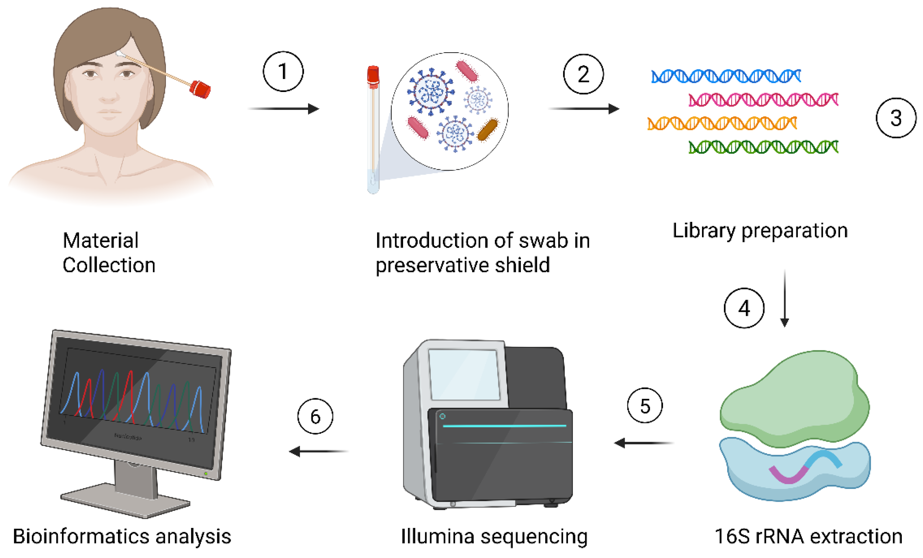
Figure 1. Metagenomic collection. Step 1—collection in the frontal region of the face; Step 2—introduction of swab in preservative shield; Step 3—library preparation; Step 4—16S rRNA extraction; Step 5—Illumina® Sequencing; Step 6—bioinformatics analysis. Created in BioRender. Siqueira, R. (2025) https://BioRender.com/8kf7jtl (accessed on 1 August 2025).
Cosmetics 12 00165 g001
Figure 2. Analysis of biometrological data. Above is the image of the participating patient’s collection and below the corresponding graphs: (a,e) Corneometer® (hydration), (b,f) Sebumeter® (oiliness), (c,g) pH-meter® (pH), (h) pH variation between groups, (d,i) transepidermal water loss (TEWL). Means ± SD were analyzed: Skin hydration: G1 (61.75 ± 19.76); G2 (60.05 ± 7.12). Oiliness: G1 (30.52 ± 14.12); G2 (35.82 ± 4.36). pH: G1 (5.34 ± 0.30); G2 (5.68 ± 0.29). TEWL: G1 (9.68 ± 0.54); G2 (12.74 ± 0.54).
Cosmetics 12 00165 g002
Figure 3. Phyla. (a) Barplot with the relative abundance of the phyla separated by group: G1 and G2; (b) heatmap grouped by variability; (c) heatmap grouped by abundance; (d) table with the relative abundance separated by group: G1 and G2.
Cosmetics 12 00165 g003
Figure 4. Boxplots of the phyla: (a) Actinobacteria, (b) Bacteroidetes, (c) Firmicutes and (d) Proteobacteria separated by group: G1 and G2.
Cosmetics 12 00165 g004
Figure 5. Families found in the study were separated by group. (a) Families in group G1 and (b) in group G2. (c) Table of relative abundance of the families.
Cosmetics 12 00165 g005
Figure 6. The abundance of the 5 main families found in the study: (a) Propionibacteriaceae, (b) Staphylococcaceae, (c) Corynebacteriaceae, (d) Micrococcaceae and (e) Streptococcaceae.
Cosmetics 12 00165 g006
Figure 7. Barplot of the most abundant genera found in the study.
Cosmetics 12 00165 g007
Figure 8. Heatmap of the genera found in the study: (a) Variability and abundance and (b) with top 10 taxa. (c) Barplot with the top 10 found and (d) table with the relative abundance of the top 5.
Cosmetics 12 00165 g008
Figure 9. Boxplot of genus results for Propionibacterium (a), Staphylococcus (b), Corynebacterium (c), Streptococcus (d) and Micrococcus (e).
Cosmetics 12 00165 g009
Figure 10. Dotplot of the differential abundance of the genus.
Cosmetics 12 00165 g010
Figure 11. Species found in the study separated by groups: (a) Barplot of the top 10 species results found; (b) abundance of P. acnes; (c) table with the 4 most abundant species.
Cosmetics 12 00165 g011
Figure 12. Boxplot of alpha diversity results by ASV richness (a), Faith’s PD (b), Gini-Simpson (c) and Shannon (d).
Cosmetics 12 00165 g012
Figure 13. Beta-diversity PCA by (a) UniFrac weighted, (b) UniFrac unweighted, (c) Jaccard weighted and (d) Bray–Curtis weighted.
Cosmetics 12 00165 g013
Figure 14. Heatmap showing patients who do not express or express little C. acnes in blue.
Cosmetics 12 00165 g014
Disclaimer/Publisher’s Note: The statements, opinions and data contained in all publications are solely those of the individual author(s) and contributor(s) and not of MDPI and/or the editor(s). MDPI and/or the editor(s) disclaim responsibility for any injury to people or property resulting from any ideas, methods, instructions or products referred to in the content.