Systematic Review

Skin Aging and Type I Collagen: A Systematic Review of Interventions with Potential Collagen-Related Effects

by
Ofek Bar
1,* and
Skaidra Valiukevičienė
1,2
1
Faculty of Medicine, Lithuanian University of Health Sciences, 44307 Kaunas, Lithuania
2
Department of Skin and Venereal Diseases, Medical Academy, Lithuanian University of Health Sciences, 44307 Kaunas, Lithuania
*
Author to whom correspondence should be addressed.
Cosmetics 2025, 12(4), 129; https://doi.org/10.3390/cosmetics12040129
Submission received: 28 April 2025 / Revised: 8 June 2025 / Accepted: 11 June 2025 / Published: 20 June 2025
(This article belongs to the Special Issue Feature Papers in Cosmetics in 2025)

Abstract

:
Aging leads to a decline in skin function due to intrinsic factors (genetics, hormones) and extrinsic factors (sun exposure, pollutants). Type I collagen plays a vital role in maintaining skin integrity and elasticity. As aging progresses, collagen synthesis diminishes, resulting in weakened skin structure and wrinkle formation. This systematic review explores the role of type I collagen in skin aging by summarizing key clinical findings. A systematic search was conducted using PubMed and ScienceDirect as the primary databases, including studies published between 2014 and 2025 that addressed type I collagen and skin aging. Eleven clinical studies were selected following PRISMA guidelines. The results consistently show the decline of type I collagen as a central contributor to dermal thinning, loss of elasticity, and the appearance of wrinkles and sagging. Clinical trials demonstrate that collagen supplementation, particularly from hydrolyzed fish cartilage and low-molecular-weight peptides, enhances collagen production, improves skin hydration and texture, and reduces signs of photoaging. Overall, the evidence emphasizes the critical role of type I collagen in skin aging and suggests that targeted collagen supplementation may serve as an effective strategy to maintain skin structure and combat visible signs of aging.

1. Introduction

Aging is an inexorable time-dependent deterioration of biological function and regenerative capacity of higher organisms, resulting in a progressive increase in the risk of disease and death [1,2]. Being the largest organ in the body, skin undergoes structural changes over time due to the combination of complex biological processes caused by intrinsic as well as extrinsic factors [3,4,5,6,7]. Signs of aging skin include wrinkles, loss of elasticity, sagging, laxity, dullness, rough texture, and pigmentation changes [8,9,10,11,12,13,14]. Of these, the occurrence of deep wrinkles merits special mention [15].
Aging is an interdependent process that involves integrated molecular, cellular, and tissue-level changes. At the cellular level, mitotically active cells enter into senescence, a state characterized as irreversible G1 phase cell cycle arrest yet remain metabolically active. Cellular senescence acts not only as a model for human tissue aging research but plays a critical role itself in systemic aging [16]. In skin, aging is associated with an insidious loss of regenerative potential in almost all components of its structural and functional integrity. Significantly, as the first line of defense of the body, the skin is especially vulnerable to intrinsic biological processes, as well as to extrinsic physical damage, so it can be one of the first parts of the body to show signs of aging [17].
Intrinsic aging, which is a genetic process including hormonal behavior and natural physiological processes that occur over time, has been associated with decreased collagen production, specifically with the decrease in type I collagen leading to a loss of skin elasticity and the presence of wrinkles [18].
Extrinsic aging is driven by chronic exposure to environmental agents, such as ultraviolet light (UV) radiation, pollution, and microorganisms (Figure 1). These environmental stressors contribute to the formation of reactive oxygen species (ROS), which increase the expression of matrix metalloproteinases (MMPs), leading to the degradation of type I collagen in the dermal extracellular matrix. Specifically, UV radiation induces oxidative stress and upregulates MMPs while simultaneously suppressing new collagen synthesis. Air pollution exacerbates this process by sustaining chronic inflammation, damaging fibroblasts, and impairing the skin’s reparative capacity. Moreover, microbial imbalance on the skin surface may activate local immune responses and promote inflammatory mediators, further enhancing collagen breakdown. Altogether, these extrinsic factors contribute to visible signs of skin aging, such as wrinkles, dryness, and loss of elasticity, through their cumulative detrimental effects on collagen homeostasis [3,13,19]. These outside stressors also speed up the damage to the DNA, leading to the continued structural and functional decline of our skin, compounding with intrinsic aging [20,21,22,23]. These mechanisms work together to drive the visible and biological aspects of skin aging by impacting the rates of collagen breakdown and degradation while inhibiting tissue regeneration, as shown by numerous studies that have examined the role of UV radiation on the breakdown of collagen [24,25,26,27]. These changes are responsible for visible aging: wrinkles, dryness, decreased barrier function, and epidermal thinning [28,29,30]. The skin serves as an essential protective barrier against threats, such as temperature extremes, UV, infections, mechanical damage, and skin water loss [31].
Collagen, the most abundant protein in the extracellular matrix (ECM) and connective tissue of vertebrates, is most abundant in mammals [32,33,34]. The ECM is a large and complex structure formed by proteins and glycosaminoglycans within the dermis, which provides necessary structural support to fibroblasts, keratinocytes, and endothelial cells that maintain tissue architecture to the cell and has an important influence on maintaining skin structure and function, which in turn will affect skin aging [35,36,37,38,39]. Keratinocytes, the major cell type within the epidermis, utilize the ECM for structural stability and effective skin barrier function [40,41,42,43]. In previous studies it has been confirmed that collagen of skin decreases with aging, which is one of the important molecules in the aging process of skin [32,33,44,45,46,47,48,49]. So far, 28 different forms of collagen have been characterized, and they are distinguished based on their structural characteristics, supramolecular arrangement, and functional traits [34,35,50,51,52,53,54,55]. Many forms of collagen, such as type I, are abundant through a range of tissues and are fundamental structural building blocks [56].
Type I collagen is the most abundant collagen type in the skin, accounting for 80–85% of the dermal ECM [57]. Other subtypes, like type III and type V collagen, can be found in skin, but type I collagen is fundamental to skin structure; it supplies considerable tensile strength and helps to determine the structure and durability of the dermis [58,59]. As skin ages, there is a progressive loss and fragmentation of dermal collagen fibrils, leading to reduced skin thickness and biomechanical strength [60].
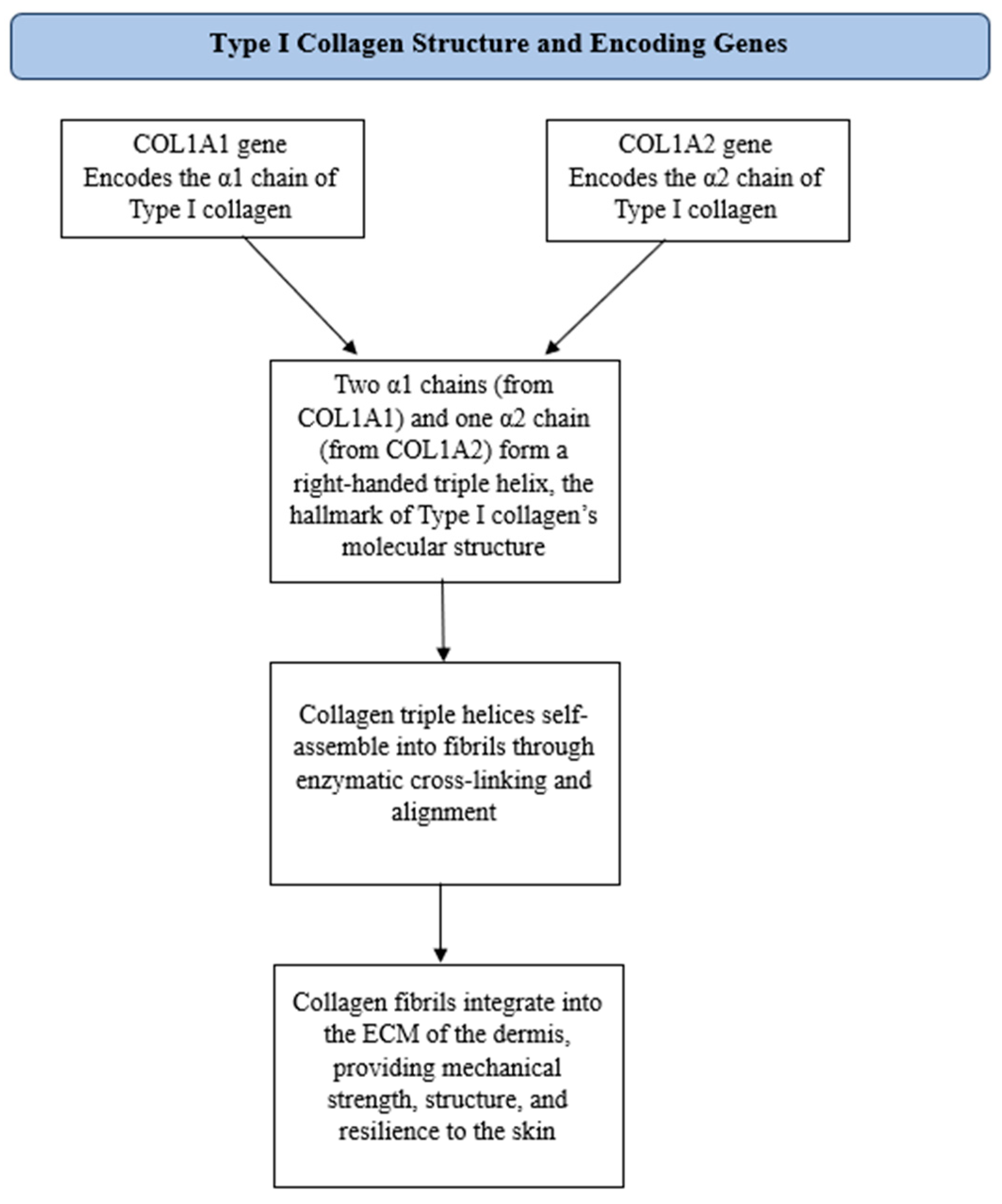
Type I collagen, the most abundant long-lived protein in humans, is a major fibrillar component of connective tissues such as skin, bone, and tendons [61]. It has a triple-helix structure composed of two α1 chains encoded by the Collagen type I alpha 1 chain (COL1A1) gene and one α2 chain encoded by the Collagen type I alpha 2 chain (COL1A2) gene (Figure 2). Among these, COL1A1 expression has been identified as a biomarker of skin aging, as its levels decline with age. This organization endows connective tissues with mechanical strength and elasticity [62,63,64,65,66,67]. This complex architecture is essential for preserving the integrity of the tissue by modulating biophysical properties, thermal stability, mechanical strength, and association capabilities with biomolecules and cells. Collagen fragmentation, a hallmark of skin aging, results from the interactions between dermal fibroblasts and the ECM that are aberrantly altered during the course of aging [68,69,70,71,72]. Fibroblasts are the main collagen-producing cells and synthesize the connective tissue matrix. These cells use specialized surface receptors, known as integrins, that specifically bind matrix proteins such as type I collagen. Integrins are transmembrane proteins that bind to the ECM, after which they aggregate and associate with the actin cytoskeleton of the cell to create focal adhesions. These focal adhesion complexes orchestrate key regulatory and mechanical functions that link the ECM to intracellular signaling pathways [32].
These dynamic changes use signal transduction pathways that are closely correlated with the structural and functional states of the cell. Even so, with advancing age, collagen homeostasis is perturbed, resulting in a decrease in collagen production, skin structural weakening, and diminished dermal strength [60]. Due to collagen degradation, hallmark features of aging, such as loss of elasticity and wrinkle formation, can be observed, highlighting the importance of maintaining skin function through collagen. MMPs are able to digest the human skin’s endogenous type I collagen.
In young, healthy skin, MMP expression is low (Figure 1), and their activity is stringently regulated by tissue inhibitors of metalloproteinases (TIMPs). This is responsible for a very slow turnover, with collagen replaced every ~30 years [72]. This gradual process is reflected in the collection of age-related changes, for example, the development of cross-links through sugars that allow them to survive degradation. The cross-links are involved in the accumulation of degraded collagen in the ECM, which disturbs its architecture and mechanical properties. It is known that, with the destruction of collagen, the basic interaction between the ECM and fibroblasts that keeps the dermis mechanically tense is disrupted [32]. Fibroblasts attach to intact collagen via integrins and create tension through their cytoskeleton. In youthful skin, this active mechanical stress enhances collagen synthesis while suppressing MMP function. However, in aged skin, the fragmented collagen reduces the sites of attachment of fibroblasts, which consequently reduces mechanical resistance. Loss of tension leads to fibroblast collapse, decreased collagen synthesis, and increased MMP activity. This creates a self-reinforcing feedback loop that makes skin age faster, resulting in decreased elasticity and thinning, and ultimately, wrinkles. Furthermore, transforming growth factor-beta (TGF-β) is a chief modulator of ECM components (i.e., collagen and elastin) that shape its architecture [43,73,74].
However, with age, the expression of TGF-β type II receptor (TβRII) is decreased in dermal fibroblasts, which attenuates downstream TGF-β signaling [75,76,77]. This impairment of collagen synthesis is not just accompanied by increased activity of MMPs, the enzymes responsible for collagen degradation [78,79,80,81]. Moreover, impaired TGF-β signaling promotes the process of inflammaging, which is associated with decreased skin elasticity, firmness, and resilience, resulting in sagging, wrinkles, and general skin aging [82,83,84,85]. Specific interventions targeting these impairments may increase collagen synthesis, stabilize the ECM, and decrease inflammation, thus representing promising strategies to fight skin aging [86,87,88,89,90].
The aim of this systematic review is to evaluate the role of type I collagen in skin aging, synthesizing findings from clinical studies to provide a comprehensive understanding of its significance.

2. Materials and Methods

2.1. Study Design

This comprehensive systematic review was conducted to evaluate the role of type I collagen in skin aging based on clinical studies. The review adhered to the Preferred Reporting Items for Systematic Reviews and Meta-Analyses (PRISMA) guidelines to ensure methodological transparency and reproducibility [91]. The search focused on identifying high-quality, peer-reviewed clinical studies published between 2014 and 2025.

2.2. Search Strategy

A comprehensive literature search was performed using PubMed and ScienceDirect which were selected as primary databases due to their extensive coverage of biomedical and dermatological research. The following keywords and Boolean operators were applied to refine the search: “Type I collagen AND skin aging”, “Collagen AND anti-aging”, and “Collagen type I AND clinical study”. The search was limited to clinical studies published in English, ensuring relevance to human applications in dermatology.

2.3. Inclusion Criteria

Studies were included if they met the following criteria:
-
Clinical studies assessing the impact of type I collagen on skin aging.
-
Published between 2014 and 2025.
-
Full-text availability in English.
-
Investigated collagen-related changes in skin elasticity, hydration, or wrinkle formation in human participants.

2.4. Exclusion Criteria

Studies were excluded if they
-
Were not clinical studies (e.g., in vitro, ex vivo, or animal studies);
-
Did not specifically evaluate type I collagen’s role in skin aging;
-
Were literature reviews, meta-analyses, or opinion pieces;
-
Were duplicates or superseded by more recent research.

2.5. Study Selection and PRISMA Framework

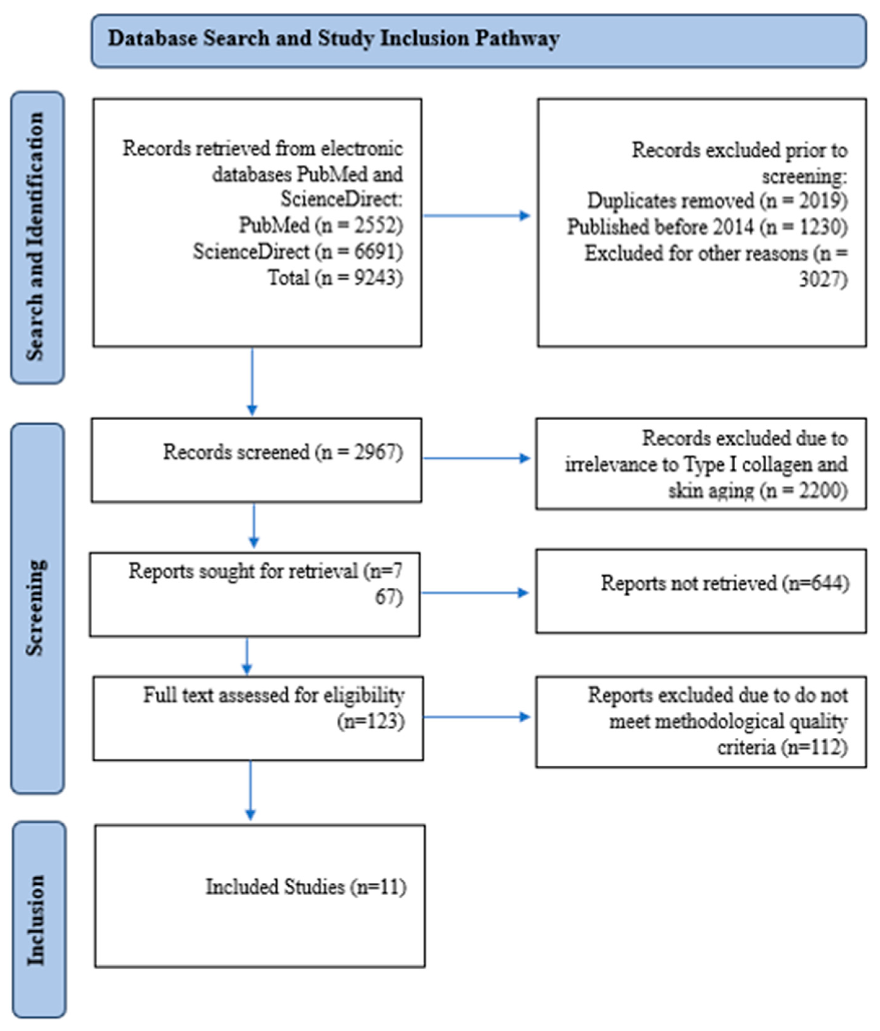
The PRISMA framework was applied to systematically screen and select eligible studies. Initially, all retrieved articles were screened based on titles and abstracts for relevance. Full-text reviews were conducted for studies meeting the inclusion criteria. The PRISMA flowchart (Figure 3) provides an overview of the selection process, ensuring a structured and reproducible approach.

2.6. Data Extraction and Analysis

A systematic and transparent approach was used to identify, screen, and select clinical studies evaluating the role of type I collagen in skin aging. The search was conducted across PubMed and ScienceDirect using predefined keywords, yielding a total of 9243 records. To ensure relevance and methodological rigor, the following steps were applied: duplicate records (n = 2019) were removed; articles published before 2014 (n = 1230) were excluded; and studies that were deemed irrelevant to the clinical investigation of type I collagen in the context of skin aging were excluded, including filtering out those that investigated collagen type I in non-cutaneous tissues or lacked specific focus on the COL1A1 gene (n = 3027), leaving 2967 records for further screening. Subsequently, these 2967 records underwent title and abstract screening, resulting in the exclusion of 2200 articles that did not directly examine the role of type I collagen in skin aging. This process left 767 reports that were sought for retrieval, of which 123 full-text articles were assessed for eligibility. Reports excluded due to insufficient methodological quality (n = 110). Ultimately, 11 studies met all inclusion criteria and were included in the final systematic review. These selected studies served as the foundation for evaluating the impact of type I collagen on skin aging and were analyzed in detail. The structured methodology, illustrated in the PRISMA flowchart (Figure 3), ensured that only high-quality, clinically relevant studies were incorporated, thereby strengthening the validity of the findings. A total of 11 studies were included in the final analysis. These articles were systematically examined to extract key information related to the effect of type I collagen on skin aging parameters (elasticity, hydration, wrinkle formation) and the clinical relevance of collagen supplementation. The extracted data were synthesized into a comprehensive table to facilitate comparison and interpretation, forming the basis for the Results and Discussion sections.

2.7. Ethical Considerations

This study is a systematic review based on previously published studies and does not involve new studies with human or animal participants. Therefore, ethical approval was not required.

3. Results and Discussion

3.1. Type I Collagen and Its Impact on Skin Aging

To ensure a structured and comprehensive evaluation of type I collagen in skin aging, a summary of evidence obtained from 11 selected studies (Table 1). Due to the scope of this systematic review, clinical studies were selected that investigated the influence of type I collagen on skin elasticity, hydration, and wrinkle generation. The table summarizes core components of each study, such as design, methods, intervention characteristics, key findings, and clinical implications (Table 1).

3.2. Individual Study Analysis

Yoon et al. [92] evaluated the potential benefit of topical estrogen treatment in photoaged skin by measuring wrinkle severity and collagen levels. What the investigators noted was that compared to placebo, the topical estrone significantly enhanced skin elasticity and decreased wrinkle severity but there were no statistically significant differences between the groups observed on physical examination, with the exception that topical estrone resulted in a marked increase in matrix metalloproteinase-1 (MMP-1), a collagen-degrading enzyme. Collectively, the data indicate that, against the expectation, estrogen treatment adversely impacts photoaged skin by negatively modulating collagen homeostasis. The results of this study may not be widely applicable because the duration of the study was short and the target population was limited to a specific age group, post-menopausal women. Studies on other doses, formulations, or concomitant use with other drugs could expand upon our findings by preventing the negative effects of MMP-1 activation and promoting the effects on collagen synthesis. This study highlights the equilibrium that exists between collagen synthesis and degradation in skin aging and that pharmaceutical interventions should be approached with caution.
This study employed both objective skin measurements (Visiometer and Cutometer) and molecular analyses (qPCR and immunohistochemistry), revealing that while type I procollagen mRNA levels increased, protein expression did not, likely due to a significant 10.3-fold upregulation of MMP-1, which underscores the complex role of topical estrogen in collagen homeostasis under UV exposure.
Humbert et al. [93] investigated the impacts of Mécano-Stimulation, a method of mechanical stimulation, on age-related skin deterioration and collagen synthesis. The trial concluded that skin firmness significantly increases and skin sagging decreases with 24 sessions of Mécano-Stimulation, and the activity of dermal fibroblasts also increases, leading to an increase in collagen deposition. Though the study did not identify significant improvements in elasticity, the authors note that mechanical stimulation effects may be more apparent in firmness and collagen synthesis than elasticity. Another limitation of this study is that there was no longer follow-up to determine the lasting effects of changes. Moreover, while the effect was stronger at the inferior and lateral region of the cheek, there was limited change for the superior region and nasolabial fold—suggesting possible pitfalls of a one-size-fits-all approach that should be avoided in future studies. As Mécano-Stimulation may be considered a safe, non-invasive technique to enhance skin trophicity, its long-term efficacy and broader applicability warrant further validation.
In the study by Humbert et al. [93], the primary clinical outcomes were assessed using objective non-invasive tools, such as cutometry for skin firmness and histological analysis of collagen content, which validated the observed improvements and supported the mechanistic findings on fibroblast stimulation and collagen synthesis.
Kim et al. [94] investigated the effect of oral supplementation with low-molecular-weight collagen peptides (LMWCP) on skin hydration, elasticity, and wrinkling. The researchers concluded that LMWCP supplementation significantly improved skin hydration and was shown to reduce wrinkle severity after 12 weeks in the study population, particularly those with lower baseline skin hydration. One strength of this study is that multiple skin parameters have been evaluated in the skin and time with each compound, giving an overall assessment of the LMWCP and skin effects on these parameters. The study found no noteworthy improvement in skin elasticity, which implied that LMWCP has limited efficacy with regard to skin elasticity and works better with regard to hydration and wrinkling. Although the presence of a placebo group and the randomized nature of the study boost the reliability of the findings, the absence of histological findings or objective analysis of imaging methods limits the evaluation of deeper structural changes in the skin. More studies are needed to determine how LMWCP affects low-Richardson skin types and younger people as well, since this is a major determinant of how well the product works.
In this study, skin hydration was assessed using the Corneometer CM 825 (Courage and Khazaka, Cologne, Germany), while wrinkling was evaluated both visually and through replica analysis with the SkinVisiometer SV 600 (Courage and Khazaka, Cologne, Germany). Elasticity was measured using the Cutometer MPA 580 (Courage and Khazaka, Cologne, Germany), which analyzes skin deformation and recovery. All assessments were standardized by location and environmental conditions, ensuring reliable measurements across timepoints.
Schütz et al. [95] studied the impact of 10-hydroxystearic acid (HSA) on age spots and pores on the human face. The results revealed that HSA significantly decreased facial pore area and pigmentation of age spots after 8 weeks of application. The strength of the study lies in the use of a multi-level approach to address the effect of HSA on skin aging, including in vitro, ex vivo, and clinical evaluations. Importantly, significant biochemical changes, including modulation of MMP-1 and collagen expression, supported the clinical findings and indicated that HSA improved skin texture via modulation of collagen metabolism. The relatively short follow-up time of the study makes the long-term effect of HSA difficult to discern, and longer follow-up studies are needed to validate whether these improvements were maintained. Recent research may have shown the potential for HSA as a new key skincare ingredient; more research is still needed to fully understand its mechanism of action and its specific application in varying skin types.
In this study, type I collagen expression was measured in vitro using immunofluorescence imaging in fibroblasts and supported by ex vivo analysis in human skin. The mechanism was further explored via PPARα activation assays and proteomic profiling.
Kang et al. [96] examined the anti-wrinkle effect of Angelica gigas Nakai root extract (ARE) in vitro and in clinical trials. This study showed that ARE upregulated type I collagen synthesis in fibroblast cells and significantly ameliorated the appearance of crow’s feet wrinkles in 8-week treatment participants. Strength of the study: both in vitro and clinical trials, and the overall effects of ARE look good. ARE, particularly when used with mineral-enriched water, appears to have potential as a protective agent to reduce the development of crow’s feet wrinkles. Nonetheless, though this study led to some promising results, it did not investigate the mechanism of how ARE can stimulate collagen synthesis, and there is still a need for more studies to identify the bioactive components. As a result, generalization is limited due to the shorter treatment period and small number of patients; further studies of larger sizes and longer durations will be helpful to back ARE as an anti-aging agent.
The anti-wrinkle effect of Angelica gigas Nakai root extract was assessed using objective 3D skin surface analysis via PRIMOS Lite, which measured parameters such as total roughness (R1) and maximum roughness (R2), alongside global photodamage scoring by dermatologists. In vitro, type I procollagen synthesis was quantified using a procollagen type I C-peptide ELISA kit.
Evans et al. [97] investigated the effects of the supplementation of hydrolyzed marine collagen (Vinh Wellness Collagen, VWC) on skin health in women aged from 45 to 60. Following the 12-week VWC supplementation, the study noted clinically meaningful increases in skin hydration and skin elasticity and decreases in wrinkle severity. The randomized, triple-blind, placebo-controlled design of this study is one of its major strengths, thus meeting some of the highest standards in clinical trial methodology. A reduction in wrinkles over time and improvement in skin texture and radiance indicate that VWC supplementation can target various aspects of skin aging. It is important to notice, however, there was quite a degree of potential bias with the reliance on self-reported measures of the appearance of skin and subjective assessments of skin radiance rather than objective measurement of outcomes such as histological analysis. Further studies should assess the long-term effects of VWC and also the effects of combining VWC with other skin health-promoting nutrients for optimal effects.
In this clinical trial, skin wrinkles were assessed objectively using the VISIA skin analysis system and the Modified Fitzpatrick Wrinkle Scale, while skin elasticity was measured instrumentally using the Cutometer® MPA 580 (Courage and Khazaka electronic GmbH, Cologne, Germany). These validated tools provided quantitative, reproducible evaluations of facial skin condition following 12 weeks of hydrolyzed collagen supplementation.
Laing et al. [98] a randomized, placebo-controlled trial to demonstrate the effects of a food supplement containing collagen peptides, vitamins, and other nutrients on skin quality. A remarkable enhancement of collagen structure in the facial skin was found with the confocal laser scanning microscopy at 12 weeks of Pb supplementation, suggesting that skin smoothness and elasticity potential of collagen were affected positively. The triple-blind, expert-assessed design of the study supports the validity of the results. But the research focused specifically on facial skin in the cheeks, and so the findings may not translate into similar results for the rest of the body. Beyond this, although the supplement was found to be safe and tolerated, the study did not examine long-term effects, indicating it might be worthwhile to further explore whether these benefits are sustainable in the long term vs. a short burst of activity.
In this trial, changes in dermal collagen structure were evaluated using in vivo confocal laser scanning microscopy VIVASCOPE 1500 (Lucid, Inc., Rochester, NY, USA). This method enabled high-resolution visualization of collagen fiber organization and fragmentation in the reticular dermis, which was scored by blinded expert graders using a visual analog scale comparing baseline and post-intervention images.
Campos et al. [99] examined the impact of oral supplementation with hydrolyzed fish cartilage on skin aging variables (wrinkle formation, dermal thickness, collagen morphology). Findings: Supplementation for 90 days led to significant improvement in skin microrelief, dermis echogenicity, and collagen structure. Results indicate that supplementation with hydrolyzed fish cartilage can increase skin density and decrease wrinkles. The specific collagen source studied and the relatively low dosage (500 mg/day) used leave us wondering how this would compare with other sources or higher doses of the same product in terms of optimal dosage to ensure maximum benefits. Although the study’s use of non-invasive imaging techniques was able to gain valuable insights into how supplementation had impacted tissue and skin changes, larger sample sizes and longer follow-up periods are needed to determine the longer-term role of hydrolyzed fish cartilage on skin health.
In this study, the structural characteristics of dermal collagen were evaluated using in vivo reflectance confocal microscopy Vivascope® 1500 (Lucid, Inc., Rochester, NY, USA). Changes in collagen morphology—such as the presence of thin reticulated collagen, coarse and huddled fibers, and elastosis—were scored before and after treatment, providing a semi-quantitative assessment of collagen remodeling.
Abe et al. [33] explored the role of 2 kDa hyaluronan (HA2K) oligosaccharides in collagen remodeling and wrinkle reduction. This study showed that HA2k penetrated the stratum corneum, activated collagen metabolism, and improved dermal collagen density. The randomized controlled trial performed by them showed that after 8 weeks, the injection of HA2k visibly reduced wrinkles in the nasolabial fold and crow’s feet areas. However, additional mechanistic investigation will be required, given that while the study’s authors conclude that HA2k is likely a good ingredient to include for wrinkle improvement, it also upregulates collagen synthesis (COL1A1) and degradation (MMP-1) at the same time. The 0.1% HA2k solution appeared promising but required further elucidation for the appropriate dosage with maximal efficacy. A major highlight of this study is that it is a real-life application, as the formulated lotion can serve as a safe, non-invasive alternative to invasive procedures such as filler injections. Nonetheless, the short duration of the study (4 weeks) and small sample size warrant careful consideration of the observations, necessitating larger, longer-term studies to corroborate these findings.
Skin parameters were evaluated using validated tools: collagen density via ultrasound (DermaLab®. Cortex Technology, Aalborg, Denmark), wrinkles via PRIMOS® systems, elasticity via Cutometer® (R2 parameter, Courage and Khazaka electronic GmbH, Cologne, Germany), and sagging via Moiré topography (F-ray®, Beyoung, Udaipur, India). Measurements were performed at baseline and 4 and 8 weeks under controlled conditions. Additionally, COL1A1 and MMP-1 levels were assessed in a 3D skin model using ELISA and immunofluorescence.
Žmitek et al. [100] investigated oral supplementation of hydrolyzed collagen (HC) or its combination with hyaluronic acid (HA) on skin aging. Results showed improvements in dermal density, wrinkle severity, and skin roughness for both the HC and the HC-HA supplemented groups. However, there were no significant differences between collagen and collagen with HA, so adding HA to collagen may not provide additional benefits. A strength of the study is its rigorous, randomized, placebo-controlled design and the fact that skin imaging techniques were used to objectively assess skin parameters. Nonetheless, the studies’ focus on a single dose of HC and HA makes it difficult to conclude which formulation is optimal for increased efficacy. Larger sample sizes and prolonged treatment times are warranted to confirm our finding, especially regarding the prospect of synergism between HC and HA. A more detailed overview of the specific processes involved in type I collagen degradation in aging and potential targets for therapeutic interventions are illustrated in a summary table (Table 2). Like we discussed before, collagen breakdown is induced by different factors, which consist of MMP activities, collagen fiber fragmentation, and perturbation of signaling pathways like TGF-β signaling, which is crucial in collagen synthesis and maintenance. Here, we summarize the former mechanisms, together with therapeutic strategies such as oral supplementation, topical treatments, and mechanical stimulation, which counteract the degradation of collagen and favor its regeneration.
In this article, wrinkle volume, depth, and roughness were measured using the Antera 3D CS multispectral topographic analyzer. Dermal density, thickness, hydration, and viscoelasticity were assessed using DermaLab Combo ultrasound and elasticity probes under standardized environmental conditions.
Iwahashi et al. [101] focused on screening materials that enhance Endo180, a collagen receptor implicated in collagen degradation and synthesis. They reported that lemon balm (Melissa officinalis) leaf extract (MOLE) enhanced Endo180 production and thereby type I collagen synthesis in dermal fibroblasts. In a clinical trial, a MOLE-containing cream notably decreased eye-corner wrinkles at 8 weeks of use. The findings of this study indicate the versatility of natural extracts to enhance collagen turnover and dermatological esthetics in the skin. The study is strong in that the researchers screened for rosmarinic acid being the active compound responsible for the effects of MOLE. However, the precise molecular events mediating the effects of MOLE on collagen metabolism are yet to be fully elucidated and warrant further exploration. For one, while the clinical data were encouraging, the study involved a small number of people, and additional research with bigger populations is needed to verify these results and expand upon their long-term effects.
Wrinkle severity was visually evaluated by a trained assessor, with photographic documentation taken at baseline, week 4, and week 8. In vitro, type I collagen expression in human fibroblasts (NB1RGB cells) was quantified by ELISA after treatment with MOLE and conditioned medium from UVB-irradiated keratinocytes. COL1A1 protein concentration was calculated based on a standard curve and normalized per cell using nuclear staining.

3.3. Mechanisms Involved in Type I Collagen Degradation

A more detailed overview of the specific processes involved in type I collagen degradation in aging and potential targets for therapeutic interventions are illustrated in a summary table (Table 2). Collagen breakdown is induced by different factors, which consist of MMP activities, collagen fiber fragmentation, and perturbation of signaling pathways like TGF-β signaling, which is crucial in collagen synthesis and maintenance. Here, we summarize the former mechanisms, together with therapeutic strategies such as oral supplementation, topical treatments, and mechanical stimulation, which counteract the degradation of collagen and favor its regeneration.

3.4. Study Limitations

Although the reviewed studies offer important insights into understanding the role of type I collagen in skin aging, as well as additional potential benefits of collagen supplementation as humans age, there were weaknesses that must be noted. Some of the studies had smaller sample sizes, for example, the ones by Kim et al. [94] (n = 64) and Iwahashi et al. [101] (n = 20), which may restrict the generalizability of the results to larger and more heterogeneous populations. Finally, most interventions are between 8 and 12 weeks in duration, which is relatively short and may not allow for the long-term evaluation of collagen supplementation and its effects on skin aging. Some studies, like the one by Yoon et al. [92], do not have long-term follow-up, making it impossible to determine if the treatment effects would be sustained. Additionally, differences in populations studied, such as age or other cutaneous conditions, may render findings less relevant to the general population. Even those studies had some limitations—for example, Humbert et al. [93] based their findings largely on visual assessments that may be subjective. Fifthly, some of the studies reviewed, such as those performed by Žmitek et al. [100], only focused on the effect of collagen supplementation, without exploring potential interactions with other skin treatments or environmental factors that could have influenced outcomes. Further shortcomings in the studies that may be addressed in future research include smaller sample sizes, lack of long-term follow-up, or just vague definitions for skin aging. All these disadvantages aside, however, the strengths of the present review are that it encompasses a variety of studies, those regarding topical versus oral collagen supplementation, and as such provides a broader perspective on the therapeutic potential of collagen when it comes to skin wellness and rejuvenation. The effectiveness of collagen supplementation on skin hydration, elasticity, and wrinkle reduction has been repeatedly demonstrated in studies evaluating both topical collagen preparations and oral collagen peptides, emphasizing the key role collagen plays in the quest for retaining youthful skin. Additional studies should investigate the long-term effects of collagen supplementation, especially in diverse populations, to determine its optimal doses and treatment durations [94,95,96,97,98,99,100]. Moreover, potential interactions and synergistic effects between collagen peptides and other anti-aging treatments such as antioxidants or hyaluronic acid could also be explored. In addition, gaining more knowledge about the molecular mechanisms controlling collagen synthesis and degradation in various skin types and environmental settings may shed further light on how to more effectively tailor therapies to target skin aging in a more precise manner.

4. Conclusions

Type I collagen plays a very important role in skin aging, as highlighted in this extensive systematic review. Skin aging is a multifactorial process driven by both intrinsic (genetic) and extrinsic (environmental) factors, with collagen degradation being one of the major players behind the appearance of wrinkles, sagging, and fluid loss in skin. As the most abundant structural protein in the dermis, type I collagen is important for preserving skin integrity, and its degradation plays a key role in the process of aging. This detailed systematic review demonstrates the role of collagen in skin aging and presents results of multiple studies indicating that both topical and oral collagen supplementation increase collagen synthesis, skin hydration, and elasticity and diminish wrinkles. Collagen supplementation has thus emerged as a non-invasive potential treatment, particularly peptides from marine and hydrolyzed collagen, which appear to have high therapeutic value in improving skin elasticity. As with all the studies presented to date, future research is needed to overcome the limitations in sample size, the duration of the study, and lack of follow-up. Continuing the investigation of the synergistic effects of collagen with other anti-aging interventions as well as elucidating the molecular mechanisms involved in the regulation of collagen synthesis and degradation will be important for the optimization of the strategies used to slow down aging and improve skin health.

Author Contributions

Conceptualization, O.B. and S.V.; methodology, O.B.; formal analysis, O.B.; investigation, O.B.; resources, O.B.; data curation, O.B. and S.V.; writing—original draft preparation, O.B.; writing—review and editing, O.B. and S.V.; supervision, S.V. All authors have read and agreed to the published version of the manuscript.

Funding

This research received no external funding.

Institutional Review Board Statement

Not applicable.

Informed Consent Statement

Not applicable.

Data Availability Statement

The original contributions presented in the study are included in the article, further inquiries can be directed to the corresponding author.

Conflicts of Interest

The authors declare no conflicts of interest.

Abbreviations

AREAngelica gigas Nakai root extracts
COL1A1Collagen type I alpha 1 chain
COL1A2Collagen type I alpha 2 chain
ECMExtra cellular matrix
HA2khyaluronan oligosaccharides
HASHydroxystearic acid
LMWCPlow-molecular-weight collagen peptides
MMPMatrix metalloproteinase
MOLEMelissa officinalis Lemon
PRISMAPreferred Reporting Items for Systematic Reviews and Meta-Analyses
ROSReactive oxygen species
TGF βTransforming Growth Factor Beta
TIMPTissue inhibitors of metalloproteinase
TβRIITransforming Growth Factor Beta Type II receptor

References

  1. Csekes, E.; Aging, L.R.S. Cellular Senescence and Natural Polyphenols. Int. J. Mol. Sci. 2021, 22, 12641. [Google Scholar] [CrossRef] [PubMed]
  2. Quan, T. Human Skin Aging and the Anti-Aging Properties of Retinol. Biomolecules 2023, 13, 1614. [Google Scholar] [CrossRef]
  3. Chin, L.-C.; Kimura, K.; Nakahata, Y.; Huang, L. Human Placental Extract Delays In Vitro Cellular Senescence through the Activation of NRF2-Mediated Antioxidant Pathway. Antioxidants 2022, 11, 1545. [Google Scholar] [CrossRef]
  4. Panichakul, T.; Srisanga, K.; Tupchiangmai, W.; Haritakun, W.; Ponnikorn, S. Skin Anti-Aging Potential of Ipomoea pes-caprae Ethanolic Extracts on Promoting Cell Proliferation and Collagen Production in Human Fibroblasts (CCD-986sk Cells). Pharmaceuticals 2022, 15, 969. [Google Scholar] [CrossRef]
  5. Boismal, F.; Serror, K.; Dobos, G.; Zuelgaray, E.; Bensussan, A.; Michel, L. Skin aging: Pathophysiology and innovative therapies. Med. Sci. 2020, 36, 1163–1172. [Google Scholar] [CrossRef]
  6. Wang, S.; Belmonte, J.C.I.; Esteban, C.R.; Long, X.; Yu, N.; Song, M.; He, Y.; Huang, J.; Zheng, Y.; Zou, Z.; et al. A Single-Cell Transcriptomic Atlas of Human Skin Aging. Dev. Cell 2021, 56, 383–397.e8. [Google Scholar] [CrossRef]
  7. Jourdain, E.; Morgado-Carrasco, D.; Piquero-Casals, J.; Gil-Lianes, J. Oral Supplementation and Systemic Drugs for Skin Aging: A Narrative Review. Actas Dermo-Sifiliogr. 2022, 114, T114–T124. [Google Scholar] [CrossRef]
  8. Rorteau, J.; Chevalier, F.P.; Fromy, B.; Lamartine, J. Functional integrity of aging skin, from cutaneous biology to an-ti-aging strategies. Med. Sci. 2020, 36, 1155–1162. [Google Scholar] [CrossRef]
  9. Pelletier, M.; Le Fur, M.; Granotier, F.; Couturaud, V. Reverse skin aging signs by red light photobiomodulation. Ski. Res. Technol. 2023, 29, e13391. [Google Scholar] [CrossRef]
  10. Kang, H.Y.; Park, T.J.; Kim, J.C. Skin-Aging Pigmentation: Who Is the Real Enemy? Cells 2022, 11, 2541. [Google Scholar] [CrossRef]
  11. Ge, C.; Xiao, Z.; Cao, C.; Wu, Y. Diet and Skin Aging—From the Perspective of Food Nutrition. Nutrients 2020, 12, 870. [Google Scholar] [CrossRef] [PubMed]
  12. Ho, C.Y.; Dreesen, O. Faces of cellular senescence in skin aging. Mech. Ageing Dev. 2021, 198, 111525. [Google Scholar] [CrossRef]
  13. Ansary, T.M.; Hossain, M.R.; Kamiya, K.; Komine, M.; Ohtsuki, M. Inflammatory Molecules Associated with Ultraviolet Radiation-Mediated Skin Aging. Int. J. Mol. Sci. 2021, 22, 3974. [Google Scholar] [CrossRef]
  14. Aguilera-Aguirre, L.; Sreedhar, A.; Singh, K.K. Mitochondria in skin health, aging, and disease. Cell Death Dis. 2020, 11, 444. [Google Scholar] [CrossRef]
  15. Damiani, E.; Brugè, F.; Cirilli, I.; Marcheggiani, F.; Olivieri, F.; Armeni, T.; Cianfruglia, L.; Giuliani, A.; Orlando, P.; Tiano, L. Modulation of Oxidative Status by Normoxia and Hypoxia on Cultures of Human Dermal Fibroblasts: How Does It Affect Cell Aging? Oxid. Med. Cell. Longev. 2018, 2018, 5469159. [Google Scholar] [CrossRef]
  16. Park, E.-Y.; Oh, Y.S.; Kim, K.; Baek, D.-J.; Kim, C.-E. Prevention of UVB-Induced Photoaging by an Ethyl Acetate Fraction from Allomyrina dichotoma Larvae and Its Potential Mechanisms in Human Dermal Fibroblasts. Int. J. Mol. Sci. 2024, 25, 7850. [Google Scholar] [CrossRef]
  17. Park, J.; Jang, Y.-P.; Lee, C.; Cho, H.; Kim, M.; Kim, B. Evaluating the Dermatological Benefits of Snowberry (Symphoricarpos albus): A Comparative Analysis of Extracts and Fermented Products from Different Plant Parts. Int. J. Mol. Sci. 2024, 25, 9660. [Google Scholar] [CrossRef]
  18. Kang, W.; Choi, D.; Park, T. Dietary Suberic Acid Protects Against UVB-Induced Skin Photoaging in Hairless Mice. Nutrients 2019, 11, 2948. [Google Scholar] [CrossRef] [PubMed]
  19. Choi, S.-Y.; Choi, Y.-J.; Yoo, J.; Kim, E.; Jung, J. Antiphotoaging Effect of AGEs BlockerTM in UVB-Irradiated Cells and Skh:HR-1 Hairless Mice. Curr. Issues Mol. Biol. 2023, 45, 4181–4199. [Google Scholar] [CrossRef]
  20. Lee, S.B.; Seo, W.-Y.; Hwang, S.J.; Shin, M.C.; Jeong, Y.; Choi, Y.J.; Ryu, D.; Kim, S.H.; Kim, K. Effects of human collagen α-1 type I-derived proteins on collagen synthesis and elastin production in human dermal fibroblasts. BMB Rep. 2021, 54, 329–334. [Google Scholar] [CrossRef]
  21. Hu, L.; Man, M.; Yu, H.; Zhang, J. Aging in the dermis: Fibroblast senescence and its significance. Aging Cell 2023, 23, e14054. [Google Scholar] [CrossRef]
  22. Young, K.; Trowbridge, J.J.; SanMiguel, J.M. Hand in hand: Intrinsic and extrinsic drivers of aging and clonal hematopoiesis. Exp. Hematol. 2020, 91, 1–9. [Google Scholar] [CrossRef]
  23. Mysliwiec, H.; Kakareko, K.; Hryszko, T.; Rydzewska-Rosolowska, A.; Bielach-Bazyluk, A.; Zbroch, E.; Flisiak, I. Sirtuin 1 and Skin: Implications in Intrinsic and Extrinsic Aging—A Systematic Review. Cells 2021, 10, 813. [Google Scholar] [CrossRef]
  24. Lee, J.H.; Jang, H.; Jo, Y.; Choi, S. Aging of hair follicle stem cells and their niches. BMB Rep. 2022, 56, 2–9. [Google Scholar] [CrossRef]
  25. Di Micco, R.; Krizhanovsky, V.; Baker, D.; d’Adda di Fagagna, F. Cellular senescence in ageing: From mechanisms to therapeutic opportunities. Nat. Rev. Mol. Cell Biol. 2021, 22, 75–95. [Google Scholar] [CrossRef]
  26. Trowbridge, J.J.; Mistry, J.J.; Díaz, P.A.C. Hematopoietic stem cell aging and leukemia transformation. Blood 2023, 142, 533–542. [Google Scholar] [CrossRef]
  27. Tyrrell, D.J.; Goldstein, D.R. Ageing and atherosclerosis: Vascular intrinsic and extrinsic factors and potential role of IL-6. Nat. Rev. Cardiol. 2020, 18, 58–68. [Google Scholar] [CrossRef]
  28. Lee, Y.H.; Shin, S.H.; Park, K.Y.; Rho, N.-K. Skin aging from mechanisms to interventions: Focusing on dermal aging. Front. Physiol. 2023, 14, 1195272. [Google Scholar] [CrossRef]
  29. Schmitt, C.A.; Wang, B.; Demaria, M. Senescence and cancer—Role and therapeutic opportunities. Nat. Rev. Clin. Oncol. 2022, 19, 619–636. [Google Scholar] [CrossRef]
  30. Ratanapokasatit, Y.; Laisuan, W.; Rattananukrom, T.; Petchlorlian, A.; Thaipisuttikul, I.; Sompornrattanaphan, M. How Micro-biomes Affect Skin Aging: The Updated Evidence and Current Perspectives. Life 2022, 12, 936. [Google Scholar] [CrossRef]
  31. Puzzi, M.B.; Lago, J.C. The effect of aging in primary human dermal fibroblasts. PLoS ONE 2019, 14, e0219165. [Google Scholar] [CrossRef]
  32. Potekaev, N.N.; Borzykh, O.B.; Medvedev, G.V.; Petrova, M.M.; Gavrilyuk, O.A.; Karpova, E.I.; Trefilova, V.V.; Demina, O.M.; Popova, T.E.; Shnayder, N.A. Genetic and Epigenetic Aspects of Skin Collagen Fiber Turnover and Functioning. Cosmetics 2021, 8, 92. [Google Scholar] [CrossRef]
  33. Seino, S.; Kurihara, H.; Kage, M.; Abe, Y.; Tokudome, Y. 2-kDa hyaluronan ameliorates human facial wrinkles through increased dermal collagen density related to promotion of collagen remodeling. J. Cosmet. Dermatol. 2022, 22, 320–327. [Google Scholar] [CrossRef]
  34. Gupta, J.; Repici, M.; Gross, S.R.; Lancaster, T.; Tabrizi, M.E.A. An Extracellular/Membrane-Bound S100P Pool Regulates Motility and Invasion of Human Extravillous Trophoblast Lines and Primary Cells. Biomolecules 2023, 13, 1231. [Google Scholar] [CrossRef]
  35. Liang, L.; Yang, C.; Zhou, Y.; Liu, L.; Chen, Y. Extracellular vesicles of Fusobacterium nucleatum compromise intestinal barrier through targeting RIPK1-mediated cell death pathway. Gut Microbes 2021, 13, 1902718. [Google Scholar] [CrossRef]
  36. Hao, H.; Zhang, X.; Tong, L.; Liu, Q.; Liang, X.; Bu, Y.; Gong, P.; Liu, T.; Zhang, L.; Xia, Y.; et al. Effect of Extracellular Vesicles Derived From Lactobacillus plantarum Q7 on Gut Microbiota and Ulcerative Colitis in Mice. Front. Immunol. 2021, 12, 777147. [Google Scholar] [CrossRef]
  37. Greiner, M.A.; Jiao, C.; Pouw, A.E.; Coussa, R.G.; Skeie, J.M.; Fingert, J.H.; Sohn, E.H.; Mullins, R.F.; Han, I.C. Cell–Matrix Interactions in the Eye: From Cornea to Choroid. Cells 2021, 10, 687. [Google Scholar] [CrossRef]
  38. Wu, Y.; Fan, J.; Wu, Z.; Li, J.; Wang, Z.; Yao, Y.; Tao, S.; Xiao, Y.; Wei, H.; Liu, X. Extracellular vesicles derived from Lactobacillus johnsonii promote gut barrier homeostasis by enhancing M2 macrophage polarization. J. Adv. Res. 2025, 69, 545–563. [Google Scholar] [CrossRef]
  39. Kumar, A.; Shipman, T.; Le, L.Q.; Xing, C.; Yu, Z.; Wang, Y.; McKay, R.M.; Jiang, C. Basement membrane proteins in extracellular matrix characterize NF1 neurofibroma development and response to MEK inhibitor. J. Clin. Investig. 2023, 133, e168227. [Google Scholar] [CrossRef]
  40. Baldomà, L.; Díaz-Garrido, N.; Badia, J. Microbiota-derived extracellular vesicles in interkingdom communication in the gut. J. Extracell. Vesicles 2021, 10, e12161. [Google Scholar] [CrossRef]
  41. Mira, E.; Mañes, S.; Carmona-Rodríguez, L.; Fernández-Aceñero, M.J.; Martínez-Rey, D. Extracellular Superoxide Dismutase, the Endothelial Basement Membrane, and the WNT Pathway: New Players in Vascular Normalization and Tumor Infiltration by T-Cells. Front. Immunol. 2020, 11, 579552. [Google Scholar] [CrossRef]
  42. Chai, P.; Lebedenko, C.G.; Flynn, R.A. RNA Crossing Membranes: Systems and Mechanisms Contextualizing Extracellular RNA and Cell Surface GlycoRNAs. Annu. Rev. Genom. Hum. Genet. 2023, 24, 85–107. [Google Scholar] [CrossRef] [PubMed]
  43. Kaji, I.; Goldenring, J.R.; Engevik, A.C. The Physiology of the Gastric Parietal Cell. Physiol. Rev. 2020, 100, 573–602. [Google Scholar] [CrossRef]
  44. Martínez-Puig, D.; Gálvez-Martín, P.; Costa-Larrión, E.; Rubio-Rodríguez, N. Collagen Supplementation for Joint Health: The Link between Composition and Scientific Knowledge. Nutrients 2023, 15, 1332. [Google Scholar] [CrossRef] [PubMed]
  45. Lin, Y.-F.; Lee, S.-Y.; Liu, S.-H.; Pei, D.; Lin, W.-C.; Wang, C.-S.; Liao, E.-C.; Hsu, C.-H. Aquaporin-8 promotes human dermal fibroblasts to counteract hydrogen peroxide-induced oxidative damage: A novel target for management of skin aging. Open Life Sci. 2024, 19, 20220828. [Google Scholar] [CrossRef]
  46. Thorseth, M.-L.; Madsen, D.H.; Rømer, A.M.A. Immune Modulatory Properties of Collagen in Cancer. Front. Immunol. 2021, 12, 791453. [Google Scholar] [CrossRef]
  47. Wei, M.; Yu, L. Biomineralization of Collagen-Based Materials for Hard Tissue Repair. Int. J. Mol. Sci. 2021, 22, 944. [Google Scholar] [CrossRef]
  48. Huang, L.; Sun, J.; Wen, D.; Zhang, Y.; Liu, Y.; Ho, C.; Gao, Y.; Li, Q. Targeting the stem cell niche: Role of collagen XVII in skin aging and wound repair. Theranostics 2022, 12, 6446–6454. [Google Scholar] [CrossRef]
  49. Kokkonen, N.; Tuusa, J.; Tasanen, K. BP180/Collagen XVII: A Molecular View. Int. J. Mol. Sci. 2021, 22, 12233. [Google Scholar] [CrossRef]
  50. Haverkamp, R.G.; Sizeland, K.H.; Wells, H.C.; Kamma-Lorger, C. Collagen dehydration. Int. J. Biol. Macromol. 2022, 216, 140–147. [Google Scholar] [CrossRef]
  51. Li, Q.; Hsu, J.J.; Demer, L.L.; Vazquez-Padron, R.I.; Bendeck, M.P.; Tintut, Y. Collagen VIII in vascular diseases. Matrix Biol. 2024, 133, 64–76. [Google Scholar] [CrossRef] [PubMed]
  52. Hintze, V.; Halfter, N.; Schnabelrauch, M.; Anderegg, U. Collagen/glycosaminoglycan-based matrices for controlling skin cell responses. Biol. Chem. 2021, 402, 1325–1335. [Google Scholar] [CrossRef]
  53. Oosterlaken, B.M.; Vena, M.P.; de With, G. In Vitro Mineralization of Collagen. Adv. Mater. 2021, 33, 2004418. [Google Scholar] [CrossRef] [PubMed]
  54. Hagemann, I.S.; Krigman, H.R.; Weiss, M.C.; Sun, L.; Chapagain, U. COL1A1::PDGFB fusion-associated uterine sarcoma and response to Imatinib: A case report. Gynecol. Oncol. Rep. 2023, 49, 101270. [Google Scholar] [CrossRef]
  55. Szoka, L.; Karna, E.; Palka, J.A.; Huynh, T.Y.L. Proline-dependent regulation of collagen metabolism. Cell. Mol. Life Sci. 2020, 77, 1911–1918. [Google Scholar] [CrossRef]
  56. Devos, H.; Zoidakis, J.; Roubelakis, M.G.; Latosinska, A.; Vlahou, A. Reviewing the Regulators of COL1A1. Int. J. Mol. Sci. 2023, 24, 10004. [Google Scholar] [CrossRef]
  57. González, S.; Dierckx, S.; Mullor, J.L.; Nergiz-Unal, R.; Merino, M.; Patrizi, M. Collagen peptides affect collagen synthesis and the expression of collagen, elastin, and versican genes in cultured human dermal fibroblasts. Front. Med. 2024, 11, 1397517. [Google Scholar] [CrossRef]
  58. Amirrah, I.N.; Lokanathan, Y.; Fauzi, M.B.; Zulkiflee, I.; Wee, M.F.M.R.; Motta, A. A Comprehensive Review on Collagen Type I Development of Biomaterials for Tissue Engineering: From Biosynthesis to Bioscaffold. Biomedicines 2022, 10, 2307. [Google Scholar] [CrossRef]
  59. Reichetzeder, C.; Irawati, F.; Putra, S.E.D.; Kok, T.; Sulistomo, H.W.; Mulyanata, L.T.; Humardani, F.M.; Liamry, J.N.; Chandra, G. Exploring the impact of diabetes on aging: Insights from TERT and COL1A1 methylation. Turk. J. Biol. 2024, 48, 257–266. [Google Scholar] [CrossRef]
  60. Kim, J.H.; Lee, S.J.; Jeong, D.H.; Kim, S.H.; Jung, M.S.; Lee, K.H. Minimally invasive skin sampling and transcriptome analysis using microneedles for skin type biomarker research. Ski. Res. Technol. 2022, 28, 322–335. [Google Scholar] [CrossRef]
  61. Guilbert, M.; Manfait, M.; Terryn, C.; Garnotel, R.; Dinten, J.-M.; Koenig, A.; Sockalingum, G.D.; Jeannesson, P.; Roig, B.; Piot, O.; et al. Highlighting the impact of aging on type I collagen: Label-free investigation using confocal reflectance microscopy and diffuse reflectance spectroscopy in 3D matrix model. Oncotarget 2016, 7, 8546–8555. [Google Scholar] [CrossRef] [PubMed]
  62. Paolini, B.; Casali, P.; Sanfilippo, R.; Alessi, A.; Padovano, B.; Greco, F.G.; Franza, A.; Fabbroni, C.; Rota, S. COL1A1::PDGFB fusion-associated uterine fibrosarcoma: A case report and review of the literature. Cancer Rep. 2024, 7, e1969. [Google Scholar] [CrossRef]
  63. Xia, M.; Wang, X.-H.; Li, X.-M.; Yao, M.-D.; Li, D.; Yao, J.; Tong, M.; Jiao, L.; Zhao, P.-Q.; Yan, B. Single-cell RNA sequencing reveals a unique pericyte type associated with capillary dysfunction. Theranostics 2023, 13, 2515–2530. [Google Scholar] [CrossRef]
  64. Xu, P.; Wang, Y.; Wu, X.; Wang, W.; Wang, Q.; Lin, W.; Zhang, Z.; Li, M. The COL1A1 rs1800012 polymorphism is associated with osteoporosis or fracture risk: A meta-analysis of 30 studies. Int. J. Burns Traum. 2024, 14, 148–159. [Google Scholar] [CrossRef]
  65. Chen, W.; Wu, X.; Hu, J.; Liu, X.; Guo, Z.; Wu, J.; Shao, Y.; Hao, M.; Zhang, S.; Hu, W.; et al. The translational potential of miR-26 in atherosclerosis and development of agents for its target genes ACC1/2, COL1A1, CPT1A, FBP1, DGAT2, and SMAD7. Cardiovasc. Diabetol. 2024, 23, 21. [Google Scholar] [CrossRef] [PubMed]
  66. Kowalewski, J.; Chrzanowska, N.M.; Lewandowska, M.A. Use of Fluorescence In Situ Hybridization (FISH) in Diagnosis and Tailored Therapies in Solid Tumors. Molecules 2020, 25, 1864. [Google Scholar] [CrossRef]
  67. Liu, L.; Huang, W. hsa_circ_0020378 regulating miR-339-3p/COL1A1 promotes osteosarcoma progression. Cancer Biol. Ther. 2023, 24, 2274120. [Google Scholar] [CrossRef]
  68. Harsanyi, S.; Zamborsky, R.; Krajciova, L.; Kokavec, M.; Danisovic, L. Developmental Dysplasia of the Hip: A Review of Eti-opathogenesis, Risk Factors, and Genetic Aspects. Medicina 2020, 56, 153. [Google Scholar] [CrossRef]
  69. Ren, J.; Da, J.; Hu, N. Identification of COL1A1 associated with immune infiltration in brain lower grade glioma. PLoS ONE 2022, 17, e0269533. [Google Scholar] [CrossRef]
  70. Steiner, R.D.; Basel, D. COL1A1/2 Osteogenesis Imperfecta. In GeneReviews®; Adam, M.P., Feldman, J., Mirzaa, G.M., Pagon, R.A., Wallace, S.E., Amemiya, A., Eds.; University of Washington: Seattle, WA, USA, 2025; Available online: https://www.ncbi.nlm.nih.gov/books/NBK1295/ (accessed on 9 March 2025). [PubMed]
  71. Vankevičienė, R.; Matulevičienė, A.; Ramašauskaitė, D.; Mazgelytė, E.; Vankevičienė, K.; Paliulytė, V. A Sporadic Case of COL1A1 Osteogenesis Imperfecta: From Prenatal Diagnosis to Outcomes in Infancy—Case Report and Literature Review. Genes 2023, 14, 2062. [Google Scholar] [CrossRef]
  72. Nishikawa, S.; Hosoi, J.; Amano, S.; Ogura, Y.; Iriyama, S. Regeneration of collagen fibrils at the papillary dermis by reconstructing basement membrane at the dermal–epidermal junction. Sci. Rep. 2022, 12, 795. [Google Scholar] [CrossRef]
  73. Hasebe, Y.; Hasegawa, S.; Akamatsu, H.; Iwata, Y.; Arima, M.; Ishii, Y.; Sugiura, K.; Yamada, T.; Sanada, A. Enhanced Type I Collagen Synthesis in Fibroblasts by Dermal Stem/Progenitor Cell-Derived Exosomes. Biol. Pharm. Bull. 2022, 45, 872–880. [Google Scholar] [CrossRef]
  74. Cheong, H.; Park, C.; Park, J.; Kim, W.-J.; Kim, W.; Kim, S.-J. Malonic Acid Isolated from Pinus densiflora Inhibits UVB-Induced Oxidative Stress and Inflammation in HaCaT Keratinocytes. Polymers 2021, 13, 816. [Google Scholar] [CrossRef]
  75. Kim, S.-O.; Yun, S.-J.; Lee, J.-B.; Choi, J.-Y.; Lee, J.-A.; Cho, K.-A.; Choi, D.-I.; Lee, S.-C. Methyl-β-cyclodextrin up-regulates collagen I expression in chronologically-aged skin via its anti-caveolin-1 activity. Oncotarget 2015, 6, 1942–1953. [Google Scholar] [CrossRef]
  76. Kong, R.; Cui, Y.; Fisher, G.J.; Wang, X.; Chen, Y.; Schneider, L.M.; Majmudar, G. A comparative study of the effects of retinol and retinoic acid on histological, molecular, and clinical properties of human skin. J. Cosmet. Dermatol. 2016, 15, 49–57. [Google Scholar] [CrossRef]
  77. Shim, J.H. Prostaglandin E2 Induces Skin Aging via E-Prostanoid 1 in Normal Human Dermal Fibroblasts. Int. J. Mol. Sci. 2019, 20, 5555. [Google Scholar] [CrossRef]
  78. Kim, J.H.; Lee, S.J.; Choi, Y.M.; Park, C.O.; Jang, E.Y.; Jeong, D.H.; Kim, S.H.; Jung, Y.W.; Roh, Y.; Lee, K.H.; et al. Development of a biomarker-based platform for comprehensive skin characterization using minimally invasive skin sampling and quantitative real-time PCR. Ski. Res. Technol. 2024, 30, e13908. [Google Scholar] [CrossRef]
  79. Kishi, K.; Asou, T.; Takaya, K. Aging Fibroblasts Adversely Affect Extracellular Matrix Formation via the Senescent Humoral Factor Ependymin-Related Protein 1. Cells 2022, 11, 3749. [Google Scholar] [CrossRef]
  80. Gecyte, E.; Gecys, D.; Sepetiene, R.; Patamsyte, V.; Skipskis, V.; Stanioniene, Z.; Barakauskas, S.; Valiukevicius, P. Genetical Signature—An Example of a Personalized Skin Aging Investigation with Possible Implementation in Clinical Practice. J. Pers. Med. 2023, 13, 1305. [Google Scholar] [CrossRef]
  81. Wei, Y.; Peng, D.; Wang, M.; Fu, M.; Wei, X. Targeting TGF-β signal transduction for fibrosis and cancer therapy. Mol. Cancer 2022, 21, 104. [Google Scholar] [CrossRef]
  82. Deng, Z.; Fan, T.; Xiao, C.; Tian, H.; Zheng, Y.; Li, C.; He, J. TGF-β signaling in health, disease, and therapeutics. Signal Transd. Target. Ther. 2024, 9, 61. [Google Scholar] [CrossRef]
  83. Tzavlaki, K.; Moustakas, A. TGF-β Signaling. Biomolecules 2020, 10, 487. [Google Scholar] [CrossRef]
  84. Lee, J.H.; Massagué, J. TGF-β in developmental and fibrogenic EMTs. Semin. Cancer Biol. 2022, 86, 136–145. [Google Scholar] [CrossRef]
  85. Orwoll, E.; Esposito, P.; Jiang, M.-M.; Munivez, E.; Gannon, F.H.; Song, I.-W.; Grafe, I.; Huang, S.; Lee, B.; Nagamani, S.C.; et al. Targeting TGF-β for treatment of osteogenesis imperfecta. J. Clin. Investig. 2022, 132, e152571. [Google Scholar] [CrossRef]
  86. Lou, J.; Campbell, M.G.; Jespersen, J.M.; Yu, Z.; Nishimura, S.L.; Ito, S.; Cossio, P.; Baron, J.L.; Marks, J.; Wen, W.; et al. Dynamic allostery drives autocrine and paracrine TGF-β signaling. Cell 2024, 187, 6200–6219.e23. [Google Scholar] [CrossRef]
  87. Zhao, X.; Wang, J.; Wan, Y.Y. Intricacies of TGF-β signaling in Treg and Th17 cell biology. Cell. Mol. Immunol. 2023, 20, 1002–1022. [Google Scholar] [CrossRef]
  88. Seoane, J.; Lan, Y.; Wang, X.; Dussault, I.; Barcellos-Hoff, M.H.; Schlom, J.; Audhuy, F.; Gulley, J.L.; Moustakas, A. Dual inhibition of TGF-β and PD-L1: A novel approach to cancer treatment. Mol. Oncol. 2022, 16, 2117–2134. [Google Scholar] [CrossRef]
  89. Zeng, Q.; Wang, S.; Xu, H.; Wu, D.; Shi, X.; Wu, F.; Zhou, H.; Yang, J.; Deng, S.; Hu, T. TGF-β signaling in the tumor metabolic microenvironment and targeted therapies. J. Hematol. Oncol. 2022, 15, 135. [Google Scholar] [CrossRef]
  90. Gough, N.R.; Mishra, L.; Xiang, X. TGF-β Signaling in Liver, Pancreas, and Gastrointestinal Diseases and Cancer. Gastroenterology 2021, 161, 434–452.e15. [Google Scholar] [CrossRef]
  91. Page, M.J.; McKenzie, J.E.; Bossuyt, P.M.; Boutron, I.; Hoffmann, T.C.; Mulrow, C.D.; Shamseer, L.; Tetzlaff, J.M.; Akl, E.A.; Brennan, S.E.; et al. The PRISMA 2020 statement: An updated guideline for reporting systematic reviews. BMJ 2021, 372, n71. [Google Scholar] [CrossRef]
  92. Lee, S.; Chung, J.; Yoon, H. Long-term Topical Oestrogen Treatment of Sun-exposed Facial Skin in Post-menopausal Women Does Not Improve Facial Wrinkles or Skin Elasticity, But Induces Matrix Metalloproteinase-1 Expression. Acta Dermato-Venereol. 2014, 94, 4–8. [Google Scholar] [CrossRef]
  93. Humbert, P.; Fanian, F.; Lihoreau, T.; Jeudy, A.; Elkhyat, A.; Robin, S.; Courderot-Masuyer, C.; Tauzin, H.; Lafforgue, C.; Haftek, M. Mécano-StimulationTM of the skin improves sagging score and induces beneficial functional modification of the fibro-blasts: Clinical, biological, and histological evaluations. Clin. Interv. Aging 2015, 10, 387–403. [Google Scholar] [CrossRef] [PubMed]
  94. Sakai, Y.; Kim, D.-U.; Chung, H.-C.; Choi, J.; Lee, B.-Y. Oral Intake of Low-Molecular-Weight Collagen Peptide Improves Hydration, Elasticity, and Wrinkling in Human Skin: A Randomized, Double-Blind, Placebo-Controlled Study. Nutrients 2018, 10, 826. [Google Scholar] [CrossRef]
  95. Schütz, R.; Rawlings, A.V.; Wandeler, E.; Jackson, E.; Trevisan, S.; Monneuse, J.; Bendik, I.; Massironi, M.; Imfeld, D. Bio-derived hydroxystearic acid ameliorates skin age spots and conspicuous pores. Int. J. Cosmet. Sci. 2019, 41, 240–256. [Google Scholar] [CrossRef]
  96. Kang, J.-W.; Cho, H.-E.; Choi, H.-M.; Lee, I.-C. Anti-wrinkle properties of Angelica gigas Nakai root extracts using miner-al-rich water. J. Cosmet. Dermatol. 2023, 22, 328–334. [Google Scholar] [CrossRef]
  97. Guthrie, N.; Zakaria, N.; Pelipyagina, T.; Evans, M.; Lewis, E.D. A randomized, triple-blind, placebo-controlled, parallel study to evaluate the efficacy of a freshwater marine collagen on skin wrinkles and elasticity. J. Cosmet. Dermatol. 2020, 20, 825–834. [Google Scholar] [CrossRef]
  98. Laing, S.; Wilhelm, K.-P.; Bielfeldt, S.; Ehrenberg, C. A Dermonutrient Containing Special Collagen Peptides Improves Skin Structure and Function: A Randomized, Placebo-Controlled, Triple-Blind Trial Using Confocal Laser Scanning Microscopy on the Cosmetic Effects and Tolerance of a Drinkable Collagen Supplement. J. Med. Food 2020, 23, 147–152. [Google Scholar] [CrossRef]
  99. Campos, P.M.B.G.M.; Franco, R.S.B.; Kakuda, L.; Cadioli, G.F.; Maria, D.G.; Bouvret, E. Oral Supplementation with Hydrolyzed Fish Cartilage Improves the Mor-phological and Structural Characteristics of the Skin: A Double-Blind, Placebo-Controlled Clinical Study. Molecules 2021, 26, 4880. [Google Scholar] [CrossRef]
  100. Butina, M.R.; Žmitek, J.; Pogačnik, T.; Hristov, H.; Žmitek, K.; Keršmanc, P. The Effects of Dietary Supplementation with Collagen and Vitamin C and Their Combination with Hyaluronic Acid on Skin Density, Texture and Other Parameters: A Randomised, Double-Blind, Placebo-Controlled Trial. Nutrients 2024, 16, 1908. [Google Scholar] [CrossRef]
  101. Kawashima, Y.; Iwahashi, H.; Masaki, H.; Taga, A. Lemon Balm (Melissa officinalis L.) Leaf Extract Promotes Endo180 Production in Dermal Fibroblasts and has Antiwrinkle Effect on Human Skin. Photodermatol. Photoimmunol. Photomed. 2025, 41, e70006. [Google Scholar] [CrossRef]
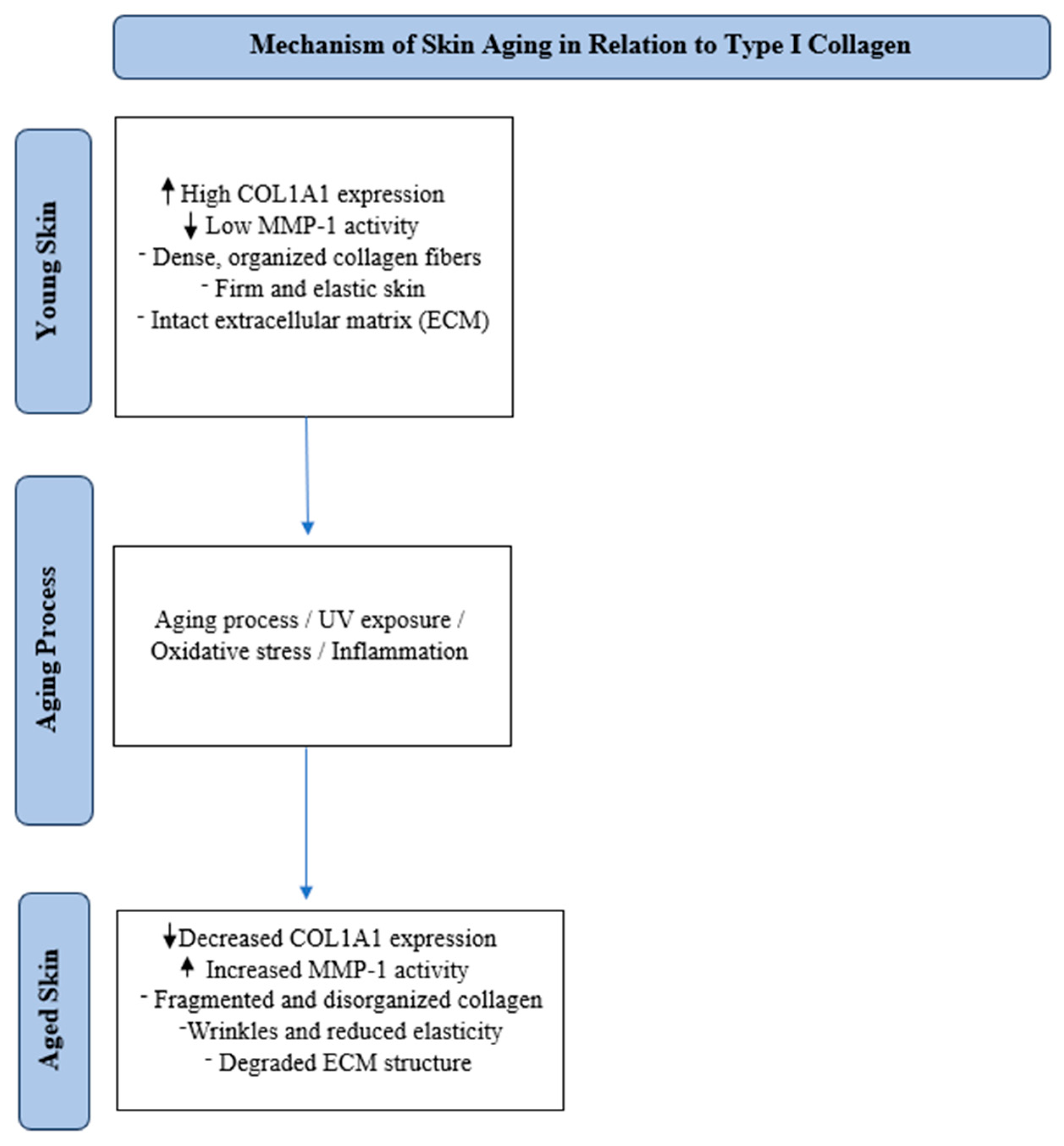
Figure 1. Schematic overview of the role of type I collagen (COL1A1) in the aging process of human skin. The transition from youthful to aged skin involves reduced collagen production, increased enzymatic degradation by MMPs (especially MMP-1), and structural deterioration of the extracellular matrix.
Cosmetics 12 00129 g001
Figure 2. Schematic representation of type I collagen structure and its formation from COL1A1 and COL1A2 gene products.
Cosmetics 12 00129 g002
Figure 3. PRISMA 2020 flow diagram for new systematic reviews that only included searches of databases and registers.
Cosmetics 12 00129 g003
Table 1. Summary of articles examining type I collagen and its impact on skin aging.
References
(Author, Year)
Study DesignSample Size and
Population
InterventionsKey FindingsClinical Implications
Yoon et al., 2014 [92]Randomized, double-blind, vehicle-controlled study80 post-menopausal women (mean age: 55.2 ± 2.2 years) with photoaged skin1% estrone cream (topical estrogen) vs. vehicle, applied daily for 24 weeksNo significant increase in type 1 collagen (COL1A1) protein despite a 5.0-fold increase in mRNA. MMP-1 increased 10.3-fold, leading to collagen breakdown. No improvement in wrinkles or elasticity, with some worsening. Topical estrogen failed to protect or restore collagen in sun-exposed skin. The treatment was applied to the crow’s feet area.Collagen synthesis alone is insufficient if MMP-1 is upregulated, causing collagen breakdown. Sun-exposed skin may respond differently to estrogen, leading to increased collagen degradation. Estrogen-based treatments may have negative effects on UV-exposed aging skin, highlighting the need for alternative collagen-preserving approaches.
Humbert et al., 2015 [93]Double-blinded, controlled, and randomized study30 subjects (20 females, 10 males), aged 35–50, with mild-to-moderate facial sagging30 subjects (20 females, 10 males), aged 35–50, with mild-to-moderate facial sagging24 sessions of Mécano-StimulationTM (mechanical skin stimulation) over 8 weeks, applied to one side of the face.Increased type 1 collagen synthesis in fibroblasts (107.8% increase, not statistically significant) and enhanced fibroblast migration and dermal remodeling. Higher MMP-9 expression facilitated collagen turnover. Ultrastructural improvements in dermal structures, with better-aligned collagen bundles. 70–73% of subjects showed improvements in skin sagging, firmness, and elasticity. Applied on one hemiface, covering multiple sun-exposed facial areas (chin, nasolabial folds, cheeks, cheekbones, temples, and forehead). Subjects had Fitzpatrick skin types I–IV.Shows that mechanical stimulation activates fibroblasts, enhancing type 1 collagen synthesis and dermal restructuring. Supports collagen remodeling as an anti-aging target, reversing fibroblast dysfunction. A non-invasive alternative for improving collagen density and skin firmness.
Kim et al., 2018 [94]Randomized, double-blind, placebo-controlled64 women (aged 40–60) with photoaged skinDaily oral supplementation of 1 g low-molecular-weight collagen peptide (LMWCP) for 12 weeks vs. placeboSignificant increase in skin hydration at 6 weeks (p < 0.001) and 12 weeks (p = 0.003). Reduction in wrinkle severity (crow’s feet) after 12 weeks (p = 0.013). Improved skin elasticity (p = 0.025 for overall, p = 0.027 for net). LMWCP reduced collagen degradation, downregulating MMPs. No adverse effects; safety parameters normal. Supports type 1 collagen peptides in improving skin structure and function. Demonstrates that collagen peptides inhibit MMPs, reducing collagen breakdown. Suggests LMWCP as an effective oral supplement for hydration, elasticity, and anti-wrinkle effects.
Schütz et al., 2019 [95]Double-blind, vehicle-controlled, parallel-group study (in vivo)37 Caucasian women (aged 38–65) with visible signs of skin aging and 38 in the placebo group (aged 34–65)Topical application of 1% 10-hydroxystearic acid (HSA) twice daily for 8 weeksSignificant increase in type 1 collagen (+96%). HSA inhibited UVB-induced MMP-1 expression by 83% (p < 0.01) and reduced collagen degradation. Mitigated UVB-induced stress markers (p53 −46%, sunburn cells −34%, p < 0.01). Reduced pore size and age spot pigmentation after 8 weeks (p < 0.05).HSA stimulates collagen synthesis and prevents degradation, showing potential as an anti-aging agent. Inhibiting MMP-1 protects type 1 collagen from UV-induced breakdown, supporting skin structure. Potential for topical HSA as a non-invasive alternative to preserve dermal integrity.
Kang et al., 2023 [96]Double-blind, randomized, placebo-controlled, in vivo clinical study This clinical test was conducted on volunteers who met the inclusion criteria and not the exclusion criteria.Topical application of Angelica gigas Nakai root extract (ARE) in mineral-rich water vs. placebo for 8 weeksARE increased type 1 collagen production by 40% (p < 0.05). Significantly reduced crow’s feet wrinkles and inhibited photodamage (p < 0.05). Suppressed MMP-3 expression, reducing collagen breakdown. Improved skin roughness parameters (R1–R5) after 8 weeks (p < 0.05).Confirms that ARE stimulates type 1 collagen synthesis and prevents degradation. Demonstrates anti-aging effects, reducing wrinkles and photodamage. Suggests botanical extracts may enhance collagen production and protect against ECM degradation.
Evans et al., 2020 [97]Randomized, triple-blind, placebo-controlled, parallel study50 women (aged 45–60) with visible signs of natural and photoagingDaily oral supplementation of 10 g freshwater marine collagen (VWC) vs. placebo for 12 weeks35% reduction in wrinkles after 12 weeks (p = 0.035). 24% greater reduction on the right side vs. placebo. Improved elasticity and hydration, with self-reported benefits in radiance (+22%), firmness (+25%), and wrinkles (+15%). Well tolerated with no adverse events. Anatomical locations: Cheeks and nasolabial folds.Oral collagen supplementation enhances collagen integrity and elasticity in aging skin, providing essential amino acids for dermal repair and stimulating collagen synthesis. The study highlights marine collagen as a potential nutritional intervention for skin aging. More research is needed to determine long-term effects and optimal dosing strategies.
Laing et al., 2020 [98]Randomized, placebo-controlled, triple-blind trial60 healthy female participants (aged 40–70)Daily oral supplementation of 2.5 g collagen peptides (with acerola, vitamin C, E, biotin, and zinc) vs. placebo for 12 weeks.Significant improvement in collagen structure (p < 0.05), reduced fragmentation, and increased fiber organization, enhancing dermal integrity. Better subjective ratings in elasticity, firmness, hydration, and skin appearance. No adverse effects; supplement well tolerated.Confirms that collagen supplementation supports type 1 collagen structure, reduces fragmentation, and improves ECM integrity. Suggests collagen supplementation as a safe, effective anti-aging strategy, especially with essential micronutrients.
Campos et al., 2021 [99]Randomized, double-blind, placebo-controlled study46 women (aged 45–59) with visible signs of agingDaily oral supplementation of 500 mg hydrolyzed fish cartilage collagen peptides for 90 days vs. placeboSignificant increase in dermis echogenicity (p < 0.05), indicating improved collagen density. RCM showed better collagen morphology and reduced elastosis. Wrinkle reduction: Nasolabial (−31%, p < 0.05), periorbital (−26%, p < 0.05), and frontal (−14%, NS). Increased dermis thickness (p < 0.05), suggesting better hydration and collagen regeneration. Participants reported improvements in firmness, hydration, and skin tone.Oral collagen supplementation improves skin structure, reduces wrinkles, and increases dermal density. Supports collagen supplementation as a non-invasive anti-aging strategy. Suggests low-dose hydrolyzed fish collagen enhances collagen integrity and reduces aging signs.
Abe et al.,
2022 [33]
Randomized21 women
participants
Topical 0.1% HA2k lotion, applied daily for 8 weeksHA2k penetrates the stratum corneum and upregulates COL1A1 and MMP-1 promoting collagen remodeling. After 4 weeks, it increases dermal collagen density, reduces wrinkle depth, and improves skin elasticity and hydration.Supports collagen remodeling in aging skin, offering a non-invasive anti-aging alternative to fillers/micro-needling. Has potential for use in cosmetic formulations targeting collagen synthesis and wrinkle reduction.
Žmitek et al., 2024 [100]Randomized, double-blind, placebo-controlled study87 women (aged 40–65)Daily oral supplementation of 5 g hydrolyzed collagen with 80 mg vitamin C (CP) vs. 5 g hydrolyzed collagen + 30 mg hyaluronic acid + 80 mg vitamin C (CPHA) vs. placebo for 16 weeks.Significant increase in dermal density (16.3% for CP, 16.0% for CPHA, p < 0.001). Significant reduction in wrinkle volume (−13.8% for CP, −13.9% for CPHA, p < 0.001) and maximum wrinkle depth (−16.9% for CP, −19.2% for CPHA, p < 0.05). No significant impact on skin elasticity or hydration compared to placebo. No additional benefits from adding hyaluronic acid to collagen supplementation.Collagen supplementation improves skin density and reduces wrinkles. Demonstrates collagen’s role in ECM maintenance, particularly type 1 collagen. Supports collagen supplementation as an effective anti-aging intervention. Hyaluronic acid offers no additional benefits in collagen supplementation.
Iwahashi et al., 2025 [101]Double-blind, randomized, placebo-controlled clinical trial (RCT)20 Japanese women (mean age: 44.5 ± 3.4 years) with photoaged skinTopical application of Lemon Balm Extract (MOLE) or placebo twice daily for 8 weeksMOLE significantly increased type 1 collagen production in fibroblasts, improving collagen remodeling and reducing MMP-1 levels. UVB-induced collagen reduction in keratinocytes was reversed by MOLE. Wrinkle severity at the eye corner was significantly lower in the MOLE group after 8 weeks (p < 0.05). MOLE may preserve type 1 collagen and improve skin aging signs.
Table 2. Key mechanisms involved in type I collagen degradation and the corresponding interventions aimed at counteracting skin aging.
Collagen Degradation MechanismInterventionOutcome
Increased MMP (Matrix Metalloproteinases) Activity [72,93]Topical collagen peptides, mechanical stimulation (e.g., Mécano-StimulationTM), oral collagen supplementation. Reduced MMP activity, improved collagen synthesis, enhanced skin firmness and elasticity.
Collagen Fragmentation and Cross-link Formation [97]Hydrolyzed collagen supplements, marine collagen peptides. Improved collagen structure, reduction in wrinkles, increased collagen density.
Decreased TGF-β (Transforming Growth Factor Beta) ActivityOral collagen supplementation, topical treatments (e.g., MOLE).Enhanced collagen synthesis, reduced collagen breakdown, and better skin hydration and elasticity.
Reduced Fibroblast Attachment Sites to Collagen [101]Mechanical stimulation, collagen supplementation.Improved skin firmness, increased collagen synthesis, reduction in skin sagging.
Inflammaging (Oxidative Stress and Inflammation) [3,15,45,74]Oral collagen supplements, topical treatments with antioxidants or hyaluronic acid.Stabilization of collagen structure, reduced inflammation, improved skin texture and firmness.
Disclaimer/Publisher’s Note: The statements, opinions and data contained in all publications are solely those of the individual author(s) and contributor(s) and not of MDPI and/or the editor(s). MDPI and/or the editor(s) disclaim responsibility for any injury to people or property resulting from any ideas, methods, instructions or products referred to in the content.