Article

Oxidative-Inflammatory Modulation of Skin Lipid Metabolism by Squalane, Oleic Acid, and Linoleic Acid

1
Guangzhou Xika Technology Co., Ltd., Guangzhou 510653, China
2
Guangdong Provincial Key Laboratory of Bioengineering Medicine, Institute of Biomedicine, Jinan University, Guangzhou 510630, China
3
Biopharmaceutical R&D Center, Jinan University, Guangzhou 510630, China
*
Authors to whom correspondence should be addressed.
These authors contributed equally to this work.
Cosmetics 2025, 12(4), 130; https://doi.org/10.3390/cosmetics12040130
Submission received: 18 March 2025 / Revised: 6 June 2025 / Accepted: 18 June 2025 / Published: 20 June 2025

Abstract

:
Squalane (SQ, a saturated, sebum-mimetic hydrocarbon), oleic acid (OA, a monounsaturated fatty acid), and linoleic acid (LA, a polyunsaturated essential fatty acid) belong to the category of “lipids and fats” in cosmetic materials, and are widely employed as skin-conditioning emollients. However, they present differences in UV stress. In this study, we compared their effects on UV-induced oxidative damage, inflammation, and lipid metabolism using a mouse model and human sebaceous gland cells (SZ95). Results showed that 10% SQ did not worsen oxidative damage or inflammation after 6 weeks of UV exposure. In contrast, the 5% and 10% OA/LA groups showed increased skin wrinkling (p < 0.01), epidermal thickening (p < 0.05), and sebaceous gland atrophy. Transcriptome analysis indicated OA/LA upregulated arachidonic acid-related cytokine pathways (PTGS2/IL-1β; p < 0.001). In SZ95 cells, 0.006% OA/LA significantly increased lipid droplet formation (p < 0.001), free fatty acid (FFA) levels (p < 0.001), and pro-inflammatory gene expression (p < 0.001). Conversely, SQ neither promoted lipid droplet/FFA secretion nor induced oxidative stress. These findings suggest that high concentrations of unsaturated fatty acids in skincare may worsen lipid dysregulation and inflammation, while formulations based on saturated hydrocarbons like SQ could provide superior photoaging management by stabilizing skin barrier function.

Graphical Abstract

1. Introduction

Lipids and fats are indispensable core raw materials in cosmetic formulation design. As mandated by the EU CosIng database’s official classification standard, their technical functions are categorized into six core classes: Skin Conditioning—Emollient, Skin Protecting, Occlusive Agent, Surfactant/Cleansing, Viscosity Controlling, and Emulsifying Agent [1,2]. Precise selection of lipids tailored to skin type needs—such as occlusive agents for dry skin or regulating lipids for oily skin—is critical for achieving the desired efficacy [3]. This study focuses on three liquid emollients: squalane (SQ), oleic acid (OA), and linoleic acid (LA). Their primary mechanism involves supplementing or mimicking natural epidermal lipids to reinforce the skin’s physical barrier [4]. Owing to their excellent biocompatibility, cost-effectiveness, and formulation compatibility, they are widely used [5,6]. Despite sharing this functional classification, SQ, OA, and LA exhibit profound molecular divergence: SQ is a fully saturated isoprenoid hydrocarbon (derived from squalene hydrogenation), OA is a monounsaturated ω-9 fatty acid (18:1 n-9), and LA is a polyunsaturated ω-6 essential fatty acid (18:2 n-6) [7,8].
The skin, as the primary environmental interface, is continuously exposed to ultraviolet (UV) radiation. UV exposure induces lipid peroxidation, disrupting the stratum corneum’s lamellar structure, impairing barrier integrity, activating inflammation, and dysregulating epidermal lipid metabolism [9,10]. Crucially, a lipid’s chemical class and degree of unsaturation are key determinants of its photostability and biological interactions [11]. SQ’s oxidative inertness stems directly from its saturated structure; it enhances barrier function physically (e.g., reducing TEWL) without engaging in enzymatic metabolism [12]. In stark contrast, the double bonds in OA and LA represent vulnerable sites for UV-induced peroxidation. UV exposure initiates free radical chain reactions, generating cytotoxic lipid peroxidation products (e.g., MDA, 4-HNE) [13]. These reactive aldehydes compromise membrane integrity and activate inflammatory pathways (e.g., NF-κB), promoting pro-inflammatory cytokine release (e.g., IL-6, TNF-α) and exacerbating lipid dysregulation and barrier dysfunction [14,15,16].
The fundamental disparity in molecular saturation between SQ and unsaturated OA/LA strongly suggests divergent efficacy in maintaining the skin barrier, resisting photo-oxidation, and regulating lipid metabolism under UV stress. This holds significant clinical relevance for precision skincare in oily/acne-prone populations. Despite widespread use based on safety and compatibility, the precise mechanisms by which these structurally diverse lipids regulate UV-induced oxidative damage, inflammation, and lipid metabolic networks remain unclear, demanding urgent validation of the traditional ‘universal suitability’ assumption. This study integrates murine models and human sebaceous gland cells (SZ95) to systematically evaluate SQ, OA, and LA effects under UV exposure, aiming to decipher their regulatory mechanisms. The findings will provide a scientific basis for optimizing cosmetic formulations targeting oily/acne-prone skin, thereby promoting innovative strategies for photoprotection, acne inflammation modulation, and barrier homeostasis restoration.

2. Materials and Methods

2.1. Materials

Oleic acid, Linoleic acid (yuanye, Shanghai, China), Squalane (Amyris, Emeryville, CA, USA), Oil Red O Dye (Beyotime, Shanghai, China), Free Fatty Acid (FFA) Content Detection Kit (Solarbio, Beijing, China), HiPure Total RNA Mini Kit (Magen, Guangzhou, China), Evo M—MLV Reverse Transcription Reagent Premix, SYBR Green Pro Taq HS Premixed qPCR Kit (Takara, Shiga, Japan), Carbomer 2020 (Lubrizol, Wickliffe, OH, USA), AGE (Advanced Glycation End Product), ELISA Kit (ELK biotechnology, Wuhang, China)
Animals: eight-week-old female Kunming (KM) mice (28–32 g) were obtained from the Zhuhai BesTest Bio-Tech Co., Ltd. (Zhuhai, China) (qualification certificate no. 44822700044786). The animal trials adhered to the ARRIVE criteria for animal research.

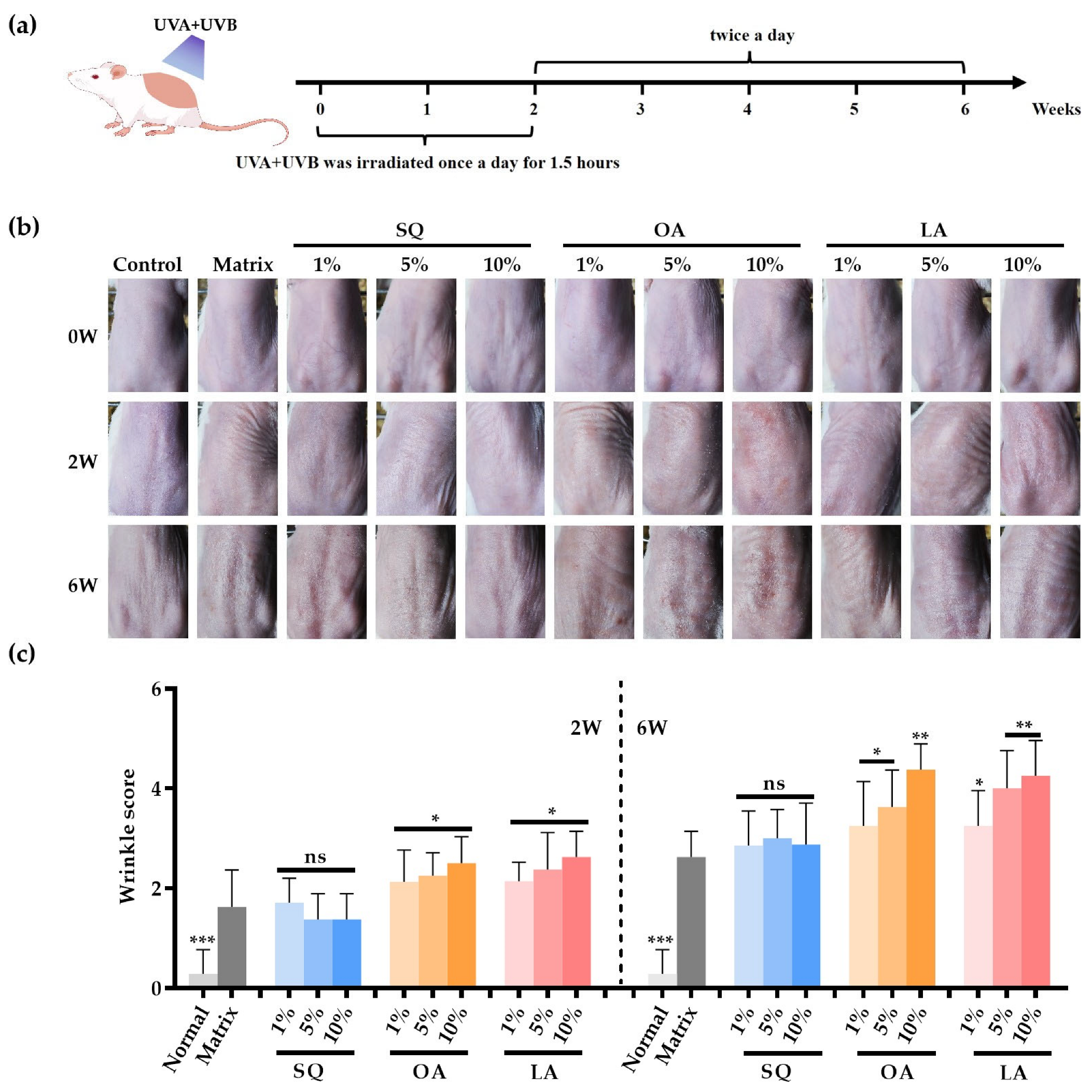
2.2. UV Irradiation and Substance Administration Regimen

KM mice were randomly divided into eleven groups (n = 6 per group): normal, matrix (0.6% carbomer 2020), SQ (1%, 5%, and 10%), OA (1%, 5%, and 10%), and LA (1%, 5%, and 10%). Before and throughout the experiment, a 2 × 2 cm dorsal area of each mouse was shaved with a hair trimmer and depilated with depilatory cream. All groups except the normal received topical application (0.1 mL per application) followed by UV lamp irradiation for 1.5 h. Irradiation was performed once daily for the first 2 weeks and twice daily for the next 4 weeks, accumulating total doses of 26.5 J/cm2 UVA and 11.7 J/cm2 UVB over the 6-week period. The backs of the mice were photographed at weeks 0, 2, and 6, and skin damage was assessed according to the Skin Wrinkle Rating Scale [17]. Six weeks after exposure, mice were systemically anesthetized and euthanized. Skin samples from the irradiated area were excised and either fixed in 10% formaldehyde for histological analysis or snap frozen in liquid nitrogen (−80 °C) for transcriptomic studies. Both epidermis and dermis were preserved for analysis.

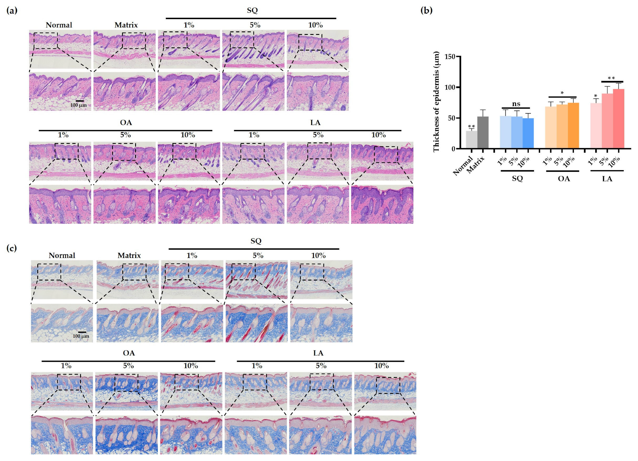
2.2.1. Histological Analysis

At the conclusion of the experiment, KM mice were euthanized via intraperitoneal injection of sodium pentobarbital (180 mg/kg). UV-exposed skin areas were fixed in 10% neutral buffered formalin, dehydrated through a graded ethanol series, paraffin-embedded, and sectioned at 4 μm thickness for histological examination. Histological evaluations included hematoxylin and eosin (H&E) staining to assess epidermal stratification. Epidermal thickness was quantified using ImageJ software (v1.53t; National Institutes of Health, Bethesda, MD, USA).

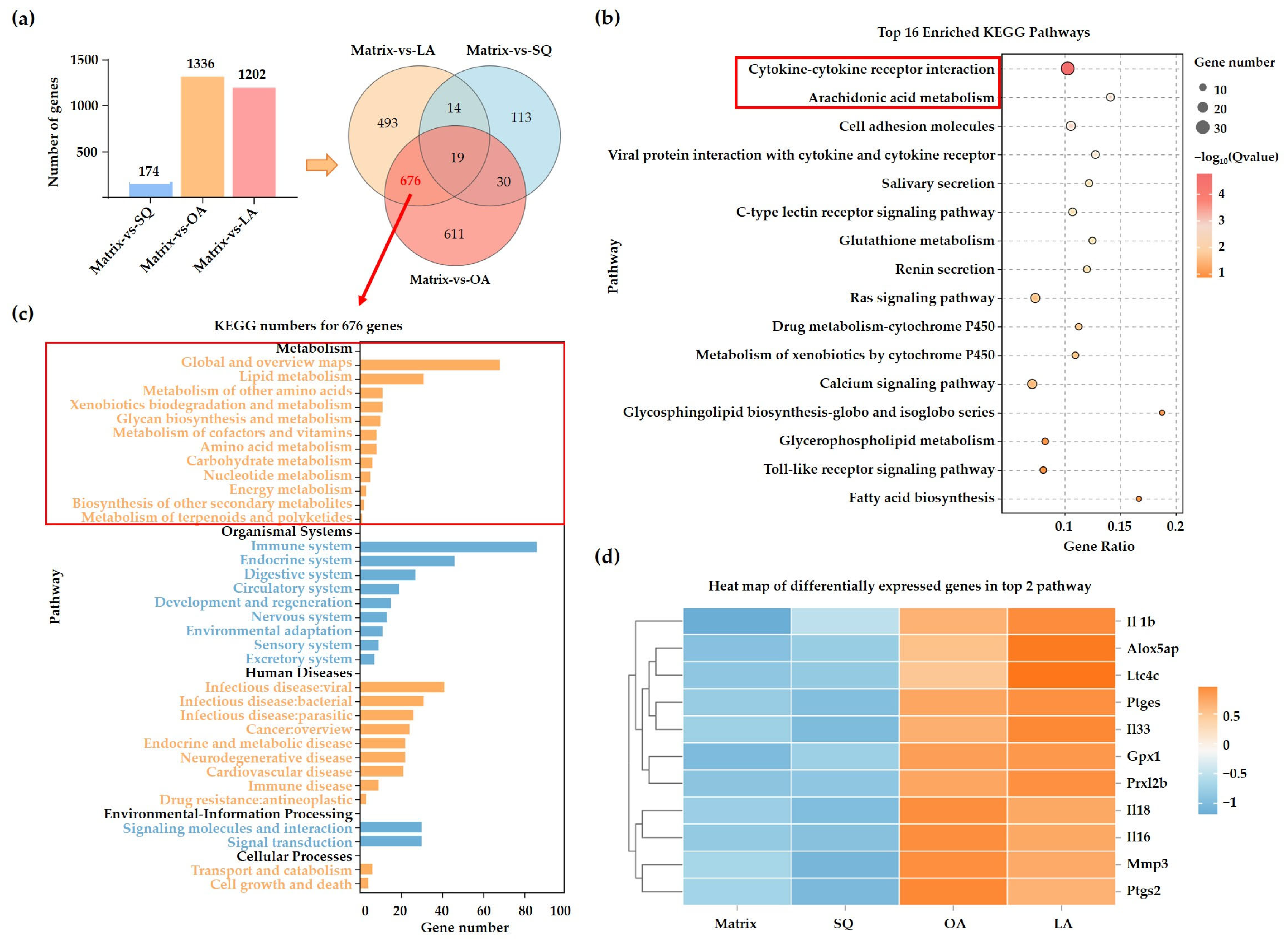
2.2.2. Transcriptomics Analysis

Total RNA was extracted from each group of frozen skin tissues using the TRIzol reagent (following the manufacturer’s instructions) and subsequently reverse-transcribed into cDNA. cDNA libraries were constructed and sequenced on the Illumina NovaSeq 6000 platform by Gene Denovo Biotechnology Co., Ltd. (Guangzhou, China). Raw sequencing data underwent standard quality control procedures prior to gene quantification. Differentially expressed genes in filtered samples were subsequently analyzed through KEGG pathway enrichment analysis.

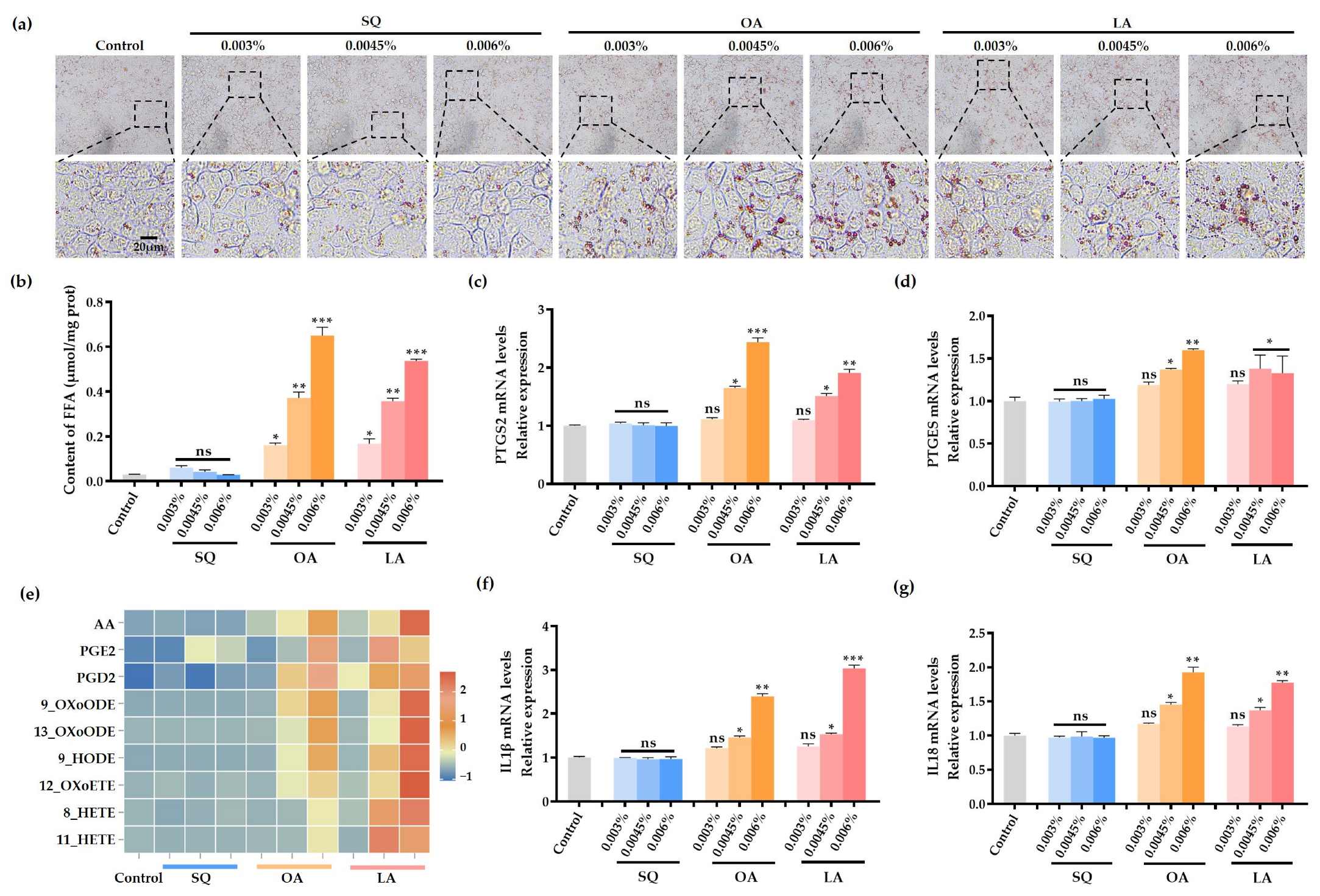
2.3. Lipid Droplet Staining of SZ95 Cells

When cells reached 80% confluence, SZ95 sebocytes were seeded in 12-well plates at a density of 2.5 × 10⁴ cells/well. Following 24 h incubation at 37 °C with 5% CO2, cells were divided into control and experimental groups. The control group received 1 mL of complete medium per well, while experimental groups were treated with three concentrations (0.003%, 0.0045%, and 0.006%) of either SQ, OA, or LA dissolved in culture medium (1 mL/well). Post-treatment, plates were maintained in the incubator for 72 h with daily medium replacement. Cells were processed through sequential steps: PBS washing, 4% paraformaldehyde fixation, incubation with 500 μL Oil Red O working solution (15 min), and PBS rinsing. Lipid accumulation was assessed through microscopic observation.

2.4. Detection of Free Fatty Acids in SZ95 Cells

The cell culture and treatment procedure was performed as described in Section 2.3. After 72 h of treatment, cells were harvested using a cell scraper. Approximately 1 × 10⁶ SZ95 cells were resuspended in 200 μL of ultrapure water and sonicated for 1 min (200 W, 2-s pulses with 1-s intervals). The lysate was then centrifuged at 12,000× g for 10 min at 4 °C. Subsequently, 30 μL of the supernatant was collected for protein quantification via the BCA assay, while FAA levels were quantified using a commercial assay kit following the manufacturer’s protocol.

2.5. LC-MS/MS Analysis of Oxidized Lipid in SZ95 Cells

Cell culture and drug treatment were performed as described in Section 2.3. After 72 h of drug treatment, SZ95 cells (1 × 10⁷ cells per group) were harvested and subjected to metabolome analysis via mass spectrometry (performed by Gene Denovo Biotechnology Co., Ltd. (Guangzhou, China)).

2.6. Expression of Genes Involved in sz95 Lipid Secretion

Cell culture and drug treatment were performed as described in Section 2.3. After 72 h of drug treatment, cells were harvested for qPCR analysis. Primer sequences used for qPCR are listed below (Table 1):

2.7. Statistical Analysis

Data are expressed as mean ± standard deviation (SD) of at least three independent replicates. Statistical analyses were performed using Student’s t-test (for two-group comparisons) or one-way ANOVA (for multi-group comparisons) in GraphPad Prism 8.0. Significance levels were set at p < 0.05 (*), p < 0.01 (**), and p < 0.001 (***).

3. Results

3.1. Apparent Characteristics of Mouse Skin After Ultraviolet Irradiation

As shown as Figure 1a, to evaluate the effects of squalane (SQ), oleic acid (OA), and linoleic acid (LA) on UV-induced skin damage, mice were exposed to UV irradiation at specific wavelengths with escalating doses. As demonstrated in Figure 1b, compared with normal mice, matrix-treated mice exhibited characteristic photodamage manifestations, including cutaneous roughness, xerosis, and loss of skin glossiness. SQ treatments (1%, 5%, 10%) did not exacerbate UV-induced damage compared to the matrix group; however, the 10% SQ group exhibited a partial mitigation of these effects. Conversely, both OA and LA groups (1%, 5%, 10%) demonstrated aggravated oxidative skin damage post-UV exposure. Two weeks after UV irradiation, acute erythema was observed in all treatment groups. Six weeks after UV irradiation, there was progressive dermal structural disorganization, characterized by disrupted skin surface texture, transverse striations, hyperpigmentation, and pathological sebum overproduction. These manifestations were most severe in the 10% OA and 10% LA groups. Quantitative skin damage scoring (Figure 1c) revealed statistically significant differences between the matrix group and the 10% OA, 5% LA, and 10% LA groups (p < 0.01).

3.2. Changes in Skin Pathology Content in Mice After Ultraviolet Irradiation

The skin of mice after 6 weeks of UV modeling was selected for section staining and pathological observation. The results are shown in Figure 2a,c. Compared with the normal group, the epidermal cell structure in the matrix group was altered, exhibiting disorganized cell arrangement, increased stratum corneum thickness, and epidermal hyperplasia. Although stratum corneum thickening and epidermal hyperplasia were also observed in the squalane group, no significant differences were detected compared to the matrix group (Figure 2b). In the UV-induced skin damage experiment, the experimental results of polydimethylsiloxane (PDMS) were consistent with those of squalane. Specifically, PDMS (1%, 5%, 10%) showed no exacerbation of UV-induced skin damage in appearance (Figure S1a), but histological analysis revealed stratum corneum thickening and epidermal hyperplasia (Figure S1b). However, epidermal thickness remained statistically similar to the matrix group (Figure S1c). In contrast, stratum corneum thickening was more pronounced in the oleic acid (OA) and linoleic acid (LA) groups, accompanied by stratum corneum cell proliferation, relative to the matrix group. Both the 5% and 10% LA groups significantly promoted epidermal hyperplasia (p < 0.01). Additionally, OA and LA induced follicular obstruction, with severity escalating at higher concentrations. These treatments also disrupted lipid metabolism. Notably, 10% LA group exhibited severe atrophy of the sebaceous glands.
Consistent with the histopathological changes, the accumulation of advanced glycation end products (AGEs) showed a striking divergence between lipid types (Figure S2). The content of AGEs in the skin of mice in the control group was low. AGEs were increased in the matrix group compared to the control group. There was no significant difference in the levels of AGEs between the squalane group (1%, 5%, and 10%) and the matrix group. The oleic and linoleic acid groups significantly promoted the production of AGEs in a concentration-dependent manner compared with the matrix group. Notably, the 10% LA group significantly promoted the production of AGEs (p < 0.001). Unsaturated fatty acids may exacerbate UV-induced damage through carbonyl stress pathways.

3.3. Transcriptome Analysis

Figure 3a presents the histogram and Venn diagram comparing differentially expressed genes (DEGs) between treatment groups and the matrix group. A total of 695 DEGs were shared between the OA and LA groups, with 19 overlapping those in the SQ group. After excluding these overlapping genes, 676 unique DEGs were retained for subsequent analysis. KEGG pathway enrichment analysis of these 676 DEGs revealed significant enrichment in lipid metabolism pathways (31 genes; Figure 3c). The bubble plot in Figure 3b highlights key enriched pathways, including arachidonic acid metabolism and cytokine–cytokine receptor interaction. Correspondingly, Figure 3d displays a heatmap of expression profiles for key DEGs within these two pathways.

3.4. SQ, OA and LA on Lipid Droplet Synthesis in SZ95 Cells

Mechanistically, the lipid-stimulating effects of unsaturated fatty acids were associated with upregulated expression of lipogenic genes (SREBP1, SCD1, ACC, and PPARγ) (Figure S3a–d). Compared to the normal group, saturated lipid-treated groups showed no significant differences in the mRNA levels of these genes. In contrast, unsaturated fatty acids at concentrations of 0.0045% and 0.006% significantly upregulated their expression. Specifically, OA and LA at 0.006% increased the expression of SREBP1 and SCD1 (p < 0.01), as well as ACC and PPARγ (p < 0.001). Similarly, ALA at 0.006% significantly elevated the expression of SREBP1, SCD1, and ACC (p < 0.01) and induced a more pronounced upregulation of PPARγ (p < 0.001).
SZ95 sebocytes were incubated with SQ, OA, and LA at concentrations of 0.003%, 0.0045%, and 0.006%, respectively, for 72 h. Oil Red O staining results (Figure 4a) demonstrated that SQ-treated cells contained minimal lipid droplets across all tested concentrations. In contrast, OA- and LA-treated groups showed significant increases in both quantity and size of intracellular lipid droplets compared to the control group.
Free fatty acid (FFA) content analysis (Figure 4b) revealed no significant differences between control and SQ-treated groups. However, OA and LA treatments induced concentration-dependent elevation of intracellular FFA levels. Notably, 0.006% concentrations of both OA and LA significantly enhanced FFA production (p < 0.001). After treating SZ95 cells with squalane (SQ), oleic acid (OA), and linoleic acid (LA) for 72 h, the intracellular oxidized lipids were analyzed. As shown in the heatmap of Figure 4e, the levels of arachidonic acid (AA), prostaglandins (PGE2, PGD2), hydroxyeicosatetraenoic acids (12-OXO-ETE, 8-HETE, 11-HETE), and oxidative stress-related products (9-OXO-ODE, 13-HETE) were significantly increased in the OA (0.0045%) and LA (0.006%) groups. Notably, no significant differences in the concentrations of these oxidized lipid metabolites were observed compared to the squalane (SQ) control groups at various concentrations.
As shown in Figure 4c,d,f,g, compared to the control group, different concentrations of SQ and 0.003% OA/LA exhibited no significant differences in the expression levels of PTGS2, PTGES, IL1β, or IL18. However, at 0.0045% concentration, both OA and LA upregulated the expression of PTGS2, PTGES, IL1β, and IL18 (p < 0.05). When the concentration reached 0.006%, OA significantly upregulated PTGS2 (p < 0.001), PTGES, IL-1β, and IL-8 (p < 0.01), while LA markedly increased the expression of PTGS2, IL-8 (p < 0.01), PTGES (p < 0.05), and IL-1β (p < 0.001). The dose-dependent upregulation of pro-inflammatory genes (PTGS2/PTGES/IL1β/IL-8) suggests a potential inflammatory cascade triggered by high concentrations of OA/LA.
The experiment on lipid droplet secretion in SZ95 sebocytes further confirmed that synthetic saturated oils (PDMS and HDA) did not alter lipid metabolism or induce inflammation, whereas unsaturated fatty acid (ALA) promoted lipid synthesis and triggered an inflammatory cascade. SZ95 sebocytes treated with polydimethylsiloxane (PDMS, 0.003–0.006%) or HDA for 72 h showed minimal intracellular lipid droplets (Oil Red O staining, Figure S4a) and no significant changes in free fatty acid (FFA) levels compared to controls (Figure S3b). In contrast, ALA treatment markedly increased lipid droplet accumulation and induced a concentration-dependent rise in FFA content, with significant enhancement at 0.0045% and 0.006% concentrations (p < 0.01). As shown in Figure S3c–f, PDMS, HDA, and 0.003% ALA had no significant effect on pro-inflammatory genes (PTGS2, PTGES, IL1β, IL18) compared to control group. Furthermore, 0.0045% ALA upregulated PTGS2, PTGES, IL1β, and IL18 (p < 0.05), and 0.006% ALA induced a further increase of these genes (p < 0.01).

4. Discussion

The cosmetic industry faces two fundamental conflicts: the efficacy-risk paradox of unsaturated lipids (e.g., OA and LA) and the mechanistic gaps in understanding lipid-inflammatory crosstalk under photostress. As emphasized in the introduction regarding their core molecular divergence—saturated squalane (SQ) versus unsaturated fatty acids (OA, LA) with double bonds—this study confirms that these structural differences lead to starkly contrasting biological effects under ultraviolet (UV) exposure. While low concentrations of OA/LA enhance skin barrier function, their photochemical instability at higher concentrations or under UV irradiation generates cytotoxic peroxidation products (e.g., MDA, 4-HNE), disrupting lipid metabolism and accelerating skin aging [18,19,20]. Existing research predominantly focuses on the in vitro antioxidant capacity of individual lipids, lacking systematic analysis of lipid peroxidation-inflammatory networks in the complex skin microenvironment [15,21]. To resolve these conflicts and validate the structure-dependent efficacy differences, this study employed UV-irradiated mouse models and human sebaceous gland cells (SZ95) to investigate the regulatory effects of SQ, OA, and LA at cosmetically relevant concentrations on photo-induced oxidative damage, lipid metabolism, and inflammatory responses.
SQ’s saturated hydrocarbon backbone confers exceptional oxidative inertness, making it a photostable barrier enhancer. Under UV exposure, SQ effectively prevented the formation of reactive aldehydes (e.g., 4-HNE) and suppressed the activation of matrix metalloproteinases (MMPs) and advanced glycation end products (AGEs) by halting free radical chain reactions, thereby resisting UV-aggravated skin damage [12]. Parallel experiments with synthetic saturated lipids like polydimethylsiloxane (PDMS) further reinforced the conclusion that saturated structures provide photoprotection. In stark contrast, the double bonds in OA and LA molecules served as key sites for UV-induced peroxidation, triggering dose-dependent epidermal hyperplasia and AGE accumulation. Due to possessing two double bonds (18:2 n-6), LA exhibited higher oxidative susceptibility than monounsaturated OA (18:1 n-9), with 10% LA treatment even causing severe sebaceous gland atrophy—consistent with follicular damage mediated with oxidized sebum observed in clinical and model systems [22]. Lipid peroxidation was identified as the common pathway underlying these damages: the oxidized lipid profiles observed in SZ95 cells strongly correlated with AGE accumulation in mouse skin. Notably, LA’s higher oxidation index significantly amplified the production of cytotoxic eicosanoids (e.g., PGD2/PGE2, HETEs), which not only directly degrade collagen but also potently recruit inflammatory cells, establishing a self-perpetuating damage cycle [23].
Transcriptomic analysis further elucidated the shared pro-inflammatory mechanism of OA and LA, revealing their co-activation of core inflammatory hubs including arachidonic acid metabolism (key enzyme PTGS2/COX-2), cytokine–receptor interactions (e.g., IL-1β, IL-6), and NF-κB signaling. These activated pathways provide the molecular basis for the observed phenotypes: Firstly, peroxidized OA/LA generated under UV exposure directly serve as substrates for cyclooxygenase (COX-2) and lipoxygenase, driving the synthesis of pro-inflammatory eicosanoids PGE2 and HETEs, thereby exacerbating inflammation. Secondly, even extremely low concentrations of LA (0.006%) potently induced IL-1β expression—a key trigger for comedogenesis in oily skin—directly corroborating clinical observations that oxidized sebum exacerbates acne inflammation [24,25]. Thirdly, in the SZ95 sebocyte model, OA/LA at 0.0045–0.006% significantly upregulated the core lipid synthesis regulatory axis (SREBP1-PPARγ-ACC), synergistically promoting fatty acid biosynthesis, excessive lipid droplet formation, and free fatty acid (FFA) hypersecretion. Excess FFAs disrupt the skin’s acidic mantle, creating an environment conducive to Cutibacterium acnes proliferation [26,27]. The concentration-dependent upregulation of PTGS2 (COX-2) and IL-1β by OA/LA jointly activated the NLRP3 inflammasome, driving follicular hyperkeratosis and inflammatory cell infiltration. Concurrently, pro-oxidative metabolites (e.g., PGE2, HETEs) exerted additional chemotactic effects, attracting more immune cells and ultimately leading to characteristic acne inflammatory lesions like erythema and pustules [28]. Although squalene was not directly tested here, its well-established comedogenic mechanism, mediated by oxidative degradation [29], further supports the risks of unsaturated lipids under UV exposure for acne-prone skin. Conversely, saturated SQ and synthetic saturated lipids like PDMS (which also demonstrated photostability in this study), lacking vulnerable double bonds, resist peroxidation and thus avoid stimulating FFA overload or activating these inflammatory pathways, offering a safer option for maintaining barrier homeostasis in oily and acne-prone skin.
These findings hold significant clinical implications for cosmetic formulation design, particularly for products targeting oily and acne-prone skin, and challenge the traditional assumption of “universal suitability” for OA/LA based solely on safety and compatibility. OA and LA exhibit a pronounced duality under UV exposure: beneficial for barrier repair at low concentrations, yet posing significant pro-acnegenic risks at higher doses or under photostress. Saturated emollients, especially SQ, owing to their inherent photostability and metabolic inertness, effectively prevent lipid peroxidation and its ensuing inflammatory cascades, providing a superior solution for lipid replenishment in UV-exposed, oily/acne-prone skin. Therefore, formulations rich in highly unsaturated lipids like OA/LA warrant careful risk assessment, especially for daytime use or products intended for acne-prone populations exposed to sunlight. Conversely, selecting photostable saturated lipids such as SQ or silicones offers safer barrier support.
This study, while preliminarily elucidating the regulatory mechanisms of lipid metabolism under photodamage using UV-irradiated mouse models and SZ95 sebocytes and clearly demonstrating the core differences between saturated (SQ) and unsaturated (OA/LA) lipids, has limitations requiring future investigation. The observed biological effects may result from the synergistic interplay between lipid saturation state and the inherent properties of their chemical classes (e.g., saturated hydrocarbon SQ vs. unsaturated fatty acid OA/LA). Future studies will require more controlled intra-class comparisons (e.g., saturated vs. unsaturated triglycerides) to precisely isolate the contribution of saturation itself. Human skin is a complex ecosystem involving dynamic interactions among keratinocytes, microbiota, immune cells, and the extracellular matrix; its functional regulation surpasses the simulation capacity of current single-cell models (e.g., SZ95) or animal experiments. To comprehensively dissect the coordinated regulation of epidermal barrier function and sebum secretion under photostress, future work will integrate keratinocyte models and establish UV-damaged keratinocyte–sebocyte co-culture systems to decipher the regulatory patterns of intercellular signaling on sebum secretion within photodamaged microenvironments.

5. Conclusions

This study systematically reveals the differential roles of squalane (SQ) and unsaturated fatty acids (OA, LA) in regulating UV-induced skin damage, lipid metabolism, and inflammation. It clarifies how high concentrations or UV exposure drive skin injury, inflammation, and metabolic dysregulation through lipid peroxidation. These findings provide critical theoretical foundations for balancing efficacy and risk in cosmetic formulations, guiding the development of safer and more effective products. Additionally, they offer novel insights for understanding and treating lipid metabolism-related skin disorders such as oily skin and acne, advancing both dermatological research and clinical practice.

Supplementary Materials

The following supporting information can be downloaded at: https://www.mdpi.com/article/10.3390/cosmetics12040130/s1, Figure S1: Effect of PDMS on skin appearance of UV—Irradiated mice; Figure S2: Changes of AGE content in mice skin after ultraviolet irradiation; Figure S3: Effects of saturated hydrocarbon/unsaturated fatty acids on the expression of SREBP1, SCD1, ACC, and PPARγ genes in SZ95 cells. Table S1: The primer sequences of SREBP1, ACD1, ACC, PPARγ; Figure S4: Effect of different concentrations of PDMS, HDA and ALA on lipid droplet synthesis and Lipid metabolism in SZ95 cells.

Author Contributions

Conceptualization, L.-F.T. and Q.X.; methodology, Y.-F.Z.; software, X.-W.L.; validation, H.-Y.S., Q.-R.Z. and W.-R.Z.; formal analysis, H.-Y.S. and Q.-R.Z.; investigation, X.-W.L.; resources, L.-F.T.; data curation, Z.-Y.Z.; writing—original draft preparation, Q.-R.Z.; writing—review and editing, W.-R.Z.; visualization, Q.-R.Z.; supervision, Y.-F.Z.; project administration, Z.-Y.Z.; funding acquisition, Q.X. All authors have read and agreed to the published version of the manuscript.

Funding

This research was funded by the Natural Science Foundation of Guangdong Province, China (2021A1515012480); the National Key Research and Development Program of China (2022YFC2403102); Guangzhou Research and Development Plan in Key Fields, China (202103030003); R&D Plan Project in Key Fields of Guangdong Province, China (2022B1111080007).

Institutional Review Board Statement

The animal study protocol was approved by the Institutional Animal Care and Use Committee of the Jinan University (protocol code 20241115-04 and approved on 15 November 2024).

Informed Consent Statement

Not applicable.

Data Availability Statement

Data will be made available on request.

Acknowledgments

SZ95 human sebocytes (generously provided by Christos C. Zouboulis, Honorary Professor h.c., Department of Dermatology, Venereology, Allergology and Immunology, Dessau Medical Center, Brandenburg Medical School, Auenweg 38, 06847 Dessau-Rosslau, Germany).

Conflicts of Interest

Authors Wen-Rong Zhang, Zi-Yan Zhou, Xue-Wan Li, and Li-Feng Tang were employed by Guangzhou Xika Technology Co., Ltd. The remaining authors declare that the research was conducted in the absence of any commercial or financial relationships that could be construed as a potential conflict of interest.

Abbreviations

The following abbreviations are used in this manuscript:
SQsqualane
OAoleic acid
LAlinoleic acid
PTGS2Prostaglandin-Endoperoxide Synthase 2
IL-1βInterleukin-1 beta
FFAsFree Fatty Acids
UVultraviolet
ALAα-linolenic acid
NF-κBNuclear factor kappa B
IL-6Interleukin-6
TNF-αTumor Necrosis Factor alpha
PDMSpolydimethylsiloxane
HDAhexyldecyl alcohol
KMKunming
H&Ehematoxylin and eosin
PTGESProstaglandin E Synthase
GAPDHGlyceraldehyde-3-phosphate dehydrogenase
DEGsdifferentially expressed genes
AAArachidonic Acid
PGE2Prostaglandin E2
PGD2Prostaglandin D2
12-OXO-ETE12-oxo-eicosatetraenoic acid
8-HETE8-hydroxyeicosatetraenoic acid
11-HETE11-hydroxyeicosatetraenoic acid
9-OXO-ODE9-oxo-odecatetraenoic acid
13-HETE13-hydroxyeicosatetraenoic acid
SREBP1sterol regulatory element-binding protein 1
SCD1stearoyl-CoA desaturase 1
ACCacetyl-CoA carboxylase
PPARγperoxisome proliferator-activated receptor gamma

References

  1. Ferrara, F.; Woodby, B.; Pecorelli, A.; Schiavone, M.L.; Pambianchi, E.; Messano, N.; Therrien, J.P.; Choudhary, H.; Valacchi, G. Additive effect of combined pollutants to UV induced skin OxInflammation damage. Evaluating the protective topical application of a cosmeceutical mixture formulation. Redox Biol. 2020, 34, 101481. [Google Scholar] [CrossRef] [PubMed]
  2. Tang, X.; Yang, T.; Yu, D.; Xiong, H.; Zhang, S. Current insights and future perspectives of ultraviolet radiation (UV) exposure: Friends and foes to the skin and beyond the skin. Environ. Int. 2024, 185, 108535. [Google Scholar] [CrossRef]
  3. Bacellar, I.O.L.; Baptista, M.S. Mechanisms of Photosensitized Lipid Oxidation and Membrane Permeabilization. ACS Omega 2019, 4, 21636–21646. [Google Scholar] [CrossRef]
  4. Jung, Y.R.; Shin, J.M.; Kim, C.H.; Kim, S.; Kim, C.D.; Seo, Y.J.; Lee, J.H.; Im, M.; Lee, Y.; Lee, Y.H. Activation of NLRP3 Inflammasome by Palmitic Acid in Human Sebocytes. Ann. Dermatol. 2021, 33, 541–548. [Google Scholar] [CrossRef]
  5. Flori, E.; Mastrofrancesco, A.; Ottaviani, M.; Maiellaro, M.; Zouboulis, C.C.; Camera, E. Desaturation of sebaceous-type saturated fatty acids through the SCD1 and the FADS2 pathways impacts lipid neosynthesis and inflammatory response in sebocytes in culture. Exp. Dermatol. 2023, 32, 808–821. [Google Scholar] [CrossRef]
  6. Prakoeswa, C.R.S.; Damayanti Anggraeni, S.; Umborowati, M.A.; Sari, M.; Hendaria, M.P.; Thahir, T.F. The Role of Moisturizer Containing Anti-inflammatory on Skin Hydration in Mild-Moderate Atopic Dermatitis Patients. Dermatol. Res. Pract. 2024, 2024, 3586393. [Google Scholar] [CrossRef] [PubMed]
  7. Szymańska, E.; Wojasiński, M.; Czarnomysy, R.; Dębowska, R.; Łopianiak, I.; Adasiewicz, K.; Ciach, T.; Winnicka, K. Chitosan-Enriched Solution Blow Spun Poly (Ethylene Oxide) Nanofibers with Poly (Dimethylsiloxane) Hydrophobic Outer Layer for Skin Healing and Regeneration. Int. J. Mol. Sci. 2022, 23, 5135. [Google Scholar] [CrossRef] [PubMed]
  8. Douguet, M.; Picard, C.; Savary, G.; Merlaud, F.; Loubat-bouleuc, N.; Grisel, M. Spreading properties of cosmetic emollients: Use of synthetic skin surface to elucidate structural effect. Colloids Surf. B Biointerfaces 2017, 154, 307–314. [Google Scholar] [CrossRef]
  9. Atalay Ekiner, S.; Gęgotek, A.; Skrzydlewska, E. Inflammasome activity regulation by PUFA metabolites. Front. Immunol. 2024, 15, 1452749. [Google Scholar] [CrossRef]
  10. Ding, Y.; Jiratchayamaethasakul, C.; Lee, S.H. Protocatechuic Aldehyde Attenuates UVA-Induced Photoaging in Human Dermal Fibroblast Cells by Suppressing MAPKs/AP-1 and NF-κB Signaling Pathways. Int. J. Mol. Sci. 2020, 21, 4619. [Google Scholar] [CrossRef]
  11. Das, U.N. Essential Fatty Acids and Their Metabolites in the Pathobiology of Inflammation and Its Resolution. Biomolecules 2021, 11, 1873. [Google Scholar] [CrossRef] [PubMed]
  12. Grujić-Milanović, J.D.; Miloradović, Z.Z.; Mihailović-Stanojević, N.D.; Banjac, V.V.; Vidosavljević, S.; Ivanov, M.S.; Karanović, D.J.; Vajić, U.V.; Jovović, D.M. Excesive consumption of unsaturated fatty acids leads to oxidative and inflammatory instability in Wistar rats. Biomed. Pharmacother. 2021, 139, 111691. [Google Scholar] [CrossRef]
  13. Romana-Souza, B.; Saguie, B.O.; Pereira de Almeida Nogueira, N.; Paes, M.; Dos Santos Valença, S.; Atella, G.C.; Monte-Alto-Costa, A. Oleic acid and hydroxytyrosol present in olive oil promote ROS and inflammatory response in normal cultures of murine dermal fibroblasts through the NF-κB and NRF2 pathways. Food. Res. Int. 2020, 131, 108984. [Google Scholar] [CrossRef] [PubMed]
  14. Ottaviani, M.; Alestas, T.; Flori, E.; Mastrofrancesco, A.; Zouboulis, C.C.; Picardo, M. Peroxidated squalene induces the production of inflammatory mediators in HaCaT keratinocytes: A possible role in acne vulgaris. J. Investig. Dermatol. 2006, 126, 2430–2437. [Google Scholar] [CrossRef]
  15. Ishikawa, A.; Ito, J.; Shimizu, N.; Kato, S.; Kobayashi, E.; Ohnari, H.; Sakata, O.; Naru, E.; Nakagawa, K. Linoleic acid and squalene are oxidized by discrete oxidation mechanisms in human sebum. Ann. N. Y. Acad. Sci. 2021, 1500, 112–121. [Google Scholar] [CrossRef]
  16. Cheng, L.C.; Li, Y.T.; Ji, C.; Liu, Y.W.; Wang, L.R.; Cheng, P.; Yu, J. Determine content of linoleic acid and squalene of acne patients sebum by HPLC. Chin. J. Aesthetic Med. 2016, 25, 5. [Google Scholar]
  17. Kong, S.Z.; Shi, X.G.; Feng, X.X.; Li, W.J.; Liu, W.H.; Chen, Z.W.; Xie, J.H.; Lai, X.P.; Zhang, S.X.; Zhang, X.J.; et al. Inhibitory effect of hydroxysafflor yellow a on mouse skin photoaging induced by ultraviolet irradiation. Rejuvenation Res. 2013, 16, 404–413. [Google Scholar] [CrossRef]
  18. Kováčik, A.; Kopečná, M.; Hrdinová, I.; Opálka, L.; Boncheva Bettex, M.; Vávrová, K. Time-Dependent Differences in the Effects of Oleic Acid and Oleyl Alcohol on the Human Skin Barrier. Mol. Pharm. 2023, 20, 6237–6245. [Google Scholar] [CrossRef]
  19. Wang, X.; Jia, Y.; He, H. The Role of Linoleic Acid in Skin and Hair Health: A Review. Int. J. Mol. Sci. 2024, 26, 246. [Google Scholar] [CrossRef]
  20. Manosalva, C.; Bahamonde, C.; Soto, F.; Leal, V.; Ojeda, C.; Cortés, C.; Alarcón, P.; Burgos, R.A. Linoleic Acid Induces Metabolic Reprogramming and Inhibits Oxidative and Inflammatory Effects in Keratinocytes Exposed to UVB Radiation. Int. J. Mol. Sci. 2024, 25, 10385. [Google Scholar] [CrossRef]
  21. Ito, J.; Komuro, M.; Parida, I.S.; Shimizu, N.; Kato, S.; Meguro, Y.; Ogura, Y.; Kuwahara, S.; Miyazawa, T.; Nakagawa, K. Evaluation of lipid oxidation mechanisms in beverages and cosmetics via analysis of lipid hydroperoxide isomers. Sci. Rep. 2019, 9, 7387. [Google Scholar] [CrossRef] [PubMed]
  22. Brami-Cherrier, K.; Chernavsky, A.; You, H.; Grando, S.A.; Brideau-Andersen, A.; Sondergaard, B. Botulinum Neurotoxin Type A Directly Affects Sebocytes and Modulates Oleic Acid-Induced Lipogenesis. Toxins 2022, 14, 708. [Google Scholar] [CrossRef]
  23. Kovács, D.; Camera, E.; Póliska, S.; Cavallo, A.; Maiellaro, M.; Dull, K.; Gruber, F.; Zouboulis, C.C.; Szegedi, A.; Törőcsik, D. Linoleic Acid Induced Changes in SZ95 Sebocytes-Comparison with Palmitic Acid and Arachidonic Acid. Nutrients 2023, 15, 3315. [Google Scholar] [CrossRef] [PubMed]
  24. Dahlan, N.H.; Sitohang, I.B.S.; Indriatmi, W.; Wibowo, H.; Enggy, L.E. Correlation Between Reduced IL-1β Levels in Acne Lesions and the Decrease in Acne Inflammatory Lesions Following Topical Vitamin D Administration: A Double-Blind Randomized Controlled Trial. Clin. Cosmet. Investig. Dermatol. 2024, 17, 2183–2195. [Google Scholar] [CrossRef]
  25. Liu, S.; Luo, X.H.; Liu, Y.F.; Zouboulis, C.C.; Shi, G. Emodin exhibits anti-acne potential by inhibiting cell growth, lipogenesis, and inflammation in human SZ95 sebocytes. Sci. Rep. 2023, 13, 21576. [Google Scholar] [CrossRef]
  26. Wan, K.; Li, J.; Ma, L.; Chen, T.; Chen, Y.; Li, Z.; Zouboulis, C.C.; Wang, G.L.; Wang, J. Camellia saponin modulates oleic acid/linoleic acid-induced lipogenesis in human sebocytes through lipophagy activation. Int. J. Cosmet. Sci. 2025, in press. [Google Scholar] [CrossRef] [PubMed]
  27. Yang, L.; Lin, X.Y. Research progress on acne and skin barrier. Chin. J. Integr. Tradit. West. Med. Dermatol. Venereol. 2021, 20, 416–419. [Google Scholar]
  28. Kawamoto, A.; Kuwano, T.; Watarai, E.; Igarashi, T.; Katayama, Y.; Kushida, K.; Nakamura, S.; Murase, T.; Yoshida, H.; Ishikawa, J. Oleic acid-induced interleukin-36γ: A possible link between facial skin redness and sebum. J. Cosmet. Dermatol. 2023, 22, 2308–2317. [Google Scholar] [CrossRef]
  29. Condrò, G.; Sciortino, R.; Perugini, P. Squalene Peroxidation and Biophysical Parameters in Acne-Prone Skin: A Pilot “In Vivo” Study. Pharmaceuticals 2023, 16, 1704. [Google Scholar] [CrossRef]
Figure 1. Effect of SQ, OA, and LA on skin appearance of UV-irradiated mice. (a) Schematic representation of the UV irradiation model. (b) Characteristics of mouse skin after oxidative damage. (c) Visual scores of skin photoaging grading by SQ, OA, and LA in mice. n = 6, mean ± SD, ns: no significant, * p < 0.05, ** p < 0.01, *** p < 0.001 vs. Matrix.
Cosmetics 12 00130 g001
Figure 2. Effects of SQ, OA, and LA on skin pathology and AGE content in the skin of UV-irradiated mice. (a) HE & Masson staining of skin sections induced by UV. Scale bar = 100 μm. (b) Thickness of epidermal skin. (c) HE & Masson staining of skin sections induced by UV. Scale bar = 100 μm. n = 3, mean ± SD, ns: no significant, * p < 0.05, ** p < 0.01 vs. Matrix.
Cosmetics 12 00130 g002
Figure 3. Transcriptome analysis of the effects of 10% SQ, OA, and LA on the skin of ultraviolet-irradiated mice. (a) Number of differential genes and Wayne’s diagram display in SQ, OA, LA vs. matrix group. (b) Statistical chart of KEGG numbers for 676 genes. Key enriched pathways are highlighted (red box). (c) KEGG-enriched significant bubble plots for 676 genes. The metabolism pathway is marked with a red box. (d) Heatmap of differential genes in the cytokine–cytokine receptor interaction and Arachidonic acid metabolism pathways.
Cosmetics 12 00130 g003
Figure 4. Effect of different concentrations of SQ, OA, and LA on lipid droplet synthesis and lipid metabolism in SZ95 cells. (a) Oil red O staining of intracellular lipid droplets in SZ95 cells. Scale bar = 20 μm. (b) Detection of total free fatty acids in SZ95 cells. (c) PTGS2 mRNA levels relative expression. (d) PTGES mRNA levels relative expression. (e) Oxidized lipids in SZ95 cells cultured with SQ, OA, and LA for 72 h were analyzed and visualized via a differential metabolite clustering heatmap (The vertical columns in the heatmap corresponding to each treatment group represent concentrations of 0.003%, 0.0045%, and 0.006% from left to right). (f) IL1β mRNA levels relative expression. (g) IL18 mRNA levels relative expression. n = 3, mean ± SD, ns: no significant, * p < 0.05, ** p < 0.01, *** p < 0.001 vs. Control.
Cosmetics 12 00130 g004
Table 1. The primer sequences of key genes.
Primer NamePrimer Sequence
PTGS2F:5′-TCTGAAACCCACTCCAAACACA-3′
R:5′-CATTTCGAAGGAAGGGAATGTTATT-3′
PTGESF:5′-GCTGATCACACCCACAGTTG-3′
R:5′-CCAGGAAAAGGAAGGGGTAG-3′
IL-6F:5′-CATCCTCGACGGCATCTCAG-3′
R:5′-TCACCAGGCAAGTCTCCTCA-3′
IL-18F:5′-CAGAAGRACTGADCTCGCC-3′
R:5′-CAGAAGTACCTGAGCTCGCC-3′
GAPDHF:5′-CAGGAGGCATTGCTGATGAT-3′
R:5′-GAAGGCTGGGGCTCATTT-3′
Disclaimer/Publisher’s Note: The statements, opinions and data contained in all publications are solely those of the individual author(s) and contributor(s) and not of MDPI and/or the editor(s). MDPI and/or the editor(s) disclaim responsibility for any injury to people or property resulting from any ideas, methods, instructions or products referred to in the content.

